comparison of phits, geant4, and hibrac simulations of depth-dependent yields of β + -emitting...
TRANSCRIPT

This content has been downloaded from IOPscience. Please scroll down to see the full text.
Download details:
IP Address: 130.217.227.3
This content was downloaded on 01/07/2014 at 07:01
Please note that terms and conditions apply.
Comparison of PHITS, GEANT4, and HIBRAC simulations of depth-dependent yields of β+-
emitting nuclei during therapeutic particle irradiation to measured data
View the table of contents for this issue, or go to the journal homepage for more
2013 Phys. Med. Biol. 58 6355
(http://iopscience.iop.org/0031-9155/58/18/6355)
Home Search Collections Journals About Contact us My IOPscience

IOP PUBLISHING PHYSICS IN MEDICINE AND BIOLOGY
Phys. Med. Biol. 58 (2013) 6355–6368 doi:10.1088/0031-9155/58/18/6355
Comparison of PHITS, GEANT4, and HIBRACsimulations of depth-dependent yields of β+-emittingnuclei during therapeutic particle irradiation tomeasured data
Heide Rohling1, Lembit Sihver2, Marlen Priegnitz3,Wolfgang Enghardt1,3 and Fine Fiedler3
1 Technische Universitat Dresden, OncoRay-National Center for Radiation Research inOncology, Fetscherstr. 74, PO Box 41, D-01307 Dresden, Germany2 Chalmers University of Technology, Nuclear Engineering, Applied Physics, Fysikgrand 3,SE-41296 Gothenburg, Sweden3 Helmholtz-Zentrum Dresden-Rossendorf, Institute of Radiation Physics, PO Box 510119,D-01314 Dresden, Germany
E-mail: [email protected]
Received 10 April 2013, in final form 12 July 2013Published 2 September 2013Online at stacks.iop.org/PMB/58/6355
AbstractFor quality assurance in particle therapy, a non-invasive, in vivo rangeverification is highly desired. Particle therapy positron-emission-tomography(PT-PET) is the only clinically proven method up to now for this purpose.It makes use of the β+-activity produced during the irradiation by the nuclearfragmentation processes between the therapeutic beam and the irradiated tissue.Since a direct comparison of β+-activity and dose is not feasible, a simulation ofthe expected β+-activity distribution is required. For this reason it is essentialto have a quantitatively reliable code for the simulation of the yields of theβ+-emitting nuclei at every position of the beam path. In this paper results ofthe three-dimensional Monte-Carlo simulation codes PHITS, GEANT4, andthe one-dimensional deterministic simulation code HIBRAC are comparedto measurements of the yields of the most abundant β+-emitting nuclei forcarbon, lithium, helium, and proton beams. In general, PHITS underestimatesthe yields of positron-emitters. With GEANT4 the overall most accurateresults are obtained. HIBRAC and GEANT4 provide comparable results forcarbon and proton beams. HIBRAC is considered as a good candidate for theimplementation to clinical routine PT-PET.
(Some figures may appear in colour only in the online journal)
0031-9155/13/186355+14$33.00 © 2013 Institute of Physics and Engineering in Medicine Printed in the UK & the USA 6355

6356 H Rohling et al
1. Introduction
Therapeutic irradiation with protons or ions is preferable to conventional radiotherapy withphotons with respect to dose distribution. The primary particles are stopped in the tumourregion, where most of the dose is deposited at the so-called Bragg peak close to the endof the particle range. The position of the Bragg peak can be precisely adjusted in depthby varying the initial energy of the protons or ions. An extended volume is covered by thesuperposition of several Bragg peaks from particles with different energies; a so-called spreadout Bragg peak is formed. Using an active beam energy variation and magnetic scanning, anexact dose shaping can be done in three dimensions (Haberer et al 1993). Therefore, normaltissue can be spared much better compared with conventional radiotherapy. Especially, theirradiation with ions or protons facilitates the treatment of tumours neighbouring organs atrisk.
However, due to the steep dose gradient at the distal edge of the depth dose distribution,uncertainties in the particle range may have large impact on the dose distribution and,consequently, on the treatment outcome. Differences between the planned and delivered dosecan be patient-specific e.g. due to setup errors, organ motions and anatomical changes, or canresult from systematic errors in the treatment planning. To ensure that the dose is correctlydelivered to the tumour region an irradiation monitoring is highly desired.
Up to now, the only clinically proven technique for a monitoring of therapeutic particleirradiation is particle therapy positron-emission-tomography (PT-PET). This technique wasused for positron-radioactive ion beams at Lawrence Berkeley National Laboratory in Berkeley,USA (Llacer 1988) and at Heavy-Ion Medical Accelerator at Chiba, Japan (Iseki et al 2003).For stable carbon ions the feasibility of PT-PET during irradiation (in-beam) was provenat GSI Helmholtzzentrum fur Schwerionenforschung Darmstadt, Germany (Enghardt et al2004). For proton radiotherapy the clinical on-line monitoring by PT-PET was studied byNishio et al (2010). In Parodi et al (2007) it was demonstrated by a clinical study that PET-CTmeasurements performed after proton irradiation provide a suitable control.
PT-PET makes use of the production of β+-active nuclides, e.g. 10C, 11C, 13N and 15O (withhalf-lives of 19 s, 20 min, 10 min, and 2 min, respectively) produced by nuclear reactionsof the incident particle (IP) beam with the atoms of tissue. A direct comparison betweendelivered dose and measured activity is intrinsically not feasible with PT-PET because dosedeposition and the creation of activity are different physical processes. While the dose isdeposited by interaction with the electrons of the tissue the activity is induced via nuclearinteractions.
A tool dedicated to the verification of the spatial distribution of the delivered dose hasto fulfil the following requirements. (i) It has to simulate all processes leading to the PETsignal according to the treatment plan with a precision as high as possible. This includesthe electronic stopping and the nuclear interactions of the IPs and all massive products ofnuclear interactions (fragments) in the tissue, the decay and annihilation of the β+-emitters, aswell as the propagation and detection of the annihilation photons. The necessary anatomicalinformation can be extracted from CT scans. (ii) For a reliable simulation the washout ofthe β+-emitters in the patient has to be considered (e.g. Tomitani et al 2003, Mizuno et al2003, Fiedler et al 2008) and (iii) patient motion during PET data acquisition (Stutzer et al2013a, 2013b). The simulation results are finally compared to the PET-measurement anda qualitative statement on the correctness of dose deposition can be provided. Concerningthe achievable accuracy of PT-PET, in Fiedler et al (2010) PT-PET has been provenfeasible to detect range differences of 6 mm in water with a sensitivity and specificity largerthan 90%.

Comparison of PHITS, GEANT4, and HIBRAC simulations of depth-dependent yields of β+-emitting nuclei 6357
Table 1. List of experimental setups used in the PHITS and HIBRAC simulations.
Beam E/A MeV Target Yields Published simulation data
1H 140.0, 175.0 PMMA 11C, 15O7Li 162.3 Water 11C, 15O, 13N, 10C GEANT4 (Priegnitz 2012b)3He 130.0 Water 11C, 15O, 13N GEANT4 (Pshenichnov et al 2007),
SHIELDHIT (Fiedler 2008)12C 266.1 Water 11C, 15O, 13N, 10C12C 266.1 PMMA 11C, 15O, 13N, 10C POSGEN (Priegnitz 2012b)
In this context, the abilities of the three-dimensional general purpose Monte-Carlo (MC)simulation code PHITS (Niita et al 2010) and the one-dimensional deterministic transportcode HIBRAC, developed by Sihver et al (1996, 1998) and Sihver and Mancusi (2009) areevaluated: their abilities to simulate the depth-dependent yields of the produced β+-emittingnuclei are compared to experimental results. Furthermore, GEANT4 simulations (Agostinelliet al 2003) were used for an additional comparison.
2. State of the art
2.1. Experiments
Depth-dependent yields of β+-emitting nuclei can be deduced from activity measurementsperformed during and directly after the irradiation. The number of nuclides can be derived bymeans of a fitting procedure based on the half-lives of the nuclides and subsequent modellingof the build-up of activity during irradiation. This procedure was first used in Parodi et al(2002), details are given furthermore in Priegnitz et al (2008, 2012).
In table 1, the selection of experiments to be reproduced by PHITS, HIBRAC andGEANT4 are compiled. All these experiments were performed at GSI Helmholtzzentrumfur Schwerionenforschung Darmstadt, Germany (Parodi et al 2002, Fiedler et al 2006, Fiedler2008, Priegnitz et al 2008, Priegnitz 2012). References to already published simulations withGEANT4 (Agostinelli et al 2003), SHIELDHIT (Gudowska et al 2004) and POSGEN (Hasch1996, Ponisch et al 2004) in comparison with these experimental data are given in the lastcolumn of the table. A detailed simulation study of the tool SHIELDHIT using data from theseexperiments at GSI with different incident energies can be found in Luhr et al (2013).
2.2. PHITS
The three-dimensional general purpose MC transport code PHITS (Particle and Heavy-IonTransport code System) (Niita et al 2010) calculates the transport of particles through anymaterial in a wide energy range. PHITS has proven its applicability in various fields of researche.g. shielding of accelerators (e.g. Sato et al 2005), space radiation dosimetry (e.g. Sihver et al2010), and radiotherapy (e.g. Sato et al 2009).
Simulations of the production of 11C and 15O during proton irradiation of water andPMMA targets have already been performed with PHITS; see Seravalli et al (2012). Comparedto simulation results obtained by GEANT4 and FLUKA insufficient results were obtained.However, for these PHITS simulations only the default settings of the code have been used.On the other hand, PHITS was applied successfully in related fields of research, e.g. PHITS iscapable to simulate the secondary neutron production during proton irradiation quite accurately(Schardt et al 2010).

6358 H Rohling et al
In PHITS, version 2.30, the following nuclear reaction models are available: the Jet AAMicroscopic Transport Model (JAM) developed by Nara et al (1999), the quantum moleculardynamics (QMD), see Niita et al (1995) and three different versions of the Bertini model: theFree Bertini, the old Cugnon model according to Cugnon (1980), and the new Cugnon modeldescribed in Cugnon et al (1981). In order to simulate the statistical decay of the excitednuclei, which are the result from one of the mentioned models, three evaporation modelsare incorporated in PHITS; the statistical decay model (SDM), the Dressner (Dres) modelDressner (1962), and the generalized evaporation model (GEM) (Furihata 2000), which is thedefault one. A review of the current status of PHITS can be found in Niita et al (2011). Theionization process can be simulated by the models SPAR (Armstrong and Chandler 1973) andATIMA (Geissel and Scheidenberger 1998, Scheidenberger and Geissel 1998), with SPARas the default. Angular and energy straggling is included (Niita et al 2011). Two differenttotal reaction cross section models can be applied. These are Shen (Shen et al 1989), whichis implemented in PHTIS with a slight error (Sihver et al 2012) and the NASA systematicsdeveloped by Tripathi et al (1996, 1997, 1999) as default.
2.3. HIBRAC
HIBRAC is a one-dimensional deterministic particle transport code developed by Sihver et al(1996, 1998) and Sihver and Mancusi (2009), which is optimized for treatment planning ofion-beam tumour therapy. In several ion-beam facilities (e.g. at IMP in China, HIMAC,HIBMC and GHMC in Japan and at GSI in Germany) the code or parts of the codeare included in the treatment planning system (Sihver and Mancusi 2009, Li and Sihver2011).
HIBRAC is based on semi-empirical total and fragmentation reaction cross sectionformulas for proton–nucleus and nucleus–nucleus reactions and models for calculatingstopping power, energy straggling, momentum loss, etc developed by Sihver et al (1993,1996, 1998) and Sihver and Mancusi (2009).
Furthermore, HIBRAC calculates two generations of projectile fragments up to a massnumber A = Aprojectile −1 whereas the fragmentation cross sections are deduced by scalingthe corresponding proton-nucleus target fragmentation cross sections (Sihver et al 1993,Sihver and Mancusi 2009). These proton–nucleus reference cross sections are derived by avalidated semi-empirical code for the target fragmentation during proton irradiation (Sihveret al 1998, Sihver and Mancusi 2009) and considered in terms of reversed kinematics asprojectile fragmentation cross section of an ion beam hitting a hydrogen target in HIBRAC.
For ion beams, this reference data is then scaled by using the weak factorization propertyof projectile fragmentation using a scaling parameter of Bradt-Peters-type (Olson et al 1983).Since the weak factorization property is not feasible for a hydrogen target, as shown in LaTessa et al (2007), further corrections are applied.
Sihver et al (1998) remark the imprecision of the simulation of the production of fragmentswith Z = 1 and Z = 2 due to the lack of experimental data. Nevertheless, the total and likewisethe partial charge changing cross section for particles with Z = Zprojectile − 3, . . . , Zprojectile − 1agree quite well with experiments of a carbon beam for the energy range of interest, cf Toshitoet al (2007).
The HIBRAC code has not been used before to calculate the depth-dependent yield ofone specific nuclide resulting from nuclear reactions, only for specific atomic numbers. Toachieve this goal, changes were necessary because of the following aspects. First, only thefluence of produced secondary and tertiary particles is automatically simulated by HIBRAC.Secondly, this information is summarized for each Z with Z = 1, . . . ,Zprojectile. An even more

Comparison of PHITS, GEANT4, and HIBRAC simulations of depth-dependent yields of β+-emitting nuclei 6359
Figure 1. Comparison of β+-emitter distribution arising from different nuclear reaction modelsimplemented in PHITS for a 175 MeV proton beam on PMMA: depth-dependent yields of 15Oand 11C are depicted.
crucial point is the neglected target fragmentation. In section 3.2 the changes in the HIBRACcode are described.
2.4. GEANT4
There have already been several evaluation studies of the MC simulation framework GEANT4with respect to the production of β+-emitting nuclei during therapeutic irradiation with protonsand ions. In Pshenichnov et al (2006, 2007), the yields of β+-emitting nuclei during carbonand helium irradiation are simulated, and in Pshenichnov et al (2007) even compared to theexperimental data mentioned above. In both papers the GEANT4 application MCHIT wasused. It is concluded that GEANT4 models the β+-activity with reasonable accuracy. In thatstudy 30–50% deviation for a selected nuclide is considered as accurate. The same order ofdiscrepancies was found by Bohlen et al (2010), describing benchmarks for a carbon beam. Inthe latter it is stated that for carbon beams the QMD is preferable to the intra-nuclear cascademodel in GEANT4 due to a higher accuracy in comparison to experiments.
3. Simulation study
Depth-dependent yields of the β+-emitters 11C, 15O, 10C, and 13N, which are the most importantones during therapeutic irradiation of human tissue, are simulated with PHITS, HIBRAC andGEANT4. The simulated yields presented in this paper are given per IP and per millimetre.
3.1. PHITS simulations
Version 2.30 of PHITS was used in this study. To guarantee that the beam and all the fragmentswere stopped within the target, the size of the target was set to 10 × 10 × 40 cm3 with the

6360 H Rohling et al
Figure 2. Comparison of β+-emitter distribution arising from the different evaporation modelswhich are available in PHITS: production of 11C, 15O, and 10C during 12C irradiation with266 AMeV of a water target.
beam direction on the z-axis. Since the simulation was aimed at the calculation of selectednuclides at the position where they stop, the output file T-Yield with the option cutoff waschosen. For the simulation of proton beams 107 IPs were used and 106 for all other ions.Both available total cross section models, SHEN and TRIPATHI, were tested. Concerningthe nuclear reaction model, QMD, JAM, and the three Bertini models were applied. BothATIMA and SPAR, which simulate the ionization processes, were employed. Additionally, allevaporation models that are available in PHITS were tested.
3.2. HIBRAC simulations
The functionality of the HIBRAC implementation was extended to calculate the productionyields of specific nuclides. First of all, each projectile fragment of interest was saved in asingle variable for each slap. When a fragmentation process takes place in a specific bin,the remaining range of this particle was calculated and the yield for the corresponding binupdated. Furthermore, the target fragmentation process was included. With the revised versionof HIBRAC, the following types of fragments can be calculated.
• First generation: projectile fragments.• First generation: target fragments.• Second generation: projectile fragments, that means fragments of projectile fragments.
Comparison of PHITS, GEANT4, and HIBRAC simulations of depth-dependent yields of β+-emitting nuclei 6361
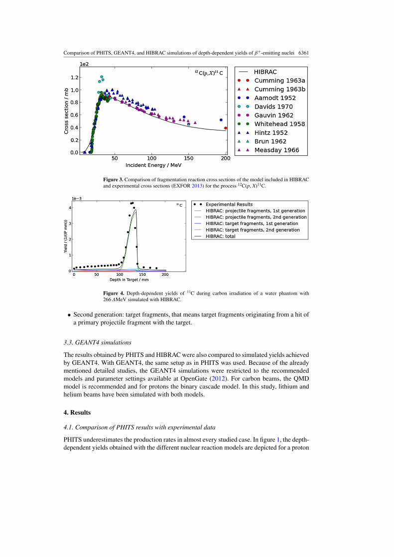
Figure 3. Comparison of fragmentation reaction cross sections of the model included in HIBRACand experimental cross sections (EXFOR 2013) for the process 12C(p, X)11C.
Figure 4. Depth-dependent yields of 11C during carbon irradiation of a water phantom with266 AMeV simulated with HIBRAC.
• Second generation: target fragments, that means target fragments originating from a hit ofa primary projectile fragment with the target.
3.3. GEANT4 simulations
The results obtained by PHITS and HIBRAC were also compared to simulated yields achievedby GEANT4. With GEANT4, the same setup as in PHITS was used. Because of the alreadymentioned detailed studies, the GEANT4 simulations were restricted to the recommendedmodels and parameter settings available at OpenGate (2012). For carbon beams, the QMDmodel is recommended and for protons the binary cascade model. In this study, lithium andhelium beams have been simulated with both models.
4. Results
4.1. Comparison of PHITS results with experimental data
PHITS underestimates the production rates in almost every studied case. In figure 1, the depth-dependent yields obtained with the different nuclear reaction models are depicted for a proton

6362 H Rohling et al
Figure 5. Depth-dependent yields of 15O during 7Li irradiation of a water phantom with162.3 AMeV simulated with HIBRAC: influence of primary projectile fragments on the yieldof target fragments of second generation.
beam. Only minor differences can be recognized between the models. Further simulations showthat the two available total cross section models lead to the same production rate. Moreover, theavailable stopping power models SPAR and ATIMA produce a slight deviation in range, thiscould be very important in the context of monitoring therapeutic particle irradiation. However,tuning of the calculated ranges can easily be done by adjusting the ionization potential usedfor the target material. Finally, the choice of the evaporation model has much impact. Thisinfluence is independent of the combination of the evaporation models with reaction models.In some cases a good agreement with the experimental data is achieved by applying noevaporation model, see figure 2. In other cases the default model behaves best. Unfortunately,no definite statement can be made concerning a recommendation of the evaporation modelsin PHITS for this kind of application.
4.2. Comparison of HIBRAC results with experimental data
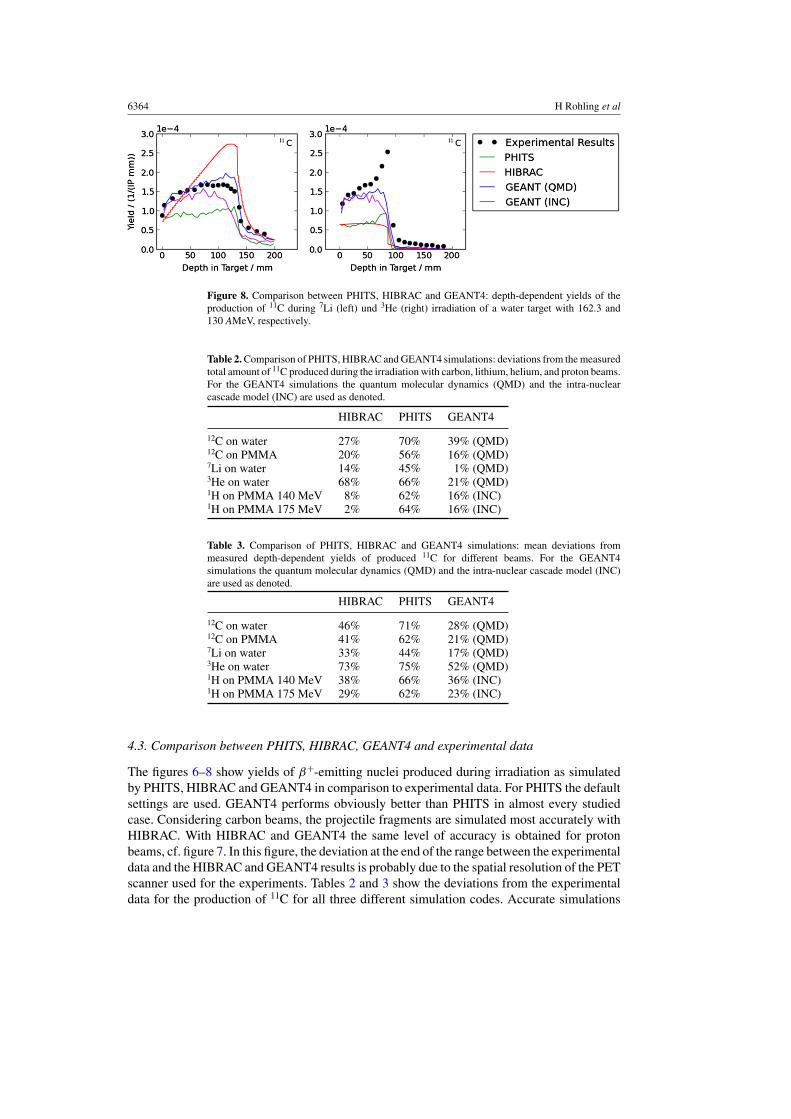
In figure 3, the cross sections for 12C(p, X)11C reactions used in HIBRAC in comparisonwith published experimental data according to EXFOR (2013) are presented. These simulatedresults are in good agreement with the measured values. The same applies to the reaction16O(p, X)15O. Figures 4 and 6 show the simulated yields produced during carbon irradiationtogether with experimental data. For 11C the peak resulting from projectile fragmentation iswell reproduced, however, the plateau resulting from target fragments is too low. For heliumand lithium beams partially large deviations are visible, compare figures 5 and 8. In thehelium, lithium and proton cases, the yields of β+-emitters result only from targetfragmentation.
For the irradiation of a water target with a 162.3 AMeV 7Li beam the fragmentation of thetarget nuclei into 15O is analysed in detail. Figure 5 shows the first and secondary generation oftarget fragments i.e. created via collisions of projectile and projectile fragments, respectively.It seems that the target fragments of first generation are consistent with the experimental datadue to the agreement at the entrance point of the target. The yields of the secondary targetfragments are too high. As shown in figure 5 these secondary target fragments mainly resultfrom collisions of primary projectile fragments with atomic number 1 or 2 with the watertarget. It can be concluded that the cross section for the production of protons and heliumout of 7Li may not be accurate enough, which is discussed in section 2.3. In contrast tothis for proton beams the yields are consistent with the experimental results as shown infigure 7.

Comparison of PHITS, GEANT4, and HIBRAC simulations of depth-dependent yields of β+-emitting nuclei 6363
Figure 6. Comparison between PHITS, HIBRAC, and GEANT4: depth-dependent yields ofproduction of 11C, 15O, and 10C during 12C irradiation of a PMMA target with 266 AMeV.
Figure 7. Comparison between PHITS, HIBRAC and GEANT4: depth-dependent yields of theproduction of 15O and 11C during proton irradiation of a PMMA target with 140 MeV.
6364 H Rohling et al
Figure 8. Comparison between PHITS, HIBRAC and GEANT4: depth-dependent yields of theproduction of 11C during 7Li (left) und 3He (right) irradiation of a water target with 162.3 and130 AMeV, respectively.
Table 2. Comparison of PHITS, HIBRAC and GEANT4 simulations: deviations from the measuredtotal amount of 11C produced during the irradiation with carbon, lithium, helium, and proton beams.For the GEANT4 simulations the quantum molecular dynamics (QMD) and the intra-nuclearcascade model (INC) are used as denoted.
HIBRAC PHITS GEANT4
12C on water 27% 70% 39% (QMD)12C on PMMA 20% 56% 16% (QMD)7Li on water 14% 45% 1% (QMD)3He on water 68% 66% 21% (QMD)1H on PMMA 140 MeV 8% 62% 16% (INC)1H on PMMA 175 MeV 2% 64% 16% (INC)
Table 3. Comparison of PHITS, HIBRAC and GEANT4 simulations: mean deviations frommeasured depth-dependent yields of produced 11C for different beams. For the GEANT4simulations the quantum molecular dynamics (QMD) and the intra-nuclear cascade model (INC)are used as denoted.
HIBRAC PHITS GEANT4
12C on water 46% 71% 28% (QMD)12C on PMMA 41% 62% 21% (QMD)7Li on water 33% 44% 17% (QMD)3He on water 73% 75% 52% (QMD)1H on PMMA 140 MeV 38% 66% 36% (INC)1H on PMMA 175 MeV 29% 62% 23% (INC)
4.3. Comparison between PHITS, HIBRAC, GEANT4 and experimental data
The figures 6–8 show yields of β+-emitting nuclei produced during irradiation as simulatedby PHITS, HIBRAC and GEANT4 in comparison to experimental data. For PHITS the defaultsettings are used. GEANT4 performs obviously better than PHITS in almost every studiedcase. Considering carbon beams, the projectile fragments are simulated most accurately withHIBRAC. With HIBRAC and GEANT4 the same level of accuracy is obtained for protonbeams, cf. figure 7. In this figure, the deviation at the end of the range between the experimentaldata and the HIBRAC and GEANT4 results is probably due to the spatial resolution of the PETscanner used for the experiments. Tables 2 and 3 show the deviations from the experimentaldata for the production of 11C for all three different simulation codes. Accurate simulations

Comparison of PHITS, GEANT4, and HIBRAC simulations of depth-dependent yields of β+-emitting nuclei 6365
of 11C are most important, since today mostly off-line PET or in-room PET starting with thedata acquisition after the irradiation is used for monitoring. At that time the vast majorityof β+-emitters with half-lives shorter than that of 11C (half-life of 20 min) have alreadydecayed. Tables 2 and 3 confirm that GEANT4 shows considerably better agreement withthe experiments than PHITS, both for the total and the depth-dependent 11C-yields and,furthermore, the comparability of HIBRAC with GEANT4 simulations for proton beams.
5. Conclusion
PHITS and HIBRAC were evaluated with respect to their capability to reproduce the productionof β+-emitters during particle therapy. This simulation study includes different types of ionbeams. An extended version of HIBRAC was used for this purpose. With PHITS, the differentimplemented models have been analysed in detail.
5.1. PHITS
The selection of models can be important for simulations of production rates of β+-emitters.Especially, the choice of the evaporation model has much impact on the final results. Forhigher energies this fact has already been shown by Mancusi et al (2011). In general, it can bestated, that PHITS underestimates the yields confirming the results of Seravalli et al (2012)for protons. Recently, PHITS version 2.52, which incorporates the latest intra-nuclear cascademodel INCL4.6 (Cugnon et al 2011) was released. As a next step, this version of PHITSshould be evaluated and compared to the results obtained in this paper.
5.2. HIBRAC
As expected due to the experiences with POSGEN (Ponisch et al 2004) that uses the samecross section parameterizations (Sihver et al 1993) as HIBRAC, the simulated projectilefragmentation to 11C during an irradiation of water and PMMA targets with 12C beams isquite accurate. Furthermore, the experimental yields of 11C and 15O produced during protonirradiation are well reproduced by HIBRAC. Concerning the target fragmentation for helium,lithium and carbon beams, HIBRAC would need improvement. One reason for the founduncertainties is the secondary target fragmentation. This deviation is caused most likely byimprecise production cross sections of protons and helium fragments. However, there is a lackof experimental data for the production of protons and light ions produced from therapeuticproton and ion beams in tissue equivalent material that would be necessary to carefullybenchmark the currently used cross sections models. If more accurate measurements of thesecross sections and yields will be performed (see e.g. Henriquet et al 2012), the cross sectionmodels in HIBRAC could easily be improved. With respect to the implementation in clinicalroutine PT-PET, HIBRAC is a possible candidate due to its robustness and speed comparedto GEANT4 and PHITS. To apply HIBRAC to PT PET in routine therapy monitoring morebenchmarking and adjustment of the applied cross sections is needed. However, for protontherapy, the obtained results are promising.
5.3 GEANT4
GEANT4 shows overall the best results. Due to the mentioned successful application ofGEANT4 in this field of research (Pshenichnov et al 2006, 2007), Bohlen et al (2010), theresults first of all confirm the experimental data. In order to explain the visible deviations from

6366 H Rohling et al
the experimental data, e.g. of the 11C and 10C yields in figure 6, an advanced analysis includingthe different models implemented in GEANT4 would be needed. Furthermore, in the case of10C, the measurement statistics is not optimal.
Acknowledgments
This work was supported by the EU FP7 ENVISION project (grant agreement number241851). Heide Rohling would like to thank Prof Lembit Sihver and his group for hostingher at the Department of Nuclear Engineering at Chalmers University of Technology inGothenburg/Sweden for six month in 2012. Furthermore, we thank Frank Verhaegen, FredericStichelbaut and Katia Parodi for the helpful explanations concerning Seravalli et al (2012).
References
Agostinelli S et al 2003 GEANT4—a simulation toolkit Nucl. Instrum. Methods Phys. Res. A 506 250–303Armstrong T W and Chandler K C 1973 A Fortran program for computing stopping powers and ranges for muons,
charged pions, protons, and heavy ions Oak Ridge Report ORNL-4869 (Oak Ridge, TN: Oak Ridge NationalLaboratory)
Bohlen T T, Cerutti F, Dosanjh M, Ferrari A, Gudowska I, Mairani A and Quesada J M 2010 Benchmarking nuclearmodels of FLUKA and GEANT4 for carbon ion therapy Phys. Med. Biol. 55 5833–47
Cugnon J 1980 Monte Carlo calculation of high-energy heavy-ion interactions Phys. Rev. C 22 1885–96Cugnon J, Mancusi D, Boudard A and Leray S 2011 New features of the INCL4 model for spallation reactions
J. Korean Phys. Soc. 59 955–8Cugnon J, Mizutani T and Vandermeulen J 1981 Equilibration in relativistic nuclear collisions. A Monte Carlo
calculation Nucl. Phys. A 352 505–34Dressner L 1962 EVAP—A FORTRAN program for calculating the evaporation of various particles from excited
compound nuclei Report No ORNL-TM-196 (Oak Ridge, TN: Oak Ridge National Laboratory)Enghardt W, Crespo P, Fiedler F, Hinz R, Parodi K, Pawelke J and Ponisch F 2004 Charged hadron tumour therapy
monitoring by means of PET Nucl. Instrum. Methods Phys. Res. A 525 284–8EXFOR 2013 Experimental nuclear reaction data Database Version of 1st March 2013 (www.nudc.bnl.gov/
exfor/exfor00.htm)Fiedler F 2008 Anwendung des in-beam PET Therapiemonitorings auf Prazisionsbestrahlungen mit Helium-Ionen
PhD Thesis Technische Universitat Dresden, Fakultat fur Mathematik und Naturwissenschaften, Dresden.Wissenschaftlich-Technischer Bericht FZD-494
Fiedler F, Crespo P, Parodi K, Sellesk M and Enghardt W 2006 The feasibility of in-beam PET for therapeutic beamsof 3He IEEE Trans. Nucl. Sci. 53 2252–9
Fiedler F, Priegnitz M, Julich R, Pawelke J, Crespo P, Parodi K, Ponisch F and Enghardt W 2008 In-beam PETmeasurement of biological half-lives of 12C irradiation induced beta+activity Acta Oncol. 47 1077–86
Fiedler F, Shakirin G, Skowron J, Braess H, Crespo P, Kunath D, Pawelke J, Ponisch F and Enghardt W 2010 On theeffectiveness of ion range determination from in-beam PET data Phys. Med. Biol. 55 1989–98
Furihata S 2000 Statistical analysis of light fragment production from medium energy proton-induced reactions Nucl.Instrum. Methods Phys. Res. B 171 251–8
Geissel H and Scheidenberger C 1998 Slowing down of relativistic heavy ions and new applications Nucl. Instrum.Methods Phys. Res. B 136–138 114–24
Gudowska I, Sobolevsky N M, Andreo P, Belkic D and Brahme A 2004 Ion beam transport in tissue-like media usingthe Monte Carlo code SHIELD-HIT Phys. Med. Biol. 49 1933–58
Haberer T, Becher W, Schardt D and Kraft G 1993 Magnetic scanning system for heavy ion therapy Nucl. Instrum.Methods Phys. Res. A 330 296–305
Hasch B G 1996 Die physikalischen Grundlagen einer Verifikation des Bestrahlungsplanes in der Schwerionen-Tumortherapie mit der Positronen-Emissions-Tomographie Dissertation Technische Universitat Dresden,Dresden
Henriquet P et al 2012 Interaction vertex imaging (IVI) for carbon ion therapy monitoring: a feasibility study Phys.Med. Biol. 57 4655–69
Iseki Y et al 2003 Positron camera for range verification of heavy-ion radiotherapy Nucl. Instrum. Methods Phys.Res. A 515 840–9

Comparison of PHITS, GEANT4, and HIBRAC simulations of depth-dependent yields of β+-emitting nuclei 6367
La Tessa C, Sihver L, Mancusi D, Zeitlin C, Miller J, Guetersloh S and Heilbronn L 2007 Weak and strong factorizationproperties in nucleus-nucleus collisions in the energy region 290–2100 MeV/n Nucl. Phys. A 791 451–72
Li Q and Sihver L 2011 Therapeutic techniques applied in the heavy-ion therapy at IMP Nucl. Instrum. MethodsPhys. Res. B 269 666–70
Llacer J 1988 Positron emission medical measurements with accelerated radioactive ion beams Nucl. Sci. Appl.3 111–31
Luhr A, Priegnitz M, Fiedler F, Sobolevsky N and Bassler N 2013 Dependence of simulated positron emitter yieldsin ion beam cancer therapy on modeling nuclear fragmentation Appl. Radiat. Isot. at press
Mancusi D, Boudard A, Cugnon J, David J-C and Leray S 2011 Influence of nuclear de-excitation on observablesrelevant for space exploration Adv. Space Res. 47 1194–99
Mizuno H et al 2003 Washout measurement of radioisotope implanted by radioactive beams in the rabbit Phys. Med.Biol. 48 2269–81
Nara Y, Otuka N, Ohnishi A, Niita K and Chiba S 1999 Relativistic nuclear collisions at 10 AGeV energies fromp+Be to Au+Au with the hadronic cascade model Phys. Rev. C 61 024901
Niita K, Chiba S, Maruyama T, Takada H, Fukahori T, Nakahara Y and Iwamoto A 1995 Analysis of the (N,xN’)reactions by quantum molecular dynamics plus statistical decay model Phys. Rev. C 52 2620–35
Niita K, Iwase H, Sato T, Iwamoto Y, Matsuda N, Sakamoto Y, Nakashima H, Mancusi D and Sihver L 2011 Recentdevelopments of the PHITS code Proc. 5th Int. Symp. on Radiation Safety Detection and Technology; Prog.Nucl. Sci. Technol. 1 1–6
Niita K, Matsuda N, Iwamoto Y, Iwase H, Sato T, Nakashima H, Sakamoto H and Sihver L 2010 PHITS: particleand heavy ion transport code system JAEA-Data/Code 2010–022 (version 2.23)
Nishio T, Miyatake A, Ogino T, Nakagawa K, Saijo N and Esumi H 2010 The development and clinical use of abeam ON-LINE PET system mounted on a rotating gantry port in proton therapy Int. J. Radiat.Oncol. Biol.Phys. 76 277–86
Olson D L, Berman B L, Greiner D E, Heckman H H, Lindstrom P J and Crawford H J 1983 Factorization offragment-production cross sections in relativistic heavy-ion collisions Phys. Rev. C 28 1602–13
OpenGate 2012 Radiation Therapy and Dosimetry as in Dec. 2012 www.opengatecollaboration.org/Parodi K, Enghardt W and Haberer T 2002 In-beam PET measurements of β+ radioactivity induced by proton beams
Phys. Med. Biol. 47 21–36Parodi K et al 2007 Patient study of in vivo verification of beam delivery and range, using positron emission tomography
and computed tomography imaging after proton therapy Int. J. Radiat. Oncol. Biol. Phys. 68 920–34Ponisch F, Parodi K, Hasch B G and Enghardt W 2004 The modelling of positron emitter production and PET imaging
during carbon ion therapy Phys. Med. Biol. 49 5217–32Priegnitz M 2012 Ein neues Konzept zur Modellierung der Positronenemitter-Produktion bei der Partikeltherapie
PhD Thesis Technische Universitat Dresden, DresdenPriegnitz M, Fiedler F, Kunath D, Laube K and Enghardt W 2012 An experiment-based approach for
predicting positron emitter distributions produced during therapeutic ion irradiation IEEE Trans. Nucl. Sci.59 77–87
Priegnitz M, Mockel D, Parodi K, Sommerer F, Fiedler F and Enghardt W 2008 In-beam PET measurement of7Li3+ irradiation induced β+-activity Phys. Med. Biol. 53 4443–53
Pshenichnov I, Larionov A, Mishustin I and Greiner W 2007 PET monitoring of cancer therapy with 3He and 12Cbeams: a study with the GEANT4 toolkit Phys. Med. Biol. 52 7295–312
Pshenichnov I, Mishustin I and Greiner W 2006 Distributions of positron-emitting nuclei in proton and carbon-iontherapy studied with GEANT4 Phys. Med. Biol. 55 6099–112
Sato T, Kase Y, Watanabe R, Niita K and Sihver L 2009 Biological dose estimation for heavy ion therapy using animproved PHITS code coupled with the microdosimetric kinetic model Radiat. Res. 171 107–17
Sato T, Sihver L, Iwase H, Nakashima H and Niita K 2005 Simulations of an accelerator-based shielding experimentusing the particle and heavy-ion transport code system PHITS Adv. Space Res. 35 208–13
Schardt D, Iwase H, Martino G, Kaderka R, La Tessa C, Meer D, Safai S and Sihver L 2010 Energy spectrum of fastneutrons produced by 130 MeV protons in water GSI Scientific Report (Darmstadt: GSI) p 481
Scheidenberger C and Geissel H 1998 Penetration of relativistic heavy ions through matter Nucl. Instrum. MethodsPhys. Res. B 135 25
Seravalli E et al 2012 Monte Carlo calculations of positron emitter yields in proton radiotherapy Phys. Med.Biol. 57 1659–73
Shen W, Wang B, Feng J, Zhan W, Zhu Y and Feng E 1989 Total reaction cross section for heavy-ion collisions andits relation to the neutron excess degree of freedom Nucl. Phys. A 491 130–46
Sihver L, Lantz M, Bohlen T T, Mairani A, Cerutti A F and Ferrari A 2012 A comparison of total reaction crosssection models used in FLUKA, GEANT4 and PHITS IEEE Aerospace Conf. pp 1–10
Sihver L and Mancusi D 2009 Present status and validation of HIBRAC Radiat. Meas. 44 38–46

6368 H Rohling et al
Sihver L, Sato T, Puchalska M and Reitz G 2010 Simulations of the MATROSHKA experiment at the internationalspace station using PHITS Radiat. Environ. Biophys. 49 351–7
Sihver L, Schardt D and Kanai T 1998 Depth-dose distribution of high energy carbon, oxygen and neon beams inwater Japan J. Med. Phys. 18 1–21
Sihver L, Tsao C H, Silberberg R, Barghouty A F and Kanai T 1996 Calculations of depth dose distributions, crosssections and momentum loss Adv. Space Res. 17 105–8
Sihver L, Tsao C H, Silberberg R, Kanai T and Barghouty A F 1993 Total reaction and partial cross section calculationsin proton-nucleus (Zt � 26) and nucleus-nucleus reactions (Zp and Zt � 26) Phys. Rev. C 47 1225–36
Stutzer K, Bert C, Enghardt W, Helmbrecht S, Parodi K, Priegnitz M, Saito N and Fiedler F 2013b Experimentalverification of a 4D reconstruction algorithm used for in-beam PET measurements in particle therapy Phys.Med. Biol. 58 5085–5111
Stutzer K, Menkel S, Bert C, Enghardt W, Helmbrecht S, Saito N and Fiedler F 2013a 4D particle therapy PETsimulation for moving targets irradiated with scanned ion beams Phys. Med. Biol. 58 513–33
Tomitani T et al 2003 Washout studies of 11C in rabbit thigh muscle implanted by secondary beams of HIMAC Phys.Med. Biol. 48 875–89
Toshito T et al 2007 Measurements of total and partial charge-changing cross sections for 200- to 400-MeV/nucleonC12 on water and polycarbonate Phys. Rev. C 75 054606
Tripathi R K, Cucinotta F A and Wilson J W 1996 Accurate universal parameterization of absorption cross sectionsNucl. Instrum. Methods Phys. Res. B 117 347–9
Tripathi R K, Cucinotta F A and Wilson J W 1997 Accurate universal parameterization of absorption cross sections:II—neutron absorption cross sections Nucl. Instrum. Methods Phys. Res. B 129 11–15
Tripathi R K, Cucinotta F A and Wilson J W 1999 Accurate universal parameterization of absorption cross sections:III—light systems Nucl. Instrum. Methods Phys. Res. B 155 349–56