comparison of human mesenchymal stem cells derived from...
TRANSCRIPT
Biomed Pap Med Fac Univ Palacky Olomouc Czech Repub. 2014 Sep; 158(3):373-377.
373
Comparison of human mesenchymal stem cells derived from dental pulp, bone marrow, adipose tissue, and umbilical cord tissue by gene expression
Peter Stankoa,b, Katarina Kaiserovab, Veronika Altanerovab, Cestmir Altanerb,c
Aims. Our aims were to characterize human mesenchymal stem cells isolated from various tissues by pluripotent stem cells gene expression profile.Methods. Four strains of dental pulp stem cells (DP-MSCs) were isolated from dental pulp tissue fragments adhered to plastic tissue culture dishes. Mesenchymal stem cells derived from umbilical cord tissue (UBC-MSCs) were isolated with the same technique. Bone marrow derived mesenchymal stem cells (BM-MSCs) were isolated from nucleated cells of bone marrow obtained by density gradient centrifugation. Human mesenchymal stem cells from adipose tissue (AT-MSCs) were isolated by collagenase digestion. All kinds of MSCs used in this study were cultivated in low glucose DMEM containing 5% or human platelet extract. All stem cell manipulation was performed in GMP conditions. Expression of 15 pluripotent stem cells genes on the level of proteins was measured by Proteome Profiler Human Pluripotent Stem Cell Array. Induction of MSCs to in vitro differentiation to adipocytes, osteoblasts, chondroblasts was achieved by cul-tivation of cells in appropriate differentiation medium. Results. All MSCs tested were phenotypically similar and of fibroblastoid morphology. DP-MSCs and UBC-MSCs were more proliferative than bone marrow BM-MSCs and AT-MSCs. Protein expression of 15 genes typical for pluripotent stem cells distinguished them into two groups. While the gene expression profiles of BM-MSC, AT-MSCs and UBC-MSCs were similar, DP-MSCS differed in relative gene expression on the level of their products in several genes.Conclusions. Dental pulp mesenchymal stem cells cultivated in vitro under the same conditions as MSCs from bone marrow, adipose tissue and umbilical cord tissue can be distinguished by pluripotent stem cell gene expression profile.
Key words: dental pulp, bone marrow, adipose tissue, umbilical cord tissue, mesenchymal stem cells, gene expression
Received: September 5, 2013; Accepted: September 25, 2013; Available online: October 18, 2013http://dx.doi.org/10.5507/bp.2013.078
aDepartment of Stomatology and Maxillofacial Surgery, Faculty of Medicine, Comenius University in Bratislava, Bratislava, Slovak Republic bSt. Elisabeth Cancer Institute, Heydukova 10, 812 50 Bratislava, Slovak Republic cCancer Research Institute, Slovak Academy of Sciences, Vlarska 7, 833 91 Bratislava, Slovak RepublicCorresponding author: Peter Stanko, e-mail: [email protected]
INTRODUCTION
Adult stem cells are present in a variety of tissues in the human body. The most studied adult stem cell popula-tion is the mesenchymal stem cells (MSCs). MSCs pos-sess multi-lineage differentiation potential; they produce a variety of cytokines, chemokines, growth factors that mostly in paracrine fashion are involved in the regenera-tion of used and damaged tissues. MSCs can be readily isolated from several tissues like bone marrow, adipose tissue, umbilical cord tissue and from post-natal dental pulp tissues and other tissues. Multipotent mesenchymal progenitor cells known as dental pulp stromal/stem cells (DP-MSCs), with high proliferative potential for self-renewal are intensively studied because of their neural characteristics1,2. DP-MSCs can play a potential role in peripheral neural regeneration3. DP-MSCs as cells of ec-tomesenchymal origin can serve as an excellent source for generation induced pluripotent cells4. Recently it was shown that DP-MSC are a promising source for cell-based therapy for immune diseases such as systemic lupus ery-thematosus5.
Generally, mesenchymal (stromal) stem cells have several interesting properties responsible for the induc-tion of endogenous reparatory processes in the body. For instance human bone marrow mononuclear concentrates containing MSCs were shown to be an effective thera-peutic strategy for “no option“ patients with critical limb ischemia preventing limb amputations6. The quality of the BM-MSCs as gene expression and cell surface mark-ers, determined the success in the ischemic ulcers treat-ment7. Discoveries of the immunomodulatory functions of MSCs have suggested that they might have therapeutic use in treating immune diseases8,9. MSCs also possess tumor tropic property; they can be recruited by tumors and metastases10. Genetically modified MSCs are the ba-sis for cancer gene therapy using suicide genes (reviewed in11,12). MSCs represent novel promising therapeutic tools in emerging regenerative medicine. MSCs of different ori-gin are being tested in a large number of clinical trials for at present untreatable diseases. As of August 2013, the public clinical trials database http://clinicaltrials.gov showed 344 clinical trials using MSCs for a very wide range of therapeutic applications.
Biomed Pap Med Fac Univ Palacky Olomouc Czech Repub. 2014 Sep; 158(3):373-377.
374
MATERIALS AND METHODS
Cell cultures All donors of adipose tissue, bone marrow, teeth, um-
bilical cord tissue and blood platelets provided informed written consent.
Human BM-MSCs were isolated from bone marrow aspirates taken from the iliac crest of normal adult donors by procedures described previously7. Briefly, BM-MSCs were isolated from nucleated bone marrow cells obtained by density gradient centrifugation by adherence to plas-tic dishes. The separated fraction was resuspended in a complete culture medium DMEM low glucose (1 g/L) supplemented with 5% human platelet extract (PE) and incubated at 37 °C in humidified atmosphere with 5% CO2.
Human AT-MSCs were isolated from lipoaspirates using a collagenase type VII digestion and plastic adher-ence technique as described previously9. The material was obtained from healthy individuals undergoing elective li-poaspiration.
Dental pulp stem cells (DP-MSCs) were isolated from dental pulp tissue fragments adhered to plastic tissue cul-ture dishes. The same technique was used to obtain um-bilical cord tissue mesenchymal stem cells (UBC-MSCs).
For expansion of all kinds of MSCs, the cells were seeded at 4000 cells/cm2 to plastic flasks and grown with medium exchange every 2-3 days. The cells were then har-vested with trypsin/EDTA, resuspended at 1×10⁶ cells/ml in 10% dimethylsulfoxide and 30% human serum albumin and frozen in 1 ml aliquots in liquid nitrogen.
Platelet extract preparations Platelet extract was prepared from human platelets of
healthy blood donors by procedures described previous-ly10. Briefly, platelets in bags contained 5x10¹¹/L platelets in blood plasma were twice frozen at -80 ºC and subse-quently thawed at 37 ºC. Lysed platelets and platelets bod-ies were eliminated by centrifugation and the supernatant was filtered through a 0.22 μm GP Millipore Express Plus Membrane Stericup and designated as platelet extract
(PE). PE isolates were negative for bacterial, fungal and mycoplasma contaminations.
Differentiation of MSCs in vitroThe ability of MSCs to differentiate to adipocytes, os-
teoblasts and chondrocytes in vitro was evaluated using Human Mesenchymal Stem Cell Functional Identification Kit (R&D SYSTEMS Minneapolis, MN 55413).
Protein concentration was measured by Pierce™ BCA Protein Assay Kit (Thermo SCIENTIFIC) according to the manufacturer’s instructions.
Expression of MSCs genesExpression of 15 pluripotent stem cell genes on the
level of proteins was m easured by Proteome Profiler Human Pluripotent Stem Cell Array (R&D SYSTEMS Minneapolis, MN 55413). Relative expression levels of individual genes were detected according to the manufac-turer’s instructions. Briefly, cultured MSCs were rinsed with PBS, disintegrated by lysis buffer and the cell extract was prepared by centrifugation according to the recom-mendations of the provider. Total protein concentration was determined. The same amount of protein was used for incubation of all arrays and procedure was performed as directed by the manufacturer. Membranes were exposed to Western HRP Substrate (LuminataTM Forte), covered with plastic wrap and exposed to X-ray film. Pluripotent stem cell array data on developed X-ray film was quanti-fied by scanning the film by a transmission mode scan-ner. ImageJ software (NIH) was used for the quantitative evaluation.
StatisticsSigmaPlot 11.0 software for Windows (Systat Software,
Germany) was used for statistical analysis. Student's t-tests were used for the data that passed the Shapiro-Wilk normality test and the Mann-Whitney Rank Sum test for non parametric data.
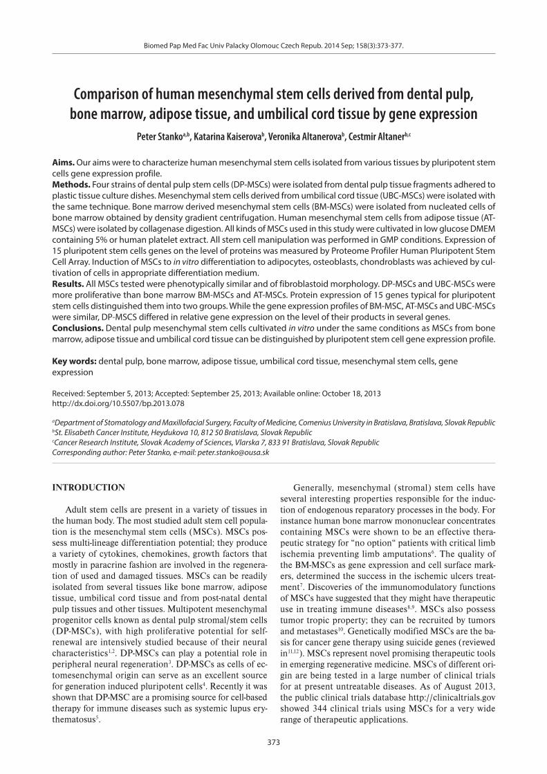
Fig. 1. Protein expression of 15 pluripotent stem cell genes. Cell extracts of BM-MSCs (n=10), AT-MSCs (n=4), UBC-MSCs (n=4) and DP-MSCs (n=4) were examined by Proteome Profiler Human Pluripotent Stem Cell Array and quantified by ImageJ program.

Biomed Pap Med Fac Univ Palacky Olomouc Czech Repub. 2014 Sep; 158(3):373-377.
375
RESULTS
Relative expression of 15 pluripotent stem cell genes on the level of proteins
Tissue cultures of BM-MSCs derived from early pas-sage of ten bone marrow donors, four AT-MSCs, four um-bilical tissue derived MSCs and four MSCs prepared from dental pulp tissue were examined for relative expression of 15 genes by Proteome Profiler Human Pluripotent Stem Cell Array. The data quantified by ImageJ software are presented in Fig. 1.
Relative expression of all tested genes in MSCs de-rived from bone marrow, adipose tissue and umbilical tissues did not differ significantly. In DP-MSCs higher relative expression was found for the following genes: E-cadherin, Goosecold (GSC), PDX-1/IPF1 (statistical significance P≤0.001) and, Sox2 (P=0.013) in comparison with expressions in BM-MSCs. Lower relative expression
of genes Snail (P≤0.001) Sox17 (P=0.006) and gene Oct-3/4 (P=0.006) was detected in the same comparison. Thus DP-MSCs differed substantionally from MSCs derived from bone marrow, adipose tissue and umbilical cord tis-sue.
Differentiation of MSCs in vitroDP-MSCs can be induced to all differentiation lineages
required for MSCs characterization in vitro. Interestingly, while BM-MSCs and AT-MSCs showed a gradual loss of osteogenic differentiation potential with increasing num-ber of passages in vitro, DP-MSCs on the other hand had an increasing potential (Fig. 2.).
Presence of cell surface markers All tested MSCs were examined for expression of cell
surface markers by flow cytometry. All MSCs tested ex-pressed CD44, CD90, CD105 markers and were negative
Fig. 2. Differentiation of BM-MSCs, AT-MSCs, UBC-MSCs and DP-MSCs in vitro. Human Mesenchymal Stem Cell Functional Identification Kit was used for in vitro induction of differentia-tion of all MSCs into osteogenic, adipogenic and chondrogenic lineages.
Biomed Pap Med Fac Univ Palacky Olomouc Czech Repub. 2014 Sep; 158(3):373-377.
376
for expression of CD34, CD45 surface antigens (data not shown).
DISCUSSION
Adult human MSCs are factories that produce a large number of bioactive factors that induce in the body, the molecular processes of regenerative paths. Bone marrow-derived MSCs (BM-MSCs) secrete factors that act in a paracrine manner to promote complex endogenous repa-ratory processes. MSCs act through interactions with the endogenous cells and tissues. They are also respon-sive to their environment and can modify their activities and functions depending on the biomolecular context. MSCs can accelerate wound closure by modulating the inflammatory environment, promoting the formation of a well-vascularized granulation matrix, encouraging the migration of keratinocytes, and inhibiting apoptosis of wound healing cells. Numerous in vivo studies provide overwhelming evidence that the biologically active com-pounds secreted by BM-MSCs play the main role in their therapeutic potential. An important part of the healing process is suppression of the immune response and/or immunomodulation induced by MSCs. There is no evi-dence that applied cells are transformed into target tissue. Therefore, understanding the gene expression profile that indicates their lineage-specific proclivity is fundamental to the development of successful cell-based therapies.
Our comparison of human mesenchymal stem cells derived from different tissues revealed no differences in cell morphology or in the expression of surface markers typical for mesenchymal stem cells. However, a compari-son of the protein levels of several genes typical for pluri-potent stem cells led to classifying these MSCs into two groups. There were no significant differences in the level of 15 pluripotent stem cell genes of BM-MSCs, AT-MSCs and UBC-MSCs. We found highly significant differences in the amount of protein products in DP-MSCs. All cells were cultivated in the same culture fluid with human growth factors from platelets.
Observed differences in gene expression of pluripotent stem cell genes in DP-MSCs might reflect their embryonic stem cell origin. Dental pulp is made of ecto-mesenchymal elements, containing neural crest-derived cells, which dis-play plasticity and multipotential capabilities13. Mixed em-bryonic origin of DP-MSCs might be connected with our observation of the lower level of transcription factor char-acteristic for embryonic stem cells Oct-3/4 in comparison with BM-MSCs. Determination of Oct3/4 on protein level rules out the role of several Oct-4 pseudogenes present in normal genome. Higher expression of gene Goosecoid, which acts as a transcription factor is obviously connected with its involvement in embryogenesis, where plays a role in craniofacial and rib cage development14.
We found in DP-MSCs higher expression of prod-uct of gene E-cadherin - calcium dependent adhesive molecule and lower expression of gene Snail. Snail acts as the E-cadherin repressor. Snail is a zinc finger tran-
scriptional repressor that downregulates the expression of ectodermal genes within the mesoderm. Cadherins and transcription factor Snail are specific markers of Epithelial-Mesenchymal Transition (EMT). EMT plays a role in maintenance of embryonic mesoderm, growth ar-rest, survival and cell migration15,16. Higher expression of E-cadherin and lower expression of Snail favor DP-MSCs for reparative functions where EMT plays an important role. This might also reflect our observation of the abil-ity of DP-MSCs to undergo osteogenic differentiation despite the large number of passages in vitro that differ from the other MSCs. This is in agreement with observa-tion of Hara and coworkers17,18 that expression of bone morphogenic protein 4 in DP-MSCs is much higher than in BM-MSCs.
The transcription factor PDX1 plays a critical role in glucose-induced insulin gene transcription in adult β-cells. High expression of Pdx1 in DP-MSCs suggests that those cells might be useful for treatment of diabetes and/or could be a good source of cells for transdifferentiation to beta cells19.
The product of Sox2 gene and Sox 17 both impor-tant transcriptional regulators are required for stem-cell maintenance in the central nervous system20. Sox2 may function as a switch in neuronal development. Higher expression of Sox2 reflects probably the ability of DP-MSCs to be induced in vitro to undergo neuronal differ-entiation – a property that differs from the other MSCs. This supports our observation that DP-MSCs cultivated in neurobasal medium form spheroids.
MSCs isolated from different tissues have heterolo-gous populations of clones with various growth and/or regenerative potential. Gene expression profiles might be one of characterizing for use in future clinical applica-tions. Microarray analysis was used to compare the global gene expression profiles of high growth/multipotential clones with low growth potential cell clones derived from 3 stromal tissues21.
Whether the difference in gene expressions in DP-MSCs compared with BM-MSCs, AT-MSCs and UBC-MSCs is reflected in their secretome, remains to be analysed.
ACKNOWLEDGMENTS
The study was supported by a grant project obtained from the Slovak League against Cancer. We wish to thank to L. Hunakova, Ph.D. for statistical analysis.
Authorship contribution: All authors contributed equally to the study.
REFERENCES
1. Wang S, Qu X, Chunhua Zhao R. Clinical applications of mesenchy-mal stem cells Journal of Hematology & Oncology 2012;5:19.
2. Suchanek J, Soukup T, Visek B, Ivancakova R, Kucerova L, Mokry J. Dental pulp stem cells and their characterization. Biomed Pap Med Fac Univ Palacky Olomouc Czech Repub 2009;153:31-36.
Biomed Pap Med Fac Univ Palacky Olomouc Czech Repub. 2014 Sep; 158(3):373-377.
377
3. Martens W, Bronckaers A, Politis C, Jacobs R, Lambrichts I. Dental stem cells and their promising role in neural regeneration: an update. Clin Oral Investig 2013; Jul 12. [Epub ahead of print] doi: 10.1007/s00784-013-1030-3
4. Xing Y, Haiyan Q, Cunye Q, Rocky STuan, Songtao Shi.George T.,Huang J. iPS cells reprogrammed from human mesenchymal-like stem/progenitor cells of dental tissue origin. Stem Cells Dev 2010;19(4):469-80. doi: 10.1089/scd.2009.0314
5. Makino Y, Yamaza H, Akiyama K, Ma L, Hoshino Y, Nonaka K, Terada Y, Kukita T, Shi S, Yamaza T. Immune Therapeutic Potential of Stem Cells from Human Supernumerary Teeth. J Dent Res 2013;92:609-15.
6. Klepanec A, Mistrik M, Altaner C, Valachovicova M, Olejarova I, Slysko R, Balazs T, Urlandova T, Hladikova D, Liska B, Tomka J, Vulev I, Madaric J. No Difference in Intraarterial and Intramuscular Delivery of Autologous Bone-Marrow Cells in Patients with Advanced Critical Limb Ischemia. Cell Transplant 2012;21:1909-18.
7. Altaner C, Altanerova V, Cihova M, Hunakova L, Kaiserova K, Klepanec A, Vulev I, Madaric J. Characterization of Mesenchymal Stem Cells of “No-Options” Patients with Critical Limb Ischemia Treated by Autologous Bone Marrow Mononuclear Cells. PLoS One. 2013;8(9):(in press). doi:10.1371/journal.pone.0073722
8. Le Blanc C, Tammik K, Rosendahl E, Zetterberg Ringd´en O. HLA expression and immunologic properties of differentiated and un-differentiated mesenchymal stem cells. Experimental Hematology 2003;10:890-6.
9. Kucerova L, Altanerova V, Matuskova M, Tyciakova S, Altaner C. Adipose tissue-derived human mesenchymal stem cells mediated prodrug cancer gene therapy. Cancer Res 2007;67:6304-13.
10. Altanerova V, Cihova M, Babic M, Rychly B, Ondicova K, Mravec B, Altaner C. Human adipose tissue-derived mesenchymal stem cells expressing yeast cytosinedeaminase: uracil phosphori-bosyltransferase inhibit intracerebral rat glioblastoma. Int J Cancer;2012;130:2455-63.
11. Altaner C. Prodrug cancer gene therapy. Review. Cancer Lett 2008;270:191-201.
12. Cihova M, Altanerova V, Altaner C. Stem cell based cancer gene therapy. Mol Pharm 2011;8:1480-7.
13. Sinanan AC, Hunt NP, Lewis MP. Human adult craniofacial muscle-de-rived cells: neural-cell adhesion-molecule (NCAM; CD56)-expressing cells appear to contain multipotential stem cells. Biotechnol Appl Biochem 2004;40:25-34.
14. Takumi Era. Pluripotent stem cells as a source of mesenchymal stem cells Review article. Inflammation and Regeneration 2013;33:19-28.
15. Carver EA, Jiang R, Lan Y, Oram KF, Gridley T. The Mouse Snail Gene Encodes a Key Regulator of the Epithelial-Mesenchymal Transition. Mol Cell Biol 2001;21:8184-8.
16. Jiao W, Miyazaki K, Kitajima Y. Inverse correlation between E-cadherin and Snail expression in hepatocellular carcinoma cell lines in vitro and in vivo. Br J Cancer 2002:86:98-101.
17. Hara K, Yamada Y, Nakamura S, Umemura E, Ito K, Ueda M. Potential characteristics of stem cells from human exfoliated deciduous teeth compared with bone marrow-derived mesenchymal stem cells for mineralized tissue-forming cell biology. J Endod 2011;37:1647-52.
18. Nakamura S, Yamada Y, Katagiri W, Sugito T, Ito K, Ueda M. Stem cell proliferation pathways comparison between human exfoliated de-ciduous teeth and dental pulp stem cells by gene expression profile from promising dental pulp. J Endod 2009;35:1536-42.
19. El-Badri N, Ghoneim MA. Mesenchymal Stem Cell Therapy in Diabetes Mellitus: Progress and Challenges. J Nucleic Acids 2013;2013:194858. doi: 10.1155/2013/194858
20. Lee H J, Wu J, Chung J, Wrathall JR. SOX2 expression is upregulated in adult spinal cord after contusion injury in both oligodendrocyte lineage and ependymal cells. J Neurosci Res 2013;91:196-210.
21. Menicanin D, Bartold PM, Zannettino AC, Gronthos S. Identification of a common gene expression signature associated with immature clonal mesenchymal cell populations derived from bone marrow and dental tissues. Stem Cells Dev 2010;19:1501-10.