comparison of 3 and 7 tesla magnetic resonance imaging of … · 2016-11-01 · obstructive...
TRANSCRIPT

150
INTRODUCTION
Tectal gliomas are a distinct form of pediatric brainstem tu-mor. Patients typically present with symptoms related to in-creased intracranial pressure, which results from obstructive hydrocephalus [1]. We report 7.0 Tesla magnetic resonance imaging (7T MRI) results in a case of obstructive hydroceph-alus caused by tectal glioma.
The recent developments in 7T MRI have been fueled by the promise of increased signal-to-noise ratio (SNR) compared with that of 1.5T and 3T MRI. Several pilot studies have used 7T MRI to image brain tumors [2-6]. Phase-contrast cine MRI provides important information on the hemodynamics of ce-rebrospinal fluid (CSF) circulation [7]. CSF flow physiology and pathology have been investigated with the phase-contrast cine MRI method over the last 15 years [8-10]. 7T brain MRI can potentially improve our understanding of CSF flow in cas-
Comparison of 3 and 7 Tesla Magnetic Resonance Imaging of Obstructive Hydrocephalus Caused by Tectal GliomaHyeong Cheol Moon1, Hyeon-Man Baek2,3, Young Seok Park1,4
1Medical Neuroscience and Neurosurgery, 4Department of Neurosurgery, Chungbuk National University Hospital, Cheongju, Korea2Bioimaging Research Team, Korea Basic Science Institute, Cheongju, Korea3Department of Bio-Analytical Science, Korea University of Science and Technology, Daejeon, Korea
Received August 5, 2016Revised August 31, 2016Accepted September 27, 2016
CorrespondenceYoung Seok ParkDepartment of Neurosurgery, Chungbuk National University, College of Medicine, 1 Chungdae-ro, Sewon-gu, Cheongju 28644, KoreaTel: +82-43-268-6080Fax: +82-31-273-1614E-mail: [email protected]
Obstructive hydrocephalus caused by tectal glioma, which relived by neuroendoscopy, have been de-scribed using 3.0 Tesla magnetic resonance imaging (3T MRI) so far, we present the results obtained from 3T and 7T MRI in this patient. A 21-year-old woman presented at our hospital with gait disturbance, hormonal insufficiency, and urinary incontinence that began prior to 6 years of age. 3.0T MRI revealed a non-enhancing tectal mass along with obstructive hydrocephalus. The mass measured approximately 1.1×1.0×1.2 cm. An endoscopic third ventriculostomy was performed to relieve the hydrocephalus. We compared hydrocephalus and cerebrospinal fluid (CSF) flow findings from 3T and 7T MRI, both preoper-ative and postoperative at 1, 6 months. Intraventricular CSF voiding on T2-weighted images obtained with 7T MRI showed greater fluid inversion than those obtained with 3T MRI. This study shows that 7T brain MRI can provide detailed information on hydrocephalus caused by tectal glioma. Further studies are needed to develop refined 7T MRI protocols for better images of hydrocephalus.
Key Words Magnetic resonance imaging; Brain neoplasms; Hydrocephalus; Glioma.
es of hydrocephalus by providing detailed anatomical and pathophysiological data.
In this study, we report the results of 7T brain MRI in a pa-tient with obstructive hydrocephalus caused by tectal glioma to document the safety and highlight the potential benefits of 7T MRI for the clinical diagnosis of hydrocephalus. We also report the results of a comparison between 3T and 7T MRI in this patient.
CASE REPORT
A 21-year-old woman presented at our hospital with gait disturbance and urinary incontinence persisting since before the age of 6. We observed exophthalmos and papilledema, and the patient complained of diplopia, primary amenorrhea, and cognitive decline over several years. A computed tomog-raphy scan showed obstructive hydrocephalus with transep-endymal edema. MRI revealed a non-enhancing mass of tec-tum with obstructive hydrocephalus. The mass measured approximately 1.1×1.0×1.2 cm (Fig. 1). We performed an endoscopic third ventriculostomy (ETV) and fenestration at the floor of the third ventricle into the prepontine cistern, but
CASE REPORT Brain Tumor Res Treat 2016;4(2):150-154 / pISSN 2288-2405 / eISSN 2288-2413http://dx.doi.org/10.14791/btrt.2016.4.2.150
This is an Open Access article distributed under the terms of the Creative Commons Attribution Non-Commercial License (http://creativecommons.org/licenses/by-nc/3.0) which permits unrestricted non-commercial use, distribution, and reproduction in any medium, provided the original work is properly cited.Copyright © 2016 The Korean Brain Tumor Society, The Korean Society for Neuro-Oncology, and The Korean Society for Pediatric Neuro-Oncology

HC Moon et al.
151
Fig. 2. Preoperative 7T MR axial T2-weighted image with hydrocephalus (A), axial T2-weighted image showing tectal glioma (B), and sagit-tal T1-weighted image (C). Postoperative 6 month MRI imaging (D-F). The arrow showing tectal glioma.
A B C
D E F
A B C
D E FFig. 1. Preoperative 3T MRI imaging. A: Axial T2-weighted 3T MR image confirming the presence of significant hydrocephalus. B: Axial T2-weighted 3T MR imaging confirming the tumor in the tectal plate. C: Axial T1-weighted gadolinium enhance 3T MR imaging confirming the presence of significant hydrocephalus. D: Axial T1-weighted gadolinium enhance 3T MR imaging confirming the the tumor in the tectal plate. E: Coronal T1-weighted gadolinium enhance 3T MR imaging. F: Sagittal T1-weighted 3T MR imaging hydrocephalus with non enhancing mass.

152 Brain Tumor Res Treat 2016;4(2):150-154
7T MRI of Tectal Glioma
we did not obtain biopsy samples from the tectal glioma. These procedures were successful in ameliorating the patient’s neu-rological symptoms and hydrocephalus. We performed 3T and 7T brain MRI, both preoperatively and postoperatively at 1, 6 months (Fig. 1, 2, 3). At the postoperative follow-up, the patient reported that she had begun menstruating for the first time, and also reported a dramatic improvement of her diplopia.
We tested whether CSF flow dynamic parameters were dif-ferent in 3T and 7T phase-contrast cine MRI, and whether ETV had an effect on MRI findings. 3T phase-contrast cine MRI was performed on a 3T Philips MR system (Philips Healthcare, Cleveland, OH, USA). The standard scanning protocol included a fluid attenuation inversion recovery im-age (repetition time 8,000 ms; nominal echo time 388 ms; in-version time 2,200 ms; matrix size 344×342; voxel size
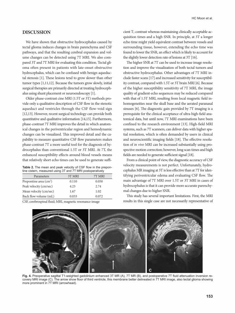
0.7×0.7×0.7 mm3; scan duration 8 min) and a T2-weighted image (repetition time 3,000 ms; echo time 56 ms; matrix size 488×396; voxel size 0.5×0.5×0.5 mm2; scan duration 6 min 48 s), which were used to assess small vessel pathology (Fig. 2, 3, Table 1). The floor of third ventricle, this mem-brane better delineated in 7T MRI image, also tectal glioma showing more prominent in 7T MRI (Fig. 4). We measured the mean and peak velocity of CSF flow in the prepontine cistern postoperatively. Postoperative 3T phase-contrast cine MRI showed increased CSF flow in the prepontine area (Ta-ble 2). This confirm flow of CSF through endoscopic fenes-tration site using both 3T and 7T MRI. 7T scans were acquired using a whole-body 7T MR system (Philips Healthcare, Cleveland, OH, USA) with a volume transmit and 16- or 32-channel receive head coil (Nova Medical, Wilmington, MA, USA).
Fig. 3. Preopeartive imaging (A and D) and postoperative 1 month (B and E), postop 6 month 7T (C and F) fluid attenuation inversion re-covery (FLAIR) MR studies showing parenchymal change and ventricle size. Preoperative (D) and postoperative (E and F) sagittal FLAIR MR image demonstrating a decrease in the ventricle scalloping and stable appearance of the tectal glioma.
A B C
D E F
Table 1. Imaging protocol for 7T MRI
Parameters T2 T1 FLAIRTR 3,000 4.6 8,000/2,200 (TI)TE 56 2.3 388FA 110 8 40FOV 240×240×200 240×240×160 240×240×168Voxel size 0.5×0.5×0.5 0.5×0.5×0.5 0.7×0.7×0.7Matrix size 488×396 480×480 344×342Acquisition time 6:48 min 5:53 min 8:00 minTR, repetition time; TE, echo time; FA, flip angle; FOV, field of view; FLAIR, fluid attenuation inversion recovery; TI, the inversion time
HC Moon et al.
153
DISCUSSION
We have shown that obstructive hydrocephalus caused by tectal glioma induces changes in brain parenchyma and CSF pathways, and that the resulting cerebral expansion and vol-ume changes can be detected using 7T MRI. We also com-pared 3T and 7T MRI for evaluating this condition. Tectal gli-oma often present in patients with late-onset obstructive hydrocephalus, which can be confused with benign aqueduc-tal stenosis [1]. These lesions tend to grow slower than other tumor types [1,11,12]. Because the tumors grow slowly, initial surgical therapies are primarily directed at treating hydroceph-alus using shunt placement or neuroendoscopy [1].
Older phase-contrast cine MRI (1.5T or 3T) methods pro-vide only a qualitative description of CSF flow in the stenotic aqueduct and ventricles through the CSF flow-void sign [12,13]. However, recent surgical technology can provide both quantitative and qualitative information [14,15]. Furthermore, phase-contrast 7T MRI improves the detail in which anatom-ical changes in the periventricular region and hemodynamic changes can be visualized. This improved detail and the ca-pability to measure quantitative CSF flow parameters makes phase-contrast 7T a more useful tool for the diagnosis of hy-drocephalus than conventional 1.5T or 3T MRI. At 7T, the enhanced susceptibility effects around blood vessels means that relatively short echo times can be used to generate suffi-
cient T2 contrast whereas maintaining clinically acceptable ac-quisition times and a high SNR. In principle, at 3T a longer echo time might yield equivalent contrast between vessels and surrounding tissue, however, extending the echo time was found to lower the SNR, an effect which is likely to account for the slightly lower detection rate of lesions at 3T [16].
The higher SNR at 7T can be used to increase image resolu-tion and improve the visualization of both tectal tumors and obstructive hydrocephalus. Other advantages of 7T MRI in-clude faster scans [17] and increased sensitivity for susceptibil-ity contrast, compared with 1.5T or 3T brain MRI [6]. Because of the higher susceptibility sensitivity of 7T MRI, the image quality of gradient-echo sequences may be reduced compared with that of 1.5T MRI, resulting from local magnetic field in-homogeneities near the skull base and the aerated paranasal sinuses [6]. The diagnostic gain provided by 7T imaging is a prerequisite for the clinical acceptance of ultra-high-field ana-tomical data, but until now, 7T MRI examinations have been confined to the research environment [13]. High-field MRI systems, such as 7T scanners, can deliver data with higher spa-tial resolution, which is often demanded by users in clinical and neuroscientific imaging fields [18]. The effective resolu-tion of in vivo MRI can be increased substantially using pro-spective motion correction; however, long scan times and high fields are needed to generate sufficient signal [18].
From a clinical point of view, the diagnostic accuracy of CSF velocity measurements is not perfect. Unfortunately, hydro-cephalus MR imaging at 3T is less effective than at 7T for iden-tifying periventricular edema and evaluating CSF flow. The main advantage of 7T MRI over 1.5T or 3T MRI in cases of hydrocephalus is that it can provide more accurate parenchy-mal changes due to higher SNR.
This study has several important limitations. First, the MRI results in this single case are not necessarily representative of
Table 2. The mean and peak velocity of CSF flow in the prepon-tine cistern, measured using 3T and 7T MRI postoperatively
Parameters 3T MRI 7T MRIPrepontine area (cm2) 0.110 0.050Peak velocity (cm/sec) 4.23 2.74Mean velocity (cm/sec) 1.67 1.02Back flow volume (mL) 0.033 0.072CSF, cerebrospinal fluid; MRI, magnetic resonance image
Fig. 4. Preoperative sagittal T1-weighted gadolinium enhanced 3T MR (A), 7T MR (B), and postoperative 7T fluid attenuation inversion re-covery MRI image (C). The arrow show floor of third ventricle, this membrane better delineated in 7T MRI image, also tectal glioma showing more prominent in 7T MRI (arrowhead).
A B C

154 Brain Tumor Res Treat 2016;4(2):150-154
7T MRI of Tectal Glioma
those for hydrocephalus resulting from other medical condi-tions. Second, we did not perform cine MRI at 7T. Further technological advances are necessary to measure CSF velocity at 7T. Third, we did not perform any contrast-enhanced studies at 7T, and further research is necessary to determine whether 7T MRI can be useful for diagnosis of obstructive hydrocepha-lus with tectal glioma. Fourth, we lacked MR hardware, such as more efficient Radiofrequency coils coils and stronger or faster gradient coils, that are necessary for effective 7T MRI, accord-ing to Feinberg and Mark [18] and Stucht et al. [19].
We have shown that 7T brain MRI can be performed safely in a patient with hydrocephalus and intra-axial brain tumors. 7T brain MRI offers a more detailed view of brain parenchy-ma compressed by hydrocephalus than 3T MRI. We also re-port the first use of phase-contrast 7T cine MRI. 7T MRI and phase-contrast cine 7T MRI are feasible and valuable tools in the diagnosis and treatment of hydrocephalus with tectal gli-oma. The benefits of 7T MRI over conventional 1.5T or 3T MRI for hydrocephalus include the quantitative measurement of CSF flow parameters and enhanced anatomical detail. How-ever, further study is needed to develop refined MRI proto-cols for better imaging of hydrocephalus.
Conflicts of InterestThe authors have no financial conflicts of interest.
AcknowledgmentsThis work was supported by through grant of Chungbuk national uni-
versity, Chungbuk national university hospital and the National Research Foundation of Korea (NRF NRF 2014K1A3A1A21001372).
REFERENCES
1. Li KW, Roonprapunt C, Lawson HC, et al. Endoscopic third ventricu-lostomy for hydrocephalus associated with tectal gliomas. Neurosurg Focus 2005;18(6A):E2.
2. Lupo JM, Banerjee S, Hammond KE, et al. GRAPPA-based suscepti-bility-weighted imaging of normal volunteers and patients with brain tumor at 7 T. Magn Reson Imaging 2009;27:480-8.
3. Moenninghoff C, Maderwald S, Theysohn JM, et al. Imaging of adult astrocytic brain tumours with 7 T MRI: preliminary results. Eur Radi-ol 2010;20:704-13.
4. Cho ZH, Kang CK, Han JY, et al. Observation of the lenticulostriate arteries in the human brain in vivo using 7.0T MR angiography. Stroke
2008;39:1604-6.5. Dashner RA, Kangarlu A, Clark DL, RayChaudhury A, Chakeres DW.
Limits of 8-Tesla magnetic resonance imaging spatial resolution of the deoxygenated cerebral microvasculature. J Magn Reson Imaging 2004;19:303-7.
6. Paek SL, Chung YS, Paek SH, et al. Early experience of pre- and post-contrast 7.0T MRI in brain tumors. J Korean Med Sci 2013;28:1362-72.
7. Chen G, Zheng J, Xiao Q, Liu Y. Application of phase-contrast cine magnetic resonance imaging in endoscopic aqueductoplasty. Exp Ther Med 2013;5:1643-1648.
8. Akay R, Kamisli O, Kahraman A, Oner S, Tecellioglu M. Evaluation of aqueductal CSF flow dynamics with phase contrast cine MR imaging in idiopathic intracranial hypertension patients: preliminary results. Eur Rev Med Pharmacol Sci 2015;19:3475-9.
9. Forner Giner J, Sanz-Requena R, Flórez N, et al. Quantitative phase-con-trast MRI study of cerebrospinal fluid flow: a method for identifying pa-tients with normal-pressure hydrocephalus. Neurologia 2014;29:68-75.
10. Fin L, Grebe R. Three dimensional modeling of the cerebrospinal fluid dynamics and brain interactions in the aqueduct of sylvius. Comput Methods Biomech Biomed Engin 2003;6:163-70.
11. Conner M, Gillespie J, Schiff E, Holmes D, Frey M, Quick S. Experi-mental infection of horses and ponies by oral and intranasal routes with New York State reovirus type 3 and German reovirus types 1 and 3 equine isolates. Zentralbl Veterinarmed B 1984;31:707-17.
12. Conner ES, Foley L, Black PM. Experimental normal-pressure hydro-cephalus is accompanied by increased transmantle pressure. J Neuro-surg 1984;61:322-7.
13. Schroeder HW, Schweim C, Schweim KH, Gaab MR. Analysis of aq-ueductal cerebrospinal fluid flow after endoscopic aqueductoplasty by using cine phase-contrast magnetic resonance imaging. J Neurosurg 2000;93:237-44.
14. Hayashi N, Matsumae M, Yatsushiro S, Hirayama A, Abdullah A, Ku-roda K. Quantitative analysis of cerebrospinal fluid pressure gradients in healthy volunteers and patients with normal pressure hydrocepha-lus. Neurol Med Chir (Tokyo) 2015;55:657-62.
15. Yatsushiro S, Hirayama A, Matsumae M, Kuroda K. Visualization of pulsatile CSF motion separated by membrane-like structure based on four-dimensional phase-contrast (4D-PC) velocity mapping. Conf Proc IEEE Eng Med Biol Soc 2013;2013:6470-3.
16. Tallantyre EC, Morgan PS, Dixon JE, et al. A comparison of 3T and 7T in the detection of small parenchymal veins within MS lesions. Invest Radiol 2009;44:491-4.
17. Schmitt F, Grosu D, Mohr C, et al. [3 Tesla MRI: successful results with higher field strengths]. Radiologe 2004;44:31-47.
18. Feinberg DA, Mark AS. Human brain motion and cerebrospinal fluid circulation demonstrated with MR velocity imaging. Radiology 1987; 163:793-9.
19. Stucht D, Danishad KA, Schulze P, Godenschweger F, Zaitsev M, Speck O. Highest resolution in vivo human brain MRI using prospec-tive motion correction. PLoS One 2015;10:e0133921.