comparing bacteria, archaea and...
TRANSCRIPT

1
Basic unit of living organisms is the cell; the smallest unit capable of life.
“Features” found in all cells:
Comparing Bacteria, Archaea and Eucarya
! Ribosomes ! ATP Energy! Cell Membrane ! External Stimuli! Genetic Material ! Regulate Flow! Cytoplasm ! Reproduce
A Bacterial Cell
Escherichiacoli

2
Saccharomycescerevisiae
Elements of cellular structure
E. coli and S. cerevisiae

3
The size range of cells
Size relationship among Bacteria

4
A Million times bigger than E. coli!
Titanospirillumvelox
Up to 40 m long
Thiomargaritanamibiensis
Up to 500 m wide
5
The machine/coding functions of the cell
Central Dogma
M l
Chemical features of a “typical” bacterial cell (E. coli)
Mostly rRNA
Take Home Message:
Proteins are #1 by weight
Lipids are #1 by number
Peptidoglycan is 1 jumbo molecule
RNA is mostly ribosomes
DNA is also a huge polymer

6
Macromolecules in a typical bacterial cell:
Not just “soup” – highly ordered cytoplasm
Contents (e.g., proteins) will vary, depending on conditions.
Locations of macromolecules in the cell
All over
2 typesmostly
Cell Wallmostly
Cell WallCell Mem
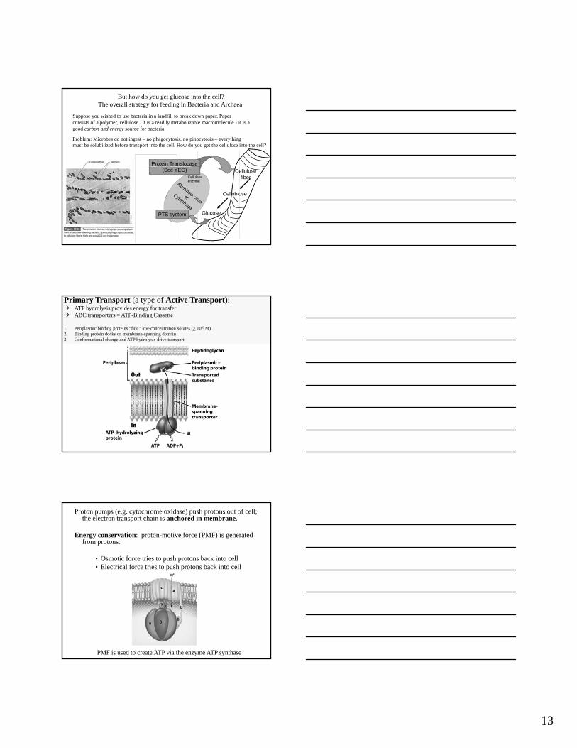
Classification of microbial cellular features:Invariant (or common to all)
Ribosomes: Sites for protein synthesis –
Comparing Bacteria, Archaea and Eucarya
p yaka the grand translators.
Cell Membranes: The barrier between order and chaos.
Nucleoid Region: Curator of the Information.

7
Ribosome structure

8
Most Complex
S= Svedberg; a sedimentation coefficient that is NOT ADDITIVE!!!
Protein synthesisDNA
Ribosomes attached to mRNA = polysomes
Classification of microbial cellular features:Invariant (or common to all)
Ribosomes: Sites for protein synthesis –
Comparing Bacteria, Archaea and Eucarya
p yaka the grand translators.
Cell Membranes: The barrier between order and chaos.
Nucleoid Region: Curator of the Information.

9
8 nmthick
The Cytoplasmic Membrane
Membrane has similar viscosity as oil: Fluid Mosaic Model
Stabilized by H bonds, hydrophobic interactions, and by Mg++ and Ca++
binding to phosphate heads
Ester linkages
Hydrophilic
Phospholipids are “Amphipathic”
Ester linkages
Hydrophobic
Functions of the cytoplasmic membrane
Charge separation:Potential energyAnalogous to a battery

10
Free diffusion of water (passive transport)assisted by aquaporins
Active or passive transport (depends on conditions), but proteins aid movement
Sterol Cholesterol Hopanoid(e.g., Diploptene)
All rigid planar molecules
O2 -
Few Bacteria Many Bacteria
11
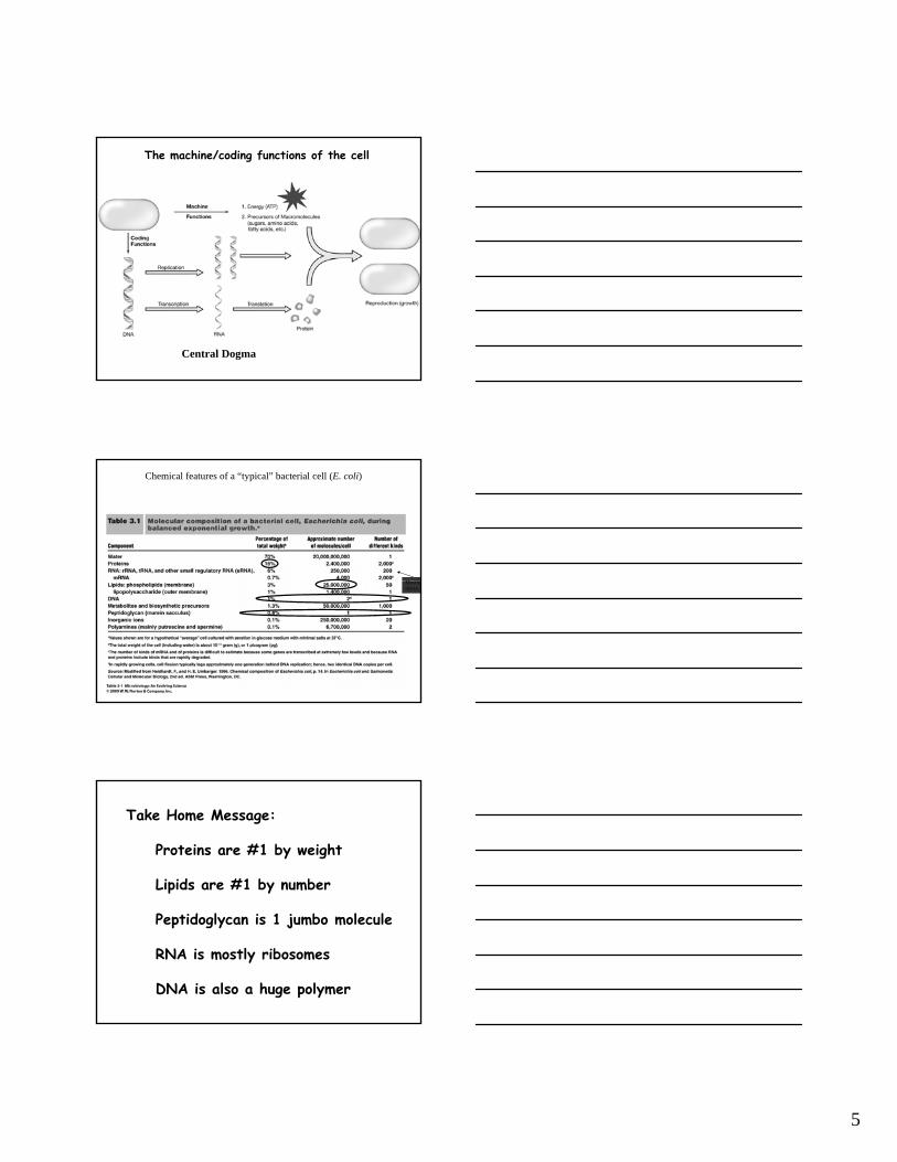
Ester Linkage Ether Linkage Isoprene Unit
Fatty Acid
Major lipids of Archaea and the structureof archaeal membranes
Lipid monolayer, not a bilayer: mostly found in extremophiles
Major lipids of Archaea and the structureof archaeal membranes

12
Archaeal cell membrane structure
Passive Diffusion:-small, uncharged molecules (O2, CO2, H2O)-weak acids and bases in protonated form (more hydrophobic)
Facilitated Diffusion (a type of passive tranport):Powered by solute’s own concentration gradient
Aquaporins, glycerol transporters
13
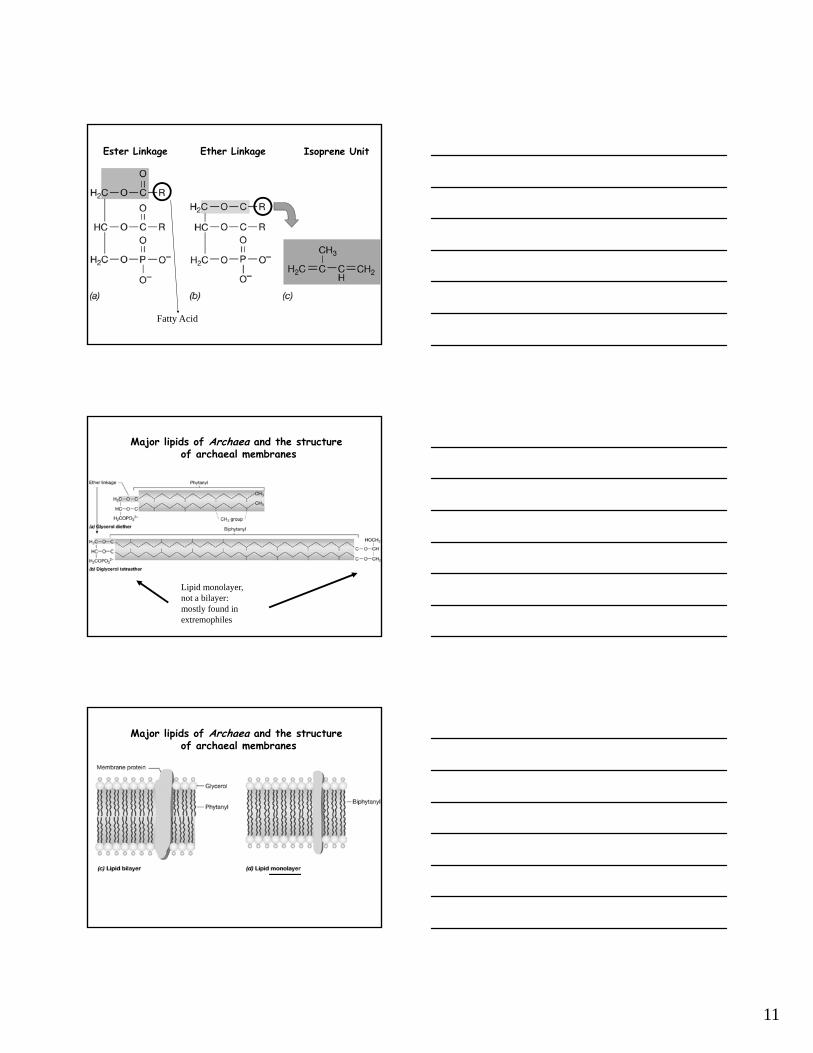
But how do you get glucose into the cell?The overall strategy for feeding in Bacteria and Archaea:
Suppose you wished to use bacteria in a landfill to break down paper. Paper consists of a polymer, cellulose. It is a readily metabolizable macromolecule - it is a good carbon and energy source for bacteria
Problem: Microbes do not ingest – no phagocytosis, no pinocytosis – everything must be solubilized before transport into the cell. How do you get the cellulose into the cell?
Protein Translocase
Cellulosefiber
(Sec YEG)
Cellobiose
Glucose
Cellulaseenzyme
PTS system
Primary Transport (a type of Active Transport): ATP hydrolysis provides energy for transfer ABC transporters = ATP-Binding Cassette
1. Periplasmic binding proteins “find” low-concentration solutes (> 10-6 M)2. Binding protein docks on membrane-spanning domain3. Conformational change and ATP hydrolysis drive transport
Proton pumps (e.g. cytochrome oxidase) push protons out of cell; the electron transport chain is anchored in membrane.
Energy conservation: proton-motive force (PMF) is generated from protons.
• Osmotic force tries to push protons back into cell• Electrical force tries to push protons back into cell
PMF is used to create ATP via the enzyme ATP synthase
14
Classification of microbial cellular features:Invariant (or common to all)
Ribosomes: Sites for protein synthesis –
Comparing Bacteria, Archaea and Eucarya
p yaka the grand translators.
Cell Membranes: The barrier between order and chaos.
Nucleoid Region: Curator of the Information.
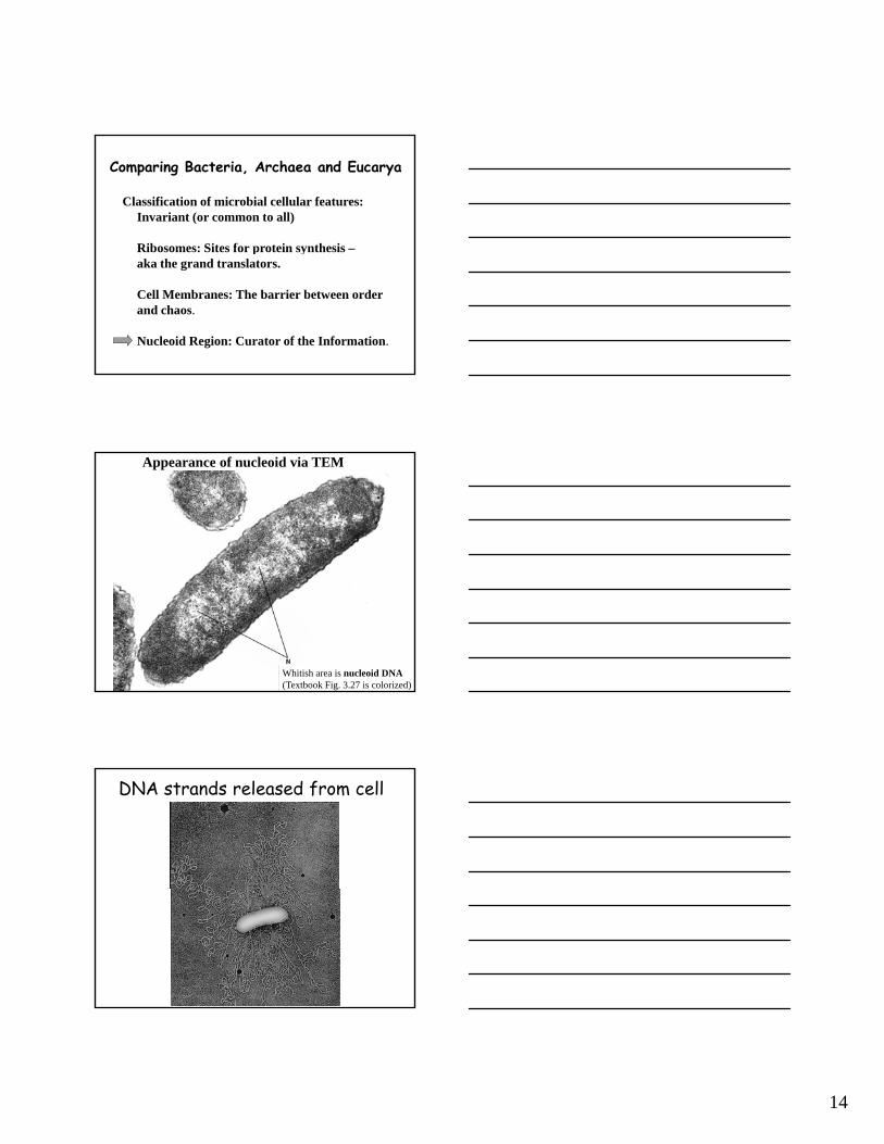
Appearance of nucleoid via TEM
Whitish area is nucleoid DNA(Textbook Fig. 3.27 is colorized)
DNA strands released from cell

15
Bacterial & Archaeal DNAStatistics:Chromosomes: ~1; bears essential genesPlasmids: 0 to hundreds; helpful genesCircumference: ~ 1 mm
Enigma:How to fit 1 mm long chromosome into a 1 m wide cell?Condensation: 30 to 50 loops of DNA emerging from a denser coreSupercoiling: tight twistingOrganization: wrapped around histone-like proteins (in Bacteria) or histones (in Archaea)
Condensation: 30 to 50 loops of DNA emerging from a denser core centered on ori at cell equator
Supercoiling
Stabilization by binding to proteins
Chapter 3.5

16
Nucleosomes in Archaea & Eucarya
Overview of DNA replication
Theta Structure
See Chapter 7.3, 7.5 - 7.7
Overview of DNA replication
40 minutes to replicate E. colichromosomechromosome.
20 minutes for cell division.
How???
http://www.wwnorton.com/college/biology/mbio/content/ch03/animation.asp

17
Overview of DNA replication
Bacteria have cytoskeletons!
Bacteria divide by binary fission. FtsZ marks the spot.
a. Phase contrastb. Nucleoid stainc. Antibody to FtsZd. Overlay of b & c
a.
b.
c.
d.
FtsZ is a structural analog (ancestor?) of eukaryotic tubulin
forms Z-ring at future site of cytokinesis only 17% amino acid identity to tubulin but similar 3D structures and assembly properties

18
MreB is a homolog (ancestor?) of actin
-cell shape determinant-present in rod- and spiral-shaped cells but absent from cocci-only 15% amino acid identity but similar 3D structure
A-C: WT and mreB mutants of B. subtilis (note cell shapes)D-F: Helical filaments formed by MreB-like proteins in B. subtilisG&H: MreB filamentsI &J: Actin and MreB structures overlaid. Only 15% amino acid identity but similar 3D structure.
Gemmataobscuriglobus
Membrane encompassednucleoidnucleoid