comparative pathophysiology and management of protein
TRANSCRIPT


R E V I EW
Comparative pathophysiology and management of
protein-losing enteropathy
Melanie D. Craven1 | Robert J. Washabau2
1Small Animal Clinical Sciences, Western
College of Veterinary Medicine, University of
Saskatchewan, Saskatoon, Canada
2Department of Veterinary Clinical Sciences,
College of Veterinary Medicine, University of
Minnesota, St. Paul, Minnesota
Correspondence
Melanie D. Craven, Small Animal Clinical
Sciences, Western College of Veterinary
Medicine, University of Saskatchewan,
Saskatoon, Canada S7N 5B4.
Email: [email protected]
Protein-losing enteropathy, or PLE, is not a disease but a syndrome that develops in numerous
disease states of differing etiologies and often involving the lymphatic system, such as lymphan-
giectasia and lymphangitis in dogs. The pathophysiology of lymphatic disease is incompletely
understood, and the disease is challenging to manage. Understanding of PLE mechanisms
requires knowledge of lymphatic system structure and function, which are reviewed here. The
mechanisms of enteric protein loss in PLE are identical in dogs and people, irrespective of the
underlying cause. In people, PLE is usually associated with primary intestinal lymphangiectasia,
suspected to arise from genetic susceptibility, or “idiopathic” lymphatic vascular obstruction. In
dogs, PLE is most often a feature of inflammatory bowel disease (IBD), and less frequently intes-
tinal lymphangiectasia, although it is not proven which process is the true driving defect. In cats,
PLE is relatively rare. Review of the veterinary literature (1977-2018) reveals that PLE was life-
ending in 54.2% of dogs compared to published disease-associated deaths in IBD of <20%,
implying that PLE is not merely a continuum of IBD spectrum pathophysiology. In people, diet
is the cornerstone of management, whereas dogs are often treated with immunosuppression
for causes of PLE including lymphangiectasia, lymphangitis, and crypt disease. Currently, how-
ever, there is no scientific, extrapolated, or evidence-based support for an autoimmune or
immune-mediated mechanism. Moreover, people with PLE have disease-associated loss of
immune function, including lymphopenia, severe CD4+ T-cell depletion, and negative vaccinal
titers. Comparison of PLE in people and dogs is undertaken here, and theories in treatment of
PLE are presented.
KEYWORDS
crypt, hypoalbuminemia, lymphangiectasia, lymphatic, panhypoproteinemia, PLE
1 | COMPARATIVE PATHOPHYSIOLOGY OF
PROTEIN-LOSING ENTEROPATHY
In dogs and people, protein-losing enteropathy (PLE) is associated
with numerous related or unrelated diseases (Table 1), but there are a
limited number of mechanisms by which plasma protein is lost
through the gastrointestinal (GI) tract1–3:
• Physical or functional lymphatic obstruction leading to “overflow”
lymph leak, for example, congenital or acquired lymphatic disease.
• Release of cellular mediators affecting vascular permeability and
causing fluid egress into tissues, for example, widespread mast
cell activation, eosinophilic gastroenteropathy.
Abbreviations: APCs, antigen presenting cells; ATIII, antithrombin III; BECs, blood
endothelial cells; CCECAI, canine chronic enteropathy clinical activity index; CD,
Crohn's disease; CE, chronic enteropathy; CIBDAI, canine IBD activity index;
CRP, C-reactive protein; CT/MRI, computerized tomography/magnetic resonance
imaging; EFA, essential fatty acid; α1 PI, alpha 1 protease inhibitor; FISH, fluores-
cence in situ hybridization; GALT, gut-associated lymphoid tissue; GI, gastrointes-
tinal; GL, granulomatous lymphangitis; HS, heparan sulfate; IBD, inflammatory
bowel disease; IL, intestinal lymphangiectasia; IMPDH, inosine monophosphate
dehydrogenase; IP, immunophenotyping; LCFA, long-chain fatty acids; LECs, lym-
phatic endothelial cells; LN, lymph node; MCFA, medium-chain fatty acids; MCT,
medium-chain triglycerides; PARR/PCR, PCR for antigen-receptor rearrange-
ments; PIL, primary intestinal lymphangiectasia; PLE, protein-losing enteropathy;
PLN, protein-losing nephropathy; RER, resting energy requirement; PTE, pulmo-
nary thromboembolism; SCFA, short-chain fatty acids; SCWT, Soft Coated
Wheaten Terrier; SI, small intestinal; TE, thromboembolism; UC, ulcerative colitis;
UFH, unfractionated heparin sulfate; YT, Yorkshire Terrier; YT-PLE, Yorkshire
Terrier protein-losing enteropathy.
Received: 16 February 2018 Accepted: 30 November 2018
DOI: 10.1111/jvim.15406
This is an open access article under the terms of the Creative Commons Attribution-NonCommercial License, which permits use, distribution and reproduction in any
medium, provided the original work is properly cited and is not used for commercial purposes.
© 2019 The Authors. Journal of Veterinary Internal Medicine published by Wiley Periodicals, Inc. on behalf of the American College of Veterinary Internal Medicine.
J Vet Intern Med. 2019;1–20. wileyonlinelibrary.com/journal/jvim 1

• Mucosal inflammation (nonerosive or erosive/ulcerative), for
example, inflammatory bowel disease (IBD).
In each mechanism, protein-rich fluid accumulates in the intersti-
tium and passes into the GI tract via mucosal tight junctions, without
requiring mucosal epithelial disruption or ulceration (Figure 1).3,4 Albu-
min is the most affected protein in people because of its slow turn-
over rate, which is why protein loss in PLE is diagnosed by detection
of hypoalbuminemia.5 As a water-soluble protein in dogs and people,
albumin maintains plasma oncotic pressure and transports hormones,
fatty acids, ions, and bilirubin. In health, enteric losses in people occur
as sloughed enterocytes and normal secretions, and account for
around only 1-2% of the entire protein pool and less than 10% of total
albumin.6 In steady state, albumin broken down approximates albumin
synthesized in people, but in PLE, protein wasting can amount to 60%
of the total protein pool.7,8 The most severely diminished are long
half-life proteins with limited ability to respond rapidly—albumin,
immunoglobulins, and ceruloplasmin.5 Hepatic synthesis in PLE is nor-
mally accelerated so that proteins with shorter half-lives such as IgE,
clotting factors, prealbumin, and transferrin are preserved near
normal.1,5
Protein-losing enteropathy can be asymptomatic but can also pre-
sent as a life-threatening condition with effusions, lymphedema, and
in people, severe disfigurement. Immune defenses are disrupted
because of loss of lymphocytes, globulins, iron, calcium, and other
serum components (lipids, fat soluble vitamins).2,4,5,7,8
The immunological deficits documented in PLE are numerous in
people, including impaired antibody function, B-cell depletion, dimin-
ished IgG, IgA, and IgM, and T-cell depletion, especially CD4+ T cells.2
Despite profoundly low CD4+ counts, opportunistic infection appears
to be uncommon in people.9,10 Similar studies are lacking in dogs, but
depletion in these key components is to be similarly expected.
Sudden death caused by thromboembolism (TE) occurs in PLE and
can manifest in dogs that have clinically “silent” disease.11 The mecha-
nism is not well defined, but suggested causes of PLE-associated throm-
bosis include systemic inflammation, altered vitamin K absorption, loss
of antithrombin III (ATIII), hyperaggregation of platelets, hyperfibrino-
genemia, and vascular compromise.11–13
2 | LYMPHATIC STRUCTURE AND
FUNCTION
The lymphatics are a unidirectional vascular network forming an
extensive drainage system throughout the body with roles of extracel-
lular fluid homeostasis, fat absorption and transport, and immune sys-
tem function.3 Running parallel with the venous system, the lymphatic
system is broadly divided into lymphatic capillaries (where interstitial
fluid enters), prenodal and postnodal vessels, and collecting vessels.3
Collecting vessels drain to the thoracic duct via discrete regional
lymph nodes (LNs) before joining the confluences of large veins.3,10
Specialized lymphatic endothelial cells (LECs), with structural similarity
to blood endothelial cells (BECs), form lymphatic capillary walls.
Derived embryologically from the outbranching of cardinal vein BECs,
LECs are under control of genes such as Prospero Homeobox Protein
1, Lymphatic Vessel Endothelial Receptor 1, Podoplanin, and Vascular
Endothelial Growth Factor 3.14–18
2.1 | Lymphatic capillaries
The LECs of capillary walls are not tightly joined, but only the cell edges
overlap, forming “flap-like” valves (Figure 1).19 Lymphatic endothelial
cells have no support (mural) cells but attach to the extracellular matrix
via fine collagen filaments that anchor to surrounding tissues. This
allows LECs to operate like swinging doors, receiving interstitial fluid in
a strictly 1-way system.3 When pressure in the lymphatic capillary
exceeds interstitial pressure, the endothelial “door” flap remains shut,
preventing lymph from leaving.3,20 But when interstitial pressure
exceeds lymphatic capillary pressure, the “doors” flap open and intersti-
tial fluid moves to the lymph system.20 Not only fluid but also other sub-
stances such as proteins and pathogens, inflammatory and neoplastic
TABLE 1 Causes of PLE in people and dogs
People Dogs
1. Mucosal injury
a. Erosive
Inflammatory bowel diseases(Crohn's disease, ulcerative
colitis)
Inflammatory bowel diseases(lymphoplasmacytic,
eosinophilic, granulomatous)
Infections: Giardia, Clostridium,
Campylobacter, Salmonella,
rotavirus, Whipple's disease,
intestinal tuberculosis
Infections: Parvovirus,
Clostridium, Campylobacter,
Salmonellosis, Histoplasmosis,
Schistosomiasis (Heterobilharziaamericana)
Neoplasia Neoplasia
Nonsteroidal enteropathy Nonsteroidal enteropathy
b. Non-Erosive
Menetrier's disease(hypertrophic gastritis)
Diet-induced enteropathy
Eosinophilic gastritis Immunoproliferative enteropathy
Celiac disease Hypoadrenocorticism
Lactose or other food
intolerance
Intestinal crypt disease
Systemic lupus erythematosus
Intestinal crypt disease
2. Infectious
Lymphatic filariasis Lymphatic filariasis (RARE)
Hookworms
Strongyloides stercoralis
3. Lymphatic disease
Idiopathic primary IL (Waldmann'sdisease)
Idiopathic primary IL
Secondary IL: Crohn's disease,neoplasia sarcoidosis, congestive
heart failure, restrictive
pericarditis
Secondary IL: IBD, neoplasia,lymphatic infections, right-sided
congestive cardiac failure
Fontan surgery Lymphangitis (granulomatous/
inflammatory)
Genetic: Lymphodysplasia
(Hennekam's syndrome)
Lymphangitis
Abbreviations: IBD, inflammatory bowel disease; IL, intestinal
lymphangiectasia.
2 CRAVEN AND WASHABAU

cells can join lymph in this manner.21,22 Interstitial proteins enter lymph
capillaries easily, despite being too large to enter blood capillaries.22
2.2 | Collecting lymphatics and lymphangions
Lymphatic capillaries merge and drain into precollecting vessels. The
spindle-shaped LECs of precollectors are continuously adherent, with
“zipper”-like junctions.23 Intraluminal valves prevent backflow of
lymph, and smooth muscle cells enable intrinsic contractility.20,21,24
Precollectors merge into larger collecting vessels, made up of a series
of subunits called “lymphangions” (the section between 2 intraluminal
valves).24,25 The collecting vessel wall is organized into a layer of con-
tinuously adherent LECs (intima), a basement membrane comprising
laminin and type IV collagen, and circumferential smooth muscle
(media).26,27 Lymphangions act individually and in groups, forming
contractile units that propel lymph via intraluminal valves made of
connective tissue covered with LECs.23,25,26
2.3 | Intestinal lymphatic networks
The microanatomy of the intestinal lymphatics is described in ultra-
structural studies, and in dogs and people there are 3 layers: central
villus lymphatics (lacteals), submucosal lymphatics, and smooth muscle
lymphatics.19,27 The lacteals connect to the submucosal lymphatic net-
work with abundant interconnections. Injection of tracers shows free
flow of fluid throughout the submucosal network and into adjacent villi,
suggesting a valveless system lacking in smooth muscle and spontaneous
contraction.20 The lymphatic vessels draining the muscle wall form a sep-
arate and noncommunicating network. Both networks transport lymph
to collecting lymphatics24 with 1-way valves and spontaneous contrac-
tions propelling lymph away from the intestine to the mesentery.3,20
2.4 | Tissue fluid homeostasis
Lymphatic capillaries maintain tissue fluid homeostasis by absorbing
and transporting extravasated fluid to the venous circulation. Starling
FIGURE 1 Diagrammatic representation of “PLE syndrome” diseases in dogs (lymphangiectasia, lymphangitis, crypt lesions, IBD, mucosal
ulceration) and the effect of Starling's forces on intestinal osmolality and the interstitial-lumenal oncotic gradient. Protein leaks into the small
intestine passively, via an oncotic gradient from the interstitium to the lumen. If osmotic pressures equilibrate, a reduction in passive leak is
expected. When the intestinal lumen has a higher oncotic pressure than the interstitium, such as postprandially, the gradient may be
reversed, depending on the underlying pathology. Top: Magnification of lymphatic capillary structure, showing overlapping endothelial cells
that act as flap-like doors allowing interstitial fluid to enter while preventing lymph exit. IBD, inflammatory bowel disease; PLE, protein-losing
enteropathy
CRAVEN AND WASHABAU 3

demonstrated that lymphatics play an important role in the regulation
of hydrostatic and oncotic pressure throughout the entire body.28
From the capillary blood, fluid filters continuously into the interstitium
and a dynamic equilibrium is reached between filtration, reabsorption,
and tissue fluid clearance by lymphatics.3 The extravasation of fluid is
dictated by Starling forces, and in people there can accumulate up to
8 L during 24 hours.21 Traditionally, venous reabsorption was thought
to clear extravasated fluid, but there is now comprehensive published
scientific evidence21,22,24,28 that venous reabsorption is nonexistent
in most vascular beds during steady-state conditions.21,22 Lymph
transport is both passive and active. Extrinsic factors such as arterial
pulsation, skeletal muscle contraction, and respiration cause passive
compression and expansion of the small capillary lymphatics. Intrinsic
spontaneous contractions are generated in larger vessels, and in people
intraluminal lymph pressure can reach up to 100 mm Hg.21 Lymphatic
vessel contractions are influenced by many different stimuli: humoral,
neuronal, pressure, temperature, and shear stress.21,24 They show reac-
tivity, either inhibitory or stimulatory, to a vast number of vasoactive
substances such as substance P, nitric oxide, noradrenaline, histamine,
endothelin, and prostaglandins.21,24,25 Despite evidence of lymphatic
innervation, most consistently described as sympathetic, the exact role
of the nerve supply in lymphatic function is unclear.3
The drainage function of lymphatic vessels is crucial for resolving
inflammatory reactions in the interstitium, and during inflammation,
lymphatics can proliferate through lymphangiogenesis to remove
debris directly or via phagocytic cells.3,17,21 Development and prolifer-
ation of lymphatic vessels is primarily mediated by vascular endothe-
lial growth factor C in pathological states.29
2.5 | Lymph nodes
The LNs, spleen, and other secondary lymphoid organs (eg, intestinal
Peyer's patches) are anatomically located to capture foreign antigen
and antigen presenting cells (APCs) for immune surveillance.30 Affer-
ent lymphatic vessels traverse the fibrous capsule of the LN and
drain lymph into the subcapsular sinus and cortex. The cortex has a
paracortex, populated by T cells (for antigen presentation by dendritic
cells), and a germinal center, comprised B cells (for humoral immune
responses). Lymph drains through the cortex via trabecular sinuses
and into the central medullary region via medullary sinuses. The sys-
temic blood circulation interacts with the LN via high endothelial
venules in the cortex.25,31 Lymph then drains from the medulla toward
the hilus and exits the LN via an efferent lymph vessel, effectively
clearing and filtering extravascular fluid from tissues throughout the
entire body.
2.6 | Immune surveillance
Lymphatic vessels function in immune surveillance as the major con-
duit of antigens and immune cells from the periphery, through the tis-
sues of the immune system. The highly organized microarchitecture
of LNs hosts peripheral APCs to meet and inform naive T cells.25,32
The largest immunologic organ is the GI tract, where immunologic bar-
rier function plays a vital role in maintaining homeostasis. The gut-
associated lymphoid tissue (GALT) consists of the intestinal lamina
propria cells, intraepithelial lymphocytes, Peyer's patches, and mesen-
teric LNs.33 Lymphocytes continuously recirculate from the blood-
stream into intestinal lymph through GALT, to exert immune control
in almost all tissues.34 Mature lymphocytes migrate from blood to tis-
sues, and back to the bloodstream via lymph, once or twice a day, until
the cell either finds its antigen or is removed by apoptosis.35
2.7 | Intestinal lymphatics
Dietary fat is absorbed into lacteals (Figure 1) and transported as chyle
to the bloodstream. Resin casts of canine small intestinal (SI) lymphatics
have been produced using methacrylate resin and imaging by scanning
electron microscopy.19 The casts show the lymphatic canal of the jeju-
num as a single, blind-ending, central tubule, with circular constrictions
at the villus tip, and oval indentations corresponding to impressions by
endothelial nuclei (Figure 2). The central lacteals of the ileum in dogs are
smaller, slender, leaf-like shapes and drain to the lymphatic plexus,
above the muscularis layer.19
2.8 | Fat absorption
The lacteals absorb essential fatty acids (EFAs) from the bowel. Most
ingested fat in a normal diet is taken up by the lacteals and trans-
ported to the venous circulation.36 Fatty acids are classified as short-
chain, (SCFAs, <6 carbons), medium-chain, (MCFAs, 6-12 carbons), or
long-chain fatty acids (LCFAs, 12 carbons).36 Most dietary fat is in
the form of LCFAs and is absorbed by the enterocyte to form chylomi-
crons, which enter lymph via the lacteal. In addition to lymphatic
absorption, medium-chain triglycerides (MCTs) are water soluble and
can pass through enterocytes into the portal blood, bypassing the
lymphatics.37 In people and dogs, MCTs are more efficiently digested
and absorbed in this way, and the resulting MCFAs are transported
directly to the liver to be metabolized.36–39
2.8.1 | PLE in people
In people, PLE is usually caused by primary intestinal lymphangiectasia
(PIL),3 described in 1961 by Dr. Thomas A. Waldmann’s 18 cases of
“idiopathic hypercatabolic hypoproteinemia.”4 These patients had
edema associated with hypoproteinemia, low serum albumin, and serum
gammaglobulins. The total exchangeable albumin pool, assessed with
radiolabeled 131I-albumin, was low in all patients, and daily fecal excre-
tion of 131I was twice that of healthy controls. Intestinal histology
showed dilatation of lymph vessels in the mucosa and submucosa and
the authors proposed the term “intestinal lymphangiectasia” (IL). Known
since as Waldmann's disease, PIL can also be asymptomatic.40
The tortuous and dilated lacteals of the mucosa and submucosa in
PIL continually leak protein-rich lymph into the bowel. Patients usually
present in childhood with edema, weight loss, diarrhea, and lymphope-
nia with no obvious cause. Edema is pitting because oncotic pressure is
low, and moderate serous effusions (peritoneal, pleural, pericardial) are
common.40,41 The worldwide incidence of PIL in people is unknown,
but familial forms are rare and there is no predilection for sex or
race.2,40 Pathophysiology is unclear, but theories include genetic anom-
aly and lymphatic obstruction.42 Lymphatic obstruction theory says that
lymphatic hypoplasia or idiopathic malformation causes obstruction
4 CRAVEN AND WASHABAU
and pressure-induced rupture of cystically dilated lymphatic vessels.42
Genetic theory implicates genes with vital roles in lymphogenesis, that
is, VEGF3, prospero-related homeobox-transcriptional factor, forkhead
transcriptional factor, and SOX18.43Mutation of CCBE1, a gene that pro-
duces a protein with functions in extracellular matrix remodeling and
migration, is known to cause generalized lymphatic dysplasia (lymph-
edema and lymphangiectasia) in Hennekam syndrome, an inherited
autosomal recessive disorder.44
Secondary IL occurs when a primary disease process obstructs
lymphatic vessels or when increased venous pressure induces lym-
phatic hypertension (Table 1). In people, secondary IL has been associ-
ated with constrictive pericarditis, lymphoma, Whipple's disease,
sarcoidosis, intestinal tuberculosis, and Crohn's disease (CD).2,40 In
CD, numerous studies describe lymphangitis, lymphangiectasia, lym-
phatic bacterial infiltration, and LN infection.45 Some authors suggest
that the core driving pathology in CD is lymphatic disease, with sec-
ondary vasculitis and transmural inflammation.46–48 In other diseases
where PLE occurs, lymphatics are not the root cause and protein is
lost via mucosal epithelium by intercellular leak or exudation,2 for
example, eosinophilic gastroenteropathy, Menetrier's disease, autoim-
mune enteropathy, and systemic lupus erythematosus.40
2.8.2 | PLE in dogs
In contrast to people, PLE in dogs is usually associated with lympho-
plasmacytic enteritis (LPE) rather than PIL (Table 1). In the last
30 years, published data on PLE includes 23 articles, spanning from
1977 to 2018 (Table 2).12,13,49–69 Taken together, they describe in total
469 dogs of 61 different breeds, most prevalent the Yorkshire Terrier
(YT), Border Collie, German Shepherd, and Rottweiler (Figures 3 and 4).
Historical features commonly described are ascites, vomiting, diarrhea,
weight loss, polyuria and polydipsia, anorexia, weight loss, and lethargy.
Clinical examination findings commonly include abdominal distension
(ascites), cachexia, muscle wasting, weakness, depression, dyspnea/
tachypnea, and abdominal pain. Clinical scoring systems including
CIBDAI70 (canine IBD activity index) and CCECAI71 (canine chronic
enteropathy clinical activity index) were compared in 8 studies and did
not always concur: CIBDAI scores indicated “mild IBD” (score 4-5) or
“severe IBD” (score ≥ 9), whereas CCECAI scores indicated “clinically
insignificant disease,” (score 0-3) “moderate disease”(score 6-8) or “very
severe disease” (score ≥ 12).
The cumulative median serum albumin level, reported in 21 of
23 studies, was 1.49 g/dL, globulins 1.95 g/dL, and total serum pro-
teins 3.35 g/dL (Figure 5), and low serum cobalamin was reported in
8 of 23 studies.13,52,54,56,64,66,67,69 Small bowel diseases were LPE, in
319 of 469 dogs (68%) and lymphangiectasia in 214 dogs (58%,
Figure 3). Inflammatory lymphatic disease, including lipogranuloma-
tous foci and lymphangitis, was present in 38 dogs (8%). Crypt disease
was present in 34 dogs (7.2%). Histology was scored according to the
standards of the World Small Animal Veterinary Association (WSAVA)
GI Standardization Group72 in 6 studies, with detailed scores pub-
lished in 3 (Figure 6).
Treatment was described in 16 of 23 reports.49,51–57,59,61,62,64–67,69
An immune-mediated cause was nearly always presumed, as shown
by the use of immune suppressive medication in 15 of 16 publi-
cations (Figure 7).49,51–57,59,61,62,64–67,69 Prednisolone was most
frequently used (15 studies) followed by azathioprine (9 studies),
cyclosporine (9 studies), chlorambucil (4 studies), and methotrexate
(1). Metronidazole and other antimicrobials were each prescribed in
8 studies. Dietary change to low-fat, limited antigen, hydrolyzed,
or home-cooked food was implemented in 14 studies and MCT pro-
vision in 2.
Median survival times, reported in 8 studies, were highly variable
(months: 1.0, 2.2, 5, 5, 8.4, 16, 18, and 28). Complicating factors
included TE in 26 dogs and hypocalcemia in 42 dogs. Follow-up infor-
mation was available for 445 of 469 cases (94.8%) from 23 publica-
tions, and disease-associated death occurred in 54.2%, (204 survivors
and 241 dead or euthanized because of PLE). Various negative prog-
nostic factors were reported in 6 separate studies and are listed in
Table 3. Factors repeatedly identified include medium bodyweight
(2 studies),65,69 altered serum urea (3 studies),52,56,57 and decreased
serum albumin (4 studies).52,56,57,69
In summary, published reports to date show that PLE in dogs is
associated with lymphangiectasia in around 50% of cases and LPE in
approximately 66%. Lymphangitis and crypt disease each occur in
<10%, and other types of IBD (granulomatous, eosinophilic) are rare
causes of PLE. Immune suppressive treatment is often prescribed,
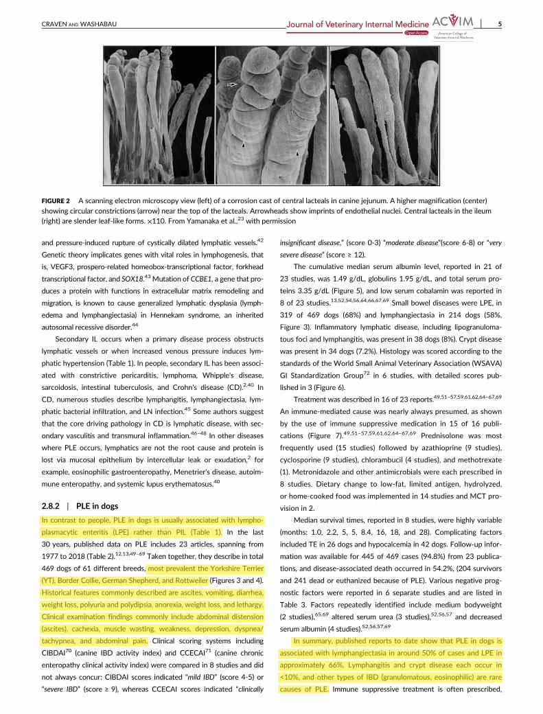
FIGURE 2 A scanning electron microscopy view (left) of a corrosion cast of central lacteals in canine jejunum. A higher magnification (center)
showing circular constrictions (arrow) near the top of the lacteals. Arrowheads show imprints of endothelial nuclei. Central lacteals in the ileum
(right) are slender leaf-like forms. ×110. From Yamanaka et al.,23 with permission
CRAVEN AND WASHABAU 5
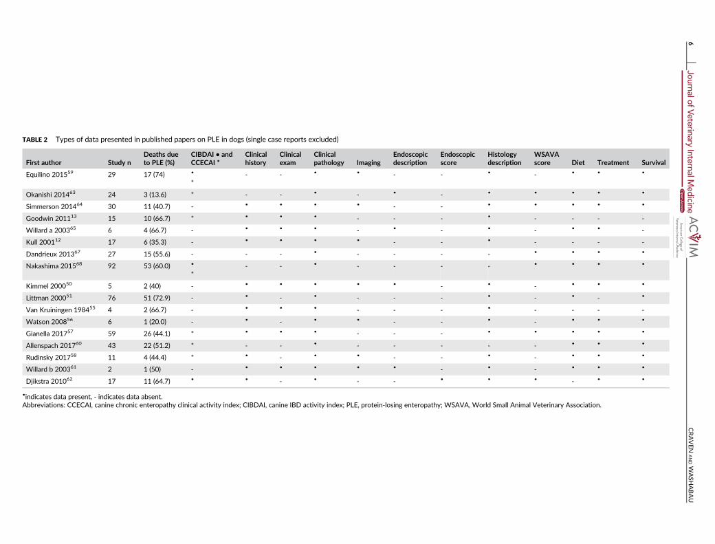
TABLE 2 Types of data presented in published papers on PLE in dogs (single case reports excluded)
First author Study nDeaths dueto PLE (%)
CIBDAI • andCCECAI *
Clinicalhistory
Clinicalexam
Clinicalpathology Imaging
Endoscopicdescription
Endoscopicscore
Histologydescription
WSAVAscore Diet Treatment Survival
Equilino 201559 29 17 (74) •
*
- - • • - - • - • • •
Okanishi 201463 24 3 (13.6) * - - • - • - • • • • •
Simmerson 201464 30 11 (40.7) - • • • • - - • • • • •
Goodwin 201113 15 10 (66.7) * • • • - - - • - - - -
Willard a 200365 6 4 (66.7) - • • • - • - • - • • -
Kull 200112 17 6 (35.3) - • • • • - - • - - - -
Dandrieux 201367 27 15 (55.6) - - - • - - - - • • • •
Nakashima 201568 92 53 (60.0) •
*
- - • - - - - • • • •
Kimmel 200050 5 2 (40) - • • • • • - • - • • •
Littman 200051 76 51 (72.9) - • - • - - - • - • - •
Van Kruiningen 198455 4 2 (66.7) - • • • - - - • - - - -
Watson 200856 6 1 (20.0) - • - • • - - • - • • •
Gianella 201757 59 26 (44.1) * • • • - - - • • • • •
Allenspach 201760 43 22 (51.2) * - - • - - - - - • • •
Rudinsky 201758 11 4 (44.4) * • - • • - - • - • • •
Willard b 200361 2 1 (50) - • • • • • - • - • • •
Djikstra 201062 17 11 (64.7) • • - • - - • • • - • •
•indicates data present, - indicates data absent.
Abbreviations: CCECAI, canine chronic enteropathy clinical activity index; CIBDAI, canine IBD activity index; PLE, protein-losing enteropathy; WSAVA, World Small Animal Veterinary Association.
6CRAVEN
ANDW
ASHABAU

usually corticosteroids, and sometimes 2 or 3 immune suppressive
agents concurrently. Dietary modification is frequently but not always
implemented. Although prolonged survival can occur, over half of
dogs (54.2%) with PLE die from their disease or from pulmonary
thromboembolism (PTE) as a complicating factor. It is interesting to
note that, in contrast, disease-associated deaths from 2 published out-
come studies of IBD in dogs71,73 were considerably lower at 13%73
and 18%,71 suggesting that the PLE underlying disease mechanisms
are not merely a more severe continuum of IBD spectrum pathophysi-
ology. Perhaps, this is not unexpected when considering the functions
of lymph fluid in immune defense, and the role of albumin in systemic
immunomodulatory and anti-inflammatory activities, antioxidant prop-
erties, and endothelial stabilization.74
2.8.3 | Lymphangiectasia in dogs
Primary IL, a diagnosis of exclusion, is a relatively rare problem of
unknown etiology in dogs. Breed predispositions support a genetic
susceptibility in the Soft Coated Wheaten Terrier (SCWT), Norwegian
Lundehund, Yorkshire and Maltese Terriers, and Shar-Pei.11 Studies of
PIL with scanning electron microscopy in dogs show grossly dilated
lacteals that rupture and leak protein-rich fluid into the lumen and
interstitium (Figure 1).19 Lymph is supposedly a local tissue irritant,
causing inflammation and granuloma formation,46 which presents a
unique challenge in the histological determination of primary versus
secondary IL, also lymphangitis and vasculitis. Other potential diagnos-
tic pitfalls are the similar appearance of blood and lymphatic endothe-
lium on routine stains, and that the drainage function of lymphatics
during inflammation stimulates the appearance of new vessels (lym-
phangiogenesis).3,75,76 Consequently, diagnosis of IL is not straightfor-
ward, especially without the use of differentiative histological markers.
The immunohistochemical markers Prox-1 and CD31 separate lym-
phatic from blood capillary endothelium.75 In the SCWT, lymphatic dila-
tation (17% prevalence) is reported to be strongly associated with
lymphangitis (35% prevalence), and less so with IBD, whereas the pres-
ence of all 3 lesions concurrently was rare.49
Secondary IL results from direct blockade to lymphatics, for
example, neoplasia, or an indirect block, for example, dense inflamma-
tory reaction and impaired drainage. It is reported that IL exists in
a high proportion of endoscopic biopsies (44/83; 53%) from dogs
with chronic GI disease, and in this patient population, there was a
Disea
se
Out
com
eM
ST
Breed
s0
100
200
300
400
500
600
700
800
Variable
Nu
mb
er
of
do
gs
Yorkshire Terrier
75
SCWT76
Mixed breed 36
Dachshund 15 Shiba Inu 12Maltese 11
Papillon 10
Survivors 204
Euthanased or died
241
Lym
phopla
sm
acytic
ente
ritis
319
Lym
phangie
cta
sia
214
Villus blunting57
Crypt abscess 34
Fibrosis 15Lymphangitis 8
T-embolism 26
HypoCa 42
28 months53 o
ther
bre
eds
with
n<
10
Lipogranulomatous lymphangitis 30
18 months
16 months
Rottweiler 21
Border Collie 10
FIGURE 3 Cumulative data from 23 PLE articles spanning from 1977 to 2017, involving a total of 469 dogs. HypoCa, hypocalcemia; MST,
median survival time; PLE, protein-losing enteropathy; SCWT, Soft Coated Wheaten Terrier; T-embolism, thromboembolism12,13,49–69
CRAVEN AND WASHABAU 7

significant association of IL with hypoalbuminemia (28/37; 76%).77 In
close agreement, on full thickness biopsy, 59% (38/64) of dogs with
chronic GI disease were shown to have IL.78
Published evidence showing an immune-mediated etiology of IL
in dogs (and people) is totally lacking.11,79 The obvious place to seek
an anomaly is in the lymphatic endothelium or in the fine collagen
fibers that anchor lymphatic capillaries to surrounding connective tis-
sue, but none has ever been described.
2.8.4 | Granulomatous lymphangitis
Lymphangitis and granuloma formation are rare in dogs with PLE.11
Clinical presentation is typical for PLE but can also include fever and
abdominal pain.51 The histological description is of transmural intestinal
granulomas containing large “multinucleate giant cells,” neutrophils,
macrophages, and lymphoplasmacytic infiltrates surrounding lym-
phatics.50,80 The lesions of granulomatous lymphangitis (GL) are not
always localized to the bowel. In some cases involvement of the entire
mesentery, LNs, and intrathoracic lymphatics goes undetected until
laparatomy or necropsy because lymphatic vascular imaging and mag-
netic resonance imaging (MRI) are not routine procedures.50,80 Across
species, granulomatous bowel disease is particularly associated with
infectious agents, for example, Whipple's Disease in people and Tro-
pheryma whipplei, Mycobacterium paratuberculosis in Johne's disease of
cattle, and Escherichia coli in Granulomatous Colitis of the Boxer dog.
Villus blun
ting
Epith
elial injur
y
Muc
osal fibr
osis
Lactea
l dila
tion
IELs
L-P in
filtra
tes
Eosinop
hils
Neu
troph
ils
Cry
pt le
sion
s
0
20
40
60
80
HISTOLOGICAL VARIABLE
Nu
mb
er
of
do
gs
0
1
23
Severity score
FIGURE 6 Cumulative histology scores from 3 canine studies of PLE,
per the WSAVA GI standardization group scoring system.48,53,65GI,
gastrointestinal; PLE, protein-losing enteropathy. Numbers on bars
are the number of dogs
Dieta
ry cha
nge
Paren
tera
l nut
rition
Med
ium
cha
in tr
iglyce
rides
Predn
isolon
e
Azath
iopr
ine
Cyc
losp
orine
Chlor
ambu
cil
Plasm
a tra
nsfu
sion
Antim
icro
bials
Met
ronida
zole
Acid
redu
cers
Sucra
lfate
Anti-e
met
ics
Low d
ose
aspirin
Diure
tics
Vitam
in B
120
5
10
15
20
Type of treatment
Nu
mb
er
of
stu
die
s
>176
>166
3323
21
823
4 28
53 53
5158
FIGURE 7 Cumulative treatment data in PLE across studies; white
numbers on each bar indicate the minimum number of dogs that
received each specific treatment.49,51–57,61,64–67,69 PLE, protein-losing
enteropathy
Cas
e fa
tality
Album
in
Tota
l pro
teins
Globu
lins
0
20
40
60
80
100
0.0
1.0
2.0
3.0
4.0
5.0
per
cen
t d
eath
s d
ue t
o P
LE S
eru
m p
rote
ins (g
/dL
)
FIGURE 5 Cumulative disease-associated deaths and serum proteins
reported in PLE publications (solid line median, dotted line
mean).12,13,49–57,61,64–67,69 PLE, protein-losing enteropathy
York
shire
Ter
rier
Mixed
bre
ed
Borde
r Collie
GSD
Rot
tweiler
Malte
se Ter
rier
Toy Poo
dle
Dac
hshu
nd
Sheltie
Englis
h Sp
Span
SCW
T0
5
10
15B
reed
fre
qu
en
cy a
cro
ss s
tud
ies
36
610
116
76
15
75
217 5
FIGURE 4 Breed frequencies represented across canine PLE
publications; numbers on bars indicate the number of dogs within the
breed.12,13,49–69 PLE, protein-losing enteropathy
8 CRAVEN AND WASHABAU
In GL, an etiology is rarely found but could include infectious, parasitic,
and neoplastic causes.11,51 An immune-mediated basis is often also
cited but not proven. A report of 10 dogs with GL uncovered no evi-
dence of a bacterial cause; however unusual bacteria can evade (fluo-
rescence in situ hybridization; FISH) detection.80
2.8.5 | Intestinal crypt pathology
Crypt disease is increasingly recognized as a cause of PLE in dogs
although by an unknown mechanism. The YT is overrepresented in the
United States and Europe for PLE,56,64,81,82 and is particularly suscepti-
ble to crypt pathology.56 The lesions are described as dilated cystic
crypts containing sloughed epithelial cells, debris, and leucocytes67 and
are often called “abscess” by pathologists but are not known to be asso-
ciated with a specific pathogen (Figures 1 and 8). Their histological
appearance, packed with proteinaceous and cellular debris, seems
inconsistent, however, with simple cystic malformation. Parallels drawn
with parvovirus infection, that is, villus collapse and fusion and crypt
distension, led researchers to undertake intestinal immunostaining for
parvovirus antigen in 2 dogs, which was negative.67
In people, crypt lesions are reported to be highly predictive of
ulcerative colitis (UC).83 Increased numbers of neutrophils and mucosa-
associated bacteria are found to colonize crypts in UC, suggesting
an opportunist role for bacteria in “cryptitis” or crypt “abscess” forma-
tion.83,84 Investigation for a similar etiology in YT crypt PLE (YT-PLE)
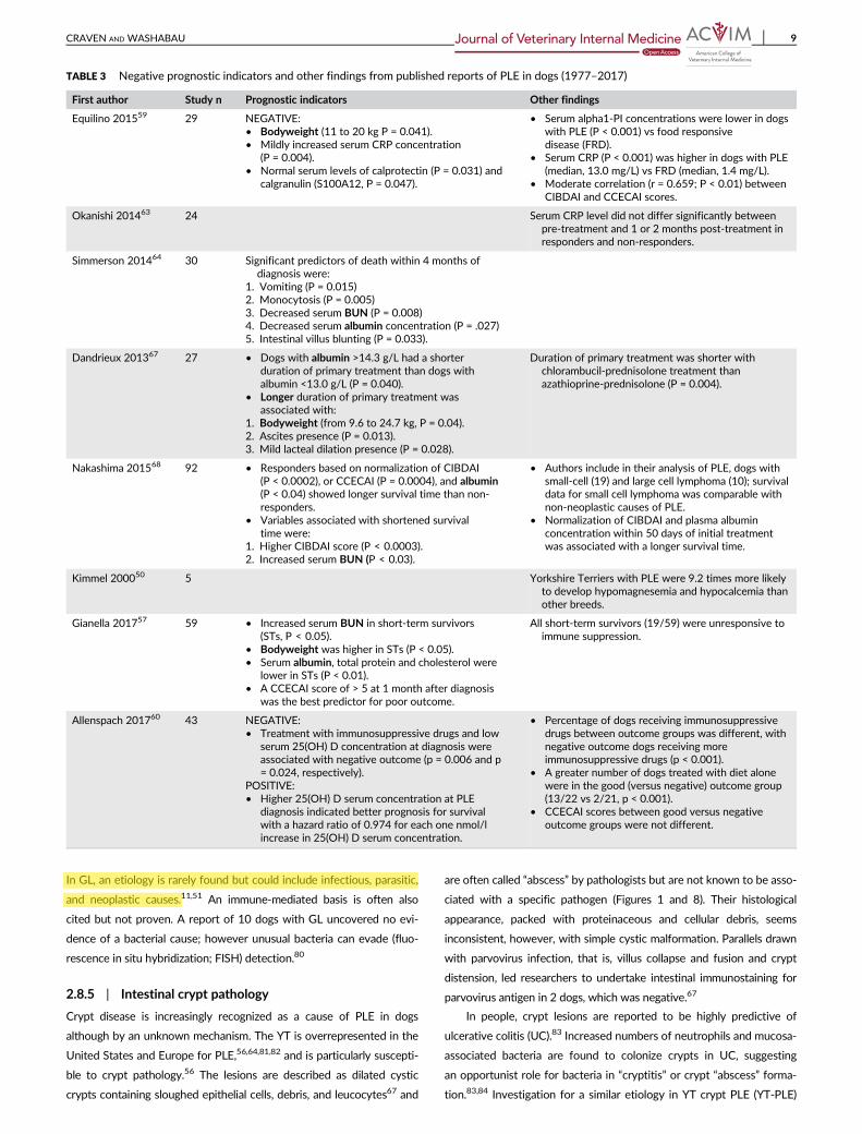
TABLE 3 Negative prognostic indicators and other findings from published reports of PLE in dogs (1977–2017)
First author Study n Prognostic indicators Other findings
Equilino 201559 29 NEGATIVE:
• Bodyweight (11 to 20 kg P = 0.041).
• Mildly increased serum CRP concentration(P = 0.004).
• Normal serum levels of calprotectin (P = 0.031) and
calgranulin (S100A12, P = 0.047).
• Serum alpha1-PI concentrations were lower in dogs
with PLE (P < 0.001) vs food responsive
disease (FRD).• Serum CRP (P < 0.001) was higher in dogs with PLE
(median, 13.0 mg/L) vs FRD (median, 1.4 mg/L).
• Moderate correlation (r = 0.659; P < 0.01) between
CIBDAI and CCECAI scores.
Okanishi 201463 24 Serum CRP level did not differ significantly between
pre-treatment and 1 or 2 months post-treatment inresponders and non-responders.
Simmerson 201464 30 Significant predictors of death within 4 months ofdiagnosis were:
1. Vomiting (P = 0.015)
2. Monocytosis (P = 0.005)
3. Decreased serum BUN (P = 0.008)4. Decreased serum albumin concentration (P = .027)
5. Intestinal villus blunting (P = 0.033).
Dandrieux 201367 27 • Dogs with albumin >14.3 g/L had a shorter
duration of primary treatment than dogs with
albumin <13.0 g/L (P = 0.040).
• Longer duration of primary treatment wasassociated with:
1. Bodyweight (from 9.6 to 24.7 kg, P = 0.04).
2. Ascites presence (P = 0.013).
3. Mild lacteal dilation presence (P = 0.028).
Duration of primary treatment was shorter with
chlorambucil-prednisolone treatment than
azathioprine-prednisolone (P = 0.004).
Nakashima 201568 92 • Responders based on normalization of CIBDAI
(P < 0.0002), or CCECAI (P = 0.0004), and albumin(P < 0.04) showed longer survival time than non-
responders.
• Variables associated with shortened survival
time were:1. Higher CIBDAI score (P < 0.0003).
2. Increased serum BUN (P < 0.03).
• Authors include in their analysis of PLE, dogs with
small-cell (19) and large cell lymphoma (10); survivaldata for small cell lymphoma was comparable with
non-neoplastic causes of PLE.
• Normalization of CIBDAI and plasma albumin
concentration within 50 days of initial treatmentwas associated with a longer survival time.
Kimmel 200050 5 Yorkshire Terriers with PLE were 9.2 times more likelyto develop hypomagnesemia and hypocalcemia than
other breeds.
Gianella 201757 59 • Increased serum BUN in short-term survivors
(STs, P < 0.05).
• Bodyweight was higher in STs (P < 0.05).
• Serum albumin, total protein and cholesterol werelower in STs (P < 0.01).
• A CCECAI score of > 5 at 1 month after diagnosis
was the best predictor for poor outcome.
All short-term survivors (19/59) were unresponsive to
immune suppression.
Allenspach 201760 43 NEGATIVE:
• Treatment with immunosuppressive drugs and low
serum 25(OH) D concentration at diagnosis wereassociated with negative outcome (p = 0.006 and p
= 0.024, respectively).
POSITIVE:
• Higher 25(OH) D serum concentration at PLEdiagnosis indicated better prognosis for survival
with a hazard ratio of 0.974 for each one nmol/l
increase in 25(OH) D serum concentration.
• Percentage of dogs receiving immunosuppressive
drugs between outcome groups was different, with
negative outcome dogs receiving moreimmunosuppressive drugs (p < 0.001).
• A greater number of dogs treated with diet alone
were in the good (versus negative) outcome group
(13/22 vs 2/21, p < 0.001).• CCECAI scores between good versus negative
outcome groups were not different.
CRAVEN AND WASHABAU 9
has been undertaken.81 In this work, involving 12 YT (median age
8 years) presented for investigation of hypoalbuminemia (median albu-
min 1.6 g/dL), duodenal biopsies were evaluated (endoscopic n = 7
and surgical n = 5). Histology was dominated by large crypt lesions
(median severity score 2.5/3), alongside lymphangiectasia (median
score 2/3) and lymphoplasmacytic plus eosinophilic inflammation
(median score 2.5/3) Analysis of intestinal biopsies using FISH with
probes targeting “all bacteria” revealed no evidence of bacteria within
or adjacent to crypt cysts, and the mucosal surface was infrequently
colonized by bacteria (Figure 8). Most dogs were treated with immune
suppressive prednisolone/dexamethasone (11/12) or azathioprine
(2/12), antimicrobials (9/12), and dietary change. Within 3 months of
diagnosis, 7 of 12 (58%) had died or were euthanized due to PLE (4/7)
and suspected TE (3/7). Longer term survival occurred in 3 of 12 dogs,
(10, 24, and 36 months) before death or euthanasia because of PLE.
Two dogs were alive at 24 and 26 months after diagnosis, and 1 was
still hypoalbuminemic (albumin 1.4-1.9 mg/dL). The authors con-
cluded that crypt lesions in YT-PLE do not contain bacteria (aseptic
abscess or “cyst”) and found no support for localized intestinal dysbio-
sis in the initiation of crypt lesions. The material contained within
crypts was not precisely determined but appeared to be sloughed
cells and mucus/debris. The high frequency of cysts in the biopsies
examined would represent a vast number if present throughout the
SI, and their rupture to the lumen could plausibly cause overwhelming
protein loss. A disease histologically similar to YT-PLE is “tufting
enteropathy” of people, a form of epithelial dysplasia characterized by
partial villus atrophy, crypt hyperplasia/pseudocyst formation, and
epithelial disorganization. Mutations in the gene encoding epithelial
cell adhesion molecule are associated with tufting enteropathy.85
2.8.6 | Lymphoplasmacytic enteritis
In dogs, IBD is currently thought to arise from immune system dys-
function, adverse reaction to dietary components, intestinal bacteria
(dysbiosis), and genetic susceptibility.86,87 In PLE, IBD (“IBD-PLE”)
causes protein loss either via leaky intercellular junctions or severe
mucosal exudation (Figure 1). Although inflammatory processes can
alone increase protein turnover, this would not usually cause hypo-
proteinemia unless very severe and extensive. Secondary IL can
accompany IBD, although defining primary versus secondary IL is
problematic.46,47 Now that antibodies to lymphatics are available, it
might become possible to better define primary lymphatic disease ver-
sus IBD as the driving force in IBD-PLE.
There are numerous published accounts of IBD-PLE in dogs that
describe the absence of lymphatic disease.11,77,88 It is unclear whether
lymphatic changes are interlinked with IBD severity; however, the
idea that IBD-PLE could lack a lymphatic response deserves careful
consideration when structural studies have confirmed that every villus
contains a thin-walled lymphatic capillary connected to a valveless
vascular network (Figures 1 and 2). The bowel is a huge organ, and in
most cases the ileum and majority of the jejunum are never sampled,
so that mucosal biopsies collected from approximately 10% to 15% of
intestinal surface area inevitably falsely assume the full picture of
pathology. Because many (>50%) dogs with PLE die caused by their
disease soon after diagnosis, it becomes important to consider all pos-
sible confounding mechanisms when managing IBD-PLE. A safe
assumption is a multifactorial process with varying degrees of lym-
phatic and intercellular losses and mucosal exudation. Targeted treat-
ment would ideally encompass each contributing aspect, that is,
cellular restoration and protection, mucosal barrier repair, resolving
lymphatic obstruction, stasis and hypertension, and guarding against
bacterial translocation across damaged mucosa.
3 | DIAGNOSIS OF PLE
Differential diagnosis of moderate to severe hypoalbuminemia in dogs
includes hepatic failure, protein-losing nephropathy (PLN), and GI dis-
ease. In hypoalbuminemic dogs with normal preprandial and postpran-
dial bile acids and normal urine protein : creatinine ratio, PLE is probable.
Ultrasound appearance of mucosal thickening and duodenal striations/
speckling are suggestive but not specific for IBD/lymphoma and lym-
phatic disease, respectively.89 Presently there are no noninvasive tests
for definitive diagnosis of PLE, which necessitates intestinal biopsy. The
exception is lymphoma which can occasionally be diagnosed by
ultrasound-guided fine needle aspiration cytology of bowel mucosa,
abdominal LNs, and liver/spleen. Routine tests in PLE include serum
chemistry, CBC, fecal culture and parasitology, urinalysis, abdominal and
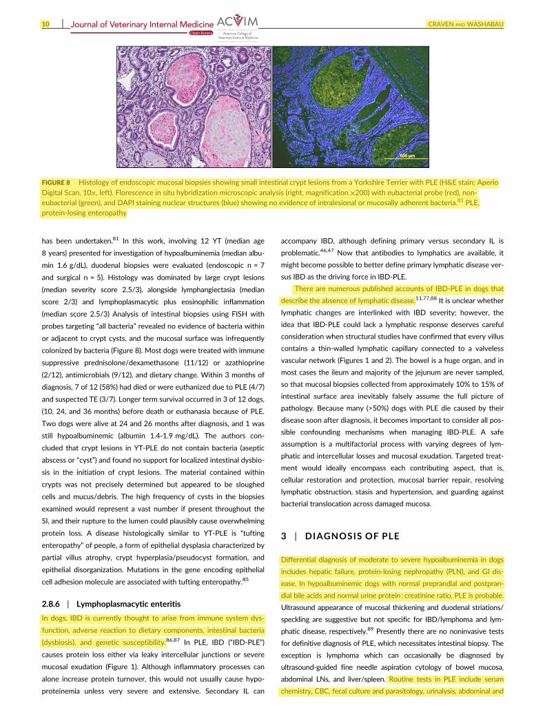
FIGURE 8 Histology of endoscopic mucosal biopsies showing small intestinal crypt lesions from a Yorkshire Terrier with PLE (H&E stain; Aperio
Digital Scan, 10×, left). Florescence in situ hybridization microscopic analysis (right, magnification ×200) with eubacterial probe (red), non-
eubacterial (green), and DAPI staining nuclear structures (blue) showing no evidence of intralesional or mucosally adherent bacteria.81 PLE,
protein-losing enteropathy
10 CRAVEN AND WASHABAU

thoracic imaging (for PTE and comorbidities), and GI endoscopy and
biopsy.11 In dogs, PLE involving severe hypoalbuminemia and effusions
can occur in hypoadrenocorticism, and measuring basal serum cortisol
concentration is also a consideration.90–92
3.1 | Noninvasive “biomarker” tests
Various potential biomarkers of IBD and PLE have been studied, such
as cobalamin, C-reactive protein (CRP), perinuclear anti-neutrophil
cytoplasmic antibodies, S100A12 (calgranulin-C), calprotectin, and
fecal alpha-1 protease inhibitor (α1 PI).11,87,93–95 Cobalamin, CRP, and
α1 PI are the only commercially available tests at this time. Hypocoba-
laminemia is an inconsistent finding, reported in 7 of 19,96 8 of 13,13
4 of 18,69and 15 of 20 dogs65 with PLE in separate studies but was
associated with a negative outcome.71 CRP was reported to be a
strongly correlated marker for systemic inflammatory activity in dogs
with IBD97 but in later studies was increased in only 43% of cases98
and did not correlate with disease severity by histologic grade or clini-
cal scoring.99,100 More recently it was found that a high CRP concen-
tration is a negative prognostic indicator in PLE (P = .004),65 but in
other work, there was no difference between responders and nonre-
sponders.55 There is no clear consensus on the diagnostic utility of
CRP, and as a nonspecific inflammatory marker, its validity across
studies may depend on factors such as geographical region (endemic
disease, parasitism, animal welfare standards), and laboratory method-
ology. More recently, fecal α1 PI, a measurable serum protease inhibi-
tor lost into the bowel in PLE, was reported to distinguish dogs with
moderate to severe intestinal crypt disease and lacteal dilatation
(n = 54) from those without (n = 66) with sensitivity of 44%-74% and
specificity of 57%-93%.100 Being a noninvasive surveillance tool for
screening PLE-susceptible breeds, α1 PI is also helpful to detect PLE in
animals with concurrent PLN or hepatic insufficiency.
Fecal calgranulin-C (S100A12) is a noninvasive marker of IBD in
people.101,102 A calcium-binding protein, S100A12, is secreted by acti-
vated neutrophils, a cell type abundant in the intestinal mucosa of IBD
in people. When tested and validated for accuracy in distinguishing
dogs with IBD from healthy controls, fecal S100A12 showed a sensi-
tivity of 65% and specificity of 84%, with a misclassification rate of
20%.100 In another study, serum S100A12 did not identify dogs with
PLE; however, reference range serum S100A12 and serum calprotec-
tin concentrations (P = .04 and P = .03, respectively), were found to
be negative prognostic indicators in dogs with PLE. In people, calpro-
tectin is another useful IBD marker,103 corresponding to a neutrophil
cytosol protein with resistance to bacterial degradation. In dogs,
serum calprotectin ≥296 μg/L showed sensitivity of 82.4% and speci-
ficity 68.4% for IBD detection, although it did not correlate with CIB-
DAI, CRP, or histology.104–106 The “gold standard” biomarkers for IBD
and PLE in dogs have yet to be realized. The dissimilarities in histologi-
cal appearance of human and canine diseases, for example, neutro-
philic/granulomatous inflammation (people) versus lymphoplasmacytic
inflammation (dogs and cats), and transmural (people) versus mucosal
(dogs and cats) distribution, imply probable species specificity in a high
performing biomarker.
3.2 | Intestinal biopsy in PLE
Definitive diagnosis of PLE necessitates histological evaluation of
intestinal biopsies. Endoscopic biopsy carries lower procedural risk
than surgical biopsy owing to specific comorbidities: (1) PLE is a risk
for thrombosis, a “silent” killer, with risk of clot embolization or exten-
sion during anesthesia and surgical manipulations; (2) hypovitaminosis D
and hypocalcemia have known potential adverse effects on wound heal-
ing, intestinal repair, cardiac and vascular function107; (3) hypoalbumine-
mia was significantly associated with death or euthanasia in 2 separate
IBD outcome studies in dogs,71,73 and 4 studies of PLE.52,56,57,69 Logi-
cally, posing increased physiologic demands for protein (intraoperative
hemorrhage and wound healing) might be reasonably expected to
increase the albumin-associated risk of negative outcome. When, how-
ever, a focal intestinal mass is detected, (eg, lipogranulomatous lymphan-
gitis, lymphoma), endoscopic reach can be a limiting factor in achieving a
noninvasive definitive diagnosis, wherein surgical biopsy (with/without
mass resection) might be required to facilitate an accurate diagnosis
(assuming that fine needle aspirates were nondiagnostic).50,51,80,108
Historically, it has been queried whether endoscopic biopsy is
adequate for diagnosis of IL because of failure to sample submucosal
lymph vessels.77,78,87,88,96 More recently, histological diagnosis of IL
was reported as equivalent by endoscopic (44/83; 53%) and surgical
biopsy (38/64; 59%).77,78 The latter report, describing 38 dogs with IL,
found transmural lymphatic pathology in 29 of 38 (76%) cases, sug-
gesting that mucosal biopsies would usually be of adequate depth.
Furthermore, the interconnected structure of the “web-like” and
valveless intestinal network implies that IL would not usually be con-
fined to submucosal lymphatics without some degree of transference
of lymph capillary hypertension to the lacteal. Diagnostic accuracy of
endoscopic biopsy inevitably depends on sample quality88 and 6 (high
quality) to 15 (moderate quality) duodenal samples are advised in
dogs, except for diagnosis of crypt lesions, which reportedly requires
13-20 biopsies. The sensitivity of endoscopy for lymphoma diagnosis
was >90%, regardless of biopsy quality.88
The gross appearance of the duodenal mucosa and particularly
the presence of white villus tips are inadequate to accurately predict
IL, with reported sensitivity of 68% and specificity of 42% for endo-
scopic appearance alone in IL diagnosis.96
The endoscopic appearance of PIL in people reveals obvious,
creamy yellow villi corresponding to marked dilatation of intestinal
lymphatics.40 Histology confirms the presence of dilated mucosal and
submucosal lymphatics, from a few millimeters to centimeters. Lac-
teals can be dilated in many villi or only a few. Mucosal abnormalities
vary, but the absence of villous atrophy, significant mucosal/submuco-
sal defect, and microorganisms, is key to diagnosis. Endoscopy can be
negative when intestinal lesions are segmental or localized. In these
cases, video capsule endoscopy or enteroscopy can be diagnostic.40
Ultrasonography can show dilatation of intestinal loops, wall thicken-
ing, severe mesenteric edema, and ascites.109 Contrast computerized
tomography (CT) identifies localized IL, showing typically diffuse, nod-
ular, small bowel thickening with or without dilatation, and a “halo
sign” due to bowel swelling and edema.42,110 Magnetic resonance
imaging has been successfully applied in people with IL to evaluate for
CRAVEN AND WASHABAU 11
localized and systemic disease.111 The use of CT and MRI for the pur-
pose of PLE diagnosis in dogs is not reported.
4 | SPECIAL TESTS
4.1 | Mucosal biopsy FISH and culture
A pathogenic role of the intestinal microbiome is not recognized in PLE;
however, intestinal dysbiosis with a Gram-negative shift is emerging as
a contributor to IBD pathophysiology. It should not be forgotten that
the loss of immune system components in people with PLE depletes
immune barrier function,79 and so potentially, intestinal dysbiosis could
be of greater severity and detriment in IBD-PLE.83,112–115 Disrupted
mucosal immunity and dysbiosis in PLE are further implied by the
observation that mucosal neutrophils were significantly more often
present (P = .003) in dogs with chronic enteropathy (CE) and hypoalbu-
minemia (17/37, 46%) than CE-normoalbuminemia (7/46, 15%).77 In
dogs at greater risk of mucosal immune compromise, for example, lym-
phangiectasia, ulceration, granulomatous or predominantly neutrophilic
inflammation, and chronic intussusception,116,117 analysis by FISH can
be applied to search mucosae for bacterial adherence/invasion. Used
in molecular microbiology to identify bacteria within formalin fixed
tissues, FISH is commercially offered, at Cornell University. Where his-
tology or FISH reveal intramucosal or mucosally adherent bacteria,
intestinal specimens can be submitted for culture and sensitivity (biop-
sies can be stored frozen in Luria-Bertani culture medium).
4.2 | Mucosal biopsy immunophenotyping and
polymerase chain reaction for antigen-receptor
rearrangements
When routine histology fails to differentiate a benign, polyclonal lym-
phoid proliferation from a monoclonal, neoplastic process, clarification
may be achieved using immunophenotyping (IP) for lymphocyte
markers118 or polymerase chain reaction (PCR) to detect clonal B and
T cell populations (PCR for antigen-receptor rearrangements).118 Both
methods utilize the paraffin wax-embedded histology block, without
need for further sampling. The consensus of the WSAVA International
GI Standardization Group is that IP retains precedence over clonality
testing as a diagnostic procedure.72
5 | MANAGEMENT OF PLE
In treating PLE, defining the precise nature of protein loss, that is,
inflammatory, exudative, or structural (lymphatic or crypt distension)
determines the selection of appropriate therapeutics. To optimize
recovery rates, an otherwise safe assumption is that the processes of
lymph fluid loss, active protein exudation, and leaky cell junctions
could be present at some juncture in every affected bowel.
5.1 | Diet
A low-fat diet with supplementary MCT is the cornerstone of PLE
management in people, proven to be the most effective, most widely
prescribed approach and with minimal adverse effects.40,119 Desai
et al. showed that within a few weeks, a high-protein, low-fat diet
with MCT not only improves clinical signs but also reduces mortality
in people.37 The need for dietary control in people appears to be per-
manent, because clinical and biochemical findings reappear after low-
fat diet withdrawal.40 Dietary fat restriction prevents engorgement of
intestinal lymphatics with chyle, preventing rupture of grossly dis-
tended lacteals. Medium-chain triglycerides are directly absorbed into
the portal venous circulation and provide nutrient fat without lacteal
engorgement.40 The benefit of MCT should not be assumed to be
restricted to lymphatic bypass. Medium-chain triglyceride digestion is
rapid and simple, without stimulating cholecystokinin secretion or uti-
lizing the actions of bile and lipase for absorption. Absorption of MCT
occurs via passive diffusion along the GI tract into the portal system
bound to albumin. No further packaging or modification of MCT mole-
cules is required. Moreover, MCTs are not dependent on the carnitine
acyltransferase system for transport into the mitochondria for β-oxi-
dation. This provides the ability for more rapid metabolism of MCTs
and improved utilization even in states of protein deficiency.120
Suggested dietary proportions for dogs with PLE are calories from
carbohydrate 55-60%, total fat calories 10-15%, and protein calories
25-30%, using a highly digestible protein (>87%).36 The canine diet
must always contain LCFAs for provision of EFAs, and supplementa-
tion of fat-soluble vitamins (A, D, E, K) is particularly important when
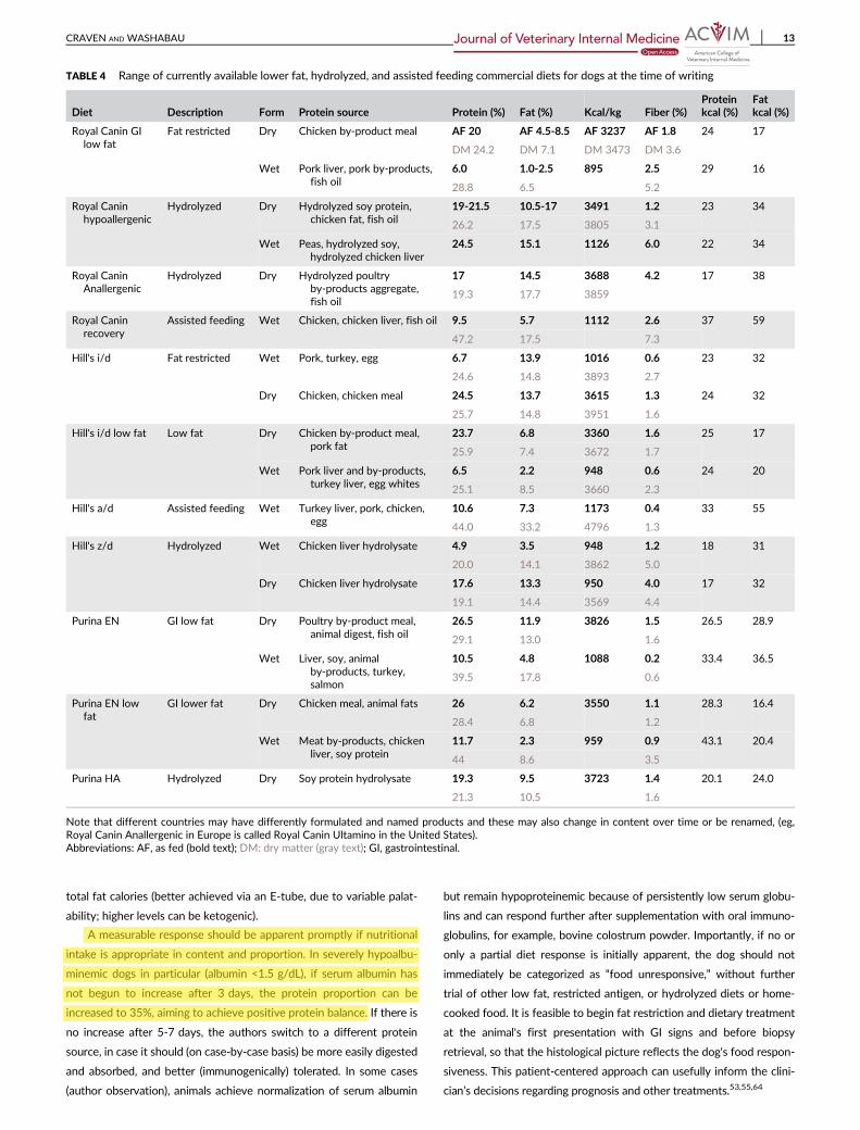
dietary fat content is low. Available low-fat commercial diets are listed
in Table 4; however, the choice of diet in PLE cases is often dictated
by patient appetite and preference. Many (especially YT) are histori-
cally fussy eaters (perhaps reflecting more chronic subclinical GI dis-
ease), and are frequently anorexic. Nutritional management in PLE is
paramount and if not effectively implemented, the cycle of protein
leak and catabolism will continue until eventually death is inevitable.
In selective eaters or those with unreliable appetite, the authors rou-
tinely place an esophagostomy tube (E-tube) during the endoscopy
anesthesia. This step seems to be the key to success in PLE manage-
ment for both patient (particularly YT) and owner. Often very unwell
and with critically low serum proteins, these patients need every
modicum of support. The pet-owner bond can be disrupted when
owners force their pet to take medications and a specific diet, and life
can become unbearably stressful and anxious for both. The authors'
experience is that in retrospect, the feeding tube was not infrequently
a life-saving measure. When tube feeding is initiated, a commercial
low fat, blenderized canned food is supplemented up to 30% protein
calories (with whey or pea protein powder) and divided into 6-8 meals
per day (resting energy requirement, RER, in kcal/day = 70 × [ideal
weight in kg]0.75). It should be recognized that multiple daily feedings
might not be possible in every clinical scenario. If feeding is well
accepted, in underweight dogs, a gradually increasing amount over
the RER may be possible. In tube fed dogs, oral alimentation is encour-
aged, for example, lean protein (cooked turkey/fish), low fat prescrip-
tion foods, and tube feeding is adjusted according to appetite. Free
amino acid preparations (eg, Vivonex) can be useful during critical
stages but should not replace or delay provision of complete nutri-
tional support. To supplement caloric density, MCT can be given in
the form of mixed C8 and C10 triglycerides to provide up to 30% of
12 CRAVEN AND WASHABAU
total fat calories (better achieved via an E-tube, due to variable palat-
ability; higher levels can be ketogenic).
A measurable response should be apparent promptly if nutritional
intake is appropriate in content and proportion. In severely hypoalbu-
minemic dogs in particular (albumin <1.5 g/dL), if serum albumin has
not begun to increase after 3 days, the protein proportion can be
increased to 35%, aiming to achieve positive protein balance. If there is
no increase after 5-7 days, the authors switch to a different protein
source, in case it should (on case-by-case basis) be more easily digested
and absorbed, and better (immunogenically) tolerated. In some cases
(author observation), animals achieve normalization of serum albumin
but remain hypoproteinemic because of persistently low serum globu-
lins and can respond further after supplementation with oral immuno-
globulins, for example, bovine colostrum powder. Importantly, if no or
only a partial diet response is initially apparent, the dog should not
immediately be categorized as “food unresponsive,” without further
trial of other low fat, restricted antigen, or hydrolyzed diets or home-
cooked food. It is feasible to begin fat restriction and dietary treatment
at the animal's first presentation with GI signs and before biopsy
retrieval, so that the histological picture reflects the dog's food respon-
siveness. This patient-centered approach can usefully inform the clini-
cian’s decisions regarding prognosis and other treatments.53,55,64
TABLE 4 Range of currently available lower fat, hydrolyzed, and assisted feeding commercial diets for dogs at the time of writing
Diet Description Form Protein source Protein (%) Fat (%) Kcal/kg Fiber (%)Proteinkcal (%)
Fatkcal (%)
Royal Canin GI
low fat
Fat restricted Dry Chicken by-product meal AF 20 AF 4.5-8.5 AF 3237 AF 1.8 24 17
DM 24.2 DM 7.1 DM 3473 DM 3.6
Wet Pork liver, pork by-products,
fish oil
6.0 1.0-2.5 895 2.5 29 16
28.8 6.5 5.2
Royal Canin
hypoallergenic
Hydrolyzed Dry Hydrolyzed soy protein,
chicken fat, fish oil
19-21.5 10.5-17 3491 1.2 23 34
26.2 17.5 3805 3.1
Wet Peas, hydrolyzed soy,hydrolyzed chicken liver
24.5 15.1 1126 6.0 22 34
Royal CaninAnallergenic
Hydrolyzed Dry Hydrolyzed poultryby-products aggregate,
fish oil
17 14.5 3688 4.2 17 38
19.3 17.7 3859
Royal Canin
recovery
Assisted feeding Wet Chicken, chicken liver, fish oil 9.5 5.7 1112 2.6 37 59
47.2 17.5 7.3
Hill's i/d Fat restricted Wet Pork, turkey, egg 6.7 13.9 1016 0.6 23 32
24.6 14.8 3893 2.7
Dry Chicken, chicken meal 24.5 13.7 3615 1.3 24 32
25.7 14.8 3951 1.6
Hill's i/d low fat Low fat Dry Chicken by-product meal,
pork fat
23.7 6.8 3360 1.6 25 17
25.9 7.4 3672 1.7
Wet Pork liver and by-products,
turkey liver, egg whites
6.5 2.2 948 0.6 24 20
25.1 8.5 3660 2.3
Hill's a/d Assisted feeding Wet Turkey liver, pork, chicken,egg
10.6 7.3 1173 0.4 33 55
44.0 33.2 4796 1.3
Hill's z/d Hydrolyzed Wet Chicken liver hydrolysate 4.9 3.5 948 1.2 18 31
20.0 14.1 3862 5.0
Dry Chicken liver hydrolysate 17.6 13.3 950 4.0 17 32
19.1 14.4 3569 4.4
Purina EN GI low fat Dry Poultry by-product meal,
animal digest, fish oil
26.5 11.9 3826 1.5 26.5 28.9
29.1 13.0 1.6
Wet Liver, soy, animal
by-products, turkey,salmon
10.5 4.8 1088 0.2 33.4 36.5
39.5 17.8 0.6
Purina EN lowfat
GI lower fat Dry Chicken meal, animal fats 26 6.2 3550 1.1 28.3 16.4
28.4 6.8 1.2
Wet Meat by-products, chicken
liver, soy protein
11.7 2.3 959 0.9 43.1 20.4
44 8.6 3.5
Purina HA Hydrolyzed Dry Soy protein hydrolysate 19.3 9.5 3723 1.4 20.1 24.0
21.3 10.5 1.6
Note that different countries may have differently formulated and named products and these may also change in content over time or be renamed, (eg,
Royal Canin Anallergenic in Europe is called Royal Canin Ultamino in the United States).
Abbreviations: AF, as fed (bold text); DM: dry matter (gray text); GI, gastrointestinal.
CRAVEN AND WASHABAU 13
5.2 | Drug treatment
5.2.1 | Immune modification
Immune suppressive agents are infrequently utilized in people for the
treatment of most types of PLE.2,40–42 In contrast, in dogs an
immune-mediated etiology of PLE is usually presumed11; however,
there is no scientific or convincing volume of case-based evidence
that IL (also lymphangitis and crypt disease) is autoimmune or primar-
ily immune-mediated. Rather, the immune system can suffer the loss
of components9 such that the induction of immune suppression
becomes potentially a precarious step. Lymphatic disease is a particu-
larly unknown entity in dogs, and it is interesting to note the differing
numbers of disease-associated deaths in PLE (~54%) versus IBD
(<20%),71,73 when in both diseases immune suppression with cortico-
steroids is the mainstay of treatment worldwide. The adverse effects
of corticosteroid treatment in dogs, that is, muscle protein catabolism,
thrombosis,121–123 and hyperlipidemia124 (with potential for lymphatic
expansion) are particularly disadvantageous in PLE, and their use is
somewhat counterintuitive. Recently, immunosuppressive treatment
of PLE was significantly associated with a negative outcome (P < .001,
2/21 dogs alive), versus those treated with dietary modification alone
(13/22 dogs alive).53 Clinical response to diet alone (various foods) is
also reported in in 10 of 11 YT with PLE64 Interestingly, in a recent
report52 all “short-term” survivors (19 of 59 dogs with PLE) appeared
unresponsive to immune suppressive treatment, which brings forth
enquiry on how best to rationalize their use in this setting.
Immune suppression is often suggested to be reserved as “last
resort” treatment, for IBD87 and some animals with moderate to severe
change are reported to clinically respond completely to dietary man-
agement alone.71,87,125 The clinical severity of IBD does not always
correlate with the histological severity score,72 and neither variable is
demonstrated to predict the requirement for immune suppressive
treatment.71,73 Commonly used immune suppressive medications in
dogs are prednisolone, cyclosporine, azathioprine, chlorambucil, and
mycophenolate. In a small study, improved survival of dogs with PLE
was associated with the use of prednisolone plus chlorambucil (10/14
alive) versus prednisolone plus azathioprine (2/13 alive, P = .004).69
Consideration of the type of inflammation present and agent pharma-
codynamics should help to guide drug choice. Mycophenolate, chlorambucil,
and azathioprine can all cause severe GI toxicosis and myelosuppression.
Azathioprine is a purine antimetabolite that inhibits nucleic acid syn-
thesis causing chromosomal breaks and disruption of cellular metabo-
lism. Azathioprine is poorly absorbed PO and clinical response can take
up to 6 weeks.126,127 Chlorambucil is a cytotoxic alkylating agent with
delay of up to 4 weeks before clinical response is appreciated.126,127 It
is well absorbed PO and highly bound to plasma proteins, which is of
note when used in hypoproteinemic animals. Cyclosporine suppresses
cell-mediated immunity and might be more efficacious in suppressing
T-cell-macrophage interactions in granulomatous inflammation.127,128
However, it is variably absorbed PO and can be less well absorbed
in the diseased bowel.129 Mycophenolate mofetil, a prodrug of myco-
phenolic acid, is an inhibitor of inosine monophosphate dehydroge-
nase (IMPDH), a rate-limiting enzyme in nucleotide synthesis.130
Lymphocytes are particularly dependent on IMPDH, unlike other cell
types (eg, neutrophils) that recycle nucleotides via a salvage pathway.
Mycophenolate mofetil is thought to suppress proliferation specifi-
cally of lymphocytes130 and has many desirable properties in treating
IBD: absorbed PO, “neutrophil-sparing,” and without extensive time
delay.127 However, it causes significant GI irritation, and in people has
a “black box” warning (serious or life-threatening risk) for lymphoma.126
Regarding corticosteroids, budesonide is a preparation with topical
activity in the GI tract but has not been specifically evaluated for PLE
treatment.127 Some practitioners use parenteral dexamethasone due to
concerns that prednisolone is insufficiently PO absorbed, but in studies
of people with Crohn's disease, this theory is not well supported.131,132
5.3 | Heparan sulfate
A common histological feature of PLE in people is loss of heparan sul-
fate (HS) from the basolateral surface of intestinal epithelial cells
(in numerous seemingly unrelated PLE etiologies).133 A complex proteo-
glycan, HS is a large, highly sulfated glycosaminoglycan composed of
alternating units of α-N-acetylglucosamine and α-glucuronic acid.
Heparan sulfate functions as an anticoagulant by binding ATIII and is a
component of basement membranes in numerous organs, including
intestinal mucosa. In the kidney, HS molecules are an important barrier
against protein leak and are deficient in the glomerulus of PLN
(nephrotic syndrome). Interestingly, in children born with severe PLE,
deficiency of HS has been discovered in the basolateral surface of the
enterocyte.134 Exogenous heparin treatment to these children reversed
enteric protein loss at doses that are considered subtherapeutic for
anticoagulation in people (5000 U/day SC).135 The precise mechanism
by which HS relieves protein loss across the mucosa is unknown, but
the effect has been reported in several other PLE-associated
diseases.135–137 The ability of HS treatment to reverse PLE in dogs is
hopeful but has not yet been evaluated (see antithrombotics).
5.4 | Octreotide
Octreotide is a long-acting somatostatin analogue that suppresses GI
motility and hormone secretion in the pituitary gland, pancreas, and
intestine. In 1998, the efficacy of octreotide in 1 human PIL patient
was first reported, and it has since, as monotherapy with MCT diet,
been shown to lead to long-term clinical, biochemical, and histological
remission of PLE of differing etiologies in people.138 The mechanism
of action of octreotide in diminishing protein loss through the GI tract
is unclear. Somatostatin receptors exist in normal gut lymphoid tissue
and on veins and venules.139 Both somatostatin and octreotide nor-
mally decrease splanchnic blood flow via vasoconstriction and also
decrease intestinal motility, gastric emptying, gallbladder contraction,
pancreatic secretion, and triglyceride absorption.140,141 Theorized
mechanisms of octreotide's action in PIL include decreased intestinal
fat absorption, inhibition of GI vasoactive peptides, and stimulation of
the autonomic nervous system.138 Octreotide is expensive and admin-
istered parenterally, and in responsive patients discontinuation usually
causes PLE recurrence.142 Reported adverse effects in human clinical
trials were diarrhea, abdominal pain, nausea, headache, cholelithiasis,
hyperglycemia, and constipation.40,119,140,143 Octreotide has been
used in dogs for the treatment of insulinoma.144
14 CRAVEN AND WASHABAU
5.5 | Antithrombotics
The published data described here suggests that TE occurs in around
6% of PLE cases but is almost certainly underestimated, being a
“silent” killer.13,145,146Prophylaxis with antithrombotic medication
could prevent death and is usually well tolerated, inexpensive, and
with minimal adverse effect. There is no consensus on optimal antith-
rombotic treatment in PLE, and options include unfractionated heparin
sulfate (UFH), low-molecular-weight heparin, clopidogrel, and low-
dose aspirin. In consideration of the markedly beneficial effect on PLE
in people, UFH would be a logical choice but is poorly tolerated by
painful SQ injection q8h. For years, UFH was thought to be minimally
absorbed PO, supported by minimal changes in plasma APTT levels
after oral administration, but sublingual and oral UFH absorption has
since been demonstrated in numerous species including dogs.147–149
There is now published evidence of reliable oral UFH absorption in
healthy dogs, evidenced by ATIII inhibition and anti-Xa activity.150
Recovery of UFH from both plasma and urine in people and dogs also
supports that UFH administered PO is absorbed and widely distrib-
uted.148,150 In dogs, no negative effects were observed to indicate
UFH-induced hemorrhage, excessive ATIII consumption, or thrombo-
cytopenia during oral heparin administration.150 Further studies deter-
mining optimal oral dosage in dogs, and clinical benefit in disease
states has not yet emerged. Clopidogrel and low-dose aspirin are fre-
quently used as antiplatelet agents in dogs with PLE, either alone or in
combination.145 The optimal combination, dosages, and required dura-
tion of treatment is unknown; however, dogs in complete clinical
remission of PLE can die suddenly from TE,145 which suggests a bene-
fit of prolonged treatment.
5.5.1 | Other
Other medications used empirically to treat IBD and PLE are antimi-
crobials (metronidazole, tylosin, doxycycline), gut protectants (acid
reducers, sucralfate), antiemetics, and probiotics.11,36 The use of gut
protectants might help to maintain a lumenal environment that favors
ongoing repair of the mucosal barrier. If mucosal ulceration or neutro-
philic inflammation are present, broad spectrum antimicrobial treatment
might be warranted, for example, doxycycline, amoxycillin-clavulanate,
cephalexin, and FISH analysis alongside culture data would also serve
to guide antimicrobial treatment. Cobalamin deficiency has been asso-
ciated with a negative prognosis,93 and supplementation by oral and
parenteral routes is described.151 Although rare, folate deficiency can
be corrected by oral supplementation. Corrections of deficits in cal-
cium and magnesium in PLE have been described by Kimmel et al.,61
by administration of “constant rate infusions of magnesium sulfate
(1.0 mEq/kg/d [0.5 mEq/lb/d], IV) and calcium gluconate solution
(10 mg/kg/h, IV) until correction of the electrolyte abnormalities was
achieved.” Specific dosages and preparations of fat soluble vitamins A,
D, E, and K in PLE are not described; however, it is unlikely that the
use of a reputable veterinary supplement at the recommended daily
dosage would result in vitamin toxicosis. Diagnosis of specific vitamin
deficiencies and their resolution after supplementation would necessitate
individual quantification of vitamin levels. Addressing concurrent prob-
lems such as nausea/emesis (eg, maropitant, ondansetron), abdominal
pain (eg, tramadol, oral buprenorphine, cautious paracetamol), intestinal
dysmotility (eg, metoclopramide, cisapride) is also important. The use of
appetite stimulants in animals with GI disease is controversial. In dogs,
reduced appetite is nearly always an indication of another problem such
as nausea, pain, dysmotility, and use of appetite stimulants can mask a
debilitating untreated problem. Antioxidant treatment and reduction of
environmental stressors might also be helpful.152–155 Complications of
peripheral limb edema can be avoided by using support wraps, vigilant
monitoring for skin breakdown, gentle grooming and cleaning, frequent
ambulation, and massage. A deep, conforming mattress will provide
greater comfort when ascites and limb edema are present. Diuretics are
not generally useful because the edema relates to a reduction in plasma
oncotic pressure.41
5.5.2 | Oncotic support
Measures to increase plasma oncotic pressure such as the use of
hydroxyethyl starch (Hetastarch 6%, HS), plasma transfusion, and
purified canine albumin are only transiently helpful in PLE. Proteins
administered IV are leaked into the GI tract, and this can in itself be
detrimental due to the potential for aggravated loss of lymph and
immune system components. However, this can also be life-saving in
management of critically unwell patients and facilitates anesthesia and
diagnostic procedures. Plasma volume expansion produced by HS
approximates that of 5% albumin, for 24-36 hours. When TE is sus-
pected, HS or purified canine albumin is preferable over plasma trans-
fusion to avoid thrombus extension. The provision of parenteral
nutrition via a central venous catheter is another means of increasing
plasma oncotic pressure while providing nutritional support, with the
unfortunate caveat of higher risks of sepsis, local infection/abscess,
and thrombosis.13,74,156,157
5.6 | Theories on PLE management
5.6.1 | Stopping the leak
The lymphatic circulation is devoid of any central pump. To achieve
continuous lymph flow external compression is necessary, via intesti-
nal peristalsis, movement of body parts, arterial pulsations, and tissue
compression by external forces (eg, gravitational, weight bearing, mas-
sage). When lacteals are grossly dilated, the overlapping LECs might
not be able to maintain their door-like function to ensure the unidirec-
tional flow of interstitial fluid into lymphatic capillaries (Figure 1).
Without this 1-way system, the external compressive “pumps” could
merely contribute to lymph entering interstitial tissues and the bowel
lumen, so that an unrelenting cycle of lymph leakage, local irritation,
protein loss, and edema formation develops. Anterograde lymph flow
then becomes largely dependent on passive (Starling) forces. A steady
state could be reached by catabolism of interstitial tissue proteins or
lymphangiogenesis to restore lymph drainage, until these compensa-
tory mechanisms are also exhausted.158 Animals that present with
panhypoproteinemia and effusions are frequently in a late stage of a
dangerous “PLE cycle” disease.
It follows that diminishing the protein leak might be achieved by
(1) emphasis on Starling forces (2) by modifying intestinal peristalsis,
and (3) bypass of the affected intestinal area, if PLE is confirmed to be
segmental.
CRAVEN AND WASHABAU 15
5.6.2 | Emphasis on Starling forces
Osmolality of the human duodenum in fasted state is 200-250 mOsm/kg,
versus serum osmolality, 290-300 mOsm/kg.159 In healthy people, the SI
environment changes on a time-after-food basis. The effect of different
“fed states” on SI intraluminal osmolality was evaluated by administering
the nutritional drink Ensure Plus.159 Fasted state osmolality values were
in general hypo-osmotic, with overall median value of 224 mOsm/kg,
whereas hyperosmotic values (as high as 600 mOsm/kg, median
285 mOsm/kg) were found in fed states during the first 3 hours of
digestion.159 In dogs, fasted intestinal lumen osmolality is around
200-250 mOsm/kg and serum osmolality 290-310 mOsm/kg.160
Applying Starling's forces to maintain an osmotic gradient between
the bowel lumen and the interstitium should be helpful in PLE man-
agement. The bowel is continually leaking across a large surface area,
with no means to “plug” the leak. But by continually matching intesti-
nal intraluminal with interstitial osmolality, an iso-osmotic oncotic
gradient develops and the rate of protein loss by passive Starling
forces must slow or stop. Depending on the underlying pathology (eg,
lymphatic obstruction versus functional stasis), maintaining a mildly
hyperosmolar lumen reverses the oncotic pressure gradient to stimu-
late anterograde lymph flow and protein absorption (Figure 1). It is
not practicable to continually monitor lumen and serum osmolality,
but an informative baseline could be measured at endoscopy. An
iso- or mildly hyperosmotic lumen could theoretically be achieved by
feeding small, frequent, high-protein/low-fat meals every 2-3 hours
so that the bowel is in the fed state more of the time. Protein require-
ments in people are normally 0.6-0.8 g/kg/day, and in PLE this value
may increase 5-fold (3.0-4.0 g/kg/day) before a positive protein bal-
ance is reached.42 The optimal level of protein supplementation in
dogs with PLE has never been determined. A warmed, liquid consis-
tency, low-fat diet appears logically advantageous over dried or
dense wet food to reduce GI “work” and to optimize enterocyte con-
tact and surface tension for maximal nutrient absorption.
5.6.3 | Modifying intestinal peristalsis
Intestinal peristalsis passively facilitates lymph transport and a hypo-
motile bowel can develop lymph stasis. Studies of GI motility in PLE
are lacking; however, bowel swelling and edema might alter GI transit
times. Motility can be assessed radiographically using barium impreg-
nated polyethylene spheres, barium fluoroscopy, radionuclide transit,
or video capsule endoscopy. When dysmotility is present, prokinetic
treatment, for example, metoclopramide (0.2-0.5 mg/kg PO q8h) or
cisapride (0.1-0.5 mg/kg PO q8h), should be considered, with careful
monitoring for signs of worsening hypoproteinemia or deteriorating
clinical condition. Enhanced bowel contractility could also serve to
accelerate protein and lymph loss and is likely to be more safely initi-
ated alongside a well implemented feeding plan.
5.6.4 | Bypass of segmental PLE
Enterectomy for localized areas of lymphangiectasia has been curative
in people with segmental PLE.161 The reasons for disease localization
to a particular anatomical area are unknown, but improvement can
be only transient because of disease recurrence. Segmental PLE has
been diagnosed using (99 m) Tc-labeled albumin scintigraphy.162,163
Segmental presentations in dogs and breed-specific anatomical locali-
zation are not recognized so far but are unexplored phenomena. Serial
monitoring using scintigraphy (with radiolabeled canine albumin) could
help to identify dogs with lesion localization that might be safely ame-
nable to surgical treatment.
6 | CONCLUSIONS
In dogs, PLE is most frequently associated with lymphangiectasia and
IBD and is managed empirically with immune suppressive agents,
most frequently prednisolone, azathioprine, cyclosporine, and chlor-
ambucil, as well as dietary change and antimicrobials. The use of
immunosuppressive agents remains controversial in any of these dis-
orders and is not utilized in people for the treatment of most types of
PLE. Lifelong dietary management is the cornerstone of treatment,
and recent studies in dogs support this approach.53,64 Current treat-
ment is associated with >50% fatality, which logically drives manage-
ment toward the safest approach until pathophysiology is better
understood and specific, nonempirical treatment can be formulated.
The investigative history of E. coli-associated granulomatous colitis in
the Boxer dog (GC) highlights exceptionally well that the rationale for
exerting immune suppression, albeit as a “last resort” treatment,
invites careful consideration. Historically, for around a half century,
GC was often treated with immune suppression and with usually fatal
outcome. When clinical observations with antimicrobials drove the
discovery of an infectious etiology, GC became largely curable.164,165
An excellent relationship with the internist and a high level of owner
support are also a crucial aspect of managing PLE.
CONFLICT OF INTEREST DECLARATION
Authors declare no conflict of interest.
OFF-LABEL ANTIMICROBIAL DECLARATION
Authors declare no off-label use of antimicrobials.
INSTITUTIONAL ANIMAL CARE AND USE COMMITTEE
(IACUC) OR OTHER APPROVAL DECLARATION
Authors declare no IACUC or other approval was needed.
ORCID
Melanie D. Craven https://orcid.org/0000-0002-1759-0778
REFERENCES
1. Braamskamp MJAM, Dolman KM, Tabbers MM. Clinical practice.
Protein-losing enteropathy in children. Eur J Pediatr. 2010;169(10):
1179-1185.
2. Freeman HJ, Nimmo M. Intestinal lymphangiectasia in adults. World J
Gastrointest Oncol. 2011;3(2):19-23.
3. Rovenská E, Rovenský J. Lymphatic vessels: structure and function.
Isr Med Assoc J. 2011;13(12):762-768.
16 CRAVEN AND WASHABAU

4. Waldmann TA, Steinfeld JL, Dutcher TF, Davidson JD, Gordon RS.
The role of the gastrointestinal system in idiopathic hypoproteinemia.
Gastroenterology. 1961;41:197-207.
5. Takeda H, Ishihama K, Fukui T, et al. Significance of rapid turnover
proteins in protein-losing gastroenteropathy. Hepatogastroenterology.
2003;50(54):1963-1965.
6. Schmidt PN, Blirup-Jensen S, Svendsen PJ, Wandall JH. Characteriza-
tion and quantification of plasma proteins excreted in faeces from
healthy humans. Scand J Clin Lab Invest. 1995;55(1):35-45.
7. Wochner RD, Weissman SM, Waldmann TA, Houston D, Berlin NI.
Direct measurement of the rates of synthesis of plasma proteins in
control subjects and patients with gastrointestinal protein loss. J Clin
Invest. May 1, 1968;47(5):971-982.
8. Waldmann TA, Morell AG, Wochner RD, Strober W, Sternlieb I. Mea-
surement of gastrointestinal protein loss using ceruloplasmin labeled
with copper. J Clin Invest. 1967;46(1):10-20.
9. Magdo HS, Stillwell TL, Greenhawt MJ, et al. Immune abnormalities
in Fontan protein-losing enteropathy: a case-control study. J Pediatr.
2015;167(2):331-337.
10. Winkler S, Linke A, Adams V, Schuler G, Erbs S. How to improve
endothelial repair mechanisms: the lifestyle approach. Expert Rev Car-
diovasc Ther. 2010;8(4):573-580.
11. Dossin O, Lavoué R. Protein-losing enteropathies in dogs. Vet Clin
North Am Small Anim Pract. 2011;41(2):399-418.
12. Kull PA, Hess RS, Craig LE, Saunders HM, Washabau RJ. Clinical, clin-
icopathologic, radiographic, and ultrasonographic characteristics of
intestinal lymphangiectasia in dogs: 17 cases (1996-1998). J Am Vet
Med Assoc. 2001;219(2):197-202.
13. Goodwin LV, Goggs R, Chan DL, Allenspach K. Hypercoagulability in
dogs with protein-losing Enteropathy. J Vet Intern Med. 2011;25(2):
273-277.
14. Harvey NL, Srinivasan RS, Dillard ME, et al. Lymphatic vascular
defects promoted by Prox1 haploinsufficiency cause adult-onset
obesity. Nat Genet. 2005;37(10):1072-1081.
15. Johnson NC, Dillard ME, Baluk P, et al. Lymphatic endothelial cell
identity is reversible and its maintenance requires Prox1 activity.
Genes Dev. 2008;22(23):3282-3291.
16. Srinivasan RS, Dillard ME, Lagutin OV, et al. Lineage tracing demon-
strates the venous origin of the mammalian lymphatic vasculature.
Genes Dev. 2007;21(19):2422-2432.
17. Oliver G, Alitalo K. The lymphatic vasculature: recent progress and
paradigms. Annu Rev Cell Dev Biol. 2005;21(1):457-483.
18. Birke K, Drecoll E, Kerjaschki D, Birke MT. Expression of podoplanin
and other lymphatic markers in the human anterior eye segment.
Investig Opthalmol Vis Sci. 2010;51(1):344.
19. Yamanaka Y, Araki K, Ogata T. Three-dimensional organization of
lymphatics in the dog small intestine: a scanning electron microscopic
study on corrosion casts. Arch Histol Cytol. 1995;58(4):465-474.
20. Unthank JL, Bohlen HG. Lymphatic pathways and role of valves in
lymph propulsion from small intestine. Am J Physiol. 1988;254(3 Pt
1):G389-G398.
21. Adamczyk LA, Gordon K, Kholová I, et al. Lymph vessels: the forgot-
ten second circulation in health and disease. Virchows Arch. 2016;
469:3-17.
22. Ikomi F, Hunt J, Hanna G, Schmid-Schönbein GW. Interstitial fluid,
plasma protein, colloid, and leukocyte uptake into initial lymphatics.
J Appl Physiol. 1996;81(5):2060-2067.
23. Baluk P, Fuxe J, Hashizume H, et al. Functionally specialized junc-
tions between endothelial cells of lymphatic vessels. J Exp Med.
2007;204(10):2349-2362.
24. Miller MJ, McDole JR, Newberry RD. Microanatomy of the intestinal
lymphatic system. Ann N Y Acad Sci. 2010;1207:E21-E28.
25. Weitman E, Cuzzone D, Mehrara BJ. Tissue engineering and regener-
ation of lymphatic structures. Future Oncol. 2013;9(9):1365-1374.
26. Määttä M, Liakka A, Salo S, Tasanen K, Bruckner-Tuderman L, Autio-
Harmainen H. Differential expression of basement membrane com-
ponents in lymphatic tissues. J Histochem Cytochem. 2004;52(8):
1073-1081.
27. Ohtani O, Ohtani Y. Organization and developmental aspects of lym-
phatic vessels. Arch Histol Cytol. 2008;71:1-22.
28. Starling EH. On the absorption of fluids from the connective tissue
spaces. J Physiol. 1896;19(4):312-326.
29. Nurmi H, Saharinen P, Zarkada G, Zheng W, Robciuc MR, Alitalo K.
VEGF-C is required for intestinal lymphatic vessel maintenance and
lipid absorption. EMBO Mol Med. 2015;7(11):1418-1425.
30. Drayton DL, Liao S, Mounzer RH, Ruddle NH. Lymphoid organ devel-
opment: from ontogeny to neogenesis. Nat Immunol. 2006;7(4):
344-353.
31. von Andrian UH, Mempel TR. Homing and cellular traffic in lymph
nodes. Nat Rev Immunol. 2003;3(11):867-878.
32. Randolph GJ, Angeli V, Swartz MA. Dendritic-cell trafficking to lymph
nodes through lymphatic vessels. Nat Rev Immunol. 2005;5(8):
617-628.
33. Garden OA. Gastrointestinal Immunology. In: Washabau RJ, Day MJ,
eds. Canine & Feline Gastroenterology. St Louis, MO: Elsevier Saun-
ders; 2013:42-53.
34. Young AJ, Hay JB. Rapid turnover of the recirculating lymphocyte
pool in vivo. Int Immunol. 1995;7(10):1607-1615.
35. Salmi M, Jalkanen S. How do lymphocytes know where to go: current
concepts and enigmas of lymphocyte homing. Adv Immunol. 1997;
64(8):139-218.
36. Sanderson SL. Nutritional strategies in GI disease. In: Washabau RJ,
Day MJ, eds. Canine and Feline Gastroenterology. St Louis, MO: Else-
vier Saunders; 2013:409-428.
37. Desai AP, Guvenc BH, Carachi R. Evidence for medium chain tri-
glycerides in the treatment of primary intestinal lymphangiectasia.
Eur J Pediatr Surg. 2009;19(4):241-245.
38. Alfano V, Tritto G, Alfonsi L, Cella A, Pasanisi F, Contaldo F. Stable
reversal of pathologic signs of primitive intestinal lymphangiectasia
with a hypolipidic, MCT-enriched diet. Nutrition. 2000;16(4):
303-304.
39. Beynen AC, Kappert HJ, Lemmens AG, Van Dongen AM. Plasma lipid
concentrations, macronutrient digestibility and mineral absorption in
dogs fed a dry food containing medium-chain triglycerides. J Anim
Physiol Anim Nutr (Berl). 2002;86(9–10):306-312.
40. Vignes S, Bellanger J. Primary intestinal lymphangiectasia
(Waldmann's disease). Orphanet J Rare Dis. 2008;3:5.
41. Umar SB, DiBaise JK. Protein-losing enteropathy: case illustrations
and clinical review. Am J Gastroenterol. 2010;105(1):43-49.
42. Siddeswari R, Manohar S, Reddy T, Suryanarayana B, Abhilash T,
Devender. Primary intestinal lymphangiectasia. Indian Acad Clin Med.
2016;17(1):60-63.
43. Hokari R, Kitagawa N, Watanabe C, et al. Changes in regulatory mol-
ecules for lymphangiogenesis in intestinal lymphangiectasia with
enteric protein loss. J Gastroenterol Hepatol. 2008;23(7 PT2):
e88-e95.
44. Alders M, Hogan BM, Gjini E, et al. Mutations in CCBE1 cause gener-
alized lymph vessel dysplasia in humans. Nat Genet. 2009;41(12):
1272-1274.
45. Von Der Weid P-Y, Rehal S, Ferraz JG. Role of the lymphatic system
in the pathogenesis of Crohn's disease. Pathology. 2002;34(6):
335-341.
46. Sura R, Colombel J-F, Van Kruiningen HJ. Lymphatics, tertiary lym-
phoid organs and the granulomas of Crohn's disease: an immunohis-
tochemical study. Aliment Pharmacol Ther. 2011;33(8):930-939.
47. Van Kruiningen HJ, J-FJ-F C. The forgotten role of lymphangitis in
Crohn's disease. Gut. 2008;57(1):1-4.
48. Van Kruiningen HJ, Hayes AW, Colombel J-F. Granulomas obstruct
lymphatics in all layers of the intestine in Crohn's disease. APMIS.
2014;122(11):1125-1129.
49. Yuki M, Sugimoto N, Takahashi K, et al. A case of protein-losing
enteropathy treated with methotrexate in a dog. J Vet Med Sci. 2006;
68(4):397-399.
50. Kimmel SE, Waddell LS, Michel KE. Hypomagnesemia and hypocalce-
mia associated with protein-losing enteropathy in Yorkshire Terriers:
five cases (1992-1998). J Am Vet Med Assoc. 2000;217(5):703-706.
51. Littman MP, Dambach DM, Vaden SL, Giger U. Familial protein-
losing enteropathy and protein-losing nephropathy in soft coated
wheaten terriers: 222 cases (1983-1997). J Vet Intern Med. 2000;
14(1):68-80.
CRAVEN AND WASHABAU 17

52. Lane IF, Miller E, Twedt DC. Parenteral nutrition in the management
of a dog with lymphocytic-plasmacytic enteritis and severe protein-
losing enteropathy. Can Vet J. 1999;40(10):721-724.
53. Olson NC, Zimmer JF. Protein-losing enteropathy secondary to intes-
tinal lymphangiectasia in a dog. J Am Vet Med Assoc. 1978;173(3):
271-274.
54. Milstein M, Sanford SE. Intestinal lymphangiectasia in a dog. Can Vet
J. 1977;18(5):127-130.
55. Van Kruiningen HJ, Lees GE, Hayden DW, Meuten DJ, Rogers WA.
Lipogranulomatous lymphangitis in canine intestinal lymphangiecta-
sia. Vet Pathol. 1984;21(4):377-383.
56. Watson VE, Hobday MM, Durham AC. Focal intestinal lipogranulo-
matous lymphangitis in 6 dogs (2008-2011). J Vet Intern Med. 2014;
28(1):48-51.
57. Gianella P, Lotti U, Bellino C, et al. Clinicopathologic and prognostic
factors in short- and long-term surviving dogs with protein-losing
enteropathy. Schweiz Arch Tierheilkd. 2017;159(3):163-169.
58. Rudinsky AJ, Howard JP, Bishop MA, Sherding RG, Parker VJ,
Gilor C. Dietary management of presumptive protein-losing enterop-
athy in Yorkshire terriers. J Small Anim Pract. 2017;58(2):103-108.
59. Equilino M, Théodoloz V, Gorgas D, et al. Evaluation of serum bio-
chemical marker concentrations and survival time in dogs with
protein-losing enteropathy. J Am Vet Med Assoc. 2015;246(1):91-99.
60. Allenspach K, Rizzo J, Jergens AE, Chang YM. Hypovitaminosis D is
associated with negative outcome in dogs with protein losing enter-
opathy: a retrospective study of 43 cases. BMC Vet Res. 2017;
13(1):96.
61. Willard MD, Zenger E, Mansell JL. Protein-losing enteropathy associ-
ated with cystic mucoid changes in the intestinal crypts of two dogs.
J Am Anim Hosp Assoc. 2003;39(2):187-191.
62. Dijkstra M, Kraus JS, Bosje JT, Den Hertog E. Protein-losing enterop-
athy in Rottweilers. Tijdschr Diergeneeskd. May 15, 2010;135(10):
406-412.
63. Okanishi H, Yoshioka R, Kagawa Y, Watari T. The clinical efficacy of
dietary fat restriction in treatment of dogs with intestinal lymphan-
giectasia. J Vet Intern Med. 2014;28(3):809-817.
64. Simmerson SM, Armstrong PJ, Wünschmann A, Jessen CR, Crews LJ,
Washabau RJ. Clinical features, intestinal histopathology, and out-
come in protein-losing enteropathy in Yorkshire Terrier dogs. J Vet
Intern Med. 2014;28(2):331-337.
65. Willard MD, Helman G, Fradkin JM, et al. Intestinal crypt lesions
associated with protein-losing enteropathy in the dog. 14(3):
298-307.
66. Tamura Y, Ohta H, Kashiide T, et al. Case report: protein-losing
enteropathy caused by Mesocestoides vogae (syn. M. corti) in a dog.
Vet Parasitol. 2014;205(1–2):412-415.
67. Dandrieux JRS, Noble P-JM, Scase TJ, Cripps PJ, German AJ. Com-
parison of a chlorambucil-prednisolone combination with an
azathioprine-prednisolone combination for treatment of chronic
enteropathy with concurrent protein-losing enteropathy in dogs:
27 cases (2007-2010). J Am Vet Med Assoc. 2013;242(12):1705-
1714.
68. Nakashima K, Hiyoshi S, Ohno K, et al. Prognostic factors in dogs
with protein-losing enteropathy. Vet J. 2015;205(1):28-32.
69. Parker VJ, Jergens AE, Whitley EM, Frana TS. Isolation of Cokero-
myces recurvatus from the gastrointestinal tract in a dog with
protein-losing enteropathy. J Vet Diagn Invest. 2011;23(5):1014-
1016.
70. Jergens AE. Clinical assessment of disease activity for canine inflam-
matory bowel disease. J Am Anim Hosp Assoc. 2004;40(6):437–45.
71. Allenspach K, Wieland B, Gröne A, Gaschen F. Chronic enteropathies
in dogs: evaluation of risk factors for negative outcome. J Vet Intern
Med. 2007;21(4):700-708.
72. Washabau RJ, Day MJ, Willard MD, et al. Endoscopic, biopsy, and
histopathologic guidelines for the evaluation of gastrointestinal
inflammation in companion animals. J Vet Intern Med. 2010;24(1):
10-26.
73. Craven M, Simpson JW, Ridyard AE, Chandler ML. Canine inflamma-
tory bowel disease: retrospective analysis of diagnosis and outcome
in 80 cases (1995-2002). J Small Anim Pract. 2004;45(7):336-342.
74. Ferrer R, Mateu X, Maseda E, et al. Non-oncotic properties of albu-
min. A multidisciplinary vision about the implications for critically ill
patients. Expert Rev Clin Pharmacol. 2018;11(2):125-137.
75. Sleeckx N, Van Brantegem L, Fransen E, et al. Evaluation of immuno-
histochemical markers of lymphatic and blood vessels in canine mam-
mary tumours. J Comp Pathol. 2013;148(4):307-317.
76. Van den Eynden GG, Van der Auwera I, Van Laere SJ,
et al. Distinguishing blood and lymph vessel invasion in breast cancer:
a prospective immunohistochemical study. Br J Cancer. 2006;94(11):
1643-1649.
77. Wennogle SA, Priestnall SL, Webb CB. Histopathologic characteris-
tics of intestinal biopsy samples from dogs with chronic inflammatory
enteropathy with and without hypoalbuminemia. J Vet Intern Med.
2017;31(2):371-376.
78. Kleinschmidt S, Meneses F, Nolte I, Hewicker-Trautwein M. Retro-
spective study on the diagnostic value of full-thickness biopsies from
the stomach and intestines of dogs with chronic gastrointestinal dis-
ease symptoms. Vet Pathol. 2006;43(6):1000-1003.
79. Strober W, Fuss IJ. Protein-losing Enteropathies. Mucosal Immunol-
ogy. 4th ed. Boston, MA: Elsevier Inc.; 2015:1667-1694.
80. Lecoindre A, Lecoindre P, Cador JL, et al. Focal intestinal lipogranulo-
matous lymphangitis in 10 dogs. J Small Anim Pract. 2016;57(9):
465-471.
81. Craven MD, Duhamel GE, Sutter NB, McDonough SP, Simpson KW.
Absence of bacterial association in Yorkshire terriers with protein-
losing enteropathy and cystic intestinal crypts. J Vet Intern Med.
2009;23:757.
82. Peterson PB, Willard MD. Chapter 58: protein-losing enteropathies.
Vet Clin North Am Small Anim Pract. 2003;33(5):1061-1082.
83. Sokol H. Crypt abscess-associated microbiota in inflammatory bowel
disease and acute self-limited colitis. World J Gastroenterol. 2010;
16(5):583-587.
84. Swidsinski A, Loening-Baucke V, Vaneechoutte M, Doerffel Y. Active
Crohn's disease and ulcerative colitis can be specifically diagnosed
and monitored based on the biostructure of the fecal flora. Inflamm
Bowel Dis. 2008;14(2):147-161.
85. Sivagnanam M, Mueller JL, Lee H, et al. Identification of EpCAM as
the gene for congenital tufting Enteropathy. Gastroenterology. 2008;
135(2):429-437.
86. Jergens AE. Inflammatory bowel disease. Current perspectives. Vet
Clin North Am Small Anim Pract. 1999;29(2):501-521. vii.
87. Simpson KW, Jergens AE. Pitfalls and progress in the diagnosis and
management of canine inflammatory bowel disease. Vet Clin North
Am Small Anim Pract. 2011;41(2):381-398.
88. Willard MD, Mansell J, Fosgate GT, et al. Effect of sample quality on
the sensitivity of endoscopic biopsy for detecting gastric and duode-
nal lesions in dogs and cats. J Vet Intern Med. 2008;22(5):1084-1089.
89. Sutherland-Smith J, Penninck DG, Keating JH, Webster CRL. Ultraso-
nographic intestinal hyperechoic mucosal striations in dogs are asso-
ciated with lacteal dilation. Vet Radiol Ultrasound. 2007;48(1):51-57.
90. Lyngby JG, Sellon RK. Hypoadrenocorticism mimicking protein-losing
enteropathy in 4 dogs. Can Vet J. 2016;57(7):757-760.
91. Lennon EM, Boyle TE, Hutchins RG, et al. Use of basal serum or
plasma cortisol concentrations to rule out a diagnosis of hypoadreno-
corticism in dogs: 123 cases (2000–2005). J Am Vet Med Assoc.
2007;231(3):413-416.
92. Wakayama JA, Furrow E, Merkel LK, Armstrong PJ. A retrospective
study of dogs with atypical hypoadrenocorticism: a diagnostic cut-off
or continuum? J Small Anim Pract. 2017;58(7):365-371.
93. Allenspach K, Culverwell C, Chan D. Long-term outcome in dogs with
chronic enteropathies: 203 cases. Vet Rec. 2016;178(15):368.
94. Ruaux CG, Steiner JM, Williams DA. Protein-losing enteropathy in
dogs is associated with decreased fecal proteolytic activity. Vet Clin
Pathol. 2004;33(1):20-22.
95. Allenspach K. Diagnosis of small intestinal disorders in dogs and cats.
Vet Clin North Am Small Anim Pract. 2013;43(6):1227-1240.
96. Larson RNN, Ginn JAA, Bell CMM, Davis MJJ, Foy DSS. Duodenal
endoscopic findings and histopathologic confirmation of intestinal
lymphangiectasia in dogs. J Vet Intern Med. 2012;26(5):1087-1092.
18 CRAVEN AND WASHABAU
97. Jergens AE, Schreiner CA, Frank DE, et al. A scoring index for disease
activity in canine inflammatory bowel disease. J Vet Intern Med.
2003;17(3):291-297.
98. Jergens AE, Crandell J, Morrison JA, et al. Comparison of oral predni-
sone and prednisone combined with metronidazole for induction
therapy of canine inflammatory bowel disease: a randomized-
controlled trial. J Vet Intern Med. 2010;24(2):269-277.
99. McCann TM, Ridyard AE, Else RW, Simpson JW. Evaluation of dis-
ease activity markers in dogs with idiopathic inflammatory bowel dis-
ease. J Small Anim Pract. 2007;48(11):620-625.
100. Heilmann RM, Parnell NK, Grützner N, et al. Serum and fecal canine
α1-proteinase inhibitor concentrations reflect the severity of intesti-
nal crypt abscesses and/or lacteal dilation in dogs. Vet J. 2016;207:
131-139.
101. Foell D, Kucharzik T, Kraft M, et al. Neutrophil derived human
S100A12 (EN-RAGE) is strongly expressed during chronic active
inflammatory bowel disease. Gut. 2003;52(6):847-853.
102. Kaiser T, Langhorst J, Wittkowski H, et al. Faecal S100A12 as a non-
invasive marker distinguishing inflammatory bowel disease from irri-
table bowel syndrome. Gut. 2007;56(12):1706-1713.
103. Tibble JA, Bjarnason I. Non-invasive Investigation of Inflammatory
Bowel Disease. World JGastroenterol. 2001;7:460-465.
104. Heilmann RM, Suchodolski JS, Steiner JM. Purification and partial char-
acterization of canine calprotectin. Biochimie. 2008;90(9):1306-1315.
105. Heilmann RM, Jergens AE, Ackermann MR, Barr JW, Suchodolski JS,
Steiner JM. Serum calprotectin concentrations in dogs with idiopathic
inflammatory bowel disease. Am J Vet Res. 2012;73(12):1900-1907.
106. Heilmann RM, Berghoff N, Mansell J, et al. Association of fecal cal-
protectin concentrations with disease severity, response to treat-
ment, and other biomarkers in dogs with chronic inflammatory
enteropathies. J Vet Intern Med. 2018;32(2):679-692.
107. Mukherjee K, Kavalukas SL, Barbul A. Nutritional aspects of gastroin-
testinal wound healing. Adv Wound Care. 2016;5(11):507-515.
108. Meschter CL, Rakich PM, Tyler DE. Intestinal lymphangiectasia with
lipogranulomatous lymphangitis in a dog. J Am Vet Med Assoc. 1987;
190(4):427-430.
109. Maconi G, Molteni P, Manzionna G, Parente F, Bianchi Porro G.
Ultrasonographic features of long-standing primary intestinal lym-
phangiectasia. Eur J Ultrasound. 1998;7(3):195-198.
110. Mazzie JP, Maslin PI, Moy L, Price AP, Katz DS. Congenital intestinal
lymphangiectasia: CT demonstration in a young child. Clin Imaging.
2003;27(5):330-332.
111. Liu NF, Lu Q, Wang CG, Zhou JG. Magnetic resonance imaging as a
new method to diagnose protein losing enteropathy. Lymphology.
2008;41(3):111-115.
112. Darfeuille-Michaud A, Boudeau J, Bulois P, et al. High prevalence of
adherent-invasive Escherichia coli associated with ileal mucosa in
Crohn's disease. Gastroenterology. 2004;127(2):412-421.
113. Sartor RB. Microbial influences in inflammatory bowel diseases. Gas-
troenterology. 2008;134(2):577-594.
114. Mann E a. Gastrointestinal infection as a trigger for inflammatory
bowel disease. Curr Opin Gastroenterol. 2011;10:1-6.
115. Vázquez-baeza Y, Hyde ER, Suchodolski JS, Knight R. Dog and
human inflammatory bowel disease rely on overlapping yet distinct
dysbiosis networks. Nat Microbiol. 2016;1(October):1-5.
116. Jergens AE, Pressel M, Crandell J, et al. Fluorescence in situ hybridi-
zation confirms clearance of visible helicobacter spp. associated with
gastritis in dogs and cats. J Vet Intern Med. 2009;23(1):16-23.
117. Kornreich BG, Craven M, McDonough SP, et al. Fluorescence in-situ
hybridization for the identification of bacterial species in archival
heart valve sections of canine bacterial endocarditis. J Comp Pathol.
2012;146(4):298-307.
118. Vernau W, Moore PF. An immunophenotypic study of canine leuke-
mias and preliminary assessment of clonality by polymerase chain
reaction. Vet Immunol Immunopathol. 1999;69(2–4):145-164.
119. Troskot R, Jur�ci�c D, Bili�c A, Pal�ci�c MG, Težak S, Brajkovi�c I. How to
treat an extensive form of primary intestinal lymphangiectasia? World
J Gastroenterol. 2015;21(23):7320-7325.
120. Shah ND, Limketkai BN. Nutrition issues in gastroenterology, series
#160. The Use of Medium-Chain Triglycerides in Gastrointestinal
Disorders.
121. Rose LJ, Dunn ME, Allegret V, Bédard C. Effect of prednisone admin-
istration on coagulation variables in healthy beagle dogs. Vet Clin
Pathol. 2011;40(4):426-434.
122. Van Winkle TJ, Bruce E. Thrombosis of the portal vein in eleven
dogs. Vet Pathol. 1993;30(1):28-35.
123. O'Kell AL, Grant DC, Panciera DL, Troy GC, Weinstein NM. Effects
of oral prednisone administration with or without ultralow-dose
acetylsalicylic acid on coagulation parameters in healthy dogs.
Am J Vet Res. 2012;73(10):1569-1576.
124. Xenoulis PG, Steiner JM. Lipid metabolism and hyperlipidemia in
dogs. Vet J. 2010;183:12-21.
125. Jergens AE, Simpson KW. Inflammatory bowel disease in veterinary
medicine. Front Biosci (Elite Ed). 2012;4:1404-1419.
126. Plumb Donald C. Plumb's Veterinary Drug Handbook. Eighth
ed. Hoboken, New Jersey: Blackwell Wiley; 2015.
127. Viviano KR. Update on immununosuppressive therapies for dogs and
cats. Vet Clin North Am Small Anim Pract. 2013;43(5):1149-1170.
128. Archer TM, Boothe DM, Langston VC, Fellman CL, Lunsford KV,
Mackin AJ. Oral cyclosporine treatment in dogs: a review of the liter-
ature. J Vet Intern Med. 2014;28(1):1-20.
129. Archer TM, Fellman CL, Stokes JV, et al. Pharmacodynamic monitor-
ing of canine T-cell cytokine responses to Oral cyclosporine. J Vet
Intern Med. 2011;25(6):1391-1397.
130. Allison AC, Eugui EM. Mycophenolate mofetil and its mechanisms of
action. Immunopharmacology. 2000;47:85-118.
131. Shaffer JA, Williams SE, Turnberg LA, Houston JB, Rowland M.
Absorption of prednisolone in patients with Crohn's disease. Gut.
1983;24(3):182-186.
132. Tanner AR, Halliday JW, Powell LW. Serum prednisolone levels in
Crohn's disease and coeliac disease following oral prednisolone
administration. Digestion. 1981;21(6):310-315.
133. Bode L, Eklund EA, Murch S, Freeze HH. Heparan sulfate depletion
amplifies TNF-induced protein leakage in an in vitro model of
protein-losing enteropathy. AJP Gastrointest Liver Physiol. May
1, 2005;288(5):G1015-G1023.
134. Murch SH, Winyard PJ, Koletzko S, et al. Congenital enterocyte
heparan sulphate deficiency with massive albumin loss, secretory
diarrhoea, and malnutrition. Lancet. May 11, 1996;347(9011):1299-
1301.
135. Donnelly JP, Rosenthal A, Castle VP, Holmes RD. Reversal of
protein-losing enteropathy with heparin therapy in three patients
with univentricular hearts and Fontan palliation. J Pediatr. 1997;
130(3):474-478.
136. Kelly AM, Feldt RH, Driscoll DJ, Danielson GK. Use of heparin in the
treatment of protein-losing enteropathy after fontan operation for
complex congenital heart disease. Mayo Clin Proc. 1998;73:777-779.
137. Westphal V, Murch S, Kim S, et al. Reduced heparan sulfate accumu-
lation in enterocytes contributes to protein-losing enteropathy in a
congenital disorder of glycosylation. Am J Pathol. 2000;157(6):1917-
1925.
138. Ballinger AB, Farthing MJ. Octreotide in the treatment of intestinal
lymphangiectasia. Eur J Gastroenterol Hepatol. 1998;10(8):699-702.
139. Reubi JC, Laissue JA, Waser B, Steffen DL, Hipkin RW,
Schonbrunn A. Immunohistochemical detection of somatostatin
sst2a receptors in the lymphatic, smooth muscular, and peripheral
nervous systems of the human gastrointestinal tract: facts and arti-
facts. J Clin Endocrinol Metab. 1999;84(8):2942-2950.
140. Al Sinani S, Rawahi YA, Abdoon H. Octreotide in Hennekam
syndrome-associated intestinal lymphangiectasia. World J Gastroen-
terol. 2012;18(43):6333-6337.
141. Sari S, Baris Z, Dalgic B. Primary intestinal lymphangiectasia in chil-
dren: is octreotide an effective and safe option in the treatment?
J Pediatr Gastroenterol Nutr. 2010;51(4):454-457.
142. Kuroiwa G, Takayama T, Sato Y, et al. Primary intestinal lymphan-
giectasia successfully treated with octreotide. J Gastroenterol. 2001;
36(2):129-132.
143. Bac DJ, Van Hagen PM, Postema PTE, ten Bokum AMC,
Zondervan PE, van Blankenstein M. Octreotide for protein-losing
enteropathy with intestinal lymphangiectasia. Lancet. 1995;345:1639.
144. Robben JH, van den Brom WE, Mol JA, van Haeften TW, Rijnberk A.
Effect of octreotide on plasma concentrations of glucose, insulin,
CRAVEN AND WASHABAU 19
glucagon, growth hormone, and cortisol in healthy dogs and dogs
with insulinoma. Res Vet Sci. 2006;80(1):25-32.
145. Jacinto AML, Ridyard AE, Aroch I, et al. Thromboembolism in dogs
with protein-losing Enteropathy with non-neoplastic chronic small
intestinal disease. J Am Anim Hosp Assoc. 2017;53(3):185-192.
146. Futterman LG, Lemberg L. A silent killer--often preventable.
Am J Crit Care. 2004;13(5):431-436.
147. GonzeMD, Manord JD, Leone-Bay A, et al. Orally administered heparin
for preventing deep venous thrombosis. Am J Surg. 1998;176:176-178.
148. Hiebert LM, Wice SM, Ping T. Increased plasma anti-Xa activity and
recovery of heparin from urine suggest absorption of orally adminis-
tered unfractionated heparin in human subjects. J Lab Clin Med.
2005;145(3):151-155.
149. Ueno M, Nakasaki T, Horikoshi I, Sakuragawa N. Oral administration
of liposomally-entrapped heparin to beagle dogs. Chem Pharm Bull
(Tokyo). 1982;30(6):2245-2247.
150. Erickson M, Hiebert LM, Carr AP, Stickney JD. Effect of oral adminis-
tration of unfractionated heparin (UFH) on coagulation parameters in
plasma and levels of urine and fecal heparin in dogs. Can J Vet Res.
2014;78(3):193-201.
151. Toresson L, Steiner JM, Suchodolski JS, Spillmann T. Oral cobalamin
supplementation in dogs with chronic enteropathies and hypocobala-
minemia. J Vet Intern Med. 2016;30(1):101-107.
152. Marks SL, Laflamme DP, McAloose D. Dietary trial using a commer-
cial hypoallergenic diet containing hydrolyzed protein for dogs with
inflammatory bowel disease. Vet Ther. 2002;3(2):109-118.
153. Rubio CP, Martínez-Subiela S, Hernández-Ruiz J, Tvarijonaviciute A,
Cerón JJ, Allenspach K. Serum biomarkers of oxidative stress in dogs
with idiopathic inflammatory bowel disease. Vet J. 2017;221:56-61.
154. Xu J, Verbrugghe A, Lourenço M, et al. Does canine inflammatory
bowel disease influence gut microbial profile and host metabolism?
BMC Vet Res. 2016;12(1):114.
155. Cerquetella M, Spaterna A, Laus F, et al. Inflammatory bowel disease
in the dog: differences and similarities with humans. World J Gastro-
enterol. 2010;16(9):1050-1056.
156. Lippert AC, Fulton RB, Parr AM. A retrospective study of the use of Total
parenteral nutrition in dogs and cats. J Vet Intern Med. 1993;7:52-64.
157. Queau Y, Larsen JA, Kass PH, Glucksman GS, Fascetti AJ. Factors
associated with adverse outcomes during parenteral nutrition admin-
istration in dogs and cats. J Vet Intern Med. 2011;25(3):446-452.
158. Olszewski WL. Lymph Stasis. Boca Raton: CRC press; 1991.
159. Clarysse S, Tack J, Lammert F, Duchateau G, Reppas C, Augustijns P.
Postprandial evolution in composition and characteristics of human
duodenal fluids in different nutritional states. J Pharm Sci. 2009;
98(3):1177-1192.
160. Eade MN, Pybus J, Ready J. No evidence of a countercurrent multi-
plier in the intestinal villus of the dog. Gastroenterology. 1990;98(1):
3-10.
161. Kim NR, Lee S-K, Suh Y-L. Primary intestinal lymphangiectasia suc-
cessfully treated by segmental resections of small bowel. J Pediatr
Surg. 2009;44(10):e13-e17.
162. Engelmann N, Ondreka N, von Pückler K, Mohrs S, Sicken J,
Neiger R. Applicability of 99m Tc-labeled human serum albumin scin-
tigraphy in dogs with protein-losing Enteropathy. J Vet Intern Med.
2017;31(2):365-370.
163. Chiu N-T, Lee B-F, Hwang S-J, Chang J-M, Liu G-C, Yu H-S. Protein-
losing enteropathy: diagnosis with 99m Tc-labeled human serum
albumin scintigraphy. Radiology. 2001;219(1):86-90.
164. Hostutler RA, Luria BJ, Johnson SE, et al. Antibiotic-responsive his-
tiocytic ulcerative colitis in 9 dogs. J Vet Intern Med. 2004;18(4):
499-504.
165. Simpson KW, Dogan B, Rishniw M, et al. Adherent and invasive
Escherichia coli is associated with granulomatous colitis in boxer
dogs. Infect Immun. 2006;74(8):4778-4792.
How to cite this article: Craven MD, Washabau RJ. Compara-
tive pathophysiology and management of protein-losing enter-
opathy. J Vet Intern Med. 2019;1–20. https://doi.org/10.
1111/jvim.15406
20 CRAVEN AND WASHABAU