comparative in vitro toxicity profile of electronic and...
TRANSCRIPT

Int. J. Environ. Res. Public Health 2014, 11, 11325-11347; doi:10.3390/ijerph111111325
International Journal of
Environmental Research and
Public Health ISSN 1660-4601
www.mdpi.com/journal/ijerph
Article
Comparative In Vitro Toxicity Profile of Electronic and Tobacco
Cigarettes, Smokeless Tobacco and Nicotine Replacement
Therapy Products: E-Liquids, Extracts and Collected Aerosols
Manoj Misra *, Robert D. Leverette, Bethany T. Cooper, Melanee B. Bennett and Steven E. Brown
Lorillard Tobacco Company, A.W. Spears Research Center, 420 North English Street, Greensboro,
North Carolina 27405, USA; E-Mails: [email protected] (R.D.L.);
[email protected] (B.T.C.); [email protected] (M.B.B.);
[email protected] (S.E.B.)
* Author to whom correspondence should be addressed; E-Mail: [email protected];
Tel.: +1-336-335-6679; Fax: +1-336-335-6640.
External Editor: Konstantinos Farsalinos
Received: 1 August 2014; in revised form: 16 October 2014 / Accepted: 24 October 2014 /
Published: 30 October 2014
Abstract: The use of electronic cigarettes (e-cigs) continues to increase worldwide in
parallel with accumulating information on their potential toxicity and safety. In this study,
an in vitro battery of established assays was used to examine the cytotoxicity,
mutagenicity, genotoxicity and inflammatory responses of certain commercial e-cigs and
compared to tobacco burning cigarettes, smokeless tobacco (SLT) products and a nicotine
replacement therapy (NRT) product. The toxicity evaluation was performed on e-liquids
and pad-collected aerosols of e-cigs, pad-collected smoke condensates of tobacco
cigarettes and extracts of SLT and NRT products. In all assays, exposures with e-cig
liquids and collected aerosols, at the doses tested, showed no significant activity when
compared to tobacco burning cigarettes. Results for the e-cigs, with and without nicotine in
two evaluated flavor variants, were very similar in all assays, indicating that the presence
of nicotine and flavors, at the levels tested, did not induce any cytotoxic, genotoxic or
inflammatory effects. The present findings indicate that neither the e-cig liquids and
collected aerosols, nor the extracts of the SLT and NRT products produce any meaningful
toxic effects in four widely-applied in vitro test systems, in which the conventional cigarette
smoke preparations, at comparable exposures, are markedly cytotoxic and genotoxic.
OPEN ACCESS

Int. J. Environ. Res. Public Health 2014, 11 11326
Keywords: e-cigarette; snus; snuff; e-liquid; aerosol; cytotoxicity; mutagenicity;
inflammation; condensate; in vitro
1. Introduction
The typical commercial electronic cigarette (e-cig) is comprised of three major components:
a rechargeable or disposable battery, a heating element that generates an inhalable aerosol, and an
associated switch or puff-activated circuitry. The circuitry serves to produce the aerosol only during
the active puffing cycle, essentially eliminating sidestream emissions from the device during usage.
The typical commercial e-cig also contains a liquid solution containing aerosol-forming excipients
such as glycerol and/or propylene glycol, flavoring materials and, optionally, nicotine. This solution is
usually delivered from a small reservoir by capillary wicking to the heating zone to affect the
generation of an aerosol that superficially resembles cigarette smoke in appearance. A great variety of
e-cig sizes, configurations, liquid formulations and designs are emerging on a continual basis in this
rapidly-developing worldwide marketplace in response to users’ evolving personal preferences.
As such, the popularity and sales volume of e-cigs continue to increase worldwide [1,2], and there
is a need for a fuller scientific understanding of the potential benefits or risks that e-cigs may have,
both to individual users as well as the general smoking and nonsmoking populations. A contemporary
framework for assessing the relative risks and benefits to both individuals and populations must
necessarily include the characterization of any potential toxicological hazard inherent to a product, and
a consideration of those properties against those of other available alternative products. The individual
risks to smokers and the harm to populations resulting from conventional cigarette smoking are very
well understood and extensively documented [3]; however, the use of alternative products, such as
electronic cigarettes, holds potential as an effective approach to advancing the public health amongst
adult smokers in the near term [4–6].
It is well understood that tobacco cigarettes produce a multitude of harmful and toxic constituents
that together induce deleterious health effects including chronic obstructive pulmonary disease
(COPD), cardiovascular disease (CVD) and cancer [3]. Conversely, e-cigs do not burn tobacco and do
not deliver harmful constituents in the numbers or in nearly the quantities that are found in the smoke
of conventional tobacco cigarettes [7]. A recent review reporting on chemical, toxicological (mostly
cytotoxicity studies on established cell lines) and clinical studies clearly indicates that e-cig liquids and
aerosols contain far less and fewer chemicals, induce significantly less cytotoxicity or adverse effects,
and result in considerably reduced cardiovascular and respiratory functional effects than are reported
for tobacco cigarettes [8]. In prior investigations of aqueous extract of e-cig aerosols in mammalian
fibroblast cells [9] and myocardial cells [10], some moderate cytotoxicity was observed for certain
flavoring compounds found in the tested products. However, all such studies, to date, have reported the
tested e-cig liquids and aerosols to be markedly less toxic than extracts prepared from the smoke of
conventional cigarettes.
This study utilized an in vitro battery of established assays to examine the cytotoxicity (Neutral Red
Uptake; NRU), mutagenicity (reverse bacterial mutagenicity test; Ames), genotoxicity (micronucleus

Int. J. Environ. Res. Public Health 2014, 11 11327
formation; MN) and inflammatory effect (cytokine IL-8 release: IL-8) in cells exposed to preparations
of various tobacco cigarettes, smokeless tobacco (SLT), nicotine replacement therapy (NRT) products,
and commercial electronic cigarettes. Pre-incubation and in-media exposure methods were adopted for
e-liquids, aqueous extracts of SLT, NRT products, and pad-collected aerosols from e-cigs as well as
pad-collected smoke from tobacco cigarettes.
To date, no systematic toxicity studies have been reported that directly compared e-cig with SLT,
NRT, and tobacco cigarettes. Therefore, the present comprehensive multi-endpoint study of a variety
of tobacco and nicotine delivery products that have been previously assigned different positions on a
risk continuum [11,12] was designed to address the following:
1. Toxicity of e-cig liquids;
2. Toxicity of SLT products;
3. Toxicity of a NRT lozenge product;
4. Toxicity of pad-collected particulate matter from freshly-generated smoke and aerosols
from tobacco cigarettes and e-cigs, respectively.
2. Materials and Methods
2.1. Chemicals and Methods
All chemicals were purchased from Sigma-Aldrich (St. Louis, MO, USA) unless otherwise stated.
Recommendations from Cooperation Centre for Scientific Research Relative to Tobacco (CORESTA)
were followed for the selection of toxicological assays used in this study [13].
2.2. Product Characterization
Commercial blu e-cigs containing glycerol-based e-liquids, with and without nicotine and two
market leader flavors (Classic Tobacco and Magnificent Menthol), were used in this study. For
comparative purposes, tobacco burning cigarettes (Kentucky Reference 3R4F, 1R5F and Marlboro
Gold), SLT products (Marlboro Snus, Copenhagen Snuff) and a NRT product (Nicorette Lozenge)
were also tested. The products used in the study, their general specifications and the level of nicotine
measured in test samples are detailed in Table 1.
2.3. E-Liquid Extraction
The e-liquids were extracted from the wicking material located inside the cartomizer for both
rechargeable and disposable e-cigs under aseptic conditions. The mouth-end plug of e-cigs was
removed and the polyester wicking material was removed with sterilized stainless steel forceps. The
wet wicking material was then placed in a sterile 20 mL plastic syringe tipped with a sterile 0.45 µm
pore size syringe filter. The e-liquids from the wet wicking material were extracted by pushing the
syringe plunger and collected in a sterilized test tube. About 1.0 mL of e-liquid was extracted from
each e-cig. Subsequently, the e-liquids were diluted and delivered to the respective test systems.

Int. J. Environ. Res. Public Health 2014, 11 11328
Table 1. Product Characterization.
Product Class Name/Description Abbreviation Lot # Product Label
Nicotine (mg)
Nicotine Measured in
Samples (mg/mL)
Tobacco
Cigarettes
Kentucky Reference
Cigarettes
3R4F -- 0.8 2.08 *
1R5F -- 0.2 1.27 ± 0.10
Marlboro Gold, 72 mm Marlboro Gold V128Z33B4 0.68 1.96 ± 0.08
Electronic
Cigarettes
(e-cig)
Control e-cig N/A -- 0 0 ± 0
blu™ e-cigs Classic Tobacco
No Nicotine (Rechargeable) blu CT-Ø 0248 0 0 ± 0
blu™ e-cigs Classic Tobacco
High Nicotine (Cigalike) blu CT-High 270/404 24.0 17.93 ± 0.34
blu™ e-cigs Magnificent
Menthol No Nicotine
(Rechargeable)
blu MM-Ø 237 0 0 ± 0
blu™ e-cigs Magnificent
Menthol High Nicotine
(Cigalike)
blu MM-High 404 24.0 20.43 ± 0.41
Smokeless
Tobacco (SLT)
Marlboro® Snus N/A N335X0X50 15.7 0.42 ± 0.14
Copenhagen® Snuff N/A NEI31755H 10.6 0.46 ± 0.03
Nicotine
Replacement
Therapy (NRT)
Nicorette® Lozenge N/A 13780 4.0 0.10 ± 0.03
* Only one sample value.
2.4. Pad-Collected Aerosols for Tobacco Cigarettes and E-Cigs
All tobacco cigarettes were conditioned at 60% relative humidity at 24°C for at least 18 h prior to
machine smoking. E-Cig batteries were charged immediately prior to use (rechargeable only). The
e-liquid from the control e-cig contained a glycerol/water mixture, without flavors or nicotine, similar
to the tested commercial products.
It has been suggested that realistic tobacco cigarette smoking, as well as the e-cig vaping profile, are
more intense than the ISO machine smoking profile (35 mL puff volume, 2 s draw, 60 s puff interval).
In the absence of a standardized vaping profile for e-cigs and our intention to compare e-cig toxicity
with conventional cigarette toxicity, this study employed the Canadian Intense (CI) puffing conditions.
Both tobacco and e-cigs were smoked on a VITROCELL® VC10 smoking robot (VITROCELL
Systems, Waldkirch, Germany) under the CI puffing conditions: 55 mL puff volume, 2 s draw, 30 s puff
interval, and 100% blocked air dilution in the case of tobacco cigarettes [14]. Wet Total Particulate
Matter (WTPM) and e-cig aerosols were collected on Cambridge glass fiber filter pads, which capture
in excess of 99% of cigarette smoke particulate matter. The filters were extracted into either
dimethylsulfoxide (DMSO) for tobacco smoke or phosphate buffered saline (PBS) for e-cig aerosols,
both to a final concentration of 40 mg/mL (w/v) and stored at −80 °C prior to analysis.

Int. J. Environ. Res. Public Health 2014, 11 11329
2.5. Aqueous Extract of Smokeless Tobacco (SLT) and Nicotine Replacement Therapy (NRT) Products
Aqueous extracts from commercially available products obtained at retail outlets were prepared
based on previously reported methods [15]. Products were suspended in PBS at 80 mg/mL (Dulbecco’s
PBS, #14040, +MgCl2 +CaCl2, Gibco, Grand Island, NY, USA). The suspension was incubated at
37 °C for 21–24 h, shaking at 150 rpm on a shaker incubator. The final suspension was then
centrifuged at 12,000 g for 10 min to remove particulates, filter sterilized, aliquoted and stored at
−80 °C prior to analysis.
2.6. Nicotine Measurement
The level of nicotine in e-liquids and pad-collected smoke and aerosols was quantified using Gas
Chromatography-Flame Ionization Detection (GC-FID) instrumentation with a six point calibration
utilizing a nicotine standard concentration range [16]. The method precision “variability” was 0.3%–0.7%,
method accuracy was 97.4%–98.6%, method LOD was 0.0524 mg/g and method LOQ was 0.1040 mg/g.
2.7. Cell Culture
Human lung epithelial carcinoma cells A549 (ATCC# CCL-185) were plated in 96-well plates in
200 μL per well of complete medium (Ham’s F-12K medium with 10% heat-inactivated fetal bovine
serum (FBS), 2 mM L-glutamine and 0.01 mg/mL gentamicin) at a seeding density of 75,000 cells/mL
and allowed to attach and grow overnight at 37 °C in an atmosphere of 5% CO2 prior to exposures.
Chinese hamster ovary cells CHO-K1 (ATCC# CCL-61) were seeded in 96-well plates at 2500
cells/well in complete growth medium (Ham’s F-12K medium with 10% FBS and 0.01 mg/mL
gentamicin) and allowed to attach and grow overnight (37 °C, 5% CO2) prior to exposures.
2.8. Cell Treatment
Cells were treated for approximately 24 h with increasing levels of e-liquids, aqueous extracts,
WTPM or pad-collected e-cig aerosols in fresh complete cell media prior to any toxicological
evaluations. The cellular treatment dose range used for e-cigs (e-liquids and pad-collected aerosols)
was 0–20 mg/mL and for tobacco cigarettes 0–0.5 mg/mL. The doses utilized for tobacco burning
cigarette samples were based on dose range finding experiments that demonstrated high cytotoxicity
occurring at or above 0.5 mg/mL. Solubility limitations of e-liquids were observed at doses beyond 20
mg/mL; therefore, doses above 20 mg/mL were not utilized in this study. The cellular treatment dose range
used for SLT and NRT samples was 0–27 mg/mL, which incorporated the dose range previously utilized
for smokeless tobacco products [15]. The toxicological responses were normalized with their respective
vehicle controls, either DMSO for tobacco burning cigarettes or culture medium for all other samples.
2.9. Cytotoxicity and IL-8 Assay
Following cellular treatment with samples, a 200 µL aliquot of the exposure medium was taken
from each well and processed for IL-8 analysis, and the cells adhered to the wells were processed for
the cytotoxicity assay.

Int. J. Environ. Res. Public Health 2014, 11 11330
Cytotoxicity was measured in A549 cells by the NRU method [17,18]. In brief, the cell treatment
medium was replaced with 1.5% (v/v) neutral red dye in fresh serum-free complete medium and
incubated for 2.5 h. The plates were then washed and the cell-incorporated neutral red dye was released
and quantified by measuring absorbance at 540 nm on an Infinite M200 Pro spectrophotometer
(TECAN, Morrisville, NC, USA). The EC50 for NRU (mg/mL) was calculated and compared using
GraphPad Prism v. 5.02 (two tailed; for comparisons, statistical significance @ p < 0.05).
The release of cytokine IL-8 was quantified [19] in cellular medium with an ELISA detection kit
(Abazyme, Inc., MA, USA) by measuring absorbance at 450 on an Infinite M200 Pro spectrophotometer
(TECAN). Results for the IL-8 release are reported as % vehicle control and compared using
GraphPad Prism v. 5.02 (two tailed; for comparisons, statistical significance @ p < 0.05).
2.10. Bacterial Mutagenesis Assay
Ames reverse bacterial mutagenicity assays were conducted with the pre-incubation
modification [20,21] in strains TA98 and TA100 with S9 activation. Aroclor-induced Sprague-Dawley
rat liver S9 post-mitochondrial supernatant (Moltox, Inc., Boone, NC, USA), in 0.154 M KCl, was used for
the S9-cocktail (0.1 M phosphate buffer, pH 7.4; 8 mM MgCl2, 33 mM KCl, 5 mM glucose-6-phosphate
(G-6-P), 4 mM nicotinamide adenine diphosphate (NADP), 5% (v/v) S9-fraction).
The e-cig and SLT/NRT sample dose ranges utilized for the Ames reverse bacterial mutagenicity
assay were 0–48 mg/mL and 0–3.2 mg/mL, respectively, due to the sample solubility and sample
volume limits of the Ames exposure system. This SLT/NRT dose range is similar to the dose range
previously reported for smokeless tobacco product extracts tested in the Ames assay [15]. The control
sample for e-cig liquids, e-cig pad-collected aerosols, SLTs and NRT was PBS and the control sample for
WTPM from tobacco burning cigarettes was DMSO.
Exposures of Salmonella tester strains were performed as follows: 100 μL of an overnight culture,
500 μL of the S9-mix, plus 25 µL sample were combined in a sterile tube, capped and shaken at
250 rpm for 20 min at 37 °C prior to the addition of 2.5 mL of histidine/biotin top agar and plated onto
minimal glucose agar plates. Revertant colonies were counted after 48 hours of incubation at 37 °C.
All exposures were conducted in triplicate in a minimum of two independent experiments. All colonies
were counted with an automated colony counter, Synbiosis ProtoCOL3 (Frederick, MD, USA).
Activity reported as revertants per mg was calculated from the linear portion of the dose response
curve and compared using GraphPad Prism v. 5.02 (slope analysis, two tailed; for comparisons,
statistical significance @ p < 0.05).
2.11. Micronucleus Assay
The in vitro MN assay was performed in CHO-K1cells as previously described [22], utilizing the
MN HitKit-K11-0001-1 (Thermo Fisher Scientific, Pittsburgh, PA, USA). Cells were exposed to
samples in the absence of S9 for 20 ± 2 h followed by treatment with the cytokinesis blocking agent,
cytochalasin B. Cell viability was determined by the cytokinesis-block proliferation index (CBPI). MN
frequency (%MN) was determined on a Cellomics® ArrayScan® VTi (Pittsburg, PA, USA) using the
Micronucleus Bioapplication, V.4 software. Activity is reported as % Control and compared using
GraphPad Prism v. 5.02 (two tailed; for comparisons, statistical significance @ p < 0.05).

Int. J. Environ. Res. Public Health 2014, 11 11331
3. Results and Discussion
This in vitro comparative toxicological study was designed to evaluate e-liquids extracted from
commercial e-cig products and pad-collected aerosols and smoke delivered by laboratory machine
smoking of e-cigs and combustible tobacco cigarettes. Although several in vitro tests are routinely
used and accepted by regulatory authorities, there are inherent limitations which affect the usefulness
of the assays to predict toxicity potential of a substance in vivo, and especially in humans. Given that
no single in vitro test can fully replicate in vivo test results, a battery of in vitro tests with a high
concordance to in vivo models has the potential to establish a weight-of-evidence approach for
evaluating the biological impact associated with e-cigs. In addition, the in vitro toxicological analysis
of appropriate comparative product types further provides context for results that otherwise may be
misleading or lack relevancy to the determination of biological activity.
A battery of toxicological endpoints that have been amply demonstrated to appropriately
characterize the responses to cigarette smoke preparations was selected to provide points of reference
in terms of the cytotoxicity, mutagenicity, genotoxicity, and inflammatory responses elicited by the
tested e-cigs.
Genotoxicity (Ames and MN formation) testing is an important part of the hazard assessment of a
chemical for regulatory purposes and has been demonstrated to have very high concordance with
rodent carcinogenicity or in vivo genotoxicity when tested together [23]. Additionally, inadequate
resolution of inflammation and uncontrolled inflammatory reactions can evoke a state of chronic
inflammation, which is a common etiologic factor for various human respiratory lesions, including
cancer [24,25].
A list of all products evaluated in this study is detailed in Table 1. This study utilized the CI
smoking profile to smoke both e-cigs as well as tobacco cigarettes. As a basis for comparative
analysis, this study evaluated the toxicological impact of traditional tobacco products, commercial
Marlboro Gold and two Kentucky reference cigarettes, four blu e-cigs, Copenhagen Snuff, Marlboro
Snus, and Nicorette Lozenge. In addition to comparing products in different classes (tobacco cigarette,
e-cig, SLT and NRT), the toxicological impact of nicotine using e-cigs with and without nicotine was
also investigated.
3.1. E-Liquids, Smokeless Tobaccos (SLTs) and Nicotine Replacement Therapy (NRT): Cytotoxicity
The toxicological response of e-liquids and aqueous extracts of SLT and NRT products was evaluated
in A549 cells and is shown in Figures 1 A–E. No cytotoxicity was observed for any of the e-liquids,
as well as for all SLT and NRT products tested up to their respective highest sample doses (Figure 1A).
Similarly, Bahl et al. reported little or no cytotoxicity for most of the 35 commercial refill liquids
for e-cigs previously tested in human lung fibroblast cells [26]. This study utilized two blu e-cig
market leading flavors, classic tobacco and magnificent menthol. To compare the dose levels, the
maximum e-liquid dose utilized by Bahl et al. [26] was 12.6 mg/mL, equivalent to the highest dose of
1% (v/v), assuming a 100 µL MTT assay volume and an e-liquid density of about 1.22 g/mL.
Additionally, Bahl et al. [26] also reported toxicity to adjacent wells at 10% (v/v) e-liquid dilutions,
equivalent to approximately 126 mg/mL; however, the present study did not reveal any vapor toxicity

Int. J. Environ. Res. Public Health 2014, 11 11332
from e-liquids at doses as high as 27 mg/mL to adjacent wells. The observed cytotoxicity in adjacent
wells [26] may be a result of higher e-liquid concentrations used by Bahl et al. or the volatility of specific
e-liquid ingredients or flavors released while incubating at 37 ºC, or the possibility of differential
susceptibility of the lung fibroblasts used by Bahl et al. and the A549 cells used in this study.
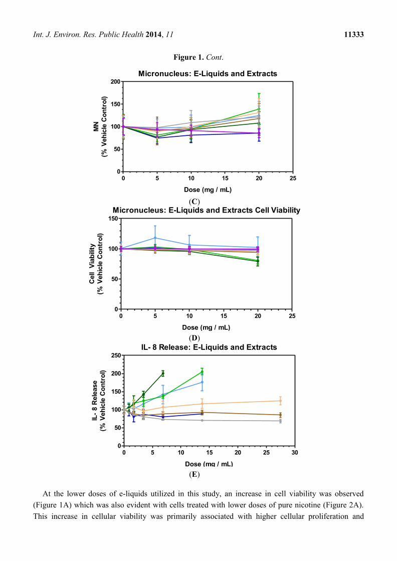
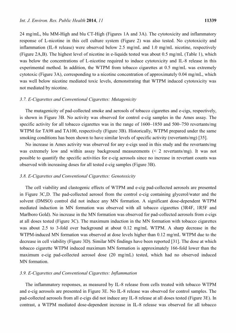
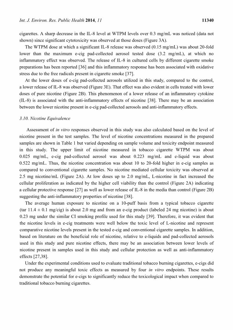
Figure 1. In vitro activity of e-cig liquids, smokeless tobacco and lozenge aqueous extracts
in NRU (A), Ames (B), MN (C and D) and IL-8 (E). NRU, MN and IL-8 data reported as
% vehicle control, PBS in the case of e-liquids, SLT and NRT aqueous extracts. Data
points in each plot represent the mean values ± SD from a minimum of two (2) independent
experiments. MN cell viability (D) shown to verify lack of MN induction is not due to
cytotoxicity at higher doses. ( ) blu CT-Ø; ( ) blu CT-High; ( ) blu MM-Ø;
( ) blu MM-High; ( ) Marlboro Snus; ( ) Copenhagen Snuff; ( ) Nicorette
Lozenge; ( ) Control e-cig.
(A)
(B)
NRU: E-Liquids and Extracts
0 5 10 15 20 25 300
50
100
150
Dose (mg / mL)
Cell V
iab
ilit
y
(% V
eh
icle
Co
ntr
ol)
Ames: E-Liquids and Extracts
Revert
an
ts /
mg
Contr
ol E-c
ig
blu C
T-
blu C
T-Hig
h
blu M
M-
blu M
M-H
igh
Mar
lboro
Snus
Copen
hagen
Snuff
Nic
orette
Lozenge
0
1
2
3
4
5TA98
TA100
Int. J. Environ. Res. Public Health 2014, 11 11333
Figure 1. Cont.
(C)
(D)
(E)
At the lower doses of e-liquids utilized in this study, an increase in cell viability was observed
(Figure 1A) which was also evident with cells treated with lower doses of pure nicotine (Figure 2A).
This increase in cellular viability was primarily associated with higher cellular proliferation and
Micronucleus: E-Liquids and Extracts
Dose (mg / mL)
MN
(% V
eh
icle
Co
ntr
ol)
0 5 10 15 20 250
50
100
150
200
Micronucleus: E-Liquids and Extracts Cell Viability
Dose (mg / mL)
Cell
Via
bilit
y
(% V
eh
icle
Co
ntr
ol)
0 5 10 15 20 250
50
100
150
IL- 8 Release: E-Liquids and Extracts
0 5 10 15 20 25 300
50
100
150
200
250
Dose (mg / mL)
IL-
8 R
ele
ase
(% V
eh
icle
Co
ntr
ol)

Int. J. Environ. Res. Public Health 2014, 11 11334
cellular protection mediated by the low level of nicotine exposure [27]. Therefore, there may be an
association between the lower nicotine present in e-cig liquids and increased cellular viability, thus
cellular protection.
Figure 2. Effects of L-nicotine on cytotoxicity (A) NRU and inflammation (B) IL-8 in
A549 cells. Data points in each plot represent the mean values ± SD from a minimum of
two (2) independent experiments.
(A)
(B)
3.2. E-Liquids, Smokeless Tobaccos (SLTs) and Nicotine Replacement Therapy (NRT): Mutagenicity
The Ames test, also known as the bacterial reverse mutation assay, is widely used for the
determination of a compound’s ability to induce mutations and has been shown to have a high
predictive value with rodent carcinogenicity tests [28].
The activity of e-liquids, SLT and NRT extracts in the Ames assay is shown in Figure 1B. The control
e-cig sample containing water/glycerol and the PBS control sample (SLT and NRT) did not induce any
revertants above baseline and were within the variability range of this assay. No significant induction in
the activity over respective controls was observed for all e-liquids and extracts (Figure 1B). The level of
L- Nicotine Mediated Cytotoxicity
L- Nicotine (mg / mL)
Cell
Via
bilit
y (
% C
on
tro
l)
0 5 10 15 20 25 30 35
0
25
50
75
100
125
150
175
200
225
L- Nicotine Mediated IL - 8 Release
L- Nicotine (mg / mL)
IL-
8 R
ele
ase (
%C
on
tro
l)
0 5 10 15 20 25 30 35
0
25
50
75
100
125

Int. J. Environ. Res. Public Health 2014, 11 11335
e-liquids as high as 15-fold higher than SLTs and NRT extracts did not induce any activity. No evidence
of cytotoxicity, as determined by the background bacterial lawn, was observed for all e-liquid, SLT and
NRT samples at all doses tested.
3.3. E-Liquids, Smokeless Tobaccos (SLTs) and Nicotine Replacement Therapy (NRT): Genotoxicity
The results of in vitro MN formation assay are shown in Figure 1C,D). The MN assay conducted in
CHO-K1 cells, identifies clastogenic and aneugenic chemicals which essentially cause a DNA-damaging
event that leads to the disruption or breakage of chromosomes and ultimately results in sections of the
chromosome being deleted, added, or rearranged upon cell division (mitosis) [29].
A sample dose range which does not produce cytotoxicity of more than 55 ± 5% (compared to
control) was utilized in the MN formation assay [24]. The control e-cig, e-liquids, SLT and NRT
extracts did not induce any significant cytotoxicity at all dose levels tested since cell-viability
remained around 100% of control at all concentrations (Figure 1D). No significant induction in the
MN formation over respective controls was observed for all e-liquids and SLT and NRT extracts
(Figure 1C).
3.4. E-Liquids, Smokeless Tobaccos (SLTs) and Nicotine Replacement Therapy (NRT): Inflammation
Airway epithelial cells are the first line of defense in the airways to respond to any external stimuli
and secrete specific chemo-attractants and pro-inflammatory cytokines, for example IL-8, monocyte
chemotactic protein-1, and IL-1ß, in order to activate the secondary response for neutrophils and
macrophage infiltration [30,31]. Instead of measuring downstream acute or chronic phase
inflammation specific cytokines, this study measured an upstream pro-inflammatory cytokine, IL-8.
The effects of 24 h exposures of e-liquids and SLT and NRT extracts on IL-8 release in A549 cells
are shown in Figure 1E. The control samples for e-cig, SLTs and NRT did not induce any significant
IL-8 release. No significant IL-8 release was observed for most of the products, with the exception of
the blu MM-Ø, blu MM-High and blu CT-Ø treatments which resulted in higher IL-8 release only at
extremely high doses of 6.9–13.8 mg/mL. When compared to the IL-8 release induced by conventional
cigarette samples (Figure 3E), any significant IL-8 release as a result of the blu MM e-liquid
treatments occurred at doses approximately 42-fold higher than the conventional tobacco cigarettes. It
has been suggested that the toxicity of e-liquids may change when the same e-liquids are heated to
produce the inhaled aerosol [26]. The evaluation of e-cig aerosol toxicity is essential since the intended
use of e-cigarettes is through aerosol inhalation. Additionally, it is proposed that different e-liquid
formulation ingredients may evaporate differently, leading to changes in concentrations in the
generated aerosols as well as the possibility that components may undergo modification when
subjected to the heat used to generate the aerosol; therefore, the final composition of the aerosol may
be different when compared to the e-liquid [9]. In the light of that, the purpose of the study was to also
characterize the aerosol toxicity as delivered by heating the e-liquid.

Int. J. Environ. Res. Public Health 2014, 11 11336
Figure 3. In vitro activity of pad-collected WTPM from tobacco cigarettes and pad-collected
e-cig aerosols in NRU (A), Ames (B), MN (C and D), and IL-8 (E). NRU, MN and IL-8 data
is reported as % vehicle control; PBS in the case of e-cigarette pad-collected aerosols,
DMSO for tobacco-burning cigarette pad-collected WTPM. Control e-cig exposures in NRU
and IL-8 were at the highest deliverable dose, resulting in no observable cytotoxicity or IL-8
release above background levels (data not shown). Data points in each plot represent the
mean values ± SD from a minimum of two (2) independent experiments. MN cell viability
(D) shown to verify lack of MN induction is not due to cytotoxicity at the higher doses.
( ) 3R4F; ( ) 1R5F; ( ) Marlboro Gold; ( ) blu CT-Ø; ( ) blu CT-High;
( ) blu MM-Ø; ( ) blu MM-High; ( ) Control e-cig.
(A)
(B)
NRU: Pad-Collected
0.0
0.5
1.0
1.5
2.0
2.5
3.0
3.5
4.0
0
50
100
150
200
Dose (mg / mL)
Cell V
iab
ilit
y
(% V
eh
icle
Co
ntr
ol)
Ames: Pad-Collected
3R4F
1R5F
Mar
lboro
Gold
Contr
ol E-c
ig
blu C
T-
blu C
T-Hig
h
blu M
M-
blu M
M-H
igh
0
1
2250
500
750
1000
1250
1500
1750
2000TA98
TA100
Revert
an
ts /
mg

Int. J. Environ. Res. Public Health 2014, 11 11337
Figure 3. Cont.
(C)
(D)
(E)
3.5. E-Cigarettes and Conventional Cigarettes
In order to study the comparative toxicities of the e-cig aerosols and tobacco smoke, all products
were smoked by the standardized CI profile with the aerosols or smoke from each product being
collected on a pad as described in the Experimental Section. The pad-collected tobacco smoke matter
was extracted in DMSO because it has been widely applied as a vehicle for in vitro assays of test
Micronucleus Induction: Pad-Collected
Dose (mg / mL)
MN
In
du
cti
on
(% V
eh
icle
Co
ntr
ol)
0.0
0
0.0
2
0.0
4
0.0
6
0.0
8
0.1
0
0.1
2
0.1
4
0
100
200
300
400
5
10
15
20
25
Micronucleus: Pad-Collected Cell Viability
Dose (mg / mL)
Cell V
iab
ilit
y
(% V
eh
icle
Co
ntr
ol)
0.0
0
0.0
2
0.0
4
0.0
6
0.0
8
0.1
0
0.1
2
0.1
4
0
50
100
150
2005
10
15
20
25
IL- 8 Release: Pad-Collected
0.0
0.5
1.0
1.5
2.0
2.5
3.0
3.5
4.0
0
100
200
300
400
Dose (mg / mL)
IL-
8 R
ele
ase
(% V
eh
icle
Co
ntr
ol)

Int. J. Environ. Res. Public Health 2014, 11 11338
articles of limited water solubility due to its excellent solvent properties for both polar and non-polar
compounds and its moderate toxicity to test organisms [32,33]. The reasoning for the concentration
ranges utilized in this study was to limit the level of DMSO in order to avoid any solvent specific
effects on the assays [32]. The toxicological responses of the pad-collected e-cig aerosols and cigarette
smoke are shown in Figure 3A–E.
3.6. E-Cigarettes and Conventional Cigarettes: Cytotoxicity
The cytotoxicity of e-cig pad-collected aerosol is shown in Figure 3A. The e-cig pad-collected
aerosol was not cytotoxic at all tested levels. For comparative purposes, different levels of WTPM
from tobacco cigarettes were also tested. A dose-dependent increase in cell death was observed for
3R4F, 1R5F and Marlboro Gold cigarettes with up to 90% cell death at the 0.5 mg/mL maximum
applied dose. The WTPM mediated cytotoxicity results are in agreement with previously reported
studies [12,34]. It was not possible to quantify the comparative cytotoxicity in terms of traditional EC50
values (a dose which induces 50% cell death) since no cell death was observed at any concentration
used for all e-cig samples (Table 2 and Figure 3A).
There was an observed increase in cellular viability in cells treated with e-liquids (Figure 1A) and
lower doses of pure nicotine (Figure 2A). Also treatment with e-cig aerosols resulted in a similar
increase in cellular viability (Figure 3A). This observed increase in cellular viability for pure nicotine
and both e-cig liquids and pad-collected aerosols could be related to nicotine’s effect on cellular
proliferation and protection [27].
Table 2. NRU EC50 values for WTPM only (mean ± SE). EC50 expressed in mg/mL to
correct for differences in dose volumes between exposure methods. † ND: e-cig pad-collected
aerosols EC50 not determined since cytotoxicity was not detected at doses tested.
Pad-Collected Matter: Smoke and Aerosols
Sample NRU EC50 (mg/mL) S.E.
3R4F 0.196 0.010
1R5F 0.237 0.014
Marlboro Gold 0.204 0.009
Control e-cig ND † --
blu CT-Ø ND † --
blu CT-High ND † --
blu MM- Ø ND † --
blu MM-High ND † --
Similar findings were reported for aqueous extracts of aerosols from various commercial e-cigs
studied in cultured mammalian fibroblast and myocardial cells [9,10]. Both studies also reported that
some aqueous extracts of e-cig aerosols showed cytotoxicity related to flavors, but were significantly
less cytotoxic than cigarette smoke extracts. This study evaluated two flavored e-cigs, with and
without nicotine (Table 1).
With e-liquids and pad-collected aerosols, no nicotine or flavor specific cytotoxic effects were
evident with e-cigs without nicotine, blu MM-Ø, and blu CT-Ø, and with nicotine as high as
Int. J. Environ. Res. Public Health 2014, 11 11339
24 mg/mL, blu MM-High and blu CT-High (Figures 1A and 3A). The cytotoxicity and inflammatory
response of L-nicotine in this cell culture system (Figure 2) was also tested. No cytotoxicity and
inflammation (IL-8 release) were observed below 2.5 mg/mL and 1.0 mg/mL nicotine, respectively
(Figure 2A,B). The highest level of nicotine in e-liquids tested was about 0.5 mg/mL (Table 1), which
was below the concentrations of L-nicotine required to induce cytotoxicity and IL-8 release in this
experimental method. In addition, the WTPM from tobacco cigarettes at 0.5 mg/mL was extremely
cytotoxic (Figure 3A), corresponding to a nicotine concentration of approximately 0.04 mg/mL, which
was well below nicotine mediated toxic levels, demonstrating that WTPM induced cytotoxicity was
not mediated by nicotine.
3.7. E-Cigarettes and Conventional Cigarettes: Mutagenicity
The mutagenicity of pad-collected smoke and aerosols of tobacco cigarettes and e-cigs, respectively,
is shown in Figure 3B. No activity was observed for control e-cig samples in the Ames assay. The
specific activity for all tobacco cigarettes was in the range of 1600–1850 and 500–750 revertants/mg
WTPM for TA98 and TA100, respectively (Figure 3B). Historically, WTPM prepared under the same
smoking conditions has been shown to have similar levels of specific activity (revertants/mg) [35].
No increase in Ames activity was observed for any e-cigs used in this study and the revertants/mg
was extremely low and within assay background measurements (< 2 revertants/mg). It was not
possible to quantify the specific activities for e-cig aerosols since no increase in revertant counts was
observed with increasing doses for all tested e-cig samples (Figure 3B).
3.8. E-Cigarettes and Conventional Cigarettes: Genotoxicity
The cell viability and clastogenic effects of WTPM and e-cig pad-collected aerosols are presented
in Figure 3C,D. The pad-collected aerosol from the control e-cig containing glycerol/water and the
solvent (DMSO) control did not induce any MN formation. A significant dose-dependent WTPM
mediated induction in MN formation was observed with all tobacco cigarettes (3R4F, 1R5F and
Marlboro Gold). No increase in the MN formation was observed for pad-collected aerosols from e-cigs
at all doses tested (Figure 3C). The maximum induction in the MN formation with tobacco cigarettes
was about 2.5 to 3-fold over background at about 0.12 mg/mL WTPM. A sharp decrease in the
WTPM-induced MN formation was observed at dose levels higher than 0.12 mg/mL WTPM due to the
decrease in cell viability (Figure 3D). Similar MN findings have been reported [31]. The dose at which
tobacco cigarette WTPM induced maximum MN formation is approximately 166-fold lower than the
maximum e-cig pad-collected aerosol dose (20 mg/mL) tested, which had no observed induced
MN formation.
3.9. E-Cigarettes and Conventional Cigarettes: Inflammation
The inflammatory responses, as measured by IL-8 release from cells treated with tobacco WTPM
and e-cig aerosols are presented in Figure 3E. No IL-8 release was observed for control samples. The
pad-collected aerosols from all e-cigs did not induce any IL-8 release at all doses tested (Figure 3E). In
contrast, a WTPM mediated dose-dependent increase in IL-8 release was observed for all tobacco
Int. J. Environ. Res. Public Health 2014, 11 11340
cigarettes. A sharp decrease in the IL-8 level at WTPM levels over 0.3 mg/mL was noticed (data not
shown) since significant cytotoxicity was observed at those doses (Figure 3A).
The WTPM dose at which a significant IL-8 release was observed (0.15 mg/mL) was about 20-fold
lower than the maximum e-cig pad-collected aerosol tested dose (3.2 mg/mL), at which no
inflammatory effect was observed. The release of IL-8 in cultured cells by different cigarette smoke
preparations has been reported [36] and this inflammatory response has been associated with oxidative
stress due to the free radicals present in cigarette smoke [37].
At the lower doses of e-cig pad-collected aerosols utilized in this study, compared to the control,
a lower release of IL-8 was observed (Figure 3E). That effect was also evident in cells treated with lower
doses of pure nicotine (Figure 2B). This phenomenon of a lower release of an inflammatory cytokine
(IL-8) is associated with the anti-inflammatory effects of nicotine [38]. There may be an association
between the lower nicotine present in e-cig pad-collected aerosols and anti-inflammatory effects.
3.10. Nicotine Equivalence
Assessment of in vitro responses observed in this study was also calculated based on the level of
nicotine present in the test samples. The level of nicotine concentrations measured in the prepared
samples are shown in Table 1 but varied depending on sample volume and toxicity endpoint measured
in this study. The upper limit of nicotine measured in tobacco cigarette WTPM was about
0.025 mg/mL, e-cig pad-collected aerosol was about 0.223 mg/mL and e-liquid was about
0.522 mg/mL. Thus, the nicotine concentration was about 10 to 20-fold higher in e-cig samples as
compared to conventional cigarette samples. No nicotine mediated cellular toxicity was observed at
2.5 mg nicotine/mL (Figure 2A). At low doses up to 2.0 mg/mL, L-nicotine in fact increased the
cellular proliferation as indicated by the higher cell viability than the control (Figure 2A) indicating
a cellular protective response [27] as well as lower release of IL-8 in the media than control (Figure 2B)
suggesting the anti-inflammatory properties of nicotine [38].
The average human exposure to nicotine on a 10-puff basis from a typical tobacco cigarette
(tar 11.4 ± 0.1 mg/cig) is about 2.0 mg and from an e-cig product (labeled 24 mg nicotine) is about
0.23 mg under the similar CI smoking profile used for this study [39]. Therefore, it was evident that
the nicotine levels in e-cig treatments were well below the toxic level of L-nicotine and represent
comparative nicotine levels present in the tested e-cig and conventional cigarette samples. In addition,
based on literature on the beneficial role of nicotine, relative to e-liquids and pad-collected aerosols
used in this study and pure nicotine effects, there may be an association between lower levels of
nicotine present in samples used in this study and cellular protection as well as anti-inflammatory
effects [27,38].
Under the experimental conditions used to evaluate traditional tobacco burning cigarettes, e-cigs did
not produce any meaningful toxic effects as measured by four in vitro endpoints. These results
demonstrate the potential for e-cigs to significantly reduce the toxicological impact when compared to
traditional tobacco burning cigarettes.

Int. J. Environ. Res. Public Health 2014, 11 11341
3.11. Comparable Human Exposure: Conventional and E-Cig
The comparative human exposure to tobacco cigarette smoke and e-cig aerosol is important in order
to assess e-cig mediated reduced exposure and reduced harm. It has been reported that a smoker with one
pack-a-day tobacco cigarette consumption inhales on average about 261 mg/m3 cigarette tar [40],
equivalent to about 271 µg/mL or 0.271 mg/mL [40]. Internal study indicated the range of e-cig
aerosol delivery to be in the range of 0.5–1.5 mg/puff under Canadian Intense conditions (39).
Assuming similar e-cig use as a conventional tobacco cigarette (200 puffs), the upper limit of human
exposure to e-cig aerosol is approximately 300 mg or approximately 250 µg/mL or 0.25 mg/mL. The
range of e-cig pad-collected aerosol used in the present study was 3.2–20 mg/mL. No adverse
toxicological events were observed in this study even when the e-cig aerosol levels used were about
12–78 times higher than expected with normal e-cig use.
3.12. Contribution of Findings to Tobacco Harm Reduction and E-Cigs
The concept of Tobacco Harm Reduction (THR) has been advanced as a pragmatic approach to
achieving reductions in the adverse public health impacts of cigarette smoking in the near term; in
parallel with social, educational, and regulatory strategies intended to reduce and discourage cigarette
smoking, particularly among adolescents [4,41–43].
The use of non-combustible SLT products such as Swedish-style snus and traditional moist snuff
are demonstrably on the order of 98% less harmful in terms of risks for lung cancer, COPD, CVD, and
other cancers, (including oral cancers) as compared to cigarette smoking [11,41,44,45].
Similarly, NRT products such as dermal patches and chewing gums have been shown to be safe and
efficacious in clinically-managed and over-the-counter consumer usage. Therapeutic nicotine vapor
inhaler devices and aerosol sprays for nasal and oral use have to date demonstrated similar benefits and
low risks in facilitating smoking cessation [46]. The efficacy of such conventional NRT cessation aids
has, however, proven in practice to fall short (~7% cessation success) of what is needed by
a considerable number of smokers [47]. These smokers consistently report that taste, sensory and
behavioral components experienced in the act of cigarette smoking are substantial motivators of the
smoking behavior and may well comprise a population that could achieve substantially higher success
in quitting through use of products that mimic the behavior element of smoking as well as the delivery
of nicotine. Therefore, the use of e-cigs that provide some of the taste, sensory and behavioral
components of conventional tobacco cigarette smoking may hold substantial promise in defining the
potential benefits of the THR paradigm [48].
Despite the absence of long-term epidemiologic data on any chronic disease risk, a growing body of
recent literature is consistent with an expectation that the use of e-cigs is unlikely to raise serious
health concerns [49,50], particularly in comparison to those that result from the smoking of
conventional cigarettes [7]. This conclusion is currently based, and further supported by this study and
on a growing number of independent analyses of commercially-available e-cig liquids and product
aerosols from markets around the world, that have consistently reported very low or undetectable
levels of most tobacco smoke constituents that are known or suspected to play a prominent role in the
etiology of serious tobacco-related diseases. [7,51–59].
Int. J. Environ. Res. Public Health 2014, 11 11342
There are various essential and contributory components to the THR framework, including product
use and behavior, taste, nicotine delivery, product chemistry, toxicity and clinical safety. This study
shows that neither the e-cig liquids nor collected aerosols produced any meaningful toxic effects in
widely used in vitro test systems. These findings add additional value to the increasing body of
scientific weight-of-evidence supporting the potential inclusion of e-cigs into THR paradigm.
4. Conclusions
In summary, this comparative in vitro toxicity study of e-cigs, SLT, NRT and tobacco cigarette
products demonstrates the following:
(1) E-cigs vs. Tobacco WTPM: At doses up to approximately 100-fold higher than typical
cigarette smoke exposures, blu e-cig liquids and pad-collected aerosols had no-to-extremely
low in vitro activity (NRU, Ames, MN and IL-8) when compared to WTPM from tobacco
burning cigarettes. WTPM activity was up to approximately 6,000 times higher than e-cigs.
(2) E-cigs vs. SLT and NRT: blu e-cig liquids demonstrated similar no-to-extremely low
in vitro activity as aqueous extracts from a commercial nicotine lozenge (NRT) and
commercial SLT products (snus and snuff).
(3) Effect of Nicotine: In vitro activities (NRU, Ames, MN and IL-8) measured for blu e-cig
exposures, with and without nicotine, were similar for all sample types, indicating that the
presence of nicotine, at the levels tested, did not contribute to any toxicological effects,
confirmed by the lack of cytotoxicity and inflammation response of L-nicotine at
comparative levels.
(4) Effect of Flavors: In vitro activities (NRU, Ames and MN) for the commercial blu e-cigs
were indistinguishable from control (glycerol/water); indicating these flavors (CT and
MM), at the levels tested, had no detectable impact on the cytotoxicity and genotoxicity
endpoints utilized in this study. There was some observed IL-8 induction for some
e-liquids, albeit at the highest doses tested.
(5) Liquid vs. Pad-Collected Aerosol: In vitro results for blu e-cigs, in this study, were similar
for the different exposure methods (e-liquids and pad-collected aerosol); demonstrating no
detectable impact on the in vitro toxicological responses when the e-liquids were
aerosolized.
(6) SLT vs. Tobacco WTPM: SLT extracts added to the test systems at levels up to 54-fold
higher than those used for Tobacco-WTPM generated by burning cigarettes was markedly
less cytotoxic and mutagenic, and evoked a significantly lower IL-8 response at all dose
levels evaluated. The effects of the SLT extracts in the assays were statistically
indistinguishable from those of the e-cig and NRT preparations.
With respect to the study, lack of any meaningful in vitro acute toxicity for blu e-cigs and extremely
low levels of chemical constituents measured in blu [39] and the analysis of known reduced risk
products such as NRT and SLT has the potential to demonstrate a decreased human health impact as
compared to conventional tobacco-burning cigarettes.

Int. J. Environ. Res. Public Health 2014, 11 11343
Acknowledgments
The authors would like to thank Edward Robinson, Dan J. Heck, Sherwin Yan and Carl D’Ruiz for
critical review of the manuscript and Mark Sackfield for technical assistance in sample preparation for
toxicological testing.
Author Contributions
Manoj Misra: Participated in study design and execution of the study, conducted cytotoxicity (NRU)
and IL-8 release measurements (e-liquids and pad-collected matter from e-cigs and tobacco cigarettes,
aqueous extracts of SLTs and NRT) and related data collection and interpretation, and constructed
manuscript as principal writer.
Robert D. Leverette: Participated in study design and execution of the study, coordinated sample
preparation (e-liquids, pad-collected aerosols and smoke condensates, SLT and NRT aqueous extracts),
conducted mutagenesis Ames test measurements (e-liquids and pad-collected matter from e-cigs and
tobacco cigarettes, aqueous extracts of SLTs and NRT) and related data collection, compilation and
interpretation and presentation of data (graphs and tables).
Bethany T. Cooper: Conducted MN formation measurements (e-liquids and pad-collected matter
from e-cigs and tobacco cigarettes, aqueous extracts of SLTs and NRT) and related data collection.
Melanee B. Bennett: Maintained and provided cultured cells, A549 and CHO cells, for the study
and assisted in conducting cytotoxicity (NRU) measurements (e-liquids and pad-collected matter from
e-cigs and tobacco cigarettes, aqueous extracts of SLTs and NRT).
Steven E. Brown: Participated in study design and critical review of the manuscript.
Conflicts of Interest
The authors are Lorillard Tobacco Company employees and declare no conflict of interest with
respect to the research, authorship, and/or publication of this article.
References
1. Ayers, J.W.; Ribisl, K.M.; Brownstein, J.S. Tracking the rise in popularity of electronic nicotine
delivery systems (electronic cigarettes) using search query surveillance. Am. J. Prev. Med. 2011,
40, 448–453.
2. Etter, J.-F.; Bullen, C.; Flouris, A.D.; Laugesen, M.; Eissenberg, T. Electronic nicotine delivery
systems: A research agenda. Tob. Control. 2011, 20, 243–248.
3. Levitz, J.S.; Bradley, T.P.; Golden, A.L. Overview of smoking and all cancers. Med. Clin. North
Am. 2004, 88, 1655–1675.
4. Polosa, R.; Rodu, B.; Caponnetto, P.; Maglia, M.; Raciti, C. A fresh look at tobacco harm reduction:
The case for the electronic cigarette. Harm Reduct. J. 2013, 10, doi:10.1186/1477-7517-10-19.
5. Brown, B.; Beard, E.; Kotz, D.; Michie, S.; West, R. Real-world effectiveness of e-cigarettes
when used to aid smoking cessation: A cross-sectional population study. 2014, 109, 1531–1540.
6. Statement from Specialists in Nicotine Science and Public Health Policy. Available online:
http://www.nicotinepolicy.net/documents/letters/MargaretChan.pdf (accessed on 27 October 2014).
Int. J. Environ. Res. Public Health 2014, 11 11344
7. Goniewicz, M.L.; Knysak, J.; Gawron, M.; Kosmider, L.; Sobczak, A.; Kurek, J.; Prokopowicz, A.;
Jablonska-Czapla, M.; Rosik-Dulewska, C.; Havel, C.; et al. Levels of selected carcinogens and
toxicants in vapour from electronic cigarettes. Tob. Control 2013, 23, 1–7.
8. Farsalinos, K.E.; Polosa, R. Safety evaluation and risk assessment of electronic cigarettes as
tobacco cigarette substitutes: A systematic review. Ther. Adv. Drug Safety 2014, 5, 67–86.
9. Romagna, G.; Allifranchini, E.; Bocchietto, E.; Todeschi, S.; Esposito, M.; Farsalinos, K.E.
Cytotoxicity evaluation of electronic cigarette vapor extract on cultured mammalian fibroblasts
(clearstream-life): Comparison with tobacco cigarette smoke extract. Inhal. Toxicol. 2013, 25,
354–361.
10. Farsalinos, K.E.; Romagna, G.; Allifranchini, E.; Ripamonti, E.; Bocchietto, E.; Todeschi, S.;
Tsiapras, D.; Kyrzopoulos, S.; Voudris, V. Comparison of the cytotoxic potential of cigarette
smoke and electronic cigarette vapour extract on cultured myocardial cells. Int. J. Environ. Res.
Public Health 2013, 10, 5146–5162.
11. Nutt, D.J.; Phillips, L.D.; Balfour, D.; Curran, V.; Dockrell, M.; Foulds, J.; Fagerstrom, K.;
Letlape, K.; Milton, A.; Polosa, R.; et al. Estimating the harms of nicotine-containing products
using the MCDA approach. Eur. Addict. Res. 2014, 20, 218–225.
12. Arimilli, S.; Damratoski, B.E.; Bombick, B.; Borgerding, M.F.; Prasad, G.L. Evaluation of
cytotoxicity of different tobacco product preparations. Regul. Toxicol. Pharmacol. 2012, 64, 350–360.
13. CORESTA In Vitro Toxicology Task Force. The Rationale and Strategy for Conducting In Vitro
Toxicology Testing of Tobacco Smoke. Available online: http://www.coresta.org/Reports/IVT_
TF_Rationale-IVT-Testing-Tob.-Smoke_Report_Jun04.pdf (accessed on 27 October 2014).
14. Determination of “Tar,” Nicotine and Carbon Monoxide in Mainstream Tobacco Smoke-Official
Method. Available online: http://laws.justice.gc.ca/en/T-11.5/SOR-2000-272/182471.html. (accessed
on 8 January 2006).
15. Rickert, W.S.; Wright, W.G.; Trivedi, A.H.; Momin, R.A.; Lauterbach, J.H. A comparative study
of the mutagenicity of various types of tobacco products. Regul. Toxicol. Pharmacol. 2007, 48,
320–330.
16. Official Method T-115. Determination of “Tar”, Nicotine and Carbon Monoxide in Mainstream
Tobacco Smoke. Available online: http://laws-lois.justice.gc.ca/eng/regulations/SOR-2000-273/
page-14.html (accessed on 29 October 2014).
17. Borenfreund, E.; Puerner, J.A. Toxicity determined in vitro by morphological alterations and
neutral red absorption. Toxicol. Letters 1985, 24, 119–124.
18. Bombick, D.W.; Doolittle, D.J. The role of chemical structure and cell type in the cytotoxicity of
low molecular weight aldehydes and pyridines. In Vitro Toxicol. 1995, 8, 349–356.
19. Human IL-8 ELISA Kit. For the Quantitative Determination of Human IL-8 Concentrations in
Serum, Plasma, Cell Culture Supernatant and Other Biological Fluids. Available online:
http://www.abazyme.com/inserts/EL10008.pdf (accessed on 20 October 2014).
20. Aufderheide, M.; Gressmann, H. A modified ames assay reveals the mutagenicity of native
cigarette mainstream smoke and its gas vapour phase. Exp. Toxicol. Pathol. 2007, 58, 383–392.
21. Maron, D.M.; Ames, B.N. Revised methods for the salmonella mutagenicity test. Mutat. Res.
1983, 113, 173–215.

Int. J. Environ. Res. Public Health 2014, 11 11345
22. Diaz, D.; Scott, A.; Carmichael, P.; Shi, W.; Costales, C. Evaluation of an automated in vitro
micronucleus assay in CHO-K1 cells. Mutat. Res. 2007, 630, 1–13.
23. Kirkland, D.; Reeve, L.; Gatehouse, D.; Vanparys, P. A core in vitro genotoxicity battery
comprising the AMES test plus the in vitro micronucleus test is sufficient to detect rodent
carcinogens and in vivo genotoxins. Mutat. Res. 2011, 721, 27–73.
24. Aggarwal, B.B.; Shishodia, S.; Sandur, S.K.; Pandey, M.K.; Sethi, G. Inflammation and cancer:
How hot is the link? Biochem. Pharmacol. 2006, 72, 1605–1621.
25. Balkwill, F.; Charles, K.A.; Mantovani, A. Smoldering and polarized inflammation in the
initiation and promotion of malignant disease. Cancer Cell 2005, 7, 211–217.
26. Bahl, V.; Lin, S.; Xu, N.; Davis, B.; Wang, Y.H.; Talbot, P. Comparison of electronic cigarette
refill fluid cytotoxicity using embryonic and adult models. Reprod. Toxicol. 2012, 34, 529–537.
27. Balharry, D.; Sexton, K.; BeruBe, K.A. An in vitro approach to assess the toxicity of inhaled
tobacco smoke components: Nicotine, cadmium, formaldehyde and urethane. Toxicology 2008,
244, 66–76.
28. Scott, K.; Saul, J.; Crooks, I.; Camacho, O.M.; Dillon, D.; Meredith, C. The resolving power of
in vitro genotoxicity assays for cigarette smoke particulate matter. Toxicol. in Vitro 2013, 27,
1312–1319.
29. Rosefort, C.; Fauth, E.; Zankl, H. Micronuclei induced by aneugens and clastogens in mononucleate
and binucleate cells using cytokinesis block assay. Mutagenesis 2004, 19, 277–284.
30. Fuke, S.; Betsuyaku, T.; Nasuhara, Y.; Morikawa, T.; Katoh, H.; Nishimura, M. Chemokines in
bronchiolar epithelium in the development of chronic obstructive pulmonary disease. Am. J.
Respir. Cell Mol. Biol. 2004, 31, 405–412.
31. Moretto, N.; Facchinetti, F.; Southworth, T.; Civelli, M.; Singh, D.; Patacchini, R.
α,β-Unsaturated aldehydes contained in cigarette smoke elicit IL-8 release in pulmonary cells
through mitogen-activated protein kinases. Am. J. Physiol. Lung Cell Mol. Physiol. 2009, 296,
L839–L848.
32. Baker, R.R.; Massey, E.D.; Smith, G. An overview of the effects of tobacco ingredients on smoke
chemistry and toxicity. Food Chem. Toxicol. 2004, 42, S53–S83.
33. Misra, M.; Leverette, R.D.; Hamm, J.T.; Vulimiri, S.V. In vitro toxicological evaluation of
cigarette smoke particulate matter: Effect of Dimethyl Sulfoxide (DMSO) as solvent. Beiträge zur
Tabakforschung International 2010, 24, 2–9.
34. Jianhua, Y.; Gao, Q.; Mi, Q.; Li, X.; Miao, M.; Cheng, P.; Luo, Y. In vitro micronucleus assay for
the analysis of total particulate matter in cigarette smoke: Comparison of flow cytometry and laser
scanning cytometry with microscopy. Mutat. Res. 2013, 755, 120–125.
35. Rickert, W.S.; Trivedi, A.H.; Momin, R.A.; Wright, W.G.; Lauterbach, J.H. Effect of smoking
conditions and methods of collection on the mutagenicity and cytotoxicity of cigarette mainstream
smoke. Toxicol. Sci. 2007, 96, 285–293.
36. Fields, W.R.; Leonard, R.M.; Odom, P.S.; Nordskog, B.K.; Ogden, M.W.; Doolittle, D.J. Gene
expression in Normal Human Bronchial Epithelial (NHBE) cells following in vitro exposure to
cigarette smoke condensate. Toxicol. Sci. 2005, 86, 84–91.
37. Pryor, W.A.; Stone, K. Oxidants in cigarette smoke. Radicals, hydrogen peroxide, peroxynitrate,
and peroxynitrite. Ann. N. Y. Acad. Sci. 1993, 686, 12–28.

Int. J. Environ. Res. Public Health 2014, 11 11346
38. Kalra, R.; Singh, S.P.; Pena-Philippides, J.C.; Langley, R.J.; Razani-Boroujerdi, S.; Sopori, M.L.
Immunosuppressive and anti-inflammatory effects of nicotine administered by patch in an animal
model. Clin. Vaccine Immunol. 2004, 11, 563–568.
39. Tayyarah, R.; Long, G.A. Comparison of select analytes in aerosol from e-cigarettes with smoke
from conventional cigarettes and with ambient air. Reg. Toxicol. Pharmacol. 2014, 2014,
doi:10.1016/j.yrtph.2014.10.010.
40. Finch, G.L.; Nikula, K.J.; Chen, B.T.; Barr, E.B.; Chang, I.Y.; Hobbs, C.H. Effect of chronic
cigarette smoke exposure on lung clearance of tracer particles inhaled by rats. Fundam. Appl.
Toxicol. 1995, 24, 76–85.
41. Rodu, B. The scientific foundation for tobacco harm reduction, 2006–2011. Harm Reduct. J.
2011, 8, doi:10.1186/1477-7517-8-19.
42. McNeill, A.; Munafò, M.R. Reducing harm from tobacco use. J. Psychopharmacol. 2012, 2012,
doi:10.1177/0269881112458731.
43. Fagerström, K.O.; Bridgmanb, K. Tobacco harm reduction: The need for new products that can
compete with cigarettes. Addict. Behav. 2014, 39, 507–511.
44. Gartner, C.E.; Hall, W.D. Should Australia lift its ban on low nitrosamine 576 smokeless tobacco
products? Med. J. Aust. 2008, 188, 44–46.
45. Phillips, C.V.; Rodu, B. Tobacco harm reduction: Opportunity and opposition. Drugs Alcohol Today
2013, 13, 73–78.
46. The Use of Nicotine Replacement Therapy to Reduce Harm in Smokers. Available online:
htpp://mhra.gsi.gov.uk (accessed on 27 October 2014).
47. Moore, D.; Aveyard, P.; Connock, M.; Wang, D.; Fry-Smith, A.; Barton, P. Effectiveness and
safety of nicotine replacement therapy assisted reduction to stop smoking: Systematic review and
meta-analysis. BMJ 2009, 338, doi:10.1136/bmj.b1024.
48. Farsalinos, K.E.; Romagna, G.; Tsiapras, D.; Kyrzopoulos, S.; Spyrou, A.; Voudris, V. Impact of
flavour variability on electronic cigarette use experience: An internet survey. Int. J. Environ. Res.
Public Health 2013, 10, 7272–7282.
49. Farsalinos, K.E.; Romagna, G.; Tsiapras, D.; Kyrzopoulos, S.; Voudris, V. Characteristics,
perceived side effects and benefits of electronic cigarette use: A worldwide survey of more than
19,000 consumers. Int. J. Environ. Res. Public Health 2014, 11, 4356–4373.
50. Hajek, P.; Etter, J.-F.; Benowitz, B.; Eissenberg, T.; McRobbie, H. Electronic cigarettes: Review
of use, content, safety, effects on smokers and potential for harm and benefit. Addiction 2014,
2014, doi:10.1111/add.12659.
51. Ruyan®E-cigarette Bench-Top Tests. Available online: http://www.healthnz.co.nz/DublinEcigBench
topHandout.pdf (accessed 20 November 2013).
52. Evaluation of e-Cigarettes. Available online: http://www.fda.gov/downloads/drugs/Scienceresearch/
UCM173250.pdf (accessed on 10 November 2013).
53. Hadwiger, M.; Trehy, M.; Ye, W.; Moore, T.; Allgire, J.; Westenberger, B. Identification of
amino-tadalafil and rimonabant in electronic cigarette products using high pressure liquid
chromatography with diode array and tandem mass spectrometric detection. J. Chromatogr. A.
2010, 1217, 7547–7555.

Int. J. Environ. Res. Public Health 2014, 11 11347
54. Cahn, Z.; Siegel, M. Electronic cigarettes as a harm reduction strategy for tobacco control: A step
forward or a repeat of past mistakes? J. Public Health Policy 2011, 32, 16–31.
55. Pellegrino, R.M.; Tinghino, B.; Mangiaracina, G.; Marani, A.; Vitali, M.; Protano, C.; Osborn, J.F.;
Cattaruzza, M.S. Electronic cigarettes: An evaluation of exposure to chemicals and fine
Particulate Matter (PM). Ann. Ig. 2012, 24, 279–288.
56. Kim, H.; Shin, H. Determination of tobacco-specific nitrosamines in replacement liquids of
electronic cigarettes by liquid chromatography-tandem mass spectrometry. J. Chromatogr. A.
2013, 1291, 48–55.
57. Etter, J.; Zäther, E.; Svensson, S. Analysis of refill liquids for electronic cigarettes. Addiction
2013, 108, 1671–1679.
58. Williams, M.; Villarreal, A.; Bozhilov, K.; Lin, S.; Talbot, P. Metal and silicate particles including
nanoparticles are present in electronic cigarette cartomizer fluid and aerosol. PLoS One 2013, 8,
doi:10.1371/journal.pone.0057987.
59. Burstyn, I. Peering through the mist: Systematic review of what the chemistry of contaminants in
electronic cigarettes tells us about health risks. BMC Public Health 2014, 14, doi:10.1186/
1471-2458-14-18.
© 2014 by the authors; licensee MDPI, Basel, Switzerland. This article is an open access article
distributed under the terms and conditions of the Creative Commons Attribution license
(http://creativecommons.org/licenses/by/4.0/).