comparative gross and histological morphology of goat
TRANSCRIPT
Tanzania Veterinary Journal Vol. 34 (2) 2019
Comparative gross and histological morphology of goat (caprine) and sheep (ovine) placentomes
I. P. Kashoma1 and C. Luziga2 1Department of Veterinary Surgery and Theriogenology, College of Veterinary and Biomedical Sciences,
Sokoine University of Agriculture, P. O. Box 3015 Morogoro, Tanzania
2Department of Veterinary Anatomy and Pathology, College of Veterinary and Biomedical Sciences,
Sokoine University of Agriculture, P. O. Box 3016 Morogoro, Tanzania
E-mail: [email protected]
SUMMARY
This study aimed to investigate the gross and micro-anatomy of the placentome of Tanzanian goats and sheep and their relationship with some foetal parameters. Forty pregnant uteri (20 from goats and 20 from sheep) were collected from slaughtered pregnant animals. Foetal age was estimated using Crown-Rump Length formula. Other parameters such as head length (HL), neck length (NL), fore and hind limb length, foetal weight, total number of placentomes and placentome size and location in the uteri were taken. All placentae exhibited the typical characteristics of a cotyledonary with discrete areas of attachment between maternal and foetal tissues. Placentome counts ranged from 70 to 92 and 74 to 104 in sheep and goats, respectively. The average length of placentomes was 20.0±4.2 and 30.7±9.0 in sheep and goats, respectively. Placentomes from does in the present study were concave in shape and the majority were of type A. Regression analysis showed a statistically significant relationship between foetal age and placental size (r2=0.3245; p<0.001); between foetal age and foetal weight (r2=0.7364; p<0.001); and between foetal age and placental weight (r2=0.6458; p<0.01); but the number of placentomes insignificantly (P>0.01) varied with foetal age. In conclusion, this study has demonstrated that the Tanzanian sheep and goat placentae are cotyledonary – synepitheliochorial type having placentomes that tend to increase in size with gestation age in response to greater nutrient and or metabolic requirement of the growing foetus.
INTRODUCTION
Goats and sheep represent the second and third largest proportion of the livestock population in Tanzania, respectively. Tanzania’s small ruminant wealth estimates 17.1 million goats and 9.2 million sheep contributing to about 22% of the national meat supply (MLFD, 2015). The popularity of small ruminants in Tanzania can be attributed largely to their hardiness, their value as social currency, high prolificacy, fertility and excellent adaptability to environmental conditions.
Female reproduction always carries a great importance in terms of economy for any livestock species (Hafez, 1993). Female
goats (does) and sheep (ewes) reach puberty between 3 and 15 months of age, depending on inherent character of different breed, body weight at birth, photo-period and nutritional status (Freitas et al., 2004), and come into heat every 21 days for 2-48 hours.The average gestation period (length of pregnancy) of doe and ewe is usually 150 days.
In ruminants, the placenta is characterized by discrete areas of attachment, the placentomes, which are formed by intimate interaction between uterine caruncles and chorionic cotyledons. Placentomes are specialized areas for haemotrophic exchange
23
The Tropical Veterinarian
of nutrients/metabolites between the foetal and maternal blood streams consisting of interdigitated foetal cotyledonary and maternal caruncular microvilli. Proper establishment of the vascularity of the placentome is essential for the maternal system to support the exponentially growing foetus in the last trimester of pregnancy (Reynolds and Redmer, 2001; Vonnahme et al., 2008).
Furthermore, there is a close relationship between foetal weight/size, placental size, and uterine and umbilical blood flows (Regnault et al., 1999). Placental size may be indicative of foetal growth, as placental restriction has been shown to decrease with foetal size (Regnault et al., 1999; Reynolds and Redmer, 2001). However, placentome morphology, size, number and distribution vary greatly between species. The distribution of placentome type and size of individual placentomes can be influenced by many factors such as, nutritional state, placental blood flow and oxygen availability (Gardner et al. 2002; Osgerby et al., 2004).
Histologically the placentome is composed of the caruncular (maternal) tissue with its crypts filled with ramified projections of the chorion known as chorionic villi (Bjorkman, 1976). In the placentome, the maternal blood circulation is separated from the foetal circulation by six tissue layers: maternal vascular endothelium, maternal connective tissue, syncytium, trophoblastic epithelium, foetal mesenchyma and foetal vascular endothelium. The caprine placenta is classified as epitheliochorial (Derivaux et al.,
1988). However, Wooding (1992) has proposed that the ruminant placenta be classified as syneptheliochorial due to the occurrence of migration of foetal cells through maternal-foetal junction and fusion of these cells with the maternal epithelial cells.
Ovine placentomes are classified into four types (Type A, B, C and D) on the basis of their gross morphological appearance (Vatnick et al., 1991). Type A placentomes being concave, type D convex, and types B and C intermediate in morphology reflecting the degree of aversion of the hemophagous zone. Furthermore, type A placentomes consist of maternal tissue surrounding a small portion of foetal tissue, type B and C placentomes are intermediate in shape, and type D placentomes consist of foetal tissue completely surrounding maternal tissue (Vatnick et al., 1991; Vonnahme et al., 2006).
Furthermore, due to variations in breeds and nutrition among different management systems, it is important for each breed to be assessed for the information to be relevant and applicable to the target population of sheep and goats. There is very little information available on the gross and histological variability of Tanzanian sheep and goat placentomes during different stage of pregnancy.Therefore, the objective of the present study was to document the gross and microscopic structure of the placentomes of the doe and ewe and their relationship to foetal age.
MATERIALS AND METHODS
Collection of Materials
The present study was conducted on 40 healthy gravid uteri (does and ewes each 20) procured from non-descript breed of Tanzanian goats (Small East African breed) and sheep (Red Maasai and Gogo breeds). The uteri were collected from local slaughter
houses. The samples were transported to the Anatomy laboratory, College of Veterinary Medicine and Biomedical Sciences. The pregnant uteri were carefully dissected using surgical blades, scissors and forceps to display the foetuses and placentomes.
24
Tanzania Veterinary Journal Vol. 34 (2) 2019
Foetal parameters
Immediately after uterine dissection, foetuses were cleaned with water soaked cotton to remove the amniotic fluid. Each foetus was measured for crown rump length (CRL), head length (HL), neck length (NL), length of fore and hind leg in centimeters using nylon tape (Banan-Khojesteh et al., 2011) and weighed in grams using digital weighing balance (Shanghai Puchun Measure Instrument Co., Ltd. , China).
Foetal age was estimated using the formula (Y=2.74x + 30.15) (Banan-Khojesteh et al., 2011). Where; X = Crown-Rump Length in centimeters (CRL), and Y = Age of foetus in days. Gestation ages were divided into three groups, namely: early gestation period (0 day-50 days), mid gestation period (51 days-100 days) and late gestation period (101 days-till term).
Placentomal morphometric parameters
Placentomal parameters such as shape, number, distribution, length and width were assessed in each gravid uterus. Ten randomly placentomes were collected from each gravid uterus and measured for length and width using a vernier caliper. The collected
placentomes were then kept in glass jar containing 10% neutral-buffered formalin until processed for histological examination. Histological tissue preparations were performed as previously described (Wooding, 2006). Briefly, random portions of placentomes were dehydrated in increasing concentrations of ethanol, cleared in xylene and embedded in paraffin wax. Sectioned of 4-5 μm thickness were cut on microtome and stained with haematoxylin and eosin (H&E).
The microscopic structures of placentomes were examined using Olympus binocular microscope and images were captured using Moticam 1000 camera (Motic China Group Ltd.).
Statistical analyses
Regression analyses (Graph Pad Prism Version 5) were used to evaluate the relationship between the estimated age and foetal parameters including the relationship between foetal age and placentome size, and foetal age and number of placentome. Fetal and placentomes mean parameters between ovine and caprine were compared using t-test.
RESULTS
Fetal measurements
The average fetal weight, crown-rump length, length of the head, neck, fore- and hind-limbs are shown in supporting table 1. In general, the foetal weight in both species studied significantly increased with advancing stage of pregnancy. Similarly, foetal heights; Crown-rump length, anus, head length, neck length, fore and hind length increased with pregnancy advancement.
However, significant difference in foetal measurements was observed between stage-3 (late pregnancy) and the other two stages of pregnancies (early and mid-pregnancy). Nevertheless no significant difference on similar parameters (foetal age and size) was observed between the two species.
Placentome parameters
Grossly, the placenta of the Tanzanian goat and sheep exhibited the typical characteristics of a cotyledonary placenta
25

The Tropical Veterinarian
with discrete areas of attachment between maternal and foetal tissues, the placentomes (Figure 1). The maternal caruncle formed the basal plate of the placentome, while the foetal cotyledon formed the chorionic plate of the placentome. Each placentome possessed two surfaces; concave and convex surfaces. In most of the placental tissues examined in this study, the basal plate was the convex surface, while the chorionic plate was the concave surface (Figure 2). In species (caprine and ovine) studied, placentomes were arranged in four rows; two rows were attached with the foetus and two rows were in the border of mesometrium.
The gross parameters of the ewe and doe placentomes are presented in table 1. Generally, the average number of placentomes in ovine gravid horn was 78.6±9.4, which ranged from 70 to 96.
The number of placentome increased as pregnancy advanced. In early pregnancy, placentomes ranged from 70 to 78; in mid gestation ranged from 64 to 94 and in late pregnancy ranged from 76 to 96.
In goats, the number of placentomes in gravid uteri ranged from 74 to 104 (average of 92). Similar to sheep, the number of
placentomes in goats was also found to increase with advancing pregnancy (Table 1).
The size of placentomes was found to vary with location in the uterus and with stages of pregnancy (Table 1). In does, during early pregnancy the average length of placentome was 16.8±3.4 mm with a range of 14 to 21 mm and width 12.8 ± 1.5 mm with a range of 11 to 15 mm. During mid-pregnancy there was slight increase in size with a range of 30 to 39 mm.
The size of placentome continued to increase in late gestation. In ewes, the length of placentome ranged from 13 to 16, 17 to 21 and 23 to 28, during early, mid and late pregnancies, respectively.
Smallest placentomes were observed in the apical portion of uterine horn and largest placentomes were observed in the vicinity of the umbilical cord in the mid ventral region which gradually became smaller towards the extremities.
However, some placentomes in body of uterus were small as compare to neighboring placentomes. In addition, there was highly significant increase in the average length, width, thickness and total number of placentome during whole pregnancy.
Figure 1: A two months gravid uterus of caprine. Placentomes are visible from the uterine serosasurface
Figure 2: Gross anatomy of caprine placentomes
26
Tanzania Veterinary Journal Vol. 34 (2) 2019
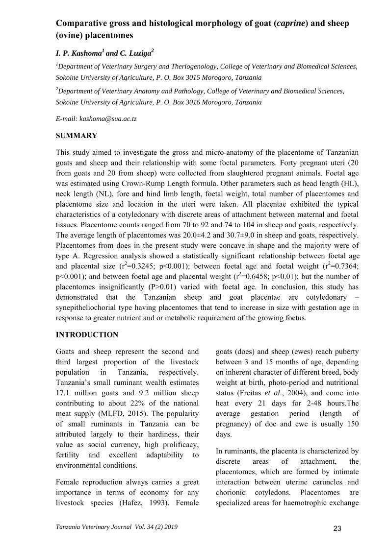
Table 1: Comparative measurements of different parameters in placentome of goats (n=20) and sheep (n=20) with regard to three different stages of pregnancy, Mean ± SE
Estimated Age of pregnancy
Number of placentomesin
thegravid horns)
Placentome parameters
Length(mm) Width (mm)
Goat Sheep Goat Sheep Goat Sheep 41 - 60 Days 88.3±10.8 74.3±3.3 16.8 ± 3.4 14.5 ± 1.3 12.8±1.5 14.3±1.0
61 - 80 Days 91.6±9.7 76.7±11.2 33.0 ± 4.1 19.1 ± 1.5 20.6±2.1 21.7±3.4
81 - 100 Days 94.3±4.6 83.2±8.1 37.9 ± 1.6 21.2 ± 1.7 26.6±1.5 26.6±1.5
Average 91.8±8.5 78.6±9.4 30.7 ± 9.0 20.0 ± 4.2 20.8±5.7 21.9±5.1
Histological findings
In goats, the basal plate was the convex surface; the chorionic plate of same placentome was shallow, resulting in a rather flat surface in the placentomes. In advance gestation, some placentome
exhibited a reversal of the typical placentomal shape, whereas the basal plate was the concave surface of the placentome, while the chorionic plate constituted the convex surface of the placentome.
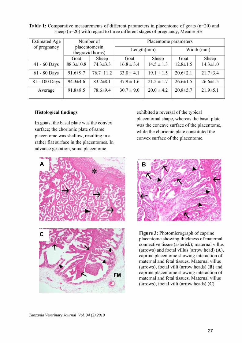
Figure 3: Photomicrograph of caprine placentome showing thickness of maternal connective tissue (asterisk); maternal villus (arrows) and foetal villus (arrow head) (A), caprine placentome showing interaction of maternal and fetal tissues. Maternal villus (arrows), foetal villi (arrow heads) (B) and caprine placentome showing interaction of maternal and fetal tissues. Maternal villus (arrows), foetal villi (arrow heads) (C).
A B
C
27

The Tropical Veterinarian
In ewes, four types of placentomes (type A, B, C, and D placentomes) were observed. Type A placentomes were concave in shape with the maternal tissue completely surrounding the fetal tissue; Type B placentomes consisted of fetal tissue beginning to grow over the surrounding maternal tissue; Type C placentomes were flat and consist of a large portion of fetal tissue that has begun to surround the maternal tissue; Type D placentomes contained mostly fetal tissue, which surrounds the maternal tissue.
However, the greatest (P= 0.02) number and percentage of placentomes in the gravid uterus were type A, followed by type B, and then types C and D. Placentome collected from does in the present study was concave in shape; each placentome possessed two surfaces, namely, a concave surface and a convex surface.
The placentomes of both caprine and ovine contained six cell layers; maternal capillary endothelium, maternal connective tissue, crypt syncytium, trophoblast, mesenchyme, and foetal capillary endothelium (Figures 3A-C). Although all 6 layers were present, considerable variation is sizes for some tissues were noted. The result also showed that all of the studied tissues tended to increase from 50 days of pregnancy but the foetal connective tissue and trophoblast epithelium decreased during the 90 days of pregnancy.
Caprine maternal tissue (maternal capillaries and fibroblasts tissue) thickness ranged from 7.5-11μm compared to that of the ovine with the thickness ranging from 2.5-4μm. Foetal connective tissue in ovine placentome ranged from 6-7μm while that of caprine was from 16-17μm.
DISCUSSION
Gross and microscopic structure of the placentomes of the doe and ewe and their relationship to foetal age were acquired and analysed. The average foetal weight of pregnant goat and sheep were 498.05 and 406.15 gm, respectively, which was significantly, lower than 4,779 gm that reported in sheep (van der Linden et al., 2013).
Furthermore, weight gradually increased significantly with advancing pregnancy. Several reasons may be associated with the variations observed in foetal weight in this study including availability of nutrients (Bauer et al., 1998), uterine blood flow and the utero-placental function especially when approaching term (Ferrell, 1991), and presence of singletons versus twins (Mellado et al., 2011). All the foetal diameters (Crown –rump length, head length, neck length, fore and hind limb length) gradually increased with advancing
pregnancy. This observation has been reported elsewhere (Lee et al., 2005; Hussein, 2008; Khanam, 2012) and may be attributed to availability of nutrients to the foetus, type of breed and parity (Bauer et al., 1998).
Linear association between foetal parameters (weight and size) and placentome parameters (length and width) was observed, indicating that both foetal and placentome parameters were much lower in early gestation but increased with age of gestation. Doize et al., (1997) also reported a linear regression relationship between foetal age in days and placentome size in does. However, poor correlation between placentome size and gestational age has been reported elsewhere in ovine (Kelly and Newnham, 1989; Gonzalez et al., 1998) and goats (Nwaogu et al., 2010). The increase in foetal parameters relative to placental parameters is probably due to
28
Tanzania Veterinary Journal Vol. 34 (2) 2019
increases in placentome vascularity, thereby increasing foeto-maternal exchange (Leiser et al., 1997).
This study has provided an insight into the assessment of the fine structures of caprine and ovine placentomes, and some of the accompanying parameters of growing foetuses over the course of gestation. Each cotyledon combined with caruncle found to form a separate unit known as placentomes as previously described in ewes (Bjorkman, 1981) and goats (Basha and Mohammad, 2013).
The placentome represents the functional unit of the placenta through which the foetus is nourished and it increases in size within certain limit to accommodate changing requirements of the foetus/foeti during intrauterine life (Noakes et al., 2001; Lee et al., 2005).
The placenta of both goat and sheep exhibited the typical characteristics of a cotyledonary type with the discrete area of attachment between maternal and foetal tissue i.e. the placentome. Similar observations were reported in West African dwarf goat (Igwebuike and Ezeasor, 2013) and non-descript breed of goat in India (Kumar et al., 2015).
The goats’ placentomes had the maternal caruncle formed the basal plate of the placentome, while the foetal cotyledon formed the chorionic plate of the placentome. These findings are in agreement with those reported in goats elsewhere (Igwebuike and Ezeasor, 2013; Basha and Mohammad, 2013).
In sheep, four types of placentomes (type A, B, C, and D placentomes) were observed although the greatest (P= 0.02) number and percentage of placentomes in the gravid uterus were type A, followed by type B, and
then types C and D. These findings are comparable to those observed elsewhere (Vonnahme et al., 2008; Steyn et al., 2001; Gardner et al., 2002; Osgerby et al., 2004).
The number of placentomes increased with advancement of pregnancy. The increase of number of placentomes with advancing pregnancy has been reported elsewhere in goats (Igwebuike, 2009; Kumar et al., 2015), cattle (Folusho, 2012) and sheep (Igwebuike, 2009). The average number of placentomes in the placenta of Tanzanian goats and sheep ranged from 74 to 104 and 70 to 92, respectively. The number of placentomes may be influenced by many factors such as maternal nutrition and body condition (Osgerby et al., 2004; Osgerby et al., 2003) and alterations in the insulin-like growth factor cascade (Igwebuike, 2010).
The size of placentomes varied in the different location of the uterus and also in different stages of pregnancy. This finding is in line with the observation recorded in Indian goats (Kumar et al., 2015), West African Dwarf goats Igwebuike and Ezeasor (2013), Yaks (Liu et al., 2010) and Fulani Zebu (Lilian et al., 2013).
The size of placentomes varied in the different region of the uterus with the smallest placentomes observed in the apical portion of uterine horn and largest placentomes in the vicinity of the umbilical cord. The increased size of placentome enhanced the rate of physiological exchange between the foetal and maternal system (Reynolds and Ferrell, 1987).
The size and number of placentomes were significantly higher in gravid horn as compared to non-gravid horn. Similar observations have been reported previously in goats (Gupta, 1984), cattle (Schlafer et
29
The Tropical Veterinarian
al., 2000; Laven and Peters, 2001) and Yak (Lui et al., 2010).
Arrangement of placentomes in four rows in both gravid and non-gravid horn has also been reported in Indian goats (Gupta, 1984; Kumar et al., 2015) and cattle (Gupta, 1984). However, Hafez (1954) in cattle observed 2 to 6 rows of placentomes in the uterine body and two rows at the extremities of horn.
Similarly, Igwebuike and Ezeasor (2013) reported randomly distributed with no defined order of arrangements of placentomes in goats.
The placenta constitutes a selective barrier and pathway for the passage of chemical substances.The present study described that the proportion of maternal connective tissue, foetal connective tissue, trophoblast
epithelium and syncytium (multi nucleated cells) had a tendency to increase from 50 days pregnancy. This observation is in agreement with previous study by Khanam, (2012) in goats, however conflicts with the ovine feature of histological structure of placentome reported by Stegeman, (1974).
Also the study described that the proportion of foetal connective tissue decreased with gestation age and this is supported by the findings reported elsewhere in sheep (Wimsatt, 1950) and goats (Wango et al., 1990).
In conclusion, this study has demonstrated that the pregnant Tanzanian sheep and goats are characterized by a cotyledonary placenta. The placentomal size increases with gestational age in response to the greater nutrient/metabolic requirements of the foetus, as pregnancy progresses
.
REFERENCES
BananKhojasteh, S.M. (2012). Prenatal development of Iranian goat foetuses. International Research Journal of Applied and Basic Sciences, 3 (10): 2022-2024
Basha, S. and Mohammad, A. G. 2013. Some anatomical and histological studies on the placenta of goats in Dhamar, Yemen. Yemeni J. Agri. Vet Sci., 1(1):26-32.
Bauer, M. K., Harding J. E., Bassett, N. S., Breier, B. H., Oliver, M. H., Gallaher, B. H. 1998. Fetal growth and placental function. Molecular and Cellular Endocrinology. 140, 115-120.
Bjorkman, N. (1981). Placentation. In, Veterinary Histology Ed) Dellmann, H and Brown, E.M. 2nd Edition. Lea &Febiger, Philadelphia, pp 337-355.
Bjorkman, N. Placentation. In: Dellmann, H.D.; Brown, E.M. (Ed.)(1976): Textbook of veterinary histology. Philadelphia : Lea and Febiger,. p.351-69.
Derivaux, J., Ectors, F., Beckers, J.F. (1988). The ruminant placenta: structure and endocrine function. Brussels : State University of Liege
Derivaux, J.; Ectors, F.; Beckers, J.F. (1988). The ruminant placenta: structure and endocrine function. Brussels : State University of Liege,.
Doize, F., Vaillancourt, D., Carabin, H., Belanger J. (1997): Determination of gestational age in sheep and goats usingtransrectalultrasonographic measurement of placentomes. Theriogenol. 48, 449-460.
30

Tanzania Veterinary Journal Vol. 34 (2) 2019
Ferrell, C. L. 1991. Maternal and fetal influences on uterine and conceptus development in the cow: Growth of tissues of the gravid uterus. J. Anim. Sci. 69:1945-1953.
Folusho, D. A. 2012. The development of bovine placentome and associated structure during gestation. PhD thesis. Massey University, New Zealand. Pp. 35.
Freitas VJF, Lopes-Junior ES, Rondina D, Salmito-Vanderley CSB, Salles HO, Simplício AA, Baril G, Saumande J. Puberty in Anglo-Nubian and Saanen female kids raised in the semi-arid of north-eastern Brazil. Small Ruminant Res. 53: 167–172, 2004.
Gardner DS, Ward JW, Giussani DA, Fowden AL 2002. The effect of a reversible period of adverse intrauterine conditions during late gestation on fetal and placental weight and placentome distribution in sheep. Placenta 23 (6): 459–466.
Gonzalez, B.A., Santiago, M.J. and López, S. A. (1998). Estimation of foetal development in Manchega dairy ewes by transrectalultrasonographic measurements. Small Rumin. Res. 27, 243-250.
Gupta, S. K. 1984. Gross histological and certain histochemical observation on the placentome of goat. M. V. Sc. Thesis, Mathura
Hafez, E. S. E. 1954. The placentome in the buffalo. Acta Zoology. 21, 176-191.
Hafez, E. S. E. 1993. Folliculogenesis, egg maturation and ovulation. In: Reproduction in Farm Animals.
Hafez E.S.E. (ed), Lea and Febiger Press, Philadelphia.PP.114-143.
Hussein A.A (2008). Determination of first pregnancy and foetal measurements in eqyptianBaladi goats (Capra hircus). VeterinariaItaliana, 44(2): 429-437.
Igwebuike, U. M. (2010): Impact of maternal nutrition on ovine foetoplacental development: a review of the role of insulin-like growth factors. Anim. Reprod. Sci. 121, 189-196.
Igwebuike, U. M. 2009. A review of uterine structureal modification that influence conceptus implantation and development in sheep and goat. AnimReprod Sci., 112: 1-7.
Igwebuike, U. M. and Ezeasor, D. N. 2013. The morphology of placentome and formation of chorionic villous tree in West African Dwarf Goat (caprahircus). Vet. Archiv., 83(3): 313- 321.
Kelly, R.W. and Newnham J.P. (1989): Estimation of gestational age in Merino ewes by ultrasound measurement of foetal head size. Aust. J. Agric. Res. 40, 1293-1299
Khanam JS (2012). Study on placentome and foetus at different stages of pregnancy in Black Bengal goat, MS Thesis, Department of Animal Breeding and Genetics, Bangladesh Agricultural University.
Kumar,V., Singh, S.P., Farooqui, M.M., Kumar, P., Prakash A. (2015). Gross and Biometrical studies of Placentome in Goat (Capra hircus) during Different Stages of
31
The Tropical Veterinarian
Pregnancy. Journal of Animal Research,5(2): 251-255.
Laven, R. A. and Peters, A. R. 2001. Gross Morphometry of the bovine placentome during gestation. Reprod. Dom. Anim., 36, 289-296.
Lee Y, Lee J, Cho H, Shin Y, Cho Y, Shim W, Choi H, Shin D, Lee G and Shin S (2005). Ultrasonic measurements of fetal parameters for estimation of gestational age in Korean black goats. Journal of Veterinary Medicine Science, 67(5): 497-502.
Leiser, R., Krebs, C., Klisch, K., Ebert, B., Dantzer, V., Schuler, G. (1997): Fetal villosity and microvasculature of the bovine placentome in the second half of gestation. J. Anat. 191, 517-527.
Lilian, O.C., Usende, I.L., Daniel, E.N. and ThankGod, O.E (2013). Gross and Micro-Anatomical Observations on Fulani Zebu Placentome and its Relationship with some Foetal Parameters. Journal of Experimental Biology and Agricultural Sciences, 1(5): 353-359.
Liu, B. Cui, Y. Yang, B. Fan, J. Zhao, Z. and Yu, S. 2010. Morphometric analysis of Yak placentomes during gestation. The anatomical record: advance in integrative anatomy and evolutionary Biology., 293; 1873-1879.
McGeady, T. A., Quinn, P. J., Fitzpatrics, E. S., Ryan, M. T. and Cahalan, S. 2006. Veterinary embryology.,Oxford: Blackwell Publishing.
Miguel Mellado M., Meza-Herrera< C.A., Arévalo, J.R., De Santiago-Miramontes, M.A., Rodríguez, A., Luna-Orozco, J.R and Veliz-Deras,
F.G. (2011). Relationship between litter birth weight and litter size in five goat genotypes. Animal Production Science, 51, 144–149
MLFD (2015). Tanzania Livestock Mordenization Initiative.July_ 20151453204967.pdf
Noakes DE, Parkinson TJ & England GCW (2001). Arthur’s Veterinary Reproduction and Obstetrics. Elsevier, China. Eight edition. Pp 69-75.
Nwaogu, I.C., Anya, K.O and Agada, P.C. (2010).Estimation of foetal age using ultrasonic measurements of different foetal parameters in red Sokoto goats (Capra hircus). Veterinarski Archives, 80 (2), 225-233,
Osgerby JC, Wathes DC, Howard D, Gadd TS 2004. The effect of maternal under nutrition on the placental growth trajectory and the uterine insulin-like growth factor axis in the pregnant ewe. Journal of Endocrinology 182(1): 89–103.
Osgerby, J.C., Gadd, T.S. and Wathes, D., C. (2003): The effect of maternal nutrition and body condition on placental and fetal growth in the ewe placenta. Placenta 24, 236-247.
Regnault TR, Orbus RJ, Battaglia FC, Wilkening RB, Anthony RV. Altered arterial concentrations of placental hormones during maximal placental growth in a model of placental insufficiency. J Endocrinol 1999; 162: 433–442.
Regnault TRH, Orbus RJ, Battaglia FC, Wilkening RB, Anthony RV. (1999). Altered arterial concentrations of placental hormones during maximal placental
32

Tanzania Veterinary Journal Vol. 34 (2) 2019
growth in a model of placental insufficiency. J Endocrinol. 162:433–442.
Reynolds LP, Redmer DA. Mini-review: angiogenesis in the placenta. BiolReprod 2001; 64:1033–1040.
Reynolds, L.P., Redmer, D.A. (1995: Utero-placental vascular development and placental function. J. Anim. Sci.,; 73: 1839-1851.
Schlafer, D. H., Fisher, P. J. and Davies, C. J. 2000. The bovine placenta before and after birth: placental development and functionin health and disease. Anim. Reprod. Sci,. 60-61: 145-160.
STEGEMAN, J.H.J. (1974). Placental development in the sheep and its relation to fetal development. Bijdragen tot de Dierkunde, 44(1):63-72.
Steyn C, Hawkins P, Saito T, Noakes DE Hanson MA (2001) Undernutrition during the first half of gestation increases the predominance of fetal tissue in late-gestation in ovinePlacentomes. European journal of Obstetrics and Gynecology and Reproductive Biology 98: 165 – 170.
van der Linden, D.S., Sciascia, Q., Sales, F., and McCoard, S.A. (2013). Placental nutrient transport is affected by pregnancy rank in sheep. J Anim Sci. 91(2):644-53.
Vatnick I, Schoknecht R, Darrigrand R, Bell AW. (1991). Growth and
metabolism of the placenta after unilateral fetectomy in twin pregnant ewes. J DevPhysiol 15:351–356.
Vonnahme K.A., Arndt W.J., Johnson M.L., Borowicz P.P. and Reynolds L.P. (2008). Effect of Morphology on Placentome Size, Vascularity, and Vasoreactivity in Late Pregnant Sheep.Biology of Reproduction 79, 976–982.
Vonnahme KA, Hess BW, Nijland MJ, Nathalielsz PW, Ford SP. (2006). Placentomal differentiation may compensate for maternal nutrient restriction in ewes adapted to harsh range conditions. J AnimSci; 84:3451–3459
Wango EO, Wooding FB, and Heap RB. (1990). The role of trophoblastic binucleate cells in implantation in the goat: a morphological study. J Anat. 171:241-257.
Wimsatt,W. A. 1950. New histological observations on the placenta of the sheep. American J.Anat. 87 (3): 391-408.
Wooding, F. B. P. (2006): Analysis of the structure of the ruminant placenta: methods of fixation, embedding, and antibody localization at light and electron microscope levels. Methods Mol. Med. 121, 315-322.
Wooding, F.B.P. (1992). Current topic: the synepitheliochorial placenta of ruminants: binucleate cell fusions and hormone production. Placenta, 13 (2): 101-13.
33

The Tropical Veterinarian
Supporting Table 1: Comparative measurements of different parameter of foetuses recovered from goats (n=20) and sheep (n=20) in three different stages of pregnancy, Mean ± SE
Stage of pregnan
cy
Fetal weight (gm) Crown –rump length (cm)
Head length (cm)
Neck length (cm)
Fore limb length (cm)
Hind limb length (cm)
Goat Sheep Goat Sheep Goat Sheep Goat Sheep Goat Sheep Goat Sheep
41 - 60 Days
93.0 ±13.61
87.51 ±11.59
8.52 ±1.59
8.45 ±0.59
2.12 ±0.24
2.48 ±0.34
1.96 ±0.32
2.00 ±0.36
4.38 ±0.39
2.29 ±0.46
3.52 ±0.28
3.55 ±0.66
61 - 80 Days
258.14 ±130.94
212.71 ±37.33
17.07 ±1.2
16.73 ±1.26
2.36 ±0.26
2.70 ±0.29
2.31 ±0.26
2.26 ±0.26
6.23 ±0.37
5.09 ±0.55
6.81 ±0.36
7.05 ±0.80
81 - 100 Days
960.75 ±77.22
941.01 ±99.65
26.91 ±2.17
25.13 ±2.33
3.61 ±0.31
3.23 ±0.18
2.36 ±0.33
2.62 ±0.25
9.00 ±0.28
9.18 ±0.52
9.11 ±0.43
9.04 ±0.38
Average 498.05 ±402.2
406.15 ±367.0
18.86 ±7.79
17.59 ±6.19
2.80 ±0.73
2.83 ±0.40
2.25 ±0.33
2.32 ±0.35
6.89 ±1.95
6.39 ±2.30
6.91 ±2.28
6.96 ±2.07
34