comparative evaluation of therapeutic - publications
TRANSCRIPT

D458_covI-IV.indd 1 2007-10-29 16:31:14

COMPARATIVE EVALUATION OF THERAPEUTIC
RADIOPHARMACEUTICALS

The following States are Members of the International Atomic Energy Agency:
AFGHANISTANALBANIAALGERIAANGOLAARGENTINAARMENIAAUSTRALIAAUSTRIAAZERBAIJANBANGLADESHBELARUSBELGIUMBELIZEBENINBOLIVIABOSNIA AND HERZEGOVINABOTSWANABRAZILBULGARIABURKINA FASOCAMEROONCANADACENTRAL AFRICAN REPUBLICCHADCHILECHINACOLOMBIACOSTA RICACÔTE D’IVOIRECROATIACUBACYPRUSCZECH REPUBLICDEMOCRATIC REPUBLIC OF THE CONGODENMARKDOMINICAN REPUBLICECUADOREGYPTEL SALVADORERITREAESTONIAETHIOPIAFINLANDFRANCEGABONGEORGIAGERMANYGHANA
GREECEGUATEMALAHAITIHOLY SEEHONDURASHUNGARYICELANDINDIAINDONESIAIRAN, ISLAMIC REPUBLIC OF IRAQIRELANDISRAELITALYJAMAICAJAPANJORDANKAZAKHSTANKENYAKOREA, REPUBLIC OFKUWAITKYRGYZSTANLATVIALEBANONLIBERIALIBYAN ARAB JAMAHIRIYALIECHTENSTEINLITHUANIALUXEMBOURGMADAGASCARMALAWIMALAYSIAMALIMALTAMARSHALL ISLANDSMAURITANIAMAURITIUSMEXICOMONACOMONGOLIAMONTENEGROMOROCCOMOZAMBIQUEMYANMARNAMIBIANETHERLANDSNEW ZEALANDNICARAGUANIGERNIGERIA
NORWAYPAKISTANPALAUPANAMAPARAGUAYPERUPHILIPPINESPOLANDPORTUGALQATARREPUBLIC OF MOLDOVAROMANIARUSSIAN FEDERATIONSAUDI ARABIASENEGALSERBIASEYCHELLESSIERRA LEONESINGAPORESLOVAKIASLOVENIASOUTH AFRICASPAINSRI LANKASUDANSWEDENSWITZERLANDSYRIAN ARAB REPUBLICTAJIKISTANTHAILANDTHE FORMER YUGOSLAV REPUBLIC OF MACEDONIATUNISIATURKEYUGANDAUKRAINEUNITED ARAB EMIRATESUNITED KINGDOM OF GREAT BRITAIN AND NORTHERN IRELANDUNITED REPUBLIC OF TANZANIAUNITED STATES OF AMERICAURUGUAYUZBEKISTANVENEZUELAVIETNAMYEMENZAMBIAZIMBABWE
The Agency’s Statute was approved on 23 October 1956 by the Conference on the Statute othe IAEA held at United Nations Headquarters, New York; it entered into force on 29 July 1957The Headquarters of the Agency are situated in Vienna. Its principal objective is “to accelerate andenlarge the contribution of atomic energy to peace, health and prosperity throughout the world’’.
f .

TECHNICAL REPORTS SERIES No. 458
COMPARATIVE EVALUATION OF THERAPEUTIC
RADIOPHARMACEUTICALS
INTERNATIONAL ATOMIC ENERGY AGENCYVIENNA, 2007

IAEA Library Cataloguing in Publication Data
Comparative evaluation of therapeutic radiopharmaceuticals. — Vienna : International Atomic Energy Agency, 2007.
p. ; 24 cm. — (Technical reports series, ISSN 0074-1914 ; no. 458)STI/DOC/010/458ISBN 92-0-115106-3Includes bibliographical references.
1. Radiopharmaceuticals — Evaluation. I. International Atomic Energy Agency. II. Series: Technical reports series (International Atomic Energy Agency) ; 458.
IAEAL 07–00496
COPYRIGHT NOTICE
All IAEA scientific and technical publications are protected by the terms of the Universal Copyright Convention as adopted in 1952 (Berne) and as revised in 1972 (Paris). The copyright has since been extended by the World Intellectual Property Organization (Geneva) to include electronic and virtual intellectual property. Permission to use whole or parts of texts contained in IAEA publications in printed or electronic form must be obtained and is usually subject to royalty agreements. Proposals for non-commercial reproductions and translations are welcomed and considered on a case-by-case basis. Enquiries should be addressed to the IAEA Publishing Section at:
Sales and Promotion, Publishing SectionInternational Atomic Energy AgencyWagramer Strasse 5P.O. Box 1001400 Vienna, Austriafax: +43 1 2600 29302tel.: +43 1 2600 22417email: [email protected] http://www.iaea.org/books
© IAEA, 2007
Printed by the IAEA in AustriaOctober 2007
STI/DOC/010/458

FOREWORD
Radionuclide therapy employing unsealed radiotherapeutic agents has emerged as an important tool for cancer management. The development of therapeutic radiopharmaceuticals based on different types of carrier molecule and a variety of radioisotopes is being actively pursued worldwide. There have been many significant advances in this field, and many of the technical problems involved in labelling biomolecules with a variety of radionuclides have been solved. However, the assessment of the relative effectiveness of different radiopharmaceuticals for cancer therapy is a difficult task owing to the large number of variables that must be considered, some related to the biological carrier and others to the radioisotope. Comparing the therapeutic efficacy in patients is not feasible in most cases for ethical and regulatory reasons. Hence, it is important to develop laboratory methods that can be used for reliable and efficient comparative evaluation of promising therapeutic radiopharmaceuticals.
The IAEA has organized several coordinated research projects (CRPs) in the field of radiopharmaceuticals that have helped Member States to acquire technologies for the production of useful radiopharmaceuticals. In one such CRP on techniques for labelling biomolecules for targeted therapy, conducted from 1998 to 2001, the participants developed several protocols and standard operating procedures for labelling peptides and antibodies with therapeutic radioisotopes. During the course of the CRP, it was recognized that successful development of therapeutic radiopharmaceuticals will require in vitro biological assays as well as appropriate tumour models for carrying out biodistribution studies of the products in order to collect data for preclinical studies. Two meetings, held in 1999 and 2001, recommended the organization of a CRP for the development of laboratory methods for comparative evaluation of therapeutic radiopharmaceuticals. Fifteen countries — Brazil, Cuba, the Czech Republic, Greece, Hungary, India, Italy, the Republic of Korea, Mexico, Pakistan, Poland, Romania, the United Kingdom, the United States of America and Uruguay — participated in this CRP.
The work plan of the CRP was developed during the first Research Coordination Meeting, held in Bucharest in October 2002. It addressed the issues related to the evaluation of new therapeutic radiopharmaceuticals, which included the development of analytical techniques to establish the chemical, radiochemical and pharmaceutical purity, and the stability of therapeutic radiopharmaceuticals. In addition, it was decided that specific bioassays and tumour models needed for the evaluation of the biological efficacy of the end products would be developed during the CRP. Participants also identified the potential lead molecules and isotopes to be used during the CRP for the development of radiopharmaceuticals.
At the second Research Coordination Meeting, held in April 2004 in Warsaw, participants reported on the progress of the work carried out during the first part of the CRP. The results of the research carried out by each laboratory were presented at the final Research Coordination Meeting, held in Vienna in November 2005. During the course of the CRP, participants successfully established several analytical techniques, biological assays, animal tumour models and protocols for evaluation of therapeutic radiopharmaceuticals, which are described in detail in this publication. Another major achievement of the CRP was the development of a reliable protocol for the preparation and evaluation of 177Lu-DOTATATE, a radiopharmaceutical useful for therapy of neuroendocrine tumours. One of the participating States, Italy, started using 177Lu-DOTATATE for therapy during the course of the CRP, and another State, Brazil, reported its clinical use after completion of the project.
The results obtained by various laboratories during the CRP are summarized in this publication. The IAEA thanks all the CRP participants for their valuable contributions, and S. Banerjee for her help in compiling and editing this report. The IAEA officer responsible for this publication was M.R.A. Pillai of the Division of Physical and Chemical Sciences.
EDITORIAL NOTE
Although great care has been taken to maintain the accuracy of information contained in this publication, neither the IAEA nor its Member States assume any responsibility for consequences which may arise from its use.
The use of particular designations of countries or territories does not imply any judgement by the publisher, the IAEA, as to the legal status of such countries or territories, of their authorities and institutions or of the delimitation of their boundaries.
The mention of names of specific companies or products (whether or not indicated as registered) does not imply any intention to infringe proprietary rights, nor should it be construed as an endorsement or recommendation on the part of the IAEA.
The authors are responsible for having obtained the necessary permission for the IAEA to reproduce, translate or use material from sources already protected by copyrights.
Material prepared by authors who are in contractual relation with governments is copyrighted by the IAEA, as publisher, only to the extent permitted by the appropriate national regulations.

CONTENTS
PART I. OVERVIEW OF THE COORDINATED RESEARCH PROJECT
CHAPTER 1. OBJECTIVES, RESULTS AND ACHIEVEMENTS OF THE COORDINATED RESEARCH PROJECT . . . . . 3
1.1. Introduction . . . . . . . . . . . . . . . . . . . . . . . . . . . . . . . . 31.2. Objectives of the coordinated research project . . . 71.3. Overview of the work plan . . . . . . . . . . . . . . . . . . . . 71.4. Scientific achievements of the coordinated
research project . . . . . . . . . . . . . . . . . . . . . . . . . . . . . 101.5. Conclusion and recommendations . . . . . . . . . . . . . . 12
CHAPTER 2. PREPARATION AND QUALITY CONTROL OF 177Lu-DOTATATE FOR TARGETED THERAPY . . . 17
2.1. Materials . . . . . . . . . . . . . . . . . . . . . . . . . . . . . . . . . . . 172.2. Preparation of 177Lu-DOTATATE . . . . . . . . . . . . 192.3. Quality control . . . . . . . . . . . . . . . . . . . . . . . . . . . . . . 202.4. Storage of radiolabelled peptide . . . . . . . . . . . . . . . 212.5. In vitro stability . . . . . . . . . . . . . . . . . . . . . . . . . . . . . 212.6. Conclusion . . . . . . . . . . . . . . . . . . . . . . . . . . . . . . . . . 22
PART II. REPORTS BY PARTICIPANTS IN THE COORDINATED RESEARCH PROJECT
CHAPTER 3. DEVELOPMENT OF SOMATOSTATIN BASED RADIOPHARMACEUTICALS FOR RECEPTOR MEDIATED RADIONUCLIDE THERAPY . . . . . . . . 27E.B. Araújo, J.S. Caldeira Filho, L.T. Nagamati, E. Muramoto, M.T. Colturato, R. Couto, K. Okasaki, M.F. Suzuki, M.I.C.C. Guimaraes
3.1. Introduction . . . . . . . . . . . . . . . . . . . . . . . . . . . . . . . . 273.2. Materials and methods . . . . . . . . . . . . . . . . . . . . . . . 293.3. Results and discussion . . . . . . . . . . . . . . . . . . . . . . . 353.4. Organ dosimetry . . . . . . . . . . . . . . . . . . . . . . . . . . . . 453.5. Micronucleus assay . . . . . . . . . . . . . . . . . . . . . . . . . . 48
CHAPTER 4. BIOLOGICAL EVALUATION OF RADIOTRACERS FOR RADIONUCLIDE THERAPY . . . . . . . . . . . . . . . 53G.J. Pimentel, R. Ravelo, M. Miranda, J. Gavilondo, L. Pérez, M. Ayala, H. Bell, H.E. Garay, O. Reyes, I. Sanchez
4.1. Introduction . . . . . . . . . . . . . . . . . . . . . . . . . . . . . . . . 544.2. Materials and methods . . . . . . . . . . . . . . . . . . . . . . . 554.3. Results and discussion . . . . . . . . . . . . . . . . . . . . . . . 614.4. Conclusion . . . . . . . . . . . . . . . . . . . . . . . . . . . . . . . . . 70
CHAPTER 5. PRECLINICAL COMPARISON OF DOTATATE LABELLED WITH DIFFERENT RADIONUCLIDES . . . . . . . . . . . . . . . . . . . . . . . . . . . . . . 73M. Laznicek, A. Laznickova, F. Trejtnar, L. Melicharova, J. Cihlo, M. Petrik
5.1. Introduction . . . . . . . . . . . . . . . . . . . . . . . . . . . . . . . . 745.2. Materials and methods . . . . . . . . . . . . . . . . . . . . . . . 745.3. Results and discussion . . . . . . . . . . . . . . . . . . . . . . . . 795.4. Conclusion . . . . . . . . . . . . . . . . . . . . . . . . . . . . . . . . . 85
CHAPTER 6. EVALUATION OF PEPTIDES LABELLED WITH BETA EMITTING RADIONUCLIDES FOR RECEPTOR TARGETED RADIOTHERAPY OF MALIGNANT TUMOURS. . . . . . . . . . . . . . . . . . . . . . . . 87T. Maina, B. Nock, A. Nikolopoulou, C. Petrou, P. Cordopatis
6.1. Introduction . . . . . . . . . . . . . . . . . . . . . . . . . . . . . . . . 886.2. Materials . . . . . . . . . . . . . . . . . . . . . . . . . . . . . . . . . . . 906.3. Methods . . . . . . . . . . . . . . . . . . . . . . . . . . . . . . . . . . . 916.4. Results . . . . . . . . . . . . . . . . . . . . . . . . . . . . . . . . . . . . 956.5. Conclusion . . . . . . . . . . . . . . . . . . . . . . . . . . . . . . . . . 100

CHAPTER 7. LABELLING AND BIOLOGICAL EVALUATION OF THERAPEUTIC RADIOPHARMACEUTICALS . . . . . . . . . . . . . . . . . . . 103G.A. Jánoki, A. Polyák, R. Király, L. Balogh, L. Kőrösi, D. Máthé
7.1. Introduction . . . . . . . . . . . . . . . . . . . . . . . . . . . . . . . . 1037.2. 188Re labelled bone seeking and tumour specific
agents for therapy . . . . . . . . . . . . . . . . . . . . . . . . . . . 1047.3. Radiolabelled receptor specific peptides for
targeted use in diagnosis and therapy . . . . . . . . . . . 112
CHAPTER 8. PREPARATION AND EVALUATION OF 177Lu-DOTATATE FOR POTENTIAL APPLICATION IN PEPTIDE RECEPTOR RADIONUCLIDE THERAPY . . . . . . . . . . . . . . . . . . . . 131S. Banerjee, S. Chakraborty, A. Korde, T. Das, A. Mukherjee, U. Pandey, H.D. Sarma, G. Samuel, P.S. Dhami, A.D. Ingle, M. Venkatesh
8.1. Introduction . . . . . . . . . . . . . . . . . . . . . . . . . . . . . . . . 1328.2. Materials . . . . . . . . . . . . . . . . . . . . . . . . . . . . . . . . . . . 1338.3. Methods. . . . . . . . . . . . . . . . . . . . . . . . . . . . . . . . . . . . 1348.4. Results and discussion . . . . . . . . . . . . . . . . . . . . . . . 1438.5. 177Lu labelling of other target specific ligands . . . . 1578.6. Conclusion . . . . . . . . . . . . . . . . . . . . . . . . . . . . . . . . . 161
CHAPTER 9. OPTIMIZATION OF LABELLING CONDITIONS AND CELL BINDING ASSAY FOR 177Lu-DOTATATE . . . . . . . . . . . . . . . . . . . . . . . . . . 163M. Chinol, Sun Ju Choi
9.1. Introduction . . . . . . . . . . . . . . . . . . . . . . . . . . . . . . . . 1639.2. Materials and methods . . . . . . . . . . . . . . . . . . . . . . . 1649.3. Results . . . . . . . . . . . . . . . . . . . . . . . . . . . . . . . . . . . . 1669.4. Conclusion . . . . . . . . . . . . . . . . . . . . . . . . . . . . . . . . . 168

CHAPTER 10. COMPARATIVE EVALUATION OF LABELLED BIOMOLECULES FOR TARGETED RADIOTHERAPY. . . . . . . . . . . . . . . . . . . . . . . . . . . . . . . 169S.J. Choi, Y.D. Hong, S.M. Choi
10.1. Introduction . . . . . . . . . . . . . . . . . . . . . . . . . . . . . . . . 16910.2. Materials and methods . . . . . . . . . . . . . . . . . . . . . . . 17110.3. Results . . . . . . . . . . . . . . . . . . . . . . . . . . . . . . . . . . . . 17310.4. Conclusion . . . . . . . . . . . . . . . . . . . . . . . . . . . . . . . . . 177
CHAPTER 11. LABORATORY METHODS TO EVALUATE THERAPEUTIC RADIOPHARMACEUTICALS . . . 181C. Arteaga de Murphy, G. Ferro-Flores, J. Rodriguez-Cortes, M. Pedraza-Lopez, M.T.Ramirez-Iglesias
11.1. Introduction . . . . . . . . . . . . . . . . . . . . . . . . . . . . . . . . 18111.2. Materials and methods . . . . . . . . . . . . . . . . . . . . . . . 18311.3. Results . . . . . . . . . . . . . . . . . . . . . . . . . . . . . . . . . . . . . 18811.4. Discussion and conclusion . . . . . . . . . . . . . . . . . . . . 192
CHAPTER 12. LABORATORY EVALUATION OF THE BETA EMITTING RADIONUCLIDES 177Lu, 131I, 153Sm AND 166Ho, AND RADIOPHARMACEUTICALS FOR RADIOTHERAPY. . . . . . . . . . . . . . . . . . . . . . . . . . . . . . . 197M.M. Ishfaq, H.J. Nizakat, K.M. Bashar, I. Haider
12.1. Introduction . . . . . . . . . . . . . . . . . . . . . . . . . . . . . . . . 19812.2. Materials . . . . . . . . . . . . . . . . . . . . . . . . . . . . . . . . . . . 19912.3. Methods . . . . . . . . . . . . . . . . . . . . . . . . . . . . . . . . . . . 19912.4. Results and discussion . . . . . . . . . . . . . . . . . . . . . . . 204
CHAPTER 13. PRECLINICAL EVALUATION OF THERAPEUTIC RADIOPHARMACEUTICALS BASED ON 90Y AND 177Lu. . . . . . . . . . . . . . . . . . . . . . . . . . . . . . . . . . . . . . . 217D. Pawlak, A. Korsak, R. Mikołajczak, B. Janota, U. Karczmarczyk, E. Jakubowska
13.1. Introduction . . . . . . . . . . . . . . . . . . . . . . . . . . . . . . . . 21713.2. Methods . . . . . . . . . . . . . . . . . . . . . . . . . . . . . . . . . . . 21713.3. Results . . . . . . . . . . . . . . . . . . . . . . . . . . . . . . . . . . . . 22113.4. Conclusion . . . . . . . . . . . . . . . . . . . . . . . . . . . . . . . . . 231

CHAPTER 14. LABELLING OF DOTATATE WITH 177Lu AND 131I FOR DIAGNOSIS AND TARGETED THERAPY: IN VITRO AND IN VIVO COMPARATIVE EVALUATION. . . . . . . . . . . . . . . . . . . . . . . . . . . . . . . . . . 233V. Lungu, D. Niculae, D. Chiper, R. Mihai, L. Danaila, S. Baiculescu
14.1. Introduction . . . . . . . . . . . . . . . . . . . . . . . . . . . . . . . . 23414.2. Materials . . . . . . . . . . . . . . . . . . . . . . . . . . . . . . . . . . . 23414.3. Methods. . . . . . . . . . . . . . . . . . . . . . . . . . . . . . . . . . . . 23514.4. Results . . . . . . . . . . . . . . . . . . . . . . . . . . . . . . . . . . . . 24014.5. Conclusion . . . . . . . . . . . . . . . . . . . . . . . . . . . . . . . . . 254
CHAPTER 15. PRECLINICAL DEVELOPMENT OF THERAPEUTIC RADIOPHARMACEUTICALS . . . 257S.J. Mather
15.1. Introduction . . . . . . . . . . . . . . . . . . . . . . . . . . . . . . . 25715.2. Radiopharmaceutical design . . . . . . . . . . . . . . . . . . 25815.3. Synthesis or purchase of precursor . . . . . . . . . . . . 25915.4. Radiolabelling development . . . . . . . . . . . . . . . . . . 26015.5. Analyses of radiochemical purity . . . . . . . . . . . . . . 26015.6. Stability assessment . . . . . . . . . . . . . . . . . . . . . . . . . 26115.7. Receptor binding . . . . . . . . . . . . . . . . . . . . . . . . . . . 26215.8. Cellular processing and metabolism . . . . . . . . . . . 26315.9. In vivo biodistribution and targeting . . . . . . . . . . . 26415.10. In vitro and in vivo measures of efficacy . . . . . . . . 265
CHAPTER 16. LABORATORY EVALUATION OF THERAPEUTIC BIOMOLECULES LABELLED WITH RADIOIODINE AND LUTETIUM. . . . . . . . . . 269H.S. Balter, P. Oliver, A. Robles, N. Berois, P. Cabral, A. Nappa, A. López, V. Trindade, G. Rodríguez, S. Lanzzeri, S. Verdera
16.1. Introduction . . . . . . . . . . . . . . . . . . . . . . . . . . . . . . . . 27016.2. Materials . . . . . . . . . . . . . . . . . . . . . . . . . . . . . . . . . . . 27116.3. Methods . . . . . . . . . . . . . . . . . . . . . . . . . . . . . . . . . . . 27116.4. Results . . . . . . . . . . . . . . . . . . . . . . . . . . . . . . . . . . . . . 27816.5. Conclusion . . . . . . . . . . . . . . . . . . . . . . . . . . . . . . . . . 290

APPENDIX I: PROTOCOLS DEVELOPED AS PART OF THE COORDINATED RESEARCH PROJECT . . . . . . . . . 295
APPENDIX II: PAPERS PUBLISHED BY THE PARTICIPANTS RELATED TO THE COORDINATED RESEARCH PROJECT . . . . . . . . . . . . . . . . . . . . . . . . . . . . . . . . . . . . . . 306
LIST OF PARTICIPANTS . . . . . . . . . . . . . . . . . . . . . . . . . . . . . . . . . . . . . . . 311

PART I
OVERVIEW OF THECOORDINATED RESEARCH PROJECT

.

Chapter 1
OBJECTIVES, RESULTS AND ACHIEVEMENTS OF THE COORDINATED RESEARCH PROJECT
1.1. INTRODUCTION
Radiotherapy is an essential mode of treatment of many cancers, either alone or in conjunction with other methods such as surgery and chemotherapy. While in most cases radiotherapy is given using external radiation sources, it is also possible to administer radiotherapy by specifically localizing radioisotopes emitting particulate radiation within the tumour tissue [1.1]. This mode of targeted therapy has several potential advantages over external beam therapy, including the possibility of delivering doses more selectively to the tumour and treating widespread multiple metastases. Such targeted therapy of tumours, as well as of other, benign conditions, was the thrust of nuclear medicine in the early years. A few therapeutic procedures for the treatment of hyperthy-roidism and thyroid cancer introduced at that time continue to be used regularly. In the light of the major scientific achievements in this field in the past decade [1.2–1.4], targeted therapy using radiopharmaceuticals is now being actively pursued at many centres around the world for the management of cancer.
1.1.1. Radiopharmaceuticals for targeted therapy
The challenges inherent in the development of radiotherapeutic agents arise from the need to strike a perfect balance between specific in vivo targeting and clearance of the radioactivity from non-target sites. The design and development of such agents have undergone a paradigm shift with respect to the molecular vectors employed: initially, small organic molecules or inorganic moieties were used, whereas present-day research makes use of antibodies, peptides, steroids, nucleotides and other small molecules that have specific receptor affinity. The selection of carrier molecules can be made bearing in mind the problems related to efficient drug delivery, in vivo metabolism of the agent and the pharmacokinetics of the radiolabelled drugs. Recent advances in this area [1.5, 1.6] exploit the diversity of receptor avid and immune derived molecular vectors, as well as a plethora of therapeutic radio-nuclides.
3

CHAPTER 1
1.1.2. Radionuclides for targeted therapy
Among the radionuclides used for cancer therapy, 131I, 90Y, 188Re, 166Ho and 153Sm have applications for the treatment of a multitude of malignant disorders by targeting specific organs and tissues. Such agents have been used for cancer therapy, palliation of bone pain arising from secondary metastases, radiosynovectomy, intravascular radiation therapy and the treatment of a host of other disorders. Extensive research in the field of radiopharmaceuticals and nuclear medicine practices have led to the identification of other radionuclides, including 177Lu, 67Cu and alpha emitters such as 211At, with promising radio-nuclidic, physical and chemical properties. An advantage of targeted radio-therapy is the wide variety of radionuclides with different radionuclidic characteristics — such as physical half-lives and radiation types — that are available for designing a therapeutic radiopharmaceutical for a specific appli-cation. An important task is to select a radionuclide that meets the require-ments of the particular clinical application.
1.1.3. Carrier molecules as targeting agents
The significant progress that has been made in a host of related scientific fields — such as the development of monoclonal antibodies (MoAbs), the identification of regulatory peptide analogues and the synthesis of a wide variety of bifunctional chelating agents (BFCAs) — has made it possible to expand the field of therapeutic radiopharmaceuticals. These achievements are being exploited for the development of more effective radiotherapeutic agents.
1.1.3.1. Monoclonal antibodies
Monoclonal antibodies directed against various antigens associated with specific tumour types serve as selective carriers of radionuclides to tumour cells expressing antigens of receptors in vivo. Applications of radiolabelled MoAbs were initially diagnostic (radioimmunoscintigraphic) [1.7], but have now progressed to therapeutic use (radioimmunotherapy) [1.8]. Radioimmuno-therapy requires a very stable attachment of the radionuclide to the carrier MoAb, because unbound radionuclides may target normal tissues, thus leading to increased doses to normal organs as well as the whole body. Considerable progress has been made in approaches to radiolabelling of MoAbs using a variety of radioisotopes. Examples of MoAbs that have been used successfully for radiotherapeutic studies are the anti-CD20 antibody for lymphoma, the anti-EGFR antibody for breast cancer and the anti-CEA antibody for colon cancer. Antibodies that internalize into tumour cells and bind with intercellular
4
CHAPTER 1
components may hold promise with regard to yielding a greater therapeutic effect.
1.1.3.2. Regulatory peptides
Regulatory peptide analogues, including somatostatin analogues and vasoactive intestinal peptides labelled with particle emitting radionuclides, constitute a new group of clinically promising agents. The studies [1.9] involving these molecules exploit a property of tumour cells such as intestinal adenocarcinomas, lymphomas and other neuroendocrine tumours, namely, the expression of higher concentrations of somatostatin and vasoactive intestinal peptide receptors on tumour cells compared with normal tissues. Octreotide (D-pheala-Cys-Tyr-D-Trp-Lys-Thr-Cys-Thr-OH), which binds to somatostatin receptor subtypes 2 and 5 when labelled with 111In, has proved highly efficient for scintigraphic detection of neuroendocrine tumours. Studies reported by Virgolini et al. [1.10] have demonstrated the use of 111In labelled lanreotide (β-Naphthyl-Ala-Cys-Tyr-D-Trp-Lys-Val-Cys-Thr-NH2), a disulphide linked cyclic octapeptide and a somatostatin analogue, in the imaging of a wide variety of tumours. The efficient targeting of neuroendocrine tumours, intestinal adenocarcinomas and lymphomas with 111In labelled lanreotide for diagnostic purposes provided impetus for further studies, wherein a lanreotide based agent radiolabelled with a therapeutic radioisotope was envisaged for radio-immunotherapy. The same analogues have been conjugated with the BFCA 1,4,7,10-tetraazacyclododecane-N,N′,N′′,N′′′-tetraacetic acid (DOTA), an excellent chelator for complexation with lanthanides and pseudolanthanides [1.11], and labelled with 90Y [1.12, 1.13], a pseudoradiolanthanide. This opens up possibilities for labelling with other metallic radiolanthanides such as 177Lu, owing to their similar chemical properties [1.14]. In animal experiments using rats bearing somatostatin receptor positive pancreatic tumours, treatment has shown complete remission in most of the animals, and early clinical trials appear to be promising. A host of other peptides have also recently been reported as potential carriers of therapeutic nuclides. Octreotide, a metaboli-cally stable analogue of native somatostatin, has been radiolabelled with 111In using a BFCA, and the resultant radiolabelled peptide, 111In-DTPA-octreotide (commonly known as OctreoScan), has been successfully employed in the clinical diagnosis of somatostatin receptor positive tumours. Recently begun clinical trials with [177Lu-DOTA-Tyr3]-octreotide (known as 177Lu-DOTATOC) and [177Lu-DOTA-Tyr3]-octreotate (known as 177Lu-DOTATATE) have shown very encouraging results in terms of tumour regression in patients suffering from different types of neuroendocrine tumour known to overexpress somato-statin receptors [1.15]. However, [DOTA-Tyr3]-octreotate is reported to have a
5

CHAPTER 1
ninefold higher affinity for somatostatin receptor subtype 2 compared with [DOTA-Tyr3]-octreotide, and therefore is expected to be more potent for carrying out targeted therapy in patients suffering from neuroendocrine tumours.
In the radiotherapy of tumours overexpressing somatostatin receptors (a method known as peptide receptor radionuclide therapy (PRRT)), 90Y is the most widely used radionuclide [1.13]. Recently, 177Lu (Eβ(max): 497 keV; Eγ: 208 keV (11%), 113 keV (6.4%); half-life: 6.71 d) has begun to be considered a viable alternative for the development of new PRRT agents [1.15, 1.16].
1.1.4. Comparative evaluation of therapeutic radiopharmaceuticals
Development of therapeutic radiopharmaceuticals is one of the areas most widely explored by researchers. The many significant advances in this field have solved many of the technical problems involved in labelling biomol-ecules with a variety of radionuclides. These studies have the potential to provide a large array of target specific therapeutic radiopharmaceuticals [1.17]. However, to assist in the identification of an ideal agent, reliable laboratory methods for screening the potential candidates are essential.
The biological carrier in a target specific agent can vary in size from a small organic or inorganic molecule to a large protein such as an antibody. Peptides are normally much smaller, with a molecular weight of a few thousand daltons, and their use may improve the delivery of the radiolabelled molecules. Parameters that must be considered in the evaluation of therapeutic radiophar-maceuticals based on peptides include avidity of binding before and after labelling, in vivo stability of the labelled molecule, tumour uptake and washout kinetics. Clearly, these variables are themselves dependent on the chemical nature of the radionuclide and the method of its attachment to the molecular carrier. Thus, the identification of the ideal targeted radiotherapeutic agent for each potential clinical application is a difficult task because of the multitude of variables that must be considered, some relating to the radioisotope and others to the biological carrier. Comparing various agents in patients is certainly useful; however, this strategy may not be ideal for several reasons. Primary among these are the significant time and resources that would be required to obtain regulatory approval for each radiopharmaceutical to be investigated. Moreover, a considerable number of patients must be studied to correct for variation in tumour characteristics among different patient populations so that statistically meaningful differences can be discerned. Thus the development of laboratory methods that can be used for reliable and efficient comparative evaluation of promising therapeutic radiopharmaceuticals is important, since it
6

CHAPTER 1
should enable more rapid identification of the optimal therapeutic agent for a given clinical application.
1.2. OBJECTIVES OF THE COORDINATED RESEARCH PROJECT
The overall objective of the coordinated research project (CRP) was to develop reliable methodologies and evaluative capabilities needed to make prudent selections among therapeutic radiopharmaceuticals of potential value for clinical treatment. The methodologies thus developed could be subse-quently used for collection and submission of preclinical data. The specific objectives identified for this CRP were to:
— Develop methods for labelling, purification and quality control of therapeutic radiopharmaceuticals for an appropriate disease model, based on suitable carrier molecules and radionuclides;
— Standardize in vitro methods for comparative evaluation of biological integrity, cell binding, serum stability, kinetics, internalization and cytotoxicity;
— Establish in vivo models for comparative evaluation of biodistribution, in vivo stability and therapeutic efficacy.
1.3. OVERVIEW OF THE WORK PLAN
The CRP work plan was proposed at the project’s first Research Coordi-nation Meeting (RCM), held in Bucharest in October 2002. The evaluation of a new therapeutic radiopharmaceutical depends on several analytical techniques to establish the product’s stability and chemical, radiochemical and pharma-ceutical purity. In addition, specific bioassays must be developed to evaluate its biological efficacy. These bioassays are product specific and thus need to be worked out separately for each radiopharmaceutical. Participants also identified potential lead molecules and isotopes to be used during the CRP for the development of therapeutic radiopharmaceuticals.
The following sections describe the major components involved in the development of therapeutic radiopharmaceuticals and the activities undertaken during the CRP aimed at developing comparative evaluation methods.
7

CHAPTER 1
1.3.1. Choice of lead molecule
The model compound chosen for study in this CRP was DOTA-Tyr3-octreotate (DOTATATE), a modified neuropeptide having an affinity for somatostatin receptor subtypes 2 and 5. DOTATATE is an eight amino acid peptide conjugated with the BFCA DOTA (Fig. 1.1). It can be easily labelled with metallic radionuclides by chelation with DOTA [1.11], or with iodine isotopes via radiohalogenation of the tyrosine residue [1.18]. DOTATATE labelled with 131I (or 125I, 123I), 90Y, 177Lu or other radiolanthanides is suitable for investigating and standardizing the techniques for in vitro evaluation of the efficacy of therapeutic radiopharmaceuticals [1.15]. The AR42J rat pancreatic tumour cell line, which expresses very high levels of somatostatin receptors, is an appropriate tumour model for evaluating the labelled compound in vitro. The peptide conjugate DOTATATE is commercially available from piCHEM R&D (Austria); therefore, participants had ready access to a well characterized peptide from a single commercial source. It was decided that, to initiate the studies, the IAEA would procure this compound and distribute it to the partic-ipants. Other analogues of DOTATATE that were identified as additional
FIG.1.1. Pictorial representation of DOTATATE.
8

CHAPTER 1
potential candidates [1.19, 1.20] for targeting somatostatin receptors were to be synthesized by a few of the participating countries and distributed among the other participants as part of the CRP.
1.3.2. Protocols for radiolabelling
From 1998 to 2001, the IAEA conducted a CRP on techniques for labelling biomolecules for targeted therapy, whose participants developed several protocols and standard operating procedures for labelling peptides with metallic radionuclides and iodine isotopes. These results are published in Ref. [1.21]. For the current CRP, it was decided to adopt the relevant results reported in that publication.
1.3.3. Analysis of radiochemical purity and stability
Standard quality control techniques were identified that would be required for ascertaining the radiochemical purity of the radiolabelled preparation after purification using SepPak cartridges. These included instant thin layer chromatography (ITLC), paper electrophoresis, paper chromato-graphy and reversed phase high performance liquid chromatography (HPLC). Analysis based on HPLC can be used for assessing the stability of a radiocon-jugate in buffer or saline and in human serum. The extent of serum protein binding needs to be determined using gel permeation chromatography.
1.3.4. Biological assays
The receptor binding affinity (Ki) of the cold conjugate was determined by a competition binding assay using cell membranes; the binding affinity (Ka) of the radiolabelled conjugate was determined by a direct binding assay using cell membranes. The kinetics of binding of the radioconjugate as well as the rate and extent of internalization and externalization of the radioconjugate were studied using live AR42J cells.
1.3.5. Biodistribution and targeting
The degree of receptor mediated uptake was determined by biodistri-bution studies of the radioconjugate in normal mice. At a minimum, biodistri-bution studies were to be done at 1, 4 and 24 h post-injection for evaluation of the products. However, for accurate dosimetry calculations, biodistribution studies were to be performed at up to seven time points post-injection.
9
CHAPTER 1
1.3.6. In vitro cytotoxicity
Three methods were considered to be suitable for the assessment of cytotoxicity: MTT (3-(4,5-dimethylthiazolyl-2)-2,5-diphenyltetrazolium bromide) assay, micronucleus assay and analysis by flow cytometry. The rationale for this sequence is outlined in detail in Appendix I.
The work plan of the CRP was based on the above points. The partici-pants reported on the progress of the work carried out during the first part of the CRP at the second Research Coordination Meeting, held in Warsaw in April 2004. The results of the research carried out by each laboratory were presented at the final Research Coordination Meeting, held in Vienna in November 2005. Section 1.4 details the overall achievements of the CRP. The work carried out by each participating laboratory is detailed in the individual reports in Part II of this publication.
1.4. SCIENTIFIC ACHIEVEMENTS OF THE COORDINATED RESEARCH PROJECT
1.4.1. Radiopharmaceuticals developed
The most significant achievement of the CRP was the development and evaluation of the radiopharmaceutical 177Lu-DOTATATE, which has been established as an agent for targeted therapy of neuroendocrine tumours. One of the participants, Italy, began using 177Lu-DOTATATE for therapy during the course of the research project, and another, Brazil, has reported its clinical use since the project’s completion.
Other radiolabelled agents with the potential for targeted therapy were also partially developed, including 131I-DOTATATE, 131I-TATE, 131I-GLUCOTATE, 90Y-DOTATOC, 90Y-DOTATATE, 125I-DOTATATE, the188Re-ior-P1394 peptide, the 131/125I-ior-CEA(seFv) diabody, 177Lu-DOTA-minigastrin, and 177Lu-P3 and 177Lu-P4 (cathepsin cleavable peptides). Details of these studies can be found in the individual reports presented in Part II of this publication.
1.4.2. Radionuclides studied
Lutetium-177 has been identified as a suitable radionuclide for targeted therapy of various malignant disorders. Some of the participants were introduced to the chemistry of 177Lu and the application of other therapeutic radionuclides for the first time. The sourcing of 177Lu, a radionuclide that can
10

CHAPTER 1
be readily produced with specific activities suitable for non-saturable targets (e.g. palliation of bone pain) as well as for low capacity targets such as peptide receptors, was specified. This included obtaining 177Lu from commercial suppliers as well as optimizing the production of 177Lu by the neutron activation (n,γ) route using the highest available flux and the optimal irradiation time. Some of the centres involved in the CRP produced 177Lu as a therapeutic isotope with an adequately high specific activity (>5 Ci/mg (185 TBq/g)) and the radionuclidic purity required for the radiolabelling of peptides [1.17]. Comparative studies of the 177Lu labelled peptide and the 131I and 90Y labelled analogues were carried out.
1.4.3. Protocols developed
Several protocols developed over the course of the CRP can help in the preparation and evaluation of therapeutic radiopharmaceuticals. These include the optimization of radiolabelling procedures for peptides that yield high specific activity tracers for targeting low capacity systems. Specific protocols for carrying out biological evaluations — in particular, for tissue culture, cell binding and animal biodistribution studies — were developed and imple-mented. Most of the participants demonstrated the use of specialized animal handling facilities for studies of therapeutic radiopharmaceuticals, as the studies were carried out in xenografted tumour bearing nude mice. New techniques were developed to study the radiobiological effects of different regimens of targeted radionuclide therapy, including fluorescence activated cell sorter (FACS) analysis of DNA ploidy. The CRP allowed expansion of research programmes concerning not only somatostatin receptors but also other receptor systems such as CCK-2, GRP and CD-20, and the application of DOTA conjugates for other systems having specific applicability. Table 1.1 provides a summary of the techniques used during the CRP.
1.4.4. Publications
The CRP participants generated a large amount of new information on the preparation of therapeutic radiopharmaceuticals and their evaluation. These data have resulted in several publications in peer reviewed journals and in international symposia. A list of publications resulting from the CRP is included in Appendix II of this report.
11

CHAPTER 1
1.5. CONCLUSION AND RECOMMENDATIONS
The use of therapeutic radiopharmaceuticals for the management of cancer will show major growth in the coming years. Many of these radiophar-maceuticals will use regulatory peptides as molecular vectors to carry the radionuclides to the target. A comprehensive package of information including the evaluation of tissue distribution and therapeutic efficacy is required to obtain regulatory, medical and scientific approval to begin clinical evaluation of these therapeutic radiopharmaceuticals. Participants of the CRP have
TABLE 1.1. TECHNIQUES USED DURING THE COORDINATED RESEARCH PROJECT
Technique Application Comment
Labelling procedures
Labelling with 177Lu, 131I, 90Y
High radiochemical yields obtained by all participants
HPLC analyses Quality control, purification and in vitro stability
Protocols established and applied by all participants
In vitro studies
Competitive and saturation binding assays
Receptor affinity (IC50) and dissociation constant (Kd )
Most participants obtained successful results; a few encountered problems owing to a lack of cell handling facilities
Internalization and externalization assays
Therapeutic potential
Most participants obtained successful results; a few encountered problems owing to a lack of cell handling facilities
In vivo studies
Biodistribution in normal and tumour bearing animals
In vivo stability, tumour uptake
All participants carried out experiments in normal mice; a few had the facilities to perform studies in tumour bearing animals
Blocking studies Specific tumour uptake
Many participants developed protocols
In vitro and in vivo cytotoxicity assessments
DNA alteration and decrease in tumour size
Two participants obtained successful results
Clinical studies Tumour imaging and therapy
Two participants demonstrated the promising efficacy of PRRT
12

CHAPTER 1
developed the skills necessary for acquiring the required information. Protocols for experiments to determine receptor binding, cell binding, internal-ization, externalization and blocking were established during the CRP. It was concluded that the use of such in vitro assays is important for minimizing the need for animal experimentation.
The physical and chemical properties of 177Lu make it an excellent radio-nuclide for the development of therapeutic radiopharmaceuticals. The specific activity and chemical purity of 177Lu are critical factors that influence stability, labelling yield and receptor mediated uptake. The field of therapeutic radio-pharmaceuticals will benefit greatly from the availability of high specific activity 177Lu at a reasonable cost. Some of the participants have begun producing 177Lu using the research reactors available in their countries.
Participants have developed reliable and reproducible methods for labelling DOTATATE with 177Lu and other therapeutic radionuclides. The expertise needed to handle small quantities of peptides during the work was an essential requirement owing to the high cost of the petides. The participants acquired this skill during the CRP. An essential requirement that has emerged is the need to develop suitable methodologies to extend this work to enable clinical investigation. The requirements will differ according to national regulations and must include radiation safety aspects, standard operating procedures for the production of radiopharmaceuticals, automatic dispensing of the finished products, stability evaluation of the products and methods for product preparation that meet good manufacturing practice. Internal dosimetry also needs to be evaluated prior to clinical application. Methods specifically for tumour models in rats and mice are required. The methods and procedures that have been developed for the preparation of 177Lu-DOTATATE could be adapted to evaluate other potential therapeutic radiopharmaceuticals.
In the light of these considerations, it was recommended that efforts to develop therapeutic radiopharmaceuticals be extended in the framework of other CRPs in the future. Such CRPs ideally will focus on extending the work towards the clinical evaluation of 177Lu-DOTATATE. All safety aspects such as radiation dosimetry and toxicology need to be developed in participating laboratories, and procedures for labelling and distribution that would enable suitable and reproducible handling of therapeutic levels of radioactivity need to be established.
The primary outcomes of this CRP are the development and quality control of 177Lu-DOTATATE for PRRT of neuroendocrine tumours and the establishment of a protocol that can be used for its preparation for clinical application. Details concerning this development and descriptions of the optimal protocols for preparation and quality control of this agent are provided in the reports included in Part II of this publication.
13
CHAPTER 1
REFERENCES TO CHAPTER I
[1.1] BREEMAN, W.A.P., et al., Somatostatin receptor-mediated imaging and therapy: Basic science, current knowledge, limitations and future perspectives, Eur. J. Nucl. Med. 28 (2001) 1421–1429.
[1.2] BOERMAN, O.C., OYER, W.J.G., CORSTENS, F.H.M., Radio-labeled receptor-binding peptides: A new class of radiopharmaceuticals, Semin. Nucl. Med. 30 (2000) 195–208.
[1.3] CHINOL, M., BODEI, L., CREMONESI, M., PAGANELLI, G., Receptor-mediated radiotherapy with 90Y-DOTA-D-Phe1-Tyr3-octreotide: The experience of the European Institute of Oncology Group, Semin. Nucl. Med. 32 (2002) 141–147.
[1.4] BODEI, L., CHINOL, M., CREMONESI, M., PAGANELLI, G., Facts and myths about radiopeptide therapy: Scylla, Charybdis and Sibyl, Eur. J. Nucl. Med. 29 (2002) 1099–1100.
[1.5] BREEMAN, W.A.P., DE JONG, M., VISSER, T.J., ERION, J.L., KRENNING, E.P., Optimising conditions for radiolabelling of DOTA-peptides with 90Y, 111In and 177Lu at high specific activities, Eur. J. Nucl. Med. Mol. Imaging 30 (2003)917–920.
[1.6] DE JONG, M., KRENNING, E., New advances in peptide receptor therapy, J. Nucl. Med. 43 (2002) 617–620.
[1.7] JAIN, M., BATRA, S.K., Genetically engineered antibody fragments and PET imaging: A new era of radioimmunodiagnosis, J. Nucl. Med. 44 (2003) 1070–1071.
[1.8] CRUDO, J.L., et al., Optimization of the labelling of antibodies with rhenium-188 using a prelabelled MAG3 chelate for general applications, Int. J. Pharm. 248(2002) 173–182.
[1.9] REUBI, J.C., et al., Affinity profiles for human somatostatin receptors subtypes SST1–SST5 of somatostatin radiotracers selected for scintigraphic and radio-therapeutic use, Eur. J. Nucl. Med. 27 (2000) 273–282.
[1.10] VIRGOLINI, I., et al., Indium-111-DOTA-Lanreotide: Biodistribution, safety and radiation absorbed dose in tumor patients, J. Nucl. Med. 39 (1998) 1928–1936.
[1.11] LIU, S., EDWARDS, D.S., Bifunctional chelators for therapeutic lanthanide radiopharmaceuticals, Bioconjug. Chem. 12 (2001) 7–34.
[1.12] DE JONG, M., et al., Somatostatin analogues labelled with different radionu-clides, J. Nucl. Med. 42 (1998) 368–371.
[1.13] PAGANELLI, G., et al., 90Y-DOTA-D-Phe1-Tyr3-Octreotide in therapy of neuroendocrine malignancies, Biopolymers 66 (2002) 393–398.
[1.14] BANERJEE, S., et al., 177Lu-DOTA-Lanreotide: A novel tracer as a targeted agent for tumour therapy, Nucl. Med. Biol. 31 (2004) 753–759.
[1.15] KWEKKEBOOM, D.J., et al., [177Lu-DOTA0,Tyr3]octreotate: Comparison with [111In-DTPA0]octreotide in patients, Eur. J. Nucl. Med. 28 (2001) 1319–1325.
14

CHAPTER 1
[1.16] PILLAI, M.R.A., CHAKRABORTY, S., DAS, T., VENKATESH, M., RAMA-MOORTHY, N., Production logistics of 177Lu for radionuclide therapy, Appl. Radiat. Isot. 59 (2003) 109–118.
[1.17] DE JONG, M., KWEKKEBOOM, D., VALKEMA, R., KRENNING, E.P.,Radiolabeled peptides for tumour therapy: Current status and future directions, Eur. J. Nucl. Med. Mol. Imaging 30 (2003) 463–469.
[1.18] BAKKER, W.H., et al., In vivo use of a radioiodinated somatostatin analog: Dynamics, metabolism and binding to somatostatin receptor-positive tumours in man, J. Nucl. Med. 32 (1991) 1184–1189.
[1.19] WESTER, H.J., et al., Comparison of radioiodinated TOC, TOCA and Mtr-TOCA: The effect of carbohydration on the pharmacokinetics, Eur. J. Nucl. Med. 29 (2002) 28–38.
[1.20] MAINA, T., et al., [99mTc]Demotate, a new 99mTc-based [Tyr3]octreotate analogue for the detection of somatostatin receptor-positive tumours: Synthesis and preclinical results, Eur. J. Nucl. Med. Mol. Imaging 29 (2002) 742–753.
[1.21] INTERNATIONAL ATOMIC ENERGY AGENCY, Labelling Techniques of Biomolecules for Targeted Radiotherapy, IAEA-TECDOC-1359, IAEA, Vienna (2003).
15

.

Chapter 2
PREPARATION AND QUALITY CONTROL OF 177Lu-DOTATATE FOR TARGETED THERAPY
The main goal of the coordinated research project (CRP) on comparative evaluation of therapeutic radiopharmaceuticals was to develop laboratory procedures for assessing the quality and efficacy of new therapeutic radiophar-maceuticals for cancer therapy. The assessment of the relative effectiveness of such agents is a difficult task owing to the multitude of variables that must be considered, some related to the radioisotope and others to the biological carrier. Comparing the therapeutic efficacy in patients is not feasible as a first line approach; thus it is essential to develop methods that can be used for reliable and efficient comparative evaluation of promising radiotherapeutic agents. The methods developed and the applications of those assays for comparative evaluation of therapeutic radiopharmaceuticals are reported in the individual reports in Part II of this book, and in Appendix I.
For developing these comparative laboratory methods, the lead molecule selected by the participants in the present CRP was [DOTA0,Tyr3]-octreotate (DOTATATE) and the mostly widely used radioisotope was 177Lu. Conse-quently, one of the achievements of the CRP was the development of 177Lu-DOTATATE as a viable product for therapy. This chapter describes the consid-erations that led to the development of 177Lu-DOTATATE and provides the details of its preparation and quality control. This protocol could be adapted for preparation and quality control of the product for clinical use.
2.1. MATERIALS
2.1.1. DOTATATE
Research aimed at identifying new somatostatin analogues labelled with therapeutic radionuclides for application in the targeted radiotherapy of tumours expressing somatostatin receptors has been boosted with the advent of the commercial radiotracer OctreoScan ([111In-DTPA0]-octreotide) for clinical application in the diagnosis and staging of neuroendocrine tumours [2.1]. The replacement of Phe3 with Tyr3 in the octreotide motif permits facile labelling with 123I, 125I and 131I, which can be used for targeted diagnosis, for in vitro studies or for radiotherapy [2.2]. The replacement of Thr(ol)8 in octreotide with
17

CHAPTER 2
Thr8 in [Tyr3]-octreotate results in higher selectivity and an affinity for somato-statin receptor subtype 2, as well as a higher internalization capacity in somato-statin receptor subtype 2 positive cells [2.3]. Accordingly, these analogues exhibit enhanced uptake in somatostatin receptor subtype 2 positive lesions, both in animal models and in patients, compared with the corresponding octreotide analogues. The addition of an N-terminal sugar moiety in TATE was also proposed by some investigators, and the resulting product showed increased internalization properties [2.4]. Conjugation of the chelating agent DOTA with the modified peptide yields DOTATATE, a peptide conjugate that can be readily labelled with metallic radionuclides and that facilitates faster excretion of the radiotracer via the kidneys and the urinary system by increasing the hydrophilicity of the agent [2.5, 2.6]. In the final analysis, DOTATATE was selected as the peptide moiety for the development of the radiopharmaceutical owing to its tumour targeting property and its availability from a commercial company at a reasonable price.
2.1.2. Lutetium-177
While 131I exhibits attractive nuclear properties for targeted radiotherapy (half-life: 8.04 d; Eβ(max): 0.97 MeV (89%), 0.096 MeV (7%) and two gamma photons, 364 keV (81%) and 284 keV (6%)), the wider application of 131I labelled somatostatin analogues in somatostatin receptor subtype 2 targeted radiotherapy is limited by certain factors, including the relatively high lipophilicity and increased hepatobiliary excretion. In addition, the prolonged intracellular retention of 131I required for a high therapeutic efficacy cannot be achieved by radioiodinated peptide analogues owing to their intracellular conversion to non-residualizing 131I carrying metabolites, which are eventually transported out of the cell. These problems can be overcome using somatostatin analogues labelled with metallic radionuclides, such as 111In, 90Y, 64Cu, 67/68Ga and the radiolanthanides. Most of these metals form very stable complexes with the universal chelator DOTA [2.7]. The most common strategy involves covalent coupling of DOTA derivatives to the N-terminal (D)Phe residue of [Tyr3]-octreotide or [Tyr3]-octreotate, yielding peptide–chelator conjugates that are able to effectively complex with a host of useful therapeutic radionuclides.
Lutetium-177 is increasingly being viewed as a potential radionuclide for use in in vivo therapy because of its favourable decay characteristics. Lutetium-177 decays with a half-life of 6.73 d by emission of beta particles with maximum energies of 497 keV (78.6%), 384 keV (9.1%) and 176 keV (12.2%) to stable 177Hf. The emission of gamma photons of 113 keV (6.4%) and 208 keV (11%) with relatively low abundances provides advantages that allow simultaneous
18

CHAPTER 2
scintigraphic studies, which helps in monitoring for proper in vivo localization of the injected radiopharmaceutical and in performing dosimetric evaluations. An important aspect for consideration is the relatively long half-life of 177Lu, which provides logistic advantages that facilitate its supply to locations far from reactors. In addition, the high thermal neutron capture cross-section (σ = 2100 b) of the [176Lu(n,γ)177Lu] reaction ensures that 177Lu can be produced with sufficiently high specific activity using moderate flux reactors [2.8]. In fact, the cross-section is the highest encountered among all (n,γ) produced radionuclides currently used for therapy. These favourable nuclear parameters also ensure minimal constraints with respect to large scale production of the isotope.
2.1.3. Other reagents
DOTATATE was purchased from piCHEM R&D (Austria). High specific activity 177Lu is available commercially or can be produced by irradiating enriched lutetium oxide (60.6% enriched in 176Lu, spectroscopic grade, >99.99% pure; Isoflex, Russian Federation). Baker-flex flexible silica gel IB-F plates (8 cm × 2.5 cm) were obtained from the Bakerflex Chemical Company (Germany). Whatman 3MM chromatography paper (Whatman, UK) was used for paper chromatography and paper electrophoresis. All radio-activity measurements were made using a NaI(Tl) scintillation counter after adjusting the baseline to 150 keV and keeping a window of 100 keV for 177Lu. The radionuclidic purity of the 177Lu was ascertained by high resolution gamma ray spectrometry using a high purity germanium (HPGe) detector coupled to a 4K multichannel analyser system after radiochemical processing.
2.2. PREPARATION OF 177Lu-DOTATATE
Radiolabelling of DOTATATE is carried out by adding 100 μL of 0.4M sodium acetate containing 40 mg/mL of 2,5-dihydroxybenzoic acid at pH4.5 (solution A) to 10 μg of DOTATATE (0.4 mg/mL in 0.4M sodium acetate at pH4.5) (solution B). The pH of the 177LuCl3 solution is adjusted to 3–4, and 25 μL of this solution (containing 0.25 μg of Lu, 20 Ci/mg) (solution C) is added to the mixture of solutions A and B. The final reaction mixture (solution A + solution B + solution C) is incubated at 80–90°C for 30 min. A protocol for the preparation of 177Lu-DOTATATE is presented in Table 2.1.
19

CHAPTER 2
2.3. QUALITY CONTROL
The following quality control methods can be adopted for assessing the radiochemical purity of the preparations.
2.3.1. Solid phase separation using SepPak C18 cartridges
The SepPak C18 cartridges are pre-conditioned with 5 mL of ethanol followed by 5 mL of 0.05M phosphate buffer at pH7.5. An aliquot of the labelled peptide mixed with 10 μL of 50mM DTPA is loaded on the cartridge. The unbound activities (177Lu) are eluted with 5 mL of phosphate buffer. The labelled peptide is then eluted with 3 mL of ethanol. The pooled fractions are counted separately, and the radiochemical yield is estimated based on the ratio of peptide fraction to the total activity eluted.
2.3.2. Thin layer chromatography
Thin layer chromatography studies are carried out on silica gel (aluminium sheets, Merck) in 10 cm strips as the stationary phase. Ammonium hydroxide:methanol:water (1:5:10) is used as the mobile phase. While the free activity remains at the point of origin (Rf = 0), the radiolabelled peptide migrates to an Rf of 0.4.
TABLE 2.1. PROTOCOL FOR PREPARATION OF 177Lu-DOTATATEa
Reagent Amount/volume
0.4M CH3COONa buffer (pH4.5) containing 40 mg/mL of 2,5-dihydroxybenzoic acid (solution A)
100 μL
0.4M CH3COONa buffer (pH4.5) containing 0.4 mg/mL DOTATATE (solution B)
25 μL (10 μg of DOTATATE)
177LuCl3 (pH adjusted to between 3 and 4) (solution C) 25 μL (0.25 μg of Lu, 5 mCi)
Addition of solution C to a mixture of solutions A and B
a The reaction mixture is incubated at 80–90°C for 30 min. The protocol involves the use of a peptide to metal ratio of 5:1.
20

CHAPTER 2
2.3.3. Paper chromatography
The paper chromatography studies are carried out using 10 cm long Whatman 3MM chromatography papers. For these studies, 5 μL of the test solution is spotted at 1.5 cm from the lower end of the paper strips, which are developed in 10% ammonium acetate in methanol (30:70 vol./vol.). The strips are subsequently dried and cut into 1 cm segments. The radioactivity associated with each segment is measured in a well type NaI(Tl) detector. While free activity remains at the point of origin, the radiolabelled peptide migrates to an Rf of 0.7–0.8.
2.3.4. High performance liquid chromatography
High performance liquid chromatography is used for radiochemical analyses and purification of the 177Lu-labelled DOTATATE conjugates. A dual pump HPLC unit with a C18 reverse phase column (25 cm × 0.46 cm) is used to purify the labelled conjugates. The elution is monitored both by UVsignals at 270 nm and by radioactivity signals. The flow rate is maintained at 1 mL/min. Mixtures of 1% trifluoroacetic acid and water (solvent A), and 0.1% trifluoroacetic acid and acetonitrile (solvent B) are used as the mobile phase. The following gradient elution technique is adopted for the separation: 0–4 min 95% A, 4–15 min 95% A to 5% A, 15–20 min 5% A, 20–25 min 5% A to 95% A, 25–30 min 95% A. The typical retention time of radiolabelled DOTATATE under the above conditions is approximately 800 s, whereas the free activity appears at a retention time of less than 200 s.
2.4. STORAGE OF RADIOLABELLED PEPTIDE
The stability of the radiolabelled peptides prepared under the conditions described above was studied. The 177Lu-DOTATATE was found to be adequately stable over a period of 7 d at room temperature. The addition of free radical scavengers such as 2,5-dihydroxybenzoic acid (40 mg/mL of the final mixture) was found to be essential for the storage of high specific activity 177Lu labelled DOTATATE preparations.
2.5. IN VITRO STABILITY
The stability studies of 177Lu-DOTATATE were carried out in a buffer solution (phosphate buffered saline) and in human serum. The studies were
21

CHAPTER 2
conducted at room temperature and at 37oC up to 7 d. The preparations showed greater than 95% retention of the radiometal in the peptide.
2.6. CONCLUSION
The research efforts of the participating countries were aimed at identifying a suitable and unique agent for the treatment of neuroendocrine tumours by peptide receptor radionuclide therapy, and resulted in a promising therapeutic agent, 177Lu-DOTATATE, worthy of undergoing clinical trials. The preparation and characterization of this agent were standardized, and the radiochemical stability was adequately demonstrated. Extensive in vitro and in vivo studies aimed at evaluating the biological efficacy and target specificity of this radiolabelled agent in the therapy of neuroendocrine tumours document its immense potential. The development of 177Lu-DOTATATE as the ideal agent in this context constitutes the main achievement of this CRP.
REFERENCES TO CHAPTER 2
[2.1] KRENNING, E.P., et al., Somatostatin receptor scintigraphy with indium-111-DTPA-D-Phe-1-octreotide in man: Metabolism, dosimetry and comparison with iodine-123-Tyr-3-octreotide, J. Nucl. Med. 33 (1992) 652–658.
[2.2] HOFLAND, L.J., et al., Internalization of [DOTA0-125I-Tyr3]octreotide by somatostatin receptor-positive cells in vitro and in vivo: Implications for somato-statin receptor-targeted radio-guided surgery, Proc. Assoc. Am. Phys. 111 (1999) 63–69.
[2.3] KWEKKEBOOM, D.J., et al., Overview of results of peptide receptor radio-nuclide therapy with 3 radiolabeled somatostatin analogs, J. Nucl. Med. 46 (2005) 62S–66S.
[2.4] WESTER, H.J., et al., Comparison of radioiodinated TOC, TOCA and Mtr-TOCA: The effect of carbohydration on the pharmacokinetics, Eur. J. Nucl. Med. 29 (2002) 28–38.
[2.5] BOERMAN, O.C., OYER, W.J.G., CORSTENS, F.H.M., Radio-labeled receptor-binding peptides: A new class of radiopharmaceuticals, Semin. Nucl. Med. 30 (2000) 195–208.
[2.6] BREEMAN, W.A.P., et al., Somatostatin receptor-mediated imaging and therapy: Basic science, current knowledge, limitations and future perspectives, Eur. J. Nucl. Med. 28 (2001) 1421–1429.
[2.7] LIU, S., EDWARDS, D.S., Bifunctional chelators for therapeutic lanthanide radiopharmaceuticals, Bioconjug. Chem. 12 (2001) 7–34.
22

CHAPTER 2
[2.8] PILLAI, M.R.A., CHAKRABORTY, S., DAS, T., VENKATESH, M., RAMAMOORTHY, N., Production logistics of 177Lu for radionuclide therapy, Appl. Radiat. Isot. 59 (2003) 109–118.
23

.

PART II
REPORTS BY PARTICIPANTS IN THECOORDINATED RESEARCH PROJECT

.

Chapter 3
DEVELOPMENT OF SOMATOSTATIN BASED RADIOPHARMACEUTICALS FOR RECEPTOR MEDIATED RADIONUCLIDE THERAPY
E.B. ARAÚJO*, J.S. CALDEIRA FILHO*, L.T. NAGAMATI*, E. MURAMOTO*, M.T. COLTURATO*, R. COUTO*, K. OKASAKI**, M.F. SUZUKI**, M.I.C.C. GUIMARAES***
* Radiopharmacy Centre, Nuclear and Energetic Research Institute, IPEN-CNEN
** Molecular Biology Centre, Nuclear and Energetic Research Institute, IPEN-CNEN
*** Nuclear Medicine Centre, InRad-HCFMUSP, University of São Paulo
São Paulo, Brazil
Abstract
The paper describes the methodology used for and the results of labelling
[Tyr3]octreotate (TATE) with radioiodine (131I) and [DOTA,Tyr3]octreotate
(DOTATATE) with 131I and lutetium (177Lu). The quality control and purification procedures are also described. Biodistribution studies were performed in normal Swiss mice and in nude mice bearing AR42J tumours. In vitro studies were used to evaluate the affinity of the radiopharmaceuticals for somatostatin receptors in rat brain cortex and tumour cells. Saturation binding and the internalization of the labelled peptides were determined. The frequency of micronuclei in peripheral blood lymphocytes
exposed to different radioactive concentrations of [131I]DOTATATE and
[177Lu]DOTATATE was evaluated by micronucleus assay.
3.1. INTRODUCTION
Peptide receptor radionuclide therapy (PRRT) and peptide receptor imaging of malignant neoplasms have become a primary focus of interest in nuclear medicine. Neuroendocrine tumours frequently overexpress
27

CHAPTER 3
somatostatin receptors. Somatostatin analogues such as octreotide, [Tyr3]octre-otide, lanreotide, [Tyr3]octreotate (TATE) and RC-160 have been labelled with radionuclides for diagnostic and therapeutic purposes [3.1].
Initially, 123I-Tyr3-octreotide was used to visualize endocrine pancreatic tumours by single photon emission computed tomography (SPECT) [3.2]. More recently, 111In-DTPA-D-Phe1-octreotide has become the dominant radiolabelled somatostatin analogue for visualization of tumours expressing somatostatin receptors [3.3, 3.4]. Therefore, research groups have aimed at developing somatostatin analogues that can be linked through a chelator, resulting in, for example, [DOTA,Tyr3]octreotide (DOTATOC) or [DOTA,Tyr3]octreotate (DOTATATE) for subsequent labelling with a therapeutic beta emitting radionuclide. DOTA is a chelator capable of forming stable complexes with metals such as 111In, 67/68Ga, 86/90Y and 64Cu, and with radiolanthanides such as 177Lu [3.5].
In most radionuclide therapies, bone marrow toxicity is the dose limiting factor. However, in PRRT using somatostatin analogues labelled with beta emitters such as 90Y and 177Lu, although the bone marrow is at risk, the radio-sensitive kidney is the dose limiting organ because of the high tubular re-uptake of the peptide analogues after glomerular filtration and the retention of the radionuclides in the tubular cells. This re-uptake process can be inhibited by positively charged amino acids such as lysine and arginine [3.5, 3.6].
The overall objective of the coordinated research project (CRP) on comparative evaluation of therapeutic radiopharmaceuticals was to develop reliable methodologies and evaluative capabilities to make prudent selections among therapeutic radiopharmaceuticals that can also be used for collection and submission of preclinical data. Specific objectives of the CRP included:
— Development of methods for labelling, purification and quality control of therapeutic radiopharmaceuticals for neuroendocrine tumours based on different carrier molecules and radionuclides;
— Standardization of in vitro methods for comparative evaluation of radio-pharmaceuticals for biological integrity, cell binding, serum stability, kinetics, internalization and cytotoxicity;
— Establishment of in vivo models for comparative evaluation of biodistri-bution, in vivo stability and therapeutic efficacy.
In the present study, the methodology used for labelling DOTATATE with 131I and 177Lu is described, along with the purification and quality control procedures. In vitro methods and in vivo procedures were also applied to elucidate the biodistribution of radiopharmaceuticals and their affinity for somatostatin receptors.
28

ARAÚJO et al.
3.2. MATERIALS AND METHODS
3.2.1. Labelling procedures
3.2.1.1. Labelling of TATE and DOTATATE with 131I
The labelling of TATE (AnaSpec) and DOTATATE (piCHEM R&D) with [131I]NaI (Nordion) was optimized using the chloramine T method [3.1]. A solution of 10 μg of peptide in 40 μL of phosphate buffer solution (PBS) (0.1M, pH7.5) was transferred to a reaction vial. After addition of the chloramine T solution (5 μg/5μL) and 5–10 μL of radioiodine solution (37–111 MBq), the vial was carefully vortexed and the reaction was allowed to proceed for 1–3 min at room temperature. The reaction was terminated by addition of the sodium metabisulfite solution (10 μg/5 μL). Studies were carried out by varying the molar ratios of the peptide, the DOTATATE and the radionuclide. Labelling procedures with high activity [131I]NaI were also evaluated employing 1110 and 2775 MBq (30 and 75 mCi), 30 and 100 μg of the peptide, 50 and 100 μg of chloramine T and metabisulfite, respectively. The stability of these prepara-tions was evaluated for 48 h.
3.2.1.2. Labelling of DOTATATE with 177Lu
The labelling of DOTATATE (10 μg) with 177LuCl3 in 0.05N HCl (>50 Ci/mg (Nordion Canada); >20 Ci/mg (IDB Holland BV)) was performed using sodium ascorbate and gentisic acid in 0.05N HCl medium [3.7, 3.8]. Alter-natively, the reaction was performed in acetate buffer (0.4M at pH4.5) [3.9]. All reagents were prepared with Chelex 100 treated metal free water. The labelling reaction was allowed to proceed for 30 min at 80–100oC. Labelling procedures employing high activities were performed by reacting 100 and 200 μg of DOTATATE with 75 and 140 mCi of 177LuCl3, respectively, under the same reaction conditions. The effect of gentisic acid on preventing radiolytic effects was also investigated. The stability of these preparations was evaluated for 48 h.
3.2.2. Quality control
Radiochemical purity was determined by HPLC using RP C18 columns (Waters, 4.2 mm × 50 mm, 5 μm) with UV (230 nm) and radioactivity (Packard) detection. The flow rate of 0.5 mL/min was maintained with a linear gradient of 40–80% (vol./vol.) methanol in 50mM sodium acetate buffer (pH5.5) for 20 min, and the composition was maintained for another 25 min [3.7, 3.10]. Free
29

CHAPTER 3
radioiodine was also determined by horizontal zone electrophoresis (Amersham) on Whatman No. 1 paper, with 0.05M barbital buffer at pH8.6, using a field of 300 V for 40 min. Instant thin layer chromatography was applied to determine free lutetium, with citrate/citric acid buffer at pH5.0 as the solvent. The Rf of the labelled peptide was found to be 0.3–0.4, and that of free lutetium was found to be 0.9–1.0 [3.7].
3.2.3. Purification
Reaction mixtures were purified on SepPak C18 cartridges (Waters). Reversed phase extraction was carried out by preconditioning the cartridges with 5 mL of ethanol (70%) and subsequently activating them with 5 mL of 2-propanol and 5 mL of distilled water. After loading the samples, the cartridges were washed with 5 mL of distilled water to remove the free radio-ionuclide and with 2.5 mL of ethanol (96%) to elute the labelled peptide [3.11]. The solvent was evaporated under a gentle stream of nitrogen, and the dry residue was dissolved in 2–5 mL of PBS or saline. To determine the radio-labelling yield, the radioactivities in the fractions eluted from the SepPak C18 cartridges and the SepPak cartridges themselves were measured in a dose calibrator (Capintec) under similar geometric conditions.
3.2.4. In vitro stability of the preparations
The stability of the preparations was evaluated after labelling the DOTATATE with 131I and 177Lu with low and high activities. After SepPak purification, the labelling mixtures were stored under refrigeration in a reaction medium and in ethanol (96%). The stability of the radioiodinated DOTATATE and TATE was also evaluated after incubation of 100 μL of each preparation (37 MBq), in triplicate, in 2.0 mL of human plasma at 37oC for 1, 4 and 24 h.
3.2.5. In vitro studies with somatostatin receptor cells
3.2.5.1. Preparation of somatostatin membrane receptor from rat brain cortex
The cerebral cortices of six rats were dissected and immediately placed in ice-cold Hank’s balanced salt solution (HBSS) at pH7.5, supplemented with 50 μL/mL penicillin, 50 μg/mL streptomycin and 10 000 KIU/L aprotinin. The cortices were then rinsed twice with ice-cold HBSS and homogenized in 10 vol. of HBSS. The suspension was centrifuged at 2600 rev./min for 10 min at 4oC in a Hitachi CF7D2 centrifuge, and the pellet was homogenized in a buffer
30

ARAÚJO et al.
consisting of 25mM tris buffer at pH7.5, 0.3M sucrose, 0.25mM PMSF, 1mM EGTA and 10 000 KIU/L aprotinin. The homogenate was centrifuged as described above, and the pellet was homogenized three more times in the same way, with the supernatant retained after each centrifugation. The combined supernatants were then centrifuged at 19 000 rev./min using a Sorvall centrifuge for 45 min at 4oC. The final pellet was resuspended in a 50mM tris buffer at pH7.5 containing 5mM MgCl2, 20 mg/L bacitracin, 0.25mM PMSF and 10 000 KIU/L aprotinin, separated into aliquots of 500 μL and stored immediately at –80oC until use [3.9, 3.12, 3.13].
3.2.5.2. Preparation of somatostatin membrane receptor from AR42J cells
Rat pancreatic carcinoma AR42J cells (ATCC, USA) were grown to confluence in F12K nutrient mixture (GIBCO) supplemented with 10% (vol./vol.) foetal bovine serum, 1mM glutamine, 100 U/mL penicillin and 100 μg/mL streptomycin in humidified air containing 5% CO2 at 37oC. Subculturing was performed employing a trypsin/EDTA (0.05%/0.02% wt/vol.) solution [3.14].
AR42J cells were mechanically disaggregated, washed twice with cold PBS at pH7.5 and resuspended in homogenization buffer containing 10mM tris buffer at pH7.4 and 145mM NaCl (~108 cells). The cells were disrupted by sonication (Ultrasonics Inc.), and the homogenized suspension was centrifuged using a Sorvall centrifuge at 2600 rev./min for 10 min at 4oC. The supernatant was reserved, the pellet was sonicated again and the mixture was centrifuged. The supernantants were combined and recentrifuged at 19 000 rev./min for 30 min at 4oC. The pellet was resuspended in homogenization buffer (100 µL/flask) and stored at –80oC until use. The protein contents of the brain and tumour cell homogenates were determined by the Lowry method [3.15], using bovine serum albumin as the standard.
3.2.5.3. Saturation binding assay
For saturation binding experiments, the radiopharmaceuticals were prepared and, if necessary, purified on a SepPak cartridge to remove free radio-isotopes. Two sets of triplicates of the following concentrations were prepared for total and non-specific binding studies: 0.78, 1.56, 3.12, 6.25, 12.5, 25, 50, 100, 250 and 500nM for DOTATATE labelled with 131I, and 1.35, 2.69, 5.39, 10.78, 21.56, 43.13, 86.25, 172.5, 345 and 690nM for DOTATATE labelled with 177Lu. Saturation binding assays were performed in rat brain cortex and AR42J membrane homogenates for [177Lu]DOTATATE and in rat brain cortex membrane homogenate for [131I]DOTATATE. Each assay tube contained 200 μL of binding buffer (50mM HEPES at pH7.6, containing 0.3% BSA,
31

CHAPTER 3
10mM MgCl2 and 14 mg/L bacitracin), 15 μL of radioligand solution of the corresponding concentration and 25 μL of membrane homogenate containing 40 or 10 μg of protein (for rat brain cortex and AR42J membrane homoge-nates, respectively). For non-specific binding studies, 175 μL of binding buffer and 25 μL of the cold peptide octreotide (Sandostatin, Novartis) (5.93 × 10⁻⁹
mol) as a competitor was used instead of 200 μL of binding buffer. Tubes were incubated for 1 h at room temperature. Binding was then interrupted by rapid filtration through glass fibre filters (Millipore MultiScreen System) that had been presoaked in 0.1% (wt/vol.) polyethylene glycol. Filters were washed ten times with 250 μL of ice-cold binding buffer and then air dried. The filters were extricated and placed in tubes for counting. The resulting filter samples, along with samples representing total counts added, were counted for radioactivity on an automatic NaI(Tl) gamma counter (Packard). For data analysis, a Scatchard plot was drawn and the Kd and βmax values were calculated using the GraphPad Prism 4 program (GraphPad Software) [3.9, 3.13, 3.14, 3.16, 3.17].
3.2.5.4. Time course and internalization binding assay
AR42J cells (between 8 × 10⁵ and 1 × 10⁶) were distributed in centrifuge tubes (in triplicate for each time, for specific and non-specific binding) containing 1 mL of internalization buffer (F12K medium containing 50 U/mL penicillin, 50 μg/mL streptomycin and 0.2% BSA). Non-specific internali-zation was assessed by addition of octreotide (Sandostatin, Novartis) (5.93 × 10⁻⁹ mol). After 1 h of incubation at 37oC, the radioligand was added (~6.9 × 10⁻¹² mol peptide) and the incubation was continued for the designated time (15 and 30 min, 1 and 2 h), after which the cells were washed extensively with PBS to remove free radioligand and incubated for 5 min in 2 mL ice-cold buffer (glycine at pH2.8). After removal of the acid buffer, the cells were rinsed once with an additional 1 mL of acid buffer and once with PBS. This acid buffer treatment enables dissociation of the surface bound ligand. Cells were then solubilized in 1 mL of 1N NaOH and transferred to tubes for quanti-fication of internalized radioactivity in a gamma counter. The radioactivities of combined acid fractions were also determined. Results were expressed as the percentage binding to 106 cells at different time intervals and as the percentage of internalization [3.16, 3.17, 3.18, 3.19].
3.2.6. Biodistribution studies in normal Swiss mice and in nude mice bearing tumours
Biodistribution studies were carried out in normal Swiss mice and in nude mice bearing AR42J rat pancreatic tumours. The mice were injected in the tail
32

ARAÚJO et al.
vein with [131I]DOTATATE, [131I]TATE or [177Lu]DOTATATE (0.74 MBq/0.1 mL PBS or saline). The thyroids of the animals were not blocked. The animals were sacrificed at designated time intervals post-injection, and the organs of interest were removed. The percentage of the injected dose per whole organ (% ID/organ) and percentage of the injected dose per gram of tissue (% ID/g) were determined. Two groups of Swiss mice were studied using [131I]DOTATATE prepared with different molar peptide to radionuclide ratios (2.73 and 0.54). The biodistribution profile of the 177LuCl3 was determined in a third group of mice.
Planar scintigraphic images of [177Lu]DOTATATE were obtained from nude mice bearing tumours using a gamma camera with a pinhole collimator. Animal experiments were carried out in the Nuclear Medicine Centre of the University of São Paulo.
3.2.7. Organ dosimetry
Internal dosimetry for radionuclides depends on the model used for dose estimation. In humans, the MIRD schema provides a general anatomic model with which the doses to all internal organs can be calculated from the organ residence times for the radionuclide under consideration [3.20]. The residence times were calculated along with doses (mGy/MBq) per injected activity (1.11 MBq) for [131I]DOTATATE using the Mirdose 3 program. Absorbed doses to an adult man (70 kg, 170 cm) were extrapolated from the data. For [177Lu]DOTATATE, the cumulative activity (kBq/h) for each organ was determined by analytically integrating a mathematical function fitted by least squares analysis of the data, as described by Lewis et al. [3.21]. This function was chosen to be a combination of exponentials. Human absorbed dose estimates were calculated using measured residence times and the MIRD S-value for 177Lu calculated using values supplied as supplementary information by the author of Mirdose 3 [3.22].
3.2.8. Micronucleus assay
Ionizing radiation can induce the formation of chromosome fragments and malsegregation of whole chromosomes. These chromosomal fragments or lagging chromosomes are not included in the nuclei of daughter cells and induce the formation of micronuclei (MN) [3.23]. The ability of the MN assay to detect both clastogenic and aneugenic effects is an advantage of the MN technique [3.24]. The analysis of the frequency of MN in peripheral blood lymphocytes has been considered as a cytogenetic bioindicator of exposure to ionizing radiation [3.25]. Studies of in vitro irradiation of human lymphocytes
33

CHAPTER 3
with low doses of gamma rays suggest that the detection limit is about 0.2–0.5 Gy [3.26]. For patients receiving radiotherapy, the doses estimated using MN assays agreed quite well with average whole body doses received [3.27]. The technique has also been used to assess the dose of long lived radionuclides to residents in the vicinity of the Chernobyl nuclear power plant [3.28]. Micro-nucleus assay has also been used to assess internal radionuclides, as in thyroid cancer patients treated with radioactive iodine, with increased frequencies of MN being observed [3.29].
3.2.8.1. Human peripheral lymphocytes
Venous blood was collected in heparinized syringes from six healthy donors (aged 26 ± 5 a, both sexes). The blood samples were fractionated in plastic tubes containing 3.0 mL of RPMI 1640 medium (Cultilab, São Paulo, Brazil) and exposed to different radioactive concentrations of [131I]DOTATATE and [177Lu]DOTATATE for 1 h at 37oC. The radioactive concentration range was between 600 and 5600 kBq/mL. These concentrations corresponded to an injected activity of 3.1–28.9 GBq in a ‘reference man’ weighing 70 kg. In the light of the relatively rapid clearance of the radiophar-maceutical, an incubation time of 1 h was chosen, as only 9% of the injected activity remained in the blood after that period of time. To evaluate the possible influence of the non-labelled molecule on the induction of chromosome damage, the blood sample of one donor was incubated with DOTATATE (1.7 μg/mL) and subjected to MN analysis. The ethics committee of IPEN-CNEN, São Paulo, approved this study, and all donors gave their consent for the use of their blood cells for the study.
After incubation, samples were washed three times with RPMI 1640 medium and then subjected to cytokinesis blocked MN assay. The cells were cultivated in 4.0 mL of RPMI 1640 medium (Cultilab, São Paulo, Brazil), 1.5 mL of foetal calf serum (Cultilab, São Paulo, Brazil) and 100 μL of phyto-hemagglutinin (GIBCO), and incubated at 37oC for 72 h. Cytochalasin B (Sigma, USA) at a final concentration of 6 μg/mL was added at 44 h to block cytokinesis. At the end of the incubation period, cells were harvested by centrifugation, given isotonic treatment and then fixed in a fresh fixative solution (methanol:acetic acid, 3:1). This fixation step was repeated twice, and the pellet was resuspended in a small volume of fixative solution. Six to eight drops of each suspension were transferred onto microscope slides, air dried and stained for 10 min with 5% Giemsa solution in Sorensen’s phosphate buffer. The frequency of MN was analysed in a minimum of 500 binucleated cells per sample. The number of mononucleated cells and the number of multinucleated cells were used for the calculation of the proliferation index:
34

ARAÚJO et al.
PI = (MC + 2BNC + 3MNC)/total number of cells × 100
where PI is the proliferation index, MC is the number of mononucleated cells, BNC is the number of binucleated cells and MNC is the number of multinu-cleated cells.
The values of PI were compared by one way ANOVA. Values of p < 0.05 were considered significant. A single experienced observer analysed all slides with a light microscope at 400× magnification. Binucleated cells were identified according to the criteria of Countryman and Heddle [3.20]. Cells with more than five MN were not considered. Data from three independent assays were analysed. The dose–response curves (frequency of binucleated cells with MN versus radioactive concentration) were obtained by a linear regression model, Y = a + bX, where a and b are model constants, Y is the frequency of binucleated cells with MN and X is the radioactive concentration. The slopes of the adjusted curves were compared using the Student’s t-test. Values of p < 0.05 were considered significant. The analyses were performed with the GraphPad Prism program (Version 2.00).
3.2.8.2. AR42J cell culture
Final cumulative doses of 22.2–29.6 GBq (600–800 mCi), with treatment intervals of 6–9 weeks, are reported in the literature. In this study, AR42J rat pancreatic tumour cells were grown in F12-K medium (GIBCO) supplemented with 10% foetal calf serum and incubated at 37oC. Cells (2.5 × 10⁵ cells/mL) were cultivated on a 60 mm diameter plate and exposed to 2400 or 5700 kBq/mL of [177Lu]DOTATATE for 1 h, washed twice with PBS and then cultured with cytochalasin B (Sigma, USA) at a final concentration of 2 μg/mL for 72 h at 37oC. After 72 h, cells were fixed, stained and analysed for the presence of MN in binucleated cells.
3.3. RESULTS AND DISCUSSION
3.3.1. Labelling procedures
3.3.1.1. Labelling of DOTATATE with 131I
Table 3.1 summarizes the radiochemical purity, determined by electro-phoresis, of the reaction mixtures prepared with different molar peptide to radionuclide ratios. Figure 3.1 shows the various HPLC profiles that were observed for the different molar peptide to radionuclide ratios.
35

CHAPTER 3
The labelling condition using a molar peptide to radionuclide ratio of 2.73 (7.4 MBq/μg of peptide) resulted in one radiochemical species (Rt = 22.7 min), probably the monoiodinated species (Fig. 3.1(a)). Under this labelling condition, high radiochemical purity (95.53 ± 0.88%) was observed. After the SepPak purification procedure, the radiochemical purity of the ethanol fraction was found to be 99.32 ± 0.09%. When a molar peptide to radionuclide ratio of 0.54 was used, a second radiochemical species (Rt = 24.3 min) was also observed (Fig. 3.1(b)), which could be related to the diiodinated species in the radioiodination of [Tyr3]octreotide, as described by Bakker et al. [3.11]. With
TABLE 3.1. RADIOCHEMICAL PURITY OF [131I]DOTATATE PREPARED WITH DIFFERENT MOLAR PEPTIDE TO RADIONUCLIDE RATIOS
131I per μg peptideMolar peptide to radionuclide ratio
Radiochemical purity (%)
7.4 MBq (0.2 mCi) 2.73 95.53 ± 0.88 (n = 6)
37 MBq (1 mCi) 0.54 93.69 ± 1.03 (n = 4)
74 MBq (2 mCi) 0.27 57.89 ± 0.6 (n = 4)
444 MBq (12 mCi) 0.045 2.25 ± 0.54 (n = 4)
22.7
3
22.7
24.3
24.3
22.7 24
.3
26.0
31.0
FIG. 3.1. HPLC profiles of [131I]DOTATATE using different molar peptide to radio-nuclide ratios (ethanol fraction of SepPak purification).
36

ARAÚJO et al.
the use of very low peptide to radionuclide ratios (0.27 and 0.045), additional radiochemical species were found in the HPLC profile (Figs 3.1(c) and (d)).
3.3.1.2. Labelling of DOTATATE with 177Lu
High radiochemical purities were obtained for reaction mixtures using low and high activity 177LuCl3 and employing an acetate buffer at pH4.5 as a reaction medium (Table 3.2). After the SepPak purification procedure, the radiochemical purity of the ethanol fraction was found to be 99.94 ± 0.01%.
3.3.2. Stability of the labelled peptides
Tables 3.3 and 3.4 summarize the results obtained from the stability studies of the radiolabelled peptides. The preparations were stored under refrigerated or frozen conditions.
TABLE 3.2. RADIOCHEMICAL PURITY OF [177Lu]DOTATATE PREPARATIONS
Peptide to radionuclide ratioRadiochemical purity of [177Lu]DOTATATE (%)
10 μg/2 mCi 98.93 ± 0.30
100 μg/75 mCi 99.37 ± 0.23
200 μg/140 mCi 98.30 ± 0.40
TABLE 3.3. STABILITY OF [131I]DOTATATE PREPARATIONS
Time post- labelling (h)
[131I]DOTATATE (%)
10 μg/2 mCia 30 μg/30 mCia 100 μg/75 mCib
0 95.53 ± 0.88 79.75 ± 3.72 84.0 ± 3.77
24 82.5 ± 8.16 58.02 ± 2.43 58.0 ± 1.2
48 65.42 ± 1.66 29.08 ± 0.34 —c
Note: n = 3; stability determined by electrophoresis.a Refrigerated storage.b Frozen storage.c Data not available.
37

CHAPTER 3
When [131I]DOTATATE labelled using 30 mCi of radioiodine was purified using the SepPak procedure and stored in ethanol (96%) under refrig-erated conditions, the radiochemical purity remained almost constant for 48 h, ranging from 98.32 ± 0.54% immediately after purification to 94.80 ± 0.78% after 48 h.
Figure 3.2(a) presents a typical HPLC profile for [177Lu]DOTATATE. The labelled peptide (Rt = 18.4 min) can be separated from free lutetium (Rt = 7.3 min) and from the unlabelled peptide (Rt = 19.14 min; UV HPLC spectrum not shown). The HPLC profile remained almost unaltered after 48 h under labelling conditions using 2775 MBq (75 mCi). However, under labelling conditions employing 5180 MBq (140 mCi), after 24 h the HPLC profile (Fig. 3.2(b)) showed evidence of the presence of free lutetium and a second radiochemical species (Rt ≈ 20 min). This additional radiochemical species was not identified. Figure 3.2(c) represents the latter preparation after SepPak purification (ethanol fraction).
Table 3.5 summarizes the results of stability studies of labelled compounds in human plasma at 37oC.
3.3.3. In vitro studies with somatostatin receptors
The Lowry method was used to determine the protein concentration in brain and tumour cell homogenates. The method was found to be reproducible for both preparations and for different dilutions of the preparations. The
TABLE 3.4. STABILITY OF [177Lu]DOTATATE PREPARATIONS
Time post-labelling (h)
[177Lu]DOTATATE (%)
10 μg/2 mCia 100 μg/75 mCia 100 μg/75 mCib 200 μg/140 mCia
0 98.93 ± 0.30 98.94 ± 0.09 98.74 ± 0.01 97.8 ± 0.45
3 —c 98.48 ± 0.09 — 97.01 ± 0.36
6 — 97.97 ± 0.09 98.05 ± 0.17 95.13 ± 0.40
9 — — 98.23 ± 0.04 94.32 ± 0.03
24 98.70 ± 0.45 95.85 ± 0.17 97.86 ± 0.02 91.55 ± 0.07
48 — — 94.75 ± 1.70 —
Note: n = 2–6; stability determined by ITLC-SG.a 0.6 mg of gentisic acid.b 3.0 mg of gentisic acid.c Data not available.
38

ARAÚJO et al.
TABLE 3.5. STABILITY OF THE PREPARATIONS IN HUMAN PLASMA AT 37oC
Time post- incubation (h)
[131I]DOTATATE (%) [131I]TATE (%)
0 95.41 ± 0.51 98.42 ± 0.02
1 93.80 ± 0.80 98.43 ± 0.53
4 92.40 ± 0.55 96.79 ± 1.01
24 91.05 ± 0.55 95.70 ± 0.10
18.40
5 10 15 20 25 30 35
5 10 15 20 25 30 35
5 10 15 20 55 30
(a) (b)
(c)
FIG. 3.2. HPLC profile of [177Lu]DOTATATE (a) immediately and (b) 48 h post-labelling using 200 μg of peptide and 140 mCi of radionuclide, and (c) 24 h after SepPak purification (ethanol fraction).
39

CHAPTER 3
protein concentration was found to be 3865 μg/mL for the brain homogenate and 865 μg/mL for the tumour cell homogenate.
3.3.3.1. Saturation assay
The experimental results obtained in saturation assays showed high non-specific binding for both radiopharmaceuticals, despite the use of a number of different protein concentrations of the competitor peptide (10, 100 and 1000 times the highest concentration of the radiopharmaceutical), protein concentrations of the cell homogenates (5, 10, 40 and 200 μg), incubation times and final washing procedures. In some experiments, the difference between specific and non-specific binding was not significant when comparing the counts per minute of each triplicate. For this reason, it was decided to analyse the experimental results using the Prism 4 program, fitting the total binding data to determine βmax and Kd taking into account that the non-specific binding is proportional to the radioligand concentration. In this case, the non-specific binding was estimated by the Prism 4 program according to the following equations:
— Specific = X*βmax/(Kd + X)— Non-specific = NS*S— Y = specific + non-specific = total binding— X = radioligand concentration
The results of βmax and Kd obtained using this method are expressed in Table 3.6 (total binding). In some experiments, despite high non-specific binding, the specific binding was experimentally determined (total binding
TABLE 3.6. SATURATION DATA FOR [177Lu]DOTATATE AND [131I]DOTATATE
Cell homogenate
[177Lu]DOTATATE [131I]DOTATATE
Total binding Specific binding Total binding Specific binding
βmax
(fmol/mg)Kd
(nM)βmax
(fmol/mg)Kd
(nM)βmax
(fmol/mg)Kd
(nM)βmax
(fmol/mg)Kd
(nM)
Brain 517.5 282.2 215.5 200.1 178.7 20.9 289.3 180.2
AR42J 93.3 118.4 648 771.9 —a — — —
a Data not available.
40

ARAÚJO et al.
minus non-specific binding); these results are also given in Table 3.6 (specific binding). Figures 3.3 and 3.4 provide a graphic representation of these studies.
3.3.3.2. Internalization binding assay
Figure 3.5 shows the percentage of the radiopharmaceuticals internalized into AR42J tumour cells at different incubation times, which indicates the percentage of the total cell binding after acid treatment to remove the
0 100 200 300 400 500 600 700 800
βmax
Kd
215.5200.1
Scatchard
0 50 100 150 200 2500.0
0.5
1.0
1.5
2.0
Bound (fmol/mg)
200
150
100
50
0
Concentration (nM)
Bou
nd: f
ree
Sp
ecifi
c b
ind
ing
(fmol
/mg)
FIG. 3.3. Saturation assay of [177Lu]DOTATATE .
0 100 200 300 400 500 600
0
50
100
150
200
250
βmax
Kd
289.3180.2
0 50 100 150 200 250 300 3500
1
2
3 Scatchard
Bound (fmol/mg)
Concentration (nM)
Sp
ecifi
c b
ind
ing
(fmol
/mg)
Bou
nd: f
ree
FIG. 3.4. Saturation assay of [131I]DOTATATE.
41

CHAPTER 3
radiopharmaceutical binding to the cell surface. Figure 3.6 shows the total binding at different incubation times.
3.3.4. Biodistribution studies in normal Swiss mice and in nude mice with tumours
Table 3.7 gives the biodistribution data of [131I]DOTATATE in two groups of Swiss mice: those that received the radiopharmaceutical obtained at a molar peptide to radionuclide ratio of 0.54, and those that received the agent obtained at a molar peptide to radionuclide ratio of 2.73.
According to Bakker et al. [3.10], the incorporation of more than one iodine atom into the tyrosine of Tyr3-octreotide results in the loss of receptor
0.25 0.5 1 205
101520253035404550556065
Time (h)
0.25 0.5 1 205
101520253035404550556065
Time (h)
(a)In
tern
aliz
atio
n p
er 1
06 ce
lls (%
)(b)
Inte
rnal
izat
ion
per
106
cells
(%)
FIG. 3.5. Internalization of (a) [131I]DOTATATE and (b) [177Lu]DOTATATE into AR42J tumour cells.
0.0 0.5 1.0 1.5 2.0 2.50
1
2
3[131I]DOTATATE
[177Lu]DOTATATE
Time (h)Bin
din
g p
er 1
06 ce
lls (t
otal
bin
din
g, %
)
FIG. 3.6. Uptake of the radiopharmaceuticals by AR42J tumour cells.
42

ARAÚJO et al.
binding. In the studies presented here, blood clearance was rapid and the uptakes in the non-target organs were similar in both groups of animals. The low thyroid uptake observed could be related to the in vivo stability of the labelled preparations.
In contrast to the [125I-Tyr3]octreotide, which was eliminated via the hepatobiliary route [3.30], [131I]DOTATATE was found to be cleared predomi-nantly by the kidney.
Table 3.8 shows the biodistribution of [177Lu]DOTATATE and 177LuCl3 in Swiss mice; Table 3.9 provides data on the biodistribution of [177Lu]DOTATATE in nude mice. Figure 3.7 shows the blood clearance of
TABLE 3.7. BIODISTRIBUTION OF [131I]DOTATATE IN SWISS MICE AT 1, 4 AND 24 h POST-INJECTION (% ID/g of tissue)
Region
Molar peptide to radionuclide ratio: 0.54
Molar peptide to radionuclide ratio: 2.73
1 h 4 h 24 h 1 h 4 h 24 h
Brain 0.084 ± 0.009 0.047 ± 0.006 0.020 ± 0.008 0.08 ± 0.03 0.038 ± 0.007 0.006 ± 0.003
Thyroida 0.37 ± 0.10 1.42 ± 0.39 3.36 ± 1.21 0.55 ± 0.07 1.23 ± 0.17 0.29 ± 0.06
Lungs 1.00 ± 0.09 0.55 ± 0.04 0.16 ± 0.05 0.80 ± 0.42 0.49 ± 0.22 0.052 ± 0.005
Heart 0.48 ± 0.03 0.27 ± 0.03 0.08 ± 0.02 0.48 ± 0.09 0.19 ± 0.05 0.019 ± 0.003
Spleen 0.65 ± 0.12 0.46 ± 0.08 0.14 ± 0.04 0.57 ± 0.06 0.27 ± 0.04 0.057 ± 0.018
Liver 0.77 ± 0.16 0.48 ± 0.05 0.17 ± 0.05 0.69 ± 0.05 0.36 ± 0.05 0.088 ± 0.018
Stomach 2.85 ± 0.47 2.61 ± 0.63 0.41 ± 0.15 3.32 ± 0.45 2.02 ± 0.59 0.19 ± 0.05
Kidneys 18.62 ± 2.51 15.35 ± 1.94 8.01 ± 1.51 12.18 ± 0.86 9.86 ± 1.00 1.60 ± 0.22
Small intestine
1.61 ± 0.12 0.84 ± 0.19 0.35 ± 0.12 1.48 ± 0.13 0.57 ± 0.12 0.41 ± 0.19
Large intestine
0.51 ± 0.04 2.77 ± 0.65 1.55 ± 1.48 0.44 ± 0.08 2.18 ± 0.25 1.90 ± 0.82
Muscle 0.27 ± 0.03 0.23 ± 0.09 0.05 ± 0.02 0.26 ± 0.02 0.13 ± 0.03 0.03 ± 0.01
Blood 3.50 ± 0.63 2.08 ± 0.29 0.48 ± 0.16 2.56 ± 0.27 1.19 ± 0.13 0.020 ± 0.004
Adrenal glandsa
—b — — 0.012 ± 0.003 0.008 ± 0.001 0.0014 ± 0.0005
Pancreas 1.84 ± 0.62 0.25 ± 0.06 — 1.11 ± 0.52 0.79 ± 0.12 0.030 ± 0.010
a Percentage of injected dose per whole organ; n = 6.b Data not available.
43

CHAPTER 3
TA
BL
E 3
.8.
BIO
DIS
TR
IBU
TIO
N O
F [
177 L
u]D
OT
AT
AT
E a
nd 17
7 LuC
l 3 I
N S
WIS
S M
ICE
AT
VA
RIO
US
TIM
ES
PO
ST-I
NJE
CT
ION
(%
ID/g
of t
issu
e)
Reg
ion
177 L
uCl 3
[177 L
u]D
OT
AT
AT
E
1 h
4 h
24 h
1 h
4 h
24 h
6 d
Bra
in 0
.22
± 0.
09 0
.10
± 0.
04 0
.07
± 0.
020.
032
± 0.
004
0.02
7 ±
0.01
20.
038
± 0.
011
0.01
6 ±
0.00
2
Lun
gs 4
.31
± 1.
12 2
.74
± 0.
39 1
.13
± 0.
344.
28 ±
0.3
82.
09 ±
0.1
61.
22 ±
0.4
10.
127
± 0.
021
Hea
rt 2
.14
± 0.
78 1
.57
± 0.
40 0
.63
± 0.
040.
26 ±
0.0
50.
081
± 0.
020.
22 ±
0.0
60.
045
± 0.
009
Sple
en 2
.32
± 0.
73 2
.92
± 0.
60 3
.26
± 0.
730.
38 ±
0.0
60.
26 ±
0.0
61.
87 ±
1.1
70.
109
± 0.
034
Liv
er 5
.65
± 0.
96 6
.39
± 0.
34 5
.83
± 1.
250.
29 ±
0.0
40.
25 ±
0.0
31.
69 ±
0.5
30.
045
± 0.
013
Stom
ach
5.5
9 ±
0.90
4.2
8 ±
0.89
2.7
9 ±
0.63
8.96
± 0
.97
5.05
± 0
.73
2.07
± 0
.18
0.27
6 ±
0.06
9
Kid
neys
5.9
3 ±
1.50
10.5
2 ±4
.43
7.1
3 ±
1.04
6.63
± 0
.58
6.04
± 0
.99
3.47
± 0
.27
0.31
9 ±
0.06
6
Smal
l int
esti
ne 3
.53
± 0.
43 1
.96
± 0.
54 1
.08
± 0.
441.
21 ±
0.3
02.
37 ±
0.4
30.
45 ±
0.1
00.
032
± 0.
006
Lar
ge in
test
ine
1.5
3 ±
0.46
3.6
7 ±
1.06
0.7
7 ±
0.11
1.22
± 0
.14
2.47
± 1
.17
0.91
± 0
.11
0.12
5 ±
0.02
1
Adr
enal
gla
ndsa
1.6
4 ±
0.43
2.5
0 ±
0.31
0.8
4 ±
0.25
1.74
± 0
.46
2.31
± 0
.43
2.59
± 0
.71
0.74
9 ±
0.18
3
Pan
crea
s 1
.49
± 0.
30 0
.95
± 0.
30 0
.32
± 0.
069.
30 ±
3.5
33.
16 ±
1.2
20.
49 ±
0.0
50.
080
± 0.
016
Mus
cle
0.8
4 ±
0.28
0.4
9 ±
0.09
0.1
6 ±
0.04
0.12
± 0
.02
0.09
3 ±
0.10
60.
051
± 0.
009
0.02
8 ±
0.00
5
Blo
od11
.62
± 7.
47 5
.87
± 3.
65 0
.22
± 0.
100.
93 ±
0.3
40.
12 ±
0.0
50.
13 ±
0.0
30.
041
± 0.
006
Bon
e11
.41
± 1.
0418
.85
± 5.
7712
.32
± 2.
07—
b—
—0.
096
± 0.
040
aP
erce
ntag
e of
inje
cted
dos
e pe
r w
hole
org
an; n
= 5
.b
Dat
a no
t ava
ilabl
e.
44

ARAÚJO et al.
[131I]DOTATATE and [177Lu]DOTATATE in normal Swiss mice; Fig. 3.8 shows the kidney uptake of the two radioconjugates in normal Swiss mice. Table 3.10 provides a comparison of the biodistribution of [131I]TATE and [131I]DOTATATE in nude mice at 1 and 24 h post-injection. The percentage of injected dose in the whole body over time is shown in Fig. 3.9 for Swiss mice; Fig. 3.10 shows the percentage of injected dose elimination in Swiss mice over the same time span.
Figure 3.11 shows the scintigraphic image of a tumour bearing nude mouse after the administration of [177Lu]DOTATATE.
3.4. ORGAN DOSIMETRY
Table 3.11 summarizes the doses for [177Lu]DOTATATE and [131I]DOTATATE.
TABLE 3.9. BIODISTRIBUTION OF [177Lu]DOTATATE IN NUDE MICE (% ID/g of tissue)
RegionTime post-injection
1 h 4 h 24 h 48 h 6 d
Brain 0.10 ± 0.05 0.05 ± 0.03 0.04 ± 0.04 0.02 ± 0.007 0.018 ± 0.002
Lungs 2.33 ± 0.30 1.10 ± 0.81 0.61 ± 0.29 0.36 ± 0.13 0.116 ± 0.031
Heart 0.68 ± 0.19 0.18 ± 0.04 0.096 ± 0.02 0.08 ± 0.01 0.076 ± 0.025
Spleen 0.82 ± 0.15 0.43 ± 0.09 0.27 ± 0.06 0.24 ± 0.04 0.136 ± 0.010
Liver 0.92 ± 0.26 0.69 ± 0.05 0.40 ± 0.05 0.36 ± 0.04 0.067 ± 0.013
Stomach 5.93 ± 1.45 3.80 ± 0.55 2.06 ± 0.40 1.38 ± 0.14 0.327 ± 0.040
Muscle 0.32 ± 0.11 0.09 ± 0.04 0.03 ± 0.004 0.03 ± 0.01 0.049 ± 0.010
Kidneys 10.84 ± 0.82 9.99 ± 1.96 4.35 ± 1.79 2.54 ± 0.65 0.868 ± 0.0367
Small intestine 1.77 ± 0.53 1.43 ± 0.08 0.26 ± 0.01 0.19 ± 0.01 0.047 ± 0.004
Large intestine 1.02 ± 0.43 5.16 ± 0.44 0.62 ± 0.08 0.50 ± 0.11 0.169 ± 0.076
Adrenal glandsa 1.14 ± 1.18 1.65 ± 1.51 2.07 ± 0.44 1.06 ± 0.22 0.806 ± 0.272
Pancreas 8.42 ± 2.94 3.95 ± 1.26 1.64 ± 0.21 0.90 ± 0.28 0.06 ± 0.01
Bone 1.34 ± 0.79 1.59 ± 0.46 1.34 ± 0.28 1.67 ± 0.32 0.143 ± 0.038
Tumour 2.45 ± 0.77 1.18 ± 0.32 0.83 ± 0.26 0.57 ± 0.09 0.053 ± 0.039
Blood 2.36 ± 0.15 0.24 ± 0.10 0.06 ± 0.01 0.052 ± 0.005 0.046 ± 0.005
a Percentage of injected dose per whole organ; n = 3.
45

CHAPTER 3
0 1 2 3 4 50
1
2
3
4
5
6
7
8
9
[177Lu]DOTATATE
[131I]DOTATATE
Time post-injection (h)
% ID
/tot
al b
lood
FIG. 3.7. Blood clearance of [131I]DOTATATE and [177Lu]DOTATATE in normal Swiss mice.
0.52 05.0 1
00.00.2
00.4 .42
00 52.005.0
00.100.2 .4
00 00.42
0
1
2
3
4
5
6
7[177Lu]DOTATATE
[131I]DOTATATE
Time post-injection (h)
% ID
/tis
sue
FIG. 3.8. Kidney uptake of [131I]DOTATATE and [177Lu]DOTATATE in normal Swiss mice.
0
20
40
60
80
100
0.5 1 4 24 48 144 168
Time post-injection (h)
% ID
[177Lu]DOTATATE
[131I]DOTATATE
FIG. 3.9. Percentage of injected dose, whole body: Swiss mice (n = 3).
46

ARAÚJO et al.
TABLE 3.10. BIODISTRIBUTION OF [131I]TATE AND [131I]DOTATATE IN NUDE MICE AT 1 AND 24 h POST-INJECTION (% ID/g of tissue)
Region[131I]TATE [131I]DOTATATE
1 h 24 h 1 h 24 h
Total blood 2.12 ± 0.49 0.085 ± 0.035 2.93 ± 0.32 0.124 ± 0.006
Liver 2.07 ± 0.73 0.17 ± 0.03 1.34 ± 0.09 0.149 ± 0.009
Small intestine 8.75 ± 1.87 0.061 ± 0.001 2.78 ± 0.60 0.067 ± 0.022
Muscle 0.37 ± 0.12 0.024 ± 0.009 0.42 ± 0.21 0.039 ± 0.013
Thyroida 0.28 ± 0.13 0.516 ± 0.031 0.54 ± 0.17 1.19 ± 0.31
Adrenal glandsa 0.021 ± 0.010 0.002 ± 0.001 0.018 ± 0.004 0.003 ± 0.001
Pancreas 0.78 ± 0.05 0.038 ± 0.001 1.15 ± 0.29 0.047 ± 0.013
Tumour 1.10 ± 0.45 0.18 ± 0.08 1.73 ± 0.01 0.131 ± 0.006
a Percentage of injected dose per whole organ; n = 3.
0
20
40
60
80
100
120
0.5 1 4 24 48 144 168
Time post-injection (h)
% ID
[177Lu]DOTATATE
[131I]DOTATATE
FIG. 3.10. Percentage of injected dose elimination: Swiss mice (n = 3).
47

CHAPTER 3
3.5. MICRONUCLEUS ASSAY
3.5.1. Human peripheral lymphocytes
The cytogenetic data obtained showed that there was a positive correlation (p < 0.05) between the percentage of binucleated cells with MN (Y)
TABLE 3.11. ABSORBED DOSE PER INJECTED ACTIVITY (1.11 MBq) FOR [177Lu]DOTATATE AND [131I]DOTATATE (doses given in mGy/MBq)
Region [131I]DOTATATE [177Lu]DOTATATE
Brain 7.69 E-6 3.00 E-4
Tumour 2.64 E-2 8.74
Lung 1.20 E-4 2.20 E-2
Heart 2.41 E-5 1.20 E-2
Spleen 3.58 E-4 4.60 E-2
Liver 5.72 E-4 3.40 E-2
Stomach 3.15 E-4 1.16 E-1
Pancreas 8.11 E-4 5.74 E-1
Muscle 1.25 E-4 2.60 E-2
Kidneys 3.29 E-2 4.54 E-1
Total blood 2.50 E-4 6.20 E-2
Small intestine 1.06 E-3 5.70 E-2
Large intestine 4.58 E-3 1.69 E-2
FIG. 3.11. Dynamic flux of [177Lu]DOTATATE in a tumour bearing nude mouse, 3 min per image.
48

ARAÚJO et al.
and the radioactive concentrations of radiopharmaceuticals (X) labelled with 131I (Fig. 3.12(a)) and with 177Lu (Fig. 3.12(b)). The best fit for these data was the linear model Y = (1.634 ± 0.263) + (0.912 ± 0.137) 10–3 X (r = 0.9025) for [131I]DOTATATE and Y = (1.715 ± 0.342) + (0.743 ± 0.135) 10–3 X (r = 0.8563) for [177Lu]DOTATATE. Apparently, both types of radiopharmaceutical induced similar chromosomal alterations at the cytogenetic level.
The proliferation index varied between 1.13 and 1.59 for the basal samples and between 1.08 and 1.53 for the cells exposed to either [131I]DOTATATE or [177Lu]DOTATATE. Thus, the proliferation index was not altered by the increase of the radioactive concentration of octreotate labelled with 131I (Fig. 3.13(a)) or 177Lu (Fig. 3.13(b)) in human peripheral lymphocytes (one way ANOVA, p > 0.05).
FIG. 3.12. Dose–response curve for the percentage of binucleated cells (BNC) with micro-nuclei (MN) after 1 h of exposure of human peripheral lymphocytes to different radio-active concentrations of (a) [131I]DOTATATE and (b) [177Lu]DOTATATE.
FIG. 3.13. Proliferation index of human peripheral lymphocytes exposed for 1 h to different radioactive concentrations of (a) [131I]DOTATATE and (b) [177Lu]DOTATATE. No statisti-cally significant differences were observed after the exposures (one way ANOVA, p > 0.05).
49

CHAPTER 3
3.5.2. AR42J cell culture
The results obtained showed the induction of MN in AR42J cells exposed to 2400 and 5700 kBq/mL of [177Lu]DOTATATE (Fig. 3.14). The frequency of binucleated cells with MN was 4.0% with exposure to 2400 kBq/mL of [177Lu]DOTATATE and 5.6% with exposure to 5700 kBq/mL of the radionu-clide. Compared with basal values of 2.8% for binucleated cells with MN, there was a 1.4- and 2.0-fold increment, respectively. No alteration of the prolifer-ation index was observed (PI = 1.23).
ACKNOWLEDGEMENTS
The authors thank C.R.P. Pizzo and J.A.M. Barbuto of the Tumour Immunology Laboratory, Immunology Department, Biomedical Sciences Institute of São Paulo University for their excellent technical assistance with cell cultures.
FIG. 3.14. Frequency of binucleated cells (BNC) with micronuclei (MN) after 72 h culture of AR42J cells exposed to 2400 and 5700 kBq/mL of [177Lu]DOTATATE.
50

ARAÚJO et al.
REFERENCES TO CHAPTER 3
[3.1] BREEMAN, W.A.P., et al., Somatostatin receptor-mediated imaging and therapy: Basic science, current knowledge, limitations and future perspectives, Eur. J. Nucl. Med. 28 (2001) 1421–1429.
[3.2] LAMBERTS, S., et al., Somatostatin imaging in localization of endocrine tumours, New England J. Med. 323 (1990) 1246–1253.
[3.3] KREENING, E.P., et al., Somatostatin receptor scintigraphy with 111In-DTPA-Dphe1-octreotide in man: Metabolism, dosimetry and comparison with 123ITyr3-octreotide, J. Nucl. Med. 33 (1992) 652–658.
[3.4] SIGNORI, A., et al., Peptide radiopharmaceuticals for diagnosis and therapy, Eur. J. Nucl. Med. 28 (2001) 1555–1565.
[3.5] DE JONG, M., KRENNING, E., New advances in peptide receptor therapy, J. Nucl. Med. 43 (2002) 617–620.
[3.6] BERNQRD, B.F., et al., D-Lysine reduction of indium-111 octreotide and yttrium-90 octreotide renal uptake, J. Nucl. Med. 38 (1997) 1929–1933.
[3.7] KWEKKEBOOM, D.J., et al., [177Lu-DOTA0,Tyr3]octreotate: Comparison with [111In-DTPA0]octreotide in patients, Eur. J. Nucl. Med. 28 (2001) 1319–1325.
[3.8] BREEMAN, W.A.P., DE JONG M., VISSER, T.J., ERION, J.L., KRENNING, E.P., Optimising conditions for radiolabelling of DOTA-peptides with 90Y, 111In and 177Lu at high specific activities, Eur. J. Nucl. Med. Mol. Imaging 30 (2003) 917–920.
[3.9] UGUR, O., et al., Ga-66 labelled somatostatin analogue DOTA-Dphe1-Tyr3-octreotide as a potential agent for positron emission tomography imaging and receptor mediated internal radiotherapy of somatostatin receptor positive tumours, Nucl. Med. Biol. 29 (2002) 147–157.
[3.10] BAKKER, W.H., BREEMANN, W.A., DE JONG, M., VISSER, T.J., KRENNING, E.P., Iodine-131 labelled octreotide: Not an option for somato-statin receptor therapy, Eur. J. Nucl. Med. 23 (1996) 775–781.
[3.11] BAKKER, W.H., et al., Receptor scintigraphy with a radioiodinated somato-statin analogue: Radiolabelling, purification, biologic activity, and in vivo appli-cation in animals, J. Nucl. Med. 31 (1990) 1501–1509.
[3.12] REUBI, J.C., PERRIN, M.H., RIVIER, J.E., VALE, W., High affinity binding sites for a somatostatin-28 analog in rat brain, Life Sci. 28 (1981) 2191–2198.
[3.13] RAYNOR, K., REISINE, T., Analogs of somatostatin selectively label distinct subtypes of somatostatin receptors in rat brain, J. Pharmacol. Exp. Ther. 252(1989) 510–517.
[3.14] SMITH-JONES, P.M., et al., DOTA-Lanreotide: A novel somatostatin analog for tumour diagnosis and therapy, Endocrinology 140 (1999) 5136–5148.
[3.15] LOWRY, O.H., ROSEBROUGH, N.J., FARR, A.L., RANDALL, J., Protein measurement with the folin phenol reagent, J. Biol. Chem. 193 (1951) 265–275.
[3.16] BRUNS, C., DIETL, M.M., PALACIOS, J.M., PLESS, J., Identification and characterization of somatostatin receptors in neonatal rat long bones, Biochem. J. 265 (1990) 39–44.
51

CHAPTER 3
[3.17] REUBI, J.C., et al., Affinity profiles for human somatostatin receptor subtypes SST1–SST5 of somatostatin radiotracers selected for scintigraphic and radio-therapeutic use, Eur. J. Nucl. Med. 27 (2000) 273–282.
[3.18] HOFLAND, L.J., VAN KOETSVELD, P.M., WAAIJERS, M., LAMBERTS, S.W.J., Internalisation of isotope-coupled somatostatin analogues, Digestion 57 (1996) 2–6.
[3.19] FROIDEVAUSX, S., et al., Neuroendocrine tumour targeting: Study of novel gallium-labelled somatostatin radiopeptides in a rat pancreatic tumour model, Int. J. Cancer 98 (2002) 930–937.
[3.20] KONIJNENBERG, M.K., BIJSTER, M., KRENNING, E.P., DE JONG, M., A stylized computational model of the rat for organ dosimetry in support of preclinical evaluations of peptide receptor radionuclide therapy with 90Y, 111In, or 177Lu, J. Nucl. Med. 45 (2004) 1260–1269.
[3.21] LEWIS, J.S., et al., Toxicity and dosimetry of 177Lu-DOTA-Y3-OCTREOTATE in a rat model, Int. J. Cancer 94 (2001) 873–877.
[3.22] STABIN, M., MIRDOSE: Personal computer software for internal dose assess-ment in nuclear medicine, J. Nucl. Med. 37 (1996) 538–546.
[3.23] COUNTRYMAN, P.I., HEDDLE, J.A., The production of micronuclei from chromosome aberrations in irradiated cultures of human lymphocytes, Mutat. Res. 41 (1976) 321–332.
[3.24] FENECH, M., The in vitro micronucleus technique, Mutat. Res. 455 (2000) 81–95.
[3.25] INTERNATIONAL ATOMIC ENERGY AGENCY, Cytogenetic Analysis for Radiation Dose Assessment: A Manual, Technical Report Series No. 405, IAEA, Vienna (2001).
[3.26] THIERENS, H., et al., Inter-laboratory comparison of cytogenetic endpoints for the biomonitoring of radiological workers, Int. J. Radiat. Biol. 75 (1999) 23–34.
[3.27] LEE, T.K., et al., Micronuclei in lymphocytes of prostate cancer patients under-going radiation therapy, Mutat. Res. 469 (2000) 63–70.
[3.28] LIVINGSTON, G.K., et al., Radiobiological evaluation of immigrants from the vicinity of Chernobyl, Int. J. Radiat. Biol. 72 (1997) 703–713.
[3.29] RAMIREZ, M.J., SURRALLES, J., GALOFRE, P., CREUS, A., MARCOS, R., Radioactive iodine induces clastogenic and age-dependent aneugenic effects in lymphocytes of thyroid cancer patients as revealed by interphase FISH, Muta-genesis 12 (1997) 449–455.
[3.30] WESTER, H.J., et al., Comparison of radioiodinated TOC, TOCA and Mtr-TOCA: The effect of carbohydration on the pharmacokinetics, Eur. J. Nucl. Med. 29 (2002) 28–38.
52

Chapter 4
BIOLOGICAL EVALUATION OF RADIOTRACERS FOR RADIONUCLIDE THERAPY
G.J. PIMENTEL*, R. RAVELO*, M. MIRANDA*, J. GAVILONDO**, L. PÉREZ**, M. AYALA**, H. BELL**, H.E. GARAY**, O. REYES**, I. SANCHEZ*
* Department of Nuclear Medicine, National Institute of Oncology and Radiobiology
** Centre for Genetic Engineering and Biotechnology
Havana, Cuba
Abstract
The paper evaluates several biomolecules for their use as radiotherapeutic agents, including ior-CEA1-(scFv)2, an anti-CEA antibody fragment, and the peptides ior-P1394 and DOTATATE. The first two agents were synthesized by the authors, and the third was supplied by the IAEA. Although the ior-P1394 and DOTATATE were labelled with 188Re and 90Y, respectively, with relatively high labelling efficiency, the biological activity of all agents under study was evaluated with 125I and 131I. The radio-iodination of all biomolecules using the chloramine T method yielded labelling efficien-cies of greater than 90%. Characterization was carried out by fast protein liquid chromatography and other chromatographic methods. Receptor binding of the resulting radiolabelled products and their biodistribution in rats are also described. In vitro stability studies indicated that the biomolecules were stable in saline, while in human serum they underwent dissociation at 24 h post-labelling. The in vitro binding properties of the divalent construct were analysed using immunoreactivity studies and Scatchard plots. The binding affinity constant (Ka) for the radioiodinated (scFv)2 was 3.5 × 10⁷M–1, which is similar to values reported by other authors. In vitro experiments using mice brain cortex membranes showed a dissociation constant (Kd) for ior-P1394 of 11.7pM, indicating that the obtained species binds with high affinity to somatostatin receptors. Nevertheless, a Kd of 30.2nM was achievable in the case of DOTATATE labelled with 125I. The biodistribution study of radiolabelled (scFv)2 was carried out in mice bearing CEA positive LS174T tumours at designated time points post-injection. The maximal tumour uptake (7.07 ± 2.2% of the injected dose per gram) was at 4 h post-injection. The tumour to blood ratio for the diabody was 5.7:1 at 24 h post-injection and increased to
53

CHAPTER 4
8:1 at 48 h post-injection. The normal tissues displayed low non-specific binding and no cross-reaction. Biodistribution studies with 125I-DOTATATE in healthy rats exhibited relatively high uptake and retention in somatostatin receptor positive tissues under physiological conditions. The results demonstrate that the ior-CEA1 diabody construct and the peptides ior-P1394 and DOTATATE have potential application in diagnosis and therapy. The most favourable result in the evaluation of the radioconjugates under study was for ior-P1394, which exhibited excellent binding properties for its receptor.
4.1. INTRODUCTION
Recently, there has been significant growth in the field of radiotherapy related to clinical nuclear medicine, and a number of new radionuclides and radiopharmaceuticals for the treatment of metastatic bone pain, neuroendo-crine lymphomas and other tumours have been introduced. In this respect, small molecular recognizing units (SMRUs) such as peptides and single chain fragments are assuming considerable importance as targeted vehicles in radio-therapy [4.1, 4.2]. The advantage of these SMRUs over monoclonal antibodies (MoAbs) lies in their pharmacokinetics and biodistribution. The transport of these agents to the exact lesion focus makes a crucial contribution to efficient diagnosis and therapy, with a significant reduction of secondary dose to normal organs [4.3].
In this connection, developments in the field of biotechnology have resulted in a wide variety of biomolecules for the localization and treatment of neoplastic diseases. The efficacy of many such molecules has already been demonstrated in diagnostic clinical practice, such as ior-CEA1 for colorectal cancer, ior-R3 for head and neck cancer and 14F7 for breast cancer [4.4–4.6]. Their use as therapeutic agents in routine practice has encountered certain restrictions.
As part of the coordinated research project (CRP) on comparative evaluation of therapeutic radiopharmaceuticals, the ior-CEA1-(scFv)2
fragment and the somatostatin peptide analogues ior-P1394 and [DOTA,Tyr3]octreotate (DOTATATE) were evaluated as targeted agents. The results obtained from in vitro and in vivo studies deomonstrate the clinical usefulness of these radiolabelled biomolecules as radiotherapeutic agents.
The work plan undertaken and carried out for this study included the following steps:
— Labelling of the ior-CEA1-(scFv)2 fragment and of ior-P1394 and DOTATATE with 131I, 125I and 90Y. Rhenium-188 was also used for studying the radiolabelling parameters.
54

PIMENTEL et al.
— Determination of the radiochemical purity using fast protein liquid chromatography (FPLC) and instant thin layer chromatography (ITLC), and purification using the SepPak procedure and size exclusion chroma-tography.
— Study of the stability of the ior-CEA1-(scFv)2 fragment and ior-P1394 and DOTATATE in buffer or saline for 24 h at room temperature and in human serum at 37oC using FPLC.
— Determination of the receptor binding affinity (Ki) of the cold conjugate by a competition binding assay using cells and antigen for the ior-CEA1-(scFv)2 fragment.
— Determination of the receptor binding affinity (Ka) of the radiolabelled conjugates under study by a direct binding assay using cell membranes, cells and antigen.
— Determination of the kinetics of binding of the radioconjugates to LS174T, A431 and AR42J cells.
— Determination of the rate and extent of internalization and externali-zation of the peptides ior-P1394 and DOTATATE using live AR42J cells.
— Evaluation of the biodistribution of the ior-CEA1-(scFv)2 fragment in normal mice and in mice bearing CEA positive LS174T tumours at designated times post-injection; evaluation of the biodistribution of DOTATATE in normal mice and in mice with blocked receptors in order to determine the degree of receptor mediated uptake.
4.2. MATERIALS AND METHODS
4.2.1. Radionuclides
Iodine-131, 125I and 90Y with high radioactive concentrations were provided by CENTIS (Havana, Cuba). The 188W/188Re generator was supplied by MAP Medical Technologies Oy (Finland) with a total activity of 3.7 GBq.
4.2.2. Biomolecules
4.2.2.1. Synthesis of ior-CEA1-(scFv)2
The ior-CEA1-(scFv)2 was supplied by the Centre for Genetic Engineering and Biotechnology (Havana, Cuba). The inducible bacterial expression vector pACR-1 was used to synthesize the diabody in the periplasm of Escherichia coli, according to the conditions described in Ref. [4.7]. Briefly, the genes encoded for the VH and VL regions of the anti-CEA specific mouse
55

CHAPTER 4
MoAb ior-CEA1 were individually amplified by reverse transcription polymerase chain reaction (RTPCR) starting from hybridoma RNA. The overlapping PCR was then used to construct the VH-linker-VL configuration of the scFv, using a polypeptide linker composed of 4 glycine and 1 serine. This scFv variant was expressed in the periplasm of the E. coli RV308 cells [4.8]. In this model, the biologically active scFv contains a hexahistidine tag at the C-terminus of the VL region that aids protein purification. Under these conditions, the scFv obtained spontaneously forms no-covalent dimer associa-tions. As verified by analytical size exclusion, the protocol produced close to 2 mg of antibody fragment per litre of bacterial culture, with less than 1% higher molecular weight component.
4.2.2.2. Synthesis of ior-P1394
The peptide ior-P1394 was synthesized using the Boc/Bzl strategy and the ‘tea bag’ method [4.9] with 200 mg of MBHA resin (substitution level 1mM/g) for each group. The Boc group was used for amino protection. The side chain protecting groups used were 4-methoxybenzyl for Cys, benzyl (Bzl) for Asp, formyl (For) for Trp, dichlorobenzyl (Cl2-Bzl) for Tyr and chlorobenzyloxycar-bonyl (Cl-Z) for Lys. Cleavage of the Boc group was carried out with 37.5% trifluoroacetic acid (TFA) in dichloromethane (DCM) for 30 min. The TFA salt was neutralized with 5% N,N-diisopropylethylamine (DIEA) in DCM three times for 2 min each time. The amino acids were coupled using N,N-diisopro-pylcarbodiimide (DIPCDI), and completion of the reaction was monitored by the ninhydrin test [4.10]. Side chain deprotection and cleavage from the resin were performed following the ‘low–high’ HF procedure [4.11] with HF:DMS:p-cresol (25:65:10) for 2 h at 0°C and with HF:DMS:anisole:thiocresol (79.8:10:10:0.2) for 1 h at 0°C, respectively. The peptide was extracted with 30% HAc in water and lyophilized.
The oxidation of thiol groups was carried out with 20% DMSO in water [4.12]. The peptide was dissolved in HAc:H2O (1:19) at 0.2mM and the pH was adjusted to 6 with ammonium hydroxide solution (25% in water). DMSO was added to achieve the desired concentration. Completion of the oxidation reaction was monitored by the Ellman test [4.13].
4.2.2.3. DOTATATE
DOTATATE is an octapeptide somatostatin analogue that can bind to the human somatostatin receptor subtypes with high affinity. It was obtained commercially from piCHEM R&D (Austria).
56

PIMENTEL et al.
4.2.3. Radiolabelling methods
4.2.3.1. Labelling with 125I and 131I
Radiolabelling of DOTATATE with 125I and 131I was achieved using chloramine T as an oxidizing agent at a molar ratio of chloramine T to tyrosine residue of 2.5–3.5. To this end, 50 µg of (scFv)2 or 10 µg of peptide in phosphate buffer solution (PBS) at pH7 was mixed with 1.48 MBq (40 µCi) of Na125I or Na131I. The pH was adjusted to 7.0 or 10, after which 4 μL of (1 mg/mL) chloramine T was added. The reaction mixture was then gently stirred for 2 min, and the reaction was finally quenched using 4 μL of sodium metabisul-phite in a fourfold molar excess of chloramine T.
4.2.3.2. Labelling with 90Y
DOTATATE was labelled with carrier free 90Y obtained from CENTIS (Havana, Cuba). Briefly, the pH of a 90Y stock solution (in 0.04M HCl) was adjusted to 6.5. The peptide was dissolved in 0.2M sodium acetate buffer at a concentration of 1 µg/µL at pH6.5. Then, 1 mCi (37 MBq) of 90Y was added and allowed to react at 90°C for 0.5–1.5 h.
4.2.4. Quality control
After radiolabelling with 125I and 131I, the resulting product was tested using ITLC (Gelman Instruments, USA), paper chromatography, a SepPak RP-C18 cartridge (Waters, USA) and size exclusion FPLC (Pharmacia). To determine the labelling efficiency of the radioactive iodine, ITLC was performed on silica gel impregnated glass (10 mm × 150 mm) using saline as the mobile phase. The mobile phase was allowed to migrate to 120 mm on the strips, which were air dried and cut into 10 mm sections. The strips were then measured in a 1272 Clinigamma gamma scintillation counter (LKB Wallac, Sweden).
In the case of 90Y labelling, quality controls were performed by paper chromatography (Whatman No. 3 filter paper), using saline as the mobile phase. Aliquots were spotted on chromatographic paper and allowed to migrate 10–12 cm from the origin.
A SepPak RP-C18 cartridge was used to purify the preparation. It was activated with 5 mL of absolute ethanol and 5 mL of 0.1mM HCl. Samples were applied and eluted with 5 mL of 0.1mM HCl or acetate buffer to remove the hydrophilic species. A second elution with ethanol was carried out to remove the labelled peptide. The radioactivity in the eluted fractions and on the
57

CHAPTER 4
SepPak RP-C18 cartridge was measured in a well counter under similar geometric conditions.
Analyses based on FPLC were performed using a Superose 12 or Superdex peptide HR 10/30 column (Pharmacia). Samples were injected into the unit and eluted using a 50mM PBS at a flow rate of 35 mL/h. Fractions of 1 mL were collected and counted in a 1272 Clinigamma gamma well counter.
4.2.5. Stability studies
Stability was assessed by incubating 5 μL (400 μg/mL) of the radioiodi-nated ior-CEA1-(scFv)2 in 500 mL of healthy donor human serum or PBS at 37°C over a 3 d period. At the designated time intervals (12, 24, 48 and 72 h), the samples were analysed by FPLC. The stability studies for preparations obtained by labelling 10 μg of peptide with 1.48 MBq (40 µCi) of 131I and 125I were carried out under similar conditions.
4.2.6. Internalization binding assays
AR42J cells (5.0 × 10⁵) were distributed in centrifuge tubes (in triplicate, for specific and non-specific binding) containing 1 mL of internalization buffer (DMEM supplemented with 30mM HEPES, 2mM L-glutamine, 1mM sodium pyruvate, penicillin (105 U/L), fungizone (0.5 mg/L) and 0.2% bovine serum albumin (BSA), at pH7.4.). Non-specific internalization was assessed by the addition of cold DOTATATE (9 μg). After 1 h of incubation at 37oC, 25 μL of 125I-DOTATATE (approximately 150 000 counts/min) was added and incubated for 1, 2, 3 and 4 h, after which the cells were washed with DMEM to remove any free radioligand and incubated for 10 min in 1 mL of HBSS-Ac at pH5. After removal of the acid buffer, cells were rinsed twice with an additional 1 mL of acid buffer, and the supernatants were pooled for assessment of the associated radioactivity. Cells were then dissolved in 1 mL of 1N NaOH and transferred to tubes for quantification of the internalized radio-activity in a gamma counter. Results were expressed as the percentage of binding and internalization [4.14, 4.15].
4.2.7. Membrane receptor preparation
Preparation of rat cortex membranes was carried out according to the standard operating procedures specified in the protocol of the CRP. The receptor concentration of the preparation was determined using the Lowry method.
58

PIMENTEL et al.
4.2.8. Immunoreactivity and affinity assays
Immunoreactivity was determined using an adaptation of the assay described by Lindmo et al. [4.16], with a fixed number of cells used as a substrate. Briefly, Costar 24 well plates were seeded with four sets of LS174T cells, at 5 × 10⁵, 2.5 × 10⁵, 1.25 × 10⁵, 0.625 × 10⁵, 0.31 × 10⁵ and 0.15 × 10⁵ cells per well. The cells were cultured for 4 d in RMPI 1640 medium (GIBCO) supplemented with 10% foetal bovine serum (Hyclone); the cells were gently washed twice with PBS, air dried, fixed with 200 µL of a 1:1 (vol./vol.) coldacetone:methanol mixture per well for 2 min, washed twice with distilled water and left to dry at 22oC. After the addition of 25 ng of the radiolabelled antibody samples per well and 2 h of incubation at 25oC, the wells were washed twice with PBS and once with distilled water, and the cells were detached and dissolved with 400 µL/well of 2N NaOH for 30 min at 25oC with gentle agitation. The contents of the wells were collected with an additional 500 µL of PBS, and the cell bound radioactivity was counted in a Clinigamma gamma well counter (LKB Wallac). The percentage of cell bound radioactivity was estimated with respect to the counts produced by a sample of 25 ng of the radiolabelled antibody fragment.
Affinity studies were used to analyse the biological integrity of the labelled biomolecule in the presence of various concentrations of the unlabelled drug. Typically, Costar plates were coated with 200 μL per well of 0.5 μg/mL pure human CEA solution for 1 h at 37°C. Phosphate buffer solution containing 1% BSA was used as a blocking agent. A series of 1:2 dilutions of 125I-(scFv)2 or cold (scFv)2, at concentrations ranging from 6.7 to 0.1 mg/mL, were made. Subsequently, 60 ng of 125I-(scFv)2 was added to each well and left to incubate for 1 h at 37°C. The plates were then washed three times using BSA (1%) in PBS and Tween 20 (0.1%) solutions. The remaining radioactivity was measured and the percentage of bound radioactivity was calculated. Similarly, 125I-(scFv)2 standard solutions with the same concentrations were prepared and measured to determine the specific binding. Non-specific binding was determined by incubating 150 μL of 0.435 mg/mL cold (scFv)2 and 60 ng of 125I-(scFv)2.
Scatchard analysis was used to estimate the affinity constant. In the case of the diabody, binding studies were carried out with its antigen as well as CEA [4.16]; in the case of the peptides, membrane receptors and the AR42J cells were used for the binding studies.
Competition binding assays of the peptides using A431 cells and membrane receptors, which express somatostatin receptors, were performed according to the standard operating procedures specified in the protocol of the CRP.
59

CHAPTER 4
4.2.9. Biodistribution studies
Biodistribution assays were carried out in healthy NMRI mice and athymic mice implanted with LS174T tumour cells expressing CEA; both types of mouse were obtained from CENPALAB (Havana, Cuba). For this study, an injection of 12 μCi of labelled molecule (with a specific activity of 2.5 Ci/g) was administered intravenously via the retro-orbital plexus. Five animals were used at each time point. Mice were sacrificed at 24, 48 and 72 h post-injection. The tissues and organs of interest (samples of the blood, heart, liver, kidney, spleen, stomach, small intestine, large intestine, muscle, femur and tumour) were collected and weighed using a Sartorius BP121S analytical balance (Sartorius, Germany). The results were expressed as the percentage of injected dose per gram of tissue (% ID/g).
Studies of the biodistribution of DOTATATE were performed using six sets of three male Wistar rats, each weighing 240–260 g. To study non-specific binding, rats in the first three sets were injected via the tail vein with 100 µL of cold octreotide (Sandostatin, Novartis) (control group) 15 min before the administration of the labelled peptide, while rats in the other three sets were injected with the same volume of sterile saline (problem group). Each rat was subsequently injected intravenously via the tail vein with 3.7 MBq (100 µCi) of 125I-DOTATATE. The animals in both groups were sacrificed at 30 min, and 4 and 24 h post-injection. The tissues and organs of interest (blood, muscle, femur, liver, kidneys, spleen, lungs, stomach, large intestine, small intestine, pancreas, adrenal glands and brain) were collected, weighed and measured in a 1272 Clinigamma gamma well counter (LKB Wallac, Sweden).
4.2.10. Imaging studies
Whole body imaging of nude mice bearing LS174T human CEA positive tumours was performed at 24, 48 and 72 h post-injection of 54 µg of (scFv)2
labelled with 11.1 MBq (300 µCi) of 131I. Four hundred counts were collected for each planar image using Imagamma software. The data acquisition was carried out in a 128 bit × 128 bit matrix on a gamma camera (General Electric, USA) equipped with a pinhole collimator.
60

PIMENTEL et al.
4.3. RESULTS AND DISCUSSION
4.3.1. 125I-ior-CEA-(scFv)2
Synthesis of the diabody was carried out by means of a modification of the method reported in Refs [4.17, 4.18] for synthesis of the ior-CEA1 murine antibody scFv fragment. A non-covalent dimer association was obtained from two identical ior-CEA1 scFv antibody fragments, in which VH and VL domains were linked via a 5 amino acid residue. A hexahistidine tag at the C-terminus of the VL domain aided protein purification.
The labelling of the anti-CEA (scFv)2 was evaluated using several chromatographic systems. The paper chromatography showed that more than 90% of the total radioactivity was associated with the protein fraction, corre-sponding with the (scFv)2. Although the radiolabelling yield was high, the fragment was purified by FPLC for carrying out immunoreactivity and biodis-tribution studies.
The in vitro stabilities of the radioiodinated compound were evaluated by incubating the radiolabelled protein in PBS and in human serum. The radio-chemical purities were evaluated at 12, 24, 48 and 72 h post-labelling. The stability of the labelled compound with low specific activity was compared with that of the labelled compound with high specific activity. The results showed complete retention of the radiochemical purity of the low specific activity preparation for up to 48 h in both media. However, in the case of the high specific activity preparation, rapid degradation of the radiolabelled protein was observed (Fig. 4.1), with a loss of 54% of the radioiodine in PBS and even higher losses in human serum. These results show a dependence of the stability on the specific activity and/or the radioactive concentration resulting from
0
20
40
60
80
100
0 5 10 15 20 25
Time (h)
PBS
Human serum
Act
ivity
in p
rote
in fr
actio
n (%
)
FIG. 4.1. In vitro stability of high activity 131I-ior-CEA1-(scFv)2 in PBS and human serum.
61

CHAPTER 4
autoradiological degradation. This degradation has been one of the significant challenges associated with the development of new therapeutic radiopharma-ceuticals, and several strategies have been proposed to circumvent the problem. Radical scavengers such as ascorbic acid, gentisic acid [4.19] and some alcohols [4.20] have been used to arrest the radiolytic damage of the radiolabelled preparation arising from the high specific activity of the products.
The in vitro immunoreactivity of the radiolabelled protein was evaluated to determine the influence of the labelling process on the antigen binding capacity of the molecule. To compare the immunoreactivity of the labelled and unlabelled diabodies, the specific binding versus the antibody concentration for both molecules was plotted and adjusted to a straight line. The immunoreac-tivity index was estimated from the ratio of the Lineweaver–Burk slope of the unlabelled diabody binding curve to that of the labelled diabody curve. The results showed that the 131I-(scFv)2 retained 85% of its immunoreactivity after labelling (Table 4.1). This decrease in immunoreactive capacity can be attributed to the iodine–tyrosine binding in the region of recognition in the molecule. Of the 16 tyrosine residues present in each scFv, 4 are in the comple-mentarity determinant regions (1 in VL CDR1 and 3 in VL CDR3). The bulky size of the iodine atom poses certain steric impediments to the formation of the antigen antibody complex. However, this effect does not seem to be a determining factor. The non-specific binding in the study was found to be 6%.
Scatchard analysis was used to calculate the affinity constant (Fig. 4.2). It was determined that the binding affinity was 3.2 × 10⁷M–1, a value similar to those reported for other biomolecules of this type [4.21].
The retention of immunoreactivity with respect to antigen binding capacities after radiolabelling was also assayed using size exclusion FPLC. Approximately 75 ng of radiolabelled 131I-(scFv)2 was incubated with a 15-fold molar excess of CEA for 1 h at room temperature, and the sample was analysed by size exclusion FPLC. Figure 4.3 shows the radiochromatograms of the diabody. The peak at the lower retention time is suggestive of the formation of the immunocomplex CEA-(scFv)2. Integration of the peak area of the radiochromatograms provides a measure of the radiochemical purity of the
TABLE 4.1. COMPARISON OF IMMUNOREACTIVITY OF (scFv)2 AND125I-(scFv)2
Species Intercept Slope R2 Immunoreactivity index
(scFv)2 0.2412 –0.003 0.9849 1.00125I-(scFv)2 0.2949 –0.001 0.9992 0.85
62

PIMENTEL et al.
immunoreactive fraction. From the corresponding radioiodinated fractions of the antigen antibody complex (CEA-(scFv)2 or
131I-(scFv)2) and the 131I diabody, the result attained was 87 ± 5%, compared with 85 ± 6% obtained by means of the classic technique (see Fig. 4.3(a) and Table 4.1). Similarly, the influence of the specific activity of the radioimmunoconjugate on its immunoreactivity is represented in Fig. 4.3(b), which shows that the reduction of the immunoreactivity from radiological effects at high specific activities is relatively low immediately post-labelling.
To study the in vivo stability and immunoreactivity of the diabody, as well as to check for possible cross-reactions with the NCA-95 antigen expressed in granulocytes (an earlier study, outlined in Ref. [4.22], was carried out using scFv as the variant), a study of the biodistribution of 125I-ior-CEA1-(scFv)2 in healthy MNRI mice and in nude mice bearing LS174T human CEA positive tumours was carried out.
y = –0.0319x + 0.3273
R2= 0.9942
0.10
0.15
0.20
0.25
0.30
0.35
0.00 2.00 4.00 6.00
B (nM)B
/F
FIG. 4.2. Scatchard analysis of the binding affinity of the ior-CEA1 diabody; specific binding (B) versus the ratio of specific binding to the concentration of free radioligand (B:F).
0 10 20 30 40
DiabodyImmunocomplexCEA-diabody
ImmunocomplexCEA-diabody
Diabody
1200
1000
800
600
400
200
00 10 20 30 40 50 60
Time (min)
70 000
60 000
50 000
40 000
30 000
20 000
10 000
0
Cou
nts/
min
Cou
nts/
min
Time (min)
(a) (b)
FIG. 4.3. Immunoreactivity analysed by size exclusion FPLC of 131I-ior-CEA1-(scFv)2 , 30 min post-labelling: (a) samples with low specific activity; (b) samples with high specific activity.
63
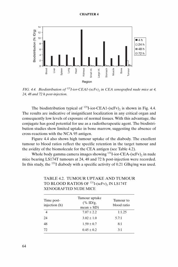
CHAPTER 4
The biodistribution typical of 125I-ior-CEA1-(scFv)2 is shown in Fig. 4.4. The results are indicative of insignificant localization in any critical organ and consequently low levels of exposure of normal tissues. With this advantage, the conjugate has good potential for use as a radiotherapeutic agent. The biodistri-bution studies show limited uptake in bone marrow, suggesting the absence of cross-reactions with the NCA 95 antigen.
Figure 4.4 also shows high tumour uptake of the diabody. The excellent tumour to blood ratios reflect the specific retention in the target tumour and the avidity of the biomolecule for the CEA antigen (see Table 4.2).
Whole body gamma camera images showing 131I-ior-CEA-(scFv)2 in nude mice bearing LS174T tumours at 24, 48 and 72 h post-injection were recorded. In this study, the 131I diabody with a specific activity of 0.21 GBq/mg was used.
TABLE 4.2. TUMOUR UPTAKE AND TUMOUR TO BLOOD RATIOS OF 131I-(scFv)2 IN LS174T XENOGRAFTED NUDE MICE
Time post-injection (h)
Tumour uptake(% ID/g,
mean ± SD)
Tumour toblood ratio
4 7.07 ± 2.2 1:1.25
24 3.02 ± 1.0 5.7:1
48 1.59 ± 0.7 8:1
72 0.45 ± 0.2 3:1
0
2
4
6
8
10
12
14
Region
4 h24 h48 h72 h
Blo
od
Live
r
Sp
leen
Lung
Hea
rt
Kid
neys
Sm
all i
nt.
Larg
e in
t.
Sto
mac
h
Mus
cle
Bon
e
Tum
our
Bio
dis
trib
utio
n (%
ID/g
)
FIG. 4.4. Biodistribution of 125I-ior-CEA1-(scFv)2 in CEA xenografted nude mice at 4, 24, 48 and 72 h post-injection.
64

PIMENTEL et al.
In Fig. 4.5, a gamma camera image of a mouse at 24 h post-injection showing the localization in the tumour demonstrates the potential of the new radioiodi-nated construct to recognize a tumour with a mass of 200 mg. An extremely hot point corresponding to the thyroid gland can also be observed. After the animal was dissected, the value obtained for the thyroid was 260.4 ± 32% ID/g (7% ID per organ). In the images recorded at 48 and 72 h post-injection, only the thyroid gland was visible.
The high uptake in the thyroid gland can be attributed to the presence of free 131I resulting from the instability of the radiolabelled preparation. This was observable in in vitro assays at high specific activities and in the in vivo pharmacokinetic pattern or metabolism. Other authors have also reported this limitation; for example, Tayan and Senekowitsch [4.23] reported a maximum accumulation in the thyroid of 1045.2% ID/g at 6 h post-injection of a radio-iodinated derivative of the epidermal growth factor. Suitable blocking of the uptake in the thyroid by pre-treatment with Lugol or a saturated solution of potassium iodide 24 h before the infusion [4.24] is a possible method to circumvent the problem and has been used for such radiotherapeutic agents.
4.3.2. ior-P1394 and DOTATATE
Radiochemical purity was ascertained by means of a combination of various chromatographic techniques. The highest labelling efficiency for the ior-P1394 labelled with 188Re (~95%) was achieved by adding 0.4μM SnF2 and heating for 2 h in the presence of citric acid. In the case of DOTATATE labelled with 90Y, a yield of around 90% was achieved. However, the corre-sponding evaluations of the agents for use in targeted therapy were carried out with the isotopes 125I and 131I.
The radioiodination yield was 90–95% for both peptides. Both were purified using reversed phase chromatography to remove the hydrophilic
FIG. 4.5. Whole body gamma camera images showing 131I-ior-CEA1-sc(Fv)2 in a mouse bearing an LS174T tumour at 24, 48 and 72 h post-injection.
65

CHAPTER 4
species in the form of non-peptide bound radioiodine and chloramine T. Size exclusion FPLC confirmed the high labelling efficiencies obtained.
The stability studies of the radioconjugates in buffer or saline and in human serum were carried out by paper chromatography and ITLC. SepPak purified 125I and 131I labelled peptides were analysed at various time intervals from 0 to 72 h post-labelling at room temperature. The results in saline showed high percentages of activity associated with the peptides, suggesting high stability of the labelled molecules under the experimental conditions. In this regard, 131I-DOTATATE was an exception, as it showed greater than 90% dissociation at 24 h. This dissociation could possibly be attributed to radiolytic damage of the peptide induced by the high beta energy of 131I during radio-iodination and purification, as reported previously by Breeman [4.25]. Incubation of the 125I labelled peptides in human serum for 24 h at 37oC led to 22.5% and 80% dissociation for ior-P1394 and DOTATATE, respectively.
4.3.3. Total binding and internalization
The total binding of 125I-ior-P1394 and 125I-DOTATATE to the AR42J tumour cells at different incubation times and the corresponding percentages of internalization after the acid treatment are shown in Fig. 4.6. The ior-P1394 showed high values of binding and internalization.
The binding affinity of both labelled peptides was tested by means of receptor binding assays. The competition binding curves of 125I-ior-P1394 and 125I-DOTATATE using rat brain cortex membranes and AR42J pancreatic tumour cells were assessed in the presence of cold peptide. The concentration of unlabelled drug that blocks half the specific binding (IC50) was determined (Fig. 4.7). For 125I-ior-P1394, the result showed a low equilibrium dissociation constant of the order of 10–12M, which is 100 times lower than the value
0
10
20
30
40
50
60
70
1 2 3 4
Time (h)
%
TB
int50.1
57.6 55.861.2
52.0 50.856.9
46.3
TB
Int50.1
57.6 55.861.2
0
5
10
15
20
1 2 3 4
Time (h)
%
TB
Int
14.438.9
25.6
38.2
9.0 9.3
13.2
8.6TB
Int
14.438.9
25.6
38.2
(a) (b)
FIG. 4.6. Total binding and internalization of (a) 125I-ior-P1394 and (b) 125I-DOTATATE in AR42J pancreatic tumour cells; TB = total binding, Int = internalization.
66

PIMENTEL et al.
reported for other somatostatin analogues [4.21–4.28], while 125I-DOTATATE displayed a value of 1.4 × 10⁻⁸M. The in vitro experiments using ior-P1394 were repeated several times with high and low receptor density, yielding the same results. No explanation could be provided for this behaviour. However, this result was indicative of a high affinity for the receptors.
The saturation binding of the labelled peptides was studied using increasing concentrations of the A431 cell line. The maximum specific binding of the radiolabelled conjugate was determined from the specific binding versus cell concentration curve (Fig. 4.8). While for 125I-ior-P1394 the findings reveal a maximum binding (immunoreactivity) of 81.4%, for 125I-DOTATATE the value achieved was approximately 1%.
02468
1012141618
1.E-13 1.E-11 1.E-09 1.E-07 1.E-05 1.E-03
[M]
0.0
0.2
0.4
0.6
0.8
1.0
1.2
1.E-16 1.E-14 1.E-12 1.E-10 1.E-08
log[M]
B/T
B/T
(a) (b)
FIG. 4.7. Competition binding curves of (a) 125I-ior-P1394 and (b) 125I-DOTATATE using rat brain cortex membranes and AR42J pancreatic tumour cells, respectively, in the presence of cold peptide.
y = 140673x + 0.8142R2 = 0.9543
0
2
4
6
8
10
12
0.E+00 2.E-05 4.E-05 6.E-05 8.E-05
[C]-1
T/B
FIG. 4.8. Double inverse plot of specific binding over total applied radioactivity as a function of increasing cell concentration (C) using the A431 cell line.
67

CHAPTER 4
Scatchard analysis was used to calculate the association constant (Ka) and the number of antigen binding sites per cell (Fig. 4.9). The association constant can be derived from the slope of the line. The affinity of ior-P1394 was found to be higher than that of DOTATATE. For both peptides, the affinity calculated by Scatchard analysis was of the same order as that obtained by the competition binding assay. The Kd of 125I-ior-P1394 with its receptor in brain membrane cells was 11.7 × 10⁻¹²M, and the corresponding value for 125I-DOTATATE was 3 × 10⁻⁸M.
The biodistribution studies can also confirm the biological activity retained by the labelled peptide and at the same time give very useful and necessary information for further clinical applications.
The results obtained showed that the distribution profile of 125I-DOTATATE and its elimination pathways in rats are similar to those of synthetic somatostatin analogues (Fig. 4.10). The low activity levels detected in the blood and in most of the organs suggest the rapid clearance of 125I-DOTATATE. This behaviour rules out the possibility of the labelled peptide’s binding to circulating plasma proteins; such binding could increase the time required for clearance of the tracer from the observed time to several hours. The radioactivity levels detected in the liver are related to the metabolism of the hepatocytes and are similar to those reported in the literature for this type of biomolecule [4.29]. Significant differences are observed between the distribution patterns of the control and problem groups at 24 h, especially in tissues where the receptor expression is positive, for example, in adrenal, pituitary and pancreatic tissues. Although the in vitro results revealed a relatively low affinity of the labelled compound for receptor binding compared with that of ior-P1394, the in vivo biodistribution pattern
y = -3E+08x + 0.1904
0.00
0.05
0.10
0.15
0.20
0 1E-10 2E-10 3E-10
B
y = -0.0855Ln(x) + 0.4983
R2 = 0.9678
0.0
0.1
0.2
0.3
0.4
0.5
0.6
1 10 100 1000
B (pM)
(a) (b)
B/F
B/F
FIG. 4.9. Scatchard analysis for (a) 125I-ior-P1394 and (b) 125I-DOTATATE: specific binding (B) versus the ratio of specific binding to the concentration of free radioligand (B:F).
68

PIMENTEL et al.
0.00
1.00
2.00
3.00
4.00
5.00
Region
Control
Problem
0
1
2
3
4
5
Problem
0
1
2
3
4
5
Control
Problem
0.00
ControlProblem
ControlProblem
ControlProblem
(a)
(b)
(c)
Bra
in
Pitu
itary
Ad
rena
ls
Hea
rt
Lung
Live
r
Sp
leen
Pan
crea
s
S. I
nt.
L. In
t.
Mus
cle
Fem
ur
Kid
neys
Sto
mac
h
Blo
od
% ID
/g%
ID/g
Region
Bra
in
Pitu
itary
Ad
rena
ls
Hea
rt
Lung
Live
r
Sp
leen
Pan
crea
s
S. I
nt.
L. In
t.
Mus
cle
Fem
ur
Kid
neys
Sto
mac
h
Blo
od
% ID
/g
Region
Bra
in
Pitu
itary
Ad
rena
ls
Hea
rt
Lung
Live
r
Sp
leen
Pan
crea
s
S. I
nt.
L. In
t.
Mus
cle
Fem
ur
Kid
neys
Sto
mac
h
Blo
od
FIG. 4.10. Biodistribution patterns in healthy Wistar rats injected with 100 µL of cold octreotide (Sandostatin, Novartis) 15 min prior to injection with 125I-DOTATATE (control group) and in untreated rats (problem group) at (a) 30 min, (b) 4 h and (c) 24 h post-injection of 125I-DOTATATE.
69

CHAPTER 4
confirms a binding affinity of labelled DOTATATE for the somatostatin receptors under physiological conditions.
4.4. CONCLUSION
The divalent scFv of the MoAb ior-CEA1 was constructed to improve the fragment’s tumour targeting properties. This construct was shown to have better functional affinity than the previous variant. The biodistribution studies in xenografted mice demonstrated that the construct exhibited high tumour accumulation with low uptake by normal organs and no cross-reactions. These properties could be exploited to develop the ior-CEA1 dimer scFv construct as a potential radiotherapeutic agent. The most favourable result for the radio-conjugates under study was found with radiolabelled ior-P1394, which exhibited excellent binding properties for its receptor.
The results obtained for ior-P1394 and DOTATATE from the in vitro and in vivo methods implemented as part of the CRP provide evidence supporting the use of these molecules to design potential agents for targeted therapy of somatostatin positive receptor tumours.
REFERENCES TO CHAPTER 4
[4.1] WALDHERR, C., et al., The clinical value of [90Y-DOTA]-D-Phe1-Tyr3-octre-otide (90Y-DOTATOC) in the treatment of neuroendocrine tumours: A clinical phase II study, Ann. Oncol. 12 (2001) 941–945.
[4.2] STEWART, P.M., JAMES, R.A., The future of somatostatin analogue therapy, Baillieres Best Pract. Res. Clin. Endocrinol. Metab. 13 (1999) 409–418.
[4.3] CREMONESI, M., et al., Dosimetry in radionuclide therapies with 90Y-conju-gates: The IEO experience, Q. J. Nucl. Med. 44 (2000) 325–332.
[4.4] OLIVA, J., et al., ior-CEA1: Un nuevo anticuerpo monoclonal anti-CEA marcado con 99mTc para la inmunogammagrafía de los tumoures colorectales: Resultados finales de un ensayo clínico fase I–II, Rev. Esp. Med. Nucl. 14 (1995) 213–221.
[4.5] OLIVA, J., et al., Detection of an unknown cerebral metastasis using radio-immunoscintigraphy with the monoclonal antibody ior-EGF-R3-Tc-99m, Rev. Esp. Med. Nucl. 16 (1997) 321–323.
[4.6] OLIVA, J., et al., Ensayo piloto con el anticuerpo monoclonal ior-c5 como agente potencial de la inmunocentelleografía del cáncer colorrectal, Rev. Esp. Med. Nucl. 20 (2001) 282–288.
[4.7] PÉREZ, L., et al., A multivalent recombinant antibody fragment specific for carcinoembryonic antigen, Biotechnol. Appl. Biochem. 43 (2006) 39–48.
70
PIMENTEL et al.
[4.8] MAURER, R., MEYER, B., PTASHNE, M., Gene regulation at the right operator (OR) of bacteriophage lambda. I. OR3 and autogenous negative control by repressor, J. Mol. Biol. 139 (1980) 147–161.
[4.9] HOUGHTEN, R.A., General methods for the rapid solid-phase synthesis of large numbers of peptides: Specificity of antigen-antibody interaction at the level of individual amino acids, Proc. Natl. Acad. Sci. U.S.A. 82 (1985) 5131–5135.
[4.10] SARIN, V.K., KENT, S.B.H., TAM, J.P., MERRIFIELD, R.B., Quantitative monitoring of solid-phase peptide synthesis by the ninhydrin reaction, Anal. Biochem. 117 (1981) 147–157.
[4.11] HOUGHTEN, R.A., BRAY, M.K., DEGRAW, S.T., KIRBY, C.J., Simplified procedure for carrying out simultaneous multiple hydrogen fluoride cleavages of protected peptide resins, Int. J. Pept. Protein Res. 27 (1986) 673–678.
[4.12] ANDREU, D., et al., “Formation of disulfide bonds in synthetic peptides and proteins”, Methods in Molecular Biology, Vol. 35, Peptide Synthesis Protocols (PENNINGTON, M.W., DUNN, B.M., Eds), Humana Press, Totowa, NJ, USA (1994) 105–109.
[4.13] ELLMAN, G.L., Tissue sulfhydryl groups, Arch. Biochem. Biophys. 82 (1959) 70–77.
[4.14] REUBI, J.C., et al., Affinity profiles for human somatostatin receptor subtypes SST1-SST5 of somatostatin radiotracers selected for scintigraphic and radiother-apeutic use, Eur. J. Nucl. Med. 27 (2000) 273–282.
[4.15] HOFLAND, L.J., VAN KOETSVELD, P.M., WAAIJERS, M., LAMBERTS, S.W.J., Internalisation of isotope-coupled somatostatin analogues, Digestion 57 (1996) 2–6.
[4.16] LINDMO, T., BOVEN, E., CUTTITTA, F., FEDORKO, J., BUNN, P.A., Jr., Determination of the immunoreactive fraction of radiolabelled monoclonal anti-bodies by linear extrapolation to binding at infinite antigen excess, J. Immunol. Methods 72 (1984) 77–89.
[4.17] FREYRE, F.M., et al., Very high level expression of an anti-CEA single-chain Fv antibody fragment in the methylothrophic yeast Pichia pastoris, J. Biotechnol. 76 (2000) 157–163.
[4.18] PÉREZ, L., et al., Production, purification and characterization of an anti-(carci-noembryonic antigen) recombinant single-chain Fv antibody fragment, Biotechnol. Appl. Biochem. 24 (1996) 79–82.
[4.19] LIU, S., EDWARDS, S., Stabilization of 90Y-labelled DOTA-biomolecule conju-gates using gentisic acid and ascorbic acid, Bioconjug. Chem. 12 (2001) 554–558.
[4.20] JAIN, M., BATRA, S.K., Genetically engineered antibody fragments and PET imaging: A new era of radioimmunodiagnosis, J. Nucl. Med. 44 (2003) 1070–1071.
[4.21] TAHTIS, K., et al., Biodistribution properties of 111In-labelled C-functionalized trans-cyclohexyl diethyletriaminepentaacetic acid humanized 3S193 diabody and F(ab')2 constructsin: A breast carcinoma xenograft model, Clin. Cancer Res. 7 (2001) 1061–1072.
71

CHAPTER 4
[4.22] PIMENTEL, G.J., et al., Hexa histidine tag as a novel alternative for one step direct labelling of a single chain Fv antibody fragment with 99mTc, Nucl. Med. Commun. 22 (2001) 1089–1094.
[4.23] TAYAN, M., SENEKOWITSCH, R., 111In or 125I labelled epidermal growth factor for the in vivo localization of EGF receptors: In vivo stability, J. Islamic Acad. Sci. 4 (1991)123–126.
[4.24] VOSE, J.M., et. al., Multicenter phase II study of iodine-131 tositumomab for chemotherapy-relapsed/refractory low-grade and transformed low-grade B-cell non-Hodgkin’s lymphomas, J. Clin. Oncol. 18 (2000) 1316–1323.
[4.25] BREEMAN, W.A.P., Aspects of Receptors Scintigraphy with Neuropeptides. PhD Thesis, Erasmus University, Rotterdam (1995).
[4.26] DECRISTOFORO, C., MATHER, J.S., Technetium-99m somatostatin analogues: Effect of labelling methods and peptide sequence, Eur. J. Nucl. Med. 26 (1999) 869–879.
[4.27] REUBI, J.C., et al., Affinity profiles for human somatostatin receptors subtypes SST1–SST5 of somatostatin radiotracers selected for scintigraphic and radio-therapeutic use, Eur. J. Nucl. Med. 27 (2000) 273–282.
[4.28] HOFLAND, L.J., et al., Crucial role for somatostatin receptor subtype 2 in determining the uptake of [111In-DTPA-D-Phe1]octreotide in somatostatin receptor-positive organs, J. Nucl. Med. 44 (2003) 1315–1321.
[4.29] LAZNICKOVA, A., LAZNICEK, M., TREJTNAR, F., MÄCKE, H.R., MATHER, S.J., “Study of pharmacokinetics and biodistribution of radiolabelled receptor specific peptides in laboratory animals”, 99mTc Labelled Peptides for Imaging of Peripheral Receptors, IAEA-TECDOC-1214, IAEA, Vienna (2001) 41–48.
72

Chapter 5
PRECLINICAL COMPARISON OF DOTATATE LABELLED WITH DIFFERENT RADIONUCLIDES
M. LAZNICEK, A. LAZNICKOVA, F. TREJTNAR, L. MELICHAROVA, J. CIHLO, M. PETRIKFaculty of Pharmacy, Charles University, Hradec Kralove, Czech Republic
Abstract
Somatostatin receptor scintigraphy is a valuable method for visualizing somato-statin receptor positive tumours and metastases, particularly those of neuroendocrine origin. Radiolabelled DOTA-Tyr3-octreotate (DOTATATE) is a new somatostatin radiolabelled peptide analogue with great potential for oncological application. The goal of the study was to evaluate the effect that radiolabelling DOTATATE with 90Y, 111In, 125I and 153Sm has on its biodistribution profile and elimination characteristics in rats. All peptides under study exhibited similar biodistribution profiles in rats at the initial time intervals. Relatively rapid clearance of radioactivity from the blood and most organs and tissues was observed. High uptake in those organs with a high density of somatostatin receptors (the pancreas and adrenal glands) and in the main elimination organ (the kidney) was found. Over longer time intervals (24 and 48 h post-injection), radioactivity in the pancreas, adrenal glands and kidneys decreased significantly in the case of the radioiodinated peptide, whereas high levels of radioactivity retention in these organs were observed with the other radiolabelled peptides at the same time inter-vals. Unlike with the other peptides, the degradation products of radioiodinated DOTATATE were externalized from the kidneys and from those organs with a high density of somatostatin receptors. Whereas somatostatin receptor mediated uptake of radioactivity in the pancreas and adrenal glands was comparable for all the complexes labelled with metallic radionuclides, the kidney uptake decreased in the following order: 111In-DOTATATE > 153Sm-DOTATATE > 90Y-DOTATATE. Differences in the coordi-nation geometries of these complexes resulted in small structural modifications of the peptide under study. These modifications influenced the distribution profiles of the radiolabelled peptides, which were reflected in altered kidney uptake.
73

CHAPTER 5
5.1. INTRODUCTION
Somatostatin receptor scintigraphy is a well established imaging procedure for the diagnosis of a variety of neuroendocrine and non-endocrine tumours. The method is based on the specific binding of a peptide with the somatostatin receptors overexpressed in tumour cells [5.1]. To date, five different somatostatin receptor subtypes have been characterized and cloned [5.2]. The majority of tumours express mainly somatostatin receptor subtype 2, although the other subtypes may be present in some human tumours. The radiolabelled peptide analogue that can be considered the gold standard for the visualization of human endocrine tumours and their metastasis is 111In-DTPA-octreotide (OctreoScan), which is available in the form of a commercial kit. In the past few years, considerable progress has been made in this area, and a number of somatostatin radioligands have been suggested as favourable alter-natives to OctreoScan. Recently, it has been shown that changing the C-terminal of octreotide from an alcoholic to a carboxylic acid group signifi-cantly increases the affinity of the peptide to somatostatin receptor subtype 2. To this end, different somatostatin derivatives have recently been introduced, with DOTA-Tyr3-octreotate (DOTATATE) being a very promising one [5.3]. The macrocyclic chelator DOTA is able to bind with different radionuclides (e.g. radioyttrium, radioindium, radiolanthanides), while tyrosine in the peptide structure allows radiolabelling with halogens. In this study, the effect that radiolabelling DOTATATE with 90Y, 111In, 125I and 153Sm has on its biodis-tribution profile and the elimination pathway in rats was determined.
5.2. MATERIALS AND METHODS
5.2.1. Radiochemicals
Carrier free 90Y in the form of yttrium chloride was obtained from Perkin-Elmer LAS (Belgium). Carrier free 111In in the form of indium chloride and 125I in the form of sodium iodide were obtained from Amersham (UK). Samarium-153 in the form of samarium chloride with a specific activity of 10–15 GBq/mg was obtained from the Nuclear Research Institute Řež plc (Czech Republic).
74

LAZNICEK et al.
5.2.2. Radiolabelling of DOTATATE
5.2.2.1. Labelling with 111In and 90Y
Radiolabelling was accomplished by adding approximately 0.5 mCi (18.5 MBq) of 90YCl3 or 111InCl3 in 10–100 μL of 40mM HCl to 200 μL of 0.4M acetate buffer at pH5 containing 0.24M gentisic acid and 10 μg of DOTATATE. After incubation at 90–95°C for 25 min, the quality of the product was tested by gradient elution high performance liquid chromatography (HPLC).
5.2.2.2. Labelling with 125I
For labelling with 125In, 5 μg of peptide in 5 μL of 0.05M acetate buffer at pH5 and 4 μL of Na125I (corresponding to 0.4 mCi) were added to 42.5 μL of 0.05M phosphate buffer at pH7.4. Thereafter, the mixture was treated with 2.5 μg of chloramine T in 2.5 μL of the same 0.05M phosphate buffer at pH7.4. This mixture was allowed to react for 1 min at ambient temperature and, after dilution with 40 μL of absolute ethanol, was subjected to HPLC analysis.
5.2.2.3. Labelling with 153Sm
For labelling with 153Sm, 10 μL of peptide solution (at a concentration of 1μg/1 μL) and no more than 2 μL (4 MBq) of SmCl3 solution in 0.05M HCl were added to 200 μL of 0.4M acetate buffer at pH5 containing 0.24M gentisic acid. The mixture was maintained at 90–95°C for 25 min. For labelling with samarium, it is necessary that the concentration ratio of c Ligand to c Sm exceed 2.5.
5.2.3. Quality control
5.2.3.1. HPLC analysis of 111In- and 90Y-DOTATATE
Analysis using HPLC was performed on a column of LiChroCART 125-4 HPLC Cartridge Purospher RP18e, 5 mm (Merck), using a Pharmacia LKB system linked to a Gradient Master GP 962 (Institute of Organic Chemistry and Biochemistry (UOCHB) Prague) and operated with a UV detector and a radioactivity monitoring analyser. Mobile phases A (0.1% TFA) and B (CH3CN) were used with the following gradient: 0–5 min 0% B, 5–25 min 0–30% B, 25–30 min 30% B, 30–35 min 30–100% B, 35–40 min 100% B. The flow rate was maintained at 0.5 mL/min. The samples for HPLC were prepared by treating 30 µL of mobile phase A with 10 µL of 10–3M DTPA and with 2 µL of
75

CHAPTER 5
the labelled peptide solution. An example of the HPLC analysis of 111In-DOTATATE is shown in Fig. 5.1
5.2.3.2. HPLC pattern of 125I-DOTATATE
Purification of 125I-DOTATATE was carried out by HPLC using the Pharmacia LKB system described above and a column of LiChroCART RP C18 (250 mm × 4.5 mm), 10 µm. The flow rate was maintained at 1 mL/min. Mobile phases A (aqueous 0.1M NaCl) and B (95% ethanol) were used with the following gradient: 0–10 min 0% B, 10–20 min 0–60% B, 20–30 min 60% B, 30–35 min 60–100% B, 35–40 min 100% B and 40–50 min 100–0% B. For biological experiments, a fraction corresponding to a retention time of 25–26.5 min was collected, the mobile phase was evaporated and the residue was diluted by addition of 5 mL of saline. An example of HPLC analysis of the product is shown in Fig. 5.2.
5.2.3.3. HPLC analysis of 153Sm-DOTATATE
HPLC analysis was carried out using a LiChroCART RP C18 column (250 mm × 4 mm), 5 μm (Merck). Mobile phases A (0.1% TFA) and B (CH3CN) were used with the following gradient: 0–10 min 0% B, 10–20 min 0–60% B, 20–30 min 60% B, 30–35 min 60–100% B, 35–40 min 100% B, 40–45 min 100–0%. The flow rate was maintained at 1 mL/min. An example of a typical HPLC analysis of the product is shown in Fig. 5.3.
5.2.4. Animal experiments
All animal experiments were approved by the Ethics Commission of the Faculty of Pharmacy of Charles University.
160 000
80 000
00 5 10 15 20 25 30 35 40
Time (min)
Act
ivity
FIG. 5.1. HPLC analysis of 111In-DOTATATE.
76
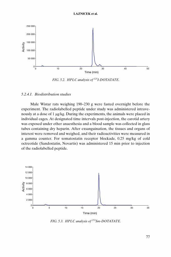
LAZNICEK et al.
5.2.4.1. Biodistribution studies
Male Wistar rats weighing 190–230 g were fasted overnight before the experiment. The radiolabelled peptide under study was administered intrave-nously at a dose of 1 μg/kg. During the experiments, the animals were placed in individual cages. At designated time intervals post-injection, the carotid artery was exposed under ether anaesthesia and a blood sample was collected in glass tubes containing dry heparin. After exsanguination, the tissues and organs of interest were removed and weighed, and their radioactivities were measured in a gamma counter. For somatostatin receptor blockade, 0.25 mg/kg of cold octreotide (Sandostatin, Novartis) was administered 15 min prior to injection of the radiolabelled peptide.
250 000
200 000
150 000
100 000
50 000
00 10 20 30 40 50
Time (min)
Act
ivity
FIG. 5.2. HPLC analysis of 125I-DOTATATE.
14 000
12 000
10 000
8 000
6 000
4 000
2 000
00 5 10 15 20 25 30 35
Time (min)
Act
ivity
FIG. 5.3. HPLC analysis of 153Sm-DOTATATE.
77

CHAPTER 5
5.2.4.2. Elimination in rats
The radiolabelled peptide under study was administered to rats as described above. The animals were then placed in separate glass metabolic cages, allowing reliable separation of urine and solid excreta. The animals had free access to standard diet and water. Two hours after injection of the peptide, the rats’ urinary bladders were emptied manually, and the urine and faeces were collected. The procedure was repeated at 24 and 48 h post-injection.
5.2.4.3. Perfusion of rat kidney
In situ rat kidney perfusion experiments were carried out as described in Ref. [5.4]. Briefly, the perfusion medium used was Krebs–Henseleit buffer at pH7.4 containing glucose (5.6 mmol/L), 5.5% bovine serum albumin (fraction V, Sigma), 5–6% washed rat erythrocytes and different amino acids. After cannulation of the ureter and the renal vein and artery, the right kidney was perfused via the renal artery at a constant arterial pressure of 14.5 kPa at 37°C. After an equilibration period of 30–35 min, the agent was added and the perfusion continued for 60 min. Urine samples were collected every 10 min, and midpoint samples of the perfusate were also obtained. Inulin was used as the standard for the measurement of the glomerular filtration rate. Elimination parameters of labelled peptides in the perfused rat kidney were characterized by the values of total renal clearance (CLR) and free fraction of the peptide in the perfusate (Fu). These values were compared with the glomerular filtration rate (GFR).
5.2.4.4. Perfusion of rat liver
An in situ perfused rat liver preparation was employed. After midline incision of the animal, the bile duct, the portal vein and the inferior vena cava were cannulated and ligated. Krebs–Henseleit buffer at pH7.4 containing glucose was used in a single pass to release the blood from the liver. The liver perfusion medium consisted of heparinized Krebs–Henseleit buffer at pH7.4 containing 10mM glucose, 4% bovine serum albumin and 10% (vol./vol.) bovine erythrocytes oxygenated with 95% O2 and 5% CO2. After a 15 min equilibration period, the flow of the perfusate was maintained at 25 mL/min and the peptide under study was added to a 150 mL reservoir. The samples of the input and outflow perfusate were removed at 10 min intervals in the middle of the 10 min periods of bile collection. The experiments were performed for 90 min after the peptide loading. Elimination rates of labelled peptides in the perfused rat liver were characterized by the values of bile clearance.
78

LAZNICEK et al.
5.2.4.5. Internalization experiments
5.2.4.5.1. Rat pancreatic carcinoma cells
Rat pancreatic carcinoma AR42J cells (ECACC, UK) were grown in RPMI-1640 medium (supplemented with 2mM glutamine and 10% foetal calf serum) in air containing 5% CO2 at 37°C [5.5]. Subculturing was performed employing a trypsin–EDTA solution. Cells were incubated at 37°C in triplicate for the designated time periods. Cellular uptake was stopped by removing the medium, and the cells were washed two times with ice-cold phosphate buffer solution. Thereafter, the cells were incubated twice at ambient temperature in acid wash buffer (50mM glycine buffer at pH2.8, 0.1M NaCl) for 5 min. The supernatant (containing the receptor bound radioligand fraction) was collected and the cells were rinsed with phosphate buffer solution. Cells were lysed by treatment with 1M NaOH, and the radioactivity associated with the cell corre-sponding to the internalized radioligand fraction was estimated.
5.2.4.5.2. Opossum kidney cells
Opossum kidney is a well established cell line known to simulate processes in the renal proximal tubule. This cell line is commonly used for uptake studies. In the study reported here, the opossum kidney cells were maintained in antibiotic-free MEM supplemented with 10% foetal calf serum, 2 mmol/L L-glutamine and 1% non-essential amino acids, and were subcultured twice a week at a ratio of 1:5. Uptake experiments were performed with 111In-DOTATATE in 25 cm2 culture flasks. The cells were seeded at a density of 4 × 10⁴ cells/cm2 and were used 6–7 d post-plating. The resulting confluent monolayers were washed twice with Ringer’s solution and incubated for the designated intervals (15 and 30 min, and 1, 2 and 3 h) with 1 nmol/L 111In-DOTATATE in Ringer’s solution. Incubation was stopped by removing the uptake medium and rinsing the monolayers with ice-cold Ringer’s solution five times. The cells were disintegrated by RIPA buffer. The intracellular radio-activity was normalized to the cell protein content by the BCA method.
5.3. RESULTS AND DISCUSSION
High performance liquid chromatography analysis of DOTATATE labelled with the radionuclides under study showed a single peak for each agent. In all cases, the labelling efficiency (radiochemical purity) was found to be greater than 98%. The distribution of radioactivity in selected organs of rats
79

CHAPTER 5
at different times post-injection of the radiolabelled peptides is depicted in Fig. 5.4.
In comparative studies of the radiolabelled agents, the radioactivity in blood and in most organs decreased for up to 2 h post-injection. While high uptake and long term retention of radioactivity were found in the kidney for DOTATATE labelled with metallic radionuclides, in the case of 125I-DOTATATE the radioactivity in the kidney decreased significantly with time. In other organs, not shown in Fig. 5.4 (i.e. the heart, spleen, testes, thyroid and brain), less than 1% of the administered radioactivity was found during the entire course of the experiments.
The radioactivity concentrations in selected tissues are shown in Fig. 5.5. The decrease of radioactivity in muscle over time was in agreement with the relatively rapid decrease of radioactivity in the blood. High uptake and long
5 min 1 h 2 h 24 h 48 h
Liver
Blood (total)Kidney
GIT
0
2
4
6
8
10
12
14
16
5 min 1 h 2 h 24 h 48 h
Liver
Blood (total)Kidney
GIT
02
4
6
8
10
12
14
16
5 min 1 h 2 h 24 h 48 h
Liver
Blood (total)Kidney
GIT
0
2
4
6
8
10
12
14
16
5 min 1 h 2 h 24 h 48 h
Liver
Blood (total)Kidney
GIT
02
4
6
8
10
12
14
16
(a) (b)
(c) (d)
% ID
/org
an
Time post-injection Time post-injection
Time post-injection Time post-injection
% ID
/org
an%
ID/o
rgan
% ID
/org
an
FIG. 5.4. Distribution of radioactivity in selected organs of rats post-injection of (a) 90Y-DOTATATE, (b) 111In-DOTATATE, (c) 125I-DOTATATE and (d) 153Sm-DOTATATE (GIT = gastrointestinal tract).
80

LAZNICEK et al.
term retention of radioactivity were found in the main elimination organ (the kidney) and in those organs with a high density of somatostatin receptors (the adrenal glands and pancreas). In inter-drug comparisons, biodistribution was similar for all agents in the initial time intervals. Over longer time intervals (24 and 48 h post-injection), however, radioactivity in the pancreas, adrenal glands and kidneys decreased significantly for the peptide labelled with radioiodine, whereas it remained high in these organs for the other radiolabelled agents at the same time intervals. In contrast to the other peptides, the degradation products of the radioiodinated DOTATATE were externalized from the kidney and those organs with high densities of somatostatin receptors. Whereas somatostatin receptor mediated uptake of radioactivity in the pancreas and adrenal glands was comparable for all of the complexes labelled with metallic radionuclides, the kidney uptake of radioactivity decreased in the following
5 min 1 h 2 h 24 h 48 h
Blood
MusclePancreas
Adrenals
0
10
20
30
40
50
5 min 1 h 2 h 24 h 48 h
Blood
MusclePancreas
Adrenals
0
10
20
30
40
50
5 min 1 h 2 h 24 h 48 h
Blood
MusclePancreas
Adrenals
0
10
20
30
40
50
5 min 1 h 2 h 24 h 48 h
Blood
Muscle
PancreasAdrenals
0
10
20
30
40
50
(a) (b)
(c) (d)
% ID
/1%
bod
y w
eigh
t
Time post-injection
% ID
/1%
bod
y w
eigh
t
Time post-injection
% ID
/1%
bod
y w
eigh
t
Time post-injection
% ID
/1%
bod
y w
eigh
t
Time post-injection
FIG. 5.5. Distribution of radioactivity in selected tissues of rats after injection of (a) 90Y-DOTATATE, (b) 111In-DOTATATE, (c) 125I-DOTATATE and (d) 153Sm-DOTATATE.
81

CHAPTER 5
order: 111In-DOTATATE > 153Sm-DOTATATE > 90Y-DOTATATE. The mechanism of renal uptake and retention of peptides involves glomerular filtration and subsequent partial reabsorption in the proximal tubular cells, where the peptides are internalized by receptor mediated endocytosis. The peptides were able to be transferred to lysosomes and digested by proteolytic enzymes. The resulting breakdown products remained in the lysosomes of the renal cells for a long period post-injection [5.6].
Cumulative excretion of radioactivity in the urine and faeces within 48 h of injection of the agents under study is shown in Fig. 5.6. The main
0
20
40
60
80
100
Faeces
Urine
0
20
40
60
80
100
Faeces
Urine
0
20
40
60
80
100
Faeces
Urine
0
20
40
60
80
100
Faeces
2 24 48
Urine
2 24 48
2 24 482 24 48
Time post-injection (h)
Time post-injection (h)
Time post-injection (h)
Time post-injection (h)
% ID
% ID
% ID
% ID
(a) (b)
(c) (d)
FIG. 5.6. Cumulative excretion of radioactivity in rats after injection of (a) 90Y-DOTATATE, (b) 111In-DOTATATE, (c) 125I-DOTATATE and (d) 153Sm-DOTATATE (in percentage of injected dose).
82
LAZNICEK et al.
elimination pathway for all peptides under study was urinary excretion. Most of the radioactivity eliminated by urine was excreted during the first 2 h post-injection.
While 90Y-DOTATATE, 111In-DOTATATE and 153Sm-DOTATATE were eliminated via urine in almost unchanged forms, in the case of 125I-DOTATATE a significant portion of the polar metabolites (degradation products) was observable in the urine 24 h post-injection (Fig. 5.7). Excretion of radioactivity via faeces was lower than that via urine, but the portion of radioactivity eliminated by this pathway was significant (12–20%) for all peptides under study. The chemical form in which the radioactivity was present in the faeces was not determined.
The renal clearance parameters of the peptides in the perfused rat kidney are presented in Table 5.1. Renal clearances of the peptides corrected for protein binding in the perfusate approached the glomerular filtration rate. Thus, the analysis of elimination mechanisms in perfused rat kidney confirmed that all agents under study were eliminated in the kidney, predominantly by glomerular filtration.
While bile clearance of the peptide labelled with 90Y, 111In and 153Sm in the perfused rat liver was negligible, that of DOTATATE labelled with 125I was markedly higher owing to its higher lipophilicity. Nevertheless, bile clearances of all peptides under study were very low compared with the perfusate flow. The results are summarized in Table 5.2.
Internalization properties of 153Sm-DOTATATE studied in AR42J rat pancreatic carcinoma cells showed that the agent is rapidly internalized into the cells. The radioactivity bound to receptors was found to reach a plateau after a 2 min incubation, with a slight increase in the internalized radioactivity over time (Fig. 5.8).
The possibility of using receptor specific peptides for therapy of certain cancer diseases is limited by nephrotoxicity caused by the uptake and retention
30 000
25 000
20 000
15 000
10 000
5000
0
80 000
60 000
40 000
20 000
0
10 000
8 000
6 000
4 000
2 000
0
Act
ivity
0 5 10 15 20 25 30 35 40
Time (min)
(a) (b) (c)
Time (min) Time (min)
0 5 10 15 20 25 30 35 40 0 5 10 15 20 25 30 35 40
Act
ivity
Act
ivity
FIG. 5.7. HPLC analysis of urine after injection of 125I-DOTATATE in rats: (a) standard, (b) 2 h post-injection and (c) 24 h post-injection.
83

CHAPTER 5
TABLE 5.1. EXCRETION PARAMETERS OF RADIOLABELLED PEPTIDES IN PERFUSED RAT KIDNEY
90Y-DOTATATE
111In-DOTATATE
125I-DOTATATE
153Sm-DOTATATE
Renal clearance (mL⋅min–1⋅g–1)
0.591 ± 0.065 0.585 ± 0.041 0.403 ± 0.060 0.778 ± 0.126
CLR/GFR × Fu 1.05 ± 0.04 1.12 ± 0.09 0.98 ± 0.04 1.06 ± 0.19
Note: CLR: renal clearance of agent; GFR: glomerular filtration rate; Fu: free fraction of peptide in perfusate.
TABLE 5.2. EXCRETION PARAMETERS OF RADIOLABELLED PEPTIDES IN PERFUSED RAT LIVER
90Y-DOTATATE
111In-DOTATATE
125I -DOTATATE
153Sm-DOTATATE
Bile clearance (mL/min)
0.0007 ± 0.0002 0.0008 ± 0.0003 0.0324 ± 0.0087 0.0009 ± 0.0003
Perfusate flow (mL/min)
25.0 25.0 25.0 25.0
0
1000
2000
3000
4000
0 10 20 30 40 50 60
Time (min)
Receptor activity
Internalized activity
Act
ivity
FIG. 5.8. Internalization of 153Sm-DOTATATE into AR42J rat pancreatic carcinoma cells as a function of time.
84

LAZNICEK et al.
of these substances in the kidney. According to recent experiments reported in the scientific literature, there are two mechanisms — receptor mediated endocytosis via megalin/cubilin and fluid phase endocytosis — that contribute almost equally to the uptake of radiolabelled somatostatin analogues by renal proximal tubular cells [5.7]. The internalization of 111In-DOTATATE into opossum kidney cells found in this study is shown in Fig. 5.9. The rate of inter-nalization was relatively slow, and internalized radioactivity increased over the course of the entire experiment.
5.4. CONCLUSION
DOTATATE can be efficiently labelled with 90Y, 111In, 125I and 153Sm with a high radiochemical yield. The biodistribution profiles of DOTATATE labelled with these different radionuclides in rats were generally similar, but some differences in the biological behaviour of the complexes were observed. All agents under study were internalized into the kidney and into those organs with a high density of somatostatin receptors (the adrenal glands and pancreas). By following the variation of the radioactivity uptake in these organs over time, it was found that DOTATATE labelled with 90Y, 111In and 153Sm exhibited slow kinetics and therefore high accumulation and long retention of radioactivity. In contrast, the degradation products of radioiodi-nated DOTATATE were externalized both from the kidney and from those organs with a high density of somatostatin receptors; moreover, the radio-activity uptake in these organs decreased significantly over longer time intervals. While somatostatin receptor mediated uptake of radioactivity in the
1.687
2.087
2.367
1.2051.162
0.0
0.5
1.0
1.5
2.0
2.5
0 50 100 150 200
Time (min)
111 In
-DO
TATA
TEfm
ol/m
g ce
ll p
rote
in
FIG. 5.9. Internalization of 111In-DOTATATE into opossum kidney cells as a function of time.
85

CHAPTER 5
pancreas and adrenal glands was comparable for all the complexes labelled with metallic radionuclides, the kidney uptake of radioactivity decreased in the following order: 111In-DOTATATE > 153Sm-DOTATATE > 90Y-DOTATATE. This implies that the different coordination geometries of these complexes influenced the distribution profiles of the radiolabelled peptides, which was reflected in their altered kidney uptake.
ACKNOWLEDGEMENTS
The authors wish to thank J. Hoderova, E. Teichmanova and I. Filipova for excellent technical assistance. The study originated within the IAEA’s CRP on comparative evaluation of therapeutic radiopharmaceuticals, Research Agreement No. 12125, and was financially supported by the grant agency of Charles University — grant Nos 146/2004, 405/2004 and 109/2005.
REFERENCES TO CHAPTER 5
[5.1] KRENNING, E.P., Somatostatin receptor scintigraphy with [111In-DTPA-D-Phe1]- and [123I-Tyr3]-octreotide: The Rotterdam experience with more than 1000 patients, Eur. J. Nucl. Med. 20 (1993) 716–731.
[5.2] REISINE, T., BELL, G.I., Molecular biology of somatostatin receptors, Endocr. Rev. 16 (1995) 427–442.
[5.3] REUBI, J.C., et al., Affinity profiles for human somatostatin receptor subtypes SST1-SST5 of somatostatin radiotracers selected for scintigraphic and radiother-apeutic use, Eur. J. Nucl. Med. 27 (2000) 273–282.
[5.4] TREJTNAR, F., LAZNICEK, M., LAZNICKOVA, A., Pharmacokinetics of 99mTc-DTPMP as compared to 99mTc-MDP in the rat, Pharm. Pharmacol. Lett. 8 (1998) 112–114.
[5.5] BEHR, T.M., GOLDENBERG, D.M., BECKER, W., Reducing the renal uptake of radiolabelled antibody fragments and peptides for diagnosis and therapy: Present status, future prospects and limitations, Eur. J. Nucl. Med. 25(1998) 201–212.
[5.6] MAINA, T., et al., [99mTc]DemoTATE, a new 99mTc-based [Tyr3]octreotate analogue for detection of somatostatin receptor-positive tumours: Synthesis and preclinical results, Eur. J. Nucl. Med. 29 (2002) 742–753.
[5.7] BARONE, R., et al., Endocytosis of somatostatin analogue, octreotide, by the proximal tubule derived opossum kidney (OK) cell line, Kidney Int. 67 (2005) 969–976.
86

Chapter 6
EVALUATION OF PEPTIDES LABELLED WITH BETA EMITTING RADIONUCLIDES FOR RECEPTOR TARGETED RADIOTHERAPY OF MALIGNANT TUMOURS
T. MAINA*, B. NOCK*, A. NIKOLOPOULOU*, C. PETROU**, P. CORDOPATIS**
* Institute of Radioisotopes and Radiodiagnostic Products, Demokritos National Centre for Scientific Research, Athens
** Pharmacy Department, University of Patras, Patras
Greece
Abstract
The paper compares the biological behaviour of three radioiodinated [Tyr3]octre-otate (TATE) analogues — TATE, DOTATATE and Sugar-TATE — in AR42J cells and in mice, using the ligand 177Lu-DOTATATE as a reference compound. The synthesis of Sugar-TATE was performed by solid phase peptide synthesis (SPPS) techniques using a properly protected Sugar-Asp unit as a building block and following standard Fmoc/Boc protection strategies. Radioiodination with either 125I or 131I was performed using the chloramine T method, followed by HPLC purification to produce single species of radioligands. Radiolabelling of DOTATATE with 177Lu was conducted in an acidic medium and under heating of the labelling reaction mixture. The binding affinities of TATE, DOTATATE and Sugar-TATE were determined by competition binding assays in somatostatin receptor subtype 2 positive AR42J cell membranes using [125I-Tyr3]octreotide as the radioligand. The internalization properties of the radioiodinated peptides and 177Lu-DOTATATE were studied by incubation in AR42J cells at 37°C. The biodistribution of the radioiodinated peptides was studied in healthy Swiss albino mice, while that of 177Lu-DOTATATE was studied in nude mice bearing AR42J experimental tumours. The Sugar-TATE synthesized by SPPS was of high purity, as verified by electron spray mass spectra and analytical HPLC. Radiolabelling of peptide analogues with either 131I or 177Lu led to single species of radiopeptides with high yields and
87

CHAPTER 6
specific activities adequate for in vitro and in vivo studies. All peptide conjugates exhibited sub-nanomolar affinities for somatostatin receptor subtype 2, while the radio-labelled peptides showed rapid and specific internalization into AR42J cells. When injected into mice, radiopeptides labelled with 131I were able to specifically localize in organs with many somatostatin receptors, such as the pancreas and the adrenals. The 131I-Sugar-TATE showed much higher specific uptake in those organs and, similar to 131I-DOTATATE, was excreted predominantly via the kidney. In contrast, 131I-TATE showed a high percentage of hepatobiliary excretion. The reference compound, 177Lu-DOTATATE, showed significant specific uptake in both the target organs and the experimental tumour, and was excreted rapidly via the renal route. Radioactivity levels remained impressively high in the tumour and in the target organs 72 h post-injection of 177Lu-DOTATATE. In contrast, the corresponding values exhibited by the radio-peptides labelled with 131I showed a rapid decline within 24 h of injection. Owing to its significantly longer residence time in normal and cancerous tissues expressing somato-statin receptor subtype 2, 177Lu-DOTATATE remains the agent of choice for radio-nuclide therapy compared with the radiopeptides labelled with 131I, which exhibited a similar affinity and efficient localizing but failed to remain at the target site for an amount of time sufficient to yield maximum radiotherapeutic impact.
6.1. INTRODUCTION
The advent of OctreoScan ([111In-DTPA0]octreotide) for clinical use in the diagnosis and staging of neuroendocrine tumours has boosted research aimed at identifying other somatostatin analogues labelled with therapeutic radionuclides for application in the targeted radiotherapy of tumours expressing somatostatin receptors. These efforts have led to powerful new therapeutic tools in the arsenal of clinical oncology [6.1–6.6].
The replacement of Phe3 with Tyr3 in octreotide permits relatively easy labelling with radiohalogens such as 123I, 125I and 131I, which can be used for targeted diagnosis, for in vitro studies or for radiotherapy [6.7]. The replacement of Thr(ol)8 in octreotide with Thr8 in [Tyr3]octreotate results in higher selectivity and an affinity for somatostatin receptor subtype 2, as well as a higher capacity for internalization into somatostatin receptor subtype 2 positive cells. Accordingly, these analogues exhibit enhanced uptake in somato-statin receptor subtype 2 positive lesions, both in animal models and in patients, compared with the corresponding octreotide analogues [6.1–6.7].
While 131I exhibits attractive nuclear properties for targeted radiotherapy (half-life: 8.04 d; Eβ(max): 0.97 MeV (89%), 0.096 MeV (7%) and two gamma photons, 364 keV (81%) and 284 keV (6%)), wider application of 131I labelled somatostatin analogues in radiotherapy targeting somatostatin receptor subtype 2 is hampered by certain drawbacks. First, the 131I based analogues
88

MAINA et al.
show a relatively high lipophilicity and increased hepatobiliary excretion [6.8]. In addition, the prolonged intracellular retention of 131I required for high therapeutic efficacy cannot be achieved by radioiodinated peptide analogues owing to their intracellular conversion to non-residualizing 131I carrying metabolites, which are eventually transported out of the cell.
These problems can be overcome by somatostatin analogues labelled with metallic radionuclides such as 111In, 90Y, 64Cu, 67/68Ga and the radiolantha-nides. Most of these metals form very stable complexes with the universal chelator 1,4,7,10-tetraazacyclododecane-1,4,7,10-tetraacetic acid (DOTA). As a rule, DOTA derivatives are covalently coupled to the N-terminal (D)Phe residue of [Tyr3]octreotide or [Tyr3]octreotate, yielding peptide–chelator conjugates that are able to effectively complex with a host of useful therapeutic radionuclides. Of particular interest is [DOTA0,Tyr3]octreotate (DOTATATE) labelled with 177Lu, which has shown promising results in the radionuclide therapy of neuroendocrine tumours in patients. The therapeutic efficacy of 177Lu-DOTATATE is a combined effect of its favourable in vivo distribution and the nuclear properties of 177Lu (half-life: 6.71 d; Eβ(max): 0.50 MeV (79%), two favourable gamma photons 208 keV (11%) and 113 keV (6.4%)) [6.1–6.10], which make it useful for external monitoring.
Compared with 131I labelled somatostatin analogues, 177Lu-DOTATATE and most other conjugates of DOTA and peptides labelled with metallic radio-nuclides show prolonged residualization within cancer cells. Several studies have shown that the major metabolic products of metallic radionuclide labelled (M3+) peptides, such as [M3+-DOTA0](D)Phe1, are trapped within the cell for several hours or even days, causing serious cell damage and eventually cell death [6.2, 6.11, 6.12].
In this study, 131I labelled TATE is compared with 131I labelled DOTATATE and a sugar modified TATE analogue (Fig. 6.1). Both DOTA and the sugar moiety were used as pendent groups to increase the hydrophilicity of the agent for excretion of the radiotracer via the kidney and the urinary system. The addition of an N-terminal sugar moiety in TATE has been proposed by many investigators and has been shown to significantly increase the internali-zation properties of the resulting radioligands [6.13]. The in vitro affinities and biodistribution profiles of the three 131I labelled TATE analogues were directly compared in this study using identical biological models. Finally, the biological properties of 177Lu-DOTATATE were also investigated using similar methods, with the aim of revealing the advantages and/or limitations of using radioiodi-nated versus metallic radionuclide labelled somatostatin based radiopeptides.
89

CHAPTER 6
6.2. MATERIALS
The somatostatin peptide analogues Tyr3-octreotide (Tide) (Bachem, Bubendorf, Switzerland) and DOTATATE (piCHEM R&D, Austria) were provided by the IAEA. TATE and Sugar-TATE were synthesized by solid phase peptide synthesis (SPPS) techniques. Reagent grade chemicals were purchased from Aldrich or Fluka and used without further purification. Iodine-125 was purchased from Nordion (Fleurus, Belgium), while 131I was provided by POLATOM (Warsaw, Poland). Lutetium-177 was obtained from IDB Holland BV (Netherlands). Analyses based on HPLC were performed on a Waters chromatograph system with a Waters model 600 solvent delivery system coupled to a PDA–UV detector, along with a GABI gamma detector from Raytest (Munich, Germany). The conditions for analyses are given below for individual products. AR42J cells were obtained from R. Kleene (Marburg University, Germany). Filtrations during competition binding assays were performed on a Brandel-48 cell harvester. Healthy Swiss albino mice were obtained from the Demokritos National Centre for Scientific Research (Greece), and Swiss nude mice were purchased from Charles River Labora-tories (France).
FIG. 6.1. Formulas of TATE, DOTA and Sugar-TATE.
90

MAINA et al.
6.3. METHODS
6.3.1. Synthesis of Fmoc-Asn(Ac4-β-D-Sug)OH building block
A mixture of 2,3,4,6-tetra-O-acetyl-b-D-glucopyranosyl isothiocyanate, 1-benzyl-N-(benzyloxy)carbonyl-L-aspartate and triethylamine (catalytic amount) in dry toluene was stirred for 3 d at room temperature [6.14]. After evaporation, the residue was dissolved in chloroform and the solution was washed with a saturated solution of sodium hydrogen carbonate and again evaporated. The residue was crystallized from ethanol and purified by flash chromatography to produce pure Z-Asn(Ac4-b-D-Glc)OBzl (62%). Cleavage of the Z and Bzl protecting groups was achieved by catalytic hydrogenation, leading to the production of H-Asn(Ac4-b-D-Glc)-OH. The amino group of H-Asn(Ac4-b-D-Glc)-OH was Fmoc protected using N-(9-Fluorenylmethoxycar-bonyloxy)succinimide (Fmoc-OSu) in alkaline solution to produce Fmoc-Asn(Ac4-b-D-Glc)-OH (Fig. 6.2). Electron spray mass spectra (ES-MS), MW of C33H36N2O14: 684.24, found: 684.10.
FIG. 6.2. Synthesis of the Fmoc-Asn(Ac4-b-D-Sug)OH building block.
91

CHAPTER 6
6.3.2. Synthesis of TATE and Sugar-TATE
TATE was synthesized using standard SPPS techniques (Fig. 6.3). The Fmoc-Asn(Ac4-b-D-Glc)OH building block was incorporated in the peptide chain using 1,3-Diisopropylcarbodiimide (DIC)/1-Hydroxybenzo-triazole (HOBt) as a coupling reagent. The protected peptide was cleaved from the resin by treatment of the peptidyl resin with a solution of dichlo-romethane:trifluoroethanol:acetic acid (6.5:3:0.5, vol./vol./vol.). Deacety-lation of the fully protected peptide was achieved in a methanolic solution of MeONa. Lateral protecting groups were removed by treatment of the peptide with a mixture of trifluoroacetic acid:1,2-ethanedithiol:triethyl-silane:dimethyl sulfate:water:anisole (85:5:1:1:2:5, vol./vol./vol./vol./vol./vol.). The free conjugate was cyclized by treatment in a dimethylsul-foxide:water solution (2:8, vol./vol.) for 36 h, and the end product was purified by reversed phase HPLC and lyophilized. ES-MS, MW C59H80N12O19S2: 1325.5, found: 1326.2 (MH+, 20), 663.2 (M + 2H+, 100).
FIG. 6.3. Synthesis of Sugar-TATE by SPPS techniques.
92

MAINA et al.
6.3.3. Competition binding assays of somatostatin analogues
The rat pancreatic carcinoma AR42J cell line, which predominantly expresses somatostatin receptor subtype 2 [6.15], was cultured and membranes were harvested as described in Ref. [6.16]. Competition binding assays were then performed using [125I-Tyr3]octreotide as the radioligand. The membranes were diluted to 10 µg of protein per assay tube in HEPES buffer (50mM, pH7.4) containing MgCl2 (5mM), bacitracin (10µM) and 0.3% BSA. The assay tubes contained each radioligand (~30 000 counts/min, 70 µL), increasing concentrations of the test peptide (30 µL) and membrane suspension (200 µL, 10 µg). The tubes were incubated, in triplicate, for 40 min at 37°C. Incubation was stopped by rapid filtration through type C glass fibre filters and rinsing with ice-cold HEPES buffer (10mM HEPES, 150mM NaCl, pH7.4, 4°C). The filters were counted in a well type gamma counter and non-specific radioligand binding was defined as the binding determined in the presence of 1µM TATE. Data from displacement experiments were analysed by non-linear regression according to a one-site model using GraphPad software (San Diego, CA, USA).
6.3.4. Radiolabelling
6.3.4.1. Radiolabelling with 125I and 131I
Radiolabelling of Tide, TATE, DOTATATE and Sugar-TATE with 125I and 131I was performed using the chloramine T method. Briefly, 50 µg of the peptide was dissolved in 25 µL of 50mM acetic acid and added to an Eppendorf tube containing phosphate buffer at pH7.4. After addition of an alkaline solution of, for example, 125I (200 µCi), the reaction was initiated by addition of a solution of chloramine T in phosphate buffer (2.5 µg, 2.5 µL). After a 60 s incubation, this mixture was diluted with MeOH (40 µL) to an end volume of 100 µL and rapidly injected into the HPLC column as a bolus. The radioiodi-nated analogue was thus collected with a high chemical and radiochemical purity, as verified by subsequent HPLC analysis. Analyses were performed on an RP-18 symmetry shield (5 mm, 3.9 mm × 150 mm) column applying a linear gradient pattern with 20% solvent B at the start, reaching 60% solvent B within 20 min (where solvent A was 0.1% aqueous TFA and solvent B was acetonitrile) at a flow rate of 1 mL/min.
93
CHAPTER 6
6.3.4.2. Radiolabelling with 177Lu
For labelling DOTATATE with 177Lu, 177LuCl3 was provided in 0.01–0.2N HCl; 1 mCi (37 MBq) was used for labelling. The labelling was carried out by placing 2 µL of a 10–3M stock solution of DOTATATE in 0.1% acetic acid:ethanol (8:2, vol./vol.) and 50 µL of 2mM Na ascorbate/20mM Na acetate at pH4.6 in a polypropylene centrifuge tube, and heating the reaction mixture at 90°C for 40 min [6.17].
6.3.5. Internalization of radiolabelled somatostatin analogues
Internalization experiments were performed using AR42J cells seeded on 35 mm diameter plates (Greiner Labortechnik, Germany) at a density of 8-9 × 10⁵ cells per well; cells remained on the plates for 48 h [6.16]. On the day of the experiment, cells were washed twice with ice-cold internalization medium (F-12K nutrient mixture supplemented with 1% (vol./vol.) foetal bovine serum). Internalization medium (1.2 mL) was added to each well together with ~300 000 counts/min 131I-DOTATATE or 177Lu-DOTATATE (in 150 µL of PBS/0.5% BSA buffer), in triplicate, and incubated for 5, 15, 30, 60 and 120 min at 37oC. The percentage of internalized radioligand was determined by acid washing the cells with a Gly buffer and subsequently lysing the cells by treatment with 1 NaOH. Non-specific internalization was determined by parallel incubations in the presence of 1µM TATE. Collected fractions were measured for radioactivity, and the percentage of specific inter-nalized activity was calculated and plotted as a function of time using the Microsoft Excel program.
6.3.6. Biodistribution of 131I labelled somatostatin analogues in healthy mice
For biodistribution experiments, Swiss albino mice weighing 30 ± 5 g were used in groups of four. Each mouse was injected through the tail vein with a bolus of labelled peptide PBS at pH7.4 (100 µL, 2.5–4 mCi). Animals were sacrificed at 1, 4 and 24 h post-injection of the radioligand by heart puncture while under a slight ether anaesthesia. Blood and urine samples were immediately collected, and the organs of interest were excised, weighed and measured for their radioactivity content in a well type gamma counter. For blocking experiments, an additional group of animals received 100 µg of TATE along with the radiopeptide under study. These animals were sacrificed at 1 or 4 h post-injection following the same procedure as before. Biodistribution data were calculated as the percentage of injected dose per gram of tissue (% ID/g) using the Microsoft Excel program.
94

MAINA et al.
6.3.7. Biodistribution of 177Lu-DOTATATE in AR42J tumour bearing athymic mice
Inocula (150 µL) containing a suspension of ~0.8 × 10⁷ AR42J cells in PBS were administered subcutaneously in the flanks of Swiss athymic mice. Palpable tumour masses developed two weeks later, and studies of the biodistribution were conducted as described in the previous subsection. The radioligand 177Lu-DOTATATE (5 µCi, 150 µL PBS) was injected alone (unblocked group) or with 100 µg of TATE (blocked group). Animals from both groups were sacrificed at 24, 48 and 72 h post-injection.
6.4. RESULTS
6.4.1. Peptides–radiopeptides
The somatostatin analogues TATE and Sugar-TATE were synthesized by SPPS techniques following standard Fmoc/Boc protection strategies (see Fig. 6.3) [6.16]. The Fmoc-Asn(Ac4-β-D-Glc)-OH building block [6.14] was used for elongation of the linear protected and resin immobilized TATE sequence. This precursor was synthesized successfully in solution (see Fig 6.2). ES-MS were found to be consistent with the expected formulas.
Radiolabelling with 125I or 131I was carried out by the chloramine T method, and radioiodinated analogues were easily isolated with high chemical and radiochemical purity by HPLC, as shown for the labelling reaction mixture of 131I-Sugar-TATE in the representative chromatogram in Fig. 6.4.
Radiolabelling of DOTATATE with 177Lu was quantitative, as shown in the radiochromatogram of a typical 177Lu-DOTATATE labelling reaction mixture in Fig. 6.5. As a result, this radiopeptide was used without further purification in the subsequent studies.
6.4.2. Affinities of somatostatin analogues for somatostatin receptor subtype 2
The binding affinities of TATE, Sugar-TATE and DOTATATE for somatostatin receptor subtype 2 were compared during competition binding assays in AR42J cell membranes expressing somatostatin receptor subtype 2. As shown by the displacement curves in Fig. 6.6, all three analogues were able to displace the radioligand [125I-Tyr3]octreotide in a monophasic and dose dependent manner. The IC50 values calculated for these analogues were all
95

CHAPTER 6
found to be in the sub-nanomolar range, revealing their high affinity for somatostatin receptor subtype 2. The order of affinity of these analogues was as follows: TATE > Sugar-TATE > DOTATATE.
FIG. 6.4. (a)Radioactive and (b) UV absorption of 131I-Sugar-TATE during HPLC analysis.
0 10 20 30 40
27.4 min177Lu-DOTATATE
Time (min)
Cou
nts/
min
FIG. 6.5. HPLC analysis of 177Lu-DOTATATE labelling reaction mixture.
96

MAINA et al.
6.4.3. Internalization of radiopeptides into AR42J cells
During incubation in AR42J cells at 37°C, 131I-DOTATATE and 177Lu-DOTATATE migrate into the cells very rapidly (see Fig. 6.7). Thus, within the first 30 min of incubation, both radioligands practically reach the plateau of the activity associated with internalization (80–85%). Intracellular radioactivity remains at this high level for up to 2 h of incubation. Internalization was shown to be mediated by somatostatin subtype 2 receptors located on the AR42J cell surface, given that the process was inhibited by the presence of 1µM TATE.
10–120
20
40
60
80
100
TATE IC50= 0.30 ± 0.03nM
Sugar-TATE IC50= 0.65 ± 0.06nM
DOTATATE IC50= 0.89 ± 0.05nM
[Peptide] (M)
10–11 10–10 10–9 10–8 10–7 10–6 10–5
Sp
ecifi
c b
ind
ing
(%)
FIG. 6.6. Comparative displacement curves of [125I-Tyr3]octreotide from somatostatin receptor subtype 2 in AR42J cell membranes with increasing concentrations of TATE, Sugar-TATE and DOTATATE.
0 30 60 90 1205 150
20
40
60
80
100
Time (min)
131I-DOTATATE
Non-specific internalization
177Lu-DOTATATE
Non-specific internalizationInte
rnal
izat
ion
(%)
FIG. 6.7. Comparative internalization curves of 131I-DOTATATE and 177Lu-DOTATATE in AR42J cells as a function of time.
97

CHAPTER 6
6.4.4. Comparative biodistribution of 131I labelled TATE, Sugar-TATE and DOTATATE in healthy mice
In Fig. 6.8, tissue distribution data for 131I-TATE, 131I-Sugar-TATE and 131I-DOTATATE in Swiss albino mice are presented as the percentage of injected dose per gram of tissue at 1, 4 and 24 h post-injection. Selected data for the pancreas and adrenal glands (target organs) as well as for the kidneys and intestines (excretory organs) have been included, whereas results of in vivo blockade are not shown. All Tyr3-radioiodinated peptides were rapidly cleared from the circulatory system. While 131I-Sugar-TATE and 131I-DOTATATE were predominantly excreted via the kidney and the urinary system, 131I-TATE displayed significantly higher hepatobiliary excretion (Fig. 6.8). It is interesting
1.0 4.0 24.00
5
10
15
20
25
Time (h)
0.0
2.5
5.0
7.5
10.0
0.0
2.5
5.0
7.5
10.0
0
2
4
6
8
10
12
131I-TATE
131I-Sugar-TATE
131I-DOTATATE
% ID
/g
1.0 4.0 24.0
1.0 4.0 24.0 1.0 4.0 24.0
Time (h)
Time (h)Time (h)
% ID
/g
% ID
/g
% ID
/g
(a) (b)
(d)(c)
FIG. 6.8. Biodistribution of 131I-TATE, 131I-Sugar-TATE and 131I-DOTATATE in Swiss albino mice (given as the percentage of injected dose per gram of tissue) at 1, 4 and 24 h post-injection; representative results for the (a) pancreas, (b) adrenal glands, (c) kidneys and (d) intestines.
98

MAINA et al.
to note that intestinal accumulation of 131I-TATE could not be blocked by co-injection of excess TATE. Conversely, intestinal accumulation of both 131I-Sugar-TATE and 131I-DOTATATE was significantly reduced in the group of blocked animals, implying a process mediated by somatostatin receptor subtype 2.
High uptake was observed in the adrenal glands and the pancreas, tissues known to express higher concentrations of somatostatin receptor subtype 2. The activity uptakes by these organs were significantly reduced in those animals treated with a high dose of TATE, thereby suggesting somatostatin receptor subtype 2 specific accumulation. Of particular interest is the signifi-cantly higher uptake of 131I-Sugar-TATE by the target organs, especially in the initial time intervals. This finding is in agreement with previous studies reporting a high internalization capacity of glycosylated somatostatin analogues in somatostatin receptor subtype 2 positive cells [6.13]. As expected, at later time intervals, externalization of radioiodine carrying metabolites from cells leads to washout of the radioactivity from the target site.
6.4.5. Biodistribution of 177Lu-DOTATATE in AR42J tumour bearing athymic mice
The biodistribution of 177Lu-DOTATATE in nude mice bearing AR42J tumours, which overexpress somatostatin receptor subtype 2, is shown in Fig. 6.9. The data reveal a high accumulation of the peptide labelled with metallic radionuclides in the experimental tumour as well as in all target organs (e.g. pancreas, adrenal glands, gastrointestinal wall). It was possible to signifi-cantly block uptake in these tissues at 4 h post-injection in animals treated with a high dose of TATE; therefore, update can be considered to be receptor specific. It is interesting to note the retention of radioactivity at the target sites several hours and even days post-injection of 177Lu-DOTATATE. This finding is in accordance with a long residualization period of 177Lu-DOTA coupled radioligand metabolites, such as 177Lu-DOTA-(D)Phe1, within the cell, as previously described for similar *M2+/3+-DOTA modified somatostatin analogues [6.2, 6.11, 6.12].
Like 131I-Sugar-TATE and 131I-DOTATATE, 177Lu-DOTATATE was excreted mainly via the kidney. However, owing to its longer renal retention, as seen in the present studies, 177Lu-DOTATATE could lead to increased radiation exposure of the kidney.
99

CHAPTER 6
6.5. CONCLUSION
The radionuclides 131I and 177Lu seem to be suitable for targeted radio-therapy of somatostatin receptor subtype 2 positive tumours. Unfortunately, 131I tagged at the o-position of Tyr3 in TATE fails to be retained within target cells and therefore is rapidly washed out of target organs in vivo. The intro-duction of DOTA or a sugar moiety at the N-terminal of TATE seems to favour renal excretion. The introduction of a sugar moiety further increases accumu-lation of the resulting 131I radiopeptides at the target, at least in the initial time intervals, most probably by enhancing cell internalization. Using 177Lu for labelling DOTATATE leads to impressively high retention in the target organs and experimental tumour. The long retention in the kidneys imposes radiation dose hazards for this organ during the therapeutic application of 177Lu-DOTATATE in cancer patients. With its presumed high internalization capacity, 131I-Sugar-TATE ideally will be tested in more detail in AR42J cells and in AR42J tumour bearing mice, and thus be directly compared with 177Lu-DOTATATE.
FIG. 6.9. Biodistribution of 177Lu-DOTATATE in AR42J tumour bearing athymic mice (given as the percentage of injected dose per gram of tissue) at 4, 24, 48 and 72 h post-injection; Bl = blood, Li = liver, He = heart, Ki = kidneys, St = stomach, In = intestine, Sp = spleen, Mu = muscle, Lu = lungs, Pa = pancreas, Ad = adrenal glands and Tu = AR42J tumour; uptake was blocked by co-injection of 100 µg of TATE.
100

MAINA et al.
ACKNOWLEDGEMENTS
We thank the IAEA for our participation in this CRP as well as for the kind offer of Tide and DOTATATE. We are grateful to W.A.P. Breeman of the Erasmus Medical Centre, Rotterdam, for his generous gift of 177LuCl3. We also thank S. Kakabakos of the Institute of Radioisotopes and Radiodiagnostic Products for providing the 125I used in the competition binding experiments.
REFERENCES TO CHAPTER 6
[6.1] KWEKKEBOOM, D.J., KRENNING, E.P., Somatostatin receptor imaging, Semin. Nucl. Med. 32 (2002) 84–91.
[6.2] BREEMAN, W.A.P., et al., Somatostatin receptor-mediated imaging and therapy: Basic science, current knowledge, limitations and future perspectives, Eur. J. Nucl. Med. Mol. Imaging 28 (2001) 1421–1429.
[6.3] DE JONG, M., KWEKKEBOOM, D., VALKEMA, R., KRENNING, E.P.,Radiolabeled peptides for tumour therapy: Current status and future directions, Eur. J. Nucl. Med. Mol. Imaging 30 (2003) 463–469.
[6.4] DE JONG, M., BREEMAN, W.A.P., VALKEMA, R., BERNARD, B.F., KRENNING, E.P., Combination radionuclide therapy using 177Lu- and 90Y-labeled somatostatin analogs, J. Nucl. Med. 46 (2005) 13S–17S.
[6.5] KWEKKEBOOM, D.J., et al., Overview of results of peptide receptor radionu-clide therapy with 3 radiolabeled somatostatin analogs, J. Nucl. Med. 46 (2005) 62S–66S.
[6.6] KRENNING, E.P., et al., Molecular imaging as in vivo molecular pathology for gastroenteropancreatic neuroendocrine tumours: Implications for follow-up after therapy, J. Nucl. Med. 46 (2005) 76S–82S.
[6.7] HOFLAND, L.J., et al., Internalization of [DOTA0,125I-Tyr3]octreotide by soma-tostatin receptor-positive cells in vitro and in vivo: Implications for somatostatin receptor-targeted radio-guided surgery, Proc. Assoc. Am. Phys. 111 (1999) 63–69.
[6.8] BAKKER, W.H., et al., In vivo use of a radioiodinated somatostatin analog: Dynamics, metabolism and binding to somatostatin receptor-positive tumours in man, J. Nucl. Med. 32 (1991) 1184–1189.
[6.9] DE JONG, M., et al., [177Lu-DOTA0,Tyr3]octreotate for somatostatin receptor-targeted radionuclide therapy, Int. J. Cancer 92 (2001) 628–633.
[6.10] KWEKKEBOOM, D.J., et al., [177Lu-DOTA0,Tyr3]octreotate: Comparison with [111In-DTPA0]octreotide in patients, Eur. J. Nucl. Med. Mol. Imaging 28 (2001) 1319–1325.
[6.11] DUNCAN, J.R., STEPHENSON, M.T., WU, H.P., ANDERSON, C.J., Indium-111-diethylenetriaminepentaacetic acid-octreotide is delivered in vivo to pancre-atic, tumour cell, renal, and hepatocyte lysosomes, Cancer Res. 57 (1997) 659–671.
101

CHAPTER 6
[6.12] AKIZAWA, H., et al., Renal metabolism of 111In-DTPA-D-Phe1-octreotide in vivo, Bioconjug. Chem. 9 (1998) 662–670.
[6.13] WESTER, H.J., et al., Comparison of radioiodinated TOC, TOCA and Mtr-TOCA: The effect of carbohydration on the pharmacokinetics, Eur. J. Nucl. Med. 29 (2002) 28–38.
[6.14] KHORLIN, Y.A., ZYRABYAN, E., MACHARADZE, R., Synthesis of glyco-sylamides and 4-N-glycosyl-L-asparagine derivatives, Carbohydr. Res. 85 (1980) 201–208.
[6.15] FROIDEVAUX, S., et al., Differential regulation of somatostatin receptor type 2 (sst 2) expression in AR4-2J tumour cells implanted into mice during octre-otide treatment, Cancer Res. 59 (1999) 3652–3657.
[6.16] MAINA, T., et al., [99mTc]Demotate, a new 99mTc-based [Tyr3]octreotate analogue for the detection of somatostatin receptor-positive tumours: Synthesis and preclinical results, Eur. J. Nucl. Med. Mol. Imaging 29 (2002) 742–753.
[6.17] BREEMAN, W.A.P., DE JONG, M., VISSER, T.J., ERION, J.L., KRENNING, E.P., Optimising conditions for radiolabelling of DOTA-peptides with 90Y, 111In and 177Lu at high specific activities, Eur. J. Nucl. Med. Mol. Imaging 30 (2003) 917–920.
102

Chapter 7
LABELLING AND BIOLOGICAL EVALUATION OF THERAPEUTIC RADIOPHARMACEUTICALS
G.A. JÁNOKI, A. POLYÁK, R. KIRÁLY, L. BALOGH, L. KŐRÖSI, D. MÁTHÉFodor József National Centre for Public Health-National,Frédéric Joliot-Curie National Research Institute
for Radiobiology and Radiohygiene,Budapest, Hungary
Abstract
The paper describes research aimed at developing radiolabelled agents using ‘bone seeking’ molecules and peptides as the target specific moieties. For the study of bone seeking molecules, hydroxyethylene diphosphonate (HEDP) and dimercaptosuccinic acid (DMSA) (V) were labelled with 188Re. For peptide radiolabelling, 99mTc and 111In were used as the diagnostic radioisotopes, and 90Y was used as the therapeutic radioisotope. The labelling yielded agents with high radiochemical purity. The labelled compounds — 188Re-HEDP, 188Re-DMSA(V), 111In-DOTATOC, 99mTc-HYNIC-TATE, 90Y-DOTATOC and 90Y-DOTATATE — were evaluated in mice, rats and healthy beagle dogs. All compounds were also tested in dogs with spontaneous tumours as pathological models. Biodistribution studies showed that the molecules accumulated in their respective target cells. Sponta-neous tumours in dogs offered a unique opportunity to investigate the diagnostic utility and therapeutic behaviour of the radiopharmaceuticals.
7.1. INTRODUCTION
Nuclear medicine is increasingly moving from the diagnosis to the therapy of diseases, with the possibility of individualizing therapy by using specific targeting molecules to deliver therapeutic radionuclides to diseased cells. These same targeting molecules labelled with diagnostic isotopes can also be used to diagnose targets, and thus it is important to focus efforts on the development of ‘matching pairs’ of diagnostic and therapeutic radiopharma-ceuticals. The studies reported here involved the radiolabelling, characteri-zation and investigation of the biological use of different compounds labelled
103
CHAPTER 7
with therapeutic radionuclides and the development of matched pairs for diagnostic and therapeutic applications.
Studies of classic bone seeking substrates were performed to assess the labelling yield and stability of the preparations. Biodistribution experiments were also carried out with these radionuclides. One of the main advantages of our group is the permission to use spontaneous animal tumours for radiophar-maceutical research, which offers the possibility to evaluate the in vivo behaviour of radiopharmaceuticals. Therefore, HEDP was labelled with 188Re, EDTMP with 177Lu and DMSA(V) with 188Re.
In addition to studies of classic bone seeking pharmaceuticals with a therapeutic potential, an evaluation was made of the receptor ligand peptide TATE to determine its potential as a diagnostic radiopharmaceutical (chelated by HYNIC and labelled with 99mT) and as a therapeutic one (chelated by DOTA and labelled with therapeutic radioisotopes (90Y and 177Lu) and a diagnostic isotope (111In)).
This report is divided into two sections. The first deals with the radio-labelling, biodistribution and biological studies of bone seeking and tumour specific molecules radiolabelled with 188Re. The second section deals with the studies of radiolabelled receptor–ligand conjugates with diagnostic and therapeutic radioisotopes.
7.2. 188Re LABELLED BONE SEEKING AND TUMOUR SPECIFIC AGENTS FOR THERAPY
7.2.1. Introduction
Rhenium-188 (half-life: 16.9 h; Eβ(max): 2.1 MeV) exhibits a soft tissue range of 11 mm and an average penetration depth of 3.8 mm, which is suitable for palliative therapy of bone metastases. An added advantage of 188Re is the 15% abundance of gamma emissions with an energy of 155 keV. This gamma photon allows imaging of the accumulation of the radiopharmaceutical in the bone and the tumour over the course of in vivo biodistribution studies. Furthermore, similar to the in-house use of the 99Mo/99mTc generator for the preparation of different 99mTc labelled diagnostic agents, 188Re can be obtained from an alumina based 188W/188Re generator, which is available at a number of nuclear medicine centres and hospitals.
Diphosphonates labelled with 99mTc have long been used as radiopharma-ceuticals for bone imaging. When labelled with beta emitting isotopes, these agents have the potential for use as therapeutic agents for the treatment of bone metastases. Rhenium-188 labelled HEDP has attractive features as a
104

JÁNOKI et al.
bone seeking agent, and it has been demonstrated that HEDP is more suitable for therapeutic applications as a bone seeking agent than are other radiolabelled diphosphonates. This study examined the conditions for labelling HEDP with 188Re, and the in vitro stability and biodistribution of the radiopharmaceutical in mice.
As a novel method for combining quality control and the profiling of side effects, dogs with spontaneous osteosarcomas were used as a pathological animal model.
Another tumour seeking molecule, DMSA, was also labelled with 188Re, and its biodistribution pattern in dogs was compared with that of its 99mTc labelled counterpart.
7.2.2. Material and methods
7.2.2.1. Preparation of 188Re-HEDP
Rhenium-188 was obtained from an alumina based 188W/188Re generator (Oak Ridge National Laboratories, USA, and MAP Medical, Finland) and eluted with 0.9% NaCl. The HEDP kit developed in-house contained Na2HEDP, 2,5-dimethoxy benzoic acid (gentisic acid) and SnCl2.2H2O, dissolved in 0.9% NaCl. The vials containing the HEDP kit were kept at temperatures between –20 and –70oC. The 188Re-HEDP was prepared by adding eluate of 188ReO4
to the kit vial, followed by addition of non-radioactive NH4ReO4 as the carrier. The mixture was heated in boiling water for 30 min and then allowed to cool to room temperature. The pH was adjusted to between 5 and 6 by addition of 2 mL of sodium acetate buffer. After prepa-ration, the solution was filtered through a 0.22 μm filter (Millipore, USA) into multidosage sterile vials (1 mL per vial).
7.2.2.2. Radiochemical analysis of 188Re-HEDP
The radiochemical purity of the 188Re-HEDP was determined by instant thin layer chromatography (ITLC) using ITLC silica gel strips developed in 95% acetone. The strips were dried and cut into 0.5 cm long segments, and the radioactivity was measured using a gamma counter (Ecko Electronics). The 188Re-HEDP remained at the origin (Rf = 0) and the 188ReO4
– moved to the solvent front (Rf = 0.9).
105

CHAPTER 7
7.2.2.3. Stability of 188Re-HEDP
The main aim of the stability studies of 188Re-HEDP was to determine its optimal storage conditions and shelf-life prior to its administration to patients. For this purpose, 188Re-HEDP was stored under different environmental conditions, namely, at 20–25oC in darkness, at 4oC in darkness, at 20–25oC in light and at 20–25oC in human serum in darkness. Instant thin layer chromato-graphic analysis of radiochemical purity was performed at 1, 3, 6, 24, 48 and 72 h after preparation of the 188Re-HEDP.
7.2.2.4. Biodistribution studies
The biodistribution studies were performed by injecting 30–50 μCi of 188Re-HEDP, in a volume of 0.2–0.3 mL, into the tail vein of laboratory mice weighing 17–18 g. The animals were sacrificed at 10 and 30 min, and at 1, 3, 6, 24, 48 and 72 h post-injection. Because of the small body dimensions, it was possible to measure the weight of the whole organ under study. The activity of the samples was measured using a gamma counter (Ecko Electronics) and calculated as the percentage of injected dose per gram of tissue (% ID/g). The activities of the blood, bone and muscle were calculated assuming blood as 5%, bone as 6% and muscle as 10.5% of total body weight. The procedures for animal handling and care were in accordance with the recommendations related to the conduct of animal experimentation.
7.2.2.5. Uptake studies in dogs with osteosarcomas
The criteria for inclusion of animals in the study were a biopsy proven osteosarcoma and a positive 99mTc-methylene diphosphonate scan for tumour localization. Fourteen dogs were used for the study. Animals that met the inclusion criteria received a single injection of 188Re-HEDP with an activity ranging from 1.1 to 2.3 GBq, depending on the body size of the animal.
Imaging of the animals was performed 3 h post-injection using a Nucline X-Ring gamma camera (Mediso, Hungary) with a high energy general purpose collimator. For each image, the activities corresponding to the tumour region and a region of normal bone of identical size were measured by a conventional region of interest (ROI) technique and given in counts per minute. The two counts per minute values were divided to obtain the tumour to normal tissue uptake ratio.
Evaluation of radiotoxicity in animals receiving 1.1–2.3 GBq of 188Re-HEDP was performed by counting two parameters, namely, the number of leucocytes and thrombocytes for bone marrow toxicity and the blood
106

JÁNOKI et al.
creatinine concentration for nephrotoxicity. Exclusion criteria were white blood cell counts of <6 G/L, platelet counts of <200 G/L and creatinine concen-trations of >140 μmol/L.
7.2.2.6. Preparation of 188Re-DMSA(V)
The 188Re-DMSA(V) was prepared using a commercially available DMSA(V) kit (Penta-DMSA, Medi-Radiopharma Ltd, Hungary). The DMSA(V) was added to 80 mg of ascorbic acid and the eluate of 1.5 GBq 188ReO4
–. The mixture was heated in boiling water for 30 min.
7.2.2.7. Radiochemical analysis of 188Re-DMSA(V)
The radiochemical purity of the 188Re-DMSA(V) was determined by ITLC, using ITLC silica gel strips developed in 95% acetone. The strips were dried and cut into 0.5 cm long segments, and the radioactivity was measured in a gamma counter (Ecko Electronics). The labelled molecule remained at the origin (Rf = 0), and the Rf for 188ReO4
– was found to be 0.9.
7.2.2.8. Biological study of 188Re-DMSA(V) in a dog
A 13 year old mixed breed dog (21 kg) was referred with a histologically confirmed fibrosarcoma in the right maxilla. The dog was injected with 400 MBq of 99mTc-DMSA(V), and a single photon emission computed tomography (SPECT) study of the region and a whole body scintigram were performed. As the tumour uptake was found to be adequate, the dog owner’s consent and the permission of the animal welfare authorities were obtained to perform a study on the dog using 1.5 GBq of 188Re-DMSA(V). SPECT studies of the head (128 frames, 30 s per frame) and whole body scintigrams were performed.
7.2.3. Results
7.2.3.1. Radiochemical purity of 188Re-HEDP
The radiochemical purity of the 188Re-HEDP, ascertained using the ITLC quality control technique, is shown in Fig. 7.1.
7.2.3.2. Stability studies
Results of stability studies for 188Re-HEDP stored under different ambient conditions are shown in Fig. 7.2.
107

CHAPTER 7
7.2.3.3. Biodistribution studies
The biodistribution data are summarized in Table 7.1.
7.2.3.4. Evaluation of tumour lesion to normal bone uptake ratio
The tumour lesion to normal bone uptake (T/N) ratio values obtained by 188Re-HEDP scans are given in Table 7.2. They were found to range between 1.19 and 8.01.
Figure 7.3 is a representative image of a dog treated with 188Re-HEDP.
–20
0
20
40
60
80
100
120
0 2 4 6 8 10 12
Rf
Act
ivity
(%)
FIG. 7.1. Radiochemical purity of 188Re-HEDP using ITLC in saline.
0
20
40
60
80
100
120
0 1 3 6 24 48 72
Time (h)
20ºC in human serum (dark)
20ºC in saline (dark)
4ºC in saline (dark)
20ºC in saline (light)
Lab
ellin
g ef
ficie
ncy
(%)
FIG. 7.2. Stability of 188Re-HEDP stored under different ambient conditions.
108

JÁNOKI et al.
TA
BL
E 7
.1. B
IOD
IST
RIB
UT
ION
PA
TT
ER
N O
F 18
8 Re-
HE
DP
IN
MIC
E
(res
ults
are
pre
sent
ed a
s th
e pe
rcen
tage
of
inje
cted
dos
e pe
r gr
am o
f tis
sue)
Reg
ion
Tim
e po
st-i
njec
tion
10 m
in30
min
1 h
3 h
6 h
24 h
48 h
72 h
Mea
nSD
aM
ean
SDM
ean
SDM
ean
SDM
ean
SDM
ean
SDM
ean
SDM
ean
SD
Blo
od1.
760.
070.
440.
040.
240.
030.
160.
010.
020.
030.
010.
040.
020.
030.
020.
06
Mus
cle
0.17
0.15
0.16
0.01
0.11
0.11
0.05
0.01
0.03
0.01
0.02
0.02
0.05
0.03
0.01
0.04
Bon
e1.
470.
081.
750.
041.
990.
132.
640.
062.
520.
031.
970.
051.
710.
071.
210.
6
Hea
rt0.
710.
120.
250.
030.
140.
020.
080.
010.
040.
090.
010.
020.
010.
020.
010.
1
Thy
roid
0.71
0.07
0.48
0.07
0.55
0.58
0.14
0.06
0.17
0.04
0.11
0.01
0.12
0.04
0.07
0.01
Lun
gs1.
170.
060.
440.
070.
260.
010.
030.
010.
020.
040.
010.
010.
010.
70.
010.
06
Liv
er0.
500.
150.
150.
010.
130.
040.
020.
040.
010.
020.
010.
050.
030.
10.
010.
6
Sple
en0.
190.
080.
090.
010.
020.
080.
040.
020.
020.
050.
030.
030.
020.
010.
020.
4
Kid
neys
3.33
0.13
2.74
0.23
1.48
0.11
1.11
0.09
0.93
0.08
0.36
0.06
0.06
0.01
0.04
0.7
Stom
ach
0.33
0.27
0.08
0.03
0.04
0.49
0.05
0.19
0.08
0.05
0.04
0.03
0.04
0.01
0.01
0.5
Smal
l in
test
ine
0.13
0.05
0.22
0.10
0.27
0.28
0.05
0.03
0.01
0.02
0.01
0.01
0.01
0.03
0.01
0.5
Lar
ge
inte
stin
e0.
200.
020.
040.
020.
060.
040.
070.
020.
020.
060.
020.
010.
030.
010.
030.
1
a SD
: sta
ndar
d de
viat
ion.
109

CHAPTER 7
7.2.3.5. Radiotoxicity and effect studies
As can be seen from Fig. 7.4, the number of leucocytes and thrombocytes decreased around the third week of therapy, without any other perceptible effect.
7.2.3.6. Radiochemical purity of 188Re-DMSA(V)
The radiochemical purity of the labelled compound 188Re-DMSA(V) as ascertained by ITLC was found to be 91%. Figure 7.5 shows the uptake of 99mTc-DMSA(V) and 188Re-DMSA(V) in a dog with a fibrosarcoma.
7.2.4. Conclusions
Various diphosphonates such as MDP, HDP and HEDP labelled with 99mTc have been investigated for use as bone seeking radiopharmaceuticals. The chemistry of rhenium is similar to that of technetium, and comparative
TABLE 7.2. TUMOUR TO NORMAL BONE UPTAKE (T/N) RATIO FOR 188Re-HEDP
Dog No. 1 2 3 4 5 6 7 8 9 10 11 12 13 14
T/N 8.01 2.1 5.58 1.96 1.63 4.68 4.43 15.1 1.87 3.2 1.46 1.19 1.46 2.55
FIG. 7.3. Ventrodorsal image of a dog with an osteosarcoma, injected intravenously with 2 GBq of 188Re-HEDP.
110

JÁNOKI et al.
Leuc
ocyt
es (1
09 /L)
0
5
10
15
20
0 1 2 3 4 5 6 Thro
mb
ocyt
es (1
09 /L)
0
200
400
600
800
0 1 2 3 4 5 6
Weeks post-therapyWeeks post-therapy
FIG. 7.4. Number of leucocytes and thrombocytes in treated dogs.
FIG. 7.5. Uptake of 99mTc-DMSA(V) and 188Re-DMSA(V) in a dog with a fibrosarcoma: (a) 99mTc-DMSA(V) AP whole body image; (b) 99mTc-DMSA(V) sagittal SPECT plane of head; (c) 188Re-DMSA(V) AP whole body image; (d) 188Re-DMSA(V) sagittal SPECT plane of head.
111

CHAPTER 7
studies have shown that HEDP is better than MDP and HDP as a bone seeking therapeutic agent when labelled with 188Re.
In this study, the initial labelling efficiency as well as the in vitro stability and biodistribution of 188Re-HEDP prepared in-house were examined. The main aim of the study was to develop a pathological animal model for examination of the therapeutic efficacy and radiotoxicity of 188Re-HEDP.
Reports in the literature have proposed various animal models for preclinical studies of therapeutic radiopharmaceuticals in the laboratory, all of which pose serious limitations for investigations. In the present case, the efficacy of radiolabelled preparations was demonstrated in other models that more closely approximate the disease in humans. In this respect, it was envisaged that animals with spontaneous tumours could provide useful populations of pathological animal models for testing the new agents while also allowing their therapeutic efficacy and toxicity to be examined.
In this study, dogs with spontaneous osteosarcomas were used as a patho-logical animal model to examine accumulation of 188Re-HEDP in bone tumour lesions. As reported, owing to in vivo oxidation of rhenium, 186Re-HEDP is washed out faster from normal bone than from abnormal bone tumour lesions. The resulting increase in the tumour to normal bone uptake ratio is a favourable feature for a therapeutic bone agent. For this reason, the tumour to normal bone uptake ratio in the image recorded was calculated approximately 3–4 h post-injection of the 188Re-HEDP. The ratio values obtained showed an extensive range — from 1.19 to 8.01. These differences in the values could depend on the nature of the individual tumour lesion, the process of bone resorption and the osteoblastic activity.
The studies with 186Re-DMSA(V) yielded good results. In the case study reported here, the tumour uptake was very favourable in the dog under study. It is important to carry out similar studies in other models. The results of the studies support reports in the literature that these agents exhibit promising potential with respect to better uptake in some tumours and bone metastases compared with diphosphonates.
7.3. RADIOLABELLED RECEPTOR SPECIFIC PEPTIDES FOR TARGETED USE IN DIAGNOSIS AND THERAPY
7.3.1. Introduction
Somatostatin receptors are known to be overexpressed in various types of human tumour, hence radiopharmaceuticals seeking those receptors are widely considered for targeted tumour therapy. To date, five somatostatin receptor
112

JÁNOKI et al.
subtypes have been identified. Many histological studies have indicated the overexpression of one or more of the receptor subtypes in a variety of human tumours. It is known from histological studies that the overexpression of somatostatin receptor subtype 2 can be utilized for targeted diagnosis and therapy of neuroendocrine malignancies such as meningioma, small cell lung cancer, medulloblastoma, carcinoid cancer, insulinoma and phaeochromo-cytoma. Clinical applications of this method are based on the high affinity binding and cellular internalization of somatostatin analogue peptides labelled with radionuclides. It is desirable to provide a means for both in vivo detection of the presence of the overexpressed somatostatin receptors and targeting of these receptors using a molecule labelled with a therapeutic radionuclide. Often, the therapeutic radionuclide will be the beta emitting 90Y or 177Lu. However, the targeting of somatostatin receptor positive tumour cells with a beta emitting nuclide facilitates ‘crossfire’, where somatostatin receptor positive cells take up and internalize the labelled peptides, and the radiations originating from these cells affect neighbouring receptor negative tumour cells.
Incorporation of the diagnostic isotope 99mTc into somatostatin analogues has great advantages because of that isotope’s wide availability, its optimal gamma imaging characteristics and its affordability. Agents labelled with 99mTc offer a method for rapid screening and staging of patients with the potential to respond to targeted radionuclide therapy.
Decristoforo et al. [7.1] have demonstrated the potential of a mixed ligand approach to the labelling of two somatostatin analogue peptides — hydrazinonicotinamide-Tyr3-octreotide (HYNIC-TOC) and HYNIC-Tyr3-octreotate (HYNIC-TATE) — with good results. The peptide conjugates were labelled with the use of the coligand ethylenediaminodiacetic acid (EDDA). In human patients with somatostatin receptor subtype 2 positive neuroendocrine tumours, both peptides labelled with 99mTc have shown rapid tumour uptake and increased sensitivity compared with their 111In labelled counterparts. The therapeutic counterparts of these 99mTc labelled diagnostic peptides can be either DOTATOC or DOTATATE labelled with 177Lu or 90Y. At the same time, there continues to be a need for appropriate animal models for determination of the diagnostic and therapeutic efficacy of new molecules for potential use in peptide receptor radionuclidic scintigraphy and therapy. Earlier studies have demonstrated the use of 111In labelled somatostatin analogues for the diagnosis of canine neuroendocrine tumours, reporting good tumour uptake.
The presence of functional somatostatin subtype 2 receptors in dog tumours has been proved. Therefore, the aims here were to study the potential applicability of 99mTc, 111In and 90Y labelled somatostatin analogues in healthy and tumour bearing dogs, and to determine the in vitro stability of the radio-labelled compound after labelling.
113

CHAPTER 7
7.3.2. Materials and methods
7.3.2.1. Labelling of and quality control procedures for diagnostic molecules
The EDDA–HYNIC-TOC kits were obtained from Kantonsspital Basel, (Switzerland). Kit 1 contained 20 µg of HYNIC-TOC, 15 mg of Tricine and 40 mg of SnCl2. Kit 2 contained 5 mg of EDDA. Labelling was typically performed by dissolving kit 2 in 0.5 mL of physiological saline, and by adding the EDDA solution to kit 1. A 99mTcO4
– solution (50 mCi, 1850 MBq) was added to the vial immediately thereafter. The reaction mixture was incubated for 10 min at 100oC.
Quality control of the labelled peptide was performed by TLC on Kieselgel plates (Merck, Germany) using acetone as the solvent. The radioac-tivities of the different parts of the plates were counted in a well type gamma counter. Reversed phase HPLC analyses were performed on a C18 column using 0.1% TFA and water as the solvents, with a flow rate of 1.5 mL/min.
A second kit formulation was also used in which kits 1 and 2 were mixed, where the coligand was present in the kit vial. The labelling procedure was similar, involving the use of 1 mL of saline and a heating time of 15 min at 90oC with strict exclusion of air.
The DOTATOC solution was obtained from the European Institute of Oncology (Milan, Italy). An aliquot of 60 μg of peptide dissolved in 30 μL of metal-free water was added to 300 μL of gentisic acid buffer at pH5. The dissolved peptide was labelled by the addition of 5 mCi of 111In (from 111InCl3
obtained from Amersham, UK) and heated at 90oC for 30 min. After heating, quality control was performed using a SepPak C18 cartridge preactivated with 5 mL of methanol and 5 mL of Na-acetate buffer at pH5.5. The labelled peptide was loaded onto the cartridge and the elution was carried out with acetate buffer followed by methanol. The activities of the acetate and methanol eluates were measured using an NK-350 gamma counter (Gamma, Hungary) calibrated for 111In measurements.
7.3.2.2. Labelling of and quality control procedures for therapeutic molecules
DOTATATE was obtained from piCHEM R&D (Austria). Carrier-free 90YCl3 solution was obtained from Perkin Elmer Ltd. (USA), POLATOM(Poland) and AEA Technologies (Germany).
The procedure for labelling DOTATOC with 90Y was as follows: 10–30 μg of peptide was dissolved in a mixture of metal-free water obtained from a Millipore water purification system and 50 μL of 0.4M Na-acetate–gentisic acid buffer (pH5.0) per 10 μg of peptide. This was added to a vial containing 10 mCi
114

JÁNOKI et al.
(370 MBq) of 90YCl3 per 10 μg of peptide. The mixture was then incubated for 25 min at 90–100oC.
The procedure for labelling DOTATATE with 90Y was as follows: 10 μg of peptide was dissolved in a mixture of metal-free water obtained from a Millipore water purification system and 50 μL of 0.4M Na-acetate–gentisic acid buffer (pH5.0) per 10 μg of peptide. This was added to a vial containing 10 mCi (370 MBq) of 90YCl3 per 10 μg of peptide. The mixture was then incubated for 25 min at 90oC.
Quality control of the labelled peptides was carried out 4 and 24 h post-labelling using C18 cartridges (SepPak and Supelco) activated with 3 mL of methanol and 3 mL of acetic acid buffer at pH5.5. After the addition of 100 μL of labelled peptide mixed with 250 μL of 50mM EDTA solution, separation of free 90Y was achieved by elution with acetic acid buffer at pH5.5. The labelled peptide was eluted with 3 mL of methanol. The activities of the eluted fractions were measured in a Capintec CRC-15 beta dose calibrator, and the labelling yield was calculated as the percentage of the activity of the methanol solution versus the total activity of both eluants.
Another quality control was performed by reversed phase HPLC analysis of the labelled peptide in 0.1% TFA in water and acetonitrile with a flow rate of 1.5 mL/min on a Chrompack-10-C18 column at 4 and 24 h post-labelling.
7.3.3. Biological experiments
For all compounds studied here, healthy beagle dogs weighing 12–18 kg were injected intravenously. Blood samples were collected at 2, 5, 10, 15 and 30 min, and 1, 6, 24 and 48 h post-injection. Pharmacokinetic curves of the injected activity in blood were obtained. To check the biodistribution, gamma camera scintigraphic and SPECT images were taken with a Mediso Nucline X-Ring camera at designated time points post-injection. Images were taken in a 256 × 256 matrix from a ventrodorsal view. For SPECT, 128 frames were taken in a 128 × 128 matrix, and reconstruction was carried out using the modified ordered subsets expectation maximization (MOSEM) algorithm in the camera’s InterView software.
In the case of 90Y-DOTATOC, a dog with a suspected insulinoma in the pancreas was injected twice, first with 10 mCi (370 MBq) and then with 5 mCi (185 MBq) of the labelled peptide, within a period of six weeks. Clinical and ultrasound imaging follow-up were provided. However, no base level diagnostic scintigraphy could be obtained prior to treatment, as the 99mTc-HYNIC-TATE was not yet available for use in the study. Follow-up with 99mTc-HYNIC-TATE was performed on the animal.
115

CHAPTER 7
7.3.3.1. 111In-DOTATOC
An eight year old female fox terrier was referred to the centre with periodic, recurrent episodes of hypoglycaemic coma and seizures. Abdominal ultrasonography revealed two masses in the body of the pancreas and hetero-geneous liver contrast two weeks before the scintigraphy. The dog received 4.5 mCi (166.5 MBq) of 111In labelled DOTATOC through the right vena saphena. Imaging was performed at 4 and 24 h post-injection. Three hours after the last scan (24 h post-injection), the animal fell into a hypoglycaemic coma and died. Whole body zoom static and SPECT images were taken.
7.3.3.2. 90Y-DOTATOC
An activity of 5 mCi (185 MBq) was injected into a healthy beagle dog. Whole body scans were obtained 1, 4 and 24 h post-injection, with an energy window centred on 140 keV but fully open to detect all bremsstrahlung. In most cases, the images could not be evaluated owing to the extremely dispersed nature of the incoming radiation originating from the animal.
A 16 kg mixed breed dog with a known 1.5 cm × 1.5 cm tumorous focus in the right lobe of the pancreas, strong hypoglycaemia, seizures and elevated serum insulin levels was injected with 90Y-DOTATOC twice, first with 370 MBq and four weeks later with 185 MBq of the labelled peptide. Scinti-graphic images were taken during both treatments at 1, 4 and 24 h post-injection, but it was not possible to evaluate them.
7.3.3.3. 99mTc-HYNIC-TATE
Three healthy dogs were injected with 10 mCi (370 MBq) of the labelled compound, and dynamic, static and SPECT images were taken. In the first 30 min, dynamic images were obtained with 3 s frames for the first 3 min and 1 min frames for the last 27 min. Static scintigrams were obtained at 1, 4 and 24 h post-injection; SPECT images were taken 2 and 5 h post-injection (see Figs 7.10–7.12 for sample scans of normal dogs).
A dog with an insulinoma was injected with 10 mCi (370 MBq) of 99mTc-HYNIC-TATE two months post-treatment and again three months thereafter, with the intention of detecting any progression of the disease. The imaging scheme described above was applied.
116

JÁNOKI et al.
7.3.3.4. Clinical follow-up of the insulinoma bearing dog
Ultrasound was used as a complementary imaging method. The size of the lesion was checked every two months. Blood samples were taken to determine red blood cell, white blood cell and platelet counts, and complete biochemistry parameters including insulin and glucose levels were measured every two weeks during the two months following therapy. The results were used to determine possible side effects of the 90Y radionuclide treatment, with particular attention given to kidney toxicity. Insulin and glucose levels were also monitored constantly.
7.3.4. Results
7.3.4.1. Quality control and HPLC studies of diagnostic molecules
The labelling yield was found to be greater than 92% in all cases, as determined by ITLC. Colloid content was less than 8% in all samples. Figure 7.6 shows ITLC and HPLC profiles of the labelled peptide.
7.3.4.2. Quality control and HPLC studies of therapeutic molecules
The labelling yields obtained by SepPak cartridge chromatography were found to be greater than 95% for both 90Y-DOTATOC and 90Y-DOTATATE. The compounds were stable 24 h post-labelling, and there was no detectable decline in labelling yield. Figure 7.7 shows the HPLC profile of a 90Y-DOTATOC solution without a stabilizing agent 24 h post-labelling.
0
200 000
400 000
600 000
800 000
1 000 000
1 200 000
0
100 000
200 000
300 000
400 000
500 000
600 000
700 000
800 000
1 2 3 4 5 6 7 8 9 10 11 12 13 14 15 16 17 18 19 20 21 22 23 24 25 26 27 28 29 30
Fractions (min)
R = 96.4%
R = 97.2%
1.8%
Cou
nts/
s
1 3 5 7 9 11 13 15 17 19
FIG. 7.6. ITLC and HPLC profiles: (a) 99mTc-HYNIC-TATE, 4 h post-labelling (acetonitrile, TLC), and (b) 99mTc-HYNIC-TATE HPLC activity pattern, 1 h post-labelling; R is activity recovery.
(a) (b)
117

CHAPTER 7
7.3.4.3. Biological studies
Figure 7.8 presents a whole body scan showing lesions in the liver and pancreas of a dog with a metastatic insulinoma imaged with 111In-DOTATOC 4 h post-injection. Figure 7.9 shows SPECT images of the intrapancreatic and intrahepatic lesions 4 h post-injection of 111In-DOTATOC. Figure 7.10 shows the ventrodorsal whole body image of a healthy dog 4 h post-injection of 185 MBq of 90Y-DOTATOC. In this figure, only the body contours and the urinary bladder can be distinguished. Figure 7.11 shows the image of a dog with an insulinoma 4 h post-injection of 370 MBq of 90Y-DOTATOC. In addition to the body contours and the urinary bladder, a focus can be distinguished in the middle of the abdomen.
Figure 7.12 shows the biodistribution of 99mTc-HYNIC-TATE in a healthy dog. At 5 min post-injection, the heart, stomach and kidneys are visible. With time, uptake by the heart and stomach gradually decreases, while activity increases in the kidney, bladder and gall bladder, which are visible in the middle of the abdomen at 30 and 60 min post-injection.
Figure 7.13 shows a ventrodorsal static image of a healthy dog 4 h post-injection of 99mTc-HYNIC-TATE. Figure 7.14 shows SPECT planes of a healthy beagle dog 2 h post-injection of the agent. Figure 7.15 shows a ventro-dorsal static scintigram of an insulinoma bearing dog 4 h post-injection of 99mTc-HYNIC-TATE. Figure 7.16 shows an abdominal SPECT reconstruction in a dog with an insulinoma 2 h post-injection of 99mTc-HYNIC-TATE.
0
10 000
20 000
30 000
40 000
50 000
60 000
70 000
80 000
1 2 3 4 5 6 7 8 9 10 11 12 13 14 15 16 17 18 19 20 21 22 23 24 25 26 27 28 29 30
R = 97%
94.6%
1.2%
Fraction
Co
un
ts/s
FIG. 7.7. HPLC profile of a 90Y-DOTATOC solution without a stabilizing agent 24 h post-labelling.
118

JÁNOKI et al.
FIG. 7.8. Ventrodorsal whole body scan of a dog with an insulinoma, injected with 185 MBq of 111In-DOTATOC. The solid arrows point to the lesions; the dashed arrows indicate the heart and kidneys.
Fig. 7.9. SPECT images of the intrapancreatic and intrahepatic lesions 4 h post-injection of 111In-DOTATOC.
119

CHAPTER 7
FIG. 7.10. Image of a healthy dog injected with 185 MBq of 90Y-DOTATOC.
FIG. 7.11. Image of a dog with an insulinoma 4 h post-injection of 370 MBq of 90Y-DOTATOC.
120

JÁNOKI et al.
Figure 7.17 shows a representative time versus blood activity curve for 90Y-DOTATOC in a healthy dog. While there was no significant difference between the pharmacokinetics of the agent in a tumour bearing dog and a healthy dog, a biphasic pattern can be observed with very similar constants for 90Y-DOTATATE.
A representative time versus blood activity curve for 99mTc-HYNIC-TATE in an insulinoma bearing dog is shown in Fig. 7.18; there was no significant difference between the healthy and the tumour bearing animals.
FIG. 7.12. Biodistribution of 99mTc-HYNIC-TATE in a healthy dog (a) 5 (b) 10 (c) 30 and (d) 60 min post-injection.
121

CHAPTER 7
FIG. 7.13. Ventrodorsal static image of a healthy dog 4 h post-injection of 99mTc-HYNIC-TATE.
FIG. 7.14. SPECT planes of a healthy beagle at the level of the gall bladder 2 h post-injection of 99mTc-HYNIC-TATE.
122
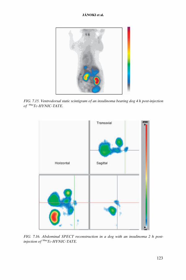
JÁNOKI et al.
FIG. 7.15. Ventrodorsal static scintigram of an insulinoma bearing dog 4 h post-injection of 99mTc-HYNIC-TATE.
FIG. 7.16. Abdominal SPECT reconstruction in a dog with an insulinoma 2 h post-injection of 99mTc-HYNIC-TATE.
123

CHAPTER 7
7.4.4. Clinical follow-up of the treated dog
The dog with insulinoma received its first treatment in September 2003. The seizures stopped immediately after the first cycle, with only one relapse after the second cycle. The animal became symptom-free two weeks after the second cycle and remained so thereafter. No abnormalities were detected in blood chemical or cellular parameters. The insulin levels are shown in
0 10 20 30 40 50
0
20
40
60
80
100
y = 93.39 exp(-2.79*t) + 6.6 exp(- 0.11*t)
Time (h)
% ID
/org
an
FIG. 7.17. Representative time versus blood activity curve of 90Y-DOTATOC in a healthy dog.
0 500 1000 15000
10
20
30
40
50
60
70
80
90
100
Time (min)
% ID
FIG. 7.18. Representative time versus blood activity curve for 99mTc-HYNIC-TATE in a dog with an insulinoma.
124

JÁNOKI et al.
Table 7.3. and in Fig. 7.19. The glucose levels were elevated after the treatment, but remained below the physiological level, indicating the presence of micrometastases undetected by SPECT imaging. The ultrasound measure-ments of the tumour size are reported in Table 7.4. At present, ultrasound gives no indication of the presence of the primary tumour in the animal.
TABLE 7.3. INSULIN LEVELS IN AN INSULINOMA BEARING DOGBEFORE AND AFTER RADIONUCLIDE THERAPY WITH 90Y-DOTATOC
Time post-therapy Insulin (µIU/mL) — Reference value: 20
— 120 (pre-therapy)
2 days 5.7
2 weeks 53.7
4 weeks 50.53 (pre–therapy cycle No. 2)
4 weeks and 2 days 7.5 (2 days post–therapy cycle No. 2)
6 weeks 44.43
8 weeks 56.46
12 weeks 50.45
0 25 50 750
20
40
60
80
100
120
Time post-therapy (d)
Insu
lin le
vels
(lU
/mL)
FIG. 7.19. Insulin levels in an insulinoma bearing dog treated with 90Y-DOTATOC; the arrows indicate therapy cycles.
125

CHAPTER 7
7.3.5. Discussion
Labelling of the peptides DOTATOC and DOTATATE with 90Y and 111In was achieved with high efficiency and good stability. The same was true for the labelling of EDDA-HYNIC-TATE with 99mTc. However, a slight in vivo instability of that labelled compound was observed in some cases, as indicated by the thyroid and salivary gland uptake of 99m TcO4
– arising from radiochemical degradation of the 99mTc labelled agents.
The scintigraphic imaging of 99mTc-HYNIC-TATE in the healthy beagle dog revealed some interesting interspecies differences between dogs and humans. Despite reports in the literature indicating that the dog somatostatin receptor subtype 2 is functionally identical to its human counterpart, the biodistribution pattern of these receptors is different in dogs. Our results correlate well with those of an earlier study by Robben et al. [7.2] based on 111In labelled octreotide indicating that dogs have a high concentration of functioning somatostatin receptors in the gastric wall, but almost no receptors expressed on the cells of the spleen. The high uptake by the heart can be attributed either to the previously unknown presence of somatostatin subtype 2 receptors in the canine heart or to the recently reported cross-reaction with growth hormone secretagogue (GHS) receptors present in large quantities on myocardial cells in humans.
Although the rate of excretion by the kidney seems to be much elevated in dogs, this route of elimination nevertheless remains the primary route in terms of dose limiting toxicity. In terms of excretion, the most interesting inter-species difference found between dogs and humans is that of biliary excretion, proven by the 99mTc-HYNIC-TATE scintigraphy in all the animals studied. This leads to uncertainty concerning the visible focus in the 90Y bremsstrahlung scintigrams in the pathological dog model, as the site of the gall bladder overlaps that of the pancreas.
TABLE 7.4. TUMOUR SIZE AS DETECTED BY ULTRASOUND
Time post-therapy (weeks) Size (mm)
— 15 × 15
2 10 × 12
4 8 × 5
8 Not detectable
126

JÁNOKI et al.
Despite the inability to detect the primary tumour or any metastases in the pathological dog model, the clinical improvement observed after the targeted radionuclide therapy supports further investigation in this direction. It was not possible to conduct immunohistological studies on tissue samples, as the animal is still alive and the primary tumour, which seems to have disappeared according to the ultrasound images, was originally inaccessible for sampling. This is also the reason why there was no primary tumour detectable with 99mTc-HYNIC-TATE in this animal.
Most human cancers are a mixture of stromal tissue and different clones of epithelial tumour cells that do not uniformly express somatostatin receptors. This contrasts sharply with the mostly monoclonal tumour models in rodents, which, in most instances, homogeneously express somatostatin receptors on all tumour cells. The use of spontaneous animal cancers having the same clonal characteristics as human cancers thus certainly has potential for evaluating the therapeutic effects and diagnostic value of radiolabelled somatostatin analogues.
The extreme clinical importance of targeted radionuclide therapy with these analogues necessitates their application as early a possible in cancer patients. Cost effective, on-site preparation of matched pairs of diagnostic and therapeutic radiopharmaceuticals clearly is the way for the future.
ACKNOWLEDGEMENTS
The authors would like to acknowledge the kind help of M. Pállai, Z. Suhajda, K. Haller and N. Fésüs for technical assistance, and M. Kovács for preparing the manuscipt.
REFERENCES TO CHAPTER 7
[7.1] DECRISTOFORO, C., MELENDEZ-ALAFORT, L., SOSABOWSKI, J.K, MATHER, S.J., 99mTc-HYNIC-[Tyr3]-octreotide for imaging somatostatin-receptor-positive tumours: Preclinical evaluation and comparison with 111In-octreotide, J. Nucl. Med. 41 (2000) 1114–1119.
[7.2] ROBBEN, J., CLAUDE REUBI, J., POLLAK, Y., VOORHOUT, G., Biodistri-bution of [111In-DTPA-D-Phe1]-octreotide in dogs: Uptake in the stomach and intestines but not in the spleen points towards interspecies differences, Nucl. Med. Biol. 30 (2003) 225–232.
127

CHAPTER 7
BIBLIOGRAPHY TO CHAPTER 7
ALTSCHUL, M., et al., Evaluation of somatostatin analogues for the detection and treatment of gastrinoma in a dog, J. Small Anim. Pract. 38 (1997) 286–291.
BODEI, L., CHINOL, M., CREMONESI, M., PAGANELLI, G., Facts and myths about radiopeptide therapy: Scylla, Charybdis and Sibyl, Eur. J. Nucl. Med. Mol. Imaging 29 (2002) 1099–1100.
BODEI, L., et al., Receptor-mediated radionuclide therapy with 90Y-DOTATOC in association with amino acid infusion: A phase I study, Eur. J. Nucl. Med. Mol. Imaging 30 (2003) 207–216.
BREEMAN, W.A., DE JONG, M., VISSER, T.J., ERION, J.L., KRENNING, E.P., Optimising conditions for radiolabelling of DOTA-peptides with 90Y, 111In and 177Lu at high specific activities, Eur. J. Nucl. Med. Mol. Imaging 30 (2003) 917–920.
CHINOL, M., BODEI, L., CREMONESI, M., PAGANELLI, G., Receptor-mediated radiotherapy with 90Y -DOTA-d-Phe1-Tyr3-octreotide: The experience of the European Institute of Oncology group, Semin. Nucl. Med. 32 (2002) 141–147.
DECRISTOFORO, C., MATHER, S.J., Technetium-99m somatostatin analogues: Effect of labelling methods and peptide sequence, Eur. J. Nucl. Med. 26 (1999) 869–876.
FUNG, L.C., GREENBERG, G.R., Characterization of somatostatin receptor subtypes mediating inhibition of nutrient stimulated gastric acid and gastrin in dogs, Regul. Pept. 26 (1997) 197–203.
GABRIEL, M., et al., An intrapatient comparison of 99mTc-EDDA/HYNIC-TOC with 111In-DTPAoctreotide for diagnosis of somatostatin receptor-expressing tumours, J. Nucl. Med. 44 (2003) 708–716.
GABRIEL, M., et al., 99mTc-N4-[Tyr3]Octreotate versus 99mTc-EDDAHYNIC-[Tyr3]Octreotide: An intrapatient comparison of two novel technetium-99m labeled tracers for somatostatin receptor scintigraphy, Cancer Biother. Radiopharm. 19 (2004) 73–79.
HEPPELER, A., FROIDEVAUX, S., EBERLE, A.N., MAECKE, H.R., Receptor targeting for tumour localization and therapy with radiopeptides, Curr. Med. Chem. 7 (2000) 7971–7994.
JAIS, P., et al., Somatostatin receptor subtype gene expression in human endocrine gastroenteropancreatic tumours, Eur. J. Clin. Invest. 27 (1997) 639–644.
KWEKKEBOOM, D., KRENNING, E.P., DE JONG, M., Peptide receptor imaging and therapy, J. Nucl. Med. 41 (2000) 1704–1713.
KWEKKEBOOM, D.J., et al., Treatment of patients with gastro-entero-pancreatic (GEP) tumours with the novel radiolabelled somatostatin analogue [177Lu-DOTA, Tyr3]octreotate, Eur. J. Nucl. Med. Mol. Imaging 30 (2003) 418–422.
LEBTAHI, R., et al., Detection of neuroendocrine tumours: (99m)Tc-P829 scintigraphy compared with (111)In-pentetreotide scintigraphy, J. Nucl. Med. 43 (2002) 889–895.
128
JÁNOKI et al.
LESTER, N.V., NEWELL, S.M., HILL, R.C., LANZ, O.I., Scintigraphic diagnosis of insulinoma in a dog, Vet. Radiol. Ultrasound 40 (1999) 174–178.
LLOYD, K.C., AMIRMOAZZAMI, S., FRIEDIK, F., CHEW, P., WALSH, J.H., Somatostatin inhibits gastrin release and acid secretion by activating sst2 in dogs, Am. J. Physiol. 272 (1997) 1481–1488.
PAGANELLI, G., et al., 90Y-DOTA-D-Phe1-Try3-octreotide in therapy of neuroendo-crine malignancies, Biopolymers 66 (2002) 393–398.
REUBI, J.C., et al., Detection of somatostatin receptors in surgical and percutaneous needle biopsy samples of carcinoids and islet cell carcinomas, Cancer Res. 50 (1990) 5969–5977.
REUBI, J.C., LAISSUE, J.A., Multiple pathways of somatostatin action in neoplastic disease, Trends Pharmacol. Sci. 16 (1995) 110–115.
REUBI, J.C., WASER, B., SCHAER, J.C., LAISSUE, J., A Somatostatin receptor sst1–sst5 expression in normal and neoplastic human tissues using receptor autoradio-graphy with subtype-selective ligands, Eur. J. Nucl. Med. 28 (2001) 836–846.
ROBBEN, J.H., et al., In vitro and in vivo detection of functional somatostatin receptors in canine insulinomas, J. Nucl. Med. 38 (1997) 1036–1042.
SIMPSON, K.W., STEPIEN, R.L., ELWOOD, C.M., BOSWOOD, A., VAILLANT, C.R., Evaluation of the long-acting somatostatin analogue octreotide in the management of insulinoma in three dogs, J. Small Anim. Pract. 36 (1995) 161–165.
VAIDYANATHAN, G., et al., Specific and high-level targeting of radiolabelled octreotide analogues to human medulloblastoma xenografts, Clin. Cancer Res. 9 (2003) 1868–1876.
VAN DIEREN, E.B., et al., Absorbed dose distribution of the auger emitters 67Ga and 125I and the beta emitters 67Cu, 90Y, 131I, and 186Re as a function of tumour size, uptake, and intracellular distribution, Int. J. Radiat. Oncol. Biol. Phys. 36 (1996) 197–204.
VIRGOLINI, I., et al., Somatostatin receptor subtype expression in human tissues: A prediction for diagnosis and treatment of cancer, Eur. J. Clin. Invest. 27 (1997) 645–647.
VIRGOLINI, I., et al., Experience with Indium-111 and Yttrium-90-labelled somato-statin analogs, Curr. Pharm. Des. 8 (2002) 1781–1807.
ZAKI, M., et al., Somatostatin receptor subtype 2 mediates inhibition of gastrin and histamine secretion from human, dog, and rat antrum, Gastroenterology 111 (1996) 919–924.
129

.
Chapter 8
PREPARATION AND EVALUATION OF 177Lu-DOTATATE FOR POTENTIAL APPLICATION IN PEPTIDE RECEPTOR RADIONUCLIDE THERAPY
S. BANERJEE, S. CHAKRABORTY, A. KORDE, T. DAS, A. MUKHERJEE, U. PANDEY, H.D. SARMA, G. SAMUEL, P.S. DHAMI, A.D. INGLE, M. VENKATESHRadiopharmaceuticals Division, Bhabha Atomic Research Centre, Trombay, Mumbai, India
Abstract
The present studies were aimed at the development of laboratory evaluation tech-niques for studying the efficacy of therapeutic radiopharmaceuticals. The primary goal was to develop radiotherapeutic agents using the peptide–BFCA conjugate DOTA-Tyr3-octreotate (DOTATATE). The radioisotopes used for the therapeutic preparations were 125I, 131I, 177Lu and 90Y. Lutetium-177 was identified as an ideal radionuclide for labelling peptides owing to the moderately high specific activity obtainable through irra-diation of the enriched targets. Lutetium-177 was produced and processed in-house for evaluation as a potential isotope for therapy using several target specific molecules. Yttrium-90 was obtained from a 90Sr/90Y generator by a supported liquid membrane separating technique, which was developed using the existing facilities. Radiolabelling procedures of the peptide–BFCA conjugate using 177Lu and 90Y were standardized to obtain maximum complexation. Studies of radiolabelling with 125I and biodistribution studies of the 125I labelled peptide were also carried out in order to extrapolate these findings for use in subsequent studies of radiolabelling with 131I as the isotope. Standard quality control techniques such as paper chromatography, paper electrophoresis and high performance liquid chromatography were used to characterize the radiolabelled species. The radiolabelling of the peptide conjugate with 90Y and its subsequent purifica-tion on a SepPak column were optimized. To evaluate the pharmacokinetic pattern, the in vivo biodistribution studies of 125I-DOTATATE and 177Lu-DOTATATE were carried out in normal Swiss mice. Further, the tumour specificity of 125I-DOTATATE was evaluated in C57BL/6 mice bearing melanoma tumours. Blocking studies of the tumour bearing animals were also carried out by injection of the unlabelled peptide DOTATATE for ascertaining the tumour specificity of the radiolabelled conjugates. In vitro studies of cells known to overexpress somatostatin receptors such as AR42J (rat pancreatic tumour), HT-29 (human colon carcinoma) and MCF-7 (human breast carcinoma) were carried out using 177Lu-DOTATATE, wherein the specificity and the
131

CHAPTER 8
binding affinity of the radiolabelled peptide were evaluated. Gamma scintigraphy of xenografted nude mice bearing HT-29 tumours injected with 177Lu-DOTATATE revealed significant activity in the tumour with no uptake in any vital organ and rapid in vivo clearance via the renal route. Lutetium-177, obtained with moderate specific activity utilizing the existing irradiation facilities, was also used for preparation of other agents such as a steroid hormone based, receptor specific agent using 17β-estradiol. Lutetium-177 was also used to design radiolabelled agents for targeting tumour hypoxia, wherein nitroimidazoles such as metronidazole and sanazol were used as the vectors. Encouraging results requiring further investigation in higher animals were obtained using 177Lu labelled EDTMP (ethylenediamine tetramethylene phosphonate) towards use as a palliative agent for the treatment of bone metastases. This agent offers consid-erable potential advantages that make it a viable alternative to existing radiopharma-ceuticals for bone pain palliation such as 153Sm-EDTMP. In each of the studies, the labelled products were evaluated by chemical and biological assays.
8.1. INTRODUCTION
The development of radiopharmaceuticals for therapeutic application using different substrates and a variety of radioisotopes is being actively pursued worldwide. The assessment of the relative effectiveness of different radiopharmaceuticals for cancer therapy is a difficult task owing to the multitude of variables that must be considered, some related to the radio-isotope and others to the biological carrier. While comparing the therapeutic efficacy in patients is a possibility, it is not feasible for obvious reasons. The development of methods that can be used for reliable and efficient comparative evaluation of promising therapeutic radiopharmaceuticals is important, because it permits more rapid identification of the optimal therapeutic parameters for a given clinical application.
Several peptide receptor specific agents, including somatostatin analogues and vasoactive intestinal peptides (VIPs), have been investigated extensively for in vivo application in tumour therapy. These studies exploit the property of tumour cells such as intestinal adenocarcinomas, lymphomas and other neuroendocrine tumours of expressing far greater numbers of somato-statin and VIP receptors on tumour cells than on normal tissues. The effec-tiveness of targeted radiotherapy of somatostatin receptor positive tumours in tumour bearing rodent models and in humans has been demonstrated using radiolabelled somatostatin analogues. In the radiotherapy of tumours over-expressing somatostatin receptors, 90Y is the most widely used radionuclide. Somatostatin analogues labelled with 90Y through suitable bifunctional chelating agents (BFCAs) have been developed and have shown promising results. One such promising agent for peptide receptor radionuclide therapy
132

BANERJEE et al.
(PRRT) is [90Y-DOTA0-Tyr3]-octreotide. Its suitable radionuclidic decay char-acteristics, availability in a no-carrier added (NCA) form and high affinity for complexation with macrocyclic chelators such as DOTA are important attributes that support the choice of 90Y as the radioisotope. Recently, 177Lu (half-life: 6.7 d; Eβ(max): 0.497 MeV; low energy, low abundance gamma emissions of 113 keV (6.4%) and 208 keV (11%)) has begun to be considered another viable alternative for the development of new agents for PRRT. It has been reported that the DOTA coupled somatostatin analogue [DOTA0–Tyr3]-octreotide labelled with 177Lu has a positive impact on tumour regression and other favourable biological characteristics in animal models. Moreover, the tissue penetration range of 177Lu (maximum range of 2 mm) is more favourable than that of 90Y (maximum range of approximately 12 mm), especially for smaller metastases, from which much of the radiation dose of 90Y will be lost to surrounding tissues. Unlike 90Y, 177Lu has the additional advantage of making it possible to perform scintigraphic and dosimetric studies with the same agent employed for therapeutic purposes.
Although 90Y is obtainable in NCA form from a 90Sr/90Y generator, there exists the stringent requirement of purification from 90Sr, a natural ‘bone seeker’ with a long half-life of 28.3 a. In contrast, 177Lu can be easily obtained with high radionuclidic purity. The high levels of radionuclidic purity of 177Lu from moderate flux reactors (5 × 10¹³ n⋅cm–2⋅s–1) using a commercially available enriched lutetium (60.6% in 176Lu) target following a 7 d irradiation procedure, its availability at a relatively high specific activity (~20 at.%, comparable with commercially available 131I) and the logistic advantages due to its long half-life (6.71 d) are desirable characteristics that make it a potential and attractive radionuclide for targeted radiotherapy.
8.2. MATERIALS
DOTATATE was obtained initially as a gift and subsequently purchased from piCHEM R&D (Austria). All chemicals and solvents used in the experiments were of analytical reagent grade and supplied by reputed chemical manufacturers. Carrier-free 125I as sodium iodide (100 mCi/mL) was obtained from the Institute of Isotopes Co. Ltd (Izotop, Hungary). Lutetium oxide (60.6% enriched in 176Lu, spectroscopic grade, >99.99% pure) was obtained from Isoflex (Russian Federation). Flexible silica gel IB-F plates (8 cm × 2.5 cm) were obtained from the Bakerflex Chemical Company (Germany). Whatman 3MM chromatography paper (Whatman, UK) was used for paper chromatography and paper electrophoresis. All radioactivity measurements were made using a NaI(Tl) scintillation counter
133
CHAPTER 8
after adjusting the baseline to 150 keV and keeping a window of 100 keV for 177Lu, 20–70 keV for 125I and 0–500 keV for 90Y to measure the brems-strahlung radiation. The radionuclidic purity of 177Lu was ascertained by high resolution gamma ray spectrometry using a high purity germanium (HPGe) detector coupled to a 4K multichannel analyser (MCA) system after radiochemical processing.
A JASCO PU 1580 high performance liquid chromatography (HPLC) system equipped with a PU 1575 UV/VIS detector (JASCO, Japan) was used in the studies. A well type NaI(Tl) scintillation detector was coupled to the system to measure activity in the eluate. All solvents used for HPLC analysis were of HPLC grade, purchased from reputed local manufacturers, and degassed and filtered prior to use.
Carrier-free Na125I (40mCi/mL) was produced at the Radiopharmaceu-ticals Division of the Bhabha Atomic Research Centre (BARC) in Mumbai, India. Yellow tetrazolium MTT (3-(4,5-dimethylthiazolyl-2)-2,5-diphenyltetra-zolium bromide) from Sigma (USA) was used for the cytotoxicity assay. Absorbance was measured at 550 nm in an ELISA reader (Bio-Tek, USA). Culture media and supplements were obtained from Sigma, and all other chemicals and solvents were of analytical reagent grade, procured from reputed manufacturers in India.
Rat pancreatic carcinoma AR42J, human colon carcinoma HT-29 and human breast carcinoma MCF-7 cell lines were procured from the National Centre for Cell Science (Pune, India). All cell cultures for in vitro studies were maintained in the in-house biosafety facility at the BARC.
Swiss and C57BL/6 mice used for bioevaluation studies were provided by the animal housing facilities of the Radiation Biology and Health Sciences Division of the BARC. Nude mice were procured and maintained at the nude mice handling facility at the Advanced Centre for Treatment Research and Education in Cancer (ACTREC) (Kharghar, Navi Mumbai, India). Scinti-graphic studies were carried out using a gamma camera (Wipro-GE, USA). All animal experiments were carried out in strict compliance with the relevant national laws relating to the conduct of animal experiments.
8.3. METHODS
8.3.1. Production of 177Lu
Lutetium-177 was produced by irradiation of isotopically enriched Lu2O3
(60.6% in 176Lu). A stock solution of the enriched target was prepared by dissolving enriched Lu2O3 powder in 0.1M HCl (at a concentration of 1 mg/
134

BANERJEE et al.
mL). A known aliquot of this solution was taken in a quartz ampoule and carefully evaporated to dryness. The ampoule was subsequently flame sealed, placed in an aluminium container and irradiated. Irradiations were carried out at different available flux positions for different durations (7–21 d) to optimize irradiation conditions to maximize specific activity. The irradiated target was dissolved in 1M HCl by gentle warming. The solution was evaporated to near dryness and reconstituted in HPLC grade water. The pH of the 177LuCl3
solution was adjusted to approximately 4 prior to complexation.The assay of the radioactivity was carried out by measuring the ionization
current obtained when an aliquot of the stock 177Lu was placed inside a pre-calibrated well type ion chamber (the calibration factor for the ion chamber for 177Lu was 15.65 A/MBq). Radionuclidic purity was determined by recording gamma ray spectra of the appropriately diluted solution of the irradiated target using an HPGe detector connected to a 4K MCA system. A 152Eu reference source was used for both energy and efficiency calibration. Several spectra were recorded for each batch at regular time intervals. Samples measured initially for the assay of 177Lu were preserved for complete decay of 177Lu (over 10–15 half-lives of 177Lu, that is, for a period of 2–3 months) and re-assayed to determine the activity of long lived 177mLu (half-life of 160.5 d). Appropriately diluted sample solutions were counted for 1 h.
8.3.2. Production of 90Y
The 90Sr/90Y generator used in these studies was developed by the Process Development Division of the BARC. The 90SrCl2 was obtained from the reprocessed fuel effluents of one of the BARC laboratories. The generator was based on a supported liquid membrane technique and consisted of a glass cell comprising two 5 mL capacity compartments — a feed and a receiver compartment — separated by a polytetrafluoroethylene membrane impregnated with 2-ethylhexyl-2-ethylhexyl phosphonic acid (KSM-17). The feed used was a suitable aliquot of high level waste with actinides, lanthanides and 137Cs removed in 0.5M HNO3 (100 mCi, 3.7 GBq) diluted to 5 mL, with the pH level adjusted to 1–2. The receiver compartment contained 5 mL of 1M HCl/HNO3. The contents of both the feed compartment and the receiver compartment were stirred constantly with small Teflon coated magnetic stirrers to enable quick and effective transport of 90Y across the membrane. After 4 h, a solution containing yttrium chloride/nitrate was drawn from the receiver compartment and used.
135

CHAPTER 8
8.3.2.1. Quality control of 90Y
8.3.2.1.1. Method 1: Spiking the feed with 85/89Sr+2
Because 90Sr is a ‘bone seeker’, the limit set for its level in 90Y prepara-tions to be used in humans is less than 2 μCi. To monitor the movement of Sr+2
across the membrane from the feed to the receiver compartment, a small aliquot of 85/89Sr chloride was added to the feed compartment along with the 90Sr chloride. A gamma ray spectrum of the 90Y chloride solution in the receiver compartment was taken to check for breakthrough of 85/89Sr into that compartment.
8.3.2.1.2. Method 2: Addition of inactive Sr+2 as a carrier in the feed compartment
The transport of Sr+2 ions across the membrane was also monitored by the addition of inactive strontium at a concentration of 5 mg/mL as a carrier in the feed compartment. Samples from the receiver compartment were drawn at regular intervals, and the Sr+2 concentrations were determined using the inductively coupled plasma atomic emission spectrophotometry (ICP-AES) method.
8.3.2.1.3. Method 3: Chromatographic methods
Chromatographic methods were also used out to analyse the relative amounts of 90Sr and 90Y in the separated 90Y. Both the strontium and yttrium used were in the form of acetates. Paper chromatography was carried out using saline as the mobile phase, and paper electrophoresis was carried out using 30 cm Whatman No. 1 paper, 0.03M NaCl and 0.15 g/L sodium citrate at 500 V for 2 h.
8.3.2.1.4. Method 4: Liquid scintillation counting
An aliquot of 90Y was taken and the decay of 90Y was followed. At present, this generator has been scaled up to 100 mCi of 90Sr to give approxi-mately 70 mCi of 90Y.
The 90Y was assayed using either a NaI(Tl) solid scintillation counter to measure the bremsstrahlung or a Tri-Carb 2100TR liquid scintillation counter (Packard Instrument Co., USA). All radioactive measurements were made using a NaI(Tl) counter.
136

BANERJEE et al.
8.3.3. Production of 125I
Production of 125I using BARC reactors began in 2003 with neutron irradiation of natural xenon gas in a special arrangement of the Dhruva reactor. A carefully standardized procedure was developed within the BARC involving one week of irradiation of 124Xe gas (4 g at 10 atm) in a 70 mL sealed capsule. To minimize the formation of 126I (which is not suitable for radioimmunoassay procedures), a short period of irradiation at medium flux using large volumes of xenon gas is desirable. Chemical processing is done in accordance with standard procedures.
8.3.4. Quality control techniques
8.3.4.1. Paper chromatography
Paper chromatography studies were carried out using 10 cm strips of chromatography paper. For each test solution, 5 µL of solution was placed at 1.5 cm from the lower end of the paper strips, which were then developed in 1mM aqueous EDTA and 50% aqueous acetonitrile solution. The strips were subsequently dried and cut into 1 cm segments. The radioactivity associated with each segment was measured in a well type NaI(Tl) detector.
8.3.4.2. Paper electrophoresis
For each of the complex solutions prepared, 5 μL of solution was placed on pre-equilibrated Whatman 3MM (35 cm × 2 cm) chromatography paper at 15 cm from the cathode. Paper electrophoresis was carried out for 1 h under a voltage gradient of approximately 10 V/cm using 0.025M phosphate buffer at a pH of around 7.5. The strips were dried and cut into 1 cm segments, and the activity was measured.
8.3.4.3. High performance liquid chromatography
The radiochemical analyses and purification of the 177Lu, 90Y and 125I labelled DOTATATE conjugates were achieved using an HPLC system. A dual pump HPLC unit with a C18 reversed phase column (25 cm × 0.46 cm) was used for purification of the labelled conjugates. The elution was monitored by UV signals at 270 nm as well as by radioactivity signals. The flow rate was maintained at 1 mL/min. Mixtures of 0.1% trifluoroacetic acid/water (solvent A) and 0.1% trifluoroacetic acid/acetonitrile (solvent B) were used as the mobile phases. The following gradient elution technique was adopted for the
137

CHAPTER 8
separation: 0–4 min 95% A, 4–15 min 95% A to 5% A, 15–20 min 5% A, 20–25 min 5% A to 95% A, 25–30 min 95% A.
8.3.5. 177Lu-DOTATATE
8.3.5.1. Preparation
DOTATATE was labelled with 177Lu by adding 20 μL of 177LuCl3
(600 μCi, 22 MBq, 1.13 nmole Lu) solution to a 200 μL solution of the conjugate (25 μg, 17.41 nmole) in 0.1M ammonium acetate buffer at pH5. The reaction mixture was incubated at 80°C for 1 h.
8.3.5.2. Optimization studies of 177Lu labelling of DOTATATE
Various parameters such as ligand concentration, incubation time and temperature were varied extensively in order to arrive at the protocol for maximum complexation. Keeping the reaction volume at 200 μL, the amount of DOTATATE was varied from 5 to 100 μg in order to determine the optimal ligand concentration for obtaining maximum complexation. The characteri-zation of the labelled conjugate and the complexation yield were determined by paper chromatography in 50% aqueous acetonitrile. The radiochemical purity of the labelled product was estimated by HPLC analysis using the gradient elution technique described above.
8.3.5.3. Complexation yields with an increase in 177Lu concentration
To obtain a 177Lu labelled DOTATATE conjugate with the highest possible specific activity, experiments were carried out to achieve maximum complexation yield of 177Lu-DOTATATE at the lowest possible ligand to metal ratio. For this, 25 μg of the peptide (the optimized ligand amount) was allowed to react with different amounts of Lu (from 0.2 to 1.6 μg) under the reaction conditions described above. In each case, the complexation yields were determined by HPLC.
8.3.5.4. Isolation of rat brain cortex membrane and competitive binding assays
Rat brain cortex membrane was isolated, and the membrane homogenate was used as a preparation containing somatostatin receptors. Four rats were sacrificed by cervical dislocation. The cerebral cortex tissue was washed three times with buffer (HEPES 20mM, pH7.3, containing 10mM MgCl2) and homogenized in ice-cold buffer, using a cortex to buffer ratio of 1:10, at
138

BANERJEE et al.
maximum speed for 10 s. The homogenate was then centrifuged at 500g for 10 min at 4°C. The supernatant was transferred to other tubes and centrifuged at 2500g for 30 min at 4°C. The supernatant was discarded, and the pellet was resuspended in HEPES buffer (with the pellet to buffer ratio kept at 1:10) and homogenized three times at maximum speed for 10 s each time. The preparation was centrifuged at 2500g for 30 min at 4°C. The three step process — resuspension in HEPES buffer, homogenization and centrifugation, as described above — was repeated twice. The last pellet was resuspended and the protein concentration was estimated using the Lowry method. The membrane preparation was diluted, aliquoted and stored at –20°C for use in receptor binding studies.
Competitive binding assays were carried out using membrane protein isolated from rat brain cortex. Rat brain cortex homogenate solution corre-sponding to 250 µg/mL of protein was prepared in assay buffer (50mM HEPES at pH7.5 containing 0.3% BSA, 10mM MgCl2). In a typical protocol, 200 µL of protein solution and 177Lu-DOTATATE corresponding to 20 ng of peptide were added per tube and incubated with varying concentrations of cold DOTATATE (0.43 ng–4.4 µg) for 1 h at ambient temperature. Next, 1 mL of 6% PEG was added per tube, and after a 30 min incubation the bound and free fractions were separated by centrifugation at 4000 rpm for 50 min. The percentage of bound tracer was calculated based on the results of gamma counting.
8.3.5.5. Maintenance of cell culture
AR42J and HT-29 cells were cultured in Ham’s F12K nutrient medium supplemented with 5% foetal bovine serum, 100 IU/mL penicillin and 50 μg/mL streptomycin. MCF-7 cells were cultured in Dulbecco’s modified Eagle’s medium (DMEM) containing 10% foetal bovine serum, 100 IU/mL penicillin, 50 μg/mL streptomycin and 10mM glutamine. All cell cultures were grown to confluence in a humidified atmosphere with 5% CO2 at 37°C. Trypsin–EDTA solution was used for subculturing and cell isolation.
8.3.5.6. In vitro cell binding studies
Studies were carried out using three different cell lines, namely, AR42J, HT-29 and MCF-7. The cells were isolated from confluent culture flasks, centrifuged and washed in plain medium as well as in assay buffer (50mM Tris HCl at pH7.5, 5mM MgCl2, 0.1M NaCl). A cell suspension of 106 cells/mL was used for all experiments. To each tube was added 5nM of 177Lu-DOTATATE, which was then incubated for 1 h at 37°C with 0.1 mL cell suspension (10–5 cells)
139

CHAPTER 8
in the presence (0.1nM – 1μM) and in the absence of cold DOTATATE. After incubation, cells were centrifuged at 3000 rev./min for 5 min, and the cell pellet was washed two times with phosphate buffer saline. The percentage of radioac-tivity associated with the cell pellet was estimated based on results of gamma counting.
8.3.5.7. In vivo distribution in normal Swiss mice
Biodistribution studies of radiolabelled DOTATATE complexes were performed in normal Swiss mice weighing 20–25 g. Between 0.15 and 0.2 mL (3–4 MBq) of the complex solution was injected through the tail vein, and the animals were then anaesthetized (using chloroform) and sacrificed by cardiac puncture at 3 and 24 h post-injection. Three mice were used at each time point. The tissues and the organs were excised, and the associated activity was measured in a flat type NaI(Tl) scintillation counter. Distribution of the activities in the different tissues and organs was calculated as the percentage of injected activity per gram (% ID/g) of tissue or organ. The percentage of activity excreted was ascertained indirectly by subtracting the accounted activity in all organs from the total activity injected.
8.3.5.8. Gamma scintigraphic studies in nude mice bearing HT-29 tumours
Human colon carcinoma (HT-29) cells were grown in culture, and approximately 106 cells were injected subcutaneously into nude mice. Solid tumours were visible after a period of two months. The animals were sacrificed, and the tumours were transplanted into additional nude mice under aseptic conditions. Solid tumours (approximately 3 mm × 3 mm) were visible within two weeks. For scintigraphic studies, tumour bearing animals were anesthetized and injected intravenously through the tail vein with approximately 500 μCi (~18 MBq) of sterile 177Lu-DOTATATE. Scintigraphic images were recorded using a single head digital SPECT gamma camera (Wipro-GE, USA) with a parallel hole, low energy high resolution (LEHR) collimator. Static images were acquired using a 256 × 256 matrix with 300 kcounts at 1, 2 and 3.5 h post-injection.
8.3.6. 90Y-DOTATATE
8.3.6.1. Preparation
DOTATATE was labelled with 90Y by adding 250 μL of 90YCl3 (500 μCi, 18.5 MBq) to an equal volume of 0.5M ammonium acetate buffer. The pH was
140

BANERJEE et al.
adjusted to 4.5 with 3N NaOH solution. This was followed by addition of 50 μL of the solution of the conjugate (50 μg) in HPLC grade water. The reaction mixture was incubated at 50°C for 1 h.
8.3.6.2. Optimization studies of 90Y labelling of DOTATATE
Various parameters such as ligand concentration, incubation time and temperature were varied extensively to arrive at the protocol for maximum complexation. The estimation of the radiochemical purity of the labelled product was carried out by HPLC analysis using the gradient elution technique described above. Complexation yields were determined by using different amounts of ligand (25–100 μg) in order to determine the optimal ligand concen-tration for obtaining maximum complexation.
8.3.7. 125I-DOTATATE
8.3.7.1. Preparation
Iodination of the DOTATATE conjugate was achieved by adding 4 μL of Na125I solution (400 μCi) to a solution of 4 μg of DOTATATE in 30 μL of 0.5M phosphate buffer at pH7.5. Chloramine T (10 μg, 10 μL) was used as the oxidizing agent. The reaction was carried out at room temperature for 90 s, after which it was quenched with 500 μL of 0.05M phosphate buffer. The radio-iodination yield was determined using paper electrophoresis.
8.3.7.2. Purification
The labelled conjugate was purified on a C18 SepPak cartridge conditioned with 2 mL 0.05M phosphate buffer at pH7.5. The column was eluted with 5 mL of 0.05M phosphate followed by 0.5 mL of ethanol.
8.3.7.3. Characterization
The radiochemical purity of the labelled conjugate was estimated by carrying out paper electrophoresis in phosphate buffer as well as by HPLC analysis using the gradient elution technique described above.
8.3.7.4. Stability studies of 125I-DOTATATE
The stability of 125I-DOTATATE was estimated in 0.05M phosphate buffer as well as in serum.
141

CHAPTER 8
8.3.7.4.1. Stability in 0.05M phosphate buffer
The purified conjugate 125I-DOTATATE was incubated in phosphate buffer at ambient temperature for 18 h, after which the stability was determined using HPLC.
8.3.7.4.2. Stability in serum
The serum stability was studied by incubation of 125I-DOTATATE in human serum overnight at ambient temperature. Determination of serum protein binding was carried out by gel permeation chromatography using a Sephadex G-25 column (2.5 cm × 30 cm) and elution using 0.05M phosphate (elution volume 1 mL fractions). The 125I-DOTATATE was incubated in serum at ambient temperature for 18 h and then treated with 10% trichloroacetic acid for precipitation of the protein bound fraction. The supernatant was tested by HPLC to estimate its purity.
8.3.7.5. In vivo biodistribution in normal Swiss mice
Biodistribution studies of 125I-DOTATATE were carried out in normal mice by injecting each animal with 37 kBq of the conjugate (corresponding to a peptide concentration of 200 ng). Animals were sacrificed at 3, 24 and 48 h post-injection. The protocol followed for in vivo distribution studies is similar to that reported in Section 8.3.5.7 for 177Lu-DOTATATE.
8.3.7.6. Biological evaluation in a murine melanoma model
C57BL/6 animals were injected subcutaneously with 106 melanoma cells. Solid tumours were visible within two weeks. Tumour bearing animals were injected with 40 kBq of 125I-DOTATATE. Biodistribution studies were carried out at 3 and 24 h post-injection, and the percentage of injected dose per organ as well as per gram of tissue was determined by gamma counting. The specificity of uptake in the tumour was studied by injecting 7.5 nmol of cold DOTATATE in addition to 50 pmol of 125I-DOTATATE into another set of tumour bearing animals. Three animals were used for each time point of experimentation.
8.3.8. Cytotoxicity studies of DOTATATE using MTT assay
Approximately 103 cells were seeded and grown on 96 well tissue culture plates in a final volume of 100 μL in a humidified atmosphere for 48 h (37°C,
142

BANERJEE et al.
5% CO2). The cells were treated with varying concentrations of DOTATATE (1–20 μg/mL) for 48 h. After incubation, 10 μL of MTT labelling agent (0.5 mg/mL in phosphate buffer solution) was added to each well. The microplate was incubated for 4 h in a humidified atmosphere. After incubation, 100 μL of solubilizing solution (10% SDS in 0.01M HCl) was added to each well. The plate was incubated overnight and the OD550 nm was measured using an ELISA reader.
8.4. RESULTS AND DISCUSSION
8.4.1. Production of 177Lu
The specific activities for 177Lu obtained from irradiation of enriched targets for different periods of time and at different thermal neutron flux positions are given in Table 8.1.
A maximum specific activity of approximately 850 GBq/mg was achieved when irradiation was carried out at a thermal neutron flux of 1 × 10¹⁴ n⋅cm–2⋅s–1
for 21 d, which corresponds to around 21% of the maximum achievable specific activity. The specific activity of the 177Lu obtained was significantly higher than the theoretically calculated value under the irradiation conditions employed (7.9 at.%), accounting for only thermal neutron capture. This could perhaps be attributed to the contribution from epithermal neutrons (resonance integral: 1087 b), which is not accounted for in theoretical calculations.
The radionuclidic purity of 177Lu produced from either a natural or an enriched target was approximately 99.99%, as was ascertained by analysing the
TABLE 8.1. REACTOR PRODUCTION OF 177Lu FROM ENRICHED TARGET
Neutron flux(n⋅cm–2⋅s–1)
Irradiation time (d)
Activity obtained (Ci/g)
Theoretical activity (Ci/g)
Experimental/ theoretical
1.4 × 10¹³ 7 2 322 ± 102 846 2.74
3.0 × 10¹³ 7 4 635 ± 269 1 814 2.56
3.0 × 10¹³ 14 7 460 ± 253 2 696 2.77
6.75 × 10¹³ 7 11 000 ± 782 4 081 2.70
6.75 × 10¹³ 14 17 750 ± 1 476 6 406 2.77
1.0 × 10¹⁴ 21 23 185 8 622 2.69
143

CHAPTER 8
gamma ray spectrum. A typical gamma ray spectrum is shown in Fig. 8.1. The average level of radionuclidic impurity burden in 177Lu due to 177mLu was found to be 5.5 kBq of 177mLu/37 MBq of 177Lu (150 nCi/1 mCi) at the end of bombardment.
8.4.2. Production of 90Y
8.4.2.1. Estimation of strontium breakthrough in 90Y
The breakthrough of Sr+2 in the receiver compartment of the generator used for sourcing of 90Y was ascertained by two methods. In one of the methods (see Section 8.3.2.1.1), the feed spiked with 85/89Sr+2 did not show any breakthrough of strontium into the receiver compartment for up to 72 h. In a second method (see Section 8.3.2.1.2), even after 12 h of operation of the generator, only 0.0008% of Sr+2 was estimated to have been transported. Since the operation of the generator is expected to be 4–6 h with 1M acid, the above breakthrough would be well within the acceptable limit.
8.4.2.2. Quality control of 90Y
In paper electrophoresis, the 85/89/90Sr+2 moved towards the cathode, while the 90Y+3 moiety moved towards the anode, as depicted in Fig. 8.2. In paper
0 1 000 2 0001
10
100
1 000
10 000
Channel number
Net
cou
nt (l
og s
cale
)
72 k
eV (17
7 Lu)
113
keV
(177 L
u)
208
keV
(177 L
u)25
0 ke
V (17
7 Lu)
321
keV
(177 L
u)
FIG. 8.1. Gamma ray spectrum of 177Lu at the end of bombardment.
144

BANERJEE et al.
chromatography using saline as the mobile phase, 85/89/90Sr+2 moved with the solvent front while 90Y+3 stayed at the point of application (Fig. 8.3).
The 90Y samples counted after 30 d did not show activity levels signifi-cantly different from the background level. The measured activity matched very well with the estimates, as was expected from the 64.1 h half-life of 90Y. Consistent results that matched the calculated values assured the high quality of the 90Y obtained and the absence of 90Sr breakthrough.
151050-5-10-15
60
40
20
0
90Y-acetate
85/89Sr-acetate
Cathode Anode
Rad
ioac
tivity
(%)
FIG. 8.2. Paper electrophoresis of 90Y-acetate and 90Sr-acetate.
0.00 0.13 0.25 0.38 0.50 0.63 0.75 0.88 1.00
1.0
0.8
0.6
0.4
0.2
0
Rf
90Y-acetate
85/89Sr-acetate
Rad
ioac
tivity
(%)
FIG. 8.3. Paper chromotography patterns of 90Y-acetate and 90Sr-acetate.
145

CHAPTER 8
8.4.3. Production of 125I
The standardized procedure developed indigenously at the BARC for the production of 125I yielded a specific activity of 15–16 mCi/µg (450–600 MBq/µg). The radiochemical purity and radioactive concentration required for various applications were also achieved.
8.4.4. 177Lu-DOTATATE
8.4.4.1. Characterization of 177Lu-DOTATATE
In paper chromatography using 50% acetonitrile in water, the activity corresponding to the 177Lu-DOTATATE complex moved towards the solvent front with Rf = 0.8–0.9, while uncomplexed 177Lu remained at the point of spotting (Rf = 0). The paper chromatography patterns of 177LuCl3 and the 177Lu-DOTATATE conjugate are shown in Fig. 8.4.
It was observed that a minimum of 25 μg was required to obtain a com-plexation yield of 98%. The effect on complexation yield of varying the ligand concentration is shown in Fig. 8.5.
Retention of radiochemical purity (>90%) after 5 d was observed using HPLC, indicating that the complex is adequately stable.
FIG. 8.4. Paper chromatography of 177Lu-DOTATATE and 177LuCl3.
146

BANERJEE et al.
The HPLC chromatograms of 177Lu-DOTATATE showing greater than 98% complexation at a concentration of 25 μg and retention of radiochemical purity after a period of 7 d are shown in Figs 8.6(a) and (b), respectively.
FIG. 8.5. Effect of variation of ligand concentration on complexation yield.
FIG. 8.6. HPLC patterns of 177Lu-DOTATATE: (a) complexation at a concentration of
25 μg of peptide and (b) radiochemical purity after 7 d.
147

CHAPTER 8
8.4.4.2. Complexation yields with an increase in the 177Lu concentration
As Table 8.2 shows, a radiolabelling yield of approximately 99% was achieved using a metal to ligand ratio as high as about 1:4.
8.4.4.3. Bioevaluation: Rat brain cortex membrane fraction
Binding studies of 177Lu-DOTATATE with rat brain cortex membraneusing 200 µL of protein solution, 177Lu-DOTATATE corresponding to 20 ng of peptide and varying concentrations of cold DOTATATE (0.43 ng – 4.4 μg) showed 6.8 ± 0.06% binding. However, the inhibition results were not conclusive, and the saturation binding assays could not be carried out owing to practical limitations on the number of animals that could be sanctioned, as well as the limited availability of the necessary instruments.
8.4.4.4. Bioevaluation: In vitro cell studies
Cell studies were carried out in three different cell lines. Experimental parameters such as cell number, tracer concentration (5nM of 177Lu-DOTATATE per tube) and incubation parameters (pH, time and temperature) were identical for all experiments. Table 8.3 shows the extent of tracer uptake in the different cell lines. AR42J cells showed the highest uptake of the three cell lines studied. No significant inhibition in binding due to the addition of cold peptide was observed. The results depicted in Table 8.3 are indicative of the higher somatostatin receptor expression on AR42J cells than on HT-29 and MCF-7 cells.
TABLE 8.2. COMPLEXATION YIELD OF 177Lu-DOTATATE AT SELECTED L/M RATIOS
DOTATATE (ng) Lu (ng) L/M Complexation yield (%)
2.5 × 10⁴ (17.41 n mole)
200 (1.13 n mole)
15.41 99.2
2.5 × 10⁴ (17.41 n mole)
400 (2.26 n mole)
7.70 99.0
2.5 × 10⁴ (17.41 n mole)
800 (4.52 n mole)
3.85 98.7
2.5 × 10⁴ (17.41 n mole)
1.6 × 10³ (9.04 n mole)
1.92 81.2
148

BANERJEE et al.
8.4.4.5. Biodistribution studies in normal mice
Biodistribution studies carried out in normal mice at 3 and 24 h post-injection (Table 8.4) showed no uptake in major organs of interest. Rapid clearance of activity via the renal route was seen at 3 h post-injection.
TABLE 8.3. CELL UPTAKE OF 177Lu-DOTATATE
Cell type Tracer uptake (%)DOTATATE (cold)
10 µg/mL 1 µg/mL 0.1 µg/mL
AR42J 44.36 ± 2.5 40.3 45.1 —a
HT-29 12.6 ± 1.4 13.8 ± 0.28 12.9 ± 0.56 13.8
MCF-7 8.15 ± 0.35 8.38 ± 0.45 — —a Not available.
TABLE 8.4. BIODISTRIBUTION PATTERN OF 177Lu-DOTATATE IN SWISS MICE AT 3 AND 24 h POST-INJECTION
Region % ID/organ (n = 3)
3 h 24 h
Blood 0.07 (0.01)a 0.01 (0.00)
Liver 0.14 (0.04) 1.76 (0.51)
Intestine 0.51 (0.02) 0.10 (0.01)
Kidney 1.98 (0.05) 1.12 (0.30)
Stomach 0.10 (0.04) 0.07 (0.03)
Heart 0.00 (0.00) 0.01 (0.00)
Lungs 0.04 (0.01) 0.09 (0.05)
Bone 0.00 (0.00) 0.00 (0.00)
Muscle 0.22 (0.02) 0.08 (0.02)
Spleen 0.00 (0.00) 0.00 (0.00)
Excretionb 96.7 (0.46) 97.7 (1.00)a Standard deviations are given in parentheses. b Calculated by subtracting the activity in all organs from the total activity injected.
149

CHAPTER 8
8.4.4.6. Gamma scintigraphic studies in nude mice bearing HT-29 tumours
Figure 8.7 shows a nude mouse bearing an HT-29 tumour in the flank region. Scintigraphic images of the mouse at different time points post-injection are shown in Fig. 8.8. Significant tumour uptake of 177Lu-DOTATATE was observed at 3.5 h with a high target to non-target ratio. Evidence from the imaging studies supports results from biodistribution studies indicating rapid renal clearance of 177Lu-DOTATATE with negligible accumulation in vital organs.
8.4.5. 90Y-DOTATATE
It was observed that a complexation yield of 70% was achievable in the labelling of DOTATATE with 90Y when 25 μg of the conjugate was used; the yield increased to 98% when 100 μg of the conjugate was used. A reasonably good complexation yield of around 87% was obtained using 50 μg of the conjugate at 50°C for 1 h; thus all studies were carried out using this amount of conjugate. The effect of variations of the ligand concentration on the complex-ation yield is shown in Table 8.5.
The HPLC chromatograms of 90Y labelled DOTATATE showing greater than 87% complexation at a concentration of 50 μg are shown in Fig 8.9.
FIG. 8.7. Nude mouse bearing an HT-29 tumour.
150
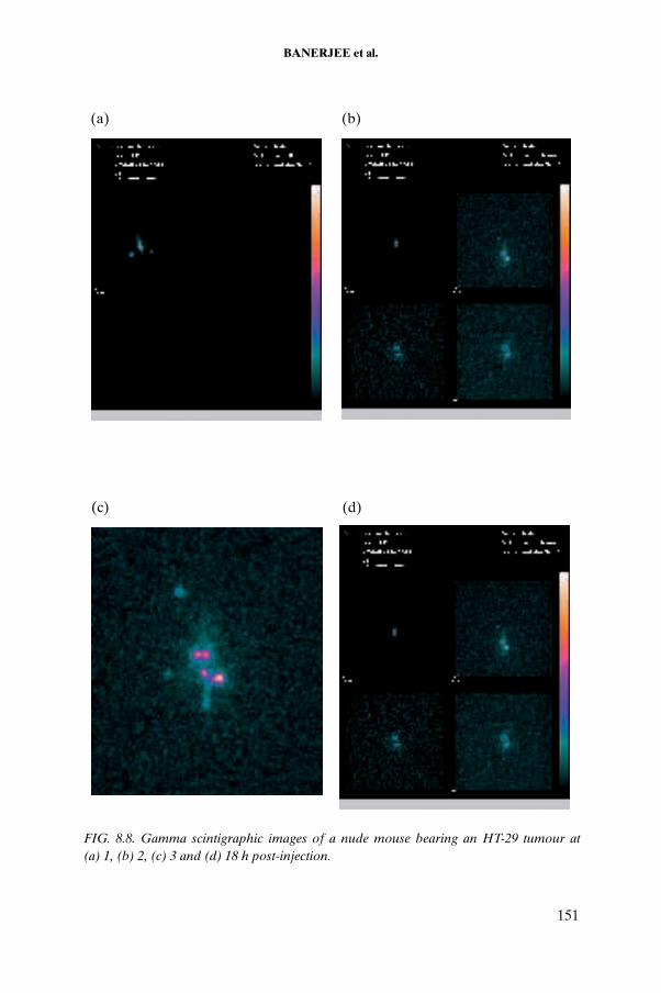
BANERJEE et al.
(a) (b)
(c) (d)
FIG. 8.8. Gamma scintigraphic images of a nude mouse bearing an HT-29 tumour at (a) 1, (b) 2, (c) 3 and (d) 18 h post-injection.
151

CHAPTER 8
8.4.6. 125I-DOTATATE
8.4.6.1. Radiolabelling with 125I
In paper electrophoresis, the 125I-DOTATATE conjugate was retained at the point of application and the free I– moved towards the anode. The radio-iodination yield obtained was approximately 90%. The paper electrophoresis pattern of the reaction mixture of 125I-DOTATATE is shown in Fig. 8.10. With SepPak separation, free I– was eluted in the phosphate buffer, while the conjugate was obtained in pure form in the ethanol fraction.
TABLE 8.5. COMPLEXATION YIELD OF 90Y-DOTATATE AT DIFFERENT PEPTIDE CONCENTRATIONS
Peptide concentration (μg) Complexation yield (%)
25 70.0
50 87.0
100 97.5
0 200 400 600 800 1000 1200 1400 1600 18000
50
100
150
200
250
90Y-acetate
90Y-DOTATATE
Retention time (s)
Cou
nts/
s
FIG. 8.9. HPLC patterns for 90Y-DOTATATE (50 μg of peptide) at 1 h post-injection and 50°C.
152

BANERJEE et al.
8.4.6.2. HPLC characterization of 125I-DOTATATE
HPLC analysis indicated that the radiochemical purity of 125I-DOTATATE was approximately 90% (Fig. 8.11).
8.4.6.3. Stability studies of 125I-DOTATATE
8.4.6.3.1. Stability in 0.05M phosphate buffer
The HPLC chromatogram of 125I-DOTATATE stored in buffer showed that the product is reasonably stable, with 90–95% retention of radiochemical purity (Fig. 8.12).
-2 -1 0 1 2 3 4 5 6 7 8 9 10 11 12 13 14 15 16 170
2 000
4 000
6 000
8 000
10 000
12 000
14 000
125I-DOTATATE
125I–
Distance from point of spotting (cm)
Cou
nts/
30 s
FIG. 8.10. Paper electrophoresis pattern of reaction mixture.
0 100 200
0
50
100
150
200
250
300
350
Retention time (s) Retention time (s)
Cou
nts/
s
Cou
nts/
s
0 500 1000 1500 2000
0
200
400
600
800
1000
125I-DOTATATE
125I–(a) (b)
FIG. 8.11. HPLC patterns of (a) 125I and (b) 125I-DOTATATE.
153

CHAPTER 8
8.4.6.3.2. Stability in serum
The gel permeation chromatography of 125I-DOTATATE incubated in human serum is shown in Fig. 8.13. The extent of serum binding was found to be around 15%.
The HPLC chromatogram in Fig. 8.14 indicates the retention of the integrity of the conjugate 125I-DOTATATE after incubation in serum.
0 200 400 600 800 1000 1200 1400 16000
20
40
60
80
100
120
140
160
180
125I-DOTATATE
125I–
Retention time (s)
Cou
nts/
s
FIG. 8.12. HPLC pattern of 125I-DOTATATE after storage in buffer.
0 5 10 15 20 25 30 35
0
20 000
40 000
60 000
80 000125I-DOTATATE
Bound fraction
Fraction number
Cou
nts/
30 s
FIG. 8.13. Gel permeation chromatography of 125I-DOTATATE incubated in human serum.
154

BANERJEE et al.
8.4.6.4. Biodistribution studies in normal Swiss mice
The biodistribution pattern of 125I-DOTATATE (Fig. 8.15) shows no uptake in major organs of interest and rapid clearance of the activity. Low thyroid uptake was indicative of the in vivo stability of the product.
0 500 1000 1500 20000
10
20
30
40
50
60
70
80125I-DOTATATE
Retention time (s)
Cou
nts/
s
125I-
FIG. 8.14. HPLC of 125I-DOTATATE (post-precipitation of serum protein).
Blood Liver Int+GB Kidney Thyroid Excreta
100
80
60
40
20
0
3 h post-injection
24 h post-injection
48 h post-injection
% ID
/org
an
FIG. 8.15. Biodistribution pattern of 125I-DOTATATE.
155

CHAPTER 8
8.4.6.5. Biodistribution studies in murine melanoma
Table 8.6 gives the results of biodistribution studies using 125I-DOTATATE in a murine melanoma tumour model. An uptake of 3% ID/g of the radiolabelled conjugate was observed in the tumour tissue within 3 h post-injection, which decreased to 2% at 24 h post-injection. The pharmacokinetics of 125I-DOTATATE in tumour bearing animals indicated significant clearance of the activity via renal excretion with negligible accumulation in other organs, including the thyroid (see Table 8.6).
Inhibition studies carried out in animals bearing melanoma tumours showed decreased uptake. The percentage of 125I-DOTATATE accumulation per gram of tumour tissue was observed to decrease by 40% at 3 h post-injection when 7.5 nmol cold DOTATATE was injected simultaneously with the 125I-DOTATATE.
8.4.7. Cytotoxicity studies of DOTATATE using MTT assay
MTT is reduced metabolically by active cells via the action of the dehydrogenase enzyme, resulting in intracellular purple formazan. Hence, the
TABLE 8.6. BIODISTRIBUTION PATTERN OF 125I-DOTATATE IN C57BL/6 MICE BEARING MELANOMA TUMOURS AT 3 AND 24 h POST-INJECTION
Region% ID/g
3 h 24 h
Blood 1.47 ± 0.89 0.92 ± 0.5
Liver 2.78 ± 0.2 2.6 ± 0.8
Intestines and gall bladder 4.61 ± 1.13 2.16 ± 0.43
Kidneys 11.8 ± 1.3 4.66 ± 0.46
Stomach 13 ± 5.7 5.9 ± 2.8
Heart 0.52 ± 0.26 0.58 ± 0.1
Lungs 11.6 ± 1.6 2.4 ± 0.57
Femurs 1.4 ± 0.4 0.125 ± 0.07
Muscles 0.17 ± 0.03 0.24 ± 0.08
Spleen 2.25 ± 1.06 1.17 ± 0.18
Tumour 3.0 ± 1.28 2.0 ± 0.3
Excreta 50 ± 2.8 71.5 ± 9.8
156

BANERJEE et al.
difference between the cell proliferation rate in untreated (control) cells and that in cells treated with DOTATATE could be estimated using MTT assay. DOTATATE at lower concentrations did not lead to significant changes in the OD, whereas 20 μg/mL DOTATATE led to an approximately 25% decrease in cell proliferation compared with the control, as shown in Table 8.7.
8.5. 177Lu LABELLING OF OTHER TARGET SPECIFIC LIGANDS
8.5.1. 177Lu labelled estradiol-BFCA conjugate
The macrocycle 1,4,7,10-tetraazacyclododecane-N,N′,N′′,N′′′-tetraacetic acid (DOTA) is known to form stable complexes with lanthanides. A steroid conjugate (Fig. 8.16) was prepared via coupling of 6α-amino-17β-estradiol with a C functionalized DOTA derivative, namely, p-thiocyanatobenzyl DOTA, as a BFCA. The synthesis of 6α-amino-17β-estradiol was carried out by a four step reaction sequence. The intermediate and target compounds were characterized using high resolution 1H-NMR spectroscopy.
In a typical radiolabelling procedure, 300 μg of the coupled product was dissolved in 0.2 mL of DMF, followed by addition of 0.2 mL of 0.1M ammonium acetate buffer at a pH of about 5.5 and 40 μL of 177LuCl3 solution (100–200 MBq). The volume of the reaction mixture was brought up to 1 mL using normal saline, and its pH was adjusted to about 5. Finally, the reaction mixture was incubated at 37°C for 2 h. The radiolabelling yields, determined by paper chromatography in saline and paper electrophoresis in phosphate buffer (pH7.5), were estimated to be 80–85%. The 177Lu-p-NCS-benzyl-DOTA-estradiol complex was purified on a SepPak column as well as by HPLC. The stability of the 177Lu labelled p-NCS-benzyl-DOTA-estradiol conjugate was studied using quality control techniques similar to those used to determine the complexation yield. It was observed that the complex exhibited good stability when stored at room temperature, as it maintained a radiochemical purity of 77% for a period of 7 d post-preparation.
TABLE 8.7. ANTIPROLIFERATIVE EFFECT OF DOTATATE ON AR42J CELLS
DOTATATE (μg/mL) OD550 nm Inhibition (%)
Control 0.77 ± 0.03 —
10 0.73 ± 0.02 5.0
20 0.57 ± 0.03 25.7
157
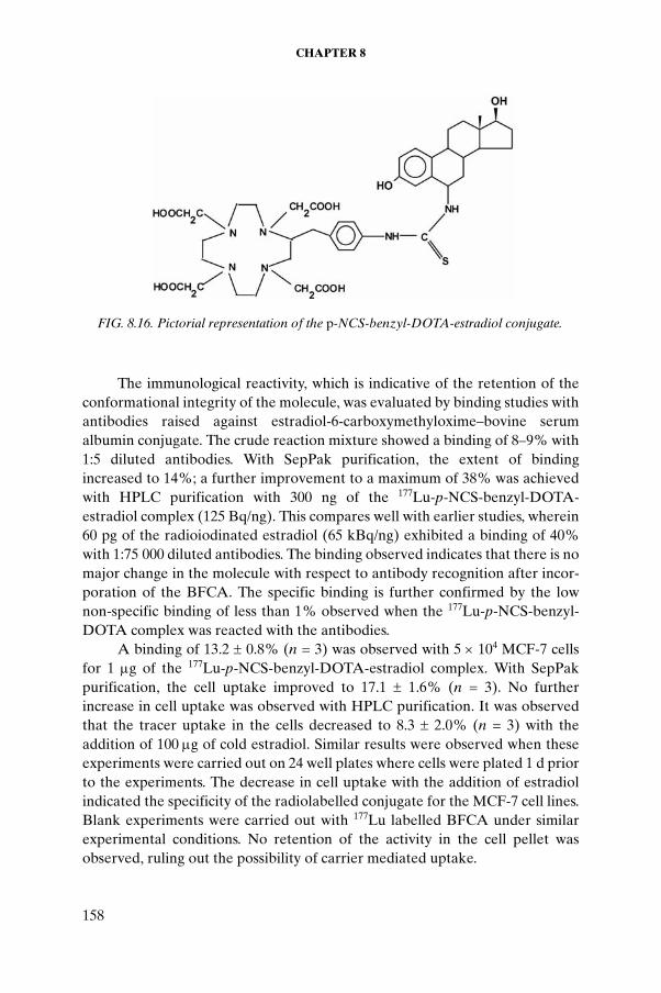
CHAPTER 8
The immunological reactivity, which is indicative of the retention of the conformational integrity of the molecule, was evaluated by binding studies with antibodies raised against estradiol-6-carboxymethyloxime–bovine serum albumin conjugate. The crude reaction mixture showed a binding of 8–9% with 1:5 diluted antibodies. With SepPak purification, the extent of binding increased to 14%; a further improvement to a maximum of 38% was achieved with HPLC purification with 300 ng of the 177Lu-p-NCS-benzyl-DOTA-estradiol complex (125 Bq/ng). This compares well with earlier studies, wherein 60 pg of the radioiodinated estradiol (65 kBq/ng) exhibited a binding of 40% with 1:75 000 diluted antibodies. The binding observed indicates that there is no major change in the molecule with respect to antibody recognition after incor-poration of the BFCA. The specific binding is further confirmed by the low non-specific binding of less than 1% observed when the 177Lu-p-NCS-benzyl-DOTA complex was reacted with the antibodies.
A binding of 13.2 ± 0.8% (n = 3) was observed with 5 × 10⁴ MCF-7 cells for 1 μg of the 177Lu-p-NCS-benzyl-DOTA-estradiol complex. With SepPak purification, the cell uptake improved to 17.1 ± 1.6% (n = 3). No further increase in cell uptake was observed with HPLC purification. It was observed that the tracer uptake in the cells decreased to 8.3 ± 2.0% (n = 3) with the addition of 100 μg of cold estradiol. Similar results were observed when these experiments were carried out on 24 well plates where cells were plated 1 d prior to the experiments. The decrease in cell uptake with the addition of estradiol indicated the specificity of the radiolabelled conjugate for the MCF-7 cell lines. Blank experiments were carried out with 177Lu labelled BFCA under similar experimental conditions. No retention of the activity in the cell pellet was observed, ruling out the possibility of carrier mediated uptake.
FIG. 8.16. Pictorial representation of the p-NCS-benzyl-DOTA-estradiol conjugate.
158

BANERJEE et al.
8.5.2. 177Lu labelled metronidazole-DOTA conjugate
The interesting observation that nitroimidazole derivatives have a tendency to accumulate in hypoxic or anaerobic regions led to the envisaging of these compounds as possible radiosensitizers, agents that enhance the lethal effect of ionizing radiations on hypoxic tissues. In the present study, 177Lu, a promising radionuclide for targeted therapy owing to its suitable nuclear decay characteristics, was chosen as the radioisotope for targeting tumour hypoxia using metronidazole, a 5-nitroimidazole, as the carrier molecule. Since direct radiolabelling of a metronidazole derivative with 177Lu is not feasible, indirect incorporation of 177Lu through a suitable BFCA was envisaged. In the choice of an ideal BFCA for complexation with 177Lu, a radiolanthanide, high thermo-dynamic stability and kinetic inertness of the resultant conjugate are guiding factors, leading to the choice of a suitable DOTA derivative, a polyazamacro-cycle, as the BFCA. Para-aminobenzyl-1,4,7,10-tetraazacyclododecane-1,4,7,10-tetraacetic acid (p-amino-benzyl-DOTA) with a suitably disposed amino substituent was covalently coupled through this substituent to the -COOH group of 2-[N-(2′-methyl-5′-nitro)imidazolyl]ethanoic acid, the carboxylic acid derivative of metronidazole. The synthesis and labelling with 177Lu of the metronidazole-p-aminobenzyl-DOTA conjugate (Fig. 8.17) and the labelled conjugate’s biological behaviour in the tumour bearing animal model were studied.
The metronidazole-BFCA conjugate was radiolabelled with 177Lu of high radiochemical purity (97%). Preliminary biodistribution studies carried out in Swiss mice bearing fibrosarcoma tumours revealed good tumour uptake (1.30% ID/g at 30 min post-injection) with rapid renal clearance (94.48% ID at 30 min post-injection) and significant tumour to blood (28.00 at 3 h post-injection) and tumour to muscle (14.00 at 3 h post-injection) ratios.
8.5.3. 177Lu labelled sanazol-DOTA conjugate
A polyazamacrocyclic-nitrotriazole conjugate for radiolabelling with the therapeutic radioisotope 177Lu was prepared. The nitroimidazole used for the
FIG. 8.17. Pictorial representation of the metronidazole-p-aminobenzyl-DOTA conjugate.
159

CHAPTER 8
study was (N-2′(carboxyethyl)-2-(3′-nitro-1′-triazolyl)acetamide, the carboxylic acid derivative of sanazole, which possesses an optimal combination of desired properties such as selective toxicity for hypoxic cells and lowered lipophilicity, resulting in lowered neurotoxicity. The BFCA used was 1,4,7,10-tetraaza-1-(4′-aminobenzylacetamido)-cyclododecane-4,7,10-triacetic acid (p-amino-DOTA-anilide), a DOTA derivative. Radiolabelling of the sanazole-BFCA conjugate (Fig. 8.18) with 177Lu was achieved under optimized conditions, yielding compl-exation of approximately 98%.
The complex was characterized by paper chromatography and HPLC studies. Bioevaluation studies in Swiss mice bearing fibrosarcoma tumours revealed good tumour uptake (0.88% ID/g at 1 h post-injection) with favourable tumour to blood (4.00 at 1 h post-injection) and tumour to muscle (4.63 at 1 h post-injection) ratios.
FIG. 8.18. Sanazole derivative-p-amino-DOTA-anilide conjugate.
160

BANERJEE et al.
8.6. CONCLUSION
Lutetium-177 has been identified as a promising isotope for therapy owing to its favourable nuclear properties and its long half-life, which provides logistic advantages with regard to its use in places with limited reactor facilities and sourcing of the radioisotopes. Production and processing of 177Lu of moderately high specific activity and high radionuclidic purity were achieved at the BARC using enriched targets and the existing irradiation facilities. The potential of this radioisotope for therapy was demonstrated using several target specific molecules. Yttrium-90 was obtained from a 90Sr/90Y generator using a supported liquid membrane separating technique developed using the existing facilities. The 125I of moderate specific activity used in the labelling studies was obtained by standardization of the existing procedure, involving irradiation of 124Xe gas in a sealed capsule for one week. The radiolabelling procedures for DOTATATE using 177Lu, 90Y and 125I were optimized, and relevant quality control parameters were standardized. In vivo biodistribution studies in normal Swiss mice revealed that the 177Lu-DOTATATE and 125I-DOTATATE had suitable pharmacokinetic properties. Blocking studies (using the peptide conjugate DOTATATE) in C57BL/6 mice bearing melanoma tumours expressing somatostatin receptors were carried out using 125I-DOTATATE in order to determine the receptor mediated uptake. The results indicate positive inhibition of binding of radiolabelled DOTATATE in the presence of the cold peptide conjugate. In vitro cell binding studies of 177Lu-DOTATATE using suitable cell lines such as rat pancreatic carcinoma AR42J and human colon carcinoma HT-29, known to overexpress somatostatin receptors, revealed significant binding. The extent of binding was found to be low with human breast carcinoma cell lines, which is indicative of the higher somatostatin receptor expression on AR42J cells compared with MCF-7 cells. Significant tumour uptake of 177Lu-DOTATATE was observed at 3.5 h with a high target to non-target ratio. Scintigraphic imaging of 177Lu-DOTATATE in xenografted nude mice bearing HT-29 tumours provides evidence supporting the rapid renal clearance with negligible accumulation in vital organs observed in biodistribution studies.
Laboratory protocols for labelling molecules with 177Lu, 90Y and 125I were optimized using the radioisotopes obtained within the BARC. Methods and experimental procedures for evaluation of the radiolabelled substrates in in vitro and in vivo animal models were also standardized.
161

CHAPTER 8
ACKNOWLEDGEMENTS
The authors gratefully acknowledge the help rendered by S.V. Thakare and K.C. Jagadeesan in irradiation studies for isotope production. We also thank P.V. Joshi and R.B. Manolkar for processing and providing 125I for our work. The constant encouragement and support provided by V. Venugopal, Director of the Radiochemistry and Isotope Group at the Bhabha Atomic Research Centre (BARC), Mumbai, N. Ramamoorthy, former Associate Director of the Radiochemistry and Isotope Group at the BARC, and M.R.A. Pillai, former Head of the Radiopharmaceuticals Division of the BARC, is gratefully acknowledged. The financial support and the samples of DOTATATE received from the IAEA are also gratefully acknowledged.
BIBLIOGRAPHY TO CHAPTER 8
CHINOL, M., BODEI, L., CREMONESI, M., PAGANELLI, G., Receptor mediated radiotherapy with Y-DOTA-D-Phe-Tyr-Oetreotide: The experience of European Institute Oncology Group, Semin. Nucl. Med. 32 (2002) 141.
DE JONG, M., KRENNING, E.P., New advances in peptide receptor radionuclide therapy, J. Nucl. Med. 43 (2002) 617.
DE JONG, M., BREEMAN, A.P., VALKEMA, R., BERNARD, B.F., KRENNING, E.P., Combination radionuclide therapy using 177Lu- and 90Y-labeled somatostatin analogs, J. Nucl. Med. 46 (2005) 13S.
PILLAI, M.R.A., CHAKRABORTY, S., DAS, T., VENKATESH, M., RAMAMOORTHY, N., Production logistics of 177Lu for radionuclide therapy, Appl. Radiat. Isot. 59 (2003) 109.
SMITH, C.J., et al., Radiochemical investigations of 177Lu-DOTA-8-Aoc-BBN[7-14]NH2: An in vitro/in vivo assessment of the targeting ability of this new radio-pharmaceutical for PC-3 human prostate cancer cells, Nucl. Med. Biol. 30 (2003) 101.
WEINER, R.E., THAKUR, M.L., Radiolabelled peptides in the diagnosis and therapy of onclogical diseases, Appl. Radiat. Isot. 57 (2002) 749.
VIRGOLINI, I., et al., In vitro and in vivo studies of three radiolabelled somatostatin analogues: 123I-Octreotide (OCT), 123I-tyr-3-Oct and 111In-DTPA-D-phe-1-OCT, Eur. J. Nucl. Med. 23 (1996) 1388.
VIRGOLINI, I., et al., Vasoactive intestinal peptide receptor scintigraphy, J. Nucl. Med. 36 (1995) 1732.
162

Chapter 9
OPTIMIZATION OF LABELLING CONDITIONS AND CELL BINDING ASSAY FOR 177Lu-DOTATATE
M. CHINOL*, SUN JU CHOI**
* Division of Nuclear Medicine, European Institute of Oncology, Milan, Italy
** Radioisotope Research and Development Laboratory, Korea Atomic Energy Research Institute, Taejeon, Republic of Korea
Abstract
The study aimed at optimizing labelling conditions for 177Lu-DOTATATE. The influence on labelling yield of pH, temperature and radical scavengers was evaluated. Under optimal conditions, labelling yields greater than 98% with a specific activity of 37–75 MBq of 177Lu/μg of DOTATATE were achieved. A maximum specific activity of about 111 MBq/nmol of peptide was obtained using a 177Lu sample with negligible traces of metal impurities. The presence of iron was found to be detrimental to obtaining good labelling yields; levels of iron greater than 1 μg/Ci of 177Lu were found to reduce labelling yields.
9.1. INTRODUCTION
This paper reports on studies carried out at the European Institute of Oncology focusing on the preparation of 177Lu-DOTATATE for clinical studies.The presence of other metal ions in the 177Lu solution can adversely affect the labelling yield. In vitro studies using membranes obtained from AR42J cells expressing somatostatin receptor subtype 2 were performed. Somatostatin-14 labelled with 125I was used at a fixed concentration of 1nM in the heterologous competition experiments. The concentration of peptide occupying 50% of receptors (Ki) was determined for the unlabelled peptide and for the peptide complexed with 90Y and 177Lu. All the Ki values were in the nanomolar range and none showed significant differences between labelled and unlabelled
163

CHAPTER 9
DOTATATE, thereby demonstrating that the presence of metal does not interfere with the receptor affinity profile.
It was recognized that the accuracy of the binding assay can be improved by ascertaining the exact concentration of the peptide. A method based on the chelation of 111In with DTPA and DOTATATE was developed, allowing the purity of the compound(s) to be determined prior to the binding assay and the molar extinction coefficient to be subsequently ascertained.
9.2. MATERIALS AND METHODS
9.2.1. Reagents
DOTA-[Tyr3]-octreotate (DOTATATE) supplied by the IAEA was complexed with YCl3 and LuCl3 (Sigma-Aldrich) by heating for 25 min at 90°C, using a 1:3 ratio of the respective DOTA peptide to the corresponding metal salt in 150 μL of 0.2M gentisic buffer at pH5. After cooling, 20 μL of 0.1M DTPA at pH5 was added to complex with the free metal ions. The compounds were purified on a SepPak C18 cartridge. The 90Y-DOTATATE and 177Lu-DOTATATE were eluted with methanol, obtaining radionuclidic purities of greater than 98% after evaporation of the methanol. The exact mass of the peptides was ascertained by electron spray ionization mass spectrometry (ESI-MS).
9.2.2. Labelling of DOTATATE with 177Lu
The 177Lu obtained from a commercial supplier was added to 50% gentisic acid in acetate buffer in order to adjust the pH to approximately 4.5. DOTATATE was added to the buffered 177Lu to achieve a specific activity of 1 mCi/μg. The mixture was then incubated at 90oC for 30 min in a thermostatic bath, after which the radiochemical purity was determined by either SepPak C18 cartridge or HPLC.
9.2.3. Cell culture
The AR42J cell line expressing somatostatin receptor subtype 2 was obtained from ATCC (Manassas, VA, USA). The cells were maintained in Kaighn’s modification of Ham’s F12 medium (F12K) with 2mM L-glutamine, 10% foetal bovine serum and 1.5 g/L of sodium bicarbonate in a humidified atmosphere at 5% CO2 and 37°C.
164

CHINOL and SUN JU CHOI
9.2.3.1. Preparation of membranes
Cells were collected in phosphate buffer solution (136.9mM NaCl, 2.7mM KCl, 8.09mM Na2HPO4 and 1.4mM KH2PO4) and centrifuged for 10 min at 2000g. The pellet was resuspended in lysis buffer (15mM TrisHCl at pH7.4, 2mM MgCl2 and 0.3mM EDTA) and homogenized with a Dounce glass apparatus. The homogenate was centrifuged at 15 000g for 20 min and the membrane pellet was resuspended in binding buffer (HEPES 50mM, pH7.6, containing 0.3% BSA, 5mM MgCl2 and 10M bacitracin).
9.2.4. Binding studies
To determine the affinity constants of each peptide, heterologous competition experiments were performed on membranes prepared from the AR42J cell line. For these binding studies, 20–25 μg of protein were incubated at a fixed concentration (0.51–1nM) of [125I]Tyr11somatostatin-14 (Amersham Biosciences) for 1 h at 30°C in the presence of increasing concentrations of unlabelled peptide (10–12 to 10–7M). Non-specific binding was determined in the presence of 1μM octreoctide. Bound and free radioactivity were separated by filtration using a Whatman GF/C filter presoaked in 10 mg/mL BSA. The binding isotherms were analysed using Prism software (GraphPad Software).
9.2.5. Determination of peptide purity
The assay used to determine peptide purity was based on the use of a trace amount of 111In to determine incorporation of the metal into DOTATATE. By checking the HPLC radiochromatogram of the reaction mixture, it was possible to ascertain the number of moles of indium that had reacted with the DOTATATE, and thus to calculate the peptide concentration. The reaction took place between 0.1 µmol of DOTATATE, 1.0 µmol of InCl3and 1.0 pmol of 111InCl3 at 90oC for 30 min for completing the incorporation of indium into the DOTATATE. Following this complexation, a molar excess of DTPA was added to complex all the unreacted and free indium in the reaction mixture.
165

CHAPTER 9
9.3. RESULTS
9.3.1. Labelling of DOTATATE with 177Lu
Following the labelling protocol described in Section 9.2, labelling yields of at least 98% were routinely achieved. Figure 9.1 depicts a typical HPLC profile showing complete labelling of DOTATATE.
However, particular attention has to be paid to the issue of the specific activity. Usually, lutetium is produced at a specific activity of 740 × 103 MBq (20 Ci/mg), which implies that 1 mCi of 177Lu corresponds to 291 pmol. Therefore, considering the theoretically feasible maximum molar ratio of 1 mole of DOTA to 1 mole of metal, for each millicurie of 177Lu solution, corre-sponding to 291 pmol of total lutetium, 291 pmol of peptide is needed. The molecular weight of DOTATATE being 1430, 291 pmol corresponds to 416 130 pg, that is, 0.41613 μg of peptide per millicurie. Therefore, assuming a 1:1 molar ratio of peptide to total lutetium, the maximum achievable practical specific activity will be 2.40 mCi/μg, equal to 3.44 mCi/nmol.
9.3.2. Binding study
Typical binding curves are shown in Fig. 9.2. The Ki values are given in Table 9.1. No statistically significant differences between the Ki value for DOTATATE and those for the 90Y-DOTATATE and 177Lu-DOTATATE samples were observed.
0 5 10
Time (min)
Cou
nts
FIG. 9.1. HPLC chromatogram of 177Lu-DOTATATE.
166

CHINOL and SUN JU CHOI
TABLE 9.1. CALCULATED Ki VALUES FOR DOTATATE, 90Y-DOTATATE AND 177Lu-DOTATATE
Sample Ki (nM)
DOTATATE 0.530.49
90Y-DOTATATE 0.330.35
177Lu-DOTATATE 0.230.21
-15.0 -12.5 -10.0 -7.5 -5.0 -2.50
5 000
10 000
15 000IC50 = 0.85nM;Ki = 0.53nM;(Kd SRIF = 1nM; conc. 125I-SRIF = 0.605nM)
[Peptide]
-15.0 -12.5 -10.0 -7.5 -5.0 -2.50
2 500
5 000
7 500
10 000
[Peptide]
-15.0 -12.5 -10.0 -7.5 -5.0 -2.50
5 000
10 000
15 000
[Peptide]
Cou
nts/
min
IC50 = 0.51nM;Ki = 0.35nM;(Kd SRIF = 1nM; conc. 125I-SRIF = 0.433nM)
Cou
nts/
min
IC50 = 0.35nM;Ki = 0.212nM;(Kd SRIF = 1nM; conc. 125I-SRIF = 0.650nM)
Cou
nts/
min
(a) (b)
(c)
FIG. 9.2. Binding affinity profiles of (a) DOTATATE,(b) 90Y-DOTATATE and (c)177Lu-DOTATATE.
167

CHAPTER 9
9.3.3. Peptide purity
A typical HPLC radiochromatogram showing the peaks of 111In-DTPA and 111In-DOTATATE is depicted in Fig. 9.3. From the area under the peak of 111In-DTPA (retention time: 1.5 min) and that of 111In-DOTATATE (retention time: 8.5 min), it was possible to calculate the purity of the DOTATATE sample used for the binding studies. Supposing the purity of DOTATATE is 100%, then 10% of the total indium (1μM) will react with DOTATATE (0.1μM), and the area under the peak of 111In-DTPA to that under 111In-DOTATATE will be in the ratio of 10:1. Experimentally, we found a sample ratio of 10.1:0.9 for our DOTATATE, indicating that the product available has a purity of 90%.
9.4. CONCLUSION
The labelling of DOTATATE with 177Lu was optimized, and high specific activities were obtained. Binding studies were carried out with somatostatin analogues as well as with the corresponding peptides labelled with metallic radionuclides. A method to determine the purity of a peptide prior to the binding assay was developed.
FIG. 9.3. HPLC radiochromatogram of 111In-DTPA and 111In-DOTATATE.
168

Chapter 10
COMPARATIVE EVALUATION OF LABELLED BIOMOLECULES FOR TARGETED RADIOTHERAPY
S.J. CHOI, Y.D. HONG, S.M. CHOI Radioisotope Research and Development Laboratory, HANARO Centre, Korea Atomic Energy Research Institute, Taejon, Republic of Korea
Abstract
The chelating agent DOTA (1,4,7,10-tetraazacyclododecane-N,N′,N′′,N′′′-tetraacetic acid) forms stable complexes with beta emitting radionuclides, and these specific complexes can be used to develop tumour targeting agents when coupled with biomolecules such as peptides and antibodies. The present study was aimed at devel-oping techniques for labelling the biomolecule DOTATATE with radionuclides such as 166Ho, 131I and 177 Lu. The radioisotope 166Ho, produced at the HANARO multipurpose reactor in the Republic of Korea, is a candidate for therapeutic use in cancer treatment because it has a 26.8 h half-life and decays with the emission of beta particles with energies of 1.77 MeV (48%) and 1.85 MeV (51%). Two other beta emitting nuclides, 131I and 177Lu, were included in the study for comparison. It was established that, of the three radionuclides studied, the best for targeted radiotherapy was 177Lu when complexed with DOTATATE. The present study focused on the development of tech-niques for labelling peptides with 166Ho, as well as quality control procedures. The labelling yield of the radiopeptide and its radiochemical purity were determined by high performance liquid chromatography and instant thin layer chromatography. In vitro assays were carried out to determine the therapeutic efficacies of 166Ho labelled peptides as a radiotherapeutic agent.
10.1. INTRODUCTION
Malignant tumours are a major cause of human mortality. A great deal of research is being conducted around the world aimed at achieving a better understanding and improved management of cancer. The concept of tumour markers has greatly aided our understanding of these diseases. These markers are biomolecules — like hormones, proteins or peptides — that are present at
169

CHAPTER 10
far higher levels in tumour tissues than in normal tissues. In vitro assays for tumour markers have made the staging, follow-up and screening of a variety of cancers possible using a simple procedure, which enables better management of the tumour [10.1–10.3]. These tumour markers can also be utilized in radio-therapy, which has emerged as a very important area of research for tumour therapy. Radiotherapy is an essential mode of treatment of all cancers, either alone or in conjunction with other methods such as surgery and chemotherapy [10.4]. While radiotherapy is usually given using external radiation sources, localizing radioisotopes in the tumour tissue also enables therapy, and their correct localization is crucial when radioisotopes emitting particulate radiation are used. Targeted therapy has been shown to have several advantages in the treatment of tumours and multiple metastases [10.5]. Well known first generation therapeutic nuclides include 131I, 89Sr, 32P and 90Y.
Increased effort has been made to label biomolecules and peptides with radionuclides because of their potential role in radioimmunotherapy [10.6, 10.7]. For a number of years, biomolecules targeting specific types of tumour were labelled with gamma emitting radioisotopes, permitting the detection of diseases. Recently, efforts have begun to label biomolecules with radionuclides emitting beta rays for use in the therapy of tumours, with promising results [10.8]. It is well known that a variety of malignant tumours overexpress somatostatin receptors; therefore, radiolabelled peptides that bind with these receptors are envisaged for targeted therapy. The radionuclides that are being used for the detection and treatment of malignant cells are gamma and beta emitters, respectively [10.9].
Somatostatin is a 14 amino acid peptide hormone containing a cyclic disulfide. It is found in the hypothalamus, cerebral cortex, brain stem, gastro-intestinal tract and pancreas, and exerts an inhibitory effect on several cell functions such as secretion of peptide hormones and growth factors [10.10, 10.11]. The clinical value of somatostatin is limited owing to its very short half-life in vivo. In recent years, considerable attention has been given to the development of somatostatin analogues (such as octreotide, RC-160 and lanreotide) for labelling with radionuclides for a variety of diagnostic applica-tions as well as for therapy of malignant tumours [10.10–10.14]. These peptide analogues bind to human somatostatin receptors with high affinity [10.15–10.17].
The current study was undertaken to develop laboratory techniques and protocols for the preparation and evaluation of therapeutic radiopharmaceu-ticals and the selection of targeted radiotherapeutic compounds with potential use in cancer treatment. For the study, DOTA-[Tyr3]-octreotate (DOTATATE), a somatostatin cyclic peptide hormone analogue (TATE coupled) with a macrocyclic chelator (DOTA), was used because it has been
170
CHOI et al.
well documented as a potential radiopharmaceutical for peptide receptor radionuclide therapy. For in vivo applications, three carrier-free radionuclides — 90Y, 177Lu and 111In — were applied; others such as 131I and 166Ho were used for a comparative study aimed at evaluating the efficacy of the radionuclides in the development of radiotherapeutic agents.
10.2. MATERIALS AND METHODS
10.2.1. Radiochemicals and somatostatin analogue used
Yttrium-90, 177Lu and 111In in carrier-free form were purchased from Perkin-Elmer Life Sciences (Belgium), and DOTATATE (2 mg/mL of lyophilized form) was supplied by the IAEA.
10.2.2. Labelling of DOTATATE with 90Y, 177Lu and 111In
The radionuclidic solutions being studied were added to 50% gentisic acid in acetate buffer to bring the pH to 4.5. DOTATATE solution was then added to achieve a specific activity of 1 mCi/μg, followed by incubation of the mixture at 90oC for 30 min in a water bath. After incubation and cooling, 20 μLof 0.1M DTPA (pH5) was added to remove the free metal ions. For quality control, an aliquot of incubated sample containing the peptide and radionu-clide after purification on a SepPak C18 cartridge was used.
The labelled compounds were purified on a SepPak C18 cartridge. The radiolabelled DOTATATE preparations were eluted with methanol, with greater than 98% purity after evaporation of the methanol. To determine its stability, the radiolabelled preparation was stored at room temperature and at 37oC for 72 h for 177Lu. The stability of the preparation, described as the radio-chemical purity, was determined by SepPak separation.
10.2.3. Radiolabelling of DOTA with 166Ho and 131I
Holmium-165 nitrate was irradiated in the 30 MW research reactor at the HANARO centre (8.755 × 10¹³ n⋅cm–2⋅sec–1) for approximately 84 h. The irradiated sample was dissolved in a solution of HCl at pH3.0, producing the solution used for the peptide labelling. For labelling, 1–5 mCi of 166Ho was used. DOTA was purchased from Fluka (Switzerland) and was solubilized in a basic solution using NaOH. The labelling parameters such as concentration of ligand, temperature and pH were varied in order to arrive at the optimal conditions for labelling DOTA with 166Ho. Quality control was subsequently
171

CHAPTER 10
carried out by high performance liquid chromatography (HPLC) and instant thin layer chromatography-silica gel (ITLC-SG) analysis.
10.2.3.1. Radiochemical purity determined by ITLC-SG and HPLC
The peptide was reconstituted in saline at pH7.2 and used for labelling. Parameters such as peptide concentration, temperature and pH were varied to arrive at the optimal conditions for labelling DOTATATE with 166Ho and 131I. The radiochemical purity of the labelled biomolecules was determined by HPLC and ITLC-SG analysis.
The samples were spotted on 1 cm × 10 cm strips (Gelman Sciences, Inc., Ann Arbor, MI, USA); the mobile phase used was 75% aqueous methanol. Uncomplexed 166Ho stays at the origin or seeding point (Rf = 0.0–0.2), and 166Ho labelled biomolecules migrate with the solvent front (Rf = 0.9).
Columns with a photodiode array detector and Millennium software were used for HPLC analysis. For reversed phase HPLC with a gradient system, a C18 μBondapak column (3.9 mm × 300 mm; Waters) and Spherisorb ODS 2 (RP18, 250 mm × 4 mm, 5 μm; Waters) with a photodiode array detector were used. The mobile phases used for 166Ho-DOTA were H2O (solvent A) and MeCN (solvent B) with an elution gradient of 0–100% B from 0 to 30 min. The mobile phases used for 166Ho-DOTATATE were 0.1% triflouroacetic acid–H2O (solvent A) and 0.1% TFA–MeCN (solvent B) with the following elution gradient: 0% B for 3 min, 0–50% B over 10 min, 50% B over 10 min, 50–70% B over 3 min, 70% B for 4 min.
10.2.4. Radiolabelling of DOTATATE with 166Ho
DOTATATE was provided by the IAEA in a lyophilized form. The peptide was solubilized in saline at pH7.2 and used for labelling. Many parameters such as peptide concentration, temperature and pH were varied to determine the optimal conditions for labelling DOTATATE with 166Ho. The radiochemical purity of the labelled biomolecules was determined by HPLC and ITLC-SG analysis.
10.2.5. Cytotoxicity
The T98G, C6 L929 and MKN45 cell lines were used for the study. Cell lines were kept frozen. All cell lines were plated on 10 cm diameter dishes in Dulbecco’s modified Eagle’s medium (DMEM) supplemented with 10% newborn calf serum and antibiotics (100 units/mL penicillin and 100 μg/mL streptomycin), and incubated at 37°C in an atmosphere with 5% CO2 and
172

CHOI et al.
100% humidity. Semi-confluent cells were detached using trypsin–EDTA and counted in a Neubauer chamber. Each well (1 × 10⁵ cells/well) received 10–100 μCi of 166Ho. A dose response study was also carried out for the cytotox-icity study, followed by 24 or 48 h of incubation.
For the study, cells were plated on 96 well plates with 2 × 10⁵ cells/well. The medium was used without phenol red. When the cells were ready, MTT (3-(4,5-dimethylthiazolyl-2)-2,5-diphenyltetrazolium bromide) stock solution (5 mg/mL) was added to each culture being assayed to equal one tenth of the original culture volume; cells were then incubated for 3–4 h. No phenol red was added to the medium during the assay. When working with attached cells, at the end of the incubation period the medium was removed and the converted dye was solubilized with acidic isopropanol (0.04–0.1N HCl in absolute isopro-panol). When working with suspension cells, the dye was added directly and dissolution was accomplished by titration. Absorbance of converted dye was measured at a wavelength of 570 nm. The percentage of cell viability was calculated from the control group.
10.3. RESULTS
10.3.1. 166Ho-DOTA
The labelling efficiency was found to be around 100% at pH7.0 with 3 h of incubation at room temperature. HPLC and ITLC-SG data for 166Ho are shown in Fig. 10.1, and data for 166Ho-DOTA are shown in Fig. 10.2. The effects of selected parameters on the labelling of DOTA with 166Ho are listed in Table 10.1. The results show that a labelling yield of 100% was obtained with pH7.0 under incubation for 180 min at 70°C. At room temperature, the highest labelling yield obtained was 98.3%. Labelling yield was poor when bicarbonate buffer was used. A Swiss rabbit was injected with 1 mCi of 166Ho-DOTA, and the image was recorded for 30 min using a gamma camera. According to imaging studies, 166Ho-DOTA was excreted via the renal pathway within 30 min of injection (Fig. 10.3).
10.3.2. 166Ho-DOTATATE
DOTATATE was radiolabelled using activities ranging from 1 to 6 mCi of 166Ho, at room temperature and pH7.0. Results showed that the retention times of labelled biomolecules were approximately 3 min. However, as the ITLC-SG data revealed (Fig. 10.4), no radiolabelling of DOTATATE with 166Ho was
173

CHAPTER 10
FIG. 10.1. HPLC and ITLC-SG analyses of 166Ho: (a) 10 μL of 166Ho injected into the HPLC system (retention time: 3.26 min); (b) ITLC (75% MeOH) with Rf = 0.2.
FIG. 10.2. HPLC and ITLC-SG analyses of 166Ho-DOTA: (a) 10 μL of 166Ho-DOTA injected into the HPLC system (retention time: 3.3 min); (b) ITLC (75% MeOH) with Rf
= 0.9 (100% labelling yield).
TABLE 10.1. EFFECTS OF CHANGES IN SELECTED PARAMETERS ON 166Ho-DOTA LABELLING YIELD
Incubation time (min)
166Ho-DOTA labelling yield (%)
Saline, pH7, room temperature 10 82.9
30 92.4
60 92.3
120 97.7
180 98.3
Saline, pH7, 37oC 30 93.2
60 72.8
Saline, pH7, 78oC 180 100.0
174

CHOI et al.
achieved. Several labelling parameters were subsequently varied to enhance the labelling efficiency, but with no beneficial effect (Table 10.2).
10.3.3. Cytotoxicity of 166Ho
To avoid unnecessary exposure to radiation from the 166Ho used in each well, adjacent wells were filled with phosphate buffer solution (PBS) for shielding purposes. The cytotoxicity study was carried out by MTT assay (Fig. 10.5). In the 166Ho treated group, the cytotoxic effect on each tumour cell line was observed; the IC50 values were as follows: 322 μCi for T98G cells,
FIG. 10.3. Gamma scintigraphic images at 30 min post-injection of 1 mCi of 166Ho-DOTA in a Swiss rabbit.
FIG 10.4. HPLC and ITLC-SG analyses of 166Ho-DOTATATE: (a) 10μL of 166Ho-DOTATATE injected into the HPLC system (retention times: 2.97 and 3.38 min); (b) ITLC (75% MeOH) with Rf = 0.9 (0% labelling yield).
175

CHAPTER 10
240 μCi for C6 cells, 173 μCi for MKN45 cells and 151 μCi for L929 cells (Fig. 10.6).
10.3.4. Labelling of DOTATATE with 111In and 177Lu
The 111In or 177Lu activity was added to 50 vol.% of a solution of gentisic acid in acetate buffer to bring the pH to 4.5. DOTATATE solution was then added to achieve a specific activity of 1 mCi/μg, followed by incubation of a reaction mixture in a water bath at 90oC for 30 min. After the incubation and subsequent cooling, 20 μL of 0.1M DTPA at pH5 was added to remove the free metal ions. For quality control, an aliquot of incubated peptide and
TABLE 10.2. EFFECTS OF CHANGES IN SELECTED PARAMETERS ON 166Ho-DOTATATE LABELLING YIELD
Parameter 166Ho-DOTATATE labelling yield
Concentration 10 mM 50 mM 100 mM
No labellingNo labellingNo labelling
166Ho activity 1 mCi 3 mCi 6 mCi
No labellingNo labellingNo labelling
(a) (b)
FIG 10.5. Effect of PBS used for shielding of gamma emission of 166Ho: (a) no shield; (b) PBS shield.
176

CHOI et al.
radionuclide was purified on a SepPak C18 cartridge, with labelling yields of greater than 98%.
10.4. CONCLUSION
DOTATATE can be efficiently labelled with different radionuclides (90Y, 111In, 177Lu and 166Ho); however, the radiochemical yields differ. The yields of 177Lu-DOTATATE were greater than those of 166Ho-DOTATATE, even using high activities. Labelling of DOTATATE with 166Ho at room temperature resulted in low yields. An increase in temperature up to 37oC resulted in a labelling yield of greater than 92%. Gamma images of 166Ho-DOTATATE in animals were different from those of 166Ho alone. While 166Ho attenuates in the body, especially in the liver and bone, 166Ho-DOTATATE was excreted via the renal pathway within 30 min post-injection. The 166Ho-DOTATATE did not result in any cytotoxic activity, possibly because of the low specific activity used. The approach was not successful and remains a subject for future investi-gation, including internalization assay and receptor binding and biodistribution studies.
T98G
Holmium (Ci)
0 50 100 150 2000
20
40
60
80
100
120C6
Holmium (Ci)
0
20
40
60
80
100
120
MKN45
Holmium (Ci)
0
20
40
60
80
100L929
Holmium (Ci)
0
20
40
60
80
100
120
48 h incubation
72 h incubation
48 h incubation
72 h incubation
48 h incubation
72 h incubation
48 h incubation
72 h incubation
0 50 100 150 200 0 50 100 150 200
0 50 100 150 200
Cel
l sur
viva
l (%
)C
ell s
urvi
val (
%)
Cel
l sur
viva
l (%
)C
ell s
urvi
val (
%)
FIG. 10.6. Cytotoxicity of 166Ho: Cells incubated with varying activities for 48 and 72 h, followed by MTT assay.
177

CHAPTER 10
ACKNOWLEDGEMENTS
The authors would like to thank the IAEA for providing the DOTATATE used in this study. Special thanks are due to M. Chinol of the IEO, Italy, for providing the opportunity to work on the development of radio-immunotherapy and to gain experience in clinical study.
BIBLIOGRAPHY TO CHAPTER 10
[10.1] SCHWARTZ, M.K., Current status of tumour markers, Scand. J. Clin. Lab. Invest., 55 (1995) 5–14.
[10.2] AZIZ, K., Tumour markers: Current status and future applications, Scand. J. Clin. Lab. Invest. 55 (1995) 153–155.
[10.3] GARNICK, M.B., FAIR, W.R., Prostate cancer: Emerging concepts: Part I & II, Ann. Int. Med. 125 (1996) 118–125, 205–211.
[10.4] ZAMORA, P.O., BENDER, H., KNAPP, F.F., Jr., BIERSACK, H.J., Radio-therapy of intrathoracic carcinoma senografts with 188Re-RC-160, a somato-statin analogue, Tumour Targeting 2 (1996) 49–59.
[10.5] FISCHMAN, A.J., BABICH, J.W., STRAUSS, H.W., A ticket to ride – Peptide radiopharmaceuticals, J. Nucl. Med. 34 (1993) 2253–2263.
[10.6] SMITH-JONES, P.M., et al., DOTA-Lanreotide: A novel somatostatin analog for tumour diagnosis and therapy, Endocrinol. 140 (1999) 5136–5148.
[10.7] INTERNATIONAL ATOMIC ENERGY AGENCY, Labelling Techniques of Biomolecules for Targeted Radiotherapy (Rep. Res. Coord. Mtg, Mumbai, India), IAEA, Vienna, Austria, 2000.
[10.8] THAKUR, M.L., Radiolabelled peptides: Now and future, Nucl. Med. Commun. 6 (1995) 724–732.
[10.9] HNATOWICH, D.J., et al., Technetium-99m labelling of DNA oligonucleotides, J. Nucl. Med. 36 (1995) 2306–2314.
[10.10] KRENNING, E.P., KWEKKEBOOM, D.J., BAKKER, W.H., Somatostatin receptor scintigraphy with [111In-DTPA-D-Phe] and [123I-Tyr]-octreotide: The Rotterdam experience with more than 1000 patients, Eur. J. Nucl. Med. 20(1993) 716–731.
[10.11] REUBI, J.C., KRENNING, E., LAMBERTS, S.W.J., KVOLS, L., Somatostatin receptors in malignant tissues, Hormonal Manipulation of Cancer: Peptides, Growth Factors and New (Anti) Steroidal Agent (Proc. 2nd Int. EORTC Symp.), Pergamon Press, Oxford, UK (1990) 1073–1077.
[10.12] OLSEN, J.O., POZDERAC, R.V., Somatostatin receptor imaging of neuro-endocrine tumours with indium-111 pentetreotide (octreoscan), Sem. Nucl. Med. 25 (1995) 251–261.
178

CHOI et al.
[10.13] VIRGOLINI, I., Receptor nuclear medicine: Vasointestinal peptide and soma-tostatin receptor scintigraphy for diagnosis and treatment of tumour patients, Eur. J. Clin. Invest. 27 (1997) 793–800.
[10.14] DE JONG, M., DE BRUIN, E., BERNARD, B., Internalization of In-111 labelled somatostatin analogue for tumour scintigraph and radionuclide therapy, J. Nucl. Med. 39 (1998) 261.
[10.15] GREGORY, S.A., et al., Bexxar (tositumomab, iodine-131 tositumomab) can be safely administered in relapsed low-grade non-Hodgkin’s lymphoma patients after prior treatment with rituximab: Initial experience from the expanded access study, Oncology 14 (2000).
[10.16] GOLDENBERG, D.M., The role of radiolabelled antibodies in the treatment of non-Hodgkin’s lymphoma: The coming of age of radioimmunotherapy, Crit. Rev. Oncol. Hematol. 39 (2001) 195–201.
[10.17] PETRYK, M., GROSSBARD, M.L., Hematologic malignancies, The Oncolo-gist 6 (2001) 317–326.
179

.

Chapter 11
LABORATORY METHODS TO EVALUATE THERAPEUTIC RADIOPHARMACEUTICALS
C. ARTEAGA DE MURPHY*, G. FERRO-FLORES**, J. RODRIGUEZ-CORTES*, M. PEDRAZA-LOPEZ*, M.T. RAMIREZ-IGLESIAS*
* Instituto Nacional de Ciencias Médicas y Nutrición Salvador Zubirán, Mexico City
** Instituto Nacional de Investigaciones Nucleares, Estado de Mexico
Mexico
Abstract
The overall aim of this coordinated research project was to develop in vivo and in vitro laboratory methods to evaluate therapeutic radiopharmaceuticals. Towards this end, the laboratory methods used in this study are described in detail. Two peptides — an 8 amino acid minigastrin analogue and octreotate — were labelled with 177Lu. Bombesin was labelled with 99mTc, and its diagnostic utility was proven. For comparison, 99mTc-TOC was used. The cell lines used in this study were AR42J cells, which over-express somatostatin receptors found in neuroendocrine cancers, and PC3 cells, which overexpress gastric releasing peptide receptors (GRP-r) found in human prostate and breast cancers. The animal model chosen was athymic mice with implanted dorsal tumours of pathologically confirmed cell cancers. The methodology described for labelling, quality control, and in vitro and in vivo assays can be easily used with other radionuclides and other peptides of interest.
11.1. INTRODUCTION
The use of somatostatin analogues labelled with radionuclides to treat tumours expressing somatostatin receptors has been clinically tested. Somato-statin is a 14 amino acid cyclic peptide involved in the regulation of several organ systems, including the nervous (brain), endocrine (pituitary gland),
181

CHAPTER 11
digestive (pancreas, stomach and intestines) and immune systems. The five somatostatin receptor subtypes are expressed in these organ systems. Because of enzymatic degradation, somatostatin has a short biological half-life; however, octreotide, a cyclic 8 amino acid peptide analogue, is more stable [11.1]. Octreotide labelled with 111In has been used clinically for several years. There are reports of somatostatin analogues coupled with bifunctional chelating agents, such as HYNIC-octreotide, that have been labelled with 99mTc for use in diagnosis [11.2].
One of the most promising analogues for peptide receptor radionuclide therapy (PRRT) is the DOTA conjugated octreotate peptide ([DOTA(0),Tyr(3)-Thr(8)]octreotate), which has a higher affinity for somato-statin receptors than does octreotide [11.3, 11.4] and can be easily labelled with 177Lu.
Lutetium-177 has very favourable characteristics for PRRT, such as a 6.71 d half-life, beta decay energy (βmax) of 497 keV (78.7%) and gamma photon energy of 208 keV (11%) and 113 keV (6%), which allows imaging [11.5, 11.6]. The conjugate [177Lu-DOTA(0),Tyr(3)]octreotate (177Lu-octreotate) has been successfully used in nude mice bearing somatostatin receptor positive tumours such as human small cell lung cancer [11.7], and in both humans and a tumour bearing rat model [11.8, 11.9]. Higher absorbed doses can be achieved in most tumours, especially small ones overexpressing somatostatin receptors [11.3] and in gastroenteropancreatic tumours [11.10].
The cholecystokinin-2 (CCK-2) receptor protein has been identified in cell membranes of medullary thyroid carcinomas (92%), astrocytomas (65%), stromal ovarian cancers (100%), small cell lung cancers and gastrointestinal neuroendocrine tumours, especially if the somatostatin receptor scintigraphy is negative [11.11, 11.12]. In vivo targeting of CCK-2 receptor positive tumours by minigastrin (MG) analogues has been demonstrated [11.13–11.16]. Specifically, [D-Glu1]-(Glu)5-Ala-Tyr-Gly-Trp-Met-Asp-Phe-NH2, a 13 amino acid peptide, has been labelled with 111In [11.13] and 99mTc [11.16], showing good stability and properties for the detection of CCK-2 receptor positive tissues, while 90Y-DTPA-[D-Glu1]-MG has been successfully used for the treatment of medullary thyroid carcinoma [11.15].
The small peptide bombesin (BN), a 14 amino acid peptide, was isolated from frog skin. BN belongs to a large group of neuropeptides with various biological functions. The human equivalent is the gastrin releasing peptide (GRP), whose receptors (GRP-r) are overexpressed in the tumour cell membrane. The strong, specific BN–GRP-r binding is the basis for labelling BN with radionuclides for the purposes of diagnosis and therapy [11.17–11.20].
The evaluation of radiopharmaceuticals for cancer therapy can be carried out in the laboratory with appropriate in vitro or in vivo methods
182

ARTEAGA DE MURPHY et al.
using cell lines or an animal model. The clinical usefulness of therapeutic radiopharmaceuticals can be evaluated by biological testing in vitro with cells that express the receptors of interest and with biodistribution studies in laboratory animals. Pancreatic AR42J cells are derived from a chemically induced rat pancreatic acinar carcinoma. These cells secrete amylase and other digestive enzymes, and express neuroendocrine markers [11.21], and therefore are useful tools for determining the specificity of the peptide in question. PC3 cells strongly bind BN [11.19, 11.20]. Athymic mice with implanted dorsal tumours are a good animal model for studying the in vivo specificity and stability of radiopharmaceuticals.
The aims of this research were to:
— Radiolabel DOTA-octreotate and DOTA-MG with 177Lu, and BN and HYNIC-TOC with 99mTc;
— Determine their radiochemical purity;— Study their specificity by in vitro studies with AR42J or PC3 cells;— Study their in vivo specificity by biodistribution studies in athymic mice
with implanted dorsal tumours.
11.2. MATERIALS AND METHODS
11.2.1. Materials
DOTA-MG (1,4,7,10-tetraazacyclodecane-1,4,7,10-tetraacetic acid–mini-gastrin octapeptide analogue, MW = 1401.5 g/mol) and DOTA-D-Phe-Cys-Tyr-D-Trp-Lys-Thr-Cys-Thr (DOTA-[Tyr3,Thr8]-octreotide, or DOTATATE, MW = 1435.63 g/mol) were synthesized by piCHEM R&D (Austria) with a purity of greater than 98% as analysed by reversed phase HPLC and mass spectroscopy. For labelling studies, 177Lu was produced by neutron capture of the highly enriched 176Lu2O3 target (176Lu, 64.3%; Isoflex, USA). Irradiations were per-formed in the central thimble of the TRIGA Mark III reactor (Instituto Nacional de Investigaciones Nucleares (ININ), Mexico) at a neutron flux of 3 × 10¹³ n⋅s–1·cm–2. Typically, 2 mg of 176Lu2O3 was irradiated for 20 h. The target was allowed to decay for 5 h, after which time 0.1 mL of 12M HCl was added and the solution was stirred for 3 min. To this solution was added 1.9 mL of injectable water; the solution was then heated for 2 min at 90°C. The average specific activity of the 177Lu chloride solution was approximately 7.4 GBq/mg. For cell and biodistribution studies, 177LuCl3 in 0.05M HCl was also obtained from Nordion, Canada (>1.85 TBq/mg, 3.2 GBq/0.1 mL).
183

CHAPTER 11
HYNIC-TOC peptide conjugate (Hynic-D-Phe-Cys-Tyr-D-Trp-Lys-Thr-Cys-Thr(ol); MW = 1170.29 g/mol) was synthesized by piCHEM R&D(Austria) with a purity of greater than 98% as analysed by reversed phase HPLC and mass spectroscopy. The 99mTc-pertechnetate was obtained from a GETEC 99Mo/99mTc generator (ININ, Mexico). All other reagents were purchased from Aldrich-Sigma Chemical Co. and used without purification.
The [Lys3]BN and succinimidyl-N-BOC-HYNIC used in the study were supplied by Bachem (USA) and ABX (Germany), respectively. The peptide was prepared at a concentration of 2 mg/mL in 0.1M HEPES (N-(2-hydroxy-ethyl)piperazine-N′-(2-ethanesulfonic acid)) buffer at pH9.0, to which 60 µL of a fresh 15 mg/mL solution of succinimidyl-N-BOC-HYNIC in dry dimethylfor-mamide (DMF) was added with agitation. The final HYNIC to peptide molar ratio was 5:1. The mixture was reacted at room temperature for 60 min and purified by solid phase extraction. BOC-HYNIC-[Lys3]BN was loaded on the preconditioned SepPak C18 cartridge followed by the addition of 5 mL of 20% CH3CN to elute BOC-HYNIC. The conjugate peptide was eluted with 1 mL of50% CH3CN, deprotected by the addition of 300 µL of trifluoroacetic acid (TFA) and dried under vacuum for 30 min (see Fig. 11.1). The sample was reconstituted with 0.8 mL of 20% CH3CN and purified by reversed phase HPLC (Waters, 1 mL loop) with a UV photodiode array in-line detector and a µBondapak C18 column (10 µm). The flow rate was maintained at 1 mL/min using 0.1% TFA/water (solvent A) and 0.1% TFA/CH3CN (solvent B). The following elution gradient was used: 100% solvent A for 3 min, 100 to 50% solvent A over 10 min, 50% solvent A for 10 min, 50 to 30% solvent A over 3 min and 30 to 100% solvent A over 4 min. In this gradient system, the retention times for HYNIC, [Lys3]BN and HYNIC-[Lys3]BN were 8, 11 and 11.4 min, respectively, as seen in the UV spectra. The collected sample containing HYNIC-[Lys3]BN was dried under vacuum and reconstituted with 0.6 mL of 10% ethanol (1 mg/mL). The conjugation yield was approximately 60%.
11.2.2. Labelling with 177Lu and 99mTc
11.2.2.1. Lutetium-177
Briefly, a 5 µL aliquot of DOTA-MG or DOTATATE (1 mg/mL) was diluted with 50 µL of 1M acetate buffer at pH5, followed by the addition of 5 µLof the 177LuCl3 (~185 MBq) solution. The mixture (<0.1 mL, pH5–6) was incubated at 90ºC in a block heater for 30 min. All solutions were prepared using deionized water.
184

ARTEAGA DE MURPHY et al.
11.2.2.2. Technetium-99m
HYNIC-TOC and HYNIC-BN were labelled from lyophilized formula-tions [11.21]. The procedure was carried out under aseptic conditions in a facility certified as following good manufacturing practice. Briefly, 1 mg of HYNIC-TOC or HYNIC-BN was dissolved in 1 mL of 10% ethanol and added to a solution of 0.8 g of EDDA, 1.6 g of tricine (N-tris[Hydroxymethyl]methyl-glycine) and 4 g of mannitol mixed in 78 mL of injectable water under mild heating and stirring. Finally, 1.6 mL of a freshly prepared stannous chloride solution (1 mg/mL in 0.012M HCl) was added in a nitrogen atmosphere. The mixture was sterilized by membrane filtration (0.22 µm, Millipore), and 1.0 mL was dispensed into 80 pre-sterilized serum vials and lyophilized for 24 h.
Additionally, 100 mL of a 0.2M phosphate buffer solution at pH7 was prepared under aseptic conditions and sterilized by membrane filtration (0.22 µm, Millipore); 5.0 mL of the solution was dispensed into pre-sterilized serum vials and stored at 4ºC. All vials were tested as sterile and apyrogenic preparations by conventional pharmaceutical procedures.
Radiolabelling was carried out by adding 1 mL of 0.2M phosphate buffer at pH7.0 to the freeze-dried kit formulation, followed immediately by addition of 555–740 MBq (1 mL) of 99mTc-pertechnetate and subsequently by incubation in a boiling water bath for 10 min.
11.2.3. Radiochemical quality control
Analyses of radiochemical purity were performed by solid phase extraction (SepPak C18 cartridge, Waters), TLC and reversed phase HPLC. An aliquot of 0.1 mL of the labelled peptide was loaded on the preconditioned SepPak cartridge, followed by addition of 5 mL of 1mM HCl to elute 177LuCl3
or 99mTc. The labelled peptide was eluted with 3 mL of ethanol:saline (1:1).Thin layer chromatography silica gel plates (aluminium backing, Merck)
in 10 cm strips were used as the stationary phase. Ammonium hydroxide:methanol:water (1:5:10) was used as the mobile phase to determine the amount of free 177Lu (Rf = 0) and 177Lu-DOTA-peptide (Rf = 0.4–0.5).
For 99mTc labelled peptides, ITLC-SG analysis was accomplished using three different mobile phases: 2-Butanone to determine the amount of free 99mTcO4
– (Rf = 1), 0.1M sodium citrate at pH5 to determine 99mTc coligand and 99mTcO4
– (Rf = 1), and methanol:1M ammonium acetate (1:1 vol./vol.) to determine 99mTc-colloid (Rf = 0). The Rf values of the radiolabelled peptide in each system were 0.0, 0.0 and 0.7–1.0, respectively.
Radiochemical purity was also determined by reversed phase HPLC on a C18 column (μBondapak C18, 10 μm, 3.9 mm × 300 mm, Waters, Milford, MA,
185

CHAPTER 11
USA) using a Waters Millennium system with an in-line radioactivity detector. The flow rate was maintained at 1 mL/min using 0.1% TFA/water (solvent A) and 0.1% TFA/acetonitrile (solvent B). The following elution gradient was used: 100% solvent A for 3 min, 100 to 50% solvent A over 10 min 50% solvent A for 10 min, 50 to 30% solvent A over 3 min and 30 to 100% solvent A over 4 min. Recovery of the radioactivity was determined.
11.2.4. Serum dilution
A volume of 10 μL of the labelled peptide solution (0.8 µg/10 µL) was incubated at 37oC in 1 mL of fresh human serum. Radiochemical purity was determined by HPLC analysis using 15 μL samples taken at designated times between 30 min and 24 h post-labelling.
11.2.5. Protein binding
Ultrafiltration (Ultrafree-PFL 30 000 NMWL, Millipore) was used to estimate the percentage of labelled peptides bound to serum proteins. A volume of 10 μL of the labelled peptide solution (0.8 µg/10 µL) was incubated at 37oC with 1 mL of fresh human serum. Samples taken at designated times up to 24 h post-labelling underwent ultrafiltration, and the percentage of the activity in the filter and the eluant was determined.
11.2.6. Cell line
AR42J murine pancreatic cancer cells (ATCC, USA) were grown in Dulbecco’s modified Eagle’s medium (DMEM) supplemented with 10% foetal bovine serum (BioWhittaker), 100 units/mL penicillin and 0.1 mg/mL strepto-mycin (GIBCO) at 37°C in an atmosphere with 5% CO2 and 95% humidity. The culture medium was changed every third day and the cells were passaged every fifth day. When cells reached 80–90% confluence, they were detached with 0.05% trypsin and 0.02% EDTA (GIBCO), and seeded into a 25 cm2
culture flask and plated in culture dishes (2 × 10⁶ cells). About 30 min prior to in vitro studies, tumour cells were washed using 0.1M phosphate buffer at pH7.5 and aliquoted at a concentration of approximately 2 × 10⁶ cells/mL (tumour cell suspension). The AR42J tumour cells used were CCK-2 receptor positive as well as somatostatin receptor positive.
The human prostate cancer cell PC3 line was originally obtained from ATCC (USA). The cells were grown at 37°C in an atmosphere with 5% CO2
and 100% humidity in DMEM supplemented with 10% newborn calf serum and antibiotics (100 units/mL penicillin and 100 μg/mL streptomycin).
186

ARTEAGA DE MURPHY et al.
11.2.7. Internalization assay and non-specific binding
PC3 cells (1 × 10⁶) supplied with fresh medium were incubated on six-well plates with approximately 200 000 counts/min of the labelled peptide (0.2 nmol total peptide) for 2 h at 4°C. Cells were then washed three times with ice-cold culture medium. Warmed culture medium was added to the plates, which were incubated in triplicate at 37°C for 2, 4, 6 and 24 h to allow internalization. Radioligand bound to the surface of the cells was removed by two 5 min acid washes (50mM glycine HCl/100mM NaCl, pH2.8) at room temperature. Cells were solubilized by incubation with 1N NaOH at 37°C to determine the inter-nalized radioligand. Results were expressed as a percentage of the total activity in the cells (surface bound and internalized). The non-specific binding was determined in parallel with the internalization assay as described above, but in the presence of 1µM Tyr4-BN (Bachem, USA) or the corresponding cold peptide.
11.2.8. Tumour induction in athymic mice
Pancreatic tumours were induced by a subcutaneous injection of AR42J or PC3 cells (1 × 10⁶), resuspended in 0.2 mL of phosphate buffered saline, into the dorsal side of 6–7 week old nude mice. The sites of injection were observed at regular intervals for the appearance of a tumour and its progression.
11.2.9. Animal studies: Biodistribution
Athymic mice with induced tumours were used to study biodistribution.For these studies, 11 MBq (30 μCi) of 99mTc-BN 1, 177Lu-DOTA-MG or 177Lu-DOTATATE in 0.04 mL was injected into the tail vein. The mice (n = 3–4 per time point) were sacrificed at 5, 15 and 30 min, and 1.5, 3 and 4 h post-injection, or, in the case of 99mTc-BN, at 2 h post-injection. The heart, spleen and kidneys, and samples of lung, liver, blood, stomach, intestines, muscle and bone were rinsed with saline, blotted with paper and placed into preweighed plastic test tubes. The activity was determined in a well type scintillation detector (Canberra) along with six 0.5 mL aliquots of the diluted standard representing 100% of the injected activity. The mean activity was used to obtain the percentage of injected dose per gram of tissue (% ID/g).
187

CHAPTER 11
11.2.10. Imaging
The nude mice with the implanted tumours were scanned using a gamma camera with a pinhole collimator 2 h post-injection of the radiopharmaceutical into the tail vein.
11.2.11. Statistical analysis
Means, standard deviations, percentages and student t tests were used for the calculation of statistical variations.
11.3. RESULTS
11.3.1. Lutetium-177
The results obtained by TLC, SepPak and HPLC analysis showed average radiochemical purity of 99.0 ± 0.15% for 177Lu-DOTA-MG and 98.6 ± 0.25% for 177Lu-DOTATATE without post-labelling purification. The average specific activity in both cases was approximately 50 GBq/µmol. Reversed phase HPLC retention time (tR) revealed similar in vitro lipophilicity for 77Lu-DOTA-MG (tR = 12.75 ± 0.03 min) and 177Lu-DOTATATE (tR = 12.59 ± 0.03 min) (Fig. 11.1). It is important to note that previous experiments in our laboratory aimed at optimizing the 177Lu labelling procedure showed that reaction volume, pH and molar ratio significantly affect the radiochemical yield. The optimal DOTA-peptide to 177LuCl3 molar ratio was found to be 4:6 in a reaction volume of less than 0.1 mL at pH5–6 under heating (data not presented).
177Lu-DOTATATE diluted in saline was found to be a stable preparation for 7 d post-labelling. The radiochemical purity and radiochromatogram profile remained practically unchanged at room temperature (Fig. 11.1(c)). However, the radio-HPLC profile of 177Lu-DOTA-MG revealed the slow formation of a second and more hydrophilic peak (tR = 10.90 ± 0.06 min), which after 1, 3 and 7 d post-labelling represented respectively 3, 14 and 23% of the total activity associated with the initial and main peak (tR = 12.75 min) (Fig. 11.1(d)). Similar behaviour was found in human serum. The radio-HPLC chromatograms showed no significant degradation of the 177Lu-DOTATATE in human serum; however, for 177Lu-DOTA-MG, conversion to the second peak (tR = 10.90 ± 0.06 min) was observed, increasing up to 15.5, 37.3, 67.8 and 98.4% of the total activity at 30 min and 1.5, 3.5 and 24 h post-labelling, respectively.
Results of the in vitro assays showed rapid internalization and specific receptor binding of 177Lu-DOTATATE and 177Lu-DOTA-MG to cells, since
188

ARTEAGA DE MURPHY et al.
there were significant differences in the percentage of uptake between blocked and unblocked cells at different times (p < 0.05) (Fig. 11.2). However, in vivo tumour uptake was minimal for 177Lu-DOTA-MG compared with 177Lu-DOTATATE (Fig. 11.3). Imaging of the mice under study showed 177Lu-DOTATATE uptake in the tumour (Fig. 11.4).
In spite of the complete formation of a less lipophilic species of 177Lu-DOTA-MG 24 h post-incubation in human serum, protein binding was not significantly different between 177Lu-DOTA-MG (31.15 ± 0.85%) and 177Lu-DOTATATE (29.75 ± 1.45%); therefore, similar in vivo biodistribution could be expected. The binding of 177Lu-DOTA-MG (tR = 10.90 ± 0.06 min) to AR42J cells was 9.20 ± 0.74% at 24 h, indicating that there was an insignificant
0
50
100
150
200
250
300
350
400
450
500
Minutes5 10 15 20 25 30
0
50
100
150
200
250
300
350
400
450
Minutes5 10 15 20 25 30
0
20
40
60
80
100
120
140
Minutes5 10 15 20 25 30
0
100
200
300
400
500
600
700
Minutes5 10 15 20 25 30
mV
mV
mV
mV
(a) (b)
(c) (d)
FIG. 11.1. Reversed phase HPLC radiochromatograms of (a) 177Lu-DOTATATE, 30 min post-labelling, (b)177Lu-DOTA-MG, 30 min post-labelling, (c) 177Lu-DOTATATE, 7 d post-labelling at room temperature and (d) 177Lu-DOTA-MG, 7 d post-labelling at room temperature.
189

CHAPTER 11
FIG. 11.2. Time dependent internalization of (a) 177Lu-DOTA-MG and (b) 177Lu-DOTATATE in blocked and unblocked AR42J cells, expressed as percentage of total activity (mean ± sd, n = 3).
0
0.1
0.2
0.3
0.4
0.5
0.6
1 2 4Time (h)
Blood
Tumor
0
2
46
8
1012
14
16
1 2 4
Time (h)
BloodTumor
DOOLB
TRAEH
GNULREVIL
NEELPS
SAERCNAP
SYENDIK
ENITSETNI WOL
ELCSUM
ENOBR
OMUT
0
2
4
6
8
10
12
14
16
177Lu-DOTATATE177Lu-DOTA-MG
% IA
/g
% IA
/g
U
% IA
/g t
issu
e
(a) (b)
(c)
TumourTumour
Blood
Heart
Lung
Liver
Spleen
Pancr
eas
Kidneys
Lower
int.
Mus
cleBon
e
Tum
our
FIG. 11.3. In vivo tumour uptake of (a) 177Lu-DOTA-MG and (b) 177Lu-DOTATATE, and (c) biodistribution of both conjugates in athymic mice.
190

ARTEAGA DE MURPHY et al.
difference with 177Lu-DOTA-MG (8.8 ± 1.21%) obtained from a fresh preparation (tR = 12.75 ± 0.03 min). Assays using these conditions did not determine the true binding affinity but were sufficient to evaluate the biological recognition or avidity compared with the control preparations.
11.3.2. Technetium-99m
The results obtained by TLC, SepPak and HPLC analysis showed a mean radiochemical purity for 99mTc-EDDA/HYNIC-TOC and 99mTc-EDDA/HYNIC-[Lys3]BN of greater than 93% without post-labelling purification. Binding with protein was 26 and 29%, respectively, at 1 h. The average specific activity was ~0.1 GBq/nmol. The radiochemical purity remained greater than 90% after 24 h in human serum.
Results of the in vitro assays showed rapid internalization and specific receptor binding of 99mTc-EDDA/HYNIC-TOC to AR42J cells and 99mTc-EDDA/HYNIC-[Lys3]BN to PC3 cells, since there were significant differences in the percentage of uptake between blocked and unblocked cells at different times (p < 0.05) (Fig. 11.5).
The biodistribution of radioactivity from 99mTc-EDDA/HYNIC-[Lys3]BN (blocked and unblocked animals) 2 h post-injection indicated rapid clearance
FIG. 11.4. Uptake of 177Lu-DOTATATE in AR42J tumour cells in an athymic mouse.
191
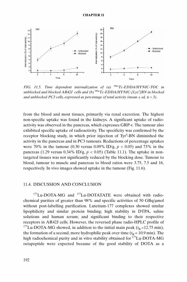
CHAPTER 11
from the blood and most tissues, primarily via renal excretion. The highestnon-specific uptake was found in the kidneys. A significant uptake of radio-activity was observed in the pancreas, which expresses GRP-r. The tumour also exhibited specific uptake of radioactivity. The specificity was confirmed by the receptor blocking study, in which prior injection of Tyr4-BN diminished the activity in the pancreas and in PC3 tumours. Reductions of percentage uptakes were 70% in the tumour (0.30 versus 0.09% ID/g, p < 0.05) and 73% in the pancreas (1.29 versus 0.34% ID/g, p < 0.05) (Table 11.1). The uptake in non-targeted tissues was not significantly reduced by the blocking dose. Tumour to blood, tumour to muscle and pancreas to blood ratios were 3.75, 7.5 and 16, respectively. In vivo images showed uptake in the tumour (Fig. 11.6).
11.4. DISCUSSION AND CONCLUSION
177Lu-DOTA-MG and 177Lu-DOTATATE were obtained with radio-chemical purities of greater than 98% and specific activities of 50 GBq/µmol without post-labelling purification. Lutetium-177 complexes showed similar lipophilicity and similar protein binding; high stability in DTPA, saline solutions and human serum; and significant binding to their respective receptors in AR42J cells. However, the reversed phase radio-HPLC profile of 177Lu-DOTA-MG showed, in addition to the initial main peak (tR =12.75 min), the formation of a second, more hydrophilic peak over time (tR = 10.9 min). Thehigh radiochemical purity and in vitro stability obtained for 177Lu-DOTA-MG octapeptide were expected because of the good stability of DOTA as a
FIG. 11.5. Time dependent internalization of (a) 99mTc-EDDA/HYNIC-TOC in unblocked and blocked AR42J cells and (b) 99mTc-EDDA/HYNIC-[Lys3]BN in blocked and unblocked PC3 cells, expressed as percentage of total activity (mean ± sd, n = 3).
192

ARTEAGA DE MURPHY et al.
lanthanide complexation agent [11.8–11.10]. It is important to point out that the advantage of small molecules offered by DOTA-MG octapeptide affords the possibility of providing a better target to background contrast because of the more rapid blood clearance.
The presence of two major peaks has already been described for 111In labelled DTPA-[D-Glu1]-MG [11.12] and 99mTc labelled HYNIC-[D-Glu1]-MG [11.8]. However, the authors of those studies reported that the two peaks were formed immediately post-labelling and were dependent on the reaction temperature and incubation time. In our findings, the single peak obtained post-labelling of DOTA-MG is converted over time to a second species. Von Guggenberg et al. [11.15] state that one of the carboxyl groups of the Glu residues at the amino terminus could be involved in the complex formation, resulting in a different species at a high temperature. This explanation is not likely for 177Lu labelling of DOTA-MG, because DOTA is a highly rigid ionizable polyaminocarboxylic macrocycle that has been proven to form very stable lanthanide complexes. To explain the experimental results obtained, several parameters of the optimized structural molecules were analysed by molecular mechanics and quantum mechanical calculation. Such
TABLE 11.1. BIODISTRIBUTION IN MICE WITH INDUCED PC3 TUMOURS, 2 H POST-ADMINISTRATION OF 99mTc-EDDA/HYNIC-[Lys3]BN (expressed as percentage of injected activity per gram of tissue (% ID/g) mean ± SD; n = 4)
Region Unblocked Blocked
Blood 0.08 ± 0.03 0.11 ± 0.06
Heart 0.05 ± 0.02 0.07 ± 0.03
Lung 0.10 ± 0.04 0.11 ± 0.05
Liver 0.17 ± 0.04 0.16 ± 0.04
Spleen 0.08 ± 0.04 0.10 ± 0.05
Pancreas 1.29 ± 0.31a 0.34 ± 0.16a
Kidney 4.70 ± 1.20 5.20 ± 1.8
Intestine 0.17 ± 0.10 0.26 ± 0.11
Muscle 0.05 ± 0.03 0.04 ± 0.02
Tumour 0.30 ± 0.11a 0.09 ± 0.03a
a Statistically significant difference (p < 0.05) between blocked and unblocked animals.
193

CHAPTER 11
considerations indicated that Lu-DOTA-MG presents two conformers with similar energies of 183.85 and 191.48 kcal/mol, possibly associated with the presence of two major HPLC peaks with the same molecular recognition properties. It was also interesting to note that in vivo and in vitro results were not in agreement with one another. AR42J cells might have enough somato-statin receptors for cell binding but not enough for binding to tumour tissues. A medullary thyroid cancer cell line may provide a better model for clarifying the problem.
Conjugation of HYNIC and BN for the preparation of 99mTc-EDDA/HYNIC-[Lys3]BN modifies the lipophilic and pharmacokinetic properties of BN, producing a radiopharmaceutical with low hepatobiliary clearance and high renal excretion. This is an important observation, because most of the 99mTc labelled BN analogues have a tendency to accumulate in the liver and intestine as a result of hepatobiliary clearance [11.17–11.20], leading to inter-ference during the detection of BN/GRPr positive cancers and their metastases in the abdominal areas.
FIG. 11.6. Uptake of 99mTc-EDDA/HYNIC-[Lys3]BN in PC3 tumour cells in an athymic mouse.
194

ARTEAGA DE MURPHY et al.
Another important advantage of the 99mTc-EDDA/HYNIC-[Lys3]BN and 99mTc-EDDA/HYNIC-TOC reported in this work is the feasibility of using instant freeze-dried kit formulations with high radiochemical purities and high specific activities for routine clinical use [11.21]. The diagnostic use of BN labelled with 99mTc has been proved; in the future, BN labelled with beta emitters should be studied for its possible use in radiotherapy.
ACKNOWLEDGEMENTS
The authors would like to thank the IAEA for its much appreciated support and the Consejo Nacional de Ciencia y Tecnología (CONACYT), Mexico, for the realization of this research.
REFERENCES TO CHAPTER 11
[11.1] LEWIS, J.S., SRINIVASAN, A., SCHMIDT, M.A., ANDERSON, C.J., In vitro and in vivo evaluation of 64Cu-TETA-Tyr3-octreotate. A new somatostatin analogue with improved target tissue uptake, Nucl. Med. Biol. 26 (1999) 267–273.
[11.2] DECRISTOFORO, C., MELENDEZ-ALAFORT, L., SOSABOWSKI, K.K., MATHER, S.J., 99mTc-HYNIC-Tyr3-octreotide for imaging somatostatin-receptor-positive tumours: Preclinical evaluation and comparison with 111In-octreotide, J. Nucl. Med. 41 (2000) 1114–1119.
[11.3] BREEMAN, W.A.P., DE JONG, M., ISSER, T.J., ERION, J.L., KRENNING, E.P., Optimising conditions for radiolabelling of DOTA-peptides with Y-90, In-111 and Lu-177 at high specific activities, Eur. J. Nucl. Med. Mol. Imaging 30(2003) 917–920.
[11.4] KWEKKEBOOM, D.J., et al., [Lu-177-DOTA(0),Tyr(3)]octreotate: Compar-ison with [In-111-DTPA(0)]octreotate in patients, Eur. J. Nucl. Med. 28 (2001) 1319–1325.
[11.5] PILLAI, M.R.A., CHAKRABORTY, S., DAS, T., VENKATESH, M., RAMAMOORTHY, N., Production logistics of Lu-177 for radionuclide therapy, Appl. Radiat. Isot. 59 (2003) 109–118.
[11.6] HASHIMOTO, K., MATSUOKA, H., UCHIDA, S., Production of no carrier-added Lu-177 via the Yb-176 (n,gamma)Yb-177- Lu-177 process, J. Radioanal. Nucl. Chem. 255 (2003) 575–579.
[11.7] SCMITT, A., et al., Biodistribution and dosimetry of Lu-177-labelled [DOTA(0),Tyr(3)]octreotate in male nude mice with human small cell lung cancer, Cancer Biotherapy Radiopharm. 18 (2003) 593–559.
[11.8] LEWIS, J.S., et al., Toxicity and dosimetry of Lu-177-DOTA-Y3-octreotate in a rat model, Int. J. Cancer 94 (2001) 873–877.
195

CHAPTER 11
[11.9] BREEMAN, W.A.P, MEARADJI, A., CAPELLO, A., BERNARD, B.F., VAN EIJCK, C.H.J., Anti-tumour effect and increased survival after treatment with [Lu-177-DOTA(0),Tyr(3)]octreotate in a rat liver micrometastases model, Int. J. Cancer 104 (2003) 376–379.
[11.10] REUBI, J.C., SCHAER, J.C., WASER, B., Cholecystokinin (CCK)-A and CCK-B/gastrin receptors in human tumours, Cancer Res. 57 (1997)1377–1386.
[11.11] GOTTHART, M., et al., Scintigraphy with In-111-DTPA-D-Glu1-Minigastrin and In-111-DTPA-D-Phe1-Octreotide in patients with gastrointestinal neuroendocrine tumours: Results of the first 60 patients, Eur. J. Nucl. Med. Mol. Imaging 30 (2003) S181.
[11.12] BÉHÉ, M., BECKER, W., GOTTHARDT, M., ANGERSTEIN, C., BEHR, T.M., Improved kinetic stability of DTPA-DGlu as compared with conven-tional monofunctional DTPA in chelating indium and yttrium: Preclinical and initial clinical evaluation of radiometal labelled minigastrin derivatives, Eur. J. Nucl. Med. Mol. Imaging 30 (2003) 1140–1146.
[11.13] BEHR, T.M., et al., Targeting of cholecystokinin-B/gastrin receptors in vivo: Preclinical and initial clinical evaluation of the diagnostic and therapeutic potential of radiolabelled gastrin, Eur. J. Nucl. Med. 25 (1998) 424–430.
[11.14] BEHR, T.M., BÉHÉ, M., Cholecystokinin-B/gastrin receptor-targeting peptides for staging and therapy of medullary thyroid cancer and other cholecystokinin-B receptor-expressing malignancies, Semin. Nucl. Med. 32 (2002) 97–109.
[11.15] VON GUGGENBERG, E., et al., 99mTc-labelling and in vitro and in vivo evalu-ation of HYNIC- and (Nα-His) acetic acid-modified [D-Glu1]-minigastrin, Bioconjug. Chem. 15 (2004) 864–871.
[11.16] LA BELLA, R., et al., In vitro and in vivo evaluation of a 99mTc(I)-labelled bombesin analogue for imaging of gastrin releasing peptide receptor-positve tumours, Nucl. Med. Biol. 29 (2002) 553–560.
[11.17] VARVARIGOU, A.D., et al., Synthesis, chemical, radiochemical and radiobio-logical evaluation of a new 99mTc-labelled bombesin-like peptide, Cancer Biotherapy Radiopharm. 17 (2002) 317–326.
[11.18] SMITH, J.C., et al., Radiochemical investigations of 177Lu-DOTA-8-Aoc-BBN[7-14]NH2: An in vitro/in vivo assessment of the targeting ability of this new radiopharmaceutical for PC-3 human prostate cancer cells, Nucl. Med. Biol. 29 (2003) 101–109.
[11.19] CHEN, X., et al., MicroPET and autoradiographic imaging of GRP receptor expression with 64Cu-DOTA-[Lys3]Bombesin in human prostate adenocarci-noma xenografts, J. Nucl. Med. 45 (2004) 1390–1397.
[11.20] MIRALLES, F., CZERNICHOW, P., SCHARFMANN, R., Pancreatic acinar AR42J cells express functional nerve growth factor receptors, J. Endocrinol. 160 (1999) 433–442.
[11.21] VON GUGGENBERG, E., et al., Radiopharmaceutical development of a freeze-dried kit formulation for the preparation of [99mTc-EDDA-HYNIC-D-Phe1, Tyr3]-octreotide, a somatostatin analogue for tumour diagnosis, J. Pharm. Sci. 93 (2004) 2497–2506.
196
Chapter 12
LABORATORY EVALUATION OF THE BETA EMITTING RADIONUCLIDES 177Lu, 131I, 153Sm AND 166Ho, AND RADIOPHARMACEUTICALS FOR RADIOTHERAPY
M.M. ISHFAQ, H. NIZAKAT, K.M. BASHAR, I. HAIDERIsotope Production Division, Pakistan Institute of Nuclear Science and Technology (PINSTECH), Islamabad, Pakistan
Abstract
Reactor produced beta emitting radionuclides such as 131I, 177Lu, 166Ho and 153Sm are emerging as important tools for the treatment of various diseases, including cancer. Among these, 177Lu is considered to be important for therapy, since it decays with a half-life of 6.71 d by the emission of beta particles (Eβ of 498 keV (78.6%), 384 keV (9.1%) and 176 keV (12.2%)) to stable 177Hf. It also emits gamma rays with energies of 208 keV (11%) and 113 keV (6.4%), which are suitable for imaging. Recently, the somatostatin analogue DOTA-Tyr3-octreotate (DOTATATE) has been identified as a very successful peptide in targeting tumours and, when radiolabelled with a therapeutic isotope, in helping reduce tumour size in patients with metastatic neuroendocrine tumours. The report describes studies of the optimization of the production of 177Lu, 166Ho and 153Sm radionuclides in PARR-1 (Pakistan Atomic Research Reactor-1), using their naturally occurring and enriched metal oxide targets. High production yields were obtained for all three radionuclides. In studies of the labelling of DOTATATE with 131I, 177Lu, 166Ho and 153Sm, different experimental variables were used to determine the optimal preparation conditions. The 131I-DOTATATE, 177Lu-DOTATATE, 166Ho-DOTATATE and 153Sm-DOTATATE prepared were of optimal purity. Thin layer chromatography was used to measure the quality of these labelled peptides. The stability of the four complexes was studied in an acetate/ascorbate buffer and in saline at 4, 12, 24 and 36 h intervals. Quality control of the complexes was performed using the radio TLC technique. The results showed that these complexes were stable in both the acetate/ascorbate buffer and in saline for the 36 h period. Results of animal studies using rats indicated that the critical organ for the labelled peptides was the pancreas and the excretion route was via the kidney. Internalization studies of the 177Lu-DOTATATE carried out with AR42J cells showed greater than 7% internalization into AR42J cells after 80 min.
197

CHAPTER 12
12.1. INTRODUCTION
For the past two decades, there has been considerable interest in the use of short lived, moderate energy, beta emitting radionuclides for targeted radio-nuclide therapy [12.1–12.3]. This therapy involves the use of a radiolabelled molecule to selectively deliver a cytotoxic level of radiation to a diseased site with maximum radiation dose to the tumour and minimum radiation dose to normal organs. A number of beta particle emitting radionuclides such as 131I, 177Lu, 153Sm and 166Ho are available for targeted radionuclide therapy for a wide range of tissues. The physical characteristics of these radionuclides are listed in Table 12.1.
The radionuclides 177Lu, with a mean tissue penetration range of 670 μm, and 131I, with a mean range of 910 μm, are more effective for therapy of small tumours, while 166Ho, with a mean range of 3200 μm, is more useful for large tumours. Samarium-153, with a mean range of 1200 μm, is considered to be effective in the therapy of medium sized tumours.
A number of radiolabelled somatostatin analogues have been reported in the literature, with potential application for targeted radiotherapy of somato-statin receptor positive tumours in tumour bearing rodent models and in humans [12.4–12.9].
This report describes studies of the optimization of the production of 177Lu, 153Sm and 166Ho radionuclides in PARR-1 (Pakistan Atomic Research Reactor-1) using the natural and isotopically enriched targets. Also described are the optimization studies of the preparation of 177Lu-DOTA-Tyr3-octreotate (177Lu-DOTATATE), 131I-DOTATATE, 153Sm-DOTATATE and 166Ho-DOTATATE under different experimental conditions. Stability studies of these complexes in saline and buffer are also described. The pharmacokinetics of these complexes in an animal model were also investigated, as was the
TABLE 12.1. PHYSICAL CHARACTERISTICS OF SELECTED THERAPEUTIC RADIONUCLIDES
Radionuclide Half-life (d)Gamma
emissions(keV)
Averagebeta emissions
(keV)
Mean tissue penetration range
(μm)
131I 8.1 364 182 910177Lu 6.7 208 133 670153Sm 1.9 103 229 1200166Ho 1.1 810 666 3200
198

ISHFAQ et al.
usefulness of 177Lu-DOTATATE with respect to its internalization in AR42J cells, which overexpress somatostatin receptors.
12.2. MATERIALS
Enriched lutetium (68.9% 176Lu) and samarium (99.06% 152Sm) oxides were obtained from Cambro Scientific (Netherlands), and natural Lu2O3,Sm2O3 and Ho2O3 were obtained from Johnson & Mathew (Germany). High specific activity 131I-NaI solution was obtained from Radioactive Products PINSTECH (Pakistan). DOTATATE (piCHEM R&D, Austria), was provided by the IAEA. All other chemicals and reagents were of analytical grade. Measurements of pH were performed using a precalibrated pH meter (Hanna Instruments model 8417) connected to a single electrode (Hanna Instruments model 1331B). The radioactivity of the radionuclides was measured using a precalibrated radioisotope calibrator (Capintec model CRC-5RH), and the gamma count rate was performed with a gamma counter (Ludlum model 261). The radionuclidic purity of various radionuclides was checked by a multichannel analyser (Canberra, USA) series 85 coupled with an HPGe detector. The radiochromatograms were analysed with the help of a Berthold 2π scanner coupled with a NaI detector. The radiopharmaceuticals were analysed by HPLC using RP C18 columns (4.2 mm × 50 mm, 5 μm, Waters, USA) with UV (230 nm) and radioactive (Packard, USA) detection.
12.3. METHODS
12.3.1. Preparation of radionuclides
12.3.1.1. Lutenium-177
Lutetium-177 was produced by irradiation of lutetium samples in the core of PARR-1, a swimming pool type reactor, at a thermal neutron flux of 1 × 10¹⁴ n⋅cm–2⋅s–1 for specified periods of time ranging from 1 to 96 h. Naturally abundant Lu2O3 and enriched 176Lu2O3 were dissolved in 1M HNO3, evaporated to dryness and reconstituted in 0.1M HNO3, and again evaporated to dryness. The Lu(NO3)3 (2.59% 176Lu) powder and enriched Lu(NO3)3
(68.9% 176Lu) powder thus obtained were sealed in quartz ampoules and cold welded in aluminium containers for irradiation. In another experiment, quartz ampoules were filled with 0.2 mL of liquid Lu(NO3)3, sealed and cold welded in aluminium containers. These targets were then irradiated in PARR-1. The
199
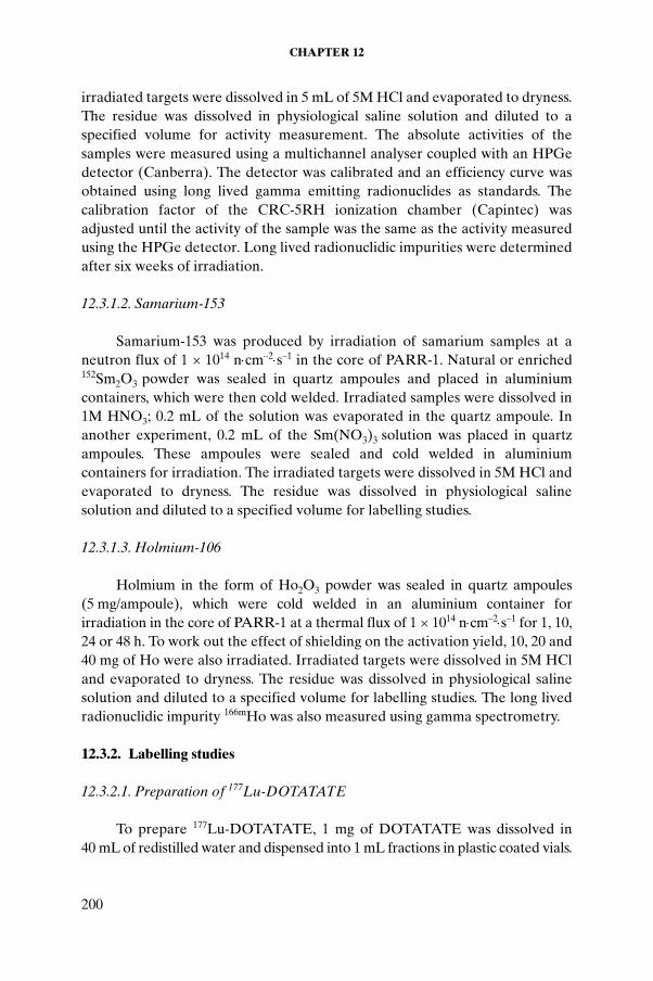
CHAPTER 12
irradiated targets were dissolved in 5 mL of 5M HCl and evaporated to dryness. The residue was dissolved in physiological saline solution and diluted to a specified volume for activity measurement. The absolute activities of the samples were measured using a multichannel analyser coupled with an HPGe detector (Canberra). The detector was calibrated and an efficiency curve was obtained using long lived gamma emitting radionuclides as standards. The calibration factor of the CRC-5RH ionization chamber (Capintec) was adjusted until the activity of the sample was the same as the activity measured using the HPGe detector. Long lived radionuclidic impurities were determined after six weeks of irradiation.
12.3.1.2. Samarium-153
Samarium-153 was produced by irradiation of samarium samples at a neutron flux of 1 × 10¹⁴ n⋅cm–2⋅s–1 in the core of PARR-1. Natural or enriched 152Sm2O3 powder was sealed in quartz ampoules and placed in aluminium containers, which were then cold welded. Irradiated samples were dissolved in 1M HNO3; 0.2 mL of the solution was evaporated in the quartz ampoule. In another experiment, 0.2 mL of the Sm(NO3)3 solution was placed in quartz ampoules. These ampoules were sealed and cold welded in aluminium containers for irradiation. The irradiated targets were dissolved in 5M HCl and evaporated to dryness. The residue was dissolved in physiological saline solution and diluted to a specified volume for labelling studies.
12.3.1.3. Holmium-106
Holmium in the form of Ho2O3 powder was sealed in quartz ampoules (5 mg/ampoule), which were cold welded in an aluminium container for irradiation in the core of PARR-1 at a thermal flux of 1 × 10¹⁴ n⋅cm–2⋅s–1 for 1, 10, 24 or 48 h. To work out the effect of shielding on the activation yield, 10, 20 and 40 mg of Ho were also irradiated. Irradiated targets were dissolved in 5M HCl and evaporated to dryness. The residue was dissolved in physiological saline solution and diluted to a specified volume for labelling studies. The long lived radionuclidic impurity 166mHo was also measured using gamma spectrometry.
12.3.2. Labelling studies
12.3.2.1. Preparation of 177Lu-DOTATATE
To prepare 177Lu-DOTATATE, 1 mg of DOTATATE was dissolved in 40 mL of redistilled water and dispensed into 1 mL fractions in plastic coated vials.
200

ISHFAQ et al.
These vials were then freeze dried for 24 h and refrigerated for further experi-ments. For labelling, 1 mL of acetate/ascorbate buffer (30mM NaOAc/25mM sodium ascorbate) at a pH ranging from 2 to 7 was added to a vial containing 25 μg of DOTATATE. To this was added 10 mCi of 177LuCl3. The vial was heated at 20–90oC for different time intervals ranging from 5 to 60 min. The radiochemical purity of 177Lu-DOTATATE was determined by radio TLC with C18 plates developed in MeOH:10% NH4OAc (70:30). Under these conditions, 177Lu-DOTATATE appears at Rf = 0.8, while 177Lu-acetate stays at Rf = 0. The labelling yields were determined by varying the pH, incubation temperature and incubation time.
12.3.2.2. Preparation of 131I-DOTATATE
To prepare 131I-DOTATATE, 1 mg of DOTATATE was dissolved in 100 mL of 0.02M acetic acid in redistilled water and dispensed into 1 mL fractions in plastic coated vials. These vials were then freeze dried for 24 h and refrigerated for use in further experiments. A vial containing 10 μg of DOTATATE was added to 40 μL of 0.1M phosphate buffer solution at pH7.5. A Na131I solution at pH7.5 with high specific activity (>2 × 10¹¹ Bq/mg) produced in our laboratory using dry distillation of irradiated natural TeO2
powder was used for the labelling studies. The labelling of DOTATATE (10 μg) with high specific activity 131I-NaI
(≥2 × 10¹¹ Bq/mg) was performed using the chloramine T method. Briefly, 10 μL (10 mCi) of radioiodine solution was added to the peptide solution in 40 μL of phosphate buffer solution (PBS). This was followed by addition of the chloramine T solution (20 μg/5 μL PBS) to a reaction vial. The vial was capped and carefully vortexed. The reaction was allowed to proceed for 2 min at room temperature and was terminated by addition of 35 μg of sodium metabisulphite solution (using 4-fold of the oxidizing agent).
The labelled 131I-DOTATATE was analysed by paper chromatography using 10 cm long Whatman 3 chromatography papers. For the paper chroma-tography studies, 5 μL of each test solution was spotted at 1.5 cm from the lower end of the paper strips, and the strips were developed in a 10% ammonium acetate:methanol (30:70 vol./vol.) mixture. Under these conditions, the radiolabelled peptide migrates to Rf = 0.7–0.8, while free 131I remains at the origin (Rf = 0). The strips were subsequently dried and cut into 1 cm segments. The radioactivity associated with each segment was measured in a well type NaI (Tl) detector.
201

CHAPTER 12
12.3.2.3. Preparation of 153Sm-DOTATATE
To prepare 153Sm-DOTATATE, 1 mg of DOTATATE was dissolved in 100 mL of 0.02M acetic acid in redistilled water and distributed into 1 mL fractions in plastic coated vials. These vials were then freeze dried for 24 h and refrigerated for use in further experiments. A vial containing 10 μg of DOTATATE was added to 1 mL of acetate/ascorbate buffer (30mM NaOAc/25mM sodium ascorbate) at a pH ranging from 2 to 8; 0.2 mCi of 153SmCl3 was then added, and the vial was heated at 20–90oC for time intervals ranging from 5 to 50 min. The radiochemical purity of the 153Sm-DOTATATE was determined by radio TLC with C18 plates developed in MeOH:10% NH4OAc (70:30). Under these conditions, 153Sm-DOTATATE appears at Rf = 0.8 while 153Sm-acetate stays at Rf = 0. The labelling yield was optimized by varying the pH, incubation time and temperature.
12.3.2.4. Preparation of 166Ho-DOTATATE
To prepare 166Ho-DOTATATE, 1 mg of DOTATATE was dissolved in 100 mL of 0.02M acetic acid in redistilled water and distributed into 1 mL fractions in plastic coated vials. These vials were then freeze dried for 24 h and refrigerated for use in further experiments. A vial containing 10 μg of DOTATATE was added to 1 mL of acetate/ascorbate buffer (30mM NaOAc/25mM sodium ascorbate) at a pH ranging from 2 to 8; 1 mCi of 166HoCl3 was then added and the vial was heated at 80oC for time intervals ranging from 5 to 60 min. The radiochemical purity of 166Ho-DOTATATE was determined by radio TLC with C18 plates developed in MeOH:10% NH4OAc (70:30). Under these conditions, 166Ho-DOTATATE appears at Rf = 0.8 while 166Ho-acetate stays at Rf = 0. The labelling yield was optimized by varying the pH, incubation time and temperature.
12.3.3. Stability of DOTATATE labelled with 177Lu, 131I, 153Sm and 166Ho
Stability studies of DOTATATE labelled with 177Lu, 131I, 153Sm and 166Ho were carried out in acetate/ascorbate buffer and saline at 4, 12, 24 and 36 h intervals at room temperature (15–18oC). The quality of the complexes was measured using TLC.
12.3.4. Animal studies
All animal experiments were performed in accordance with the protocol established by the Quality Control Group for Care and Animal Studies at
202

ISHFAQ et al.
PINSTECH. Biological studies of 177Lu-DOTATATE, 131I-DOTATATE, 166Ho-DOTATATE and 153Sm-DOTATATE in animals were performed in male Sprague-Dawley rats weighing approximately 200 g, all of which were housed separately for the duration of the experiment. Each rat was injected through the tail vein with 200 μL of the complex (~80 μCi). The rats were killed by cervical dislocation at 1, 6, 12, 24 and 72 h post-injection. A 1 mL sample of blood was drawn from the heart using a disposable syringe. The rats were then weighed and subsequently dissected, with special care taken to separate the blood and urine from the kill papers and the tissue washing. The urine was collected from the cages. Counting was performed using a Capintec dose calibrator.
12.3.5. Cell lines
The AR42J cells used in the experiments were obtained from the Department of Biological Studies, Arid University, Islamabad. The cells were incubated in a CO2 controlled oven at 37oC and were washed twice with the internalization buffer DMEM (HEPES buffer supplemented with L-glutamine, sodium pyruvate, penicillin, fungizone and bovine serum albumin). The culture medium was changed every third day, and the cells were sub-cultured every five days. When cells reached 80–90% confluence, they were dissociated with 0.05% trypsin and 0.02% EDTA.
12.3.5.1. Internalization studies
The AR42J cells were plated onto 35 mm culture dishes at a final concen-tration of 1 × 10⁶ cells and incubated for 1 h at 37 °C. Cells (1 × 10⁶ cells/tube) were incubated with 1 × 10⁴ counts/min per tube for 5, 20, 40, 80 or 120 min at 37°C. Incubation was interrupted by removal of the medium, and the cells were washed twice with PBS. After the last incubation period, 1 mL of 0.2N acetic acid/0.5M NaCl (pH2.5) was added and the cells were incubated for 5 min. The supernatant was collected (membrane bound radioligand fraction), the cells were lysed by treatment in 1N NaOH, and the radioactivity associated with the cells (internalized fraction) was determined. The percentage of internalized radioactivity was calculated. At the same time, non-specific binding was evaluated by incubating cells with tracer in the presence of a high concen-tration of unlabelled DOTATATE (150 µL of 10 µmol in each tube).
203

CHAPTER 12
12.4. RESULTS AND DISCUSSION
12.4.1. Production of radionuclides
The results of the irradiation of natural and enriched lutetium (68.9% 176Lu) in PARR-1 at a thermal neutron flux of 1 × 10¹⁴ n⋅cm–2⋅s–1 are shown in Table 12.2.
An increase of the specific activity of 177Lu was noted when irradiating 0.2 mL (liquid) of lutetium nitrate (68.9% 176Lu). More than 5.5 Ci of activity per milligram of 177Lu was obtained by irradiating this liquid target for 96 h. The probable reason why less activity was obtained using solid targets is self-shielding, which decreases neutron flux from the surface to the centre of the target matrix. Thus the actual induced activity is sometimes considerably reduced depending on the shape and volume of the target. Hence, the specific activity of 177Lu increases in the following order: Lu(NO3)3 in a solid target < Lu(NO3)3 in a liquid target. The radionuclidic impurity of 177mLu was found to be less than 2 × 10⁻³% for 96 h of irradiation.
The liquid and solid samples of 176Lu(NO3)3 (68.9% 176Lu) were also irradiated at a thermal neutron flux of 1.8 × 10¹⁴ n⋅cm–2⋅s–1. High specific activity (>8 Ci/mg) 177Lu was obtained by irradiating 0.2 mL of 40 μg of 176Lu as
TABLE 12.2. ACTIVITY YIELDS OF 177Lu FOR VARIOUS IRRADIATION TIMES AND AMOUNTS OF Lu (neutron flux = 1 × 10¹⁴ n⋅cm–2⋅s–1)
Irradiation time (h)
Amount of Lu irradiated
Activity measured
Specific activity
mCi MBq mCi/mg MBq/mg
1 4 mg 8 296 2 74
10 4 mg 78 2 886 19 721
24 4 mg 150 5 550 37 1 387
48 4 mg 384 14 208 96 3 552
48 0.2 mL, 4 mg 480 17 760 120 4 440
48 40 μg of 176Lu (68.9%) 100 3 700 2 500 92 500
48 0.2 mL, 40 μg of 176Lu (68.9%) 128 4 736 3 200 118 400
96 40 μg of 176Lu (68.9%) 184 6 808 4 600 170 200
96 0.2 mL, 40 μg of 176Lu (68.9%) 220 8 140 5 500 203 500
204

ISHFAQ et al.
176Lu2O3 (68.9% 176Lu) at a thermal neutron flux of 1.8 × 10¹⁴ n⋅cm–2⋅s–1 in PARR-I for 96 h (Table 12.3).
Samarium-153 has relatively high neutron capture cross-sections (with a thermal capture cross-section of 206 b and an epithermal capture cross-section of 3000 b), thus enabling production of high specific activity with minimal long lived radionuclidic impurities. The activities of various samarium targets obtained post-irradiation in PARR-I are given in Table 12.4.
The table shows that the specific activity of the powder target was much lower than that of the liquid target. The target of Sm(NO3)3 film prepared by evaporation in a quartz ampoule also showed higher specific activity than did the powdered form. The specific activity of 153Sm increases in the following order: Sm2O3 powder < Sm(NO3)3 film < Sm(NO3)3 liquid.
TABLE 12.3. ACTIVITY YIELDS OF 177Lu FROM 96 h OF IRRADIATION OF ENRICHED Lu AT A NEUTRON FLUX OF 1.8 × 10¹⁴ n⋅cm–2⋅s–1
Amount of Lu irradiatedActivity measured Specific activity
mCi MBq mCi/mg MBq/mg
40 μg of 176Lu (68.9%) 264 9 768 6 600 244 200
0.2 mL, 40 μg of 176Lu (68.9%) 330 12 210 8 250 305 250
TABLE 12.4. ACTIVITY YIELDS OF SELECTED SAMARIUM TARGETS (72 h of irradiation, neutron flux = 1 × 10¹⁴ n⋅cm–2⋅s–1)
Form of targetQuantity of
Sm (mg)
Yield Specific activity
mCi MBq mCi/mg MBq/mg
Sm(NO3)3 film 6.5 988 36 556 152 5 624
Sm(NO3)3 liquid 6.5 1 749 64 713 269 9 955
Sm2O3 powder 7.0 628 23 236 90 3 320152Sm(NO3)3 film
a 6.0 9 198 340 326 1 533 56 721152Sm(NO3)3 liquida 6.0 15 330 567 210 2 555 94 535152Sm2O3 powdera 6.2 6 336 234 432 1 022 37 811
a Enriched target 152Sm > 99%.
205

CHAPTER 12
The use of enriched 152Sm (>99%) minimizes the quantity of 145Sm (half-life: 340 d) and 151Sm (half-life: 93 a), which are produced by activation of stable 144Sm and 150Sm isotopes.
The activity yield of 166Ho is given in Table 12.5. As this table shows, the specific activity of 166Ho increases with an increase in the irradiation time from 1 to 48 h, whereas a decrease in specific activity is obtained with an increase in the amount of Ho from 10 to 40 mg for the same irradiation time (48 h). This decrease of radioactivity is the consequence of the self-shielding effect. The radionuclidic impurity of 166mHo (half-life: 1200 a, σ = 3.5 b) was found to be less than 2 × 10⁻³% for 48 h of irradiation.
12.4.2. Labelling studies of DOTATATE
The labelling yield of 177Lu-DOTATATE as a function of pH, incubation time and incubation temperature is presented in Tables 12.6, 12.7 and 12.8, respectively. The results indicate that high labelling yield of 177Lu-DOTATATE was obtained at pH5 with incubation for 25 min at a temperature of 90oC. The effect of varying the molar ratio of Lu to DOTATATE is shown in Fig. 12.1.
The labelling yield of 131I-DOTATATE (determined using paper chromato-graphy) as function of pH and incubation time is presented in Tables 12.9 and 12.10, respectively.
TABLE 12.5. ACTIVITY YIELDS OF 166Ho FOR SELECTED IRRADIATION TIMES AND AMOUNTS OF Ho (neutron flux 1 × 10¹⁴ n⋅ cm–2⋅ s–1)
Amount of Ho (mg)
Irradiation time (h)
Activity (mCi)
Specific activity (mCi/mg)
5 1 65 13
5 10 570 114
5 24 1 020 204
5 48 1 800 360
10 48 3 400 340
20 48 6 593 329
40 48 12 384 310
206
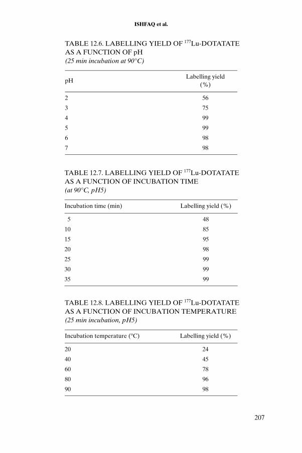
ISHFAQ et al.
TABLE 12.6. LABELLING YIELD OF 177Lu-DOTATATE AS A FUNCTION OF pH (25 min incubation at 90°C)
pHLabelling yield
(%)
2 56
3 75
4 99
5 99
6 98
7 98
TABLE 12.7. LABELLING YIELD OF 177Lu-DOTATATE AS A FUNCTION OF INCUBATION TIME (at 90°C, pH5)
Incubation time (min) Labelling yield (%)
5 48
10 85
15 95
20 98
25 99
30 99
35 99
TABLE 12.8. LABELLING YIELD OF 177Lu-DOTATATE AS A FUNCTION OF INCUBATION TEMPERATURE (25 min incubation, pH5)
Incubation temperature (oC) Labelling yield (%)
20 24
40 45
60 78
80 96
90 98
207
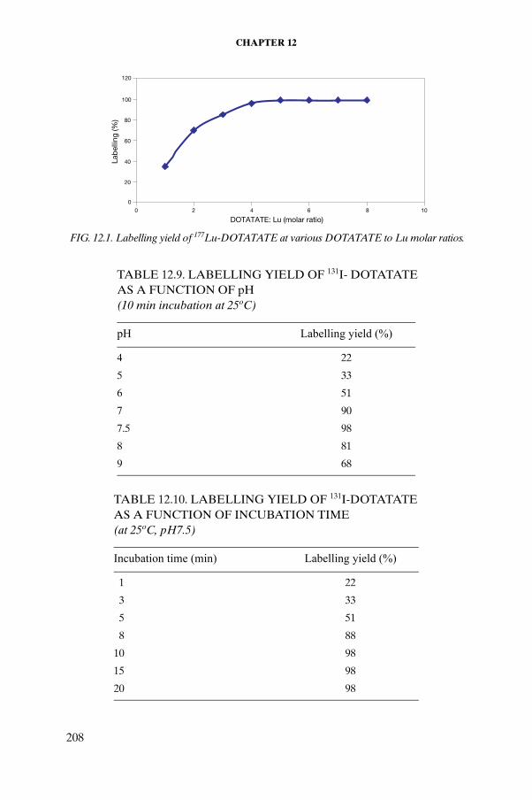
CHAPTER 12
TABLE 12.9. LABELLING YIELD OF 131I- DOTATATE AS A FUNCTION OF pH (10 min incubation at 25oC)
pH Labelling yield (%)
4 22
5 33
6 51
7 90
7.5 98
8 81
9 68
TABLE 12.10. LABELLING YIELD OF 131I-DOTATATE AS A FUNCTION OF INCUBATION TIME (at 25oC, pH7.5)
Incubation time (min) Labelling yield (%)
1 22
3 33
5 51
8 88
10 98
15 98
20 98
0
20
40
60
80
100
120
0 2 4 6 8 10
DOTATATE: Lu (molar ratio)
Lab
ellin
g (%
)
FIG. 12.1. Labelling yield of 177Lu-DOTATATE at various DOTATATE to Lu molar ratios.
208

ISHFAQ et al.
The labelling yield of 153Sm-DOTATATE as a function of pH, incubation time and incubation temperature is shown in Tables 12.11, 12.12 and 12.13, respectively. The results indicate that optimum labelling yields of 153Sm-DOTATATE were obtained at pH6 with an incubation time of 30 min at 80oC.
Similarly, the yields of 166Ho-DOTATATE as a function of pH, incubation time and incubation temperature are shown in Tables 12.14, 12.15 and 12.16, respectively. The data indicate that optimum labelling yields of 166Ho-DOTATATE were obtained at pH7 with an incubation time of 30 min at 80oC.
TABLE 12.11. LABELLING YIELD OF 153Sm-DOTATATE AS A FUNCTION OF pH (50 min incubation at 90°C)
pH Labelling yield (%)
2 34
3 48
4 71
5 86
6 98
7 98
8 98
TABLE 12.12. LABELLING YIELD OF 153Sm-DOTATATE AS A FUNCTION OF INCUBATION TIME (at 90°C, pH6)
Incubation time (min) Labelling yield (%)
5 36
10 68
15 78
20 81
25 96
30 98
40 98
50 98
209

CHAPTER 12
TABLE 12.13. LABELLING YIELD OF 153Sm-DOTATATE AS A FUNCTION OF INCUBATION TEMPERATURE (30 min incubation, pH6)
Incubation temperature (oC) Labelling yield (%)
20 38
40 68
60 92
80 99
100 99
TABLE 12.14. LABELLING YIELD OF 166Ho-DOTATATE AS A FUNCTION OF pH (30 min incubation at 80°C)
pH Labelling yield (%)
2 45
3 68
4 78
5 88
6 98
7 98
8 98
TABLE 12.15. LABELLING YIELD OF 166Ho-DOTATATE AS A FUNCTION OF INCUBATION TIME (at 80°C, pH7)
Incubation time (min) Labelling yield (%)
5 42
10 77
15 92
20 98
25 99
30 99
35 99
210

ISHFAQ et al.
12.4.3. Stability of DOTATATE labelled with 177Lu, 131I, 153Sm and 166Ho
The results of stability tests of the 131I-DOTATATE and 153Sm-DOTATATE complexes are presented in Table 12.17; those of the 177Lu-DOTATATE and 166Ho-DOTATATE complexes are presented in Table 12.18.
12.4.4. Animal studies
The data related to the biodistribution studies of the 177Lu-DOTATATE, 131I-DOTATATE, 166Ho-DOTATATE and 153Sm-DOTATATE complexes in rats are presented in Tables 12.19, 12.20, 12.21 and 12.22, respectively.
The biodistribution studies of 177Lu-DOTATATE, 131I-DOTATATE, 153Sm-DOTATATE, 166Ho-DOTATATE in a rat model indicated that the critical organ for these complexes was the pancreas and the excretion route was via the kidney. During the study, all the rats were found to show normal behaviour (movement, sleeping, eating).
TABLE 12.16. LABELLING YIELD OF 166Ho-DOTATATE AS A FUNCTION OF INCUBATION TEMPERATURE (30 min incubation, pH7)
Incubation temperature (oC) Labelling yield (%)
20 38
40 68
60 92
80 99
100 99
TABLE 12.17. STABILITY OF 131I-DOTATATE AND 153Sm-DOTATATE COMPLEXES IN BUFFER AND SALINE AT SELECTED TIME INTERVALS
Time (h)
131I-DOTATATE 153Sm-DOTATATE
Saline Buffer Saline Buffer
4 >99 >99 >99 >99
12 >98 >98 >98 >99
24 >98 >99 >98 >98
36 >98 >98 >98 >98
211

CHAPTER 12
TABLE 12.18. STABILITY OF 177Lu-DOTATATE and 166Ho-DOTATATE COMPLEXES IN BUFFER AND SALINE AT SELECTED TIME INTERVALS
Time (h)
166Lu-DOTATATE 166Ho DOTATATE
Saline Buffer Saline Buffer
4 >99 >99 >99 >98
12 >98 >98 >98 >98
24 >98 >99 >98 >98
36 >98 >98 >98 >98
TABLE 12.19. BIODISTRIBUTION OF 177Lu-DOTATATE IN MALE RATS AT SELECTED INTERVALS POST-INJECTION (% ID per organ ± SD, n = 4)
RegionTime post-injection (h)
1 6 12 24 72
Blood 2.12 ± 0.38 0.08 ± 0.03 0.05 ± 0.04 0.04 ± 0.02 0.03 ± 0.01
Lungs 0.21 ± 0.02 0.07 ± 0.01 0.05 ± 0.01 0.03 ± 0.00 0.01 ± 0.00
Liver 0.61 ± 0.08 0.32 ± 0.08 0.29 ± 0.07 0.26 ± 0.09 0.25 ± 0.06
Spleen 0.04 ± 0.01 0.02 ± 0.01 0.01 ± 0.00 0.01 ± 0.01 0.01 ± 0.00
Kidneys 2.1 ± 0.09 1.84 ± 0.09 1.78 ± 0.11 1.68 ± 0.08 1.49 ± 0.06
Heart 0.05 ± 0.01 0.01 ± 0.01 0.01 ± 0.00 0.01 ± 0.01 0.01 ± 0.00
Bone 10.55 ± 1.10 8.79 ± 1.22 7.22 ± 1.25 7.10 ± 2.10 4.85 ± 0.88
Adrenal gland
0.42 ± 0.05 0.29 ± 0.07 0.25 ± 0.04 0.24 ± 0.05 0.19 ± 0.04
Intestines 6.8 ± 1.25 5.2 ± 1.50 4.8 ± 1.23 4.12 ± 1.10 2.58 ± 0.88
Pancreas 11.25 ± 1.25 8.33 ± 1.95 5.62 ± 0.86 3.28 ± 1.10 1.75 ± 0.41
Urine 23.66 ± 12.4 —a — 31.19 ± 13.9 8.76 ± 2.42
Faeces 0.005 ± 0.001 — — 8.89 ± 1.25 4.34 ± 0.89
a—: not available.
212

ISHFAQ et al.
TABLE 12.20. BIODISTRIBUTION OF 131I-DOTATATE IN MALE RATS AT SELECTED INTERVALS POST-INJECTION (% ID per organ ± SD, n = 3)
RegionTime post-injection (h)
1 6 12 24 72
Blood 2.22 ± 0.38 0.08 ± 0.03 0.06 ± 0.04 0.04 ± 0.02 0.03 ± 0.01Lungs 0.23 ± 0.02 0.06 ± 0.01 0.06 ± 0.01 0.03 ± 0.00 0.01 ± 0.00Liver 0.59 ± 0.08 0.34 ± 0.08 0.28 ± 0.07 0.32 ± 0.09 0.25 ± 0.06Spleen 0.05 ± 0.01 0.03 ± 0.01 0.01 ± 0.00 0.01 ± 0.01 0.01 ± 0.00Kidneys 2.2 ± 0.09 1.78 ± 0.09 1.73 ± 0.11 1.78 ± 0.08 1.49 ± 0.06Heart 0.06 ± 0.01 0.01 ± 0.01 0.01 ± 0.00 0.01 ± 0.01 0.01 ± 0.00Bone 2.55 ± 1.10 1.98 ± 1.22 0.99 ± 1.25 0.80 ± 2.15 0.85 ± 0.88Adrenal gland 0.45 ± 0.05 0.31 ± 0.07 0.25 ± 0.04 0.25 ± 0.05 0.19 ± 0.04Intestines 6.6 ± 1.25 5.0 ± 1.50 4.85 ± 1.23 4.34 ± 1.10 2.19 ± 0.88Pancreas 10.25 ± 1.25 8.21 ± 2.23 5.58 ± 0.92 3.31 ± 1.10 1.75 ± 0.41Urine 22.66 ± 12.4 26.22 ± 8.90 28.19 ± 6.68 30.28 ± 12.88 8.76 ± 2.42Faeces 0.005 ± 0.001 1.08 ± 0.08 3.59 ± 1.10 6.89 ± 1.30 5.64 ± 0.89
TABLE 12.21. BIODISTRIBUTION OF 166Ho-DOTATATE IN MALE RATS AT SELECTED INTERVALS POST-INJECTION (% ID per organ ± SD, n = 3)
RegionTime post-injection (h)
1 6 12 24 72
Blood 1.85 ± 0.36 0.07 ± 0.03 0.05 ± 0.04 0.04 ± 0.02 0.03 ± 0.01
Lungs 0.25 ± 0.13 0.08 ± 0.01 0.05 ± 0.01 0.03 ± 0.00 0.01 ± 0.00
Liver 0.57 ± 0.08 0.35 ± 0.08 0.29 ± 0.07 0.25 ± 0.09 0.25 ± 0.06
Spleen 0.03 ± 0.01 0.03 ± 0.01 0.01 ± 0.00 0.01 ± 0.01 0.01 ± 0.00
Kidneys 189 ± 0.09 1.99 ± 0.09 1.78 ± 0.11 1.68 ± 0.08 1.35 ± 0.06
Heart 0.04 ± 0.01 0.01 ± 0.01 0.01 ± 0.00 0.01 ± 0.01 0.01 ± 0.00
Bone 4.22 ± 1.30 3.71 ± 1.32 1.22 ± 1.25 0.90 ± 1.10 0.30 ± 0.88
Adrenal gland
0.39 ± 0.05 0.31 ± 0.07 0.25 ± 0.04 0.23 ± 0.05 0.21 ± 0.04
Intestines 6.9 ± 1.31 5.45± 1.33 4.8 ± 1.23 4.22 ± 1.10 2.52 ± 1.12
Pancreas 10.75 ± 1.55 7.55 ± 1.95 5.62 ± 0.86 2.75 ± 1.10 1.89 ± 0.38
Urine 23.21 ± 10.4 24.85 ± 10.5 26.35 ± 9.95 31.19 ± 13.9 9.87 ± 2.35
Faeces 0.005 ± 0.001 0.99 ± 0.05 3.85 ± 1.22 7.25 ± 1.25 4.34 ± 0.89
213

CHAPTER 12
The 177Lu-DOTATATE complex showed more than 7% internalization into AR42J cells after 80 min (Fig. 12.2).
TABLE 12.22. BIODISTRIBUTION OF 153Sm-DOTATATE IN MALE RATS AT SELECTED INTERVALS POST-INJECTION (% ID per organ ± SD, n = 3)
RegionTime post-injection (h)
1 6 12 24 72
Blood 3.10 ± 0.38 0.08 ± 0.03 0.04 ± 0.04 0.04 ± 0.02 0.03 ± 0.01
Lungs 0.17 ± 0.02 0.07 ± 0.01 0.04 ± 0.01 0.04 ± 0.00 0.01 ± 0.00
Liver 0.53 ± 0.09 0.34 ± 0.08 0.22 ± 0.07 0.31 ± 0.09 0.25 ± 0.03
Spleen 0.05 ± 0.02 0.03 ± 0.01 0.01 ± 0.00 0.01 ± 0.01 0.01 ± 0.00
Kidneys 1.98 ± 0.12 1.88 ± 0.09 1.75 ± 0.11 1.62 ± 0.05 1.44 ± 0.05
Heart 0.06 ± 0.01 0.01 ± 0.01 0.01 ± 0.00 0.01 ± 0.01 0.01 ± 0.00
Bone 4.22 ± 1.50 2.12 ± 1.26 1.99 ± 1.30 1.38 ± 2.10 0.55 ± 0.58
Adrenal gland
0.45 ± 0.05 0.36 ± 0.07 0.28 ± 0.04 0.26 ± 0.05 0.29 ± 0.06
Intestines 6.24 ± 1.35 5.56± 1.50 4.85 ± 1.38 3.92 ± 1.10 2.67 ± 0.75
Pancreas 11.11 ± 1.22 8.87 ± 1.95 5.21 ± 0.86 2.88 ± 1.10 1.85 ± 0.23
Urine 21.66 ± 10.4 24.89 ± 5.22 25.56 ± 5.65 29.31 ± 13.9 7.55 ± 1.54
Faeces 0.005 ± 0.001 1.25 ± 0.05 4.68 ± 2.20 6.49 ± 1.25 4.68 ± 1.12
0
2
4
6
8
10
0 20 40 60 80 100 120 140
Incubation time (min)
Specific Non-specific
Inte
rnal
izat
ion
(%)
FIG. 12.2. Internalization of 177Lu DOTATATE into AR42J cells.
214

ISHFAQ et al.
REFERENCES TO CHAPTER 12
[12.1] HOEFNAGEL, C.A., Anticancer radiopharmaceuticals, Anticancer Drugs 2(1991) 107–132.
[12.2] VOLKERT, R.P., Therapeutic radiopharmaceuticals, Chem. Rev. 99 (1999) 2269–2292.
[12.3] WESSELS, E.K., et. al., Physical and chemical properties of radionuclide therapy, Semin. Radiat. Oncol. 10 (2000) 115–122.
[12.4] DE JONG, M., et. al., Combination radionuclide therapy using 177Lu and 90Y labelled Somatostatin analogs, J. Nucl. Med. 46 (2005) 13S–17S.
[12.5] LEWIS, J.S., et. al., Toxicity and dosimetry of 177Lu-DOTA-Y3-Octreotate in a rat model, Int. J. Cancer 94 (2001) 873–877.
[12.6] THAKUR, M.L., Radiolabelled peptides: Now and the future, Nucl. Med. Commun. 16 (1995) 724–732.
[12.7] FICHNA, J., JANECKA, A., Synthesis of target-specific radionuclide peptides for diagnosis and imaging, Bioconjug. Chem. 14 (2003) 3–17.
[12.8] KWEKKEBOOM, D.J., et. al., Treatment of patients with gastro-entero-pancre-atic (GEP) tumours with the novel radiolabelled somatostatin analogue [177Lu-DOTA-Tyr3]Octreotate, Eur. J. Nucl. Med. Mol. Imaging 30 (2003) 417–422.
[12.9] BODEI, L., et. al., Receptor radionuclide therapy with 90Y-DOTA-Tyr3-Octre-otide (90Y-DOTATOC) in neuroendocrine tumours, Eur. J. Nucl. Med. Mol. Imaging 31 (2004) 1038–1046.
215

.

Chapter 13
PRECLINICAL EVALUATION OF THERAPEUTIC RADIOPHARMACEUTICALS BASED ON 90Y AND 177Lu
D. PAWLAK, A. KORSAK, R. MIKOŁAJCZAK, B. JANOTA, U. KARCZMARCZYK, E. JAKUBOWSKARadioisotope Centre POLATOM, Otwock-Świerk, Poland
Abstract
The paper describes the procedure for labelling the DOTATATE conjugate with 90Y and 177Lu, as well as the methods used for quality control. Parameters such as stability of the radiolabelled preparation, serum stability and protein binding were assessed by internalization and competition assays. The influence of chemical contami-nants on the labelling yield of DOTATATE was investigated. Results of the first applica-tions of therapeutic doses of 90Y-DOTATATE in patients in Poland are also reported.
13.1. INTRODUCTION
DOTATATE, the somatostatin (cyclic peptide hormone) analogue TATE coupled with the macrocyclic chelator DOTA, is currently being studied for use for internal peptide receptor radionuclide therapy (PRRT). Two radionuclides, carrier-free 90Y and 177Lu obtained from enriched 176Lu, were used for labelling the peptide.
13.2. METHODS
13.2.1. Labelling of DOTATATE with 177Lu and 90Y
The radiolabelling of DOTATATE was carried out by addition of 100 μL of 0.4M sodium acetate containing 40 mg/mL dihydroxybenzoic acid at pH4.5 to 10 μg of DOTATATE (0.4 mg/mL in 0.4M sodium acetate at pH4.5). For labelling with 177Lu, a solution of 177Lu was added to the mixture to obtain a ligand to radionuclide molar ratio of 5:1. The reaction mixture was incubated at
217

CHAPTER 13
95°C for 25 min [13.1, 13.2]. For 90Y labelling, the ligand to radionuclide molar ratio required was greater than 20:1. The incubation conditions were the same as those for labelling with 177Lu.
13.2.2. Kit formulation for labelling DOTATATE
A kit formulation was developed in-house for the preparation of DOTATATE in a lyophilized, ready to use form. The kit contained 100 μg of DOTATATE, 50 mg of ascorbic acid and 6 mg of dihydroxybenzoic acid. The pH was maintained at 4.5. In a typical labelling procedure, the kit was dissolved in 1 mL of 0.9% NaCl followed by the addition of up to 200 μL of radionuclide solution (177LuCl3 or 90YCl3 in 0.05M HCl). Incubation was carried out at 95°C for 25 min. This labelling procedure resulted in a greater than 99% labelling yieldfor both radionuclides, with specific activities in the range of 1.4–3.5 mCi/μg (2.02–5.07 Ci/μmol) for 90Y-DOTATATE and 0.7–1.3 mCi/μg (1.1–1.8 Ci/μmol) for 177Lu-DOTATATE.
13.2.3. Determination of effective specific activity of radionuclides
Various quantities of the conjugate were added to a fixed radioactive concentration of both radionuclides in order to obtain ligand to 177Lu molar ratios of 1, 2.5, 5 and 7.5, and ligand to 90Y molar ratios of 1, 2, 5, 10, 25, 40, 50 and 100. The other parameters were as previously described.
13.2.4. Quality control
Three methods were used to determine the radiochemical purity of the labelled preparations: HPLC, TLC and solid phase extraction.
13.2.4.1. HPLC
A solvent module (Varian Prostar 210) with a UV detector (Varian Prostar 345) and an on-line beta ray detector (Raytest, Ramona) was used for HPLC analysis. Reversed phase HPLC was performed with a Microsorb 300 C18 column (4.6 mm × 250 mm, Varian) at a flow rate of 0.75 mL/min and UV detection at 254 nm [13.3]. The solvents used were acetonitrile (solvent A) and 0.1% TFA in water (solvent B). The following elution gradient was used: 0–5 min 95% B; 5–10 min 95 to 0% B; 10–15 min 0% B; 15–20 min 0 to 95% B; 20–25 min 95% B. Prior to injection, 25 μL of 0.4M sodium acetate solution containing 1.0 mg/mL DTPA was added to 25 μL of the test sample to bind free radionuclides. Under these conditions, the retention time of 177Lu-DTPA and
218
PAWLAK et al.
90Y-DTPA was observed to be 4.0 min, while that of 177Lu-DOTATATE and 90Y-DOTATATE was 13.8 min.
13.2.4.2. TLC
To determine the content of unbound 177Lu or 90Y, TLC analyses were performed using cellulose plates (Merck Art. 5574) and MeOH:water:25% ammonia (50:50:2) (vol./vol./vol.) as the mobile phase. Under these conditions, the unbound lutetium remains at the origin while the other components migrate with the solvent front.
13.2.4.3. SepPak C18
Solid phase extraction was used to determine radiochemical purity and to purify the labelled preparation before internalization and competition assays. The analyses were performed using the following procedure. A SepPak C18 column was activated using 5 mL MeOH or EtOH and washed with 20 mL of 0.4M sodium acetate or 0.3M ascorbic acid; 5 μL of the test sample was then dissolved in 500 μL of 0.4M sodium acetate or 0.3M ascorbic acid and loaded on the column. The column was washed with 5 mL of 0.4M sodium acetate or 0.3M ascorbic acid (fraction A), and then with 5 mL MeOH or EtOH (fraction B). The radiochemical purities of the preparations were calculated as the percentages of fraction B activity related to the sum of the activities of fractions A and B and the activity retained in the column.
13.2.5. In vitro stability
The radiolabelled preparations were stored at room temperature in 0.4M sodium acetate or 0.3M ascorbic acid solution up to 72 h post-labelling for 177Lu-DOTATATE and up to 36 h post-labelling for 90Y-DOTATATE. The influence of the radioactive concentration of the stored preparation on its stability was investigated using solutions of various radioactive concentrations: 1.0, 20 and 50 mCi/mL for 90Y-DOTATATE, and 1.0, 15 and 35 mCi/mL for 177Lu-DOTATATE. The stability of the preparations (described as the radio-chemical purity) was determined by HPLC and SepPak separation.
13.2.6. Serum stability
For each radiolabelled preparation, after the radiochemical purity was ascertained, 5 μL of the preparation was added to a 50 μL portion of aliquoted human serum. The mixture was then incubated at 37°C up to 24 h for
219

CHAPTER 13
177Lu-DOTATATE and up to 6 h for 90Y-DOTATATE. An aliquot of 50 μL of 96% ethanol and 25 μL of 0.4M sodium acetate solution containing 1.0 mg/mL DTPA was added to 25 μL of this solution. The resultant solution was centrifuged at 5000g for about 5 min. The clear supernatant solution was analysed by HPLC, TLC and SepPak column. The remaining 30 μL of the tested sample was used for the protein binding test.
13.2.7. Protein binding test
MicroSpin G-50 columns were prespun at 2000g for 1 min; 25 μL of the radiolabelled peptide in human serum (at a ratio of 1:10) and 25 μL of the radiolabelled peptide in 0.9% NaCl solution (at a ratio of 1:10) were incubated and then centrifuged at 2000g for 2 min. The activities of the column and collected eluate were measured using a NaI scintillation counter.
13.2.8. Internalization
For the internalization experiments, AR42J cells were seeded at a density of about 800 000 cells per well on six-well plates and incubated for 24–48 h. The cells were then washed twice with RPMI-1640 medium supplemented with 10% foetal calf serum (FCS) and antibiotics. The cells were supplied with fresh medium, and the radiolabelled preparation, corresponding roughly to 200 fmol peptide in 150 µL of PBS/0.5% BSA buffer, was added. To determine non-specific binding, 150 µL of 10µM Sandostatin solution was added. The cells were incubated at room temperature and at 37oC in triplicate for 5, 15, 30, 60, 90 and 120 min. Incubation was interrupted by removal of the medium and rapid rinsing twice with ice-cold medium. The cells were then incubated twice at room temperature in glycine buffer for 5 min. The supernatant was collected (membrane bound radioligand fraction), the cells were lysed by treatment in 1N NaOH and cell radioactivity was measured (internalized fraction).
13.2.9. Competition assay
For the competition assays, AR42J cells were seeded at a density about 800 000 cells per well on six-well plates and incubated for 24–48 h. Cells were then washed twice with RPMI-1640 medium supplemented with 10% FCS and antibiotics. The cells were supplied with fresh medium, and the radiolabelled preparation, corresponding roughly to 200 fmol peptide in 150 µL PBS/0.5% BSA buffer, was added. The competitor (Sandostatin) was added in ten concentrations (0.01, 0.1, 1.0, 3.0, 10, 30, 100, 300, 1 000, 10 000nM) in a total volume of 150 µL of PBS/0.5% BSA buffer. The cells were incubated for 2 h at
220

PAWLAK et al.
room temperature and at 37°C, and at two different specific activities: 1.5 and 3.0 mCi/μg. Incubation was interrupted by removal of the medium and rapid rinsing twice with ice-cold medium. The cells were then incubated twice at room temperature in glycine buffer for 5 min. The supernatant was collected and the cells were lysed by treatment in 1N NaOH. The cell radioactivity was measured (bound radioligand fraction). The IC50 value was calculated by non-linear regression using the SigmaPLOT program.
13.2.10. Influence of chemical impurities on labelling yields of DOTATATE
To study the influence of chemical impurities on labelling yields of DOTATATE, a solution of 90Y or 177Lu was added to 50 mL of 0.4M sodium acetate containing 10 μg of DOTATATE and 50 mg/mL of ascorbic acid (pH4.5) to obtain a DOTATATE to radionuclide molar ratio of about 20. These solutions were spiked with varying quantities of metallic contaminants (e.g. Ca, Cu, Cd, Fe, Zn). The quantities of metal cations were adjusted to obtain metal to radiometal molar ratios ranging from 1 to 100. The concen-tration of each analyte was verified by ICP optical emission spectrometry. The reaction mixture was incubated at 95°C for 25 min. Each point was tested in duplicate.
13.3. RESULTS
The effective specific activity (taken as the maximum specific activity that allows attainment of >98% radiochemical purity) of the radionuclides obtained for the experiments was about 5 times lower than the maximum theoretical specific activity for 177Lu and about 25 times lower than that for 90Y. The labelling yield of 177Lu-DOTATATE and 90Y-DOTATATE as a function of the DOTATATE to radionuclide molar ratio is presented in Fig. 13.1. At Lu to DOTATATE molar ratios of 1:1 and 1:2.5, the labelling yields were 8.8 and 82.9%, respectively. The labelling yield (determined by TLC, SepPak column and HPLC (Fig 13.2)) exceeded 99.0% in the presence of the 5-fold excess of the ligand at a specific activity of 285 mCi/μmol. For 90Y, the 25-fold excess of the ligand gives a labelling yield greater than 99% with a specific activity greater than 1.0 Ci/μmol.
The 90Y-DOTATATE complex is stable up to 24 h in storage at room temperature, and the radiochemical purity of the preparation is greater than 99% when the radioactive concentration is less than 20 mCi/mL. When the radioactive concentration is 50 mCi, the stability of the preparation decreases,
221

CHAPTER 13
and the complex is stable up to 18 h. The 177Lu-DOTATATE complexes are stable up to 24 h for both radioactive concentrations, 15 and 35 mCi/mL (Fig. 13.3).
The results of the serum stability study are depicted in Fig. 13.4. During the 6 h incubation in human serum, the radiochemical analysis showed high labelling yield (>98%) for 90Y-DOTATATE, as assessed by HPLC, TLC and SepPak column. The 177Lu-DOTATATE preparation was also very stable (>98% radiochemical purity) when incubated in human serum up to 24 h. The
0
20
40
60
80
100
7.5 5.0 2.5 1.0
DOTATATE : Lu molar ratio0
20
40
60
80
100
100 50 40 25 10 5 2 1
DOTATATE : Y molar ratio
(a) (b)R
adio
chem
ical
pur
ity (%
)
Rad
ioch
emic
al p
urity
(%)
FIG. 13.1. Labelling yield of (a) 177Lu-DOTATATE and (b) 90Y-DOTATATE as a function of the DOTATATE to radionuclide molar ratio.
FIG. 13.2. HPLC profile of (a) 177Lu and (b) 177Lu-DOTATATE.
222

PAWLAK et al.
protein binding was 4% for 177Lu-DOTATATE and less than 2% for 90Y-DOTATATE for up to 6 h of incubation (Fig. 13.5).
The two preparations showed similar levels of internalization (8% for90Y-DOTATATE and 7% for 177Lu-DOTATATE) into AR42J cells at room temperature after 60–90 min (Figs 13.6–13.8). In studies carried out at 37°C, the most rapid internalization was observed during the first 30 min. Total
FIG. 13.3. Results of the stability studies of 90Y-DOTATATE and 177Lu-DOTATATE at selected radioactive concentrations.
FIG. 13.4. Results of serum stability studies of (a) 90Y-DOTATATE and (b) 177Lu-DOTATATE.
223

CHAPTER 13
FIG. 13.5. Results of protein binding studies of (a) 90Y-DOTATATE and (b) 177Lu-DOTATATE.
FIG. 13.6. Results of internalization studies of 177Lu-DOTATATE into AR42J cells at room temperature.
FIG. 13.7. Results of internalization studies of 90Y-DOTATATE into AR42J cells at room temperature (RT) and 37oC.
224

PAWLAK et al.
internalization levels were similar at room temperature and at 37°C, and reached approximately 8% of the total activity. Similar results were obtained for 99mTc-HYNIC-TOC, which was used for comparison.
The 90Y-DOTATATE binds specifically to the receptors of AR42J cells (Figs 13.9, 13.10). The IC50 value depends on the specific activity of the labelled peptide. The IC50 is about 4.2 when the specific activity of 90Y-DOTATATE is 1.5 mCi/µg and 1.07 when the specific activity is 3.0 mCi/µg.
The influence of chemical impurities showed that, of the five metallic impurities investigated (Ca, Cd, Cu, Fe and Zn), the strongest competitor is Fe. For the assumed labelling yield (98% radiochemical purity), in the case of the labelling of 90Y-DOTATATE, the maximum concentration of Fe is 5 µg/Ci of 90Y (Fig. 13.11). In the case of 177Lu-DOTATATE, the maximum concentration of Fe is approximately 100 µg/Ci of 177Lu (Fig. 13.12). This difference between the two maximum concentrations is probably due to the difference in stability constants for yttrium and lutetium complexes with DOTATATE. The stability constant for 177Lu-DOTA is ten times higher than that for 90Y-DOTA (Table 13.1).
For Cu, Cd and Zn, the maximum concentration allowing the attainment of 98% radiochemical purity is 10–30 µg/Ci of 90Y and 100–600 µg/Ci of 177Lu. The maximum concentration of Ca is much higher and exceeds 60 µg/Ci of 90Y and 1000 µg/Ci of 177Lu.
FIG. 13.8. Comparision of internalization of 90Y-DOTATATE, 177Lu-DOTATATE and 99mTc-HYNIC-TOC into AR42J cells at room temperature (RT) and 37 oC.
225
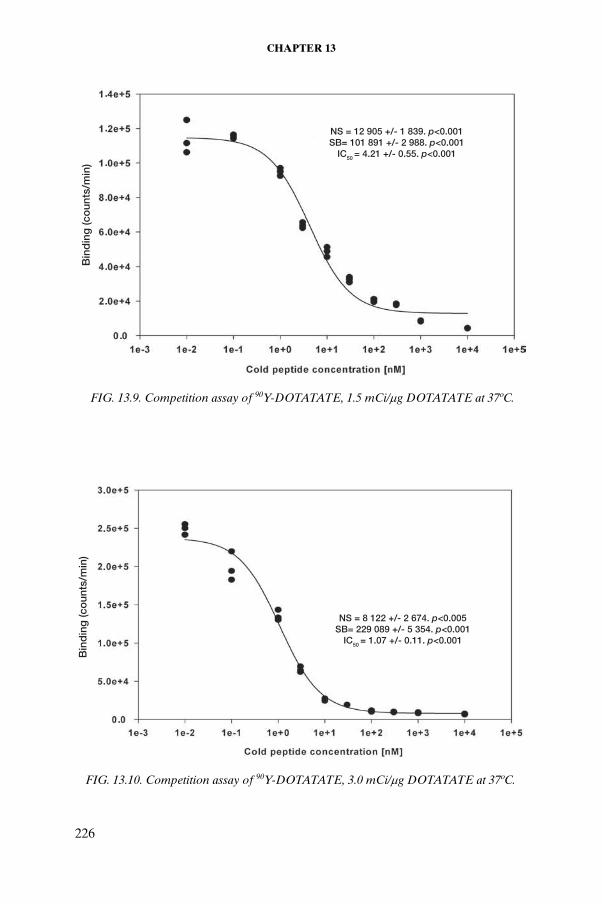
CHAPTER 13
FIG. 13.9. Competition assay of 90Y-DOTATATE, 1.5 mCi/μg DOTATATE at 37oC.
FIG. 13.10. Competition assay of 90Y-DOTATATE, 3.0 mCi/μg DOTATATE at 37oC.
226

PAWLAK et al.
FIG. 13.11. Influence of chemical impurities on labelling yield of 90Y-DOTATATE.
FIG. 13.12. Influence of chemical impurities on labelling yield of 177Lu-DOTATATE.
227

CHAPTER 13
Initial clinical trials were executed using the labelling methods described above; the results are shown in Figs 13.13–13.15. The investigations confirmed that bremsstrahlung scintigraphy can be used to estimate the effectiveness of therapy. A decrease in the uptake of 90Y-DOTATATE in neuroendocrine carcinoma metastases was observed during the course of the treatment. This
TABLE 13.1. THERMODYNAMIC STABILITY CONSTANTS (log Kml) OF SELECTED METAL–MACROCYCLIC COMPLEXES
Metallog Kml
DOTA DO3A
Y 24.3 —
La 21.7 —
Ce 23.0 19.7
Pr 23.0 —
Nd 23.0 —
Sm 23.0 —
Eu 23.5 20.69
Gd 24.0 22.02
Tb 24.2 —
Dy 24.8 —
Ho 24.5 —
Er 24.4 —
Tm 24.4 —
Yb 25.0 —
Lu 25.4 23.0
Ca 17.2 —
Co 20.2 —
Cu 22.5 —
Fe 29.4 —
Ni 20.5 —
Pb 22.7 —
Sr 15.22 —
Zn 21.05 —
228
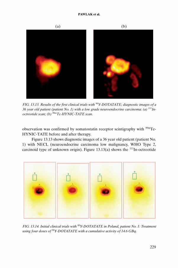
PAWLAK et al.
observation was confirmed by somatostatin receptor scintigraphy with 99mTc-HYNIC-TATE before and after therapy.
Figure 13.13 shows diagnostic images of a 36 year old patient (patient No. 1) with NECL (neuroendocrine carcinoma low malignancy, WHO Type 2, carcinoid type of unknown origin). Figure 13.13(a) shows the 111In-octreotide
FIG. 13.13. Results of the first clinical trials with 90Y-DOTATATE; diagnostic images of a 36 year old patient (patient No. 1) with a low grade neuroendocrine carcinoma: (a) 111In-octreotide scan; (b) 99mTc-HYNIC-TATE scan.
(a) (b)
FIG. 13.14. Initial clinical trials with 90Y-DOTATATE in Poland, patient No. 1: Treatment using four doses of 90Y-DOTATATE with a cumulative activity of 14.6 GBq.
229

CHAPTER 13
FIG. 13.15. Initial clinical trials with 90Y-DOTATATE in Poland, patient No. 2: (a, b) First post-treatment bremsstrahlung scan; (c, d) bremsstrahlung scan after completion of four therapy cycles.
230

PAWLAK et al.
scan; Fig. 13.13(b) shows the 99mTc-HYNIC-TATE scan. Liver deposits dominate the lesion with a high uptake of radiotracer; the patient qualified for 90Y-DOTATATE therapy.
Figure 13.14 shows initial clinical trials of 90Y-DOTATATE in patient No. 1, who was treated with four doses of 90Y-DOTATATE, using a cumulative activity of 14.6 GBq. The images show that liver deposits dominate the lesion with a high uptake of radiotracer. The bremsstrahlung images after the four courses of therapy show significant decreases of uptake with each treatment session.
Figure 13.15 shows a 48 year old patient with clinically confirmed NECLM (pancreatic gastrinoma, WHO Type 2). The first post-therapy bremsstrahlung scan is shown on the left hand side of Fig. 13.15; the brems-strahlung scan after completion of four therapy cycles is shown on the right hand side of the figure.
On the basis of the experience gained during investigation of the in vitro performance of 90Y-DOTATATE, the tracer was accepted for clinical use. Each clinical application of the complex followed the protocols established in the literature, including protection of the kidneys prior to administration of the therapeutic dose of 90Y-DOTATATE. Patients with proven metastatic neuro-endocrine tumours qualified for the treatment. Diagnostic somatostatin receptor scintigraphy was performed using 111In-octreotide and 99mTc-HYNIC-TATE. Bremsstrahlung images of 90Y-DOTATATE were taken to visualize the distribution of the tracer and to evaluate the treatment efficacy between cycles of therapy. In patient No. 1 (see Figs 13.13 and 13.14), a significant decrease of uptake was observed after the consecutive treatments, which was confirmed by the 111In-octreotide and 99mTc-HYNIC-TATE scans. Additionally, 99mTc-HYNIC-TATE was shown to be superior to 111In-octreotide scintigraphy in terms of resolution and tumour delineation.
A second example (see Fig. 13.15) showed a reduction of disease within the liver and pancreatic mass in patient No. 2 after four cycles of 90Y-DOTATATE therapy. Clinically, the patient showed no more symptoms of the disease, and the gastroscopy was normal. Therapy resulted in significant partial remission.
13.4. CONCLUSION
DOTATATE can be labelled with 177Lu and 90Y with high yields (>99% radiochemical purity). Preparations labelled with 177Lu and 90Y are stable in buffer up to 24 h. In vitro stability was observed to be very similar for both types of preparation.
231
CHAPTER 13
Our study shows that internalization into AR42J cells is 7% for 177Lu-DOTATATE and 8% for 90Y-DOTATATE after 60–90 min of incubation. The internalization rate is faster at early time points at temperatures above room temperature. The 90Y-DOTATATE binds specifically to receptors in live cells. The IC50 values obtained in our conditions were about 4nM at a radioactive concentration of 1.5 mCi/μg and 1.07nM at 3.0 mCi/μg.
High chemical purity of 90Y and 177Lu is crucial for efficient labelling of peptides for use in PRRT. The investigation confirmed that the presence of metals such as Ca, Cd, Cu, Fe and Zn significantly decreases the labelling yields of 90Y-DOTATATE and 177Lu-DOTATATE. Iron is the most harmful contaminant in the labelling procedure. The labelling yields were greater than 98% at Fe concentrations of less than 5 μg/Ci of 90Y and less than 100 μg/Ci of 177Lu.
Methods for radiolabelling 90Y-DOTATATE and 177Lu-DOTATATE were proved to be useful for the preparation of therapeutic doses for patients (>99.5% radiochemical purity). Initial clinical trials with PRRT were performed at the Clinical Hospital in Warsaw. The procedure for this treatment of patients with disseminated neuroendocrine tumours is now officially approved.
REFERENCES TO CHAPTER 13
[13.1] BREEMAN, W.A.P., DE JONG, M., VISSER, T.J., ERION, J.L., KRENNING, E.P., Optimising conditions for radiolabelling of DOTA-peptides with 90Y, 111In and 177Lu at high specific activities, Eur. J. Nucl. Med. Mol. Imaging 30 (2003) 917–920.
[13.2] DE JONG, M., et al., Somatostatin analogues labelled with different radionu-clides, J. Nucl. Med. 42 (1998) 368–371.
[13.3] INTERNATIONAL ATOMIC ENERGY AGENCY, 99mTc Labelled Peptides for Imaging of Peripheral Receptors, IAEA-TECDOC-1214, IAEA, Vienna (2001) 98–101.
232

Chapter 14
LABELLING OF DOTATATE WITH 177Lu AND 131I FOR DIAGNOSIS AND TARGETED THERAPY: IN VITRO AND IN VIVO COMPARATIVE EVALUATION
V. LUNGU*, D. NICULAE*, D. CHIPER*, R. MIHAI*, L. DANAILA**, S. BAICULESCU***
* ‘Horia Hulubei’ National Institute for Physics and Nuclear Engineering
** ‘Prof. Dr. V. Voiculescu’ Institute of Cerebro-Vascular Diseases
*** ‘Stefan Odobleja’ Academy of Cybernetics
Bucharest, Romania
Abstract
The studies focused on the radiolabelling of DOTA-Tyr3-TATE (DOTATATE) with 177Lu and 131I, on the biological behaviour of 177Lu-DOTATATE and 131I-DOTATATE and on comparative evaluation of the results obtained to select an appro-priate radiopeptide for potential application in somatostatin receptor radionuclide therapy. The radiopeptides were obtained with high purities and specific activities, but with different stabilities, with the 177Lu-DOTATATE being more stable than the 131I-DOTATATE. The results of competition and saturation binding experiments using rat brain cortex membrane homogenates show a high in vitro affinity (IC50: 4.74nM, Kd: 142.8 for 177Lu-DOTATATE; IC50: 1.28nM, Kd: 157.6 for 131I-DOTATATE). The biodis-tribution of each radiopeptide as well as the competitive biodistribution of 177Lu-DOTATATE and 131I-DOTATATE in rats bearing HRS1 (Hepatom RS1, a hepatocol-angiom carcinoma) tumours illustrate that 177Lu-DOTATATE is more stable and shows better tumour uptake than 131I-DOTATATE, and that the competitive localization index of 177Lu-DOTATATE is three times higher than that of 131I-DOTATATE. The flow cytometry measurements of the HRS1 tumour samples from rats treated with multiple doses of 177Lu-DOTATATE in combination with absorbed dose data show decreases of both the tumour cell proliferation index and DNA ploidy. These data indicate that 177Lu-DOTATATE is a good option for application in somatostatin receptor radionuclide therapy.
233

CHAPTER 14
14.1. INTRODUCTION
The peptide hormone somatostatin inhibits secretion from a wide variety of both endocrine and exocrine cells. It functions as a neurotransmitter and plays an important role in the regulation of cell proliferation and differenti-ation. Somatostatin exerts its effects through binding to specific surface membrane receptors. Somatostatin receptors are membrane glycoproteins with five different subtypes distributed in a variety of tissues throughout the body.
The overexpression of somatostatin receptors, especially subtype 2, in different neuroendocrine tumours has been studied with regard to in vivo local-ization of these tumours and their metastases using somatostatin analogues, modified somatostatin analogues or their bioconjugates radiolabelled with trivalent metallic radionuclides [14.1–14.3]. On the basis of the results obtained, including both the high quality images and the remarkable stability of the radiolabelled somatostatin under physiological conditions, it was decided to study the radiolabelling of somatostatin analogues with beta and alpha emitting therapeutic radionuclides [14.4–14.6] towards designing new radio-pharmaceuticals for peptide receptor radionuclide therapy.
Towards this end, studies were carried out which included the radio-labelling of the somatostatin receptor ligand DOTA-Tyr3-TATE (1,4,7,10-tetraazacyclotetradecane-N,N¢,N¢¢,N¢¢¢-tetraacetic acid tyrosine3-octreotate, or DOTATATE) with both 177Lu and 131I, and the quality control of the resulting complexes. In vitro studies of the biological affinity of the 177Lu-DOTATATE and 131I-DOTATATE for the somatostatin membrane receptor were carried out with the help of competition and saturation binding assays. In vivo studies of the biodistribution of 177Lu-DOTATATE and 131I-DOTATATE in animals, either alone or in competition, were also carried out. The radiotherapeutic effect of 177Lu-DOTATATE was evaluated by cytometry measurements. Estimation of absorbed doses of 177Lu-DOTATATE was carried out by mathe-matical modelling.
14.2. MATERIALS
The following radioisotopes were purchased for the studies: 131INa (Nordion Canada, specific activity of 1600 Ci/mg), 177LuCl3 (Nordion Canada, specific activity of 45 Ci/mg), 177LuCl3 (TRIGA research reactor, Pitesti, Romania, specific activity of 2.5 Ci/mg) and 177LuCl3 (POLATOM, Poland, specific activity of 7 Ci/mg). DOTATATE and Sandostatin were provided by the IAEA from piCHEM R&D (Austria). 125I-Tyr3-octreotide was received as a gift from the Institute of Oncology, Bucharest. Chemicals and reagents were
234

LUNGU et al.
purchased from Fluka Chemical and Sigma Aldrich Co. All solutions were prepared using ultrapure water (18 MΩ-cm resistivity). Radioactive samples were counted using a Picker Spectroscaler counter. Wistar rats were purchased from the Veterinary Laboratories of the ‘Cantacuzino’ Institute, Bucharest, Romania. Hepatom RS1 (HRS1) somatostatin receptor positive tumour cells and tumour bearing rats were purchased from the Institute of Oncology, Bucharest.
14.3. METHODS
14.3.1. Labelling of DOTATATE with 131I and 177Lu
The radiolabelling procedures were standardized with the help of optimi-zation studies of labelling parameters such as the peptide to radionuclide molar ratio, pH, temperature and time of reaction.
14.3.1.1. Labelling of DOTATATE with 131I
Iodination was carried out using the commonly used therapeutic dose of 100 mCi of 131I and 100 mg of DOTATATE, and a DOTATATE to 131I molar ratio of 1.7 and a DOTATATE to chloramine T molar ratio of 0.09. In a typical iodination procedure, 10 mCi of Na131I (5–10 mL with a specific activity of 22.2 × 10¹⁰ Bq/mg) was added to 10 mg of DOTATATE in 50 mL of 0.01M phosphate buffer solution (PBS) at pH7.4. To this reaction mixture, 19.7 mg of chloramine T in 20 mL of 0.1M PBS was added, followed by 1–3 min of stirring at room temperature. The reaction was stopped by addition of 39.8 mg of sodium metabisulfite in 10 mL of 0.1M PBS. A stabilizing agent, 3-hydroxy-4-aminobenzoic acid (HABA), was then added (5 mg for 10 mCi). For the experiments, 10 mCi of 131I-DOTATATE was used.
14.3.1.2. Labelling of DOTATATE with 177Lu
For the labelling of DOTATATE with 177Lu, 54 mCi of 177LuCl3/0.05N HCl (Nordion) with a specific activity of 45 Ci/mg was diluted to 100 mL with 0.05N HCl. The samples, consisting of 10 mg of DOTATATE in 50 mL of 0.4M acetate buffer at pH4.5, were labelled with 10 mCi of 177LuCl3 in 20 mL of 0.05N HCl (DOTATATE to 177Lu molar ratio of 3.7). The vials with reaction mixtures were incubated for 30 min at 80oC. After incubation and cooling, 5 mg of HABA was added as a stabilizer to prevent radiolytic damage. The stabilizer was added after radiolabelling, since thermal decomposition can occur during
235

CHAPTER 14
radiolabelling. The concentrations of the stabilizers were calculated for 10 mCi of 177Lu-DOTATATE.
14.3.1.3. Quality control and stability of 177Lu-DOTATATE and 131I-DOTATATE
The labelling yield and radiochemical purity of 177Lu-DOTATATE were checked by paper chromatography (PC) and thin layer chromatography (TLC) using different solvents such as 0.1M sodium citrate at pH5, wherein 177Lu-DOTATATE migrates to Rf = 0.62–0.70 and 177LuCl3 migrates to Rf = 1.0. In butanol:acetic acid:water (5:2:1), the radiopeptide migrates to Rf = 0.60–0.70 and free 177Lu migrates to Rf = 0.08–0.25. In the other solvent selected for quality control by TLC, 10% ammonium acetate:methanol (30:70), Rf = 0.76–0.85 for the 177Lu labelled peptide and Rf = 0.00–0.16 for free 177Lu.
The labelling yield and radiochemical purity of 131I-DOTATATE were checked with the help of PC and TLC. In water, used as one of the solvents, values of Rf = 0.65–0.70 for 131I-DOTATATE, Rf = 0 for I+ and Rf = 0.9–1.0 for I–
were observed. In the other solvent selected for quality control by TLC, butanol:acetic acid:water (5:2:1), values of Rf = 0.60–0.70 for 131I-DOTATATE and Rf = 0.9–1.0 for I+ and Rf = 0.2–0.3 for I– were observed.
The stability studies of the DOTATATE labelled with 177Lu and 131I were performed by incubation in 0.9% NaCl at room temperature for 24 h and in human serum at 37oC for 4 h. The samples were obtained by adding 0.2 mL of the labelled peptide to 2 mL of 0.9% NaCl and human serum. The radio-chemical purity was checked after each period of incubation.
14.3.2. In vitro studies
14.3.2.1. Preparation of somatostatin membrane receptor from rat brain cortex
The cerebral cortices of five rats were dissected and immediately placed in ice-cold Hank’s balanced salt solution (HBSS) at pH7.5 supplemented with 50 μL/mL penicillin, 50 μg/mL streptomycin and 10 000 KIU/L aprotinin. The cortices were then rinsed twice with ice-cold HBSS and homogenized in 10 mL of fresh HBSS.
The suspension was centrifuged at 500g for 10 min at 4oC in a Beckman J2-MC centrifuge, and the pellet was homogenized in 20 mL of homogenization buffer consisting of 25mM Tris buffer at pH7.5, 0.3M sucrose, 0.25mM PMSF, 1mM EGTA and 10 000 KIU/L aprotinine. The homogenate was centrifuged as described above, and the pellet was homogenized three more times in the same way, with the supernatant being saved after each centrifugation.
236

LUNGU et al.
The combined supernatant was then centrifuged at 48 000g for 45 min at 4oC. The final pellet was washed twice with 50mM Tris buffer at pH7.5 containing 5 mg MgCl2, 20 mg/L bacitracin, 0.25mM PMSF, 10 000 KIU/L aprotinine and 100 IU/mL RNase Inhibitor. It was then resuspended in 5 mL of washing buffer, separated into 100 mL aliquots and stored immediately at 80oC.
14.3.2.2. In vitro receptor binding assay of 131I-DOTATATE
A competition binding assay was performed using rat brain cortex membrane (50 mg protein). 125I-Tyr3-octreotide (970 Ci/mM, 35 000–40 000 counts/min) was added to each test tube in the presence of increasing concentrations of cold I-DOTATATE (synthesized under conditions similar to those used for 131I-DOTATATE). The following concentrations were used: 0.06, 0.13, 0.34, 0.68, 1.00, 2.72, 10.00, 57.80 and 100nM in a total volume of 300 mL of 50mM HEPES (pH7.6, 0.3% BSA, 5mM MgCl2, 10mM bacitracin). The samples were incubated for 2 h at room temperature, and the incubation was terminated by addition of ice-cold buffer (1 mL, 10mM HEPES, 150mM NaCl, pH7.6). The suspension was rapidly filtered over glass fibre filters (Whatman GF/B) presoaked in binding buffer using a Millipore multifiltration apparatus. The filters were rinsed four times with 2 mL of buffer, and the filter activity was measured on a NaI(Tl) gamma counter.
Saturation binding experiments for 131I-DOTATATE were performed using rat brain cortex membrane. For the total binding assay, the following concentrations of 131I-DOTATATE were prepared: 0.06, 0.13, 0.34, 0.68, 1.00, 2.72, 10.00, 57.80 and 100nM in 50 mL of binding buffer. For the binding studies, 50 mL of the radioligand solution (at the corresponding concentration) and 200 mL of rat brain cortex membrane homogenate containing 40 mg of protein were mixed. For the non-specific binding studies, instead of 50 mL of buffer, 20 mL of binding buffer and 30 mL of unlabelled peptide as the competitor (Sandostatin, 1mM in the reaction vial) were used. The tubes were incubated for 2 h at room temperature; binding was then interrupted by rapid filtration through a glass fibre filter pre-soaked with binding buffer. The filters were washed with binding buffer and then dried and counted using a NaI(Tl) gamma counter.
14.3.2.3. In vitro receptor binding assays of 177Lu-DOTATATE
The competition binding assays in this case were performed using the procedure described in the previous subsection. The binding affinity of cold Lu-DOTATATE was measured in rat brain cortex membrane. For the assay, 20 000 counts/min of 125I-Tyr3-octreotide was displaced with the following increasing concentrations of cold Lu-DOTATATE (synthesized using the same
237

CHAPTER 14
conditions as used for 177Lu-DOTATATE): 0.03, 0.14, 0.45, 0.70, 3.80, 12.80, 30.80, 170 and 500nM. The experimental data were analysed using the PRISM-2 program.
The saturation binding assays of 177Lu-DOTATATE were performed using the following concentrations: 0.05, 0.15, 4.5, 13.5, 40.5, 120.5 and 361.5nM for total and non-specific binding assays. The experimental results were analysed using the PRISM-2 program. The procedure detailed in the previous subsection was used.
14.3.3. In vivo studies
14.3.3.1. Animal models
HRS1, a hepato-colangiom carcinoma, obtained as a cell culture from the Institute of Oncology, Bucharest, was established as a solid tumour by subcuta-neous injection of 107 cells into 5 week old female Lewis rats weighing 150–200 g [14.7]. After the tumours had grown to approximately 1 cm3, serial propagation was carried out by subcutaneous injection of 0.2 mL of a 20% (wt./vol.) tumour suspension prepared by mincing the tumours and placing the material in 0.9% NaCl. Groups of HRS1 bearing rats were prepared 10 days prior to each of the following radiobiological studies: (a) biodistribution studies of 177Lu-DOTATATE and 131I-DOTATATE, (b) single dose radiotherapy experiments, (c) multiple dose radiotherapy experiments and (d) studies of the biodistribution of 177Lu-DOTATATE and 131I-DOTATATE co-injected with 150 μg of Sandostatin as a somatostatin receptor blocking agent.
14.3.3.2. Biodistribution studies of 177Lu-DOTATATE and 131I-DOTATATE
To study individual biodistribution of 177Lu-DOTATATE and 131I-DOTATATE in tumour bearing rats, animals from group (a) were injected intravenously with 0.2 mL of radioactive solutions containing 50 mCi of 177Lu-DOTATATE or 131I-DOTATATE, corresponding to approximately 0.68 mg of DOTATATE.
The tumour bearing rats in the group for competitive biodistribution studies were injected intravenously with 35 mCi of 177Lu-DOTATATE or 15 mCi of 131I-DOTATATE in a cocktail solution containing 0.68 mg of DOTATATE in 0.2 mL. At specified time points, the tumours, blood and various tissues and organs (liver, spleen, kidney, stomach, small intestine, large intestine, adrenal gland, pancreas, thyroid, pituitary gland, bone and lung) were removed and weighed, and the radioactivity was determined.
238

LUNGU et al.
The tumour bearing rats from group (d) were injected intravenously with 0.2 mL of 50 μCi of 177Lu-DOTATATE or 131I-DOTATATE, corresponding to approximately 0.68 μg of DOTATATE and 150 μg of Sandostatin as a blocking agent. After 1 h, the tumours and tissues expressing somatostatin receptors were removed and weighed, and the radioactivity was determined.
14.3.4. Radiotherapy experiments and flow cytometry measurements in HRS1 tumour bearing rats
14.3.4.1. Radiotherapy experiments with 177Lu-DOTATATE: Single dose
The tumour bearing rats (tumour volume: 0.5–2.5 cm3) from group (b) were injected intravenously with one dose of 15 mCi of 177Lu-DOTATATE prepared as described previously. Following the injection, tumour volumes were measured every 3 d for a month. At 1 month post-injection, the animals were sacrificed and the tumours were analysed using flow cytometry.
14.3.4.2. Radiotherapy experiments with 177Lu-DOTATATE: Multiple doses
In this experiment, the tumour bearing rats (tumour volume: 0.5–2.5 cm3) from group (c) were injected intravenously with 7.5 mCi of 177Lu-DOTATATE; 7 d later, the animals were again injected with 7.5 mCi of 177Lu-DOTATATE. Tumour volumes were measured and animals were sacrificed in a manner identical to that used for the single dose radiotherapy experiments.
14.3.4.3. Flow cytometry analysis of 177Lu-DOTATATE
Flow cytometry was used to analyse the DNA content of the cell cycle according to the following parameters:
— G0, G1 and G0/G1 phases, representing the (2N) diploid DNA content;— G2 + M phase, cells with (4N) tetraploid DNA content; — S phase, cells with 4(N) (tetraploid) DNA content ranging from 2(N) to
4(N).
The DNA ploidy is expressed by the DNA index (DI). For DI = 1, the DNA is normal and classified as DNA diploid; for DI ≠ 1, the DNA is abnormal and is classified as DNA aneuploid, a state usually associated with neoplastic cells. On the basis of these parameters, additional parameters for comparative estimation were calculated, including the proliferation index (PI = S + G2 + M), G2 to G1 phase ratios and the coefficient of variation (CV). The normal values
239

CHAPTER 14
of those parameters in our experiment were determined to be S ≤ 11%, PI ≤ 22%, CV < 9% and G2:G1 = 1.95–2.05.
The samples analysed were (a) control tumour samples from untreated tumour bearing rats, (b) tumour samples from tumour bearing rats treated with a single dose of 177Lu-DOTATATE and (c) tumour samples from tumour bearing rats treated with multiple doses of 177Lu-DOTATATE.
The samples were processed and stained with propidium iodide according to the method described in Ref. [14.8]. Flow cytometry DNA analysis was performed using a FACScan cytometer. The fluorescence intensity of the DNA labelled with propidium iodide was processed using CellQuest software, and the histograms of the DNA content were analysed using ModFit software. For each sample, 20 000 events were acquired. The DI was calculated with reference to the DNA diploid from the same sample.
14.3.5. Mathematical modelling
The biodistribution and radiotherapy data, together with required data for 177Lu-DOTATATE and the tumour size, were incorporated into a response model based on the linear quadratic model. This model provides data on inter-actions among important radiobiological parameters.
14.4. RESULTS
14.4.1. Labelling of DOTATATE with 131I
The DOTATATE was labelled with 131I using the method described previously, and the radiochemical purity of the radiopeptide obtained was estimated as a function of the stabilizer concentration and storage temperature, as shown in Table 14.1.
The results show that the addition of 5 mg of HABA to 131I-DOTATATE stabilizes the preparation, allowing high radiochemical purity of the radio-labelled compound to be maintained for 6 d at 4–8oC.
14.4.2. Labelling of DOTATATE with 177Lu
DOTATATE was labelled with 177Lu using the methods described previously. The radiochemical purity of the 177Lu-DOTATATE was studied in the presence of a stabilizer (HABA or GA) added post-labelling (Table 14.2).
The high radiochemical purity of the radiopeptide can be maintained for 7 d with the addition of 5 mg of HABA per 10 mCi of 177Lu-DOTATATE.
240

LUNGU et al.
14.4.3. In vitro stability of 177Lu-DOTATATE and 131I-DOTATATE
The stability of the radiopeptides in 0.9% NaCl and in human serum was tested using the method described previously. As shown in Figs 14.1 and 14.2, 131I-DOTATATE was found to be more stable than 177Lu-DOTATATE in 0.9% NaCl, while 177Lu-DOTATATE was found to be more stable than 131I-DOTATATE in human serum.
TABLE 14.1. RADIOCHEMICAL PURITY (RCP) DATA FOR 131I-DOTATATE
HABA (mg)Storage
temperature
RCP (%)
t = 0 h t = 3 d t = 7 d
5 RTa 96.5 95.2 89.2
4–8oC —b 95.2 95.7
10 RT 95.5 88.1 81.3
4–8oC — 95.8 82.1
0 RT 95.3 92.3 81.2
4–8oC — 93.1 91.1
a RT: room temperature.b —: not available.
TABLE 14.2. RADIOCHEMICAL PURITY (RCP) DATA FOR 177Lu-DOTATATE
Stabilizer (5 mg)Storage
temperature
RCP (%)
t = 0 h t = 3 d t = 7 d
GA RTa 98.5 96.3 94.1
4–8oC —b 97.1 95.4
HABA RT 98.6 98.1 96.7
4–8oC — 98.6 98.1
a RT: room temperature.b —: not available.
241

CHAPTER 14
14.4.4. In vitro studies
14.4.4.1. In vitro receptor binding assay of 131I-DOTATATE
The receptor binding data of 131I-DOTATATE were analysed using the Prism-2 program. The IC50 value for cold I-DOTATATE was determined to be 1.28nM, as shown in Fig. 14.3.
Using the same program, the saturation binding data shown in Fig. 14.4 were analysed, and the Kd value was calculated as 157pM.
FIG. 14.1. Stability of 131I-DOTATATE and 177Lu-DOTATATE in 0.9% NaCl .
FIG. 14.2. Stability of 131I-DOTATATE and 177Lu-DOTATATE in human serum.
242

LUNGU et al.
14.4.4.2. In vitro receptor assay of 177Lu-DOTATATE
The experimental results regarding the competitive and saturation binding of 177Lu-DOTATATE to somatostatin receptors were processed using Prism-2 software and are presented in Figs 14.5 and 14.6, respectively. Regarding the biological affinity and binding stability, the IC50 value obtained was 4.74nM and the Kd value was 142.8pM.
14.4.5. In vivo studies
14.4.5.1. Biodistribution of 177Lu-DOTATATE in HRS1 tumour bearing rats
The biodistribution of 177Lu-DOTATATE and 177LuCl3 for the time points studied is given in Tables 14.3 and 14.4, respectively. Rapid blood clearance was observed. The uptake of 177Lu-DOTATATE in tissue expressing somatostatin
FIG. 14.3. Competitive binding curve of cold I-DOTATATE.
FIG. 14.4. Saturation binding curve of 131I-DOTATATE.
243

CHAPTER 14
receptors is high immediately post-injection and decreases at each time point of the investigation. The uptake of 177Lu-DOTATATE increased in the tumour during the 24–72 h time range.
The uptake of 177Lu-DOTATATE in bone was high but decreased over time, indicating the possibility of decomposition post-injection, resulting in the formation of free 177Lu. In this context, the biodistribution of 177LuCl3 was studied and the uptake in bone was determined. The results show a progressive accumulation of 177Lu, with the maximum value at 4–24 h (Fig. 14.7). The high level of radioactivity was maintained at all points of time during the experiment, indicating that the pharmacokinetics of 177LuCl3 differ from those of 177Lu-DOTATATE.
FIG. 14.5. Competitive binding curve for cold Lu-DOTATATE.
FIG. 14.6. Saturation binding curve for 177Lu-DOTATATE.Linear fit: y = A + B*x : A = 0.48 ± 0.03; B = –0.007 ± 0.0008 pM–1
244

LUNGU et al.
TABLE 14.3. BIODISTRIBUTION OF 177Lu-DOTATATE (% ID/g of organ)
RegionTime post-injection (h)
3 24 48 72 168
Blood 0.72 ± 0.47 0.04 ± 0.03 0.04 ± 0.01 0.03 ± 0.01 0.03 ± 0.00
Liver 0.68 ± 0.15 0.44 ± 0.11 0.40 ± 0.05 0.28 ± 0.13 0.18 ± 0.01
Spleen 0.20 ± 0.11 0.14 ± 0.05 0.07 ± 0.01 0.03 ± 0.05 0.03 ± 0.00
Kidneys 2.12 ± 0.23 1.90 ± 0.78 1.55 ± 0.82 1.56 ± 0.02 1.23 ± 0.05
Stomach 1.95 ± 0.42 1.25 ± 0.03 1.08 ± 0.32 0.94 ± 0.08 0.79 ± 0.01
Small intestine 0.92 ± 0.37 0.89 ± 0.13 0.57 ± 0.22 0.30 ± 0.10 0.17 ± 0.01
Large intestine 2.66 ± 0.14 2.02 ± 0.10 1.73 ± 0.10 1.99 ± 0.21 0.73 ± 0.02
Adrenal gland 0.71 ± 0.32 0.76 ± 0.51 0.75 ± 016 0.50 ± 0.11 0.35 ± 0.01
Pancreas 7.79 ± 0.08 4.10 ± 0.92 3.67 ± 0.67 2.01 ± 0.31 1.50 ± 0.02
Thyroid 1.92 ± 0.21 2.09 ± 0.14 0.82 ± 0.13 0.97 ± 0.08 0.73 ± 0.10
Pituitary gland 0.37 ± 0.13 0.57 ± 0.17 0.39 ± 0.11 0.34 ± 0.02 0.12 ± 0.01
Bone 9.54 ± 0.27 5.40 ± 0.72 2.79 ± 0.53 1.72 ± 0.31 1.05 ± 0.15
Lungs 0.24 ± 0.11 0.17 ± 0.08 0.12 ± 0.05 0.05 ± 0.01 0.02 ± 0.01
Tumour 3.22 ± 2.15 6.31 ± 3.02 4.18 ± 3.42 4.07 ±2.58 0.61 ± 0.29
TABLE 14.4. BIODISTRIBUTION OF 177LuCl3
(% ID/g of organ)
RegionTime post-injection
30 min 1 h 4 h 24 h 48 h 120 h 168 h
Blood 7.90 6.21 3.84 0.84 0.10 0.03 0.00
Heart 2.80 2.14 1.82 0.65 0.04 0.03 0.00
Liver 4.10 4.80 5.34 4.73 2.22 1.09 0.35
Spleen 2.24 2.30 2.96 3.24 2.00 1.03 0.23
Kidneys 5.22 5.91 12.34 13.51 5.33 3.83 0.31
Stomach 2.04 3.04 4.53 2.87 2.37 0.61 1.12
Gastrointestinal tract 1.32 3.94 5.35 2.34 0.66 0.34 0.27
Bone 8.20 10.82 15.34 16.12 13.33 12.03 10.5
Muscle 0.16 0.48 0.52 0.26 0.26 0.17 0.08
Adrenal gland 0.53 0.84 1.84 1.41 0.54 0.36 0.16
Pancreas 1.62 1.53 1.13 0.82 0.53 0.30 0.21
Thyroid 0.56 0.67 0.58 0.32 0.16 0.10 0.08
Pituitary gland 0.03 0.09 0.12 0.19 0.02 0.00 —
245

CHAPTER 14
Figure 14.8 shows the decreased uptake of 177Lu-DOTATATE in normal tissues and the increased uptake of 177Lu-DOTATATE in the HRS1 tumour at selected times post-injection, with the maximum uptake in the 24–72 h time range.
14.4.5.2. Competitive biodistribution studies of 177Lu-DOTATATE and 131I-DOTATATE
Because the uptake of 177Lu-DOTATATE and 131I-DOTATATE in non-tumour tissues and their clearance patterns were similar, the comparative stability and bioefficacy of the two agents were studied. Tumour bearing rats were co-injected with 177Lu-DOTATATE and 131I-DOTATATE.
The competitive localization index (CLI) was defined as follows:
The results show that the CLI of 177Lu is approximately three times that of 131I (see Table 14.5 and Fig. 14.9).
FIG. 14.8. Uptake of 177Lu-DOTATATE and 177LuCl3 in bone.
CLI of Lu ID/g Lu-DOTATATE in tumour ID/g Lu-DOTAT
177177
177= %% AATE in blood
CLI of I ID/g I-DOTATATE in tumour ID/g
131131
13= %% 11I-DOTATATE in blood
246

LUNGU et al.
14.4.5.3. Biodistribution of 177Lu-DOTATATE and 131I-DOTATATE with and without co-injection of a blocking agent
Table 14.6 presents the results of a biodistribution study of 177Lu-DOTATATE and 131I-DOTATATE in HRS1 tumour bearing rats with and without co-injection of somatostatin receptor subtype 2 as a blocking agent. The results show reduced uptake of the radiopeptides in tissues expressing somatostatin receptors and in tumours in rats injected with a blocking agent compared with animals without a blocking agent (see also Table 14.3).
TABLE 14.5. COMPETITIVE LOCALIZATION INDEX (CLI) OF 177Lu-DOTATATE AND 131I-DOTATATE IN TUMOUR
Time post-injection (h)
3 24 48 72 168
177Lu-CLI 4.87 157.75 104.50 69 20.38131I-CLI 1.42 103.00 29.81 6.38 3.08
FIG. 14.8. Biodistribution of 177Lu-DOTATATE in tissue expressing somatostatin subtype 2 receptors (adrenal gland, pancreas and tumour) compared with uptake in transport, metabolic and elimination tissues at various times post-injection.
247

CHAPTER 14
TABLE 14.6. BIODISTRIBUTION OF 177Lu-DOTATATE AND 131I-DOTATATE IN HRS1 TUMOUR BEARING RATS WITH AND WITHOUT BLOCKING AGENT AT 1 h POST-INJECTION
Region
177Lu-DOTATATE 131I-DOTATATE
Without block With block Without block With block
Blood 0.96 ± 0.05 1.12 ± 0.04 1.02 ± 0.03 1.35 ± 0.01
Liver 0.82 ± 0.01 0.98 ± 0.05 0.38 ± 0.07 0.42 ± 0.03
Kidney 2.91 ± 0.03 3.41 ± 0.06 8.63 ± 0.01 9.37 ± 0.03
Pituitary 0.93 ± 0.15 0.38 ± 0.02 2.12 ± 0.12 0.51 ± 0.34
Adrenal gland 0.89 ± 0.32 0.12 ± 0.10 3.42 ± 0.24 0.48 ± 0.61
Pancreas 3.72 ± 0.33 0.95 ± 0.12 2.66 ± 0.04 0.62 ± 0.10
Tumour 3.01 ± 0.15 0.72 ± 0.04 2.24 ± 0.05 1.98 ± 0.32
FIG. 14.9. Competitive uptake of 177Lu-DOTATATE and 131I-DOTATATE.
248

LUNGU et al.
14.4.6. Radiotherapy experiments and flow cytometry measurements in HRS1 tumours
The HRS1 tumour samples from the untreated group (control), the single dose radiotherapy group (RSD) and the multiple dose radiotherapy group (RMD) were processed and measured by flow cytometry using the same conditions for all three groups (Figs 14.10–14.12).
FIG. 14.10. Flow cytometry measure of DNA content in HRS1 tumour (control).
FIG. 14.11. Flow cytometry measure of DNA content in HRS1 tumour (RSD).
249

CHAPTER 14
Figures 14.11 and 14.12 show the presence of two partially overlapping cell populations: P1, the normal population (diploid), and P2, the tumour population (aneuploid). The results given in Table 14.7 show that the efficacy of 177Lu-DOTATATE radiotherapy is greater with RMD than with RSD.
The percentage of cells in the DNA synthesis phase (S) in the control group was high (S = 34.28%), indicating that HRS1 is a very aggressive tumour. After RSD and RMD therapy with 177Lu-DOTATATE, the S values were 16 and 0%, respectively, correlating with the decreasing values of the proliferation index (PI), which fell from 34.28% in the control samples to 5.67% as an effect of RMD therapy. The DI values of 1.2 and 1.09, respectively, are an indication of dose dependent effects on DNA ploidy, indicating the diploid DNA content in the cells, after 177Lu-DOTATATE therapy.
14.4.7. Mathematical modelling of 177Lu-DOTATATE biodistribution data
The analytical forms used to represent biodistribution data in the most precise and convenient way are:
— Exponential association for the blood and liver; — Lagrangian interpolation for the spleen, kidneys, small intestine, large
intestine, thyroid, pituitary gland and tumour; — A logistic model for the stomach;
FIG. 14.12. Flow cytometry measure of DNA content in HRS1 tumour (RMD).
250

LUNGU et al.
TAB
LE
14.
7. E
FFE
CT
S O
F 17
7 Lu-
DO
TATA
TE
RA
DIO
TH
ER
APY
ON
TH
E H
RS1
TU
MO
UR
Sam
ple
P 1P 2
P 1/P
2 (%
)G
0/G1
(%)
S (%
)G
2+M
(%)
PI (%
)G
2/G1
CV
G0/G
1
(%)
S (%
)G
2+M
(%)
PI (%
)G
2/G1
CV
DI
Con
trol
——
——
——
65.7
234
.28
0.00
34.2
81.
958.
77—
—/1
00
RM
D92
.37
7.44
0.19
7.63
2.00
4.73
94.3
3 0
.00
5.67
5.6
71.
985.
101.
2188
.03/
11.9
7
RSD
98.9
70.
001.
031.
032.
002.
5283
.39
15.9
90.
6316
.62
2.00
7.56
1.09
13.6
3/86
.37
Not
e:P
1: no
rmal
cel
l pop
ulat
ion
(dip
loid
); P
2: tu
mou
r ce
ll po
pula
tion
(an
eupl
oid)
; RM
D: m
ulti
ple
dose
rad
ioth
erap
y gr
oup;
RSD
: sin
gle
dose
rad
ioth
erap
y gr
oup.
251

CHAPTER 14
— A Harris model for the lungs, bone and pancreas; — A rational function for the adrenal glands.
The coefficient data, standard error and correlation coefficient used are specific for each organ.
The curves for three tissues expressing somatostatin subtype 2 receptors are presented in Figs 14.13–14.15.
After obtaining the curves for each organ investigated, the absorbed doses in organs were calculated using the medical internal radiation dose (MIRD) relationship:
where gk is the target organ with weight mk; h is the index of the organs around the target organ; is the accumulated activity corrected using the radionu-clide decay factor for each organ in 0–336 h of investigation; is the area under the curve and is calculated for each organ; Di is the mean energy of transition; and is the energy fraction absorbed by organ k from h
FIG. 14.13. Biodistribution curve (rational function) in the adrenal glands.
D A mk h i i k h kg g g( ) = ¨( )S SD F /
Ah
Ah
F i k hg g¨( )
252

LUNGU et al.
FIG. 14.14. Biodistribution curve (Harris model) in the pancreas.
FIG. 14.15. Biodistribution curve (Lagrangian interpolation) in the tumour.
253

CHAPTER 14
organs surrounding it. The results obtained with respect to the absorbed doses to different organs are presented in Table 14.8.
On the basis of the biodistribution data, the estimated absorbed dose to the HRS1 tumour from 15 mCi of 177Lu-DOTATATE in multiple dose therapy was 225.32 rad/mCi (60.89 mGy/MBq).
14.5. CONCLUSION
The studies presented here demonstrate that the somatostatin analogue Tyr3-octreotate chelated with DOTA can easily be labelled with both 177Lu and 131I with reproducible results. The radiolabelling of DOTATATE with 131I is affected by the biomolecule to iodine molar ratio; the labelling yield is highest for a molar ratio of 1.7, resulting in the formation of a single stable final product.
In the case of labelling with 177Lu, the reactions are controlled both by the DOTATATE to 177Lu molar ratio and by the temperature and incubation time. The use of 177LuCl3 with a specific activity of 5–10 Ci/mg is required to attain the DOTATATE to 177Lu molar ratio that leads to optimal yield and radiochemical purity. The final labelled product has a radiochemical purity greater than 95% and does not require further purification.
TABLE 14.8. ABSORBED DOSES DETERMINED FROM 177Lu-DOTATATE BIODISTRIBUTION DATA IN RATS
Region rad/mCi mGy/MBq
Spleen 0.044 0.012
Kidney 1.150 0.311
Stomach 0.777 0.210
Small intestine 0.277 0.075
Large intestine 1.135 0.307
Adrenal gland 2.259 0.610
Pancreas 8.166 0.207
Thyroid 0.058 0.015
Bone 1.291 0.349
Lung 0.384 0.104
Liver 0.040 0.011
254

LUNGU et al.
The in vitro receptor binding assays of 131I-DOTATATE and 177Lu-DOTATATE using rat brain cortex membrane showed comparable affinities in the nanomolar range, placing these products among the radiopeptides with high binding affinity for somatostatin expressive receptors.
The biodistribution data show that 177Lu-DOTATATE is more stable and has better uptake than 131I-DOTATATE, and that the CLI of 177Lu-DOTATATE is three times that of 131I-DOTATATE. The biodistribution studies carried out in rats bearing tumours overexpressing somatostatin receptors show that 177Lu-DOTATATE has specific uptake and binds to the tumour with adequate stability. The adrenal gland and pancreas were determined to be dose limiting organs.
For 177Lu-DOTATATE, multiple dose therapy is more effective than single dose therapy, since a decrease of the tumour proliferation index was observed. The data obtained suggest that optimizing the time interval for multiple dose regimens leads to increases in the therapeutic efficacy. These data show that 177Lu-DOTATATE could be an effective targeted radiothera-peutic agent.
ACKNOWLEDGEMENTS
This work was financially supported by the National Research Programme of Health VIASAN (Project No. 367/2004) and by the IAEA through the CRP on comparative evaluation of therapeutic radiopharmaceu-ticals (Contract No. 12122/R0). Their help in the successful completion of these studies is gratefully acknowledged.
REFERENCES TO CHAPTER 14
[14.1] WEN, P., et al., DOTA-Tyr3–Octreotate: A somatostatin analogue for labelling with metal and halogen radionuclides for cancer imaging and therapy, Bioconjug. Chem. 13 (2002) 721–728.
[14.2] BAKKER, W.H., et al., Iodine-131 labelled octreotide: Not an option for soma-tostatine receptor therapy, Eur. J. Nucl. Med. 23 (1996) 775–781.
[14.3] MAINA, T., et al., [99mTc]Demotate, a new 99mTc-based [Tyr3]octreotate analogue for the detection of somatostatin receptor–positive tumours: Synthesis and preclinical results, Eur. J. Nucl. Med. 29 (2002) 742–753.
[14.4] KWEKKEBOOM, D.J., et al., [177Lu-DOTA-Tyr3]octreotate: Comparison with [111In-DTPA0]octreotide in patients, Eur. J. Nucl. Med. 28 (2001) 1319–1325.
[14.5] LEWIS, J.S., et al., Toxicity and dosimetry of 177Lu-DOTA-Tyr3-octreotate in a rat model, Int. J. Cancer 94 (2001) 873–877.
255

CHAPTER 14
[14.6] LEWIS, J.S., et.al., Radiotherapy and dosimetry of 64Cu-TETA-Tyr3-Octreotate in a somatostatin receptor-positive, tumour-bearing rat model, Clin. Cancer Res. 5 (1999) 3608–3616.
[14.7] LEHNERT, T., KNAEBEL, H.P., Diagnostik und Therapie von Lebermetas-tasen Neuroendocriner Tumoren, Chirurg 68 (1997) 122–131.
[14.8] VINDELOV, L.L., CHRISTENSEN, I.J., A review of techniques and results obtained in one laboratory by an integrated system of methods designed for routine clinical flow cytometric DNA analysis, Cytometry 11 (1990) 753–770.
BIBLIOGRAPHY TO CHAPTER 14
KWEKKEBOOM, D.J., et al., Overview of results of peptide receptor radionuclide therapy with 3 radiolabelled somatostatin analogs, J. Nucl. Med. 45 (2004) 1–5.
RAYNOR, K., REISINE, T., Analogs of somatostatin selectively label distinct subtypes of somatostatin receptor in rat brain, J. Pharmacol. Exp. Ther. 251 (1989) 510–517.
VALKEMA, R., et al., Long-term follow-up of renal function after peptide receptor radiation therapy with 90Y-DOTA-Tyr3–Octreotide and 177Lu-DOTA-Tyr3-Octreotate, J. Nucl. Med. 46 (2005) 995–1065.
WANG, J.H., et.al., Antineoplastic effects of octreotide on human gallbladder cancer cells in vitro, World J. Gastroenterol. 10 (2004) 1043–1046.
256

Chapter 15
PRECLINICAL DEVELOPMENT OF THERAPEUTIC RADIOPHARMACEUTICALS
S.J. MATHERCancer Research UK Nuclear Medicine Research Laboratory, St Bartholomew’s Hospital, London, United Kingdom
Abstract
The development of new and improved approaches for targeted radionuclide therapy is currently one of the most intensively pursued areas of radiopharmaceutical research. This field has, in particular, been stimulated by recent successes such as those achieved with radiolabelled anti-CD20 antibodies and radiolabelled somatostatin analogues. The main goal of this coordinated research project was to identify and validate procedures that could be used for the preclinical assessment of therapeutic radiopharmaceuticals in order to refine their performance. Additionally, the aim of the work was to compare potential lead candidates prior to undertaking clinical trials.
15.1. INTRODUCTION
This work describes in some detail those parameters that were identified, at the start of the coordinated research project, as being important in this assessment process. Since a decision was taken at the start of the project to concentrate research on the model peptide DOTA-Tyr3-octreotate (DOTATATE), examples from the field of radiolabelled neuropeptide research are included. Details concerning the following topics are discussed in this paper:
— Initial design: choice of target, ligand, radionuclide(s) and labelling strategies;
— Synthesis or purchase of cold precursors;— Radiolabelling development;— Radiochemical purity analysis;— Stability assessment;— In vitro receptor binding;
257
CHAPTER 15
— Cellular processing and metabolism; — In vivo biodistribution and targeting;— In vitro efficacy; — In vivo efficacy.
15.2. RADIOPHARMACEUTICAL DESIGN
The ideal target receptor would be restricted to diseased tissues, on which it would show high levels of expression, with no expression on normal cells. Unfortunately, such targets do not exist in real life, and therefore a compromise must be sought between relatively high and ubiquitous expression on diseased and normal cells. Some targets do show very restricted expression on normal cells; however, there is usually a trade-off between specificity and incidence. Although many other receptors (such as somatostatin) do show wide expression on normal tissues, the levels of expression on tumours are often much higher than those seen on normal cells. Specificity can also be achieved through physiological barriers. Some targets (e.g. CEA) are normally expressed on the luminal surfaces of epithelial tissues [15.1], and access of blood-borne radiotracers to such sites is therefore limited. Because of the disruption of physical structures that occurs during oncogenesis, the tumoural expression of these markers is often more heterogeneous, and angiogenesis can ensure that local delivery of the radiopharmaceutical to such sites is possible. Consideration may also be given to the consequences of irradiation of normal tissues in the event these cells are targeted. Some organs (e.g. prostate, spleen) can be regarded as redundant in a situation where life itself is threatened by disease, and extensive damage to these organs can be accepted. Other organs (e.g. liver, kidney) are, of course, essential to life, and significant levels of receptor expression on these tissues cannot be tolerated.
The initial choice of ligand on which the design of the radiopharmaceu-tical will be based can either be rational or irrational. Rational design is based on a natural ligand or modifications thereof — octreotide in the case of the somatostatin receptor, for example. Irrational design routes may be followed when there is no known natural ligand or when the natural ligand is unsuitable. These ligands are normally chosen by screening libraries such as phage display peptide or synthetic chemical combinatorial libraries [15.2].
The choice of therapeutic radionuclide will depend upon a combination of theoretical concepts such as half-life, mode and energy of particulate emission, as well as more practical issues such as cost and availability. Copper-67, for example, has an excellent profile of physical decay characteristics(half-life: 2.6 d; beta emissions 580 keV, gamma emissions 185 keV) but is very
258

MATHER
difficult to manufacture [15.3]. For this reason, if clinical trials are anticipated, then the more widely available radionuclides such as 131I or 188Re will normally be the isotopes of choice, even if the physical decay characteristics are not ideal. The recent identification of 177Lu as a radionuclide that combines promising physical decay characteristics with the potential for widespread production is a very welcome development.
The radiolabelling approach chosen will, of course, depend upon the chemical properties of the radionuclide chosen. In general, such chemistry should provide the potential for using relatively simple and rapid techniques that deliver high labelling efficiencies and highly stable complexes. Increas-ingly, these depend on the use of bifunctional chelators or other intermediates for indirect labelling of the receptor binding ligand. In addition to consider-ation of the physicochemical properties of the radioligand, the likely influence of these properties on the future biodistribution of the radiopharmaceutical must be borne in mind. Thus, hydrophilic compounds, which tend to show rapid blood clearance and renal clearance, will often be preferred to lipophilic complexes, which may show high protein binding and hepatobiliary patterns of blood clearance [15.4]. The ability of DOTA to provide such complexes of high stability with a number of useful radionuclides raises the possibility of identifying a ‘universal’ labelling approach for many applications.
15.3. SYNTHESIS OR PURCHASE OF PRECURSOR
The development process can be accelerated by the use of widely available ‘building blocks’. A wide range of natural peptides and analogues thereof are accessible at a reasonable cost, and the commercial availability of bifunctional chelators including DOTA and DTPA from companies such as Macrocylics has greatly simplified the process. In the absence of catalogue availability of a preferred peptide, a choice exists between custom commercial manufacture and in-house synthesis. Although the former option is the easier one, it is also by far the more expensive and less flexible. In-house synthesis using an automatic solid phase synthesizer is relatively simple and requires a level of expertise that can be acquired in a few months. It also allows one to make relatively small amounts of a number of different related analogues that can be compared before a decision is made regarding the compound of choice. The introduction of the chelating agent or other labelling site into the peptide sequence should ideally be made while the peptide is on the resin, since this will be both more efficient and more convenient. Cleavage from the resin will be followed by purification, normally using reversed phase HPLC. Depending on the scale of the synthesis, semi-preparative columns may be required, but small
259

CHAPTER 15
(1–2 mg) amounts can often be purified on an analytical column [15.5]. Subsequent to the purification, analysis by HPLC (preferably using a different mobile phase from that used for the purification) and mass spectrometry should be performed to establish the purity and (likely) identity of the conjugate.
15.4. RADIOLABELLING DEVELOPMENT
If a good choice of radiolabelling strategy is made at the design stage, then radiolabelling development should be relatively straightforward. It will consist of optimizing the concentration of reagents, pH, labelling time and temperature in order to achieve high labelling efficiencies. However, at this stage it makes sense not to spend too much time trying to achieve the very highest radiochemical yields, since, if the future biological performance of the compound is not good, such efforts will be largely wasted. At this stage, it is only necessary to achieve a labelling efficiency high enough to obtain material for further chemical or biological evaluation, although this may also require that the radioconjugate be purified (e.g. by solid phase purification) to obtain a high level of purity before such tests are performed.
15.5. ANALYSES OF RADIOCHEMICAL PURITY
Measurement of labelling yield and subsequent radiochemical purity requires a suitable analytical technique, and the method of choice for radio-labelled peptides is reversed phase HPLC with on-line UV and radiometric detection. It is important to use as stringent a separation method as possible with isocratic or slow mobile phase composition gradients over the peptide peak. Ideally, more than one mobile phase system should be used (e.g. a phosphate buffer–methanol system in addition to the standard water–acetonitrile system), since these may show the presence of new impurities. It is important to recognize that HPLC analyses only measure those components that elute from the column. Insoluble, highly lipophilic or positively charged species may bind to the solid phase. It is very important to verify the absence of these species by a complimentary technique such as thin layer chromatography (TLC) and to ensure that the two techniques produce similar results.
Although not an absolute requirement at this stage of development, consideration should be given to identifying the structure of the radioconjugate formed. A good indication of the structure can be obtained using electrospray
260
MATHER
mass spectroscopy, which, in experienced hands, can help to identify the structure of compounds even at low (10–6–10–9M) concentrations [15.6].
15.6. STABILITY ASSESSMENT
To be useful, a radiopharmaceutical must possess sufficient stability to retain its targeting properties in vivo for at least some hours. Instability of the complex will result not only in a diminution of targeting efficiency, but also in the generation of radioactive species that may be taken up efficiently by non-target tissues, resulting in significant radiation doses to normal organs. Perhaps the best known example is the bone accumulation of ionic 90Y+ lost from complexes, which can result in undesirable radiation of the bone marrow [15.7]. Stability should initially be assessed following dilution in simple aqueous buffers. It is unlikely that the compound can be diluted to the level that occurs in vivo, since this would result in concentrations too low to be accurately assayed, but a significant level of dilution (>1 in 10) should be used. Depending on the nature of the chemistry employed, it may be appropriate to purify the complex to remove potentially destabilizing reagents before measuring the stability. High levels of reducing agents, for example, may keep rhenium in a low oxidation state, while in their absence, as would occur in vivo, reoxidation and loss from the complex may occur. It may be necessary to add anti-oxidants such as ascorbic acid to the solutions to reduce breakdown due to radiolytic free radicals [15.8]. The same analytical technique(s) as those employed in radiolabelling development can normally be employed for this phase of stability assessment. If adequate stability (<10% loss in 6–24 h at 37°C) can be achieved in aqueous solution, then stability in serum should be evaluated. Loss of stability in the blood can result from a number of different causes such as thermodynamic instability of the complex followed by transchelation to other complexing proteins such as albumin or transferral and attack by enzymes such as peptidases. The level of dilution should again be as high as possible in order not to saturate any possible mechanisms of instability. It has been shown, for example, that the breakdown of neurotensin analogues is more rapid when the analogues are diluted to the levels encountered in vivo than when carrier levels are present [15.9]. The method(s) of analyses chosen should also be capable of identifying all the possible products of instability. These may be simple ions or small complexes, radiolabelled amino acids or peptide fragments, or labelled proteins. To perform reversed phase analysis of serum samples, it is first necessary to precipitate the serum proteins to ensure that they do not damage the HPLC column. This step may well remove radioanalytes from the solution, and it is important to measure the extent of this removal. Additional methods
261

CHAPTER 15
of analyses such as electrophoresis, TLC or size exclusion chromatography may be required to obtain a comprehensive appraisal of the pattern of instability. At this stage, it is useful to obtain a measure of the level of protein binding of the radiopharmaceutical. Highly protein bound compounds rarely perform well as targeting vehicles. A variety of methods can be used to measure protein binding including precipitation of proteins with associated radioactivity, dialysis or gel filtration. All have inherent advantages and drawbacks, but a convenient, simple method is to use Sephadex G-50 size exclusion spin columns, which provide a very simple and rapid technique for this purpose.
15.7. RECEPTOR BINDING
Although efficient in vivo targeting is dependent on numerous parameters — many still unidentified — efficient binding to the target receptor is accepted as a prerequisite. In fact, the absolute level of affinity is not really known, but it is generally felt that this level should at least be in the low nanomolar range. Radioligand binding assays have been found to be one of the most difficult techniques to establish in radiopharmaceutical laboratories, which are largely chemistry oriented. In fact, however, the amount of equipment and level of expertise required are not great and, provided a detailed procedure can be followed, success can be expected following some trial and error. Binding assays can be performed using either whole cells or cell membranes carrying the relevant receptor. Both have advantages and disad-vantages. Live cells can be readily prepared and used if tissue culture facilities are available. However, non-specific binding tends to be higher with live cells, and internalization and metabolism can occur unless the assay is performed at 4°C, at which temperature binding kinetics are slower. Cell membranes can be prepared from either living tissues or cell lines. The main disadvantage is that the preparation of the membranes, especially from tissues, can be difficult, requiring lengthy centrifugation separations. However, once a large batch of membranes has been prepared (which can be performed in a distant, suitably equipped laboratory), the membranes can be aliquoted and stored in a freezer until required for the assay. Thereafter, the assay is relatively straightforward, with the only unusual equipment required being a filtration module able to separate the membrane bound radioactivity from the unbound radioactivity [15.10].
Two main types of assay are usually undertaken, namely, ‘competition’ and ‘saturation’ assays. The former is used to measure the binding affinity of an unlabelled compound. A range of concentrations of the compound are mixed with a constant concentration of a carrier-free receptor specific radioligand and
262

MATHER
membranes, and the ability of the compound to compete with the radioligand for binding to the receptors is assessed. This assay produces a measure of binding known as the Ki. This is essentially a comparative measure of binding — useful for comparing a range of related analogues — but the actual figure obtained depends on the radioligand and the experimental set-up used. Saturation assays are used to measure the binding affinity of a radiolabelled compound. A range of concentrations of the radioligand are mixed with the membranes in the absence and presence of a large excess of an unlabelled receptor binding ligand to assess non-specific binding. This amount is subtracted from total binding to allow measurement of the receptor specific binding. A Scatchard analysis permits calculation of the Kd, or dissociation constant of the radioligand. Although the significance of this measure depends upon the assay conditions and a number of assumptions, it is generally considered to provide a good measure of the intrinsic binding affinity of the compound under investigation.
15.8. CELLULAR PROCESSING AND METABOLISM
Receptor binding peptides fall into one of two categories: agonists and antagonists. When an agonist radioligand binds to the receptor, the binding results in the transfer of a signal into the cytoplasm, and a cascade of subsequent biological events is initiated. This can be a disadvantage in that the resulting biological effects can cause undesirable side effects. However, following the initial receptor binding, the receptor–ligand complex frequently is internalized and dissociated in the lysosomes. The receptor is recycled to the cell surface, and the radioligand migrates to other cellular compartments such as the nucleus and/or is degraded by lysosomal proteases. In either event, the radionuclide is frequently retained within the cell for an extended period of time, with the consequence that the tumour is irradiated for a longer period. Antagonists have the advantage that they do not normally result in potentially toxic pharmacological effects. However, because they are not internalized and the receptor binding is essentially reversible, once the local concentration of peptide falls, the radiopharmaceutical dissociates and is cleared, resulting in a more transient irradiation of the tumour [15.11].
To predict the likely efficiency of the compound in vivo, it is useful to determine the degree of internalization and intracellular processing in vitro. This is carried out by incubating live receptor positive cells with a low concen-tration of the radioligand and then removing the unbound peptide by washing the cells. Peptide that is receptor bound but not internalized can be removed by washing the cells with an acidic buffer. Radioactivity that cannot be washed off
263

CHAPTER 15
in this way is likely to have been internalized. Subsequently, the cells can be maintained in culture and the egress of radioactivity into the supernatant measured. This allows the length of retention of the radionuclide within the cell to be measured and the likely retention time in vivo to be calculated. Using reversed phase HPLC analysis, it is possible to see if the radioligand has been metabolized or is secreted in an unchanged form. Techniques also exist that involve dissociation of the cell into its constituent organelles and their separation by centrifugation. Counting the different fractions enables the researcher to identify the intracellular fate of the radionuclide, that is, whether it remains within the lysosome or migrates to the cytoplasm, nucleus or mitochondria. These techniques are, however, prone to artefactual errors, and care should be taken to validate them before great reliance is placed on their results.
15.9. IN VIVO BIODISTRIBUTION AND TARGETING
Only those compounds that show the best performance in vitro should be chosen for studies of their biodistribution in live animals. Although the aim of these studies is to try to predict the likely pattern of biodistribution in humans, it should be borne in mind that significant differences in biodistribution patterns are likely. However, comparisons that are made in mice, for example, are also likely to be true in humans. Thus, if compound A is superior to compound B in a rodent species, it is probable that the same would also be true in patients. Owing to the level of biological variation present in such studies, biodistribution analysis has traditionally been performed by injecting the tracer into a large number of animals, killing groups of these animals at defined time points, dissecting organs and tissue of interest, and measuring the radioactivity in each sample in a counter. Recent developments in imaging devices for imaging small animals mean that it is possible to perform biodistribution analyses with smaller numbers by serial imaging of the same animals. These cameras have resolutions of 1 mm or less and allow the determination of not only macroscopic biodistribution but also intra-organ distribution. However, this method requires the presence of a gamma photon suitable for imaging.
Irrespective of how the biodistribution is determined, these studies can be performed on either normal healthy animals — usually mice or rats — or on tumour bearing animals, either normal species bearing syngeneic tumours or immunodeficient animals carrying human tumour xenografts. The rate and route of clearance of the radioactivity have an important bearing on the level of irradiation of normal tissues. Thus compounds with slow blood clearance, perhaps caused by high levels of serum protein binding, may result in high
264

MATHER
doses to well perfused normal tissues such as the bone marrow. Compounds that are cleared by hepatobiliary excretion will show extended gastrointestinal transit times, resulting in high doses to the gastrointestinal tract. Renally cleared compounds that show extensive retention of activity in the kidney are likely to result in unacceptably high radiation doses to this radiosensitive organ.
If the target receptor shows a significant level of expression on some normal tissues (e.g. pancreas and adrenal glands in the case of many neuropep-tides), then uptake in these organs can be used to give an indication of the level of uptake in receptor positive tumours. This means that normal, healthy animals can be used for the study, making it less expensive and easier to conduct. If expression of the receptor is more restricted or if it is not conserved between mice and humans, then human tumour xenografts must be established in immunodeficient mice for the biodistribution study. Again, it must be recognized that the absolute level of receptor mediated uptake in mice will be much greater than that subsequently observed in humans. However, those tracers that show the highest receptor mediated uptake in the animal model are likely to show the highest uptake in humans.
After calculating the uptake of the radionuclide in the tumour and in normal tissues over a significant period of the effective half-life of the radio-nuclide, it is possible to calculate the radiation doses that would be delivered to these regions following administration of a therapeutic activity of the radio-pharmaceutical [15.12]. If the calculated dose to the tumour significantly exceeds that to normal tissues, it would be valuable to progress to studies to assess the therapeutic efficacy of the compounds.
15.10. IN VITRO AND IN VIVO MEASURES OF EFFICACY
Such is the level of uncertainty surrounding almost every aspect of targeted radionuclide therapy that the results of efficacy studies in in vitro or in vivo models can hardly be considered to be in any way predictive of the outcome in patients [15.13]. However, it is possible to undertake such studies in order to enhance our understanding of the factors that influence efficacy in experimental models, in the hope that these developments can be extrapolated to clinical practice at a later date. One of the most important differences between the preclinical and clinical situations is geometrical. Most advanced solid tumours in humans are highly heterogeneous in terms of distribution of normal cells, dividing and quiescent tumour cells, stroma, blood supply and oxygenation. When a targeted radionuclide is delivered systemically to the tumour in vivo, it is itself distributed in a highly uneven manner, with relatively
265

CHAPTER 15
high concentrations in well perfused areas and low uptake in poorly perfused areas. It is therefore impossible to predict the radiation doses that would be received in practice and thus very difficult to extrapolate results produced in a highly controlled preclinical experiment to those that would be obtained in a very poorly controlled clinical study.
While bearing in mind these limitations of preclinical efficacy studies, there are nevertheless situations where such research can provide very useful information, especially when it is possible to replicate the characteristics of human tumours in the preclinical model. Certain types of tumour, for example, differ from the majority in being rather more homogeneous in nature. Leukaemia, for instance, consists primarily of single cells or small clumps of tumour cells suspended in a matrix of normal cells in either the blood or the bone marrow. Depending on their location, some tumours grow initially as spherical structures before they begin to spread and metastasize. It is possible to model these types of tumour in vitro or in animal models in order to answer some basic radiobiological questions such as the relative effects of isotopes with different physical decay characteristics, of radioligands with different pharmacological profiles or of tumour cells with different radiosensitivities [15.14–15.16]. Although results obtained from these studies can provide valuable insights into the changes in molecular and cell biology that follow targeted radionuclide therapy, caution should be exercised in extrapolating these results to the clinical situation.
REFERENCES TO CHAPTER 15
[15.1] BARANOV, V., YEUNG, M.M., HAMMARSTROM, S., Expression of carci-noembryonic antigen and nonspecific cross-reacting 50-kDa antigen in human normal and cancerous colon mucosa: Comparative ultrastructural study with monoclonal antibodies, Cancer Res. 54 (1994) 3305–3314.
[15.2] SZYMKOWSKI, D.E., Creating the next generation of protein therapeutics through rational drug design, Curr. Opin. Drug Discov. Devel. 8 (2005) 590–600.
[15.3] NOVAK-HOFER, I., SCHUBIGER, P.A., Copper-67 as a therapeutic nuclide for radioimmunotherapy, Eur. J. Nucl. Med. Mol. Imaging 29 (2002) 821–830.
[15.4] DECRISTOFORO, C., MATHER, S.J., The influence of chelator on the phar-macokinetics of 99mTc-labelled peptides, Q. J. Nucl. Med. 46 (2002) 195–205.
[15.5] EDREIRA, M., MELENDEZ-ALAFORT, L., MATHER, S.J., Optimization of the small-scale synthesis of DOTA-Tyr3 -octreotide, Nucl. Med. Commun. 23 (2002) 493–499.
[15.6] GREENLAND, W.E., BLOWER, P.J., Water-soluble phosphines for direct labelling of peptides with technetium and rhenium: Insights from electrospray mass spectrometry, Bioconjug. Chem. 16 (2005) 939–948.
266

MATHER
[15.7] BREEMAN, W.A., et al., Reduction of skeletal accumulation of radioactivity by co-injection of DTPA in [90Y-DOTA0,Tyr3]octreotide solutions containing free 90Y3+, Nucl. Med. Biol. 31 (2004) 821–824.
[15.8] LIU, S., EDWARDS, D.S., Stabilization of (90)Y-labelled DOTA-biomolecule conjugates using gentisic acid and ascorbic acid, Bioconjug. Chem. 12 (2001) 554–558.
[15.9] GARCIA-GARAYOA, E., et al., Preclinical evaluation of a new, stabilized neurotensin(8–13) pseudopeptide radiolabelled with (99m)Tc, J. Nucl. Med. 43 (2002) 374–383.
[15.10] BREEMAN, W.A., et al., Preclinical comparison of (111)In-labelled DTPA- or DOTA-bombesin analogs for receptor-targeted scintigraphy and radionuclide therapy, J. Nucl. Med. 43 (2002) 1650–1656.
[15.11] BREEMAN, W.A., et al., Evaluation of radiolabelled bombesin analogues for receptor-targeted scintigraphy and radiotherapy, Int. J. Cancer 81 (1999) 658–665.
[15.12] KONIJNENBERG, M.W., et al., A stylized computational model of the rat for organ dosimetry in support of preclinical evaluations of peptide receptor radio-nuclide therapy with (90)Y, (111)In, or (177)Lu, J. Nucl. Med. 45 (2004) 1260–1269.
[15.13] CARLSSON, J., et al., Tumour therapy with radionuclides: Assessment of progress and problems, Radiother. Oncol. 66 (2003) 107–117.
[15.14] CARLIN, S., AKABANI, G., ZALUTSKY, M.R., In-vitro cytotoxicity of (211)at-astatide and (131)I-iodide to glioma tumour cells expressing the sodium/iodide symporter, J. Nucl. Med. 44 (2003) 1827–1838.
[15.15] CUNNINGHAM, S.H., et al., Toxicity to neuroblastoma cells and spheroids of benzylguanidine conjugated to radionuclides with short-range emissions, Br. J. Cancer 77 (1998) 2061–2068.
[15.16] BEYER, G.J., et al., Targeted alpha therapy in-vivo: Direct evidence for single cancer cell kill using 149Tb-rituximab, Eur. J. Nucl. Med. Mol. Imaging 31 (2004) 547–554.
267

.

Chapter 16
LABORATORY EVALUATION OF THERAPEUTIC BIOMOLECULES LABELLED WITH RADIOIODINE AND LUTETIUM
H.S. BALTER*, P. OLIVER*, A. ROBLES*, N. BEROIS**, P. CABRAL*, A. NAPPA*, A. LÓPEZ*, V. TRINDADE*, G. RODRÍGUEZ*, S. LANZZERI*, S. VERDERA*
* Departamento de Radiofarmacia, Centro de Investigaciones Nucleares, Facultad de Ciencias, Mataojo
** Laboratorio de Oncología Básica y Biología Molecular (LOBBM), Facultad de Medicina
Universidad de la República, Montevideo, Uruguay
Abstract
The coordinated research project aimed at developing reliable methodologies and capabilities for the evaluation needed to make prudent selections among therapeutic radiopharmaceuticals of potential value for clinical treatment that can also be used for the collection of preclinical data. The specific objectives of the project were to develop methods for labelling, purification and quality control of therapeutic radiopharmaceuti-cals for clinically relevant disease models based on suitable carrier molecules and radio-nuclides; to standardize in vitro methods for comparative evaluation of their biological integrity, cell binding, serum stability, kinetics, internalization and externalization; and to establish in vivo models for comparative evaluation of biodistribution. The radio-pharmaceuticals selected for this research were somatostatin derivatives (DOTATATE, glycated octreotate (Gluc-TOCA), DOTA-Ahx-Oct (OCT), DOTA-Ahx-Ser-Val-Glu-Phe-Ala-Ahx-Oct (P3) and DOTA-Ahx-Gly-Ser-Val-Glu-Phe-Ahx-Oct (P4), where Ahx is epsilon amino hexyl) and the monoclonal antibody anti-CD20. The radionuclides for labelling were selected on the basis of their emission properties and local availability. The following radioisotopes were used for the present work: 131I, 177Lu and 188Re, with 125I used as a reference. In vitro assays were carried out using AR42J and D341 cells as well as membranes for somatostatin derivatives, with high levels of internalization. For anti-CD20, leucocytes and membranes obtained from them were used, with the achieve-ment of high binding values. Biodistribution studies were used to determine the normal pattern, dosimetry, kinetics of uptake and excretion pattern.
269

CHAPTER 16
16.1. INTRODUCTION
Radiolabelled somatostatin analogues are important tools for targeted therapy of neuroendocrine tumours [16.1]. DOTA-D-phe-cys-tyr-D-trp-lys-thr-cys-thr (DOTATATE), a somatostatin analogue with a high affinity for the somatostatin receptors expressed in neuroendocrine tumours, can be labelled with different beta emitting radionuclides for potential use in targeted radio-therapy. The radiolabelled products that are formed have particular chemical, radiochemical and biological properties, and interact in specific ways with cells that express somatostatin receptors. In this study, the labelling of DOTATATE with 125I, 131I and 177Lu, and the biological properties of the complexes, were investigated with the aims of studying their potential applicability as tumour seeking agents and determining the most suitable protocol for their laboratory preparation.
A key factor in the application of targeted radiotherapy is the need to maximize the tumour to normal cell radiation dose ratio. In this study, a new series of peptides — including DOTA-Ahx-Oct (OCT), DOTA-Ahx-Ser-Val-Glu-Phe-Ala-Ahx-Oct (P3) and DOTA-Ahx-Gly-Ser-Val-Glu-Phe-Ahx-Oct (P4), where Ahx is epsilon amino hexyl — developed by Whetstone and Meares of the University of California at Davis, United States of America, were evaluated. These peptides include an additional 5 amino acid sequence, which is cleavable by cathepsin. This modification helps to improve the release and trapping of labelled catabolites within the cell [16.2]. These peptides were directly compared with radioiodinated glycated octreotate (Gluc-TOCA), which was shown to have the best internalization properties of the four peptides studied. A comparison of the binding capacity, internalization, exter-nalization and stability of each peptide was carried out under optimized conditions in order to determine their properties.
The radionuclides 131I and 177Lu were chosen for radiolabelling because of their radiochemical properties and widespread availability at a reasonable cost. Iodine-125 was used as a reference isotope.
In the field of radioimmunotherapy, the anti-CD20 monoclonal antibody is widely used for the treatment of non-Hodgkin’s lymphoma. Moreover, the therapeutic efficacy of the antibody has been found to increase significantly upon labelling with beta emitting 90Y or 131I, owing to the radiological effects of ionizing radiation [16.3]. In addition to these well known radionuclides, 188Re is an attractive alternative for the labelling of anti-CD20 owing to its decay properties (half-life: 16.9 h; Eβ(max): 2.2 MeV, Eγ: 0.155 MeV) and its availability from a generator system. In the study reported here, reliable and efficient methods for labelling anti-CD20 with beta emitters of therapeutic interest were
270

BALTER et al.
developed, and the radiochemical purity, biological performance and immunoaffinity of the radiolabelled agents were investigated.
16.2. MATERIALS
Na188ReO4 was obtained from a 188W/188Re generator system (Oak Ridge National Laboratory, USA) received within the scope of the ARCAL LII Programme. High specific activity and highly concentrated Na125I was obtained from ICN (USA). Na131I (pH7–11, >100 mCi/mL) was obtained from Tecnonu-clear (Argentina); 177LuCl3 (3.4–6.4 mCi/μL, 125.8–236.8 GBq/mL; 20 Ci/mg, 740 GBq/mg) was obtained from International Isotopes Inc. (USA) and from MURR, University of Missouri (Columbia, USA).
DOTATATE was produced by piCHEM R&D (Austria) and provided by the IAEA. OCT, P3 and P4 were provided by Whetstone and Meares, University of California (Davis, USA). A chimeric human/murine anti-CD20 monoclonal antibody (Rituximab, MabThera, 10 mg/mL) was obtained from Roche (Switzerland).
The reagents used in the experiments were purchased from Aldrich Sigma Chemical Co. (USA) and used as they were received, except where otherwise stated.
A solvent module (Varian model No. 5000) with a UV detector coupled to an on-line NaI(Tl) detector was used for high performance liquid chroma-tography (HPLC) analysis. For radioactive measurements, a dose calibrator (Capintec CRC-7, USA), a solid scintillation counter (ORTEC, USA) with a plane (7.62 cm × 7.62 cm) NaI(Tl) detector, an automatic well type gamma counter (Compac-120, Picker, USA) and a multichannel analyser coupled to a NaI(Tl) detector (7.62 cm × 7.62 cm) were used.
16.3. METHODS
16.3.1. DOTATATE
16.3.1.1. Labelling and control with 125I and 131I
Labelling of DOTATATE with 125I was carried out as follows. Briefly, 1 µL of Na125I in 0.01M NaOH (pH9, 3.8 GBq/mL of 125I) was added to 5 µL of aqueous solution of peptide (1µg/µL) in 10 µL of 0.5M phosphate buffer at pH7.5. To this mixture was added 10 µL of freshly prepared solution of chloramine T (0.13 µg/µL), followed by vortexing. After 1 min of reaction time
271

CHAPTER 16
at room temperature, the labelled peptide was isolated from the reaction mixture by SepPak purification or by reversed phase HPLC.
Labelling with 131I was carried out following a procedure similar to that for 125I. However, to evaluate the yield and radiochemical purity of the labelled peptide as a function of the reaction parameters, the amounts of DOTATATE and chloramine T were varied. Briefly, 10–15 µL of Na131I in 0.01M NaOH (pH9, 3.8 GBq/mL of 131I) was added to 5 µL of aqueous solution of peptide (1µg/µL) in 10 µL of 0.5M phosphate buffer at pH7.5. To this mixture was added 10 µL of freshly prepared solution of chloramine T (0.13–60 µg/µL), followed by vortexing [16.4]. After 1 min of reaction time at room temperature, the labelled peptide was isolated from the reaction mixture by SepPak purifi-cation or by reversed phase HPLC.
Reversed phase HPLC was conducted using a C18, MCH-5-n-capp (7.5 mm × 150 mm) column eluting with the following linear gradients: (a) 10–40% B over a period of 30 min, (b) 10–90% B over a period of 30 min and (c) 10–90% over a period of 60 min. Solvent A was 0.1% trifluoroacetic acid in water and solvent B was 0.1% trifluoroacetic acid in acetonitrile. The flow rate was maintained at 1 mL/min [16.5]. Fractions of 0.5 mL each were collected, and the radioactivity was measured in a well type gamma counter. The HPLC fractions containing the labelled peptide were pooled and most of the acetonitrile solvent was removed by evaporation under a gentle stream of nitrogen. They were then re-suspended in 1 mL of saline and aliquoted for storage at 4°C and –80°C. Stability as a function of storage time was evaluated by selected chromatography systems and by reversed phase HPLC.
Chromatographic evaluation of the labelled molecules before and after purification was carried out using ITLC-SG or Whatman 3MM chromato-graphy paper in different mobile phases, namely, 2-butanone (MEK), 0.9% NaCl, EtOH:0.01N HCl (90:10 vol./vol.) and butanol:acetic acid:H2O (4:1:5) [16.6]. The labelled molecules were also characterized by electrophoresis using Whatman No. 1 paper in Tris buffer (pH7.0) at 200 V, 80 mA for a period of 30 min.
16.3.1.2. Labelling and control with 177Lu
The peptides were labelled with 177Lu via the DOTA moiety. Briefly, 2 μL of 177LuCl3 (7.2 mCi) and 7.2 μL of gentisic acid solution in 0.4M CH3COONa were added to 7.2 μL of DOTATATE solution (1 mg/mL). The mixture was then incubated at 100ºC for 30 min.
Quality control of the radiolabelled product was carried out by HPLC with a C18 column (Waters Deltapak, USA) using the same solvents as in the case of radioiodinated DOTATATE but with a gradient of 5–95% B in 15 min.
272

BALTER et al.
Chromatographic evaluation of the labelled molecule, before and after purification, was carried out using Whatman 3MM chromatography paper using the following mobile phases: 10% CH3COONH4:MeOH (30:70 vol./vol.), BuOH:CH3COOH:H2O (5:2:1 vol./vol./vol.), 0.1% TFA in ACN:H2O (1:1 vol./vol.) and 0.1M sodium citrate (pH5). The Rf values of the radiolabelled peptide in these solvents were 0.9, 0.9–1.0, 0.9–1.0 and 0.0, respectively.
The stability of the radiolabelled peptide at different time intervals was evaluated by reversed phase HPLC and chromatography in the selected solvent systems.
16.3.1.3. Biological studies
The biological behaviour of the radiolabelled peptides was evaluated by performing competition, internalization and externalization studies using AR42J cells, which are known to express somatostatin receptors. Receptor assays were done using membranes obtained from rat brain cortex tissue or from AR42J cells. Biological distribution was evaluated in mice.
16.3.1.3.1. Binding to AR42J cells
The AR42J cell line (ATCC CRL-1492) was received from S. Mather. Binding studies were carried out according to a standardized protocol. On the day of the assay, the cells, which had been incubated in a CO2 atmosphere at 37°C for several days, were washed twice with the internalization buffer DMEM (HEPES buffer supplemented with L-glutamine, sodium pyruvate, penicillin, fungizone and bovine serum albumin). The cells were then stabilized at 37°C for 1 h and their concentration was adjusted to 5 × 10⁵ cells per assay tube. A large number of tubes were prepared to test the incubation of cells with tracer over time intervals varying from 1 to 4 h as well as the non-specific binding.
Cells (5 × 10⁵ cells/tube) were incubated with 200 000 counts/min per tube (480 fmol) for 1, 2, 3 or 4 h at 37°C. At these time points, non-specific binding was determined by incubation of cells with tracer in the presence of a high concentration of cold DOTATATE (1 µmol/tube). Competition assays were carried out using increasing amounts of unlabelled peptide ranging from 7 ×10–12 to 7 × 10⁻²mM.
At the end of each incubation time, cells were centrifuged for 5 min at 500g. The supernatant was removed, the cells were washed twice with DMEM and the radioactivity was measured. The percentage of radioactivity specifically bound was determined from these data. Geometric corrections were made to
273
CHAPTER 16
ensure that the radioactivity measurements in all the tubes were carried out under similar conditions.
Evaluation of the degree of internalization of the radioconjugate at 3 and 4 h was carried out in acidic conditions in order to remove the radioactivity bound to the membrane [16.7]. Labelled DOTATATE that was bound to the membrane but not internalized was removed by incubation of the cells in 20mM sodium acetate in Hank’s balanced salt solution (HBSS-Ac) at pH5 for 10 min, followed by centrifugation. The supernatant was collected and the process was repeated once more. The pellet containing the cells was redissolved in 1 mL of 0.1N sodium hydroxide, and the radioactivity was measured.
16.3.1.3.2. Binding to membranes from rat brain cortex tissue and from AR42J cells
Rat cortex membrane preparations were obtained by a modified method using Tris buffer (25mM) and MgCl2 (0.02M) without the addition of antibiotics [16.8]. Tissues were dissected in a cold environment, homogenized with a Polytron mixer and centrifuged at 500g for 10 min to remove all big tissue debris. Tissues were centrifuged four times at 2500g for 30 min each time, and the precipitate was retained for further homogenization. Supernatants were discarded. Precipitates were resuspended in HEPES buffer. The protein concentration was determined by the Lowry method.
To optimize the conditions for the maximum binding capacity assay, different concentrations of receptors (0.83–6.63 mg/mL) were incubated with a fixed amount of purified labelled molecule (up to 50 000 counts/min per tube). Also, for the 3.3 mg/mL receptor concentration, different amounts of the 125I-DOTATATE purified by reversed phase HPLC (2000–70 000 counts/min per tube) were used, and experiments for inhibition studies were initiated.
16.3.1.3.3. Biodistribution studies
Biodistribution studies were carried out in CD-1 normal mice weighing 34–44 g and injected with 0.5–74 KBq of 125I-DOTATATE or 1.85–5.50 MBq of 177Lu-DOTATATE. Groups of three animals were sacrificed by cervical dislocation at 1, 2, 4 and 24 h post-injection. The organs and tissues of interest were removed and weighed, and the radioactivity was measured using a gamma counter. Results were expressed as the percentage of injected dose per organ (% ID/organ) and as the percentage of injected dose per gram of tissue (% ID/g). Dosimetry studies were carried out by theoretical calculation using Mirdose Olinda software.
274
BALTER et al.
16.3.2. P3, P4, OCT and Gluc-TOCA
16.3.2.1. Labelling of OCT, P3 and P4 with 177Lu, and quality control
For labelling with 177Lu, temperature, reaction time, pH and molar ratio (peptide/lutetium) were varied with the aim of optimizing labelling efficacy, radiochemical purity and stability. Quality control procedures included reversed phase HPLC (C18 column) and chromatography on various solvents and/or supports [16.9].
The peptides were dissolved in DMSO and then diluted with 0.1M ammonium acetate buffer (AAB) at pH6.5 to obtain 10mM stock solutions in AAB (10% DMSO); the peptides were stored at –126°C. Prior to labelling, the peptides were thawed and diluted to 1mM in AAB containing 10% gentisic acid. Typically, 2–5 μL of 177LuCl3 (11–229 MBq, 0.3–6.2 mCi) was added to 5–10 μL of the peptide P3, P4 or octreotide, followed by addition of 80–188 μL of 0.1M AAB with 10 mg/mL gentisic acid. The pH was measured and adjusted within a range of 4.0–5.5 with AAB; the solution was subsequently incubated at 70°C for 30 min. The effect of temperature (40–70ºC) and incubation time (10–60 min) was studied for 177Lu-P3.
High performance liquid chromatography was conducted using a Beckman System Gold HPLC system equipped with a model No. 126 program-mable solvent module, a model No. 168 diode array detector, a model No. 170 radioisotope detector and a model No. 406 analogue interface module. A Waters XTerra C18 (4.6 mm × 250 mm, 5 μm) column was used, and elution was carried out with a linear gradient of 5–95% B in 4–16 min, with 0.1% trifluoro-acetic acid in water (solvent A) and 0.1% trifluoroacetic acid in acetonitrile (solvent B) at a flow rate of 1 mL/min for analytical purposes. When HPLC was used as the purification method, a linear gradient of 10–50% B over a period of 30 min was used. The HPLC fractions containing the labelled peptide were pooled, and most of the acetonitrile solvent was removed by evaporation with a gentle stream of argon. The resulting solution was diluted to 10 mL with double distilled water and passed through an activated solid phase cartridge (tC18ENV, Waters). The cartridge was washed once with 5 mL of water and then twice with 0.5M acetic acid, and was then eluted five times with 0.25 mL of ethanol. Most of the radioiodine activity was eluted in ethanol fractions 2 to 4. These fractions were pooled, evaporated with a stream of argon at room temperature and reconstituted in saline or in culture media for cell assays.
Chromatographic evaluation of the labelled molecules was performed using Whatman 3MM chromatography paper and Silicagel IB2-F in 0.1% TFA in ACN:H2O (1:1 vol./vol.) solvent. The Rf of radiolabelled peptides and lutetium chloride were determined to be 0.9–1.0 and 0.5 for 177Lu-P3 and
275

CHAPTER 16
177Lu-P4, 0.9–1.0 and 0.3–0.4 for 177Lu-OCT, and 0.6–0.7 and 0.0 for 177LuCl3, respectively.
The stability at different time intervals was evaluated by reversed phase HPLC and by chromatography in the selected systems.
16.3.2.2. Labelling of Gluc-TOCA with radioiodine
In a reaction vial, 20 μL of a solution of peptide in 0.05M acetic acid (14 μg of peptide) was added to a solution of 125I- or 131I- (1–2 mCi in 3 μL of 0.1N NaOH solution, NEN Life Sciences, Billerica, MA, USA), followed by addition of 20 μL of 0.05M phosphate buffer at pH7.5. After vortexing, a solution of chloramine T in phosphate buffer (1.6 μg in 20 μL) was added. The reaction mixture was kept at room temperature for 1 min, and the labelled peptide was then isolated by reversed phase HPLC. A Waters XTerra C18 (5 μm, 300 mm) column was used, and elution was carried out with a linear gradient of 10–40% B over a period of 30 min, with the following solvents: 0.1% trifluoroacetic acid in water (solvent A) and 0.1% trifluoroacetic acid in acetonitrile (solvent B) at a flow rate of 1 mL/min. The HPLC fractions containing the labelled peptide were pooled, and most of the acetonitrile solvent was removed by evaporation with a gentle stream of argon. The resulting solution was passed through an activated solid phase cartridge (tC18ENV, Waters), as in the case of 177Lu labelled P3, P4 and OCT; however, in the present case the final reconstitution was in phosphate buffered saline (pH7.14) [16.10].
16.3.2.3. Biological controls
Determination of specific binding of the labelled peptides to biological materials was carried out with D341 Med human medulloblastoma cells derived from a tumour biopsy from a patient with a cerebellar medullo-blastoma, which expresses high levels of somatostatin receptors. The cells were maintained as a continuous cell line in 10% foetal calf serum and zinc option media in a humidified atmosphere (37oC, 5% CO2).
Internalization and externalization were determined as a function of time. Paired assays with each of the peptides labelled with 177Lu and radioiodi-nated (131I or 125I) Gluc-TOCA as a reference were done in order to provide a common point of comparison.
About 200 000 counts/min of each labelled peptide were incubated at 37oC in quadruplicate for 30 min and 1, 2, 3 and 4 h with 5 × 10⁵ D341 cells in tubes containing 1 mL of zinc option medium. In parallel, cells were incubated in the presence of 1μM octreotide in order to correct for non-specific uptake.
276

BALTER et al.
To determine the intracellular activity, the cells were incubated with 1 mL of 20mM medium acidified with HCl (pH2) for 10 min at 37oC. After incubation, the cells were washed twice with ice-cold internalization medium. After removing the supernatant, the cells were solubilized in 1N NaOH and the radioactivity was counted.
16.3.3. Anti-CD20
16.3.3.1. Labelling of anti-CD20 with 131I
Iodine-131 was introduced in one tyrosyl residue of the protein chain by adding 28 MBq of 131I– to 20 µg of anti-CD20 at pH7.4 and 10 μL of 0.13 µg/μL chloramine T. After incubating the reaction mixture for 1 min at room temper-ature, the radiolabelling yield was determined by protein precipitation with 10% trichloroacetic acid solution and purification was carried out by gel permeation with Sephadex G-25 (PD-10 columns, Pharmacia). Elution was carried out using 50mM phosphate saline buffer with 0.2% BSA [16.4]. The specific activity and extent of iodine incorporation were also determined.
16.3.3.2. Labelling of anti-CD20 with 188Re
For the labelling of anti-CD20 with 188Re, the antibody was first reduced by incubation with 2-mercaptoethanol to expose the sulphydryl groups and then purified by gel permeation on a PD-10 column. Fractions containing the reduced antibody were pooled and formulated as a kit for instant labelling. Each kit contained 1 mg of anti-CD20, 82.8 mg of sodium tartrate, 1.67 mg of stannous fluoride and 0.25 mg of gentisic acid. For labelling, sodium perrhenate (1.5–1.9 GBq) was acidified and added to the kit, which was then incubated for 1 h at room temperature. The radiochemical purity of 188Re-anti-CD20 was evaluated by ITLC-SG using MEK and saline as the solvents, and by saturated ITLC-SG strips (5% BSA) using EtOH:NH4OH:H2O (2:1:5) as the solvent. It was also evaluated by HPLC using a Protein Pak SW300 column and eluting with 0.01M phosphate buffer at pH7.4 at 0.5mL/min. The specific activity was also determined [16.11].
16.3.3.3. Immunoreactivity studies
Affinity studies were performed in leucocytes and in extracted membrane antigens. Isolated CD20 antigen membrane preparations were developed using a pool of concentrated leucocytes from a blood bank. After an initial centrifu-gation at 500g for 10 min at 4°C, the yellow layer containing the leucocytes was
277

CHAPTER 16
separated, homogenized and centrifuged again at 500g to eliminate debris in the precipitate from the supernatant CD20 activity. Isolation of membrane antigens was achieved by centrifugation at 26 800g for 60 min. The final protein concentration was determined by the Lowry method. The recovered pool was aliquoted and stored at –80°C.
Immunoaffinity was evaluated by estimating specific binding of the tracer to the membrane preparations with concentrations ranging from 0.25 to 30 mg/mL, using 1.5 × 10⁴ and 1 × 10⁵ times diluted unlabelled monoclonal anti-CD20, so as to determine two levels of non-specific binding and maximum binding capacity. Inhibition studies were conducted by incubating fixed membrane concentrations of 1 and 4 mg/mL for 188Re-anti-CD20 and 131I-anti-CD20, respectively, with increasing concentrations of unlabelled anti-CD20 (0.3–3 mg/mL) and 250 Bq of tracer. All incubations of membrane assays were carried out overnight at 4°C. For assays of intact leucocytes, 1 × 10⁶ and 2.5 × 10⁶ cells per tube were incubated with similar amounts of tracer and unlabelled antibody for 1 h at 37°C. Data were analysed and fitted using Prism software.
16.3.3.4. Biodistribution
Biodistribution studies were carried out in CD1 normal mice by intravenous administration of 9.3–55.5 MBq of 188Re-anti-CD20 at 4, 18 and 24 h post-injection. Organs and tissues of interest were removed and weighed, and the radioactivity was counted. Results were expressed as the percentage of injected dose per organ (% ID/organ) and as the percentage of injected dose per gram of tissue (% ID/g).
16.4. RESULTS
16.4.1. DOTATATE
The labelling yield obtained for 125I-DOTATATE was greater than 95% according to the Sep-Pak elution profile and the chromatographic behaviour of the reaction mixture using the ITLC-SG–saline system (Rf of 0.0–0.3 for 125I-DOTATATE, Rf of 0.8–1.0 for 125I–). Electrophoresis of non-purified 125I-DOTATATE showed that the radiolabelled peptide did not migrate from the point of application. Representative reversed phase HPLC radiometric and UV profiles are shown in Fig. 16.1. Two radiochemical species were found with retention times (Rt) of 23 and 25 min. The Rt of the unlabelled peptide was found to be 20 min, which allowed the collection of fractions containing the radiolabelled peptide free from cold peptide.
278
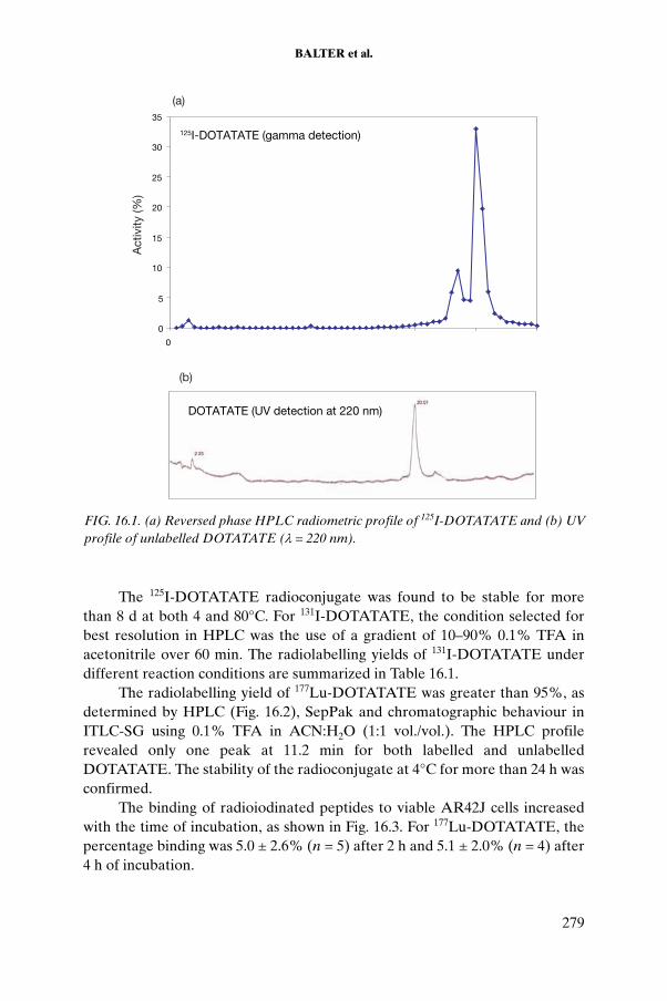
BALTER et al.
The 125I-DOTATATE radioconjugate was found to be stable for more than 8 d at both 4 and 80°C. For 131I-DOTATATE, the condition selected for best resolution in HPLC was the use of a gradient of 10–90% 0.1% TFA in acetonitrile over 60 min. The radiolabelling yields of 131I-DOTATATE under different reaction conditions are summarized in Table 16.1.
The radiolabelling yield of 177Lu-DOTATATE was greater than 95%, as determined by HPLC (Fig. 16.2), SepPak and chromatographic behaviour in ITLC-SG using 0.1% TFA in ACN:H2O (1:1 vol./vol.). The HPLC profile revealed only one peak at 11.2 min for both labelled and unlabelled DOTATATE. The stability of the radioconjugate at 4°C for more than 24 h was confirmed.
The binding of radioiodinated peptides to viable AR42J cells increased with the time of incubation, as shown in Fig. 16.3. For 177Lu-DOTATATE, the percentage binding was 5.0 ± 2.6% (n = 5) after 2 h and 5.1 ± 2.0% (n = 4) after 4 h of incubation.
125I-DOTATATE (gamma detection)
0
5
10
15
20
25
30
35
0
Act
ivity
(%)
DOTATATE (UV detection at 220 nm)
(a)
(b)
FIG. 16.1. (a) Reversed phase HPLC radiometric profile of 125I-DOTATATE and (b) UV profile of unlabelled DOTATATE (λ = 220 nm).
279

CHAPTER 16
Internalization experiments demonstrated that the radioconjugate penetrated into the cells, reaching 73.0 ± 14.0% (n = 10), 32.0 ± 9.0% (n = 3) and 62.0 ± 18.0% (n = 6) of the total bound activity in 3–4 h for the peptide labelled with 125I, 131I and 177Lu, respectively (Fig. 16.4).
TABLE 16.1. LABELLING YIELDS OF 131I-DOTATATE UNDER DIFFERENT REACTION CONDITIONS
Cl-T (μg) Activity to peptide ratio (MBq/μg) Yield (%)
1.3 0.784 72
90.0 1.776 83
91.3 3.286 93
300.0 0.821 79
300.0 1.017 22
300.0 0.496 63
600.0 0.925 24
FIG. 16.2. (a) Reversed phase HPLC radiometric profile of 177Lu-DOTATATE and (b) UV profile of unlabelled DOTATATE (λ = 220 nm).
(a)
(b)
280

BALTER et al.
Externalization experiments revealed that for the peptide labelled with 177Lu, 91.0 ± 42.0% and 61.0 ± 27.0% of the internalized activity remained inside the cell at 30 min and 60 min, respectively. In the case of 125I-DOTATATE, the figures were 58.0 ± 12.0%, 57.0 ± 15.0% and 47.0 ± 7.0% at 15, 30 and 60 min, respectively (n = 3).
FIG. 16.3. Binding of 125I-DOTATATE to whole AR42J cells calculated as percentage offactivity corrected for non-specific binding in the presence of 26μM unlabelled peptide.
FIG. 16.4. Internalization of DOTATATE labelled with 125I, 131I and 177Lu.
281

CHAPTER 16
Binding to receptors from AR42J cell membranes showed an increase of bound radiopeptide as a function of amount of receptor, reaching 1.6 and 1.1% respectively for 177Lu-DOTATATE and 125I-DOTATATE. In the case of receptors obtained from rat brain cortex, a similar response was observed, reaching 8.0 and 7.6%, respectively.
Inhibition of the binding of 125I-DOTATATE to rat brain cortex membranes (6.6 g/mL) incubated with increasing amounts of unlabelled peptide gave a displacement of up to 17.4% for 42μM DOTATATE. Inhibition of the binding to AR42J cells with increasing amounts of unlabelled DOTATATE is shown in Fig. 16.5. The IC50 value was found to be 1.67 ± 0.17nM.
The biodistribution patterns of 125I-DOTATATE and 177Lu-DOTATATE in normal mice are summarized in Tables 16.2 and 16.3, respectively.
The biological uptake pattern of the radioiodinated peptide indicates urinary as well as gastrointestinal excretion. In vivo metabolization is evidenced by the thyroid uptake (see Table 16.2), which reached 10.2 ± 8.5% of the injected dose at 24 h. In the case of the 177Lu labelled peptide, the elimination is mainly through urinary excretion. Dose estimates for the radio-iodinated peptide are shown in Table 16.4.
16.4.2. OCT, P3, P4 and Gluc-TOCA
The peptides OCT, P3 and P4 were radiolabelled with 177Lu with high yields and radiochemical purities (>98%). The effects of reaction temperature
FIG 16.5. Inhibition of the binding of 125I-DOTATATE to AR42J cells by increasing amounts of unlabelled DOTATATE.
282

BALTER et al.
(40–70ºC) and incubation time (10–60 min) for 177Lu-P3 are shown in Fig. 16.6. Radiolabelling yields of greater than 99% were obtained when reactions were carried out for either 10 min at 70ºC or 30 min at 55ºC. In contrast, 80% yield was obtained at 40ºC, even when the reaction was carried out for 60 min. The conditions selected for subsequent labelling studies were incubation for 30 min at 70ºC. Similar results were obtained for the radiolabelling of P4 and OCT with 177Lu.
The radiolabelled peptides can be separated under cold conditions by HPLC using the slow gradient, since their retention times differ by more than 10 min. Thus the labelled peptides were obtained with very high specific activity in purified form. The HPLC profile for 177Lu-OCT is shown in Fig. 16.7. Similar results were obtained for 177Lu-P3 and 177Lu-P4.
Labelling of Gluc-TOCA with 125I and 131I resulted in yields greater than 95 and 93%, respectively.
TABLE 16.2. BIODISTRIBUTION PATTERN OF 125I-DOTATATE IN NORMAL MICE AT 1, 2 AND 24 h POST-INJECTION
Region1 h 4 h 24 h
% ID % ID/g % ID % ID/g % ID % ID/g
Blood 2.54 ± 1.07 0.92 ± 0.04 3.09 ± 0.56 1.12 ± 0.21 2.22 ± 0.88 0.80 ± 0.32
Liver 1.77 ± 0.84 0.80 ± 0.11 1.02 ± 0.25 0.49 ± 0.21 3.25 ± 1.99 1.46 ± 0.83
Lungs 0.69 ± 0.38 2.63 ± 0.73 0.70 ± 0.11 2.78 ± 0.51 1.35 ± 1.48 3.38 ± 2.36
Spleen 0.16 ± 0.08 1.09 ± 0.07 0.28 ± 0.20 1.13 ± 0.54 0.33 ± 0.30 1.27 ± 1.00
Kidneys 3.62 ± 1.62 4.00 ± 0.78 2.99 ± 0.31 3.53 ± 0.75 5.83 ± 4.86 6.75 ± 4.55
Thyroid 0.12 ± 0.02 4.68 ± 0.25 1.63 ± 1.10 82.5 ± 44.6 10.20 ± 8.53 489 ± 422
Muscle 2.83 ± 1.50 0.17 ± 0.06 3.30 ± 1.01 0.20 ± 0.08 7.82 ± 5.92 0.45 ± 0.33
Brain 0.02 ± 0.01 0.11 ± 0.08 0.01 ± 0.00 0.04 ± 0.02 0.06 ± 0.04 0.34 ± 0.32
Stomach 8.9 ± 3.6 18.0 ± 6.4 10.8 ± 3.1 19.1 ± 7.8 22.9 ± 28.5 15.7 ± 17.9
Gall bladder 0.65 ± 0.60 33.0 ± 20.0 0.81 ± 0.16 26.9 ± 5.3 0.08 ± 0.04 16.6 ± 8.8
Intestines 18.1 ± 10.5 17.2 ± 5.4 19.9 ± 5.2 19.8 ± 9.4 22.1 ± 9.1 16.7 ± 4.8
Pancreas 0.69 ± 0.35 13.54 ± 2.01 0.98 ± 0.67 7.81 ± 0.60 0.31 ± 0.25 3.71 ± 1.67
Bladder and urine
26.2 ± 13.3 12.0 ± 3.4 44.6 ±7.3 33.8 ± 5.5 22.5 ± 15.6 24.9 ± 16.5
No. of mice 2 3 4
Note: Data presented as mean ± SD.
283

CHAPTER 16
The specific binding (Fig. 16.8), internalization (Fig. 16.9) and externali-zation (Fig. 16.10) studies carried out for all the peptides using the D341 cells showed that, although the total binding and internalization were higher for 125I-Gluc-TOCA (5.6 ± 0.6% and 40 ± 1% at 4 h, respectively), the percentage of activity remaining inside the cell was higher for the new peptides 177Lu-P3 and 177Lu-P4 (>80%), which bear the amino acid sequence cleavable by cathepsin inside the cell.
16.4.3. Anti-CD20
The yield of radioiodination of anti-CD20 ranged from 43 to 82%, and the specific activity of the radiolabelled product was over 30 µCi/µg, depending on the amount of monoclonal antibody and the radioactive concentration of the radionuclide. The best results were obtained when excess amounts of monoclonal antibody and high concentrations of activity were used.
The labelling of anti-CD20 with 188Re yielded a radiolabelled product with radiochemical purity greater than 95% for up to 3 h post-labelling and with a specific activity of 1.48–1.85 MBq/µg (40–50 µCi/µg).
TABLE 16.3. BIODISTRIBUTION OF 177Lu-DOTATATE IN NORMAL MICE AT 1, 2 AND 24 h POST-INJECTION
Region1 h 2 h 24 h
% ID % ID/g % ID % ID/g % ID % ID/g
Blood 4.60 ± 3.29 1.83 ± 1.55 3.31 ± 3.00 1.04 ± 0.20 3.86 ± 0.33 1.36 ± 0.12
Liver 27.8 ± 15.6 12.61 ± 6.80 18.78 ± 16.02 10.12 ± 8.64 5.39 ± 1.13 3.36 ± 1.24
Lungs 8.5 ± 6.6 40.11 ± 30.84 3.80 ± 4.09 18.76 ± 21.06 0.55 ± 0.11 2.48 ± 0.97
Spleen 1.08 ± 0.35 14.14 ± 10.48 0.95 ± 1.03 20.47 ± 26.23 0.278 ± 0.003 2.14 ± 2.79
Kidneys 3.55 ± 0.78 5.53 ± 2.29 7.31 ± 0.60 9.50 ± 1.06 7.61 ± 0.12 9.75 ± 0.04
Muscle 13.3 ± 5.7 0.84 ± 0.50 20.85 ± 16.98 1.08 ± 0.85 24.64 ± 12.19 1.48 ± 0.95
Brain 0.04 ± 0.01 0.11 ± 0.03 0.35 ± 0.37 1.34 ± 1.54 0.05 ± 0.07 0.21 ± 0.30
Stomach 2.82 ± 3.37 8.37 ± 10.71 3.75 ± 2.10 5.49 ± 3.20 1.70 ± 0.62 2.49 ± 0.61
Gallbladder 0.03 ± 0.05 0.83 ± 1.17 0.04 ± 0.02 0.77 ± 1.18 0.11 ± 0.03 17.7 ± 6.7
Intestines 5.62 ± 5.06 1.59 ± 1.46 6.38 ± 3.27 1.81 ± 1.15 6.31 ± 0.93 1.98 ± 0.40
Bladder and urine
17.44 ± 5.98 31.24 ± 6.42 26.6 ± 2.99 104 ± 38 42.96 ± 0.46 279.1 ± 25.5
No. of mice 2 2 2
Note: Data presented as mean ± SD.
284

BALTER et al.
TABLE 16.4. DOSE ESTIMATES (MGy/MBq) FOR 125I-DOTATATE AND 131I-DOTATATE
Target organ 125I 131I Target organ 125I 131I
Adrenal glands 2.74E-4 6.39E-3 Muscle 1.55E-3 1.80E-2
Brain 2.54E-5 4.06E-4 Ovaries 2.44E-2 1.08E-1
Breasts 3.21E-5 1.21E-3 Pancreas 3.25E-4 9.49E-3
Gall bladder wall 3.99E-3 2.78E-2 Red marrow 1.52E-3 2.02E-2
Lower large intestine wall
2.76E-1 1.80E-1 Bone surfaces 2.94E-3 4.01E-1
Small intestine 3.05E-2 1.84E-1 Skin 2.20E-4 5.95E-3
Stomach 1.69E-3 1.63E-2 Spleen 2.77E-4 7.53E-3
Upper large intestine wall
1.07E-1 6.91E-1 Testes 5.49E-4 2.98E-2
Heart wall 1.02E-4 2.51E-3 Thymus 6.73E-5 1.28E-3
Kidneys 3.15E-3 2.94E-2 Thyroid 9.26E-2 6.77E-1
Liver 2.69E-3 2.08E-2 Urinary bladder wall
3.76E-2 1.91E+0
Lungs 8.16E-4 6.44E-3 Uterus 5.07E-3 1.07E-1
Total body 2.90E-3 2.58E-2
FIG. 16.6. Kinetics of 177Lu-P3 labelling at different incubation temperatures, as determined by reversed phase HPLC.
285

CHAPTER 16
FIG. 16.7. HPLC purification of 177Lu-OCT: (a) UV detection at 220 nm, (b) gamma detection of the labelling mixture and (c) control of the radiochemical purity of the radio-peptide after further purification by SepPak.
286

BALTER et al.
FIG. 16.8. Specific binding of (a) 177Lu-OCT and 131I-Gluc-TOCA, (b) 177Lu-OCT and 125I-Gluc-TOCA, (c) 177Lu-P3 and 125I-Gluc-TOCA and (d) 177Lu-P4 and 125I-Gluc-TOCA to D341 cells in paired assays, expressed as percentage of bound activity minus non-specific binding at 0.5, 1, 2, 3 and 4 h.
FIG. 16.9. Internalization pattern of 177Lu-OCT and 125I-Gluc-TOCA in D341 cells in paired assays, expressed as the percentage of internalized activity over specific bound activity at 30 min, and 1, 2, 3 and 4 h.
287

CHAPTER 16
Specific binding of 131I-anti-CD20 to membrane antigens increased as a function of membrane concentration, reaching 20.2 ± 0.5% for a total protein content of 16.5 mg/mL (Fig. 16.11). Maximum binding capacity was 15 ± 2% (n= 3). Inhibition of binding was 67.2 ± 1.2% when 290 µg (5.22μM) of unlabelled anti-CD20 was added (Fig. 16.12). The IC50 value was found to be 12.7nM.
FIG. 16.10. Externalization of 177Lu-OCT, 177Lu-P3, 177Lu-P4 and 125I-Gluc-TOCA, expressed as the percentage of initial activity remaining in membrane and inside cell at different incubation times.
FIG. 16.11. Binding of 131I-anti-CD20 to increasing amounts of membrane antigen isolated from leucocytes.
288

BALTER et al.
Specific binding of 188Re-anti-CD20 to membranes reached 46.0 ± 1.0% for a protein content of 33 mg/mL. Maximum binding capacity was 17 ± 2% (n= 3). Inhibition of binding to membranes (1 mg/mL) was 66.0 ± 5.0% when 700 µg (12.6μM) of unlabelled anti-CD20 was added (Fig. 16.13). The IC50
value was determined to be 13.3nM.
FIG. 16.12. Inhibition of binding of 131I-anti-CD20 to membranes from leucocytes expressing CD20 antigen due to competition from unlabelled anti-CD20.
FIG. 16.13. Inhibition of binding of 188Re-anti-CD20 to membranes from leucocytes expressing CD20 antigen due to competition from unlabelled anti-CD20.
289

CHAPTER 16
Data on the biodistribution of 188Re-anti-CD20 are given in Table 16.5. The results are expressed as the percentage of injected dose (% ID) and the percentage of injected dose per gram of tissue (% ID/g). The main route of elimination of activity was through urinary excretion (59.7% at 24 h), while gastrointestinal excretion was 10%. Negligible uptake was observed in the thyroid and stomach.
16.5. CONCLUSION
DOTATATE was labelled with 125I, 131I and 177Lu with very high yields, even prior to purification. The two radiochemical species found for 125I-DOTATATE by reversed phase HPLC could be interpreted as mono- and diiodinated species. They exhibited a slightly higher retention time compared with the unlabelled peptide, thereby allowing the preparation of a labelled molecule with high specific activity. 131I-DOTATATE was also obtained with
TABLE 16.5 BIODISTRIBUTION OF 188Re-ANTI-CD20 IN NORMAL MICE AT 4, 18 AND 24 h POST-INJECTION
Region4 h 18 h 24 h
% ID % ID/g % ID % ID/g % ID % ID/g
Blood 12.3 ± 1.5 4.4 ± 0.3 12.6 ± 1.1 4.8 ± 0.2 2.86 ± 0.09 1.17 ± 0.14
Liver 11.1 ± 0.2 5.5 ± 0.3 16.0 ± 1.8 7.5 ± 0.6 6.3 ± 6.2 3.5 ± 3.2
Heart 0.29 ± 0.14 1.4 ± 0.7 0.4 ± 0.3 1.7 ± 0.9 0.08 ± 0.01 0.45 ± 0.17
Lungs 0.64 ± 0.17 2.2 ± 0.7 0.7 ± 0.4 2.0 ± 1.0 0.4 ± 0.4 0.9 ± 0.5
Spleen 0.4 ± 0.2 2.7 ± 1.9 0.4 ± 0.2 3.0 ± 2.4 0.2 ± 0.2 2.7 ± 2.9
Kidneys 4.3 ± 1.1 7.3 ± 3.2 17.6 ± 8.6 24.7 ± 8.1 5.4 ± 1.5 9.5 ± 3.7
Thyroid 0.07 ± 0.02 1.3 ± 0.5 0.08 ± 0.12 6.0 ± 9.1 0.02 ± 0.01 0.7 ± 0.3
Muscle 3.0 ± 2.3 0.19 ± 0.16 6.8 ± 0.9 0.40 ± 0.07 3.0 ± 1.0 0.21 ± 0.09
Bone 2.1 ± 1.3 0.57 ± 0.38 3.3 ± 0.6 0.85 ± 0.18 1.6 ± 0.6 0.44 ± 0.14
Stomach 1.8 ± 0.9 4.1 ± 2.5 2.3 ± 0.4 2.4 ± 0.1 1.9 ± 1.4 3.4 ± 1.8
Intestines 14.7 ± 5.5 6.5 ± 2.8 12.2 ± 3.5 3.1 ± 1.1 9.6 ± 8.0 3.8 ± 2.8
Bladder and urine
10.2 ± 5.2 26 ± 9 51 ± 5 25 ± 4 60 ± 11 25 ± 14
No. of mice 3 3 3
Note: Data presented as mean ± SD.
290

BALTER et al.
very high yields (>90%) prior to purification under different experimental conditions (91.3 µg of chloramine T and an activity to peptide ratio of 3.286 MBq/µg). Increasing the amounts of chloramine T did not improve the results.
The binding experiments using AR42J cells indicate that the radioconju-gates prepared are able to recognize and bind to somatostatin receptors present in viable tumour cells. Although the 131I radioconjugate is able to bind to somatostatin receptors present in live AR42J tumour cells, its internalization was lower than that of the 125I homologues in the same time interval (32 ± 9% versus 73 ± 14%). A high percentage of internalization into AR42J cells was achieved for both 177Lu-DOTATATE and 125I-DOTATATE. This observation suggests that the nature of the radionuclide used did not affect the biological properties of the peptide conjugate. Biodistribution in normal mice showed different patterns for the radioiodine and radiolanthanide labelled peptides. These findings open up the possibility of using cocktails of DOTATATE labelled with different radionuclides in order to minimize the radiation dose to organs not compromised by the tumour.
The labelling of OCT, P3 and P4 was successfully achieved using 177LuCl3
(24 Ci/mg, 2.6 Ci/mL). Ammonium acetate buffer and gentisic acid were used to provide optimal pH conditions and to protect against radiolysis, respectively. The 177Lu labelled peptides were obtained with very high specific activities after purification by HPLC and subsequent SepPak purification, as the retention time of the unlabelled peptides differs significantly from that of their labelled species.
The specific binding, internalization and externalization studies carried out for all the peptides using D341 cells showed that Gluc-TOCA exhibited higher binding to cells when labelled with 125I than when labelled with 131I. Internalization and externalization studies revealed that, although total binding and internalized activity were higher for radioiodinated Gluc-TOCA, the percentage of activity remaining inside the cell was higher (>80%) for the new peptides 177Lu-P3 and 177Lu-P4, which bear the amino acid sequence cleavable by cathepsin inside the cell. The preliminary results for the P3 and P4 peptides indicate that these new strategies for the design of somatostatin analogue peptides could play an important role in targeted radiotherapy.
The labelling of anti-CD20 with beta emitters of therapeutic potential — in the present case, 131I and 188Re — gave reliable results by simple and efficient methodologies, yielding products suitable for clinical radioimmunotherapy. In particular, the radioiodination results were satisfactory, even though purifi-cation was always needed in the case of 131I. Quality control methods for evaluation of radiochemical purity showed good reproducibility. Immuno-affinity studies showed that the extent of binding varied with membrane
291

CHAPTER 16
antigen concentration, and that good specificity of binding was demonstrated by inhibition with unlabelled anti-CD20. Both tracers gave high affinity in their binding to antigen membranes, with better results in the case of 188Re owing to its higher specific activity. Nevertheless, it is considered that, with good quality 131I and with a higher specific activity of the radioiodine than that used in the present work, both tracers would have similar results. Studies using membranes, which are stable at –80ºC for more than 6 months, instead of concentrated short lived leucocytes, have shown excellent reproducibility; therefore, the use of membranes is more convenient.
ACKNOWLEDGEMENTS
The CRP participants gratefully acknowledge S. Mather for supplying AR42J cells and M. Zalutsky for his scientific advice. We also acknowledge M. Hens, P. Welch and D. Affleck for their collaboration in the experimental work at Duke University Medical Center. We express our sincere thanks to UICC, NIH, Pedeciba Química and International Isotopes Inc. (Idaho Falls, USA).
REFERENCES TO CHAPTER 16
[16.1] BALTER, H., et al., “Labelling and control of biomolecules with 188Re and 153Sm”, Labelling Techniques of Biomolecules for Targeted Radiotherapy, IAEA-TECDOC-1359, IAEA, Vienna (2003) 183–195.
[16.2] WHETSTONE, P.A., AKIZAWA, H., MEARES, C., Evaluation of cleavable (Tyr3)-octreotate derivatives for longer intracellular probe residence, Bioconjug. Chem. 15 (2004) 647–657.
[16.3] JUWEID, M.E., Radioimmunotherapy of B-cell Non-Hodgkin’s lymphoma: From clinical trials to clinical practice, J. Nucl. Med. 43 (2002) 507–1529.
[16.4] ROBLES, A.M., BALTER, H., OLIVER, P., WELLING, M., PAUWELS, E.K.J., Improved radioiodination of biomolecules using exhaustive chloramine-Toxidation, Nucl. Med. Biol. 28 (2001) 999–1008.
[16.5] BALTER, H., et al., “Laboratory evaluation of therapeutic radiopharma-ceuticals” (Proc. IAEA Res. Coord. Mtg Warsaw, 2004).
[16.6] BALTER, H., et al., Radiolabelled DOTA-TATE: Its evaluation for targeted radiotherapy, Alasbimn J. 8 (2005),http://www2.alasbimnjournal.cl/alasbimn/CDA/sec_b/0,1206,SCID%253D15275,00.html
292
BALTER et al.
[16.7] HOFLAND, L.J., et al., Internalization of the radioiodinated somatostatin analog [125I-Tyr3]Octreotide by mouse and human pituitary tumour cells: Increase by labelled octreotide, Endocrinology 136 (1995) 3698–3705.
[16.8] VERDERA, S., et al., Labelling and quality control of 188Re-Lanreotide, Cell. Molec. Biol. 48 (2002) 741–745.
[16.9] BALTER, H.S., HENS, M., WELCH, P., AFFLECK, D., ZALUTSKY, M.R., Effect of linker sequence on internalization properties of Lu-177 labelled soma-tostatin analogues, Alasbimn J. 31 (2006), www2.alasbimnjournal.cl/alasbimn/CDA/
[16.10] VAIDYANATHAN, G., et al., Specific and high-level targeting of radiolabelled octreotide analogues to human medulloblastoma xenografts, Clin. Cancer Res. 9 (2003) 1868–1876.
[16.11] OLIVER, P., et al., Anti-CD-20-188Re: Labelling and biological performance, Nucl. Med. Rev. 8 (2005) 16.
293

.

Appendix I
PROTOCOLS DEVELOPED AS PART OF THE COORDINATED RESEARCH PROJECT
The participants developed several protocols during the course of the CRP. These protocols may be of use to other researchers involved in the development of therapeutic radiopharmaceuticals. Some of the protocols are described below.
I.1. PREPARATION OF LABELLED PEPTIDES
I.1.1 Labelling of DOTATATE with 131I
The labelling of DOTATATE (1.5–10 μg) with high specific activity [131I]NaI (≥ 22.2 × 10¹⁰ Bq/mg) was performed using the chloramine T method. A typical protocol involves the addition of 5–10 μL of radioiodine solution (37–740 MBq), maintaining a peptide to radionuclide molar ratio of 0.045–2.73, to the peptide solution in 40 μL of phosphate buffer solution (PBS) (0.1M phosphate buffered saline, pH7.5). The chloramine T solution (5–50 μg/5 μL of PBS) is then added to a reaction vial, using chloramine T to peptide molar ratios ranging from 2.5 to 3.5. The vial is capped and carefully vortexed. The reaction is allowed to proceed for 1–3 min at room temperature and is terminated by addition of the sodium metabisulphite solution (using a fourfold equimolar quantity of the oxidizing agent).
The labelled conjugate is purified on a C18 SepPak cartridge after condi-tioning with 2 mL of 0.05M phosphate buffer at pH7.5. The column is eluted with 5 mL of 0.05M phosphate followed by 0.5 mL of ethanol. While free iodide is eluted in the phosphate buffer, the conjugate is obtained in pure form in the ethanol fraction. The ethanol fraction is evaporated under nitrogen and the radiolabelled conjugate is reconstituted in saline.
I.1.2. Labelling of DOTATATE with 90Y or 177Lu
The labelling of DOTATATE with 90Y and 177Lu essentially follows an identical protocol. Usually, the radionuclide is supplied in a chloride form in 0.05N HCl. The first step involves adjustment of the pH to 4.5 using 50% volume of a solution of gentisic acid in acetate buffer at pH5. DOTATATE solution (1 mg/mL in ultrapure water) is added to the buffered solution of 177Lu
295

or 90Y; the amount of DOTATATE used is varied to achieve the desired specific activity. The reaction mixture is incubated at 90oC for 30 min in a thermostatic bath. After cooling, an aliquot is withdrawn for quality control studies.
The impurities from accompanying trace levels of metal contaminants in the radionuclide solution, mainly iron and zinc, are critical for the labelling yields. If the level of either metal contaminant exceeds 1 μg/Ci, the labelling yields will be adversely affected. With reputed commercial suppliers, specific activities of 1 mCi/μg can be routinely obtained. A value lower than this reference specific activity can be indicative of the presence of the above metal impurities. The level of impurities can be reduced by passing the 177LuCl3 or 90YCl3 solutions through a cation exchange column (such as Chelex) that retains the other metal contaminants and effects purification.
I.2. PHYSICOCHEMICAL QUALITY CONTROLS
The following quality control methods can be adopted for assessing the radiochemical purity of the preparations.
I.2.1. Purification on SepPak C18 cartridges
The SepPak cartridges are preconditioned with 5 mL of ethanol, followed by 5 mL of 0.05M phosphate buffer at pH7.5. An aliquot of the labelled peptide mixed with 10 μL of 50mM DTPA is loaded on the cartridge. The unbound activities (131I, 90Y, 177Lu) are eluted with 5 mL of phosphate buffer. The labelled peptide is then eluted with 3 mL of ethanol. The pooled fractions are counted separately, and the radiochemical yield is estimated from the ratio of activity bound to total activity eluted.
I.2.2. Thin layer chromatography
Thin layer chromatography studies are carried out on silica gel (aluminium sheets, Merck) in 10 cm strips as the stationary phase. Ammonium hydroxide:methanol:water (1:5:10) is used as the mobile phase. While the free activity remains at the point of origin (Rf = 0), the radiolabelled peptide migrates to Rf = 0.4.
I.2.3. Paper chromatography
Paper chromatography studies are carried out using 10 cm long Whatman 3 chromatography paper. For each test solution, 5 µL of the
296
solution is spotted at 1.5 cm from the lower end of the paper strips, and the strips are developed in 10% ammonium acetate in methanol (30:70 vol./vol.). The strips are subsequently dried and cut into 1 cm segments. The radioac-tivity associated with each segment is measured in a well type NaI(Tl) detector. While free activity remains at the point of origin, the radiolabelled peptide migrates to Rf = 0.7–0.8.
I.2.4. Paper electrophoresis
For paper electrophoresis, 5 μL of the complex solution (125/131I-DOTATATE) is spotted on pre-equilibrated Whatman 3MM chromatography paper (35 cm × 2 cm) at 15 cm from the cathode. Paper electrophoresis is carried out for 1 h under a voltage gradient of about 10 V/cm using 0.025M phosphate buffer at a pH of approximately 7.5. The strips are dried and cut into 1 cm segments, and the activity is measured. Free iodide moves to the anode, whereas the iodinated conjugate remains at the point of origin.
I.2.5. HPLC
The radiochemical analyses and purification of the 177Lu, 90Y and 125I labelled DOTATATE conjugates are achieved using an HPLC system. A dual pump HPLC unit with a C18 reverse phase column (25 cm × 0.46 cm) is used for purification of the labelled conjugates. The elution is monitored both by UV signals at 270 nm and by radioactivity signals. The flow rate is maintained at 1 mL/min. Water–0.1% trifluoroacetic acid (solvent A) and acetonitrile–0.1% trifluoroacetic acid (solvent B) mixtures are used as the mobile phase and the following gradient elution technique is adopted for the separation: 0–4 min 95% A, 4–15 min 95 to 5% A, 15–20 min 5% A, 20–25 min 5 to 95% A, 25–30 min 95% A.
The retention times of radiolabelled DOTATATE under the above conditions are typically found to be around 800 s, whereas the free activity appears at retention times of less than 200 s.
I.3. IN VITRO BIOLOGICAL ASSAYS
The protocols of the in vitro assays using AR42J cells are discussed below.
297

I.3.1. Maximum binding assay
The stepwise protocol adapted for the maximum binding assay is outlined below.
(a) Prior to the assay, the cells are washed twice with internalization medium comprising Dulbecco’s modified Eagle’s medium (DMEM) (or F12K or RPMI) supplemented with HEPES (30mM), L-glutamine (2mM), sodium pyruvate (1mM), penicillin (105 U/L), fungizone (0.5 mg/L) and 0.2% BSA at pH7.4.
(b) For stabilization, cells are kept in the internalization medium for 1 h at 37ºC.
(c) The cells are centrifuged at 1500 rev./min for 5 min, decanted and resuspended in DMEM to obtain a cell concentration of 1 × 10⁶ cells/mL.
(d) The cells (5 × 10⁵ cells/tube) are incubated with 200 000 counts/min of radiotracer (i.e. 125/131I-DOTATATE) at 37ºC for various time intervals (30 min, 1, 2, 3 or 4 h), and the total activity in each tube is measured.
(e) In parallel, media reagents are incubated by adding 20 μg (1mg/mL) of DOTATATE (or somatostatin) to obtain maximum displacement (non-specific binding).
(f) Cells are separated from the supernatant by centrifugation at 1500 rev./min for 5 min.
(g) The cells are washed twice with internalization buffer under the same conditions.
(h) The activities of the cells are measured, and the percentage of activity is determined with respect to total activity. For this, it is necessary to subtract non-specific binding counts (determined in step (e)) and to divide the activity in each tube by the total activity.
I.3.2. Internalization assay
Internalization assays are based on the fact that, with acid treatment, peptide that is loosely bound to the membrane dissociates from it and remains in the extracellular solution. Internalization assay is carried out by continuing the above experiments as follows:
(1) To the cells previously washed with internalization media (step (h) of the previous protocol), 1 mL of sodium acetate 20mM in Hank’s balanced salt solution (HBSS) is added, pH5.0 (see Table I.1 for the composition of HBSS). It is also possible to use 0.1M glycine buffer at pH2.8.
298

(2) The cells are incubated at 37ºC for 10 min in HBSS or for 5 min in glycine buffer.
(3) The supernatant is separated by centrifugation.(4) The cells are washed twice with cold HBSS-Ac — 20mM acetate (1.64 g/L)
in HBSS — (or glycine) and added to the other supernatants obtained in step (3).
(5) The cells are then solubilized with 1N NaOH.(6) The activity in the supernatants (non-internalized activity) and in the
solubilized cells (internalized activity) is then measured.
I.3.3. Externalization assay
(a) The cells are allowed to stabilize for 1 h at 37ºC in the internalization medium DMEM.
(b) The cells are centrifuged at 1500 rev./min for 5 min, decanted and resuspended in DMEM to obtain a cell concentration of 1 × 10⁶ cells/mL.
(c) The cells (5 × 10⁵ cells/tube) are incubated with 200 000 counts/min of radiotracer (i.e. 125/131I-DOTATATE) at 37ºC for 2 h, and the total activity in each tube is measured.
(d) Centrifugation is carried out at 1500 rev./min for 5 min, and the supernatant is transferred to an Eppendorf tube for further measurement of activity.
(e) The cells are washed with 1 mL of DMEM and step (d) is repeated.
TABLE I.1. COMPOSITION OF HANK’S BALANCED SALT SOLUTION (HBSS)a
Component Quantity (g/L)
CaCl2 anhydrous 0.14
KCl 0.40
KH2PO4 0.06
MgSO4 anhydrous 0.098
NaCl 8.00
Na2HPO4 0.048
D-glucose 1.00a The HBSS is sterilized by a 0.22 µm membrane.
299

(f) DMEM (1 mL) is added to the cells, which are incubated for 10 min at 37oC. Step (d) is then repeated.
(g) Step (f) is repeated three times.(h) The pellet is solubilized with 1 mL 1N NaOH and vortexed, and the
activity of the pellet and all the supernatants is measured.
As an alternative to this protocol, a parallel assay can be carried out in separate tubes with incubation times of 10, 20, 30 and 60 min.
I.4. PREPARATION OF SOMATOSTATIN MEMBRANE RECEPTOR
I.4.1 From rat brain cortex
Somatostatin membrane receptor is isolated from rat brain cortices by one of the two methods described below.
I.4.1.1. Method 1
Rat brain cortices are prepared according to a published method [A.1]. Briefly, the brain cortices of three Wistar rats are dissected and immediately placed in ice-cold HBSS at pH7.5 supplemented with 50 μL/mL penicillin, 50 μg/mL streptomycin and 10 000 KIU/L aprotinin. The cortices are then rinsed twice with ice-cold HBSS and minced with a surgical blade. The suspension is then centrifuged at 500g for 10 min at 4oC, and the pellet is homogenized in 5 mL of homogenization buffer consisting of 25mM Tris buffer (pH7.5), 0.3M sucrose, 0.25mM PMSF, 1mM EDTA and 10 000 KIU/L aprotinin. The homogenate is centrifuged as described above, and the pellet is homogenized three more times in the same way, with the supernatant saved after each centrifugation. The combined supernatants are then centrifuged at 48 000g for 45 min at 4oC. The final pellet is washed twice with 50mM Tris buffer (pH7.5) containing 5mM MgCl2, 20 mg/L bacitracin, 0.25mM PMSF, 10 000 KIU/L aprotinin and 100 IU/mL RNase Inhibitor, resuspended in 5 mL of washing buffer, separated into 100 μL aiquots and stored immediately at –80oC.
I.4.1.2. Method 2
All steps involved in this method must be carried out in a cold environment (0–4°C). The steps are as follows:
300

(1) The cortices (mid-brain) of adult male rats sacrificed by decapitation are dissected. It is desirable that at least five rats be used.
(2) The cortices are washed three times with HEPES buffer (20mM, pH7.3) containing 10mM MgCl2.
(3) The cortices are immediately homogenized (e.g. using a Polytron or other similar homogenizer) three times in the same buffer with a tissue to buffer ratio of 1:10 at maximum speed for 10 s.
(4) Centrifugation is carried out at 500g for 10 min at 4°C. (5) The supernatant is transferred to other tubes and centrifuged at 2500g for
30 min at 4°C. (6) The supernatant is discarded, and the pellet is resuspended in HEPES
buffer with a pellet to buffer ratio of 1:10. (7) Homogenization is carried out under the same conditions as in step (3). (8) Centrifugation is carried out at 2500g for 30 min at 4°C. (9) Steps (6) and (7) are repeated two more times.(10) The last pellet is resuspended and the protein concentration is measured
by the Lowry method.(11) The preparation is diluted to a final concentration of about 5 mg/mL for
storage.(12) The preparation is aliquoted in 200 μL portions in Eppendorf vials and
stored at –80°C.
I.4.2. Preparation of AR42J cells
AR42J cells are grown to confluence, mechanically disaggregated, washed twice with cold PBS buffer (pH7) and resuspended in homogenization buffer (1 mL/flask) containing 10mM Tris buffer, pH7.4, and 0.1mM EDTA. Cells are disrupted using a homogenizer (50 strokes/5 mL) or ultrasonication according to the manufacturer’s instructions, or by quick freezing/thawing in liquid nitrogen. The homogenized suspension is then centrifuged at 2600 rev./min for 10 min at 4oC. The supernatant is removed and centrifuged again at 26 000 rev./min for 15 min at 4oC. The pellet is resuspended in homogenization buffer (100 μL/flask) and stored at –80oC in 100 μL aliquots.
I.5. PROTOCOL FOR DETERMINATION OF MAXIMUM BINDING CAPACITY (SATURATION ASSAY)
I.5.1. Method 1
The following buffers are used:
301

— HEPES buffer A: 20mM HEPES buffer, pH7.3, containing 10mM MgCl2
and 1% BSA; — HEPES buffer B: same as HEPES buffer A, but without BSA;— Tris–saline buffer: 15mM Tris, 139mM NaCl, pH7.4.
The membrane receptor preparation (5000 μg/mL) is thawed and diluted to final concentrations of 125, 250, 500 and 1000 μg/mL with HEPES buffer B. All the steps outlined below need to be carried out in an ice bath.
The following reagents are pipetted into polystyrene tubes:
— 50 μL of bovine serum albumin, fraction V (BSA), 10% solution in all tubes;
— 50 μL of HEPES buffer A in all odd numbered tubes (Bo);— 50 μL of 40μM unlabelled somatostatin in all even numbered tubes (M);— 100 μL of membrane receptor preparation of each dilution (e.g. tubes 1
and 2: 100 μL/mL; tubes 3 and 4: 200 μg/mL, etc.);— 50 μL of labelled somatostatin (~20 000 counts/min) in all tubes.
Incubation is carried out overnight at 4°C followed by addition of 1 mL of PEG 6000 (6%) in Tris–saline buffer. The mixture is incubated for an additional 30 min at 4°C and centrifuged for 50 min at 2500g.
The supernatant is carefully aspirated so as not to disturb the precipitate, such that the tip of the aspirating pump does not touch the precipitate at any point. The activity of the precipitate is measured. This assay is to be run in duplicate.
I.5.2. Method 2
Each assay tube contains 200 μL of binding buffer (50mM HEPES, pH7.6, containing 0.3% BSA, 10mM MgCl2 and 14 mg/L bacitracin), 15 μL of radioligand solution of the corresponding concentration (ten different concen-tration levels are used) and 25 μg of membrane homogenate containing 40 or 10 μg of protein (for rat brain cortex and AR42J membrane homogenate, respectively). For non-specific series, instead of 200 μL of binding buffer, 175 μL of binding buffer along with 25 μL of unlabelled peptide as the competitor (i.e. 1μM in the reaction vial) is added. Tubes are incubated for 1 h at room temperature, at which point the binding is interrupted by rapid filtration through glass fibre filters presoaked with the binding buffer. The filters are washed with binding buffer, dried, extricated and dispensed into tubes. The filters, along with samples representing total counts added, are then counted for activity.
302

I.6. COMPETITION ASSAY
I.6.1. Method 1
The technique followed for determining maximum binding capacity is similar to that used for determining maximum binding, but with a fixed membrane receptor concentration and an increasing somatostatin concen-tration (as an inhibiting molecule). For selecting the optimal concentration of membrane receptor, the concentration that gives the best displacement of tracer needs to be established in advance.
I.6.2. Method 2
Typically, the radioligand is added to test tubes (30 000 counts/min per tube) containing rat brain cortex (50 μg of protein) or AR42J cell membranes (corresponding to 10 μg of protein) in the presence of increasing amounts of competitor in a total volume of 300 μL of 50mM HEPES (pH7.6, 0.3% BSA, 5mM MgCl2, 10μM bacitracin). The samples are incubated for 2 h at room temperature, and incubation is terminated by addition of ice-cold buffer (1 mL, 10mM HEPES, 150mM NaCl, pH7.6). The suspension is rapidly filtered over glass fibre filters (Whatmann GF/B or Millipore fibre glass, presoaked in binding buffer). The filters are rinsed thoroughly with buffer (0.25–1.0 mL), and the activity in the filter is measured. Non-specific binding is defined as the amount of activity still bound in the presence of 1μM of the competitor.
I.7. CYTOTOXICITY ASSAYS
The three methods used by the participants are described in the following paragraphs.
I.7.1. MTT assay
I.7.1.1. Protocol for MTT assay
Cell lines are kept frozen until they are cultured. Cells are plated on 10 cm diameter dishes in DMEM supplemented with 10% newborn calf serum and antibiotics (100 units/mL penicillin, 100 μm/mL streptomycin) and incubated at 37°C under a 5% CO2 atmosphere and 100% humidity. Semi-confluent cells are detached using trypsin/EDTA and counted in a Neubauer chamber. Cells are seeded with 1–2 × 10⁵ cells/well on 96 well plates. Between
303

10 and 100 μCi of labelled peptide is applied to each well. Cells are incubated for 24 or 48 h for the cell cytotoxicity study, followed by a cellular dosimetry study of the activities, applied using the EGS4 code system.
I.7.1.2. Assay
For the MTT assay, medium is used without phenol red. When the cell is ready, MTT stock solution (5 mg/mL) is added to each culture being assayed to equal one tenth of the original culture volume and incubated for 3–4 h. No phenol red is added to the medium during the assay. When working with attached cells, at the end of the incubation period the medium can be removed and the converted dye can be solubilized with acidic isopropanol (0.04–0.1N HCl in absolute isopropanol) under incubation for 30 min at 37oC. The absorbance of converted dye is measured at a wavelength of 570 nm. The cell viability of the peptide treated group is calculated compared with that of the control group, and results are expressed as the percentage of cell viability.
I.7.2. Micronuclear assay
The frequency of micronuclei (MN) in peripheral blood lympocytes is considered to be a cytogenic bioindicator for ionizing radiation exposure [I.2]. Micronuclei assay can be conducted in vitro using human blood samples incubated with the test peptide at 37oC using various activity levels (i.e. 800–4500 kBq/mL). After 1 h, the cells are washed three times with medium to eliminate the free labelled peptide. The cells are the cultured in RPMI 1640 medium, foetal calf serum and phytohemagglutinin. After 44 h of incubation, cytochalasin B (Sigma) is added to those blood cells that have completed one nuclear division and have been recognized as binucleated cells (BNCs). At 72 h from the start of the assay, cells are harvested and fixed, and slides are stained with Giemsa, as per the standard protocol.
I.7.3. Dosimetry studies
Since the radiolabelled agents developed under the framework of the CRP were intended for use in radiotherapy, calculation of absorbed dose and additional dosimetric data are also essential. The following experimental set-up supports these targets.
The protocol for pharmacokinetic studies using animals such as rats, mice and dogs stipulates that a minimum of seven time points (e.g. 30 min, 2, 12, 24, 48, 120 and 168 h for 177Lu) and three animals per time point be used. In the case of studies using dogs, region of interest techniques are to be used.
304

Determination of the percentage uptakes in the following organs and tissues is required: blood, heart, liver, spleen, kidney, stomach, gastrointestinal tract, bone, muscle, adrenal gland, pancreas, urinary bladder and tumour tissue, if applicable.
The use of MIRDDOSE 3.1 or an upgraded version is advisable for determining the absorbed dose. This software should be used to calculate the absorbed dose after drawing pharmacokinetics curves and calculating the residence time. During the evaluation of dosimetric data, doses in both tumour tissue and in critical/normal tissues must be evaluated.
REFERENCES TO APPENDIX I
[I.1] RAYNOR, K., REISINE, T., Analogs of somatostatin selectively label distinct subtypes of somatostatin receptor in rat brain, J. Pharmacol. Exp. Ther. 251(1989) 510–517.
[I.2] INTERNATIONAL ATOMIC ENERGY AGENCY, Cytogenic Analysis for Radiation Dose Assessment: A Manual, Technical Reports Series No. 405, IAEA, Vienna (2001).
305

Appendix II
PAPERS PUBLISHED BY THE PARTICIPANTS RELATED TO THE COORDINATED RESEARCH PROJECT
Journals
BALTER, H., et al., DOTATATE: a promising somatostatin derivative for targeted radiotherapy, NucMediEx (1 August 2004).
BALTER, H., et al., Radiolabelled DOTATATE: Its evaluation for targeted radio-therapy, Alasbimn J. 8 (2005).
BANERJEE, S., et al., 177Lu-DOTA-Lanreotide: A novel tracer as a targeted agent for tumour therapy, Nucl. Med. Biol. 31 (2004) 753–759.
BANERJEE, S., et al., An estradiol-conjugate for radiolabelling with 177Lu: An attempt to prepare a radiotherapeutic agent, Biorg. Med. Chem. 13 (2005) 4315–4322.
BODEI, L., et al., Receptor-mediated radionuclide therapy with 90Y-DOTATOC in association with amino acid infusion: A phase I study, Eur. J. Nucl. Med. 30 (2003) 207–216.
BODEI, L., et al., Receptor radionuclide therapy with 90Y-[DOTA]0-Tyr3-octreotide (90Y-DOTATOC) in neuroendocrine tumours, Eur. J. Nucl. Med. Mol. Imaging 31(2004) 1038–1046.
BODEI, L., et al., Receptor radionuclide therapy with 90Y-DOTATOC in patients with medullary thyroid carcinomas, Cancer Biother. Radiopharm. 19 (2004) 65–71.
CRUDO, J.L., et al., Optimization of the labelling of antibodies with rhenium-188 using a prelabelled MAG3 chelate for general applications, Int. J. Pharm. 248 (2002) 173–182.
DAS, T., et al., Preparation and preliminary biological evaluation of a 177Lu labelled sanazole derivative for possible use in targeting tumour hypoxia, Bioorg. Med. Chem. 12(2004) 6077–6084.
DAS, T., et al., 177Lu-labelled metronidazole for possible use in targeting tumour hypoxia, Radiochim. Acta (in press).
FERRO-FLORES, G., et al., An efficient, reproducible and fast preparation of 188Re-anti-CD20 for the treatment of non-Hodgkin’s lymphoma, Nucl. Med. Commun. 26 (2005) 793–799.
JÁNOKI, G.A., The potential of Re-188 labelled radiopharmaceuticals in radionuclide therapy, Nucl. Med. Review 6 (2003) 82.
306
KWEKKEBOOM, D.J., et al., Overview of results of peptide receptor radionuclide therapy with 3 radiolabelled somatostatin analogs, J. Nucl. Med. 46 Suppl. 1 (2005) 62S–66S.
MOLINA-TRINIDAD, E.M., DE MURPHY, C.A., FERRO-FLORES, G., MURPHY-STACK, E., JUNG-COOK, H., Radiopharmacokinetic and dosimetric parameters of 188Re-lanreotide in athymic mice with induced human cancer tumors, Int. J. Pharm. 310 (2006) 125–130.
PÉREZ, L., et al., A multivalent recombinant antibody fragment specific for carcinoem-bryonic antigen, Biotech. Appl. Biochem. 43 (2006) 39–48.
PILLAI, M.R.A., CHAKRABORTY, S., DAS, T., VENKATESH, M., RAMAMOORTHY, N., Production logistics of 177Lu for radionuclide therapy, Appl. Radiat. Isot. 59 (2003) 109–118.
Conference Proceedings
ARAUJO, E.B., et al., “Comparison of [131I]-Tyr3-octreotate and [131I]-DOTA-Tyr3-octreotate: The effect of DOTA on the pharmacokinetics and stability”, Trends in Radiopharmaceuticals (Proc. Int. Symp. Vienna, 2005), IAEA, Vienna (2007).
BALTER, H.S., “DOTA-TATE: Modelo para la evaluación de la unión a receptores de somatostatina”, II Simposio Científico de Actualización en Medicina Nuclear y Radio-farmacia, SUBIMN, CSIC, Montevideo, 2003.
BALTER, H.S., “Terapia de linfoma No Hodgkin con anticuerpos anti CD-20, III”, ibid.
BALTER, H., et al., “Radiolabelled DOTATATE: Its evaluatión for targeted radio-therapy”, in Proc. Int. Symp. Nuclear Oncology, Porto Alegre, Brazil, World J. Nucl. Med. 3, Suppl. 1 (2004) S39.
BALTER, H.S., et al., “DOTA-TATE alternative labellings with halogens and radio-metals”, Trends in Radiopharmaceuticals (Proc. Int. Symp. Vienna, 2005), IAEA, Vienna (2007).
ISHFAQ, M.M., HUSSAIN, N., JEHANGIR, M., “DOTA-Tyr3-octreotate: Its labelling with beta emitting radionuclides in the preparation of potential therapeutic radiophar-maceuticals”, Proc. First Int. Nucl. Chem. Cong., Kusadasi, Turkey, 2005.
ISHFAQ, M.M., HUSSAIN, N., HAIDER, I., JEHANGIR, M., “177Lu potential therapeutic agent: Preparation and quality control of 177Lu-EDTMP and 177Lu-DOTA-Tyr3-Octreotate complexes”, paper presented at 12th Eur. Symp. Radiopharmacy and Radiopharmaceuticals, Gdansk, 2004.
LAZNICKOVA, A., PETRIK, M., MELICHAROVA, L., LAZNICEK, M., ZALUTSKY, M.R., “Preclinical study of radioiodinated glucose-Tyr3-octreotate:
307

Comparison with 111In-DOTA-Tyr3-octreotate”, Trends in Radiopharmaceuticals (Proc. Int. Symp. Vienna, 2005), IAEA, Vienna (2007).
LAZNICEK, M., LAZNICKOVA, A., “The role of somatostatin receptors in normal tissues in pharmacokinetics of radiolabelled octraotates”, ibid.
LUNGU, V., NICULAE, D., CHIPER, D., RADU, M., “DOTA-Tyr3-octreotate labelled with 177Lu and 131I — comparative evaluation”, ibid.
LUNGU, V., NICULAE, D., CHIPER, D., “Radiolabelling of DOTA-TATE with 131I and 177Lu — In vitro evaluation studies”, Radiopharmacy and Radiopharmaceutical Chemistry (Proc. Int. Cong. Istanbul), World J. Nucl. Med. 3 (2004) 242.
LUNGU, V., NICULAE, D., BOUZIOTTIS, P., PIRMETTIS, I., ALBERT, P., “In vitro and in vivo evaluation of phosphonates labelled with therapeutic radionuclides”, Trends in Radiopharmaceuticals (Proc. Int. Symp. Vienna, 2005), IAEA, Vienna (2007).
LUNGU, V., NICULAE, D., CHIPER, D., ALBERT, P., “Radiosynthesis and preliminary evaluation of 131I-DOTA-TATE as a potential tracer for somatostatin receptor visualization”, Proc. 5th Int. Conf. Isotopes, Brussels, 2005.
LUNGU, V., NICULAE, D., DANAILA, L., PURICE, M., “Radiolabelling of DOTA-TATE with 177Lu — Preliminary studies with a view to somatostatin receptor radio-nuclide therapy”, ibid.
LUNGU, V., NICULAE, D., CHIPER, D., “177Lu-DOTA-octreotate in targeted therapy of the neuroendocrine tumours”, Research Days Euroaction, Bucharest, 2005 (poster).
LUNGU, V., BAICULESCU, S., CHIPER, D., NICULAE, D., DANAILA, L., “Mathe-matical modelling correlated to MIRD for calculation absorbed dose from animal biodistribution data”, Nat. Conf. Phys., Bucharest, 2005.
NAGAMATI, L.T., CALDEIRA, J.S.F., MURAMOTO, E., ARAUJO, E.B., SILVA, C.P.G., “Labelling of DOTA-Tyr3-octreotate with 131-iodine for therapy application”, suppl. to J. Nucl. Med., SNM 52nd Ann. Mtg, 2005 (abstract No. 806).
OLIVER, Y.P., et al., “Labelling and biological evaluation of Anti-CD-20 for treatment of non-Hodgkin’s lymphoma”, Trends in Radiopharmaceuticals (Proc. Int. Symp. Vienna, 2005), IAEA, Vienna (2007).
PAWLAK, D., MIKOLAJCZAK, R., PARUS, J.L., KORSAK, A., SASINOWSKA, I., “Investigation of the influence of metallic impurities on the Y-90 labelling yield of DOTA-TATE”, 5th Int. Conf. Isotopes, Brussels, 2005 (abstract).
PAWLAK, D., KORSAK, A., MIKOLAJCZAK, R., VON GUGGENBERG, E., “Preclinical in-vitro investigation of DOTA-TATE labelled with 90Y and 177Lu: Potential
308

radiopharmaceutical for receptor mediated radiotherapy”, Cong. Polish Nucl. Med. Assoc., 2004 (poster).
PAWLAK, D., KORSAK, A., MIKOLAJCZAK, R., SASINOWSKA, I., PARUS, J.L., “Investigation of the influence of metallic impurities on the 90Y and 177Lu labelling yield of DOTA-TATE”, Ann. Cong. EANM, Helsinki, 2004 (poster).
PIMENTEL, G.J., et al., “Dimeric scFv antibody construct of ior-CEA1 as potential agent for therapeutic application”, Trends in Radiopharmaceuticals (Proc. Int. Symp. Vienna, 2005), IAEA, Vienna (2007).
SUZUKI, M.F., et al., “Genotoxic evaluation of DOTA-Tyr3-octreotate labelled with 131I and 177Lu in human peripheral lymphocytes in vitro with micronucleus assay”, Ann. Int. Nucl. Atlantic Conf., Santos, Brazil (2005).
SAHAGIA, M., et al., “The standardization of 177Lu and its use in nuclear medicine, neutron measurements evaluation and applications”, paper presented at NEMEA 2, EC-JRC-IRMM Conf., Bucharest, 2004.
309

D458_covI-IV.indd 1 2007-10-29 16:31:14