comparative evaluation of the accuracy of margins …
TRANSCRIPT
*Corresponding Author Address:Dr. Dharmendra Sinha Email: [email protected]
International Journal of Dental and Health Sciences
Volume 04,Issue 05
Original Article
COMPARATIVE EVALUATION OF THE ACCURACY OF
MARGINS IN FIXED PARTIAL DENTURE USING DIFFERENT
IMPRESSION TECHNIQUES : AN IN-VITRO STUDY
Dharmendra Kumar Sinha1,Piyush Tandon2, Rajani Dable3 , Kuldeep Singh 4, Abhishek Jain5 1.MDS, Prosthodontics,Private Dental Practice 2.Reader,Department of Prosthodontics, Teerthanker Mahaveer Dental College & Research Centre, Moradabad. 3.Professor & Head,Department of Prosthodontics, Teerthanker Mahaveer Dental College & Research Centre, Moradabad. 4.Reader,Department of Prosthodontics, Teerthanker Mahaveer Dental College & Research Centre, Moradabad. 5.MDS, Prosthodontics,Private Dental Practice
ABSTRACT:
The purpose of this study was to compare the marginal accuracy of dies obtained from different impression techniques, to evaluate the marginal width of dies made by using different impression techniques, to compare the accuracy of marginal width using different impression techniques & to evaluate the most accurate impression technique among the three techniques used. The purpose of this study was to compare the marginal accuracy of dies obtained from different impression techniques, to evaluate the marginal width of dies made by using different impression techniques, to compare the accuracy of marginal width using different impression techniques & to evaluate the most accurate impression technique among the three techniques used. On comparison with metal die the overall discrepancies of the matrix impression technique was significantly smaller than those in the putty reline and multiple mix impression techniques. This was an In vitro comparative, study in which 45 impressions made by using matrix impression system, putty reline technique & multiple mix technique were measured by Travelling Microscope. On comparison with metal die the overall discrepancies of the matrix impression technique was significantly smaller than those in the putty reline and multiple mix impression techniques. Key words: Fixed partial denture, Matrix Impression system, Putty reline technique, Multiple mix technique INTRODUCTION:
The fixed partial prosthesis are one of
the well developed and well accepted
treatment modality in the field of
prosthodontics. The fabrication of an
accurately fitting fixed prosthesis is a
highly precise work. The precise work
begins right from the tooth preparation,
impression making, cast/die
preparation, wax pattern fabrication,
casting, finishing and cementation. If any
inaccuracy occurs to any step, it will be
carried through to the final stage of the
prosthodontic treatment.
Some authors claim that the
impression materials have improved on
such an extent that accuracy can be
controlled by the technique rather than
the material itself. [1] Others report that
the technique does not affect
accuracy.[1]
Impression techniques have been
categorized as monophase and dual

Sinha D.et al, Int J Dent Health Sci 2016; 4(5):979-993
980
phase. Techniques using monophase
materials are made in a single step using
a medium viscosity material. Techniques
that use dual phase materials such as
putty and light body wash method may
be accomplished in 1-step or in 2-step
(1-step and 2-step putty/light body
techniques). In the one step
technique the putty and wash material
are mixed in the same time. The light
body material is syringed around the
prepared teeth and the tray containing
the putty is seated and stabilized with
minimal pressure until the impression
materials are set and polymerized. In the
2-step putty/light body technique a
stock tray is painted with adhesive and
the putty material produces a tray
similar to that of acrylic resin, in the
second step selectively relieve the putty
and details are recorded by the light
body. One precaution is to select a tray
closely fitting the arch form thus
reducing the amount of impression
material and facilitate seating of the
loaded tray intraorally. [2]
There is a potential difficulty with this
technique as it is practically impossible
to control the bulk and even amount of
wash material. Moreover, further
modifications to this technique include
the use of polyethylene spacer.
Some authors claim that the extent of
accuracy of dies is determined more
with the technique than by the material
itself and others reporting that the
impression accuracy is governed more
with the material employed.[3,4]
However with the proven accuracy of
the material, the technique also has to
be considered, especially in cases of
fixed partial denture. The purpose of this
study was to access the accuracy of
different impression techniques in a
laboratory model that simulated the
clinical practice.
The null hypothesis of this study was
that no differences would exist in
dimensional accuracy of casts fabricated
using with the experimental group and
the control group.
MATERIAL & METHODS:
METHODS OF COLLECTION OF DATA:
Preparation of master model:
The typhodont teeth were embedded in
the maxillary Nissan model base. A
master model was selected from The
Crown Preparation Step Model. Central
incisor 11 prepared for full ceramic was
selected. In order to prevent possible
wear or fracture of master model from
repetitive impression taking procedure,
dentiform models (Nissan Dental Prod.
Inc., Japan) & 11 was duplicated with
Pattern resin (GC Dental, Japan) and
casted with NiCr and then it was
electroplated (fig.III). The measurement
of metal die was kept as control group.
The impressions were categorized into 3
groups. For all the groups before making
the impressions, the master model was
immersed in the water bath maintained
temperature. After that the master
model was taken out of the water bath

Sinha D.et al, Int J Dent Health Sci 2016; 4(5):979-993
981
and dried with air. The impressions were
made according to the individual
technique.
Group I impressions:
The Matrix Impression System (MIS)19
requires a series of three
impression procedures, using three
different types of viscosity of elastomeric
impression materials.
Firstly, an elastomeric semi-rigid
material, polyvinyl siloxane-putty, (3M
ESPE, Express XT STD) base and catalyst
was hand mixed and placed in a
modelling wax and an impression of the
prepared tooth was made to obtain a
matrix. This was allowed to set for 8
minutes.
The thickness of the matrix was
kept between 1 to 3 mm using wax
caliper. The outer portion of the matrix
was trimmed to the gingival crest with a
scalpel (No.15). The axial walls of the
prepared tooth in the matrix were
trimmed to provide space for the
impression material.[19] The internal,
incisal or occlusal aspect of the matrix
was not trimmed, as these serve as
vertical stops. The portion of the matrix
contacting the proximal surfaces of
adjacent unprepared teeth were
relieved.[19]
Secondly a definitive impression
was made using the matrix of the
prepared tooth with a high viscosity (3M
ESPE, express) elastomeric impression
material, which was injected over the
abutments with an automatic mixing
system.[9] Simultaneously, the stock tray
was loaded with medium viscosity (3M
ESPE, express) elastomeric impression
material available with an automatic
mixing system which was seated over
the master model to make an impression
of the remaining arch. The tray was held
in place for 8 minutes for the material to
set. After the setting time, the air seal
was broken with light pressure and then
the impression was recovered with a
snap. Fifteen such impressions were
made following the same procedure
were considered as Group I impressions,
and casts correspondingly.
Group II impressions :
A two-step technique Putty Reline
Technique (PRT)[1] was used.
In the first step, equal amounts of
putty base and catalyst (3M ESPE,
express STD) were hand mixed and
loaded into the perforated metal stock
tray, and the impression of the master
model was made using cellophane sheet
as spacer for light body. The impression
was allowed to set for 5 minutes. Once
the impression was set, it was removed
from the master model.[1] Then a mix of
light body (3M ESPE, express) was
injected over the prepared tooth with an
automatic mixing system, and the tray
was reseated over the master model
accurately. The tray was held in place for
8 minutes for the material to set. After
the setting time, the impression was
recovered with a snap. Fifteen such
impressions were made following the
same procedure. The impressions made
using PRT was considered as group II

Sinha D.et al, Int J Dent Health Sci 2016; 4(5):979-993
982
impressions, and the casts acquired were
considered as Group II casts.
Group III impressions :
The group impressions were made
using multiple mix technique (MMT). [21]
The irreversible hydrocolloid
impression material (Imprint - DPI) was
used to make the impression and cast
was made with, the dental plaster
(Katdent, Kalahari, and Karson-Mumbai).
Fabrication of the custom trays:
Three tissue stops of approximate
area 3 x 2mm were marked on incisal
edge of tooth No. 13, 21 & 23. The
modelling wax (Y-Dents, MDM Corp,
Delhi) was softened in hot water and
two layer thickness was adapted on to
the cast uniformly and was extended 3-
5mm beyond the marginal gingiva of the
model. The tissue stops were cut at the
marked tissue stop areas. A thin
aluminum foil was adapted onto the wax
spacer to prevent the wax
contamination of the custom tray. [22]
The custom trays were made with
acrylic resin material (DPI) and were left
on the casts for further 24 hrs. to allow
complete polymerization and to avoid
dimensional changes after the
impression procedures.[23] Fifteen
custom trays were fabricated by using
the same procedure.
The wax spacer of the custom tray
was removed along with the aluminum
foil. The custom tray was cleaned and
dried. The tray adhesive (3M ESPE, VPS
tray adhesive) was applied uniformly on
the tissue side of the custom tray and
also on the border as well as 2-3 mm
beyond the border of the custom tray.
The tray adhesive was air dried for 5
minutes.
Then a mix of regular bodied
consistency (3M ESPE, express) was
injected into the custom tray and
simultaneously a mix of light body (3M
ESPE, express) was injected over the
prepared tooth (11). The loaded custom
tray was seated completely, to make an
impression of the entire arch. The tray
was retained in position for 8 minutes
from the beginning of the mix.
After the complete setting of the
impression material, the seal was broken
by slowly pulling the tray and then the
impression was snapped out rapidly.
Fifteen such impression were made
following the same procedure and the
casts poured in these impressions were
considered as Group III casts
Preparation of the master cast:
Once the impressions were made, all
the impressions were stored at room
temperature for 30 minutes before
being poured (for complete
polymerization). Then 29 ml of distilled
water was dispensed in the bowl and
50gm of improved dental stone (type IV)
(Ultrarock, Kalabhai, Karson-Mumbai)
weighed in an electronic measuring
balance was sifted gradually into the
water and allowed to soak for 30
seconds. Later the stone was hand mixed
for 30 seconds. The bowl was placed on
the mechanical vibrator to remove all

Sinha D.et al, Int J Dent Health Sci 2016; 4(5):979-993
983
the air bubbles. The small increments of
the stone mix were placed in the
impression, which was placed on the
vibrator by using the camel hair brush
from one end of the impression to avoid
entrapment of the air voids. After
pouring the casts, the improved stone
were allowed to set for 1 hr. before
separating the casts from the
impression. [22] A total of forty five
impressions of all groups were made and
the casts were poured in the same
manner to obtain the master casts.
Measurements:
The measurements of the master
model as well as group I, II and III casts
were done in Physics Department of
Teerthanker Mahaveer Engineering
College, Moradabad using Traveling
Microscope (fig.II & fig.IV).
Measurement of the master model:
In the master model, the tooth
No.11 mesio-distal was considered as
MD, bucco-palatal as BP.
On the prepared tooth the
distance between the mesio-distal
margins was considered as
measurement No.1.
The distance between the bucco-
palatal margins was considered as
measurement No.2.
All the measurements were
recorded 3 times for the model by the
same operator and the mean was
calculated and noted.
RESULT:
The mesio-distal and bucco-lingual
measurements of the group I, group II
and group III casts and metal die were
done by using the Travelling Microscope
(Mars Communication, Lakshmi Nagar,
Delhi, serial no- 0812653). Mesio-distal
& bucco-palatal side of every model was
measured 3 times each for all the forty
five casts by the same operator. The
measurements were tabulated and
mean was calculated and statistically
analyzed. Descriptive statistics like
mean, standard deviation were
calculated for each group and also for
the differences between the groups.
Paired t-test was used to compare the
discrepancy between matrix impression
system, putty reline technique and
multiple mix technique. Multiple group
comparisons were made by one-way
analysis of variance (ANOVA). Unpaired
t-test was used for group-wise
comparisons. P-value of 0.05 or less was
considered for statistical significance.
The mean and SD for all distance
measurements were calculated and used
as the control to compare among the 3
impression techniques. The intra-
observer variability for all distance
measurements ranged between 2.639
and 1.713µm, which was 0.04% to 0.02%
of measurement errors. The percentage
of deviations from the master model for
each impression technique was
calculated of each measurement
location.
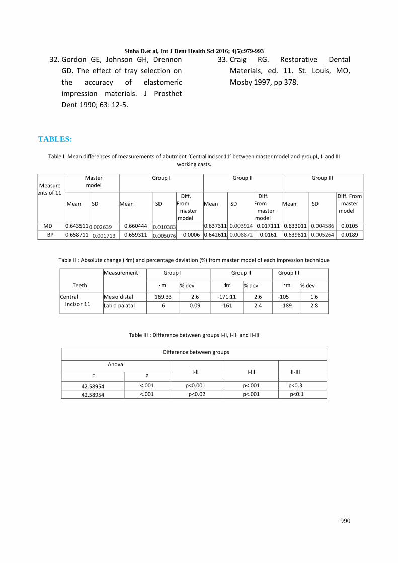
Table I :

Sinha D.et al, Int J Dent Health Sci 2016; 4(5):979-993
984
Shows the mean differences of
measurements of central incisor 11
between the master model and group I,
II and III working casts.Statistical
comparison between the master model
and group I, II, III casts measurements by
paired t-test showed significant
statistical difference (P<0.05) for all the
groups.
Table II :
Shows the percentage of deviation and
absolute change (µm) between the
metal die and master cast prepared
respectively for each impression
technique. It was found that there was
expansion in group I cast (matrix
impression technique) whereas the
Group II and Group III showed
contraction.
Table III :
Shows the difference between groups I-
II, I-III and II-III.
Statistical comparison between group I,
II and III casts measurements by one way
ANOVA (F-test) and unpaired-t-test
showed highly significant statistical
difference between the three groups
from each other. T test was used for
pair-wise comparison between the
means when ANOVA test is significant.
The significance level was set at P ≤ 0.01.
Here the p-value is ≤ 0.01, it suggests
that the observed data is inconsistent
with the assumption that the null
hypothesis is true, and hence we reject
the null hypothesis and accept the
alternative hypothesis and finally
conclude that there is significant
differences among the three impression
techniques (Table 3) for all mesio-distally
(MD) and bucco-palataly (BP) locations
(P<0.01). These analyses revealed a
significant difference between the three
techniques. Overall discrepancies of the
matrix impression technique was
significantly smaller than those in the
putty reline and multiple mix impression
techniques. Further investigation is
needed to determine the exact amount
of differences between the mentioned
impression techniques.
Graph I shows the mean mesio-distal
measurements of central incisor on
master model, Gr I, II and III casts while
Graph II shows the mean labio-palatal
measurements of central incisor on
master model, Gr I, II and III casts.
DISCUSSION:
Making an accurate impression of single
tooth or whole dentition is very vital in
obtaining accurate working casts, and for
the fabrication of the prosthesis or
restorations.
For obtaining an acceptable impression
and the working casts, various factors
have to be considered, like the proper
selection of the impression technique,
the impression material and the type of
trays. Over the past four decades,
tremendous progress has been made in
procedures for making fixed
prosthodontic impressions. These
impression procedures involve a wide
range of procedures and an even wider
range of materials. Many studies
Sinha D.et al, Int J Dent Health Sci 2016; 4(5):979-993
985
reported that the elastomeric
impression materials provide accurate
and dimensionally stable impressions.
Polyvinyl siloxane (PVS) impression
materials are extremely popular because
of their combination of excellent
physical properties, handling
characteristics and dimensional
stability.[6,24,25] In polyvinyl siloxane
impression materials, the strength of the
bond between the putty and wash is
sufficient to overcome stress that might
tend to separate the materials at their
interface and result in potential errors in
the impression.[8] The bond between the
putty material and light body is chemical
in nature and any bond failure which
occurs is a cohesive failure in the
weaker material.[26] Several factors
affect the accuracy of reproduction of
an impression material which includes
the tray,[27] tray adhesive [28] and the
impression technique.
Various impression techniques like
matrix impression system [18,19], putty
reline technique [3,17], multiple mix
technique [21] became popular for
making fixed prosthodontic
impressions. Various authors have
reported conflicting results as regard to
superiority of one technique over the
other. Livaditis GJ [18, 19] reported that
matrix impression system is more
accurate than the conventional
impression techniques.
The matrix impression system
incorporates the attributes of traditional
methods and overcomes important
deficiencies in registration of subgingival
margins, gingival retraction and relapse,
hemostasis and sulcular cleansing,
delivery of impression material
subgingivally, strengthening the sulcular
flange of the impression and
simplification for making complex
impressions [18].
The matrix forming material should
register details equal to the best
impression materials. The matrix-
forming material should be rapid setting
and compatible with the matrix
impression and tray impression
materials. Ideally, it should bond with
the other two materials without the use
of an intermediate adhesive layer.[18]
Livadatis GJ.[18] reported that
the matrix should encompass the
portions of the arch that are critical for a
fixed prosthodontic impression, which
include the prepared abutments, free
gingival margin, marginal ridges and
proximal surfaces of adjacent
unprepared teeth, and soft tissue
portions under planned pontics and
precision attachments.
For putty reline technique Fusayama T et
al6 and Wassel RM et al [14] reported that
one step putty reline technique
produced more accurate casts, whereas
Dhiman RK et al, [28] Johnson GH et al [29]
and Nissan J et al [1] reported that
dimensional accuracy was better with 2-
step technique. Hung SH et al, [3] Idris B
et al, [16] Lacy AM et al [7] and Stack
House J [5] did not find any difference
between the two techniques.

Sinha D.et al, Int J Dent Health Sci 2016; 4(5):979-993
986
Additional parameter that has to be
considered for accurate impression is the
uniformity of the wash space. [12,39]
Eames WB et al [5] reported that 2 mm
thickness of rubber base material
provided accurate impression than 4
and 6 mm thickness, because of lesser
polymerization shrinkage. Increasing the
thickness of the impression material,
produces more distortion because of
greater polymerization shrinkage. [30]
The objective of this in vitro study was to
compare the accuracy of matrix
impression system with conventional
putty reline technique and multiple mix
technique for individual die in master
model using the three impression
techniques.
A perforated metallic stock tray was
used for making the impression and
before making the impressions, tray
adhesive was applied on the tray and air
dried for 5 minutes, because the results
are enhanced both in accuracy and
consistency, when the tray adhesive is
used in a perforated stock tray. [6, 27]
After the master model was removed
from the water bath, it was dried before
making the impression because presence
of moisture affect the detail
reproduction of elastomeric
impression.[20] Equal amounts of putty
base and catalyst were hand mixed
without gloves because some brands of
latex gloves cause the setting inhibition
of elastomers. [10]
An automatic mixing system was used
for adding heavy body, medium body
and light body on the abutments in all
the three impression techniques. The
automatic mixing system is simple to
use, reduces bubbles in the mix resulting
in more precise impressions, no
spatulation required and being
economical. [11, 31]
The results showed the mesio-distal
dimensions of central incisor in Group I
was 0.660444 cm against the master
model which was 0.643511 cm. (Table
1).The percentage deviation from master
model in Gr I was 2.6, in Gr II was 2.6
and Gr III was 1.6 respectively (Table 6).
The contraction was observed in Gr II
and Gr III whereas the measurements of
Gr I was expanded as compared to that
of the master model.
The contraction in Gr II may be because
the wash material may have
hydraulically displaced the preliminary
putty impression during impression
seating and the putty may then have
exhibited some elastic recovery upon
removal of the impression and resulted
in tendency towards smaller dies. [16]
The results observed in this study are in
correlation to that of Nissan J et al, [1]
Idris B et al [15] and Gautam N et al. [4]
They stated that contraction may be due
to uncontrolled wash bulk, which
allows for differential contraction and
results in uneven dimensional change.
This may result in dies which are short
mesio-distally.
The labio-palatal dimensions of central
incisor was 0.659311 cm, against the

Sinha D.et al, Int J Dent Health Sci 2016; 4(5):979-993
987
master model which was 0.658711 cm
(Table 1).
The percentage deviation from master
model in Gr I was 0.09, Gr II was 2.4 and
Gr III was 2.8 respectively (Table 6). It
showed a considerable amount of
contraction. The contraction occurred
was more in Gr II and Gr III where as in
Gr I, the expansion was not significant
because the small bulk of the impression
material with in the matrix minimizes the
polymerization shrinkage and improves
the accuracy of the individual
abutment.[32]
The contraction in Gr II may be due to
more polymerization shrinkage or elastic
recovery of the putty. The contraction in
Gr III may be due to the uncontrolled
wash bulk, which results in uneven
dimensional change. This may lead from
narrow die in a bucco-lingual direction.
The results found in this study were
similar to those found by Gordon GE et
al [33] and Gautam N et al [4], which
revealed a slight increase in the vertical
dimension of the dies when PVS
impression material was used with stock
trays.
The putty material will get compressed if
the tray is not seated passively and the
putty material will show through after
the wash impression is made. It may
rebound to cause deformation. The
wash impression material may
hydraulically compress the putty during
the seating of the impression. The putty
could then exhibit some elastic recovery
upon removal of the impression. This
may result in an elongated die in cervico-
incisal direction. [4, 34]
The results found in this study are in
correlation with that of the findings of
Nissan J et al [1], Idris B et al [32] and
Petersen GF et al. [13]
From the above mentioned
results and discussion it can be
concluded that group I impressions
(matrix impression system) produced the
most accurate casts. Group II
impressions (putty reline technique)
produced more accurate casts than
group III impressions which were in
agreement with the findings of Gordon
GE et al. [33] Most dimensional
differences were shown in group III
impressions (multiple mix technique)
which were in agreement with the
findings of Nissan J et al [1] and Idris B et
al. [32] The matrix impression system is
more acceptable to obtain accurate dies
with polyvinyl siloxane impressions.
CONCLUSION:
The purpose of this study was to
compare the marginal accuracy of dies
obtained from different impression
techniques, to evaluate the marginal
width of dies made by using different
impression techniques, to compare the
accuracy of marginal width using
different impression techniques & to
evaluate the most accurate impression
technique among the three techniques
used.
Within the limitations of this study when
the working casts of the three groups
were compared with the master model,

Sinha D.et al, Int J Dent Health Sci 2016; 4(5):979-993
988
the conclusion was drawn that the
overall discrepancies of the matrix
impression technique was significantly
smaller than those in the putty reline
and multiple mix impression techniques.
Further investigation is needed to
determine the exact amount of
differences between the mentioned
impression techniques.
REFERENCES:
1. Nissan J, Laufer BZ, Brosh T, Assif D.
Accuracy of three polyvinyl siloxane
putty-wash impression techniques. J
Prosthet Dent 2000; 83(2) : 161-5.
2. Hung SH, Purk JH, Tira DE, Eick JD.
Accuracy of one step versus two
step putty wash addition silicone
impression technique. J Prosthet
Dent 1992;67(5) : 583-9.
3. Gautam N, Sajjan S. Matrix
impression system (MIS) versus
conventional putty reline technique
(PRT): A comparative evaluation. J of
Ind Prosthodont Soc 2003;3(2):44-8.
4. Eames WB, Sieweke JC, Wallace SW,
Rogers LB. Elastomeric impression
materials: Effect of bulk on accuracy.
J Prosthet Dent 1979;41(3):304-7.
5. Brown D. An update on
elastomeric impression materials.
Br Dent J 1981;150:35-40.
6. Lacy AM, Fukui H, Bellman T,
Jendresen MD. Time dependent
accuracy of elastomer impression
materials. Part II: polyether,
polysulfides, and polyvinyl siloxane.
J Prosthet Dent 1981;45(3): 329-33.
7. Sandrik JL, Vacco JL. Tensile and
bond strength of putty-wash
elastomeric impression materials. J
Prosthet Dent 1983;50(3)358-61.
8. Craig RG. Evaluation of an automatic
mixing system for an addition
silicone impression materials. JADA
1985;110:213-5.
9. Cook. WD, Thomas Z F. Rubber
gloves and addition silicone
materials. Aust Dent J
1986;31(2):140.
10. Chong YH, Soh G, Wickens JL. The
effect of mixing method on void
formation in elastomeric
impression materials. Int J
Prosthodont 1989;2(4):323-6.
11. Marshak B, Assif D, Pilo R. A
controlled putty-wash impression
technique. J Prosthet Dent
1990;64(6):635-6.
12. Petersen GF, Asmussen E. Distortion
of impression materials used in the
double-mix technique. J Dent Res
1991;99:343-8.
13. Wassell RM, Ibbetson RJ. The
accuracy of polyvinyl siloxane
impressions made with standard
and reinforced stock trays. J
Prosthet Dent 1991;65(4):748-57.
14. Tjan AHL, Nemetz H, Nguyen LTP,
Contino R. Effect of tray space on
the accuracy of monophasic
polyvinyl siloxane impressions. J
Prosthet Dent1992;68(1):19-28ew
CL, Chee WWL, Donovan TE. The
influence of temperature on the
Dimensional stability of Poly (vinyl
siloxane) impression materials. Int
J Prosthodont 1993;6(6):528-32.
Sinha D.et al, Int J Dent Health Sci 2016; 4(5):979-993
989
15. Idris B, Houston F, Claffey N.
Comparison of the dimensional
accuracy of one-and two-step
techniques with the use of
putty/wash addition silicone
impression materials. J Prosthet
Dent 1995;74(5):535-41.
16. Laufer BZ, Baharav H, Ganor Y,
Cardash HS. The effect of
marginal thickness on the distortion
of different impression materials. J
Prosthet Dent 1996;76:466-71.
17. Livaditis GJ. Comparison of the new
matrix system with traditional fixed
prosthodontic impression
procedures. J Prosthet Dent
1998;79(2):200-7.
18. Livaditis GJ. The matrix impression
system for fixed prosthodontics. J
Prosthet Dent 1998; 79: 208-16.
19. Johnson GH, Lepe X, Aw TC. The
effect of surface moisture on
detail reproduction of elastomeric
impressions. J Prosthet Dent
2003;90:354-64.
20. Anusavice KJ. Pillips Science of
dental materials. 11 edn Saunders,
2004, p 219-220.
21. Shillingburg HT, Hobo S, Whitsett
LD, Jacobi R, Brackett SE.
“Fundamentals of fixed
prosthodontics 1997; 3rd edn, 291-
3.
22. Goldfogel M, Harvey WL, Winter D.
Dimensional changes of acrylic resin
tray material. J Prosthet Dent 1985;
54(2): 284-6.
23. Craig RG, Urguiola NJ, Liu CC.
Comparison of commercial
elastomeric impression materials
Oper Dent 1990; 15: 94-104.
24. Cullen DR, Sandrik JL. Tensile
strength of elastomeric impression
materials, adhesive and cohesive
bonding. J Prosthet Dent 1989; 62(2)
142-5.
25. Gordon GE, Johnson GH, Drennon
DG. The effect of tray selection on
the accuracy of elastomeric
impression materials. J Prosthet
Dent 1990; 6(1): 12-15.
26. Bomberg TJ, Goldfogel MH, Hoffman
W, Bomberg SE. Considerations for
adhesion of impression materials to
impression trays. J Prosthet Dent
1988;60(6): 681-4.
27. Dhiman RK, Agarwal SK, Dhir RC.
Dimensional Accuracy of Putty/wash
one step and two step techniques
using polyvinyl siloxane impression
material : In vitro study. J of Ind
Prosthodont Soc 2001; 1(2):36-43.
28. Johnson GH, Drennon DG. Clinical
evaluation of detail reproduction
of elastomeric impression materials.
J Dent Res 1987; 66: 331.
29. Araujo PAD, Jorgensen KD. Effect of
materials bulk and undercuts on the
accuracy of impression materials. J
Prosthet Dent 1985; 54(6): 791-4.
30. Craig RG. Evaluation of an automatic
mixing system for an addition
silicone impression material. JADA
1985; 110: 213-5.
31. Dimashkieh MR, Morgano MS. A
procedure for making fixed
prosthodontic impressions with the
use of preformed crown shells. J
Prosthet Dent 1995; 73:95-6.
Sinha D.et al, Int J Dent Health Sci 2016; 4(5):979-993
990
32. Gordon GE, Johnson GH, Drennon
GD. The effect of tray selection on
the accuracy of elastomeric
impression materials. J Prosthet
Dent 1990; 63: 12-5.
33. Craig RG. Restorative Dental
Materials, ed. 11. St. Louis, MO,
Mosby 1997, pp 378.
TABLES:
Table I: Mean differences of measurements of abutment ‘Central Incisor 11’ between master model and groupI, II and III working casts.
Table II : Absolute change ( m) and percentage deviation (%) from master model of each impression technique
Table III : Difference between groups I-II, I-III and II-III
Measure
ments of 11
Master model
Group I Group II Group III
Mean
SD
Mean
SD
Diff. From
master model
Mean
SD
Diff. From
master model
Mean
SD
Diff. From master
model
MD 0.643511 0.002639 0.660444
0.010383 0.016933
0.637311 0.003924 0.017111 0.633011 0.004586 0.0105
BP 0.658711 0.001713 0.659311 0.005076 0.0006 0.642611 0.008872 0.0161 0.639811 0.005264 0.0189
Teeth
Measurement Group I Group II Group III
m % dev m % dev m % dev
Central Incisor 11
Mesio distal 169.33 2.6 -171.11 2.6 -105 1.6
Labio palatal 6 0.09 -161 2.4 -189 2.8
Difference between groups
Anova I-II
I-III
II-III
F P
42.58954 <.001 p<0.001 p<.001 p<0.3
42.58954 <.001 p<0.02 p<.001 p<0.1

Sinha D.et al, Int J Dent Health Sci 2016; 4(5):979-993
991
0.6
0.62
0.64
0.66
0.68
MM MATRIX PUTTY RELINE MULTIPLE MIX
0.6435110.660444
0.637311 0.633011
MEA
N (
cm)
CENTAL INCISOR 11 : MESIO-DISTALMEASUREMENTS
0.63
0.635
0.64
0.645
0.65
0.655
0.66
MM MATRIX PUTTY RELINE MULTIPLE MIX
0.658711 0.659311
0.6426110.639811
MEA
N (
CM
)
CENTAL INCISOR 11 : BUCCO-PALATAL MEASUREMENTS
GRAPHS:
Graph I: The mean mesio-distal measurements of central incisor on master model,
Group I, II and III casts.
Graph II: The mean labio-palatal measurements of central incisor on master model, Group I, II
and III casts.

Sinha D.et al, Int J Dent Health Sci 2016; 4(5):979-993
992
FIGURES:
FIGURE I : Materials Used In The Study
FIGURE II :Travelling Microscope & Cast

Sinha D.et al, Int J Dent Health Sci 2016; 4(5):979-993
993
FIGURE III : Master Model with Metal Die Central Incisor 11
FIGURE IV: Casts Seen Under Magnification Travelling Microscope