community eye health journal - cehjsouthasia.org · the author/s and community eye health journal...
TRANSCRIPT

S COMMUNITY EYE HEALTH JOURNAL SOUTH ASIA | VOLUME 29 ISSUE 95 | 2017 01 © The author/s and Community Eye Health Journal 2015. This is an Open Access article distributed under the Creative Commons Attribution Non-Commercial License.
Community Eye Health
JOURNALVOLUME 29 | ISSUE 95 | 2017
The ocular surface comprises the cornea, conjunctiva, eyelids and lacrimal glands and any disorder in these structures can be classified as an ocular surface disorder (OSD). Though the prevalence of OSD is quite high, unfortunately, cases often go undiag-nosed or undertreated, due to a lack of understanding of symptoms, and inaccurate evaluation. As people are living longer, these disorders are becoming more prevalent, but awareness about them is quite limited.
OSD includes conditions like Dry Eye Disease (DED), blepharitis and meibomian gland dysfunction (MDG), allergic eye diseases (AED), chemical and thermal burns and so on. Ocular surface diseases can severely affect eyesight and quality of life, and in severe cases, cause blindness. The
mode of presentation as well as the severity varies in different populations and this issue will focus on the presen-tation of OSD in South Asia.
For DED there are no population-based studies in South Asia. There are few hospital-based reports published, but the prevalence is quite variable.1-4
This is probably due to differences in geographic location as well as lack of standardized questionnaires and objective tests to confirm a diagnosis of dry eye. It’s known that increasing age is one of the risk factors for DED; so with an ageing population, we are likely to see more DED in South Asia.
Allergic conjunctivitis (AC) represents a spectrum of disorders comprising seasonal allergic conjunctivitis (SAC), perennial allergic conjunctivitis (PAC), atopic keratoconjunctivitis (AKC), vernal keratoconjunctivitis (VKC) and giant papillary conjunctivitis (GPC). The most common types are SAC and PAC, which are self-limiting conditions and rarely cause significant ocular damage. On
the other hand, AKC and VKC are severe and can affect the cornea and lead to vision loss, either due to the disease itself or due to side effects of corticos-teroids, which is one of the mainstays of therapy. Some data on AKC / VKC is available from India, Nepal and Pakistan,5-7 however it is difficult to compare studies as there is no standard validated survey instrument, and so extrapolation of this data to other populations become limited.
For ptyergium, a higher prevalence has been reported from countries with increasing geographic latitude and with age.8, 9-10 One of the major risk factors identified is ultraviolet light exposure due to outdoor occupations. Other risk factors are male gender and those residing in rural areas.8 The mainstay of therapy is surgical excision and different techniques have been described, of which covering bare sclera with conjunctival autograft probably has the lowest rate of recurrence.
Ocular surface disorders
A patient is examined for ocular surface disease. INDIA
(c) S
.B.N
. Cha
ry\ L
VPEI
EDITORIAL
Rohit C KhannaDirector, Gullapalli Pratibha Rao International Centre for Advancement of Rural Eye Care (GPR ICARE), L V Prasad Eye Institute, Hyderabad
South Asia Edition
02 COMMUNITY EYE HEALTH JOURNAL SOUTH ASIA | VOLUME 29 ISSUE 95 | 2017S
“Improving eye health through the delivery of practical
high-quality information for the eye care team”
Volume 29 | ISSUE 93
Supporting VISION 2020: The Right to Sight
EditorElmien Wolvaardt Ellison [email protected]
Editorial committeeAllen FosterClare GilbertNick AstburyDaksha PatelRichard WormaldMatthew BurtonHannah KuperPriya MorjariaGV MurthyFatima KyariDavid YorstonSally CrookSerge ResnikoffBabar QureshiPeter AcklandJanet MarsdenNoela Prasad
Regional consultantsHugh Taylor (WPR) Leshan Tan (WPR)GVS Murthy (SEAR)R Thulsiraj (SEAR)Babar Qureshi (EMR)Mansur Rabiu (EMR)Hannah Faal (AFR)Kovin Naidoo (AFR) Ian Murdoch (EUR)Janos Nemeth (EUR)Van Lansingh (AMR)Andrea Zin (AMR)
Editorial assistant Anita ShahDesign TejaProofreading Shivani Mathur Gaihaand Sridivya MukpalkarPrinting Newman ThomsonOnline edition and newsletter Sally Parsley: [email protected] Advisor for Issue 95 of the South Asia EditionRohit C Khanna
SOUTH ASIA EDITORIAL BOARD
Allen Foster Damodar Bachani Thulasiraj Ravilla Rajiv Raman Rohit C Khanna Asim Sil GV Murthy Shivani Mathur Gaiha
SOUTH ASIA ADVISORY COMMITTEE
Allen Foster Hans Limburg Elizabeth Kurian Sara Varughese Muhit Muhammed Sanduk Ruit BR Shamanna K Vishwanath M Babar Qureshi Prabhat Piyasena
Address for subscriptionsCommunity Eye Health Journal South Asia, Indian Institute of Public Health, Plot No. 1, ANV Arcade, Amar Co-operative Society, Kavuri Hills, Madhapur, Hyderabad-500033,India.Tel +91-40-49006000Email [email protected] Correspondence articlesWe accept submissions of 800 words about readers’ experiences in eye health. Contact: Community Eye Health Journal South Asia at:[email protected]
© International Centre for Eye Health, London. Articles may be photocopied, reproduced or translated provided these are not used for commercial or personal profit. Acknowledgements should be made to the author(s) and to Community Eye Health Journal. Woodcut-style graphics by Victoria Francis and Teresa Dodgson.
ISSN 0953-6833
Disclaimer
Signed articles are the responsibility of the named authors alone and do not necessarily reflect the views of the London School of Hygiene & Tropical Medicine (the School). Although every effort is made to ensure accuracy, the School does not warrant that the information contained in this publication is complete and correct and shall not be liable for any damages incurred as a result of its use.
The mention of specific companies or of certain manufacturers’ products does not imply that they are endorsed or recommended by the School in preference to others of a similar nature that are not mentioned. The School does not endorse or recommend products or services for which you may view advertisements in this Journal.
Common ocular burns are chemical or thermal, with most chemical burns being due to either acid or alkali. The majority of these chemical burns occur in young males, with lime (chuna) being an important cause. Different classification systems are available including Hughes, Roper-Hall, Dua and Holland Mannis classifications. Different modalities of treatment are available, ranging from first aid as irrigating the eye; medical
treatment with topical steroids, antibiotics and cycloplegics and surgical treatment in form of amniotic membrane graft (AMG), limbal stem cell transplantation or simple limbal epithelial cell transplantation (SLET).
This issue will provide an overview of different types of OSDs, including dry eye, allergic conjunctivitis, pterygium, corneal ulcers and ocular burns in the context of South Asia.
References
1. Sahai A, Malik P. Dry eye: prevalence and attributable risk
factors in a hospital-based population. Indian J
Ophthalmol. 2005 Jun;53(2):87-91.
2. Gupta N, Prasad I, Jain R, D'Souza P. Estimating the preva-
lence of dry eye among Indian patients attending a tertiary
ophthalmology clinic. Ann Trop Med Parasitol. 2010
Apr;104(3):247-55.
3. Basak SK, Pal PP, Basak S, Bandyopadhyay A, Choudhury
S, Sar S. Prevalence of dry eye diseases in hospital-based
population in West Bengal, Eastern India. J Indian Med
Assoc. 2012 Nov;110(11):789-94.
4. Gupta N, Prasad I, Himashree G, D'Souza P. Prevalence of
dry eye at high altitude: a case controlled comparative
study. High Alt Med Biol. 2008 Winter;9(4):327-34.
5. Pokharel N, Shah DN, Choudhary M. Vernal keratocon-
junctivitis: modes of presentation in Nepalese population.
Kathmandu University medical journal.
2007;5(4):526-30.
6. Sofi RA, Mufti A. Vernal Keratoconjunctivitis in Kashmir: A
temperate zone. International ophthalmology. 2016.
7. Saboo US, Jain M, Reddy JC, Sangwan VS. Demographic
and clinical profile of vernal keratoconjunctivitis at a
tertiary eye care center in India. Indian journal of ophthal-
mology. 2013;61(9):486-9.
8. Liu L, Wu J, Geng J, Yuan Z, Huang D. Geographical preva-
lence and risk factors for pterygium: a systematic review
and meta-analysis. BMJ Open. 2013 Nov
19;3(11):e003787.
9. Zhao L, You QS, Xu L, Ma K, Wang YX, Yang H, Jonas JB.
10-year incidence and associations of pterygium in adult
Chinese: the Beijing Eye Study. Invest Ophthalmol Vis Sci.
2013 Feb 27;54(2):1509-14.
10. Nemesure B, Wu SY, Hennis A, Leske MC; Barbados Eye
Studies Group.. Nine-year incidence and risk factors for
pterygium in the barbados eye studies. Ophthalmology.
2008 Dec;115(12):2153-8.
EDITORIAL Continued
S01
S03
S06
S08
S10
S14
S20
S24
S27
S18
Editorial
Assessment and diagnosis: a rationalapproach
Prevention, diagnosis andmanagement of dry eye inSouth Asia
Ptyergium- Epidemiologytreatment and prevention
Allergic Conjunctivits
Ocular surface injuries and management
Prevention of traumaticcorneal ulcers in SouthEast Asia
Save your eyes for 30Rupees: A case study
Common and important ocular surface conditions
POSTER
NEWS AND RESOURCES

S COMMUNITY EYE HEALTH JOURNAL SOUTH ASIA | VOLUME 29 ISSUE 95 | 2017 03 © The author/s and Community Eye Health Journal 2015. This is an Open Access article distributed under the Creative Commons Attribution Non-Commercial License.
The ocular surface is critical to the health of the eye and essential for good visual functioning. It is a complex, integrated system involving the cornea, conjunctiva, tear film, lacrimal gland, nasolacrimal
system and the eyelids (incorporating the meibomian glands and lashes). The normal physiological function of the ocular surface depends on the interaction of these different components. Working together, they maintain a clear optical surface, keep the eye from drying out, and protect it from trauma and infection. Changes in the structure and function of any of the ocular surface components can disrupt its delicate balance and lead to pathology.
Ocular surface diseases have a relatively limited set of symptoms and
signs, and a systematic approach to assessing and diagnosing these conditions is therefore necessary.
HistoryBecause patients with ocular surface problems present with a limited range of symptoms and signs, taking a detailed history is very important. Ask patients whether they have experienced, or are experiencing, any of the following:
• Reduced vision (mild blurring can occur if the tear film is disturbed; a more severe visual disturbance suggests corneal or other disease)
• Redness• Irritation or gritty sensation
(suggests epithelial disturbance)• Itching (suggests allergy)• Pain (sharp pain suggests a corneal
problem or foreign body; a duller ache may suggest uveal or scleral inflammation)
• Purulent discharge • Watering, whether from lacrimation
(increased tear production) or epiphora (decreased tear drainage)
Many diseases can affect the ocular surface. Their frequency and severity varies from region to region, often depending on the local climate. Ocular surface diseases can affect both eyesight and quality of life, and – in severe cases – cause blindness. Because they have a limited number of symptoms and signs, and can appear very similar in presentation, patients can be misdiagnosed and hence poorly managed. In this issue, we offer a systematic approach to assessing and diagnosing common ocular surface diseases and look in detail at general management principles, including how to control inflammation. Other articles discuss ocular allergy, pterygium and prevention of traumatic corneal ulcers. In the middle of the issue we also have a poster with useful information about common ocular surface conditions and their primary management.
ABOUT THIS ISSUE
Assessment and diagnosis: a rational approach
Jeremy HoffmanAcademic Clinical Fellow: International Centre for Eye Health and Specialist Registrar, Moorfields Eye Hospital, London, UK.
Matthew BurtonProfessor: International Centre for Eye Health and Consultant Ophthalmologist: Moorfields Eye Hospital, London, UK.
Examining the ocular surface. INDIA
Indi
a (c
) IIP
H H
yder
abad
/Div
yajy
othi
Hos
pita
l, S
urat

04 COMMUNITY EYE HEALTH JOURNAL SOUTH ASIA | VOLUME 29 ISSUE 95 | 2017S
It is important to take a careful note of when and how the problem developed. You need to ask if there has been a history of trauma or a foreign body. In some settings, contact lens use is common and you need to ask about this. If patients do use contact lenses, ask how they clean and use them.
ExaminationOcular surface diseases have a relatively limited set of symptoms and signs. Therefore, your approach to assessing and diagnosing these conditions needs to be systematic. A stepwise approach helps to ensure that important things are not missed.
• Vision. Start by assessing the uncorrected, pinhole and best corrected visual acuity.
• Eyelids. Examine the lid position and closure and check for entropion (when the eyelid turns in on itself), trichiasis (lashes touching the eye)and lagophthalmos (a gap between the upper and lower lid when the eyes are closed). Examine the lid margin and meibomian gland openings for abnormal positions, inflammation and plugging with secretions. Try to express the meibomian glands, using gentle pressure. The normal physiological function of the ocular surface depends on the interaction of these different components. Working together, they
maintain a clear optical surface, keep the eye from drying out, and protect it from trauma and infection. Changes in the structure and function of any of the ocular surface components can disrupt its delicate balance and lead to pathology.
• Tears. Assess the quality of the tear film by looking for discharge or debris and the tear meniscus height (to give an idea of quantity). Check the tear break-up time by instilling a drop of fluorescein and timing how long it takes for the tear film to disperse. A tear break-up time of less than 10 seconds is abnormal. Finally, perform Schirmer’s test by placing a testing strip in the inferior conjunctival fornix and asking the patient to close their eyes for five minutes. A normal result is >15 mm. Less than this suggests insufficient tear production, to varying degrees: mild is 9–14 mm, moderate is 4–8 mm and severe is <4 mm.
• Bulbar conjunctiva and sclera. Assess inflammation, scarring, haemorrhages and abnormal swellings such as pinguecula, pterygium or possible malignancies.
• Tarsal conjunctiva. Evert the upper and lower lids. Look for scarring, foreign body defects, inflammatory membranes, papillae and follicles.
• Corneal epithelium. Using a torch, look for foreign bodies, infiltrates, oedema and deposits. Is the light reflected off the
eye’s surface shiny (healthy), or rough and/or dull? Also test for corneal sensation, which may be reduced due to infection with herpes simplex or zoster.
• Corneal stroma. Look for stromal opacities. Assess the size, location, pattern and depth. Opacities may be scars or active inflammatory infiltrates. Look for blood vessels: active vessels have blood flowing, inactive have a clear, grey outline without blood.
• Corneal endothelium. Look for any guttata, Descemet folds and the presence and type of any deposits (blood, keratic precipitates or pigment).
DiagnosisProblems affecting the ocular surface broadly divide into non-infectious and infectious conditions. They present with a limited range of symptoms. The pattern of symptoms can often help to differentiate between conditions. In Table 1 we outline the typical symptom pattern for some of the commoner conditions. For example, if the person mainly complains of itching, then allergic conjunctivitis needs to be considered as a possible cause.
The symptoms of these different condi-tions can overlap. Therefore, a careful examination is critical to reaching an accurate diagnosis. Although not exhaustive, there is a list of common and important ocular surface conditions on pages 18-19, detailing their presenting features and some example photographs.
Table 1: Symptom and signs of common conditions Key: Absent Possible Present, moderate Present, severe
Bacterial conjunctivitis
Viral conjunctivitis
Allergic conjunctivitis
Microbial keratitis
Dry eye Blepharitis Rosacea Mucous membrane pemphigoid
Stephens-Johnson Syndrome
Visual impairment
Red
Pain
Itchy
Irritation or gritty sensation
Watery discharge
Purulent discharge
Condition
Symptoms/signs

S COMMUNITY EYE HEALTH JOURNAL SOUTH ASIA | VOLUME 29 ISSUE 95 | 2017 05 © The author/s and Community Eye Health Journal 2015. This is an Open Access article distributed under the Creative Commons Attribution Non-Commercial License.
The ocular surface consists of the cornea, conjunctiva, tear film, lacrimal gland, nasolacrimal system and the eyelids (incorporating the meibomian glands and lashes), each of which is described in detail below. Figure 1 shows the anatomy of the upper eyelid and anterior segment of the eye in cross-section.
CorneaThe cornea is the most powerful refracting component of the eye. Together with the lens, it focuses light on the retina. The central 4 mm zone is critical for good vision. The cornea is made up of five layers: epithelium, Bowman’s layer, stroma, Descemet’s membrane and endothelium. The normal cornea does not have blood vessels; it gains oxygen and nutrients through diffusion from the aqueous, from limbal blood vessels and from the atmosphere. The cornea is very sensitive; there is dense innervation by fine nerve fibres from the trigeminal nerve. Normal corneal sensation is essential for a healthy intact epithelial surface, tear function and protection through the blink reflex.
If damaged, the corneal epithelium can regenerate, so simple abrasion injuries can heal without scarring. However, if the stem cells that repopulate the corneal epithelial surface are damaged, for example by a chemical injury, the resulting epithelium is abnormal and clarity is lost. Corneal clarity also depends on there being a highly ordered arrangement of collagen fibres within the stroma. These deeper layers are unable to regenerate well and often heal with scarring. In addition, the cornea needs to be maintained in a relatively dehydrated state by the action of the endothelial cell layer. If this is not functioning well, the cornea becomes oedematous and opaque.
ConjunctivaThe conjunctiva is composed of an epithelial layer overlaying a loose connective tissue (stroma). It covers the eye from the edge of the cornea (limbus) to the fornices and the inside surface of the eyelids. It contains specialised goblet cells that produce the mucus layer of the tear film. In the stromal layer of the conjunctiva, there are immune system cells that defend against infection. Sometimes lymphoid cells are recruited and gather together to form visible follicles, particularly on the tarsal conjunctival surface. Papillae, which form in the tarsal conjunctiva, are dome-like swellings with inflammatory cells, oedema and a dilated blood vessel. Conjunctival scarring develops in some chronic inflammatory ocular
surface conditions, with shortened fornices, symblepharon (adhesions between the eye lid and globe) and distortion of the eyelids.
Tear filmThe tear film is made up of three layers. The outer lipid layer (produced by the meibomian glands) reduces evaporation of the middle aqueous layer (produced by the lacrimal gland), with the inner mucin layer (produced by goblet cells) helping to stabilise the aqueous layer on the corneal epithelium. A good tear film helps to maintain a well-hydrated, healthy corneal epithelium and a clear optical surface, and it protects against infection.
Lacrimal glandThe lacrimal gland sits in the supero-lateral region of the orbit. Fine ducts open into the upper fornix, delivering lacrimal fluid to the ocular surface. Secretion of tear fluid is controlled by the parasympathetic nervous system. Problems with the gland itself, obstruction of the ducts (by scarring) and neurological problems can all result in reduced aqueous tear production.
Nasolacrimal systemThe nasolacrimal system drains tear fluid from the surface of the eye. Fluid is collected through the punctae and passes along the canaliculi into the lacrimal sac. From the sac, the fluid passes down the nasolacrimal duct and drains into the nasal cavity. Obstruction at any point along the system can result in a watery eye (epiphora) and predispose the eye to infection.
EyelidsEyelids protect the eyes by covering them. They are formed of several layers: skin, the orbicularis muscle, the tarsal plate (including the meibomian glands), and the conjunctiva.
Understanding the ocular surface Jeremy Hoffman and Matthew Burton
Figure 1: Anatomy of the upper eyelid and anterior segment of the eye in cross-section
Pete
r Mal
len
ww
w.s
chep
ens.
harv
ard.
edu
Lacrimal gland
Meibomian glandUpper
fornix
Tear film
Cornea
Tarsal conjunctiva
Limbus
Lower fornix
Bulbar conjunctiva
06 COMMUNITY EYE HEALTH JOURNAL SOUTH ASIA | VOLUME 29 ISSUE 95 | 2017S
IntroductionDry eye is a condition that affects the tear film and affects the ocular surface that includes the conjunctiva and cornea.1 Dry eye, being a chronic disease, results in health related quality of life issues and economic problems due to loss of productive working days and the cost of medical treatment. Untreated dry eye may result in corneal surface ulceration and opacification leading to corneal blindness.
Definition of dry eyeIn 2007, the International Dry Eye Workshop (DEWS) report defined dry eye as a multifactorial inflammatory disease of the tears and ocular surface, resulting in discomfort and visual disturbance,
unstable tear film and ocular surface damage.1
Classification and EtiologyThe dry eye condition is classified as evaporative dry eye and aqueous tear deficient dry eye.2,3
Aqueous deficient dry eye is further subdivided as Sjogren syndrome dry eye and non-Sjogren dry eye.
Sjogren’s syndrome is a chronic inflammatory connective tissue disorder more common in females, who may be around 40 years of age. These patients may have dry eye and dry mouth. Primary Sjogren’s syndrome is without systemic disease; Secondary Sjogren’s is with systemic disease.
Non-Sjogrens’s dry eye is seen in patients having Graft versus Host disease, trachoma, conjunctival cicatrizing disorders and use of drugs such as antihistamines, decongestants, antipsy-chotic drugs, antidepressants and antihypertensives.
Evaporative dry eye is most commonly caused by meibomian gland disease.
EpidemiologyDry eye is more common in elderly
females.4 Predisposing factors include collagen vascular disease, diabetes, allergy, antihistamines, pterygium and climate.4,5
Diagnosis of Dry eyeHistory taking, clinical examination followed by investigations are done to diagnose dry eye.
SymptomsPatients with dry eye have a long history of symptoms such as of irritation and sandy or gritty sensation in the eyes. The symptoms may be mild to severe, and infrequent to long standing. The patients may have worsening of symptoms on prolonged visual work, intolerance to low humidity, feeling of dry eye and irritation. Dry eye is usually symptomatic although Sullivan et al have shown that 40% of patients having dry eye were asympto-matic and sometimes the symptoms may not correlate with the signs.6 There are various questionnaires such as Ocular Surface Disease Index (OSDI) and McMonnies questionnaire to identify, diagnose and manage dry eyes.7,8
Varsha M RathiFaculty, Tej Kohli Cornea Institute, Gullapalli Pratibha Rao International Center for Advancement of Rural Eye Care, L V Prasad Eye Institute (LVPEI), Hyderabad, India
Virender S SangwanFaculty, Tej Kohli Cornea Institute, Kallam Anji Reddy Campus, L V Prasad Eye Institute (LVPEI), Hyderabad, India.
Prevention, diagnosis & management of dry eye in South Asia
MANAGEMENT
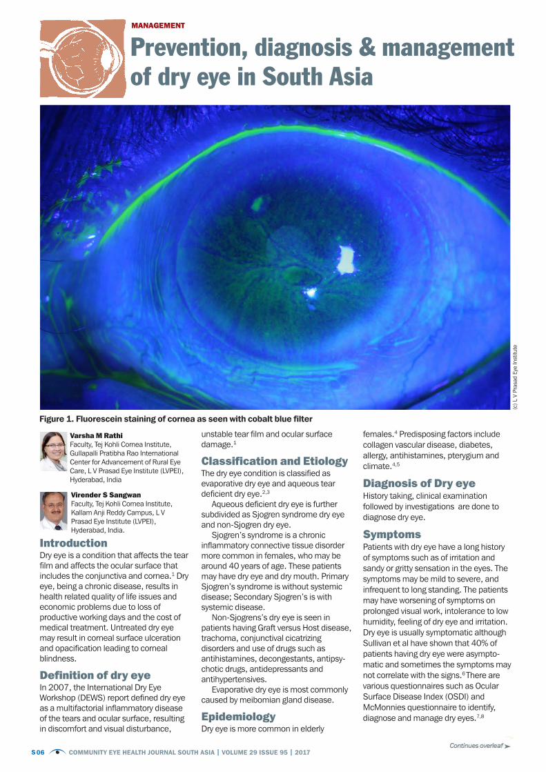
Figure 1. Fluorescein staining of cornea as seen with cobalt blue filter
(c) L
V P
rasa
d Ey
e In
stitu
te
S COMMUNITY EYE HEALTH JOURNAL SOUTH ASIA | VOLUME 29 ISSUE 95 | 2017 07 © The author/s and Community Eye Health Journal 2015. This is an Open Access article distributed under the Creative Commons Attribution Non-Commercial License.
Clinical examinationObservation of the lids, conjunctiva and cornea should be done first before performing any test. The following is the sequence of examining a patient of dry eye. 1. Initial examination of lids and the ocular surface2. TBUT – Tear film break up time after instillation of flurorescein dye3. Corneal staining with fluorescein or lissamine green (between 1-4 minutes of lissamine green instillation)4. The Schirmer 1 test (or phenol red thread test Schirmer test with anaesthesia) can be performed to determine the basal tear production. Tear osmolality should be measured after examination, if available.
Diagnostic testsSchirmer test – The test is performed by putting a filter paper strip in the middle of lower fornix.9 After five minutes, the wetting of the filter strip is assessed. A wetting of 10mm or more is considered normal. Before applying a filter strip, excess tears should be wiped out otherwise the results may be showing a false high. Repeatability of this test and correlation with patient symptoms is poor.Phenol red thread test – This test measures the tear volume. Phenol red, being pH sensitive, changes from red to yellow when exposed to tears.10 A 70mm thread is placed in lower fornix and wetting is measured after 15 seconds. The normal values range between 9mm-20mm and less than 9 mm is considered dry eye. Patel et al have shown that a value of 15 mm of wetting correlated with aqueous deficient and 22 mm with non-aqueous deficient dry eye.11
Tear osmolality – This increases in patients with dry eye disease. Tear film breakup time (TBUT)– A fluorescein strip is applied in the lower fornix and removed. The patient is asked to blink normally and then to stop blinking. The time taken from stopping blinking to the appearance of the first dark spot in the tear film indicates TBUT. A TBUT of <10 seconds is abnormal.Videokeratography and keratometry – This can also can be used to assess the TBUT. Normal values for breaking of mires during keratometry are more than 15 seconds.Meibography – Technological advances in the field of digital imaging have helped in assessing the meibomian glands, which if dysfunctional can result in evaporative dry eye. Various methods are available to do so including auto-refractometer.Corneal staining – Fluorescein staining of
cornea appears greenish and is viewed using cobalt blue filter (Figure 1). The pattern of staining gives a clue to the etiology of dry eyes e.g. inferior corneal staining in patients having lagophthalmos or inability to close the eye lids; interpalpebral staining in evaporative dry eyes. Rose Bengal stain – It stains dead and devitalized cells of cornea and conjunctiva. The patients have severe stinging when Rose Bengal stain is used. One can also use lissamine green stain.
Diagnosis is based on a combination of history, clinical examination and the investi-gations.
ManagementManagement depends on the severity of dry eye and response to the treatment.Artificial tears and lubricating eye drops– This should be given to dry eye which is aqueous deficient. Artificial tears that merely increase tear volume may worsen symptoms in patients with a lipid deficiency. Tear retention with Punctal occlusion – This may be indicated in patients who have symptomatic dry eyes, when Schirmer’s test is <5 mm and there is ocular surface staining. These can be done either with cautery or with punctal plugs (absorbable and non-absorbable). Management of lids – Treat inflammation of the meibomian glands with hot bathing over closed eyelids followed by expression of the meibomian secretions. Use of lubri-cating eye drops, oral doxycycline and tetracycline may be helpful.
Key messageDry eye is a multifactorial disease. It is important to determine whether it is aqueous deficient or evaporative dry eye or a combined one. Success of the treatment is dependent on proper understanding of the cause of dry eye and approach to the management.Determination of tear meniscus height is important. Schirmer's test will help differen-tiate aqueous deficient from evaporative dry eye. This should be done in all patients. Corneal staining with fluorescein, Rose Bengal and Lissamine green dyes will help assess damage to the ocular surface. Based on the level of damage, Schirmer’s test values and TBUT values, management can either be with lubricating eye drops, anti-inflammatory agents, environmental modifications, or treatment of inflammation of the meibomian glands. The treating physicians should modify treatment based on patients’ symptoms.
References
1. The definition and classification of dry eye
disease: report of the Definition and Classification
Subcommittee of the International Dry Eye
WorkShop (2007). Ocul Surf.2007
Apr;5(2):75-92.
2. Methodologies to diagnose and monitor dry eye
disease: report of the Diagnostic Methodology
Subcommittee of the International Dry Eye
WorkShop (2007). Ocul Surf.2007
Apr;5(2):108-52.
3. Lemp MA. Report of the National Eye Institute/
Industry workshop on Clinical Trials in Dry Eyes.
CLAO J.1995 Oct;21(4):221-32.
4. Gayton JL. Etiology, prevalence, and treatment of
dry eye disease. Clin
Ophthalmol.2009;3:405-12.
5. Kaiserman I, Kaiserman N, Nakar S, Vinker S. Dry
eye in diabetic patients. Am J Ophthalmol.2005
Mar;139(3):498-503.
6. Sullivan BD, Crews LA, Messmer EM, et al.
Correlations between commonly used objective
signs and symptoms for the diagnosis of dry eye
disease: clinical implications. Acta
Ophthalmol.2014 Mar;92(2):161-6.
7. Schiffman RM, Christianson MD, Jacobsen G,
Hirsch JD, Reis BL. Reliability and validity of the
Ocular Surface Disease Index. Arch
Ophthalmol.2000 May;118(5):615-21.
8. McMonnies CW, Ho A. Patient history in screening
for dry eye conditions. J Am Optom Assoc.1987
Apr;58(4):296-301.
9. http://www.cehjournal.org/article/schirmers-test/
accessed on 12 September, 2016.
10. McGinnigle S, Naroo SA, Eperjesi F. Evaluation of
dry eye. Surv Ophthalmol.2012
Jul-Aug;57(4):293-316.
11. Patel S, Farrell J, Blades KJ, Grierson DJ. The
value of a phenol red impregnated thread for
differentiating between the aqueous and
non-aqueous deficient dry eye. Ophthalmic
Physiol Opt.1998 Nov;18(6):471-6.
MANAGEMENT Continued

08 COMMUNITY EYE HEALTH JOURNAL SOUTH ASIA | VOLUME 29 ISSUE 95 | 2017S
EpidemiologyPterygium is a degenerative disorder of the conjunctiva. It is usually seen as a trian-gular fleshy fibrovascular proliferation from the bulbar conjunctiva onto the cornea, located mostly on the nasal side. Though it occurs worldwide, its prevalence is high in the “pterygium belt” between 30 degrees north and 30 degrees south of the equator.1 The prevalence of pterygium is reported to be 3% in Australians, 23% in blacks in United States, 15% in Tibetans in China, 18% in Mongolians in China, 30% in Japanese and 7% in Singaporean Chinese and Indians.2-7
In a population-based study from rural central India, prevalence of pterygium increased from 6.7±0.8% in the age group from 30-39 years to 25.3±2.1% in the age group of 70-79 years. Three population based studies have described the incidence of pterygium. Barbados eye study has described the 9 year incidence of pterygium to be 11.6% (95% CI,10.1-13.1), the Beijing Eye Study described the 10 year incidence of pterygium in the adult Chinese population to be 4.9%, and the 5 year cumulative incidence in Bai Chinese population in a rural community was 6.8% (95% CI, 5.2-8.4).8-10
Risk factors and pathogenesisThese population-based studies suggest that cumulative ultraviolet light exposure due to outdoor occupation is a major risk factor for the development of pterygium. Other factors associated with pterygium development are age, being male and having dry eyes.11-13 Genetic factors, tumor suppressor gene p53 and other genes may be involved in the pathogenesis of pterygium.14
A study indicated a two-stage hypothesis for pterygium pathogenesis: initial disruption of the limbal barrier and progressive active “conjunctivalization” of the cornea.15 Identification of Fuchs Flecks at the head of pinguecula, primary pterygium, recurrent pterygium, and macroscopically normal nasal and temporal limbus may represent precursor lesions to UV associated ocular surface pathology.16
PreventionAvoidance of environmental risk factors like sunlight, wind and dust by wearing UV rays protecting sunglasses and hat may prevent development of pterygium. These protective measures may help to prevent recurrence of pterygium after surgery. Similarly, wearing of eye safety equipment is recommended in environment exposed to chemical pollutants as a preventive measure for pterygium.
Indication for surgeryThe main indication for pterygium surgery is visual disturbance secondary to encroachment over the pupillary area or induced astigmatism. Other indications which can be considered are, restriction in eye movements, chronic redness and foreign body sensation, and cosmetic concerns.17
ManagementSurgery is the mainstay of treatment for pterygium causing visual disturbances. The primary complication of pterygium surgery is recurrence defined by regrowth of fibro-vascular tissue across the limbus and onto the cornea. No uniformity of opinion exists regarding the ideal pterygium excision procedure associated with lowest recur-rence rate. Bare sclera technique, which is widely used in the developing world for the ease and speed of surgery, is associated with high recurrence rates.18 Other adjunctive therapies combined with bare sclera technique have significantly reduced the recurrence rate (2% to 15%).19 Application of different agents like Strontium 90, Beta irradiation and cytotoxic drugs like Mitomycin-C and 5-Fluorouracil to the scleral bed have been tried but sight threatening complications like inflammatory scleritis, scleromalacia and loss of the eye have been occasionally reported.20 Amniotic membrane transplan-tation has been used after bare sclera technique with a reported recurrence rate of 4% to more than 60%.21,22
Currently, the most widely used procedure is pterygium excision with conjunctival autograft.23 Superior bulbar conjunctiva has been used widely since the early 1980s and is associated with recurrence rate of approximately 2% to 12% along with few complications.24-26 In the 1980s, Barraquer introduced the concept that removal of Tenon’s layer may be important in reducing recurrence rate
after pterygium removal as the tenon is the main source of fibroblasts.27 This was also emphasized by Solomon et al who combined this technique with Mitomycin-C application and amniotic membrane trans-plantation to achieve a low recurrence rate.28 A near zero recurrence rate with a good aesthetic result can be achieved by using Pterygium Extended Removal Followed by Extended Conjunctival Transplantation (P.E.R.F.E.C.T.).29-31 There is no ideal technique for conjunctival autografting which is safe, fast, easy and inexpensive. Various methods such as sutures, fibrin glue, autologous serum and electrocautery have been used for conjunctival autografting.32,33
Surgical steps for pterygium excision with conjunctival autograft that we have adopted at our hospitals under Eastern Regional Eye Care Program in the eastern part of Nepal are as follows:Anaesthesia- Peribulbar anaesthesia is preferable over the topical or subconjunc-tival to avoid pain during operation and to have smooth surgical procedure.Pterygium excision- Pterygium body is excised carefully with conjunctival scissors and the head of pterygium can be removed from cornea by using a 15 degree Bard Parker blade. Tenons and subtenon tissue must be removed carefully as much as possible. Remaining pterygium tissues from over the corneal surface can be removed with a diamond burr.Figure 1. A diamond burr is used for smoothening of
corneal surface
Conjunctival autograft preparation- The conjunctival defect created by pterygium excision should be measured
Pterygium- Epidemiology prevention and treatment
Prof Dr Sanjay Kumar SinghDirector, Eastern Regional Eye Care Programme, Biratnagar, Nepal.
PTERYGIUM
(c) S
anja
y Ku
mar
Sin
gh/ E
aste
rn R
egio
nal E
ye C
are
Prog
ram
me,
Nep
al

S COMMUNITY EYE HEALTH JOURNAL SOUTH ASIA | VOLUME 29 ISSUE 95 | 2017 09 © The author/s and Community Eye Health Journal 2015. This is an Open Access article distributed under the Creative Commons Attribution Non-Commercial License.
with a caliper and the superior bulbar conjunctiva should be marked by a marker. It is always preferable to use the marker to create exactly the same size of the graft. After marking, a subconjuctival injection of normal saline, around 2 ml, is injected on the superior bulbar conjunctiva to create the conjunctival balloon. A thin layer of conjunctival graft, devoid of tenons and subtenon tissue is prepared.Conjunctival grafting- The thin conjunc-tival graft is placed with correct orientation on the area of the conjunc-tival defect created by pterygium excision. The marker helps to identify thecorrect orientation of the graft. The conjunctival graft can be sutured with the 8’0 Vicryl or 10’0 Nylon sutures or can be
glued with fibrin glue. Conjunctival grafting with fibrin glue is a faster procedure and patients complain of less pain in the post-operative period.
Figure 2. A conjunctival autograph marking
Post-operative management- Antibiotic and steroid eye drops are given in tapering doses for one month
ConclusionMany ophthalmologists think that pterygium is a trivial condition for which not much time should be expended in surgery and for which the financial remuneration is low.34 But the patients want a cure, free of recurrence with good cosmesis after surgery. Pterygium excision with conjunctival autograft with fibrin glue offers a low recurrence rate, good cosmetic outcome with a reasonable speed of the pterygium surgery.
References
1. Detels R, Dhir SP, Pterygium: a geographical study.
Arch Ophthalmol. 1967;78: 485-491.
2. McCarty CA, Fu CL, Taylor HR. Epidemiology of
Pterygium in Victoria, Australia. Br J Ophthalmology.
2000;84:289-292.
3. Luthra R, Nemesure BB, Wu SY, et al. Frequency
and Risk Factors for Pterygium in the Barbados Eye
Study. Arch Ophthalmology. 2001;119:1827-
1832.
4. Lu P,Chen X, Kang Y, et al. Pterygium in Tibetans: a
population-based study in China. Clin Experiment
Ophthalmol. 2007;35:828-833.
5. Lu J, Wang Z, Lu P, et al. Pterygium in an aged
Mangolian Population: a population based study in
China. Eye (Lond) 2009;23:421-427.
6. Shirma H, Higa A, Sawaguchi S, et al. Prevalence
and risk factors of Pterygium in southwestern island
of Japan: the Kumejima Study. Am J Ophthalmolo.
2009;148:766-771.e761.
7. Ang M, Li X, Wong W, eta al. Prevalence of and
racial differences in pterygium a multiethnic
population study in Asians. Ophthalmology.
2012;119:1509-1515.
8. Nemesure B, Wu SY,Hennis A et al. Nine-year
incidence and risk factors for pterygium in the
Barbados eye studies. Ophthalmology.
2008;115:2153-2158.
9. Zhao L,You QS, Xu L, et al. 10 year incidence and
association of Pterygium in adult Chinese: the
Beijing Eye Study. Invest Ophthalmol Vis Science.
2013; 54:1509-1514.
10. Lan Li, Hua Zhong, Ermio Tian et al.Five-Year
Incidence and Predictors for Pterygium in a Rural
Community in China: The Unnan Minority Eye
Study. Cornea.2015;34:1564-1568.
11. Wong TY, Foster PJ, Johnson GJ, et al. The
Prevalence and risk factors for pterygium in an adult
Chinese population in Singapore: the Tanjong Pagar
survey. Am J Ophthalmol. 2001;131:176-183.
12. Saw SM, Banerjee K, Tan D. Risk factors for the
development of pterygium in Singapore: a hospital-
based case control study. Acta Ophthalmol Scand.
2000;78:216-220.
13. Ishioka M, Shimmura S, Yagi Y et al. Pterygium
and dry eye. Ophthalmologica. 2001:215:209-
211.
14. Liu T, Liu Y, Xie L, et al. Progress in the patho-
genesis of pterygium. Current Eye Res.
2013;38:1191-1197.
15. M.T.Coroneo, N. Di Girolamo and D. Wakefield.
The pathogenesis of pterygia. Current Opinion in
Ophthalmology. 1999;10:282-288.
16. Matthew H. Ip, Jeanie J Chui, Lien Tat and Minas T.
Coroneo. Significance of Fuchs Flecks in Patients
With Pterygium/Pinguecula: Earliest Indicator of
Ultraviolet Light Damage. Cornea. 2015;34:1560-
1563.
17. Lawrence W. Hirst. The Treatment of Pterygium.
Survey of Ophthalmology. 2003;48:2: 145-179.
18. Lawrence W.Hirst. Prospective Study of Primary
Pterygium surgery using Pterygium Extended
Removal Followed by Extended Conjunctival
Transplantation. Ophthalmology. 2008;115:1663-
1672.
19. Simsek T, Gunlap I, Atilla H. Comparative efficacy
of beta irradiation and mitomycin-C in primary and
recurrent pterygium. Eur J Ophthalmol.
2001;11:126-132.
20. Dougherty PJ, Hardten DR, Lindstrom RL.
Corneoscleral melt after pterygium surgery using a
single intraoperative application of mitomycin-C.
Cornea. 1996;15:537-540.
21. Ma DH, See LC, Liau SB, Tsai RJ. Amniotic
Membrane graft for primary pterygium: comparison
with conjunctival autograft and topical mitomycin C
treatment. Br J Ophthalmol. 2000;84:973-978.
22. Essex RW, Snibson GR, Daniell M, Tole DM.
Amniotic membrane grafting in the surgical
management of primary pterygium. Clin.
Experiment Ophthalmol. 2004;32:501-504.
23. Kenyon KR, Wagoner MD, Hettinger ME,
Conjunctival autograft transplantation for advanced
and recurrent pterygium. Ophthalmology.
1985;92:1461-1470.
24. Young AL, Leung GY,Wong AK et al. A randomized
trial comparing 0.02% mitomycin-C and limbal
conjunctival autograft after excision of primary
pterygium.Br J Ophthalmol. 2004;88:995-997.
25. Mejia LF, Sanchez JG, Escobar H. Management of
primary pterygia using free conjunctival and limbal-
conjunctival autografts without antimetabolites.
Cornea. 2005;24:972-975.
26. Fernandes M, Sangwan VS, Bansal AK et al.
Outcome of pterygium surgery: analysis over 14
years. Eye. 2005;19:1182-1190.
27. Barraquer JI. Etiology, pathogenesis and treatment
of the pterygium. Trans New Orleans Acad
Ophthalmol. 1980;167-178.
28. Solomon A,Pires RT, Tseng SC. Amniotic
membrane transplantation after extensive removal
of primary and recurrent pterygia. Ophthalmology.
2001;108:449-460.
29. Hirst LW. Prospective study of Primary Pterygium
Surgery using Pterygium Extended Removal
Followed by Extended Conjunctival Transplantation.
Ophthalmology. 2008;115:1663-1672.
30. Hirst LW. Cosmesis after Pterygium extended
removal followed by extended conjunctival trans-
plantation as assessed by a new, web-based
grading system. Ophthalmology. 2011;118:1739-
1746.
31. Hirst LW. Pterygium Extended Removal Followed by
Extended Conjunctival Transplantation: But on
which eye? Cornea. 2013;32:799-802.
32. Vanitha Ratnalingam, Andrew Lim Keat Eu, Gem
Leong et al.Fibrin Adhesive is Better Than Sutures
in Pterygium Surgery. Cornea.2010;29:485-489.
33. Fan Xu, Min Li, Yumei Yan et al. A Novel Technique
of Sutureless and Glueless Conjunctival
Autografting in Pterygium Surgery by Electrocautery
Pen. Cornea.2013;32:290-295.
34. Essex RW, Snibson GR, Daniell M et al. Amniotic
Membrane grafting in the surgical management of
primary pterygium. Clin Experiment Ophthalmol.
2004;32:501-504.
PTERYGIUM Continued
(c) S
anja
y Ku
mar
Sin
gh/ E
aste
rn R
egio
nal E
ye C
are
Prog
ram
me,
Nep
al
10 COMMUNITY EYE HEALTH JOURNAL SOUTH ASIA | VOLUME 29 ISSUE 95 | 2017S
Allergic Conjunctivits
The diagnosis of allergic diseases has increased in the last few decades and allergic conjunctivitis has emerged as a significant problem, which can cause severe ocular surface disease. Patients complain of itching, watering and redness. It can result in decreased quality of life, as patients with severe symptoms, if left untreated or treated poorly, may become school dropouts, unable to work outdoors and sometimes fail to sleep. The symptoms are aggravated by exposure to dry and windy climates.1,2 This article aims to provide a brief overview of the management of
allergic conjunctivitis. The most important symptom of allergic conjunctivitis is itching. Table 1 lists spectrum of disorders of allergic conjunctivitis.3
EpidemiologyThe diagnosis of allergic conjunctivitis is on the increase. SAC and PAC accounts for 15-20% of cases of allergic conjunctivitis.4 The disease is more common in hot, humid tropical climates.5 VKC has been reported from many Asian countries e.g. Nepal, Pakistan and India.2,6,7 VKC and AKC may cause corneal and ocular surface involvement leading to severe visual loss. Numerous factors such as changing climates, increasing pollution, genetics, cigarette pollutants and occurrence of allergy in early childhood have been proposed as causative agents or risk factors. Significant correlations have been observed with mixed pollen, thresher dust and raw
cotton with allergic rhinitis and allergic conjunctivitis.8 Seasonal peak is seen during April to August in patients having VKC. 9
Classification Seasonal allergic conjunctivitis-This condition is common, is seen among all ages and occurs seasonally when pollen is released in May and June. Itching followed by watering and a burning sensation is seen in these patients. Sometimes, it may be associated with a running nose (allergic rhinitis or rhinoconjunctivitis). Patients may complain of sinus pressure behind the eye.
Perennial allergic conjunctivitis-PAC has similar signs and symptoms to SAC and as the name suggests it occurs throughout the year. PAC is due to allergy to animal dander, mites and feathers. The frequency of occurrence increases as the age increases.10 The
ALLERGIC EYE DISEASE
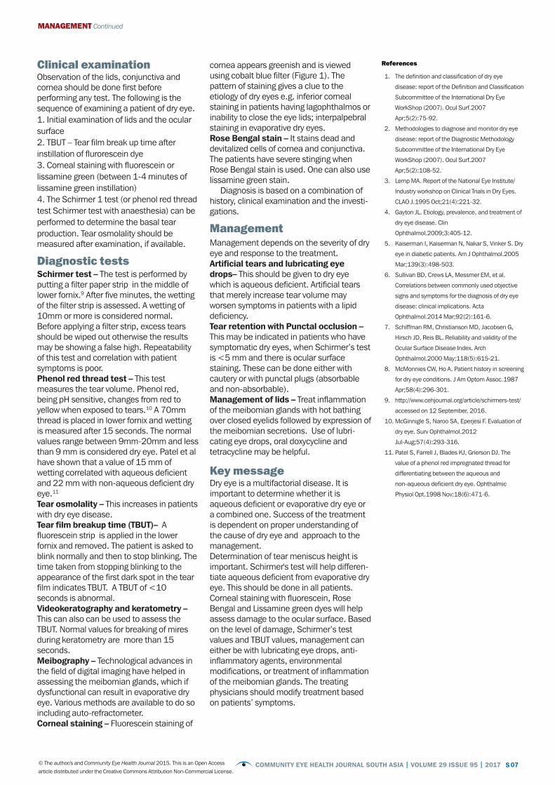
Roughness of epithelium as seen in shield ulcer
Varsha M RathiFaculty, Tej Kohli Cornea Institute, Gullapalli Pratibha Rao International Center for Advancement of Rural Eye Care, L V Prasad Eye Institute,Hyderabad, India
Somasheila I MurthyFaculty, Tej Kohli Cornea Institute, L.V. Prasad Eye Institute, Kallam Anji Reddy Campus, Hyderabad, India
(c) V
Rat
hi/L
VPEI
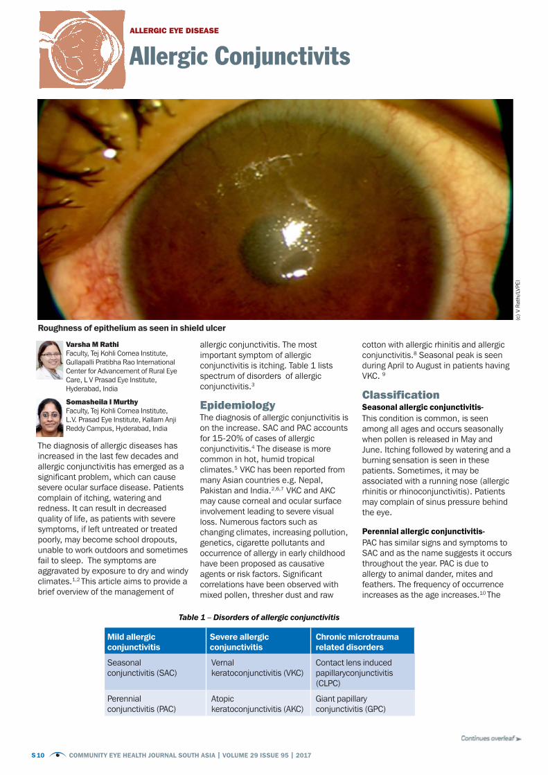
Mild allergic conjunctivitis
Severe allergic conjunctivitis
Chronic microtraumarelated disorders
Seasonal conjunctivitis (SAC)
Vernal keratoconjunctivitis (VKC)
Contact lens inducedpapillaryconjunctivitis (CLPC)
Perennial conjunctivitis (PAC)
Atopic keratoconjunctivitis (AKC)
Giant papillary conjunctivitis (GPC)
Table 1 – Disorders of allergic conjunctivitis

S COMMUNITY EYE HEALTH JOURNAL SOUTH ASIA | VOLUME 29 ISSUE 95 | 2017 11 © The author/s and Community Eye Health Journal 2015. This is an Open Access article distributed under the Creative Commons Attribution Non-Commercial License.
patients have itching, redness and swelling of conjunctiva. Corneal involvement in SAC and PAC is rare.4
Vernal keratoconjunctivitis-VKC is a disease of warm climates and occurs predominantly in young males (8-12 years of age).2,11 Although VKC is more common in children, adults may also have VKC.12,13
It is a bilateral disease and may worsen with exposure to wind, dust and sunlight. These patients may have positive history of asthma or eczema. Patients present with severe itching (rubbing of eyes usually with a knuckle), redness, discharge, and photophobia. The mucus discharge is thread-like. School-going children may drop out from going to school because of severe itching and photophobia.Three clinical forms of VKC are described: limbal or bulbar, palpebral and mixed (Figure 1). Limbal form is more common in dark skinned
individuals. In Asia, the mixed form is more common compared to the limbal form, which is seen in Africans.7
However, studies from India and Nepal have reported that the bulbar form of the disease is common in some areas.2,9
Limbal or bulbar form may present as gelatinous thickening of the limbus, presence of papillae at the limbus and yellow Horner-Tranta’s dots (Figure 1) usually at the superior limbus. These dots are seen when the disease is active and indicate severity of the disease.
The hallmark of the palpebral VKC is presence of giant papillae, which are seen on everting the upper lid – the giant papillae have a cobble stone appearance (Figure 1). This thickening of the upper lid may be associated with drooping of the lid (ptosis). Conjunctival pigmentation is common in patients having VKC. 14
The mixed form of VKC has features of both palpebral and limbal VKC.
Corneal involvement in VKC may occur as corneal epithelial punctuate keratitis, and where the epithelial erosions may coalesce and form a vernal or a shield ulcer. Presence of shield ulcer will worsen patients’ symptoms and affect vision. These ulcers are oval and are usually present in the upper part of the cornea. The shield ulcers are classified based on the presence of white material at the base of the ulcer. Based on the grades of shield ulcer, the treatment options differ.15
Atopic keratoconjunctivitis-AKC is a bilateral disease of ocular surface and lids, which occurs throughout life. The patients will have eczematous skin lesions of the body. The conjunctiva may have papillae or Trantas dots. Cataract formation can occur in these patients. Table 2 shows the differentiating features of VKC and AKC.
ALLERGY Continued
Figure 1. Clinical forms of VKC: Limbal or bulbar, palpebral and mixed
Limbal form of VKC- Gelatinous translucent appearance
Palpebral form of VKC- Cobble stone papillae seen after flipping of upper lid
Yellow Horner Trantas dots- more the dots, severe is the disease
Roughness of epithelium as seen in shield ulcer

12 COMMUNITY EYE HEALTH JOURNAL SOUTH ASIA | VOLUME 29 ISSUE 95 | 2017S
Table 2 – Differentiating features of vernal and atopic keratoconjunctivitis
VKC AKC
Age Young Old
Sex Males > Females Equal ratio
Season Spring Perennial
Duration Limited Chronic
Skin involvement No Yes, extra lid fold, maceration of canthi
Punctal stenosis No Yes
Conjunctiva Upper tarsal conjunctiva
Lower tarsal conjunctiva
Conjunctival scarring Rare Common
Cornea Shield ulcer Epithelial defects
Scarring Peripheral Central
Vascularization Rare Common
VKC – vernal keratoconjunctivitis AKC – Allergic keratoconjunctivitis
Giant papillary conjunctivitis-The presence of a contact lens, ocular prosthesis or sutures may sensitize and cause trauma to the upper tarsal conjunctiva with the formation of giant papillae. Removal of these external agents will reduce the papillae.
Toxic allergic reactions may also be due to drugs such as neomycin, atropine, epinephrine or preservatives in medicines such as thiomersol.16
Contact hypersensitivity reactions-The pattern of involvement depends upon severity of the reaction and the site of contacts. Patients may have lid swelling, redness, chemosis, follicular reaction and later sometimes cicatrisation. The corneal involvement may be in the form of superficial punctate keratitis, pseudodendrites or grayish stromal infiltrates.17
ComplicationsMost often, the complications are because of poor compliance to treatment on the part of patient, or inadequate control of the disease when it presents in its severe form. Common complications include dry eye, infection and corneal scar. Chronicity of the untreated disease may lead to vision threatening problems like limbal stem cell deficiency (LSCD) and secondary keratoconus due to rubbing of the eyes.
As the treatment involves use of corticosteroids, steroid-induced raised intraocular pressure and cataract have
been reported in these patients.7
Complications may lead to irreversible visual loss in some patients.7 Both the complications, keratoconus and LSCD need timely surgical treatment to prevent visual malfunction.
DiagnosisAppropriate management of allergic conjunctivitis needs a correct diagnosis. Figure 2 gives a guide for such diagnosis and ways to differentiate from other causes of red eyes. Presence of itching is a hallmark of ocular allergy.
ManagementThough some authors have described management protocols, there are no universally accepted protocols of management for allergic eye diseases.11,12 Various drugs are available and the treatment options vary based on the severity of the disease. It is important to avoid any known allergen or reduce exposure to it by using wrap-around glasses, by changing the environment, replacing allergen-harbouring items such as pillows and carpets. However, such recommendations may be challenging for patients. In addition, cool compresses can be done to prevent rubbing of the eye. Ocular lubricating eye drops can be used to dilute the inflammatory agents in tears and wash away the allergen to reduce itching and to prevent further worsening of symptoms. 19
The mainstay of treatment is the use of lubricants, anti-histamines and mast cell stabilizers.16,20 These are indicated in all forms of disease. Steroids are to be given under proper medical care when the cornea is involved or the disease is very severe with itching. Overuse of corticosteroids may cause steroid induced cataracts and glaucoma and may result in blindness. The drugs that are used are:Mast cell stabilizers- disodium cromoglyacate (not effective in acute stages), Nedocromil and LodoxamideAntihistamines- ketotifen, dual acting drugs such as olapatadine, azelastine, epinastine and bepostatine. Immediate symptomatic relief is possible with azelastine and epinastine, which are currently preferred.Corticosteroids- such as prednisolone are given for a short duration during acute allergic disease; oral steroids or supratarsal injection of corticosteroids is required if the disease is severe.Nonsteroidal anti-inflammatory agents (NSAIDS)- ketorolac, diclofenac can be added to antihistamines. Steroid sparing agents such as Cyclosporine A, Tacrolimus are effective in severe AKC and VKC.
ConclusionFrom a public health perspective, the number of patients being diagnosed with allergic conjunctivitis is increasing. However, not many studies are

S COMMUNITY EYE HEALTH JOURNAL SOUTH ASIA | VOLUME 29 ISSUE 95 | 2017 13 © The author/s and Community Eye Health Journal 2015. This is an Open Access article distributed under the Creative Commons Attribution Non-Commercial License.
References
1. Bonini S, Bonini S, Lambiase A, Marchi S,
Pasqualetti P, Zuccaro O, et al. Vernal keratocon-
junctivitis revisited: a case series of 195 patients
with long-term followup. Ophthalmology. 2000
Jun;107(6):1157-63.
2. Sofi RA, Mufti A. Vernal Keratoconjunctivitis in
Kashmir: A temperate zone. International ophthal-
mology. 2016 Mar 10.
3. Ehlers WH, Donshik PC. Allergic ocular disorders: a
spectrum of diseases. The CLAO journal : official
publication of the Contact Lens Association of
Ophthalmologists, Inc. 1992 Apr;18(2):117-24.
4. La Rosa M, Lionetti E, Reibaldi M, Russo A, Longo A,
Leonardi S, et al. Allergic conjunctivitis: a compre-
hensive review of the literature. Italian journal of
pediatrics. 2013;39:18.
5. Buckley RJ. Vernal keratoconjunctivitis. International
ophthalmology clinics. 1988 Winter;28(4):303-8.
6. Pokharel N, Shah DN, Choudhary M. Vernal kerato-
conjunctivitis: modes of presentation in Nepalese
population. Kathmandu University medical journal.
2007 Oct-Dec;5(4):526-30.
7. Saboo US, Jain M, Reddy JC, Sangwan VS.
Demographic and clinical profile of vernal kerato-
conjunctivitis at a tertiary eye care center in India.
Indian journal of ophthalmology. 2013
Sep;61(9):486-9.
8. Ahmed A, Minhas K, Micheal S, Ahmad F.
Prevalence of skin test reactivity to aeroallergens in
the Pakistani population. Public health. 2011
May;125(5):324-6.
9. Kansakar I. Profile of vernal keratoconjunctivitis in
Nepal: a hospital based study. Nepal Medical
College journal : NMCJ. 2011 Jun;13(2):92-5.
10. Leonardi A, Piliego F, Castegnaro A, Lazzarini D, La
Gloria Valerio A, Mattana P, et al. Allergic conjuncti-
vitis: a cross-sectional study. Clinical and
experimental allergy : journal of the British Society
for Allergy and Clinical Immunology. 2015
Jun;45(6):1118-25.
11. Katelaris CH. Ocular allergy in the Asia Pacific
region. Asia Pacific allergy. 2011 Oct;1(3):108-14.
12. Ukponmwan CU. Vernal keratoconjunctivitis in
Nigerians: 109 consecutive cases. Tropical doctor.
2003 Oct;33(4):242-5.
13. De Smedt S, Wildner G, Kestelyn P. Vernal kerato-
conjunctivitis: an update. The British journal of
ophthalmology. 2013 Jan;97(1):9-14.
14. Rao SK, Padmanabhan P. Perilimbal conjunctival
pigmentation in vernal keratoconjunctivitis: a new
sign. Cornea. 2002 May;21(4):432.
15. Cameron JA. Shield ulcers and plaques of the
cornea in vernal keratoconjunctivitis.
Ophthalmology. 1995 Jun;102(6):985-93.
16. Schmid KL, Schmid LM. Ocular allergy: causes and
therapeutic options. Clinical & experimental
optometry. 2000 Sep-Oct;83(5):257-70.
17. Dart J. Corneal toxicity: the epithelium and stroma in
iatrogenic and factitious disease. Eye.
2003;17(8):886-92.
18. Kok YO, Tan GF, Loon SC. Review: keratoconus in
Asia. Cornea. 2012 May;31(5):581-93.
19. Hingorani M, Lightman S. Therapeutic options in
ocular allergic disease. Drugs. 1995
Aug;50(2):208-21.
20. Gokhale NS. Systematic approach to managing
vernal keratoconjunctivitis in clinical practice:
Severity grading system and a treatment algorithm.
Indian journal of ophthalmology. 2016
Feb;64(2):145-8.
21. Pitt AD, Smith AF, Lindsell L, Voon LW, Rose PW,
Bron AJ. Economic and quality-of-life impact of
seasonal allergic conjunctivitis in Oxfordshire.
Ophthalmic epidemiology. 2004 Feb;11(1):17-33.
22. Gupta R, Sheikh A, Strachan DP, Anderson HR.
Burden of allergic disease in the UK: secondary
analyses of national databases. Clinical and experi-
mental allergy : journal of the British Society for
Allergy and Clinical Immunology. 2004
Apr;34(4):520-6.
available from South East Asia, which give a complete picture of allergic eye disease. Severe conjunctivitis such as VKC, being a disease of the young may increase the number of school dropouts in these countries. Economic costs for patients are high, sometimes
necessitating the need for medications to continue for years.21,22
Management of the disease is very challenging and a multipronged approach with well–trained primary and secondary care personnel to educate patients or parents about the disease,
especially about good general hygiene; avoidance of allergens; cold compression; change of environment; and judicious use of corticosteroids may improve ocular health in patients by leaps and bounds.
Figure 2. A guide to aid diagnosis of allergic conjunctivitis

14 COMMUNITY EYE HEALTH JOURNAL SOUTH ASIA | VOLUME 29 ISSUE 95 | 2017S
IntroductionThe term ‘ocular surface’ was first defined by Thoft in 1987 as a combined unit including the cornea, conjunctiva, lacrimal glands and eyelids. Gibson further described this term in 2007 to include the surface and glandular epithelia of the cornea, conjunctiva, lacrimal gland, accessory lacrimal glands, meibomian glands and the eyelashes with their associated glands of Moll and Zeis along with the nasolacrimal duct.1,2 These components of the ocular surface are connected with a continuous epithelium.
Being the most exposed part of the
eye, the ocular surface is highly prone to injury. This article covers the spectrum of various ocular surface injuries and their management.
Classification Ocular surface injury is a broad term which includes the following:• Ocular surface chemical and thermal burns or injuries
• Conjunctival laceration• Corneal perforation• Eyelid laceration
Ocular surface chemical and thermal burnsChemical injury may involve the ocular surface to a variable degree depending on the nature of the chemical agent, duration of exposure, concentration and volume of the agent. Host factors such as the nature and health of the ocular surface itself also plays an important role in deciding vulner-ability of the surface to injury.
Most of the ocular surface chemical burns are due to either acids or alkalis. Alkali burns account for two-thirds of these.3 A majority of these burns occurs in young males with increased risk of exposure to chemicals in the workplace.Alkali burns- The common alkaline sources include ammonia, lime or calcium hydroxide, lye or sodium hydroxide and magnesium hydroxide. The most common alkali causing ocular surface burns is lime
while the most severe is ammonia. Ammonia, found in fertilizers and floor cleaners, has the most rapid penetration into the surface due to its lipid as well as water solubility. Alkali burns are more severe than acid burns as they lead to saponification of cell membranes and intercellular bridges facilitating rapid penetration into the deeper layers and into aqueous and vitreous cavities. Alkali burns cause stimulation of nerve endings of cornea and conjunctiva and hence are more painful.5 Acid burns- Sulfuric acid is the most common acid implicated in acid induced ocular surface burns. Hydroflouric acid leads to the most severe burns as it is highly reactive with rapid and deep penetration just like alkalis. Acids in general cause less severe burns as compared to alkalis. They lead to coagu-lation and precipitation of proteins which in itself acts as a physical barrier, thus preventing further penetration of the agent.
Several classification systems have been suggested and proposed for ocular surface chemical injuries. Prominent ones include Hughes classification4 (1946), Roper-Hall classification5 (1965) and Dua’s classification6 (2001). These classi-fication systems hold true for cases of acute chemical injuries. In chronic cases with already established sequelae of chemical burns, the ocular surface health
Ocular Surface Injuries & Management
Anubha RathiSenior Resident, Cornea, Cataract and Refractive surgery services, Dr R P Centre of ophthalmic sciences, AIIMS
Rajesh SinhaAdditional Professor, Cornea, Cataract and Refractive surgery services, Dr R P Centre of ophthalmic sciences, AIIMS
Neelima AronSenior Resident, Cornea, Cataract and Refractive surgery services, Dr R P Centre of ophthalmic sciences, AIIMS
Namrata SharmaProfessor, Cornea, Cataract and Refractive surgery services, Dr R P Centre of ophthalmic sciences, AIIMS
MANAGEMENT
Clinical photograph of a case of conjunctival laceration with a foreign body
(c) R
P C
entr
e of
oph
thal
mic
sci
ence
s, A
IIMS

S COMMUNITY EYE HEALTH JOURNAL SOUTH ASIA | VOLUME 29 ISSUE 95 | 2017 15 © The author/s and Community Eye Health Journal 2015. This is an Open Access article distributed under the Creative Commons Attribution Non-Commercial License.
Table 1a. Roper-Hall classification
Table 1b. Dua’s classification
Grade Prognosis Cornea Conjunctiva/ limbus
I Good Corneal epithelial damage No limbal ischemia
II Good Corneal haze, iris details visible 1/2 limbal ischemia
III GuardedTotal epithelial loss, stromal haze, iris details obscured
1/3-1/2 limbal ischemia
IV Poor Cornea opaque, iris and pupil obscured >1/2 limbal ischemia
Grade Prognosis Clinical findings Conjunctival involvement
Analogue scale
I Very good 0 clock hours limbal involvement 0% 0/0%
II Good ≤3 clock hours limbal involvement ≤30% 0.1-3/ 1-29.9%
III Good >3-6 clock hours limbal involvement >30-50% 3.1-6/ 31-50%
IV Good to guarded >6-9 clock hours limbal involvement >50-75% 6.1-9/ 51-75%
V Guarded to poor >9-12 clock hours limbal involvement >75-<100% 9.1-11.9/
75.1-99.9%
VI Very poorTotal (12 clock hours) limbal involvement
100% 12/100%
may be graded using the Holland-Mannis classification system.
The two commonly used classification systems - Dua’s (2001) and Roper-Hall (1964) are summarized in Table 1a and Table 1b.5,6 The Roper-Hall classification system has classified all burns with more than 50% limbal ischemia in Grade IV. This presents as a limitation in the prognosti-cation of the burns according to grade as the prognosis is highly variable in burns
with just 50% limbal ischemia as compared to burns with total limbal ischemia. Dua’s classification in 2001 addressed this limitation and classified ocular surface chemical burns based on the clock hours of conjunctival and limbal involvement.Clinical features of ocular surface chemical burns- In the acute stage up to one week post injury, ocular surface chemical burns usually present with peri
limbal ischemia (Figure 1a), corneal and conjunctival epithelial defects (Figure 1b) and retained chemical particles especially in the fornices (Figure 1c). Milder burns show re-epithelialisation gradually with or without treatment. More severe burns may develop complications such as persistent epithelial defects, dry eye, symblepharon, ankyloblepharon, cicatricial entropion or ectropion, and in rare and severe cases corneo scleral melt.8
MANAGEMENT Continued
Figure 1. Clinical presentation of acute chemical injury
Figure 1a. Perilimbal ischemia Fig 1b. Persistent epithelial defect Fig 1c. Retained chemical particles in superior fornix
16 COMMUNITY EYE HEALTH JOURNAL SOUTH ASIA | VOLUME 29 ISSUE 95 | 2017S
Management of ocular surface chemical burns- Ocular surface chemical burn is a medical emergency. Immediate irrigation of the eye should be done with clean running water, ringer lactate or normal saline until the pH of the ocular surface is neutralized. This has to be meticulously done using double eversion of the eyelids.
Timely treatment should then be insti-tuted. Medical treatment includes topical antibiotics, cycloplegics, topical steroids, topical sodium ascorbate 10%, topical sodium citrate 10%, oral doxycycline, oral ascorbate and tear substitutes.7
Amniotic membrane transplantation is beneficial in moderate to severe chemical burns. It promotes re-epithelialisation, decreases the incidence of symblepharon formation, and decreases inflammation.8
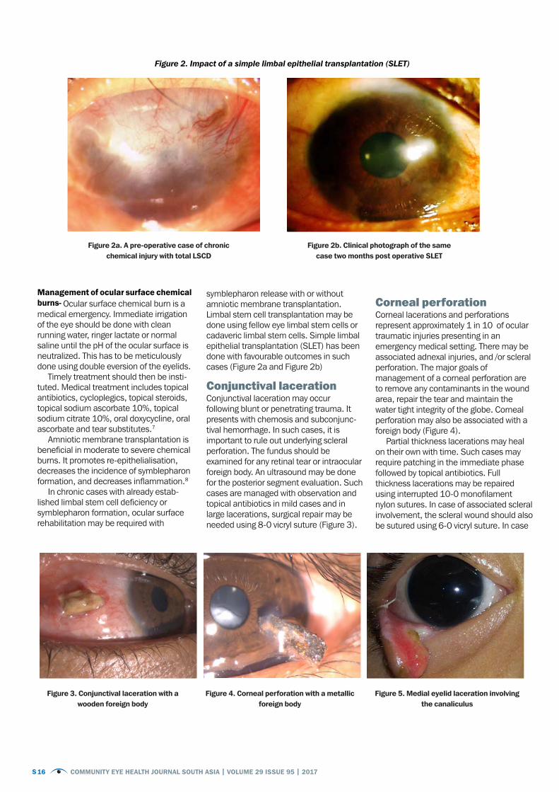
In chronic cases with already estab-lished limbal stem cell deficiency or symblepharon formation, ocular surface rehabilitation may be required with
symblepharon release with or without amniotic membrane transplantation. Limbal stem cell transplantation may be done using fellow eye limbal stem cells or cadaveric limbal stem cells. Simple limbal epithelial transplantation (SLET) has been done with favourable outcomes in such cases (Figure 2a and Figure 2b) Conjunctival lacerationConjunctival laceration may occur following blunt or penetrating trauma. It presents with chemosis and subconjunc-tival hemorrhage. In such cases, it is important to rule out underlying scleral perforation. The fundus should be examined for any retinal tear or intraocular foreign body. An ultrasound may be done for the posterior segment evaluation. Such cases are managed with observation and topical antibiotics in mild cases and in large lacerations, surgical repair may be needed using 8-0 vicryl suture (Figure 3).
Corneal perforationCorneal lacerations and perforations represent approximately 1 in 10 of ocular traumatic injuries presenting in an emergency medical setting. There may be associated adnexal injuries, and /or scleral perforation. The major goals of management of a corneal perforation are to remove any contaminants in the wound area, repair the tear and maintain the water tight integrity of the globe. Corneal perforation may also be associated with a foreign body (Figure 4).
Partial thickness lacerations may heal on their own with time. Such cases may require patching in the immediate phase followed by topical antibiotics. Full thickness lacerations may be repaired using interrupted 10-0 monofilament nylon sutures. In case of associated scleral involvement, the scleral wound should also be sutured using 6-0 vicryl suture. In case
Figure 2. Impact of a simple limbal epithelial transplantation (SLET)
Figure 2a. A pre-operative case of chronic chemical injury with total LSCD
Figure 3. Conjunctival laceration with a wooden foreign body
Figure 4. Corneal perforation with a metallic foreign body
Figure 5. Medial eyelid laceration involving the canaliculus
Figure 2b. Clinical photograph of the same case two months post operative SLET
S COMMUNITY EYE HEALTH JOURNAL SOUTH ASIA | VOLUME 29 ISSUE 95 | 2017 17 © The author/s and Community Eye Health Journal 2015. This is an Open Access article distributed under the Creative Commons Attribution Non-Commercial License.
References
1. Gipson IK. The Ocular Surface: The Challenge to
Enable and Protect Vision. Investigative ophthal-
mology & visual science.
2007;48(10):4390-4398
2. Kinoshita S, Adachi W, Sotozono C, et al.
Characteristics of the human ocular surface
epithelium. Prog Retin Eye Res.
2001;20:639–673
3. Morgan SJ: Chemical burns of the eye: causes
and management. Br J Ophthalmol 1987;
71:854-857
4. Hughes Jr WF. Alkali burns of the cornea. I. Review
of the litera. & summary of present knowledge.
Arch Ophthal. 1946; 35: 423–449
5. Roper-Hall MJ. Themal and chemical burns. Trans
Ophthalmol Soc UK 1965;85:631–53
6. Dua HS, King AJ, Joseph A. A new classification of
ocular surface burns. Br J Ophthalmol
2001;85:1379–83
7. Haddox IL. Pfister RR, Yuille-Barr D: The efficacy of
topical citrate after alkali injury is dependent on
the period of time it is administered. Invest
Ophthalmol Vis Sci 1989; 30: 1062-1068
8. Meller D, Pires RTF, Mack RJS, Figueiredo F,
Heiligenhaus A, Park WC et al. Amniotic
membrane transplantation for acute chemical or
thermal burns. Ophthalmology May;107(5):980-9
2000
9. Vote BJ, Elder MJ. Cyanoacrylate glue for corneal
perforations: a description of a surgical technique
and a review of the literature. Clinical &
Experimental Ophthalmology 2000,
28(6):437-442
10. Khalifa YM et al. Management of nontraumatic
corneal perforation with tectonic drape patch and
cyanoacrylate glue. Cornea. 2010
Oct;29(10):1173-5
MANAGEMENT Continued
of uveal prolapse, the uveal tissue that is not necrotic and has protruded for less than 24 hours may be reposited back or and any old or necrotic prolapsed tissue carefully abscised. Other than conven-tional interrupted sutures, biological glue has also been used to seal the perfora-tions especially those with tissue defect. Several studies have shown the beneficial effect of isobutyl cyanoacrylate glue for treatment of corneal perforations with tissue defect up to 3 mm.9-10
While suturing a corneal perforation, it is important to identify the major landmarks, especially limbus. It is advisable to preserve as much anatomy as possible and not over excise. The technique is to progressively halve the wound while passing sutures. They should be at 90% depth in the cornea. ‘No touch technique’ while passing sutures ensures
a maintained anterior chamber during suturing. The central suture bites should be smaller and the length of suture should increase as one goes towards periphery.
For corneo scleral lacerations, it is important to perform a 360 degree peritomy and see the extent of scleral involvement, following which the limbus is secured with vicryl or monofilament nylon suture. Corneal sutures are placed as described above. Scleral wound is closed using ‘close as you go’ or zippering technique. Disinsertion and reinsertion of recti may be required in posterior tears.
Eyelid lacerationsEyelids and lacrimal system are as much a part of the ocular surface as the cornea and conjunctiva. Simple eyelid lacera-tions, which are horizontal follow skin
lines and involve less than 25% of the lids, usually heal well even without suturing. Larger lid lacerations require surgical repair. Uncomplicated lid lacera-tions with no lacrimal system involvement can be repaired using interrupted silk sutures. In case of medial lid injuries (Figure 5) with damage to the lacrimal system, canalicular repair is required along with lid laceration repair.
ConclusionOcular surface injuries are fairly common owing to vulnerability of the exposed ocular surface to trauma. They range from ocular surface chemical burns, conjunc-tival laceration, corneal perforation and eyelid laceration. Effective and timely management of these types of injuries is essential for maintaining the integrity of the ocular surface.

18 COMMUNITY EYE HEALTH JOURNAL SOUTH ASIA | VOLUME 29 ISSUE 95 | 2017S
Com
mon
and
impo
rtan
t ocu
lar s
urfa
ce c
ondi
tions
Cond
ition
Hist
ory a
nd s
igns
Prim
ary l
evel
m
anag
emen
t In
fect
ious
con
ditio
nsM
icro
bial
ker
atiti
sH
isto
ry: P
ainf
ul, r
ed e
ye w
ith re
duce
d vi
sion
dev
elop
ing
acut
ely
over
one
or t
wo
days
(bac
teria
l) or
sub
-acu
tely
ove
r a fe
w d
ays
(fung
al).
Sig
ns: C
orne
al u
lcer
(epi
thel
ial d
efec
t) w
ith u
nder
lyin
g st
rom
al in
filtr
ate.
The
co
njun
ctiv
a w
ill b
e re
d. T
here
may
be
infla
mm
ator
y ce
lls in
the
ante
rior c
ham
ber,
prog
ress
ing
to a
hyp
opyo
n in
sev
ere
dise
ase.
Hou
rly a
ntib
iotic
eye
dr
ops
and
refe
r to
a sp
ecia
list.
Vira
l con
junc
tiviti
sH
isto
ry: R
ed, w
ater
ing
eyes
, ofte
n bi
late
ral.
Nor
mal
or r
educ
ed v
isio
n. M
ild p
ain.
M
ay h
ave
asso
ciat
ed s
ore
thro
at a
nd ru
nny
nose
.S
igns
: Wat
ery
disc
harg
e, c
onju
nctiv
al in
ject
ion,
tars
al c
onju
nctiv
al fo
llicl
es,
pre-
auric
ular
lym
phad
enop
athy
and
eye
lid o
edem
a. T
he c
orne
a m
ay b
e af
fect
ed
with
mul
tiple
sup
erfic
ial s
ub-e
pith
elia
l inf
iltra
tes
(gre
y-w
hite
spo
ts –
see
imag
e).
Avoi
d sp
read
to
othe
rs th
roug
h go
od
hygi
ene.
Sel
f-lim
iting
.
Bact
eria
l co
njun
ctiv
itis
His
tory
: Red
, unc
omfo
rtab
le e
yes
with
pur
ulen
t dis
char
ge. T
here
is u
sual
ly
redn
ess,
grit
tines
s an
d bu
rnin
g, w
hich
may
initi
ally
hav
e be
en u
nila
tera
l but
ofte
n be
com
es b
ilate
ral.
Lids
are
ofte
n st
uck
toge
ther
in th
e m
orni
ng w
ith d
ried
disc
harg
e.S
igns
: Con
junc
tival
inje
ctio
n, p
apill
ary
conj
unct
iviti
s, d
isch
arge
.
Avoi
d sp
read
to
othe
rs th
roug
h go
od
hygi
ene.
To
pica
l ant
ibio
tics
for
5–10
day
s.
Alle
rgic
con
junc
tiviti
s Vern
al
kera
toco
njun
ctivi
tis
(VKC
)
His
tory
: Alle
rgic
con
junc
tiviti
s ca
n pr
esen
t at a
ny a
ge a
s itc
hing
and
wat
erin
g du
e to
so
me
know
n or
unk
now
n al
lerg
en. A
sev
ere
form
is V
KC w
hich
pre
sent
s in
chi
ldho
od
with
sev
ere
itchi
ng, w
ater
ing,
fore
ign
body
sen
satio
n an
d th
ick
muc
us d
isch
arge
. S
igns
: The
re is
con
junc
tival
inje
ctio
n (s
ee im
age)
. Pap
illae
are
foun
d in
the
tars
al
conj
unct
iva,
whi
ch c
an b
e la
rge
and
irreg
ular
(cob
bles
tone
pap
illae
). T
rant
a’s
spot
s ar
e sm
all w
hite
dot
s at
the
limbu
s. T
he li
mbu
s ca
n be
com
e pi
gmen
ted.
The
cor
nea
can
be a
ffect
ed w
ith p
laqu
es a
nd u
lcer
atio
n of
the
uppe
r cor
nea.
Avoi
d al
lerg
ens.
O
ffer a
ntih
ista
min
es,
mas
t cel
l inh
ibito
rs,
and/
orto
pica
l ste
roid
s (s
hort
-ter
m).
Blep
harit
isH
isto
ry: I
tchi
ng, b
urni
ng, u
ncom
fort
able
eye
s, w
ith o
r with
out a
ssoc
iate
d w
ater
ing
and
dry
eye
sym
ptom
s (s
ee b
elow
). T
here
may
be
an a
ssoc
iate
d hi
stor
y of
re
curr
ent m
eibo
mia
n cy
sts.
Sig
ns: H
ard
scal
es a
nd c
rust
ing
at th
e ba
ses
of la
shes
in a
nter
ior b
leph
ariti
s. L
ook
for c
appe
d or
plu
gged
mei
bom
ian
glan
d or
ifice
s an
d hy
pera
emia
(red
ness
) of t
he
post
erio
r lid
mar
gin
in p
oste
rior b
leph
ariti
s.
Ante
rior
: Lid
cl
eani
ng to
rem
ove
crus
ts.
Post
erio
r: H
ot
com
pres
ses
and
lid
mas
sage
.
Dry e
yeDr
y eye
His
tory
: Unc
omfo
rtab
le, g
ritty
eye
s w
ith a
fore
ign
body
sen
satio
n. S
ever
e ca
ses
may
be
phot
opho
bic
and
pain
ful w
ith re
duce
d vi
sion
.S
igns
: The
tear
film
is a
bnor
mal
with
deb
ris o
n th
e su
rface
and
a te
ar b
reak
-up
time
of le
ss th
an 1
0 se
cond
s. T
he te
ar m
enis
cus
may
als
o be
thin
. Pun
ctat
e ep
ithel
ial
eros
ions
that
sta
in w
ith fl
uore
scei
n ar
e th
e ha
llmar
k of
dry
eye
dis
ease
.
Topi
cal a
rtifi
cial
tear
s (lu
bric
ants
).
Othe
r inf
lam
mat
ory c
ondi
tions
Perip
hera
l ul
cera
tive
kera
titis
(incl
udin
g M
oore
n’s
ulce
r)
His
tory
: Pai
nful
, red
eye
with
loss
of v
isio
n, d
evel
opin
g gr
adua
lly o
ver s
ever
al
wee
ks. M
ay h
ave
a hi
stor
y of
sys
tem
ic in
flam
mat
ory
dise
ase.
Moo
ren’
s ul
cer i
s an
is
olat
ed o
cula
r pro
blem
, typ
ical
ly o
ccur
ring
in y
oung
mal
es.
Sig
ns: P
rogr
essi
ve, c
ircum
fere
ntia
l str
omal
thin
ning
and
ulc
erat
ion.
The
lim
bus
is
infla
med
in th
e ar
ea n
ext t
o th
e ul
cera
tion.
Trea
t as
for
mic
robi
al k
erat
itis
(s
ee a
bove
) and
refe
r to
a s
peci
alis
t.
Mar
gina
l ker
atiti
sH
isto
ry: M
oder
ate
pain
, mild
vis
ual d
istu
rban
ce a
nd re
dnes
s.S
igns
: Ble
phar
itis,
sub
epith
elia
l mar
gina
l inf
iltra
tes
(can
be
mul
tiple
) with
an
area
of
cle
ar c
orne
a be
twee
n th
e in
filtr
ate
and
the
limbu
s. T
here
may
be
an e
pith
elia
l de
fect
, whi
ch is
usu
ally
sm
alle
r tha
n th
e in
filtr
ate.
Trea
t ini
tially
as
for
mic
robi
al k
erat
itis
. If
the
diag
nosi
s is
co
nfirm
ed, p
resc
ribe
a lo
w-d
ose
topi
cal s
tero
id.
Othe
r non
-infla
mm
ator
y con
ditio
nsNe
urot
roph
ic
kera
titis
His
tory
: Thi
s sh
ould
be
cons
ider
ed in
the
cont
ext o
f sys
tem
ic c
ondi
tions
(e.g
. le
pros
y) o
r an
ocul
ar c
ause
(e.g
. her
petic
ker
atiti
s or
her
pes
zost
er).
The
pat
ient
pr
esen
ts w
ith a
red
eye
with
redu
ced
visi
on. T
here
may
or m
ay n
ot b
e pa
in.
Sig
ns: I
nter
palp
ebra
l pun
ctat
e ep
ithel
ial e
rosi
ons,
per
sist
ent e
pith
elia
l def
ects
, st
rom
al o
edem
a an
d in
filtr
atio
n.
Trea
t the
und
erly
ing
caus
e. P
rote
ct
corn
ea w
ith
lubr
ican
ts, t
apin
g th
e ey
elid
clo
sed
at n
ight
, or
lid
clos
ure.
Ocul
ar s
urfa
ce
squa
mou
s ne
opla
sia
His
tory
: Pat
ient
s us
ually
pre
sent
with
an
awar
enes
s of
a g
row
ing
lesi
on o
n th
e oc
ular
sur
face
. Thi
s m
ay b
e un
com
fort
able
or r
ed. T
here
may
be
pain
and
redu
ced
visi
on w
hen
larg
e. T
here
may
be
an a
ssoc
iatio
n w
ith H
IV+
sta
tus.
Exam
inat
ion:
Thi
cken
ed c
onju
nctiv
al e
pith
eliu
m th
at m
ay e
xten
d on
to th
e co
rnea
w
ith p
rom
inen
t ‘fe
eder
’ ves
sels
. The
re m
ay b
e su
rface
ker
atin
isat
ion
char
acte
rised
by
whi
te p
atch
es (l
euko
plak
ia),
a ge
latin
ous
appe
aran
ce, i
nfla
mm
atio
n or
pig
men
tatio
n.
Refe
r for
wid
e su
rgic
al e
xcis
ion.
Pter
ygiu
mH
isto
ry: T
he p
atie
nt m
ay c
ompl
ain
of a
red
lum
p, o
n on
e or
bot
h si
des
of th
e co
rnea
, whi
ch c
an o
ccas
iona
lly b
ecom
e m
ore
infla
med
and
unc
omfo
rtab
le. T
here
m
ay b
e bl
urrin
g of
vis
ion,
dep
endi
ng o
n th
e ex
tent
of g
row
th a
cros
s th
e co
rnea
, and
in
duce
d as
tigm
atis
m.
Exam
inat
ion:
The
re is
a fl
eshy
, win
g-sh
aped
gro
wth
, aris
ing
from
the
conj
unct
iva,
th
at g
row
s ac
ross
the
corn
ea.
Surg
ical
exc
isio
n if
visi
on is
thre
aten
ed.
18 COMMUNITY EYE HEALTH JOURNAL | VOLUME 29 ISSUE 95 | 2016
John Dart
John Dart
John DartMatthew Burton P Vijayalakshmi
Ante
rior b
leph
ariti
sPo
ster
ior b
leph
ariti
s
Matthew Burton

S COMMUNITY EYE HEALTH JOURNAL SOUTH ASIA | VOLUME 29 ISSUE 95 | 2017 19 © The author/s and Community Eye Health Journal 2015. This is an Open Access article distributed under the Creative Commons Attribution Non-Commercial License. COMMUNITY EYE HEALTH JOURNAL | VOLUME 29 ISSUE 95 | 2016 19
Cond
ition
Hist
ory a
nd s
igns
Prim
ary l
evel
m
anag
emen
t In
fect
ious
con
ditio
nsM
icro
bial
ker
atiti
sH
isto
ry: P
ainf
ul, r
ed e
ye w
ith re
duce
d vi
sion
dev
elop
ing
acut
ely
over
one
or t
wo
days
(bac
teria
l) or
sub
-acu
tely
ove
r a fe
w d
ays
(fung
al).
Sig
ns: C
orne
al u
lcer
(epi
thel
ial d
efec
t) w
ith u
nder
lyin
g st
rom
al in
filtr
ate.
The
co
njun
ctiv
a w
ill b
e re
d. T
here
may
be
infla
mm
ator
y ce
lls in
the
ante
rior c
ham
ber,
prog
ress
ing
to a
hyp
opyo
n in
sev
ere
dise
ase.
Hou
rly a
ntib
iotic
eye
dr
ops
and
refe
r to
a sp
ecia
list.
Vira
l con
junc
tiviti
sH
isto
ry: R
ed, w
ater
ing
eyes
, ofte
n bi
late
ral.
Nor
mal
or r
educ
ed v
isio
n. M
ild p
ain.
M
ay h
ave
asso
ciat
ed s
ore
thro
at a
nd ru
nny
nose
.S
igns
: Wat
ery
disc
harg
e, c
onju
nctiv
al in
ject
ion,
tars
al c
onju
nctiv
al fo
llicl
es,
pre-
auric
ular
lym
phad
enop
athy
and
eye
lid o
edem
a. T
he c
orne
a m
ay b
e af
fect
ed
with
mul
tiple
sup
erfic
ial s
ub-e
pith
elia
l inf
iltra
tes
(gre
y-w
hite
spo
ts –
see
imag
e).
Avoi
d sp
read
to
othe
rs th
roug
h go
od
hygi
ene.
Sel
f-lim
iting
.
Bact
eria
l co
njun
ctiv
itis
His
tory
: Red
, unc
omfo
rtab
le e
yes
with
pur
ulen
t dis
char
ge. T
here
is u
sual
ly
redn
ess,
grit
tines
s an
d bu
rnin
g, w
hich
may
initi
ally
hav
e be
en u
nila
tera
l but
ofte
n be
com
es b
ilate
ral.
Lids
are
ofte
n st
uck
toge
ther
in th
e m
orni
ng w
ith d
ried
disc
harg
e.S
igns
: Con
junc
tival
inje
ctio
n, p
apill
ary
conj
unct
iviti
s, d
isch
arge
.
Avoi
d sp
read
to
othe
rs th
roug
h go
od
hygi
ene.
To
pica
l ant
ibio
tics
for
5–10
day
s.
Alle
rgic
con
junc
tiviti
s Vern
al
kera
toco
njun
ctivi
tis
(VKC
)
His
tory
: Alle
rgic
con
junc
tiviti
s ca
n pr
esen
t at a
ny a
ge a
s itc
hing
and
wat
erin
g du
e to
so
me
know
n or
unk
now
n al
lerg
en. A
sev
ere
form
is V
KC w
hich
pre
sent
s in
chi
ldho
od
with
sev
ere
itchi
ng, w
ater
ing,
fore
ign
body
sen
satio
n an
d th
ick
muc
us d
isch
arge
. S
igns
: The
re is
con
junc
tival
inje
ctio
n (s
ee im
age)
. Pap
illae
are
foun
d in
the
tars
al
conj
unct
iva,
whi
ch c
an b
e la
rge
and
irreg
ular
(cob
bles
tone
pap
illae
). T
rant
a’s
spot
s ar
e sm
all w
hite
dot
s at
the
limbu
s. T
he li
mbu
s ca
n be
com
e pi
gmen
ted.
The
cor
nea
can
be a
ffect
ed w
ith p
laqu
es a
nd u
lcer
atio
n of
the
uppe
r cor
nea.
Avoi
d al
lerg
ens.
O
ffer a
ntih
ista
min
es,
mas
t cel
l inh
ibito
rs,
and/
orto
pica
l ste
roid
s (s
hort
-ter
m).
Blep
harit
isH
isto
ry: I
tchi
ng, b
urni
ng, u
ncom
fort
able
eye
s, w
ith o
r with
out a
ssoc
iate
d w
ater
ing
and
dry
eye
sym
ptom
s (s
ee b
elow
). T
here
may
be
an a
ssoc
iate
d hi
stor
y of
re
curr
ent m
eibo
mia
n cy
sts.
Sig
ns: H
ard
scal
es a
nd c
rust
ing
at th
e ba
ses
of la
shes
in a
nter
ior b
leph
ariti
s. L
ook
for c
appe
d or
plu
gged
mei
bom
ian
glan
d or
ifice
s an
d hy
pera
emia
(red
ness
) of t
he
post
erio
r lid
mar
gin
in p
oste
rior b
leph
ariti
s.
Ante
rior
: Lid
cl
eani
ng to
rem
ove
crus
ts.
Post
erio
r: H
ot
com
pres
ses
and
lid
mas
sage
.
Dry e
yeDr
y eye
His
tory
: Unc
omfo
rtab
le, g
ritty
eye
s w
ith a
fore
ign
body
sen
satio
n. S
ever
e ca
ses
may
be
phot
opho
bic
and
pain
ful w
ith re
duce
d vi
sion
.S
igns
: The
tear
film
is a
bnor
mal
with
deb
ris o
n th
e su
rface
and
a te
ar b
reak
-up
time
of le
ss th
an 1
0 se
cond
s. T
he te
ar m
enis
cus
may
als
o be
thin
. Pun
ctat
e ep
ithel
ial
eros
ions
that
sta
in w
ith fl
uore
scei
n ar
e th
e ha
llmar
k of
dry
eye
dis
ease
.
Topi
cal a
rtifi
cial
tear
s (lu
bric
ants
).
Othe
r inf
lam
mat
ory c
ondi
tions
Perip
hera
l ul
cera
tive
kera
titis
(incl
udin
g M
oore
n’s
ulce
r)
His
tory
: Pai
nful
, red
eye
with
loss
of v
isio
n, d
evel
opin
g gr
adua
lly o
ver s
ever
al
wee
ks. M
ay h
ave
a hi
stor
y of
sys
tem
ic in
flam
mat
ory
dise
ase.
Moo
ren’
s ul
cer i
s an
is
olat
ed o
cula
r pro
blem
, typ
ical
ly o
ccur
ring
in y
oung
mal
es.
Sig
ns: P
rogr
essi
ve, c
ircum
fere
ntia
l str
omal
thin
ning
and
ulc
erat
ion.
The
lim
bus
is
infla
med
in th
e ar
ea n
ext t
o th
e ul
cera
tion.
Trea
t as
for
mic
robi
al k
erat
itis
(s
ee a
bove
) and
refe
r to
a s
peci
alis
t.
Mar
gina
l ker
atiti
sH
isto
ry: M
oder
ate
pain
, mild
vis
ual d
istu
rban
ce a
nd re
dnes
s.S
igns
: Ble
phar
itis,
sub
epith
elia
l mar
gina
l inf
iltra
tes
(can
be
mul
tiple
) with
an
area
of
cle
ar c
orne
a be
twee
n th
e in
filtr
ate
and
the
limbu
s. T
here
may
be
an e
pith
elia
l de
fect
, whi
ch is
usu
ally
sm
alle
r tha
n th
e in
filtr
ate.
Trea
t ini
tially
as
for
mic
robi
al k
erat
itis
. If
the
diag
nosi
s is
co
nfirm
ed, p
resc
ribe
a lo
w-d
ose
topi
cal s
tero
id.
Othe
r non
-infla
mm
ator
y con
ditio
nsNe
urot
roph
ic
kera
titis
His
tory
: Thi
s sh
ould
be
cons
ider
ed in
the
cont
ext o
f sys
tem
ic c
ondi
tions
(e.g
. le
pros
y) o
r an
ocul
ar c
ause
(e.g
. her
petic
ker
atiti
s or
her
pes
zost
er).
The
pat
ient
pr
esen
ts w
ith a
red
eye
with
redu
ced
visi
on. T
here
may
or m
ay n
ot b
e pa
in.
Sig
ns: I
nter
palp
ebra
l pun
ctat
e ep
ithel
ial e
rosi
ons,
per
sist
ent e
pith
elia
l def
ects
, st
rom
al o
edem
a an
d in
filtr
atio
n.
Trea
t the
und
erly
ing
caus
e. P
rote
ct
corn
ea w
ith
lubr
ican
ts, t
apin
g th
e ey
elid
clo
sed
at n
ight
, or
lid
clos
ure.
Ocul
ar s
urfa
ce
squa
mou
s ne
opla
sia
His
tory
: Pat
ient
s us
ually
pre
sent
with
an
awar
enes
s of
a g
row
ing
lesi
on o
n th
e oc
ular
sur
face
. Thi
s m
ay b
e un
com
fort
able
or r
ed. T
here
may
be
pain
and
redu
ced
visi
on w
hen
larg
e. T
here
may
be
an a
ssoc
iatio
n w
ith H
IV+
sta
tus.
Exam
inat
ion:
Thi
cken
ed c
onju
nctiv
al e
pith
eliu
m th
at m
ay e
xten
d on
to th
e co
rnea
w
ith p
rom
inen
t ‘fe
eder
’ ves
sels
. The
re m
ay b
e su
rface
ker
atin
isat
ion
char
acte
rised
by
whi
te p
atch
es (l
euko
plak
ia),
a ge
latin
ous
appe
aran
ce, i
nfla
mm
atio
n or
pig
men
tatio
n.
Refe
r for
wid
e su
rgic
al e
xcis
ion.
Pter
ygiu
mH
isto
ry: T
he p
atie
nt m
ay c
ompl
ain
of a
red
lum
p, o
n on
e or
bot
h si
des
of th
e co
rnea
, whi
ch c
an o
ccas
iona
lly b
ecom
e m
ore
infla
med
and
unc
omfo
rtab
le. T
here
m
ay b
e bl
urrin
g of
vis
ion,
dep
endi
ng o
n th
e ex
tent
of g
row
th a
cros
s th
e co
rnea
, and
in
duce
d as
tigm
atis
m.
Exam
inat
ion:
The
re is
a fl
eshy
, win
g-sh
aped
gro
wth
, aris
ing
from
the
conj
unct
iva,
th
at g
row
s ac
ross
the
corn
ea.
Surg
ical
exc
isio
n if
visi
on is
thre
aten
ed.
John Dart Matthew Burton Matthew Burton Matthew Burton Matthew Burton John Dart
Com
pile
d by
Jer
emy
Hof
fman
, Mat
thew
Bur
ton
and
Alle
n Fo
ster

20 COMMUNITY EYE HEALTH JOURNAL SOUTH ASIA | VOLUME 29 ISSUE 95 | 2017S
Prevention of Traumatic Corneal Ulcer in South East Asia
IntroductionCorneal ulceration is a leading cause of visual impairment globally, with a dispro-portionate burden in developing countries. It was estimated that 6 million corneal ulcers occur annually in the ten countries of South East Asia Region encompassing a total population of 1.6 billion.1 While antimicrobial treatment is generally effective in treating infection, “successful” treatment is often associated with a poor visual outcome. The scarring that accompanies the resolution of infection leaves many eyes blind. Thus, prevention of corneal ulcer-ation is important to reduce morbidity associated with corneal ulceration in countries grouped under South Asian Association for Regional Cooperation (SAARC). Traditional infectious causes of blindness, such as trachoma, onchocer-
ciasis, and leprosy, are declining, and soon the majority of corneal blindness will be due to microbial keratitis. Most corneal ulcers occur among agricultural workers in developing countries following corneal abrasion.
Several non-randomized prevention studies conducted before 2000 (Bhaktapur Eye Study)2 and during 2002 to 2004 in India, Myanmar, and Bhutan by World Health Organization(WHO), have suggested that antibiotic ointment applied promptly after a corneal abrasion could lower the incidence of ulcers, relative to neighbouring or historic controls.3-4 Prevention of traumatic corneal ulcer adopting the Bhaktapur model in a multicountry study in India, Bhutan, Myanmar during 2002 to 2004 was sponsored by WHO.
MethodsThe manpower utilized for this multi-country study to identify ocular injury and treat corneal abrasion is given below:Bhutan: Volunteer Village Health Workers
(VVHW) of the Government were utilized to identify ocular injury and treat corneal abrasionMyanmar: Village Health Workers (VHW) of the health department India: paid village volunteers were utilized
Inclusion criteria• Resident of study area• Corneal abrasion after ocular injury, confirmed by clinical examination with fluorescein stain and a blue torch• Reported within 48 hours of the injury• Subject aged >5 years of age
Exclusion criteria• Subject not a resident of study area• Presence of clinically evident corneal infection• Penetrating corneal injury or stromal laceration• Bilateral ocular traumaThe study was approved by Institutional Review Boards (IRB) from all the three countries.
Dr. M. SrinivasanDirector Emeritus, Aravind Eye Care,Madurai, Tamil Nadu India.
Table 1 : Study design, sample size and Results
Bhutan India Myanmar
Study design Unmasked Randomized double masked trial Unmasked
Sample size 111 corneal abrasions
338 corneal abrasions 111 corneal abrasions
Results of Multicountry study
No. of ocular injuries 135 1365 273
No. of corneal abrasions 115 409 126
No. of eligible corneal abrasions 115 374 126
Adverse events Nil 04 Nil
Corneal Ulcer
Country Principal Investigator and Lead Principal Investigator with village health workers in Bhutan
(c)M
Srin
ivas
an/ A
ECS

S COMMUNITY EYE HEALTH JOURNAL SOUTH ASIA | VOLUME 29 ISSUE 95 | 2017 21 © The author/s and Community Eye Health Journal 2015. This is an Open Access article distributed under the Creative Commons Attribution Non-Commercial License.
Treatment Protocol In general, corneal abrasions are treated with topical antibiotics and cycloplegics. In few centres bandaging the affected eye is practiced but it is controversial. 1% chloramphenicol ointment and 1% clotri-mazole ointment was used. In Bhutan only chloramphenicol was used. In Myanmar both antibiotic and antifungal ointment applied but in India it was randomized and one arm was masked to receive both and in the other arm chloramphenicol and placebo ointment were used to find out whether antifungal prophylax is needed to prevent fungal ulcer. Frequency of application of all the drugs was 3 times a day for 3 days, super-vised by village eye workers for compliance.
Conclusion of multi-country studyThis model of delivering eye care services through trained village eye workers and grass root health workers is a replicable model for any developing country, especially for South Asian Association for Regional Cooperation (SAARC) (Figure 1 a). Follow up rate on the third day at all centres were more than 98%. No case of serious adverse event was reported. Could prevent developing bacterial and fungal corneal ulcer using 1% chloram-phenicol ointment in 96% of patients if reported within 24 hours.
In Madurai, South India, a clinical trial during the same period demonstrated that abrasions randomized to topical antibacterial and antifungal prophylaxis were not significantly less likely to develop fungal ulcers than those randomized to
antibacterial ointment alone, even though the region had a high incidence of fungal infection. This same trial also found that the incidence of ulcers in villages outside the prophylaxis program one was far higher; these control villages were neigh-bouring, but not randomized. To address this issue, we proposed a community-randomised trial comparing villages randomized to receive an intervention consisting of a trained village eye worker identifying, escorting or referring the patient from intervention villages to the nearest vision centre run by Aravind Eye Care System. There a trained vision technician would confirm corneal abrasion, provide 1% chloramphenical ointment to the eligible, enrolled patients in Madurai district. Control villages received no additional intervention. The primary outcome of corneal ulcer prevention will be measured by baseline and annual population-based census performed in both intervention and control villages by masked examiners from baseline to 24 months. The examiners will examine the eyes of all household from intervention and non-intervention villages who are suspected of having a corneal ulcer or injury with torch light and magnifying loup. Each resident in the village will be examined for evidence of corneal opacity and asked about their ocular history. Annual visits will occur in villages randomized to the intervention, an active promotion campaign will be undertaken to urge residents to notify the village eye health worker within 24 hours of ocular trauma. In control villages, abrasions and ulcers will be treated if they present to the
vision centre or are found during annual monitoring visits, but active promotion of corneal abrasion care will not be offered.
Methodology – phase II studyThe study was designed in 2014 in consultation with F.I. Proctor Foundation, San Francisco. Enrollment began in January 2015 and ended in December 2016. 42 villages having approximately 92 thousand people involved in agriculture work were allotted. Randomization was done to have 50% of the population for intervention (Figure 1 b) and rest for non-intervention (Figure 1 b). Data collection, entry, and analysis will be done at Aravind Eye Care System, Madurai and the project will be completed in 2017. Legal and ethical clearance was obtained from appropriate authorities.Details of the study design are in the flow chart. (Figure 1)
Primary outcome of the current studyThe primary outcome will be the incidence of corneal ulceration in the two study arms as measured by corneal exami-nation at the base hospital or vision centre with telemedicine facility. In this study 1% chloramphenicol ointment and 1% itraconazole ointment is applied 3 times a day for 3 days and compliance would be checked by village health workers. Any adverse event will be informed to the study PI or Aravind Eye Hospital and will be taken care at no cost to the participant.
Corneal Ulcer Continued
Figure 1. Design of the study for intervention and control arms
1 a. Intervention

22 COMMUNITY EYE HEALTH JOURNAL SOUTH ASIA | VOLUME 29 ISSUE 95 | 2017S
Manpower20 paid village workers who have completed school and are able to fill study forms in English and reside in the study village. Two supervisors well-experi-enced in rural eye care work to oversee the workers.Steps in managing programme• Workers attend one week training at
Aravind Eye Hospital prior to the study to learn basic anatomy of the eye, common corneal and external diseases, ocular injuries, vision testing, use of fluorescein strip, simple eye medicines application and; attend
twice-yearly refresher trainings throughout the course of the study.
• Promote awareness of corneal abrasion intervention in intervention villages only
• Accessible by villagers via mobile phone • Conduct eye examination to diagnose
corneal abrasion and/or ulcer • Assist treatment at vision centre for
corneal abrasion, and follow up with the patient for three days at the village to ensure compliance
• Motivate patient to return for follow-up three days after treatment, assess compliance and perform examination
• The patient has developed a corneal ulcer or adverse reaction refer to
Aravind Eye Hospital for treatment immediately.Verbal consent must be obtained from
all individuals who present to the village eye worker to receive study medication. Possible risks and benefits of receiving the treatment will be explained. For patients under 18, both the child and one parent or guardian will provide consent for the child’s participation.
We believe the results of this study may emerge as a replicable model to prevent traumatic corneal ulcer, and reduce corneal blindness in South Asia.
References
1. Whitcher J, Srinivasan M, Upadhyay M. Corneal
blindness: a global perspective. Bull World Health
Organ. 2001;79(3):214-221.
2. Upadhyay M, Karmacharya P, Koirala S, et al. The
Bhaktapur eye study: ocular trauma and antibiotic
prophylaxis for the prevention of corneal ulceration
in Nepal. Br J Ophthalmol. 2001;85:388-392.
3. Getshen K, Srinivasan M, Upadhyay M, Priyadarsini
B, Mahalaksmi R, Whitcher J. Corneal ulceration in
South East Asia. I: A model for the prevention of
bacterial ulcers at the village level in rural Bhutan. Br
J Ophthalmol. 2006;90:276-278.
4. Maung N, Thant C, Srinivasan M, et al. Corneal
ulceration in South East Asia II: A strategy for the
prevention of fungal keratitis at the village level in
Burma. Br J Ophthalmol. 2006;90:968-970.
Corneal Ulcer Continued
1 b. Control

S COMMUNITY EYE HEALTH JOURNAL SOUTH ASIA | VOLUME 29 ISSUE 95 | 2017 23 © The author/s and Community Eye Health Journal 2015. This is an Open Access article distributed under the Creative Commons Attribution Non-Commercial License.
,
CONTINUING PROFESSIONAL DEVELOPMENT (CPD)
Test your knowledge and understanding
This page is designed to help you to test your own understanding of the concepts covered in this issue, and to reflect on what you have learnt. We hope that you will also discuss the questions with your colleagues and other members of the eye care team, perhaps in a journal club. To complete the activities online – and get instant feedback – please visit www.cehjournal.org
REFLECTIVE LEARNINGVisit www.cehjournal.org to complete the online ‘Time to reflect’ section.
Picture quiz
ICEH
This ten-year-old boy presents with itchy, watering eyes with a thick mucous discharge of several months’ duration. His visual acuity is 6/9 and 6/12.
Q1. Which of the following signs are visible?(tick all that apply)
a. Follicles
b. Horner-Trantas dots
c. Giant papillae
d. Pannus
e. Trachomatous inflammation
Q2. Which of the following is the most likelydiagnosis? (tick one)
a. Bacterial conjunctivitis
b. Trachoma
c. Kaposi’s sarcoma
d. Vernal conjunctivitis
e. Adenoviral conjunctivitis
Q3. Which of the following may be used in treatment? (tick all that apply)
a. Topical prednisolone
b. Topical antihistamines
c. Topical mast cell inhibitors
d. Topical acyclovir
e. Topical neomycin
1. Answer c. The slide shows giant papillae (>1.0 mm) on the upper eyelid. Horner-Trantas dots may be seen on the limbus, which is not visible in this picture. There is no evidence of follicles or trachoma.
2. Answer d. The most likely diagnosis is vernal conjunctivitis. Bacterial conjunctivitis is associated with a purulent discharge, trachoma often shows follicles, and adenovirus is self-limiting and does not have giant papillae.
3. Answer a, b and c. Treatment is to reduce inflammation from mast cell degranulation, so mast cell inhibitors, antihistamines and prednisolone may all have a role.
1. The ocular surface comprises of: Tick all that apply
a Cornea
b Conjunctiva
c Eyelids
d Lacrimal glands
2. Allergic conjunctivitis (AC) represents a spectrum of disorders comprising: Tick all that apply
a Seasonal allergic conjunctivitis
b Perennial allergic conjunctivitis
c Atopic keratoconjunctivitis
d Vernal keratoconjunctivitis
e Giantpapillary conjunctivitis
3. Ocular surface injury is a broad term which includes: Tick all that apply
a Ocular surface chemical and thermal burns or injuries
b Conjunctival laceration
c Corneal perforation
d Eyelid laceration
4. The following are useful diagnostic tests for dry eye: Tick all that apply
a Schirmer test
b Phenol red thread test
c Tear osmolality
d Tear film breakup time (TBUT)
e Videokeratography and keratometry
5. What are the major risk factors for the development of pterygium: Tick all that apply
a Ultraviolet light exposure due to outdoor occupation
b Age
c Being male
d Having dry eyes
e Being female
ANSWERS
1. All of them are correct except c.2. All of them are correct except d. 3. All of them are correct.4. All of them are correct b.5. All of them are correct except e.
ANSWERS
COMMUNITY EYE HEALTH JOURNAL SOUTH ASIA | VOLUME 29 ISSUE 95 | 2017 S23© The author/s and Community Eye Health Journal 2015. This is an Open Access article distributed under the Creative Commons Attribution Non-Commercial License.
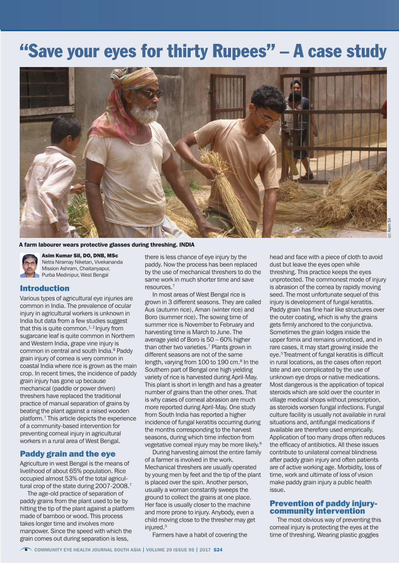
24 COMMUNITY EYE HEALTH JOURNAL SOUTH ASIA | VOLUME 29 ISSUE 95 | 2017S
IntroductionVarious types of agricultural eye injuries are common in India. The prevalence of ocular injury in agricultural workers is unknown in India but data from a few studies suggest that this is quite common.1, 2 Injury from sugarcane leaf is quite common in Northern and Western India, grape vine injury is common in central and south India.6 Paddy grain injury of cornea is very common in coastal India where rice is grown as the main crop. In recent times, the incidence of paddy grain injury has gone up because mechanical (paddle or power driven) threshers have replaced the traditional practice of manual separation of grains by beating the plant against a raised wooden platform.7 This article depicts the experience of a community-based intervention for preventing corneal injury in agricultural workers in a rural area of West Bengal.
Paddy grain and the eyeAgriculture in west Bengal is the means of livelihood of about 65% population. Rice occupied almost 53% of the total agricul-tural crop of the state during 2007-2008.7
The age-old practice of separation of paddy grains from the plant used to be by hitting the tip of the plant against a platform made of bamboo or wood. This process takes longer time and involves more manpower. Since the speed with which the grain comes out during separation is less,
there is less chance of eye injury by the paddy. Now the process has been replaced by the use of mechanical threshers to do the same work in much shorter time and save resources.7
In most areas of West Bengal rice is grown in 3 different seasons. They are called Aus (autumn rice), Aman (winter rice) and Boro (summer rice). The sowing time of summer rice is November to February and harvesting time is March to June. The average yield of Boro is 50 – 60% higher than other two varieties.7 Plants grown in different seasons are not of the same length, varying from 100 to 190 cm.8 In the Southern part of Bengal one high yielding variety of rice is harvested during April-May. This plant is short in length and has a greater number of grains than the other ones. That is why cases of corneal abrasion are much more reported during April-May. One study from South India has reported a higher incidence of fungal keratitis occurring during the months corresponding to the harvest seasons, during which time infection from vegetative corneal injury may be more likely.9
During harvesting almost the entire family of a farmer is involved in the work. Mechanical threshers are usually operated by young men by feet and the tip of the plant is placed over the spin. Another person, usually a woman constantly sweeps the ground to collect the grains at one place. Her face is usually closer to the machine and more prone to injury. Anybody, even a child moving close to the thresher may get injured.5
Farmers have a habit of covering the
head and face with a piece of cloth to avoid dust but leave the eyes open while threshing. This practice keeps the eyes unprotected. The commonest mode of injury is abrasion of the cornea by rapidly moving seed. The most unfortunate sequel of this injury is development of fungal keratitis. Paddy grain has fine hair like structures over the outer coating, which is why the grains gets firmly anchored to the conjunctiva. Sometimes the grain lodges inside the upper fornix and remains unnoticed, and in rare cases, it may start growing inside the eye.5 Treatment of fungal keratitis is difficult in rural locations, as the cases often report late and are complicated by the use of unknown eye drops or native medications. Most dangerous is the application of topical steroids which are sold over the counter in village medical shops without prescription, as steroids worsen fungal infections. Fungal culture facility is usually not available in rural situations and, antifungal medications if available are therefore used empirically. Application of too many drops often reduces the efficacy of antibiotics. All these issues contribute to unilateral corneal blindness after paddy grain injury and often patients are of active working age. Morbidity, loss of time, work and ultimate of loss of vision make paddy grain injury a public health issue.
Prevention of paddy injury- community intervention
The most obvious way of preventing this corneal injury is protecting the eyes at the time of threshing. Wearing plastic goggles
“Save your eyes for thirty Rupees” – A case study
Asim Kumar Sil, DO, DNB, MScNetra Niramay Niketan, Vivekananda Mission Ashram, Chaitanyapur, Purba Medinipur, West Bengal
A farm labourer wears protective glasses during threshing. INDIA
(c) A
sim
Sil
COMMUNITY EYE HEALTH JOURNAL SOUTH ASIA | VOLUME 29 ISSUE 95 | 2017 S24

S COMMUNITY EYE HEALTH JOURNAL SOUTH ASIA | VOLUME 29 ISSUE 95 | 2017 25 © The author/s and Community Eye Health Journal 2015. This is an Open Access article distributed under the Creative Commons Attribution Non-Commercial License.
was considered to be a cheap and easy option. Education materials were produced to propagate the use of protective glass. Posters were displayed in places that farmers visit usually. Eye health talks were organized in different occasions and festivals. One short public education video “Only 30 Rupees to save your Vision,” was developed in local language to motivate people to wear glasses (Plastic goggles in India cost Rupees 30 or half a Dollar). This video was shown in different places including the local cable network. A compact disc of this six minute film was distributed among volunteers who used it locally. This video is widely used during eye donation awareness meetings also.The most effective way of communication was interactive meeting with the farmers. Farmers’ Co-operatives were selected for the meetings. Every large village in this part of Bengal has one Co-operative where farmers get agricultural assistance and the evening is the suitable time to get them there. Interactive meetings started with the thought provoking video and was followed by discus-sions. It was found that many farmers do the threshing in the evening using electric lights. Sometimes it is overtime work, or to avoid daytime heat. Initially dark glasses (used after cataract surgery) were promoted to avoid corneal abrasion. But these were unsuitable for evening use, so the dark glasses were replaced with plain ones without increasing the cost. This white goggle had more acceptances, especially among women. The price could be kept
under INR 30. The message that any kind of spectacle is good to protect the eyes was conveyed through this intensive approach.
Measuring the impactSutahata and Mahishadal Blocks of East Medinipur district in West Bengal were selected for intensive campaigning few weeks before the harvesting time. These blocks were selected because of the proximity to the hospital. This is also the closest eye care facility for the villagers. The population of these blocks was approxi-mately 356,000.
We looked at the hospital data of all cases of corneal ulcers from these two locations as well as from Purba and Paschim Medinipur Districts served by our hospital. A decreasing trend is observed over time in those two selected blocks.
DiscussionThis awareness campaign could have
made some impact in preventing corneal injury and reduction of corneal ulcer. There could be various reasons for reduction in number of walk in patients with corneal ulcers in the clinic. Awareness campaign is possibly one contributing factor.
There are always barriers in the usage of safety eyewear amongst workers. In one study from central India about three-fourths of the workers reported using it all or most of the time during work.4 Despite knowing that protective eyewear devices offer safety from work-related injuries, workers do not tend to use them for multiple reasons. These include
some blurring of vision, discomfort, fogging, unusual appearance, people making fun of them, slipping of the goggles due to sweat and slowing work pace.
Prevention of ocular injuries in agriculture workers will reduce the incidence of microbial keratitis amongst them. Srinivasan et al demonstrated that treating corneal abrasions with antibiotic ointment by health workers at the village level significantly reduced the incidence of bacterial and fungal corneal ulcers,but primary prevention of injury is always the best.3 It is all about developing the attitude of adopting safety measures. Constant effort of educating the community will result in consciousness about eye safety and develop peer pressure to wear protective goggles. Providing protective goggles at an affordable cost should complement this effort. Also, the manufactures of the threshers have a responsibility in ensuring safety of the agricultural workers by modifying the design.
Awareness will always remain as the main strategy for prevention of eye injury. The current approach is interactive and partici-patory. The experience with a small defined population encourages us to scale up the campaign involving all stakeholders and making the goggles available locally.Acknowledgement-Dr. Samar K. Basak, Director, Disha Eye Hospitals, Barrackpore, West BengalMs. Barnali Banerji, Assistant Director of Agriculture, Directorate of Agriculture, Kolkata, West Bengal.
References
1. Nirmalan PK, Katz J, Tielsch JM, Robin AL, Thulasiraj
RD, Krishnadas R, et al. Ocular trauma in rural south
Indian population. The Aravind Comprehensive Eye
Survey.Ophthalmol2004;111:1778-81.
2. Krishnaiah S, Nirmalan PK, Shamanna BR, Srinivas M,
Rao GN, Thomas R. Ocular trauma in rural population of
southern India: The Andhra Pradesh Eye Disease Study.
Ophthalmol2006;113:1159-64.
3. Srinivasan M, Upadhyay B, Priyadarshini B, et al.
Corneal ulceration in south-east Asia III: Prevention of
fungal keratitis at the village level in south India using
topical antibiotics. Br J Ophthalmol2006;90:1472-5.
4. Samrat Chatterjee, Deepshikha Agrawal, Anupam
Sahu,Dharmender Dewangan, Primary Prevention of
Ocular Injury in Agricultural Workers with Safety Eye
Wear:Proceedings of AIOC, 2012
5. Asim Kumar Sil, Grain Found Growing in the Eye,
Community Eye Health. 2000; 13(34): 32.
6. Goel R, Malik KP, Goel A, Sharma N, Aggarwal A, Nepal
J Ophthalmol. Agriculture related corneal injuries.2013
Jan-Jun;5(1):45-9.
7. B. Adhikari et al, Rice Research Station, Govt. of West
Bengal, Status Paper on Rice in West Bengal, 2010
8. P K Mishra & Anjan Kumar Sinha, Rice Diversity in
Bankura District of West Bengal, Bioscience Discovery
3(3): 284 -287, Nov. 2012
9. Lin CC1, Lalitha P, Srinivasan M, Prajna NV, McLeod SD,
Acharya NR, Lietman TM, Porco TC Cornea. Seasonal
trends of microbial keratitis in South India. 2012
Oct;31(10):1123-7.
COMMUNITY EYE HEALTH JOURNAL SOUTH ASIA | VOLUME 29 ISSUE 95 | 2017 S25
Year Sutahata Block Mahishadal Block
Purba Medinipur District
Paschim Medinipur District
2009 48 60 687 244
2010 58 62 705 307
2011 69 69 703 312
2012 92 74 804 329
2013 126 58 852 336
2014 89 56 826 310
2015 46 48 875 360
Table 1. Number of cases of corneal ulcers in areas where the community intervention was applied
© The author/s and Community Eye Health Journal 2015. This is an Open Access article distributed under the Creative Commons Attribution Non-Commercial License.

26 COMMUNITY EYE HEALTH JOURNAL SOUTH ASIA | VOLUME 29 ISSUE 95 | 2017S
The Community Eye Health Journal South Asia invites you to subscribe on behalf of your organisation.
Write to us at [email protected] with the following details:
Tell us:
1. Name of the institution.............................................................................................................................................
2. Names and email addresses of individual members of the organisation who may wish to receive the journal
online.......................................................................................................................................................................
3. Postal Address.........................................................................................................................................................
.................................................................................................................................................................................
.................................................................................................................................................................................
PIN...........................................................................................................................................................................
Word of mouth Google Employer Email
Advertisement Community Eye Health Journal International issue
1. How did you hear about us?
2. What eye health topics you would like to know more about?
.........................................................................................................................................................................................
.........................................................................................................................................................................................
.........................................................................................................................................................................................
3. What did you find useful in previous issues?
.........................................................................................................................................................................................
.........................................................................................................................................................................................
.........................................................................................................................................................................................
4. Would you like to contribute as an author or co-author with your colleagues/collaborators?
5. Would you like to propose ideas for case studies that you are associated with?
6. Would you like to help us reach more readers in your country?
Any other comments and suggestions
.........................................................................................................................................................................................
.........................................................................................................................................................................................
.........................................................................................................................................................................................
.........................................................................................................................................................................................
.........................................................................................................................................................................................
Yes
Yes
Yes
No
No
No
COMMUNITY EYE HEALTH JOURNAL SOUTH ASIA | VOLUME 29 ISSUE 95 | 2017 S26

S COMMUNITY EYE HEALTH JOURNAL SOUTH ASIA | VOLUME 29 ISSUE 95 | 2017 27 © The author/s and Community Eye Health Journal 2015. This is an Open Access article distributed under the Creative Commons Attribution Non-Commercial License.
NEWS AND RESOURCES
VISION 2020 – INDIA annual conference: Inclusive Eye Care
From left to right: Dr R D Ravindran, Dr Taraprasad Das, Dr Mishra, Dr Deepshikha Agrawal, Shri Faggan Singh Kulaste, Shri Gauri Shanker Agrawal, Dr Promila Gupta, Dr Sara Varughese and Mr. Phanindra Babu Nukella
(c) M
GM
Eye
Inst
itute
With the theme, “Inclusive Eye Care Services,” VISION 2020: The Right to Sight organised India’s two-day annual conference on community ophthalmology. The national conference was held in Raipur, Chhattisgarh (in Central India) from 3-4 June 2017 and was hosted by MGM Eye Institute. The conference was inaugurated by the Union Minister for State for Health and Family Welfare, Government of India, Shri Faggan Singh Kulaste. Shri Gauri Shanker Agrawal, Speaker of the Legislative Assembly of Chhattisgarh and Dr Promila Gupta, Deputy Director General, National Programme for Control of Blindness, Government of India were the guests of honour for the occasion.
The conference was attended bynearly 550 delegates from across the country, including people from different sectors - non-governmental, ophthalmol-ogists, optometrists, planners, and otherservice providers who commit to the goal of preventing blindness. Delegates included Dr Promila Gupta, Deputy Director General, National Programme for the Control of Blindness, Government of India; Dr Patanjali D Nayar, WHO, South-East Asia Regional Office and from India’s premier institutes: Dr Rajendra Prasad Centre for Community Ophthalmology, Aravind Eye Care System,
L V Prasad Eye Institute, Shankar Nethralaya, SEVA Foundation, Dr Shroff’s Charity Eye Hospital, Sadguru Netra Chikitsalaya, Sankara Eye Foundation and more.
The event provided a common platform to share knowledge and learning from a community eye health perspective, which is generally not available in the more commonly held Continuing Medical Education programmes. In nearly 20 sessions, 90 research papers were presented under four main tracks: Organisational Development, Eye Care Delivery to the Unreached, Improving Patient Outcomes in Cataract Surgery and Skill Enhancement for Optometrists and Ophthalmic Assistants. The fourth track included both theory and practical training.
The sessions were a balanced blend of courses, panel discussions and presenta-tions. Some of the courses offered delegates an understanding of the concept, scope and services of vision centres, while another offered ways to overcome challenges in outreach programme management. The Course on refractive error in school children debated where we are today and the way forward.
HighlightsThe track on Organisational Development
discussed topics relevant to holistic devel-opment of an organisation. A Programme head for Corporate Social Responsibility spoke on CSR and on how organisations can partner with the corporate sector, an issue in the limelight in India. Senior Faculty from a law university discussed different aspects of medico-legal issues. An expert in Finance shared changes in law and finance bills that were applicable to NGOs. The topic on media crisis management discussed the dos and don’ts of media management.An intensive two-hour discussion on advocacy strategies to eliminate cataract blindness in India saw inputs ranging from improving quality, using appropriate technology, strengthening infrastructure, involving NGOs, building HMIS and more, including a nascent road map to elimi-nating cataract blindness in India.Maintaining quality of care as a vital aspect in all departments in a hospital was also discussed exhaustively. The clinical track at the conference, Improving Patient Outcomes in cataract surgery, discussed improving access to quality cataract care, quality in cataract surgery and infection control. A subsequent round-robin session demonstrated steps in improving safety and quality of surgical outcomes with practical examples.
VISION 2020 The Right to Sight – INDIA is a national forum of concerned stakeholders: government, INGOs, NGOs, Corporates working together for improving eye care in India through advocacy, sharing of knowledge and best practices.http://vision2020india.org/annualconference/

28 COMMUNITY EYE HEALTH JOURNAL SOUTH ASIA | VOLUME 29 ISSUE 95 | 2017S
IMPORTANT – to ensure you are informed when new issues are published, please send your email address to Shivani Mathur Gaiha at [email protected]
The International Edition will continue to be available online at www.cehjournal.org but paper copies will no longer be distributed to readers in these countries.
If your educational or training institution requires paper copies, please call
+91 40 4900 6000, write to Sridivya Mukpalkar at the Indian Institute of Public Health, Plot # 1, Rd Number 44, Kavuri Hills, Madhapur, Hyderabad, Telangana 500033, or email [email protected]
SubscriptionsContact Community Eye HealthJournal South [email protected]
Subscribe to our mailing list [email protected] or visit [email protected]
Visit us online: www.cehjournal.orgwww.facebook.comCEHJournal/https://twitter.com/CEHJournal
Useful resourcesRAAB is a free software package for data entry and analysis of rapid assessment of avoidable blindness surveys. The package contains Windows software, supporting documentation and training materials. Currently there are two versions of the RAAB package: RAAB5 and RAAB6. Please read the text below before downloading to ensure you install the version that is best suited to your needs.Features in RAAB5 include: Summary reports on key outputs, optional Diabetic Retinopathy module plus supporting materials, Spanish language version,
improved survey and IOV forms and support for Windows Vista, 7 and 8. RAAB5 also includes tables on functional low vision: people aged 50+ with best corrected VA<6/18 to PL+ in the better eye, which cannot be treated anymore. RAAB6 has an additional level of visual acuity of VA<6/12. For this, all coding, manuals, survey forms and slide sets have been updated. RAAB6 has all the functions of RAAB5, plus VA < 6/12. This means that RAAB 6 has no backward compatibility: you cannot run any data files generated in previous versions of
RAAB in RAAB6 software. Therefore, RAAB5 software is still required to provide that backward compatibility.
Authors: Hans Limburg, [email protected], and Walter Meester. Diabetic Retinopathy module, Sarah PolackPublisher: International Centre for Eye Health, London School of Hygiene & Tropical MedicinePublished: RAAB6 and RAAB5 updated September 2015.
s Community Eye Health
JOURNALSouth Asia
Editionsupported by:
The theme of the next issue of the Community Eye Health Journal is on the theme: School eye health
Next issue
supported by:
IAPB, with support from Bayer, is back with an online photo competition with the
Get featured in our next issue by sending us a high resolution original photograph (at least 300 dpi) on the theme of school eye health. Send us your entries along with a caption of maximum 20 words by 9th June 2017. This competition is for professional and amateur photographers to highlight eye health successes and document unmet need among children. The top three entries will be selected and photographer names will be published.
The format for sending us your entries is:• Name• Organisation• City, Country where the photo was taken• Caption (max 20 words)
School eye health - Online photo competition