communication between the left ventricle and atriumcommunication between the left ventricle and...
TRANSCRIPT
Thorax (1967), 22, 473.
Communication between the left ventricleand ight atrium
R. S. BARCLAY, J. M. REID, E. N. COLEMAN,J. G. STEVENSON, T. M. WELSH, AND N. McSWAN
From the Cardiothoracic Unit, Mearnskirk Hospital, and the University Department of Child Health,Royal Hospital for Sick Children, Glasgow
A further 15 patients with left ventricular/right atrial communication are reported, 14 of whomwere treated surgically. The clinical triad of a ventricular septal defect murmur, cardiacenlargement, and an arteriovenous shunt at atrial level is characteristic of the malformation.Of the 14 patients accorded surgical treatment, one died soon after operation. The remainder arewell, with both the electrocardiographic signs of left ventricular hypertrophy and the radio-graphic evidence of cardiomegaly having undergone regression.
Until recently, communication between the leftventricle and the right atrium had been regardedas an extremely rare condition and of littleclinical significance. The earliest report of thismalformation was by Thurnam (1838) and Buhl(1857), and Hillier (1859) added further contribu-tions. Thereafter there was a hIatus until Perry,Burchell, and Edwards (1949), reviewing five casesfrom the literature, added a sixth of their own.A further two cases reported by Stahlman, Kap-lan, Helmsworth, Clark, and Scott (1955) werediagnosed, as were all the preceding cases, atnecropsy, and it was not until Kirby, Johnson,and Zinsser (1957) successfully closed a leftventricular/right atrial shunt that the diagnosiswas established during the life of a patient, albeitat the time of operation. With increasing aware-ness of this entity and with more refined andprecise methods of cardiac investigation by meansof cardiac catheterization and angiocardiography,further cases have been diagnosed pre-operativelyand have been successfully operated on (Gerbode,Hultgren, Melrose, and Osborn, 1958; Braun-wald and Morrow, 1960; Levy and Lillehei, 1962;and Mellins, Cheng, Ellis, Jameson, Malm, andBlumenthal, 1964).We wish to record a further 15 patients who
had a communication between the left ventricleand right atrium; they were collected over aperiod of six years, which would indicate that theanomaly is not so rare as was previously thought.All but one had surgical treatment, and our reportis concerned with these. The fifteenth was an
infant who had multiple congenital abnormalities,who developed congestive cardiac failure when3 weeks old, and who was successfully digitalizedbut died of gastro-enteritis at 4 months. Cardio-megaly was accompanied by radiographic andelectrocardiographic evidence of left ventricularhypertrophy and by evidence at cardiac catheter-ization of an arteriovenous shunt at atrial level:necropsy demonstrated a direct communicationbetween the left ventricle and right atrium.
MATERIAL
During the period under review (1960 to 1966) 124patients with ventricular septal defect have beenoperated on with the support of cardiopulmonarybypass at the Cardiothoracic Unit, Mearnskirk Hos-pital. This total includes 14 (11%) with left ventri-cular/right atrial shunts and others with complicatedmalformations (Table I).The relevant details of the 14 patients with com-
munication between the left ventricle and the rightatrium are presented in Table II. There were fivemales and nine females, the age range being from2 to 23 years. The pre-operative diagnosis was notdefinitely established in any of our cases, inasmuchas selective angiocardiography performed in the leftventricle is the only incontrovertible means by whichthis may be achieved (Braunwald and Morrow, 1960;Taussig, 1960), and we are reluctant to perform thisinvestigation in patients with ventricular septal defect,as the majority are young children. In our first threepatients the diagnosis of this variant of ventricularseptal defect was not even suspected prior to opera-tion and was recognized only when the heart was
473
copyright. on F
ebruary 15, 2020 by guest. Protected by
http://thorax.bmj.com
/T
horax: first published as 10.1136/thx.22.5.473 on 1 Septem
ber 1967. Dow
nloaded from
474 R. S. Barclay, J. M. Reid, E. N. Coleman, J. G. Stevenson, T. M. Welsh, and N. McSwan
TABLE I124 PATIENTS WITH VENTRICULAR SEPTAL DEFECT
V.S.D. as Sole IVSD- V.DAPV.S.D..S..+ S V.S.D.+A.S.D.+ V.S.D. --Anomaly L.V.-R.A. Shunt Pulm. Stenosis
V.S.D.+P.D.A V.S.D.+A.S.D Pulm. Stenosis Aortic Stenosis
74 14 20 2 7 4 3
TABLE IIPRE-OPERATIVE AND POST-OPERATIVE DATA ON 14 PATIENTS WITH COMMUNICATION BETWEEN LEFT VENTRICLE
AND RIGHT ATRIUM
Sex
F
F
3 M
7 F
F
M
F
F
M
M
FF
IFM
P.A. Type Date of Duration I Post- Post-Pre-operative Associated Prsue of Opera- of Residual oprtv oeaieE.C.G. Defects pPressure Muru operative operative(mm. Hg) Defect tion Follow-up E.C.G. Radiograph
L.V.H. Nil 40/18 Via tri- 7.12.60 6 years Grade Isystolic Mild L.V.H. Normalcuspid
L.V.H. A.S.D. 20/10 Viatri- 29. 3.61 Died 2 days post-operatively from C.C.F. with aorticcuspid regurgitation
L.V.H. +mild Nil 35/10 Viatri- 7. 1.62 4 years Nil Mild L.V.H. Still slightR.V.H. cuspid L.V.H.
L.V.H. Complete Mild pulm. R.V. 60/0 Via tri- 29. 8.62 4 years Grade II systolic 1st degree Slight R.V.R.B.B.B. + 1st stenosis P.A. 25/8 cuspid A.V. block enlargementdegree H.B.
L.V.H. +partial A.S.D. 28/8 Direct 24. 4.63 3 years Grade I systolic Partial Still slightR.B.B.B. R.B.B.B. L.V.H.
L.V.H.+partial A.S.D. 24/9 Direct 9.10.63 3 years Nil Normal NormalR.B.B.B.
Mild L.V.H.± A.S.D. 20/10 Direct 1.11.63 3 years Grade I systolic Partial Slight L.V.H.partial R.B.B.B. R.B.B.B.L.V.H. Nil 42/4 Direct 17. 1.64 2 years Nil Slight L.V.H. Slight L.V.H.Mild L.V.H. - A.S.D. 20/8 Direct 24. 1.64 2 years Grade I systolic Partial Slight L.V.H.partialR.B.B.B.l R.B.B.B.Mild L.V.H. Nil 38/8 Direct 12. 6.64 2 years Grade I systolic Normal NormalPartial R.B.B.B. A.S.D. 20/8 Direct 7.10.64 2 years Nil Normal NormalNormal Nil 27/6 Direct 27.10.65 1 year Nil Normal NormalMild L.V.H. Nil 20/8 Tricus- 9. 2.66 6 months Nil Normal Normal
Mild L.V.H. Nil 24/8pid
Direct 10.10.66 2 months Nil
L.V.H. = Left ventricular hypertrophy; R.V.H.= right ventricular hypertrophy; R.B.B.B.= right bundle-branch block; H.B. =heart block;A.S.D. = atrial septal defect; A.V. - atrioventricular; C.C.F. =congestive cardiac failure.
opened. In the remaining 11, the diagnosis was quitefirmly predicted before operation from an associationof clinical, radiological, and cardiac catheterizationfindings, of which we had by then become aware.
CLINICAL FINDINGS Exertional dyspnoea and unduelassitude were present in only two of our patients(cases 2 and 10). The remainder were symptomless.Case 10 had been admitted to another hospital eightmonths prior to operation with a pyrexial illnesswhich subsequent investigation proved to be staphylo-coccal septicaemia and endocarditis. He made a
complete recovery following the administration ofcloxacillin. In all there was a grade IV pansystolicmurmur along the left border of the sternum, maximalin intensity at the third or fourth interspace, and thesecond sound at the pulmonary area was accentuatedbut normally split. Right ventricular pulsation ofvarying degree was noted in most. The electrocardio-gram did not prove particularly helpful; six showedpartial right bundle-branch block with left ventricularhypertrophy, a further six showed left ventricularhypertrophy alone, and in one (case 4) there was first-degree atrioventricular block.
On radiography cardiac enlargement was observed;in most the contour suggested enlargement of the leftventricle, right atrium, and pulmonary conus (Figs1 and 2).
CARDIAC CATHETERIZATION Right heart catheterizationwas carried out in all, using either the saphenous ormedian basilic vein. The outstanding and uniformfinding was the augmentation in oxygen saturationoccurring in the right atrium, although in six instancesthere was an additional but smaller increment in theright ventricle. Pulmonary hypertension was detectedin four, but in none was it more than mild. Selectiveangiocardiography carried out in the main pulmonaryartery can provide in the child an excellent pictureof the left heart chambers. Unfortunately, in manyof our earlier patients this examination was notperformed, and in another later in the series it hadto be abandoned because of the onset of seriousarrhythmia. In three patients, however, the suspecteddiagnosis was virtually established by the angio-cardiogram.
Age
5
17
No.
2
3
4
5
6
7
89
10111213
14
2
4
4
34
23576
3
copyright. on F
ebruary 15, 2020 by guest. Protected by
http://thorax.bmj.com
/T
horax: first published as 10.1136/thx.22.5.473 on 1 Septem
ber 1967. Dow
nloaded from

Communication between the left ventricle and right atrium
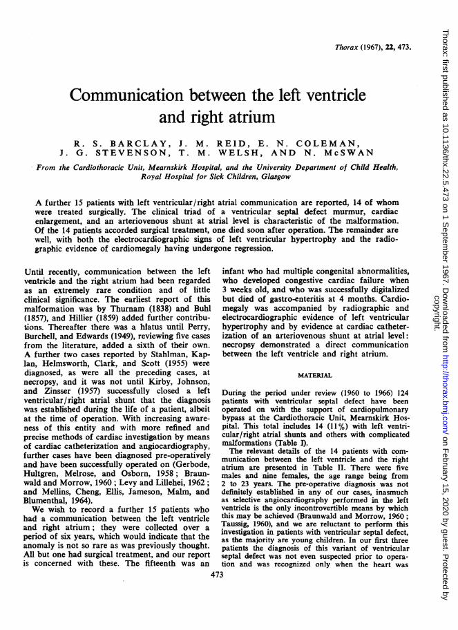
FIG. 1. Case 6. Pre-operative radio-graph showing considerable enlarge-ment of both the right atrium and theleft ventricle.
OPERATIVE DATA The 14 patients were operated on
using cardiopulmonary bypass with moderate hypo-thermia (320 C.). The correct diagnosis was unsus-pected in our first three patients, and the situationbecame apparent only after opening the ventricle andinspecting the septum. In the remaining 11, becausethe diagnosis was strongly suspected, atriotomy wasperformed which enabled repair of the defect to becarried out and also closure in six instances of anatrial septal defect; ventriculotomy was not requiredin any of these cases although two were of theindirect type. Direct suture proved adequate, and inno case was a patch considered necessary. Thepatient who had accompanying pulmonary stenosis(Table II, case 4) underwent commissurotomy afterclosure of the defect.
Recovery was uneventful in 13 patients, but one(case 2) died two days after operation from cardiacfailure with severe pulmonary congestion. It wassuspected that aortic insufficiency had been producedduring repair of the defect, the aortic cusps beingdistorted by the sutures; at necropsy this was con-firmed.
POST-OPERATIVE STATUS The 13 survivors have beensupervised by out-patient review since operation, theperiod ranging from three months to six years. Allhave remained well, although in six there remains an
unimpressive systolic murmur (grade I) at the leftsternal border. The pre-operative electrocardiographicevidence of left ventricular hypertrophy which waspresent in 12 has regressed in all but three; the first-degree atrioventricular block of case 4 has not beenaffected by operation. Radiologically, there has beena diminution in cardiac size in nine patients; but fourstill have prominence of the left ventricle, althoughthe pulmonary plethora has disappeared. We have notrepeated cardiac catheterization because we are ofthe opinion that there is sufficient information fromthe clinical findings to indicate that the shunt hasbeen either completely repaired or reduced to insigni-ficant proportions.
DISCUSSION
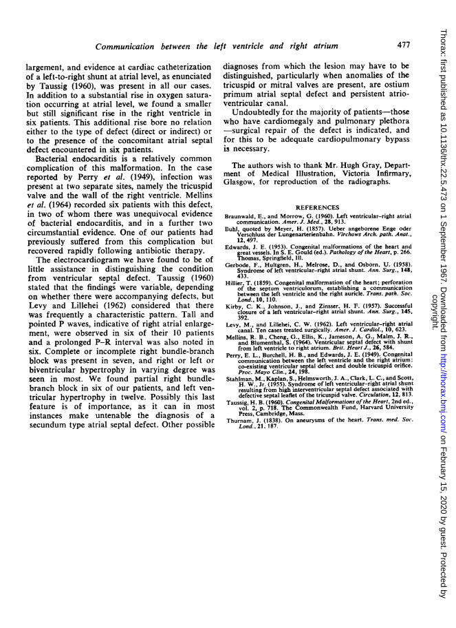
Edwards (1953) demonstrated that there are twotypes of communication between the left ventricleand the right atrium (Fig. 3). In the first the defectin the membranous portion of the ventricularseptum opens directly into the right atrium abovethe tricuspid valve (direct type). In the second, thedefect, which lies lower in the membranous partof the septum, opens first into the right ventriclebehind the septal leaflet of the tricuspid valve and
475
kM
copyright. on F
ebruary 15, 2020 by guest. Protected by
http://thorax.bmj.com
/T
horax: first published as 10.1136/thx.22.5.473 on 1 Septem
ber 1967. Dow
nloaded from
476 R. S. Barclay, J. M. Reid, E. N. Coleman, J. G. Stevenson, T. M. Welsh, and N. McSwan
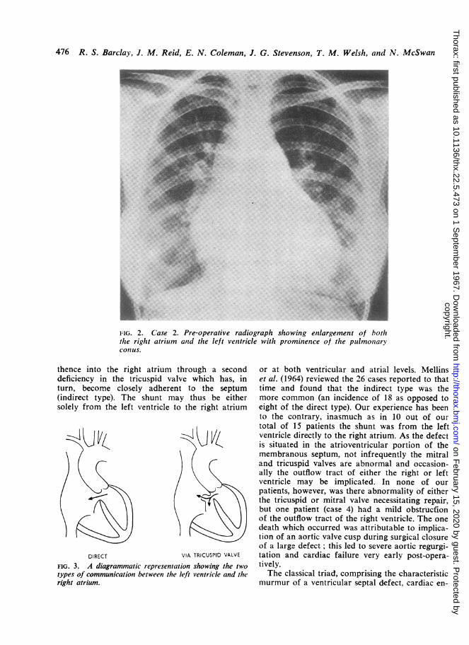
FIG. 2. Case 2. Pre-operative radiograph showing enlargement of boththe right atrium and the left ventricle with prominence of the pulmonaryconIus.
thence into the right atrium through a seconddeficiency in the tricuspid valve which has, inturn, become closely adherent to the septum(indirect type). The shunt may thus be eithersolely from the left ventricle to the right atrium
DIRECT VIA TRICUSPID VALVE
FIG. 3. A diagrammatic representation showing the twotypes of communication between the left ventricle and theright atrium.
or at both ventricular and atrial levels. Mellinset al. (1964) reviewed the 26 cases reported to thattime and found that the indirect type was themore common (an incidence of 18 as opposed toeight of the direct type). Our experience has beento the contrary, inasmuch as in 10 out of ourtotal of 15 patients the shunt was from the leftventricle directly to the right atrium. As the defectis situated in the atrioventricular portion of themembranous septum, not infrequently the mitraland tricuspid valves are abnormal and occasion-ally the outflow tract of either the right or leftventricle may be implicated. In none of ourpatients, however, was there abnormality of eitherthe tricuspid or mitral valve necessitating repair,but one patient (case 4) had a mild obstrucfionof the outflow tract of the right ventricle. The onedeath which occurred was attributable to implica-tion of an aortic valve cusp during surgical closureof a large defect; this led to severe aortic regurgi-tation and cardiac failure very early post-opera-tively.The classical triad, comprising the characteristic
murmur of a ventricular septal defect, cardiac en-
:.
copyright. on F
ebruary 15, 2020 by guest. Protected by
http://thorax.bmj.com
/T
horax: first published as 10.1136/thx.22.5.473 on 1 Septem
ber 1967. Dow
nloaded from
Communication between the left ventricle and right atrium
largement, and evidence at cardiac catheterizationof a left-to-right shunt at atrial level, as enunciatedby Taussig (1960), was present in all our cases.In addition to a substantial rise in oxygen satura-tion occurring at atrial level, we found a smallerbut still significant rise in the right ventricle insix patients. This additional rise bore no relationeither to the type of defect (direct or indirect) orto the presence of the concomitant atrial septaldefect encountered in six patients.
Bacterial endocarditis is a relatively commoncomplication of this malformation. In the casereported by Perry et al. (1949), infection waspresent at two separate sites, namely the tricuspidvalve and the wall of the right ventricle. Mellinset al. (1964) recorded six patients with this defect,in two of whom there was unequivocal evidenceof bacterial endocarditis, and in a further twocircumstantial evidence. One of our patients hadpreviously suffered from this complication butrecovered rapidly following antibiotic therapy.The electrocardiogram we have found to be of
little assistance in distinguishing the conditionfrom ventricular septal defect. Taussig (1960)stated that the findings were variable, dependingon whether there were accompanying defects, butLevy and Lillehei (1962) considered that therewas frequently a characteristic pattern. Tall andpointed P waves, indicative of right atrial enlarge-ment, were observed in six of their 10 patientsand a prolonged P-R interval was also noted insix. Complete or incomplete right bundle-branchblock was present in seven, and right or left orbiventricular hypertrophy in varying degree wasseen in most. We found partial right bundle-branch block in six of our patients, and left ven-tricular hypertrophy in twelve. Possibly this lastfeature is of importance, as it can in mostinstances make untenable the diagnosis of asecundum type atrial septal defect. Other possible
diagnoses from which the lesion may have to bedistinguished, particularly when anomalies of thetricuspid or mitral valves are present, are ostiumprimum atrial septal defect and persistent atrio-ventricular canal.
Undoubtedly for the majority of patients-thosewho have cardiomegaly and pulmonary plethora-surgical repair of the defect is indicated, andfor this to be adequate cardiopulmonary bypassis necessary.
The authors wish to thank Mr. Hugh Gray, Depart-ment of Medical Illustration, Victoria Infirmary,Glasgow, for reproduction of the radiographs.
REFERENCESBraunwald, E., and Morrow, G. (1960). Left ventricular-right atrial
communication. Amer. J. Med., 28, 913.Buhl, quoted by Meyer, H. (1857). Ueber angeborene Enge oder
Verschluss der Lungenarterienbahn. Virchows Arch. path. Anat.,12,497.
Edwards, J. E. (1953). Congenital malformations of the heart andgreat vessels. In S. E. Gould (ed.). Pathology of the Heart, p. 266.Thomas, Springfield, Ill.
Gerbode, F., Hultgren, H., Melrose, D., and Osborn, U. (1958).Syndrome of left ventricular-right atrial shunt. Ann. Surg., 148,433.
Hillier, T. (1859). Congenital malformation of the heart; perforationof the septum ventriculorum, establishing a communicationbetween the left ventricle and the right auricle. Trans. path. Soc.Lond., 10, 1 10.
Kirby, C. K., Johnson, J., and Zinsser, H. F. (1957). Successfulclosure of a left ventricular-right atrial shunt. Ann. Surg., 145,392.
Levy, M., and Lillehei, C. W. (1962). Left ventricular-right atrialcanal. Ten cases treated surgically. Amer. J. Cardiol., 10, 623.
Mellins, R. B., Cheng, G., Ellis, K., Jameson, A. G., Malm, J. R.,and Blumenthal, S. (1964). Ventricular septal defect with shuntfrom left ventricle to right atrium. Brit. Heart J., 26, 584.
Perry, E. L., Burchell, H. B., and Edwards, J. E. (1949). Congenitalcommunication between the left ventricle and the right atrium:co-existing ventricular septal defect and double tricuspid orifice.Proc. Mayo Clin., 24, 198.
Stahlman, M., Kaplan, S., Helmsworth, J. A., Clark, L. C., and Scott,H. W., Jr. (1955). Syndrome of left ventricular-right atrial shuntresulting from high interventricular septal defect associated withdefective septal leaflet of the tricuspid valve. Circulation, 12, 813.
Taussig, H. B. (1960). Congenital Malformations ofthe Heart, 2nd ed.,vol. 2, p. 718. The Commonwealth Fund, Harvard UniversityPress, Cambridge, Mass.
Thurnam, J. (1838). On aneurysms of the heart. Trans. med. Soc.Lond., 21, 187.
477
copyright. on F
ebruary 15, 2020 by guest. Protected by
http://thorax.bmj.com
/T
horax: first published as 10.1136/thx.22.5.473 on 1 Septem
ber 1967. Dow
nloaded from