common wrist injuries in sport - gp cme · common wrist injuries in sport ... helpful in excluding...
TRANSCRIPT

Common wrist injuries in Common wrist injuries in sportsport
Chris MilneChris MilneSports PhysicianSports Physician
Hamilton ,NZHamilton ,NZ

Overview / ClassificationOverview / ClassificationAcute injuries
• Simple - wrist sprain• Not so simple1 - Fracture of distal
radius/ulna2 - Scaphoid fracture3 - Fracture of hook of
Hamate4 - Scapho-lunate ligament
rupture5 - Lunate dislocation6 – Triangular fibrocartilage
(TFCC) tear
Chronic/overuse injuries1 - Missed scaphoid fracture2 - Missed scapho-lunate
ligament rupture3 - Instability of distal radio-ulnar
joint4 - TFCC tear5 - Ulnar impaction 6 - Dorsal impingement 7 – Tenosynovitis
De Quervain’s Intersection syndrome

Key pointsKey points
• 1 – Mechanism of injury is important• 2 – Specific injuries are often associated
with specific sports and age groups• 3 – A missed scaphoid fracture is the most
common missed fracture leading to litigation

HistoryHistory
1 – Mechanism of injury - FOOSH- forced flexion- forced extension
2 – High or low energyHigh energy injuries – cycling,
mountain-biking, skateboarding, rollerblading, snowboarding

Mechanism of injury

History Continued: History Continued:
3 – Location of pain – ulnar, radial sided4 – Associated clicking, snapping5 – Occupation – heavy / light work6 – Other recreational activities7 – Previous injuries + treatment

ExaminationExaminationLook – Deformity e.g – dinner fork
Swelling e.g – ganglion Feel – Tender sites – start with where they
are most symptomaticAlways check – anatomical snuffbox
- scaphoid tubercleMove – Flexion ( 80 deg ) Extension ( 70 deg )
Radial deviation (20 deg) Ulnar deviation (30 deg)Pronation (80 deg) Supination (80 deg)
Special tests 1 – Watson’s test2 – TFCC stress tests – grind, sitting hands3 – Distal radio – ulnar joint mobility4 – Impingement tests
InvestigationsInvestigationsX-rays – Routine – PA, Lateral
Scaphoid viewsClenched fist view – If suspect rupture
of scapho-lunate ligament ( gap of over 3mm is significant)
Ulnar /radial deviationUltrasound scans 1- Show tendon pathology eg tenosynovitis, or
instability of ulnar tendons 2- Useful for guidance of injection eg for
De Quervains or intersection syndrome

Investigations contd.Investigations contd.Bone Scans 1- Show active bone injury e.g scaphoid fracture (normal bone scan
helpful in excluding scaphoid fracture)2- May help in gymnasts with wrist pain, if activity in distal radial youth
plates is significantly asymmetrical ( ? Super- imposed growth plate fracture)
CT ScansThin slice in scaphoid fracture can show anatomical disruption
MRI Scans1- Scaphoid fracture2- Scapho-lunate ligament tear / disruption3- TFCC tear 4- Ulnar impaction

Bone scan scaphoid fractureBone scan scaphoid fracture

Typical case Typical case –– Wrist sprain NOSWrist sprain NOSHistory –Low energy injury
Pain, no clickingMechanism - Forced extension – Traction of flexor tendons,
Compression of dorsal capsule - Forced flexion – Traction to extensor tendonsExamination - Mild tenderness
- Minimal restriction of movement- Not tender over snuffbox or scaphoid tubercle- Watson’s test negative- TFCC stress tests negative- No pain on mobilising distal radio-ulnar joint

Typical case continuedTypical case continuedInvestigations - No Ottawa rules for wrists
- X-ray those with clinical risk factors for fracture, eg high energy injury
Treatment - Pain relief- Splintage – Off the shelf devices,
thermoplastic devices from hand therapist- Hand exercises – physio, home
based ,review if not significantly improved in three weeks

Extensor muscle exerciseExtensor muscle exercise
Fracture of Radius / UlnaFracture of Radius / Ulna• Young people – High energy required for fracture
suspect significant associated soft-tissue injury• Old People – Osteoporosis – less energy required• Treatment:1- If intra- articular involvement - step of 1mm acceptable
otherwise anatomical reduction required2- 6 weeks in cast – distal half of forearm + hand, leaving
MCP joints free3- ORIF if unstable or reduction inadequate

Colles fractureColles fracture

Fracture of scaphoidFracture of scaphoid--a minefielda minefield
• History – fall, radial sided pain• Examination – Tender in snuffbox or scaphoid tubercle, pain
with axial loading of thumb• Investigations – X-rays including scaphoid views. If need to
know yes/no rapidly. Bone scan / limited MRI positive 24 – 48 hrs post injury (cheaper than 2 weeks off work)
• Treatment – Scaphoid cast 2 weeks then re X-ray. If positive, further 4 weeks in cast .If no fracture – Velcro wrist splint and exercisesIf ongoing wrist pain – refer to orthopaedic surgeon with hand surgery interest and expertise- Likely ORIF

Scaphoid fractureScaphoid fracture
Fracture hook of HamateFracture hook of Hamate
• History – Playing golf, hit a ground shot -grip is forced against top hand. Ulnar sided wrist pain.
• Examination – Tender flexor aspect of wrist, over hamate
• Investigations – X-rays including carpal tunnel view CT or MRI scan helpful
• Treatment – Excision of hook, then 3 weeks immobilisation, or - ORIF

Fracture hook of HamateFracture hook of HamateMechanism of injuryMechanism of injury

Fracture hook of HamateFracture hook of HamateHook viewHook view
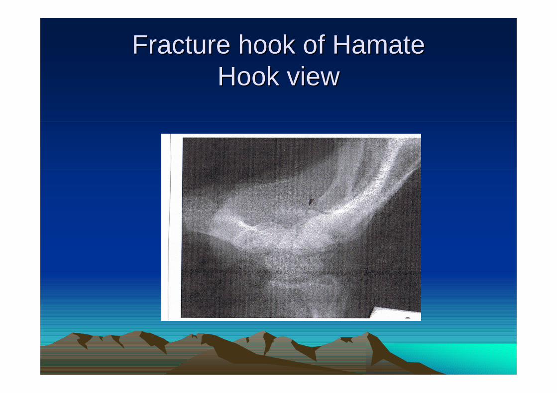
Fracture hook of HamateFracture hook of HamateHook viewHook view

ScaphoScapho--lunate ligament rupturelunate ligament rupture• History – Fall on outstretched hand, pain/clicking• Examination – Tender 2cm distal to Lister’s
tubercle, in line of middle ray. Pain, dorsal movement of scaphoid on performing Watson’s test
• Investigations – X-Rays including clenched fist view (>3mm separation) MRI scan
• Treatment – Ligament repair (at cost of some mobility of wrist)

ScaphoScapho--lunate ligament rupturelunate ligament rupture

ScaphoScapho--lunate ligament rupturelunate ligament rupture

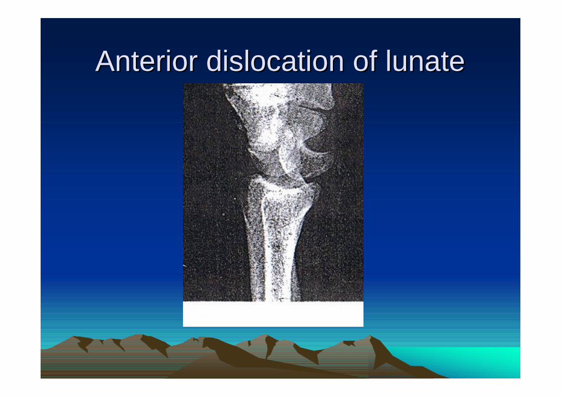
Anterior dislocation of lunateAnterior dislocation of lunate• Mechanism – FOOSH, forced extension injury • History – Severe pain, swelling in palm,
sometimes carpal tunnel syndrome• Examination – Tender swelling in palm• Investigations – X-Ray – lateral view shows
lunate tilted into palm, not articulating with capitate
• Treatment – Reduction – open or closed cast immobilisation 8 weeks
• NB – Needs to be recognised and treated within a few days to avoid complications

Alignment of carpal bonesAlignment of carpal bones
Anterior dislocation of lunateAnterior dislocation of lunate

Perilunate dislocationPerilunate dislocation• Mechanism – In association with scaphoid fracture -
lunate remains with the radius, and capitate dislocates dorsally
• History – Fall severe pain, concavity in palm• Examination – Tender swelling over dorsum of hand• Investigations – X-Ray – Lateral view shows dorsal
displacement of capitate and other distal structures• Treatment – Reduce by traction. POP with wrist in
flexion for 2 weeks, then replace with POP in neutral for a further 2 weeks
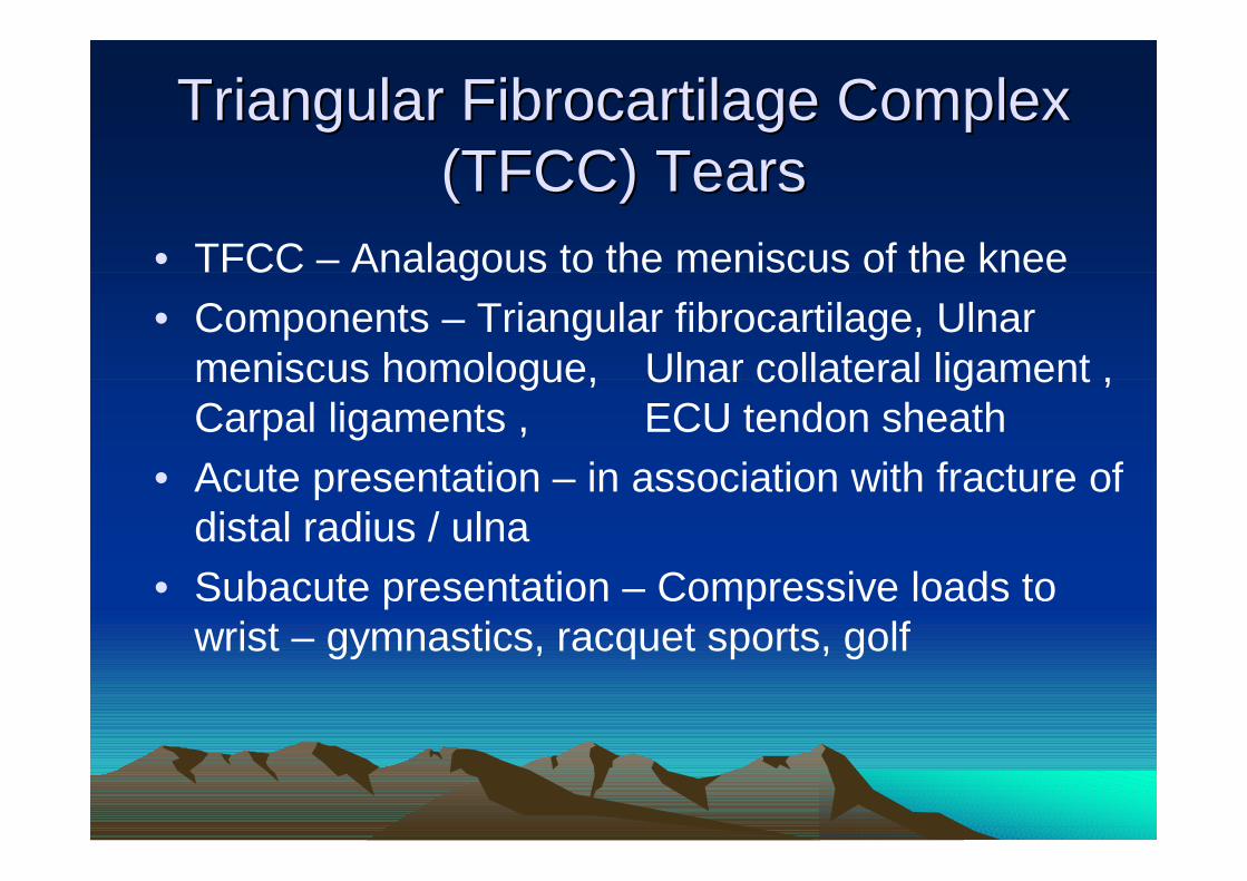
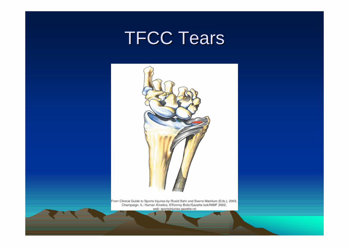
Triangular Fibrocartilage Complex Triangular Fibrocartilage Complex (TFCC) Tears(TFCC) Tears
• TFCC – Analagous to the meniscus of the knee• Components – Triangular fibrocartilage, Ulnar
meniscus homologue, Ulnar collateral ligament , Carpal ligaments , ECU tendon sheath
• Acute presentation – in association with fracture of distal radius / ulna
• Subacute presentation – Compressive loads to wrist – gymnastics, racquet sports, golf
TFCC TearsTFCC Tears

TFCC tears, continuedTFCC tears, continued
• History – Ulnar sided wrist pain +/- clicking • Examination – Tenderness, swelling ulnar
aspect of wrist, TFCC grind test, sitting hands test reproduce pain
• Investigations – X-Rays – if positive ulnar variance, increased risk of TFCC damage MRI – 60% sensitive, 90% specific
• Treatment – Brace, strengthening exercises, surgery – excision of torn fragment ,shortening of ulna (if too long)

Complications of acute injuryComplications of acute injury
• Scaphoid fracture – avascular necrosis (AVN) of proximal pole, post traumatic arthritis
• Scapho-lunate ligament disruption : -instability of proximal carpal row -SLAC wrist
• Any significant wrist injury :1- Carpal tunnel syndrome2- Complex regional pain syndrome (CRPS)3- Post traumatic arthritis

Chronic / Subacute presentationChronic / Subacute presentation
1- Review history – ask specifically about pain in snuffbox, clicking
2- Examination – look specifically for :a- Tenderness in snuffbox, scaphoid tubercleb- Watson’s testc- TFCC provocation testsd- Pain on mobilising distal radio-ulnar joint
Missed Scaphoid fractureMissed Scaphoid fracture
• History – Radial sided pain. Injury may be forgotten
• Examination – Tender in snuffbox , progressive joint stiffness
• Investigations – X-Rays – Sclerosis of proximal pole, associated degenerative change
• Treatment – ORIF and bone graft If partially heated – CT scan through long axis of scaphoid, fine cuts can show anatomic integrity
Missed scaphoMissed scapho--lunate ligament lunate ligament disruption disruption
• History – Pain + / - clicking• Examination – Tender 2cm distal to
Lister’s tubercle, pain, dorsal movement of scaphoid on performing Watson’s test
• Investigations – X-Rays incl. clenched fist view (>3mm gap), MRI scan
• Treatment – Open reduction and repair of ligament
Instability of distal radioInstability of distal radio--ulnar joint ulnar joint
• History – Pain, clicking of wrist• Examination – Tender over distal radio-
ulnar joint, pain, excessive motion of opposite side
• Investigations – True lateral in pronation may show dorsal displacement of ulnar styloid process
• Treatment – Repair of TFCC
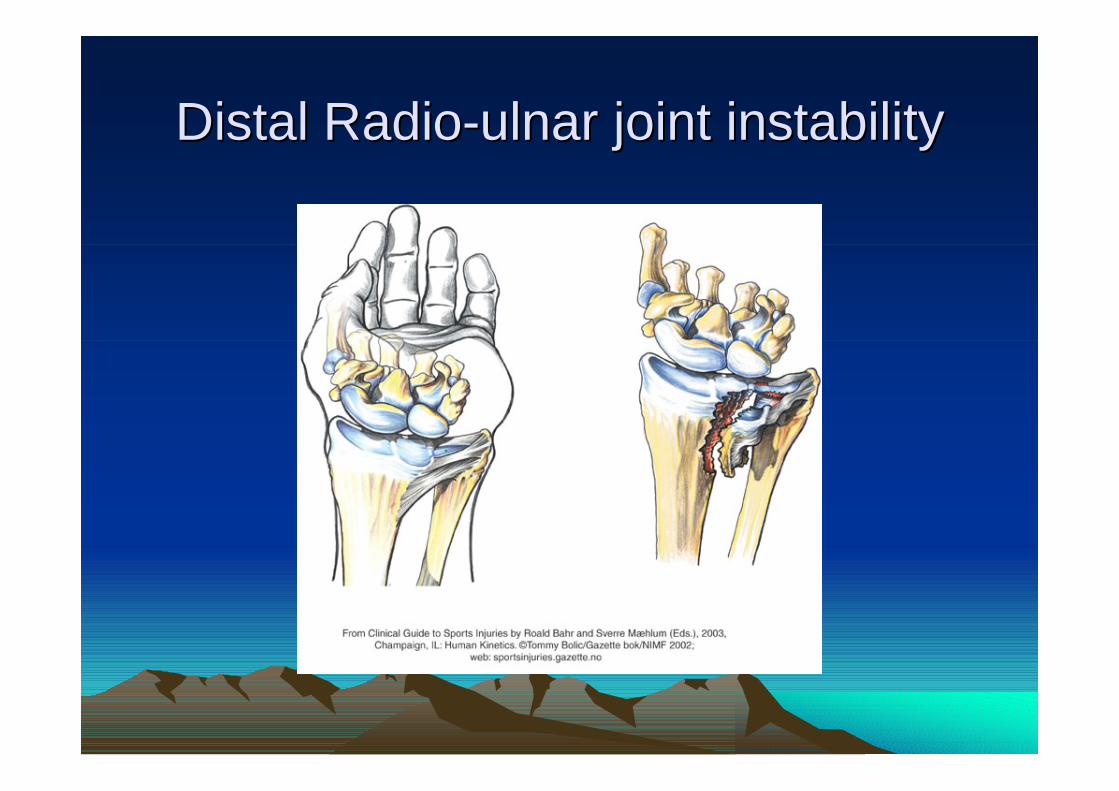
Distal RadioDistal Radio--ulnar joint instabilityulnar joint instability
Triangular Fibrocartilage (TFCC) Triangular Fibrocartilage (TFCC) TearTear
• History – Ulnar sided pain +/- clicking• Examination – Tenderness, swelling ulnar
aspect of wrist TFCC grind test, sitting hands test, reproduce pain
• Investigations – X-Rays – look for positive ulnar variance. MRI scan-60% sensitive,90% specific
• Treatment – Brace, strengthening exercises Surgery – excision of torn fragments, repair of attachments, shortening of ulna useful

Ulnar ImpactionUlnar Impaction
• Pathomechanics – Repeated impaction damages lunate and triquetrum
• History – Ulnar sided pain• Examination – Tender ulnar border of wrist• Investigations – X-Rays show: positive
ulnar variance, sclerosis of lunate• Treatment – Shortening of ulna

Dorsal impingementDorsal impingementHistory – Repeated extension loading, esp. in skeletally
immature gymnastsExamination – Tender dorsum of wrist, restricted extension,
pain at end rangeInvestigations – X-Rays may show changes in distal radial
epiphysis1- Widening of growth plate2- Cystic changes- usually affect metaphyseal aspect of
epiphyseal plate3- Haziness of normal radiolucent area of epiphyseal plate
( cf asymptomatic side )Treatment – Load reduction – modify training regime
strengthening of forearm flexors
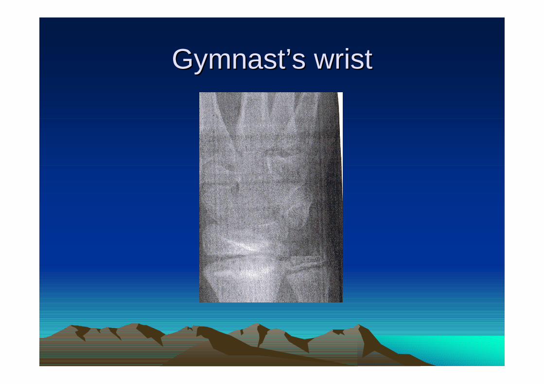
GymnastGymnast’’s wrists wrist

DE Quervains syndrome DE Quervains syndrome (tenosynovitis of APL + EPB)(tenosynovitis of APL + EPB)
History – Radial sided wrist pain, esp. with ulnar deviation e.g –L thumb of R handed golfers, racquet sports, 10 pin bowlers, rowers, canoeists
Examination – Tenderness of APL / EPB tendons as they cross the radial styloid , positive Finkelstein’s sign
Investigations – X- Ray usually normal , Ultrasound scan shows fluid in tendon sheath
Treatment – Splint (14% success) Stretches, strengthening exercises Injection – under ultrasound guidance (83% success) Injection + splintage (61% success) Rarely surgery Activity modification – after training

De QuervainDe Quervain’’s syndromes syndrome

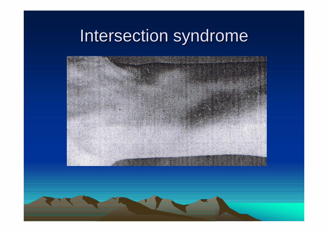
Intersection Syndrome Intersection Syndrome (crossover tenosynovitis)(crossover tenosynovitis)
• History – Radial sided distal forearm pain + crepitus in rowers or canoeists(oarsman’s wrist)
• Examination – Tender in crossover region (where APL / EPB cross wrist extensors) ,Crepitus on flexion / extension of wrist
• Investigations – Ultrasound scan shows fluid in tendon sheath • Treatment – Load reduction: off water 1-2 weeks, Injection
(under ultrasound guidance), If recurrent symptoms – surgery •Technique modification:1- Row with oar square all the time2- Rotate oar by rolling fingers not twisting the wrist3- Check adequate travel on seat

Intersection syndromeIntersection syndrome
Intersection syndromeIntersection syndrome
SummarySummary1- Be careful before you diagnose a simple wrist
sprain2- Scaphoid fractures and scapho-lunate ligament
rupture can have long term complications -have a high index of suspicion for these injuries
3- TFCC injuries are analogous to meniscal tears in knees. A trial of bracing and exercises is worthwhile
4- Tendon problems respond well to local steroid injection under ultrasound guidance