combined f-fdg pet/mr for enhanced imaging of active ... · enhanced imaging of active cardiac...
TRANSCRIPT

Combined 18F-FDG PET/MR for Enhanced Imaging of Active Cardiac Sarcoidosis
Nicolas A. Karakatsanis, Ph.D., MEng; Marc R. Dweck, M.D., Ph.D.; Ronan Abgral, M.D., Ph.D.; Maria Giovanna Trivieri, M.D., Ph.D.; Philip M. Robson, Ph.D.; Jason C. Kovacic, M.D., Ph.D.; Zahi A. Fayad, Ph.D.
Translational and Molecular Imaging Institute, Icahn School of Medicine at Mount Sinai, New York, NY, USA
Introduction
Sarcoidosis is a multisystem disease characterized by granuloma formation, inflammation and fibrosis most commonly affecting the lungs and mediastinal lymph nodes [1]. Cardiac involvement is under-diagnosed but is the leading cause of death amongst patients with sarcoidosis [2-6]. Early intervention with steroids appears to improve prognosis [7], making the accurate and early diagnosis of subclinical but active cardiac sarcoidosis an important clinical goal. Unfortunately establishing this important diagnosis remains a major clinical challenge [8, 9].
Cardiac magnetic resonance (MR) imaging with late gadolinium enhancement (LGE) has recently been introduced for visualizing the pattern of myocardial injury due to cardiac sarcoidosis [10, 11]. However, LGE cannot differentiate between active disease and old chronic scarring, thus limiting the specificity of CMR-based active sarcoidosis assessments. On the other hand, positron emission tomography (PET) imaging with 18F-Fludeoxyglucose (18F-FDG)1, a glucose analog and radiotracer widely used to assess the inflammatory response, has recently been used to identify regions of increased myocardial inflammation in patients with active cardiac sarcoidosis [12-14]. However, glucose
is the predominant source of energy consumed by the myocardium, and high non-specific physiological uptake of 18F-FDG can often lead to false positive identification of active myocardial disease. Although dietary restrictions in the 12 hours prior to PET imaging may switch the heart from glucose to free-fatty acid metabolism and effectively suppress physiological 18F-FDG uptake in the myocardium, this strategy is not always successful [14-16].
Recently, hybrid PET/MR systems have become clinically available [17-19]. The simultaneous hybrid PET/MR Biograph mMR system (Siemens Healthcare, Erlangen, Germany) combines a sensitive PET scanner with a 3T MR system to enable spatial co-registration of complementary imaging data from the two modalities [20]. Simultaneous acquisition of PET and MR data allows disease activity measured by PET to be precisely overlaid on the pattern of injury in the myocardium determined by MR from a single scan session [21]. Moreover, by replacing CT with MR, PET/MR is associated with a lower radiation dose, which is important, especially in chronic conditions such as cardiac sarcoidosis, where follow up would be desirable [17].
Recent studies in our institution have investigated the use of MR/PET for evaluating cardiac disease [21, 22] including cardiac sarcoidosis by assessing the overlap between 18F-FDG-PET activity and the pattern of myocardial injury on LGE MR. We have investigated the potential of combined PET/MR to differentiate between active and inactive cardiac sarcoid as well as identifying false-positive PET indications,
1 The full prescribing information for the Fludeoxyglucose F18 injection can be found at page XX.
LGE LGE/18F-FDG LGE LGE/18F-FDG1A 1D1B 1C
Figure 1: From left to right: (1A, C) LGE MR images showed elevated LGE signal at the lateral and anteroseptal wall for patient 1 (62-year-old male) and patient 2 (63-year-old female) respectively. (1B, D) Matched 18F-FDG PET fused with previous LGE MR images showed high 18F-FDG uptake overlapping with the LGE pattern of injury.
Pati
ent 2
Pati
ent 1
MAGNETOM Flash | (67) 1/2017 www.siemens.com/magnetom-world
2 Clinical | Cardiology
due to inadequate physiological 18F-FDG myocardial uptake suppression [22, 23].
Optimized PET/MR imaging protocol for cardiac sarcoidosis assessment
In this article, we present four clinical exams where initial diagnosis for active cardiac sarcoidosis was unclear when either LGE MR or 18F-FDG PET exams were evaluated independently [22, 23]. All participating subjects had a previous history of proven extra-cardiac sarcoidosis and/or clinical symptoms to suggest cardiac sarcoid involvement. Patients gave written informed consent and were screened for contra-indications before undergoing PET/MR imaging.
Dynamic PET data were acquired across a single bed position centered over the heart in list mode for a period of 90 min beginning 10 min after 5 MBq/kg 18F-FDG injection. The collected PET data were then histogrammed into a single scan time window corresponding to a delayed 40-100 min post-injection period. Subsequently the PET data were reconstructed with an iterative ordinary Poisson ordered subset expectation maximization (OP-OSEM) algorithm using 3 iterations, 21 subsets and a resolution modeling method optimized for the Biograph mMR system. An MR-based attenuation correction method was employed for the PET data based on 4-tissue class segmentation of a standard breath-hold 3D Dixon-VIBE MR sequence. Attenuation from the body transmit coil and spine array, but not from the flexible chest array, were included in the attenuation map.
Cardiac MR, performed simultaneously with the dynamic PET acquisition, included
i) TrueFISP cine images, acquired in the long-axis (2-chamber, 4-chamber) of the left ventricle, followed by
ii) a complete short-axis stack for assessment of cardiac volume and function, and
iii) inversion recovery-prepared spoiled gradient echo late gadolinium enhanced imaging, 10-15 minutes post injection of 0.2 mmol/kg Multi Hance (Bracco imaging, Milan, Italy) in short- and long-axis views [24]. Inversion
times were optimized to null normal myocardium with images repeated in two separate phase-encoding directions to exclude artifact.
Enhancing cardiac sarcoid diagnosis with simultaneous MR/PET imaging
In patients 1 and 2, elevated 18F-FDG uptake (at later times >60 min post tracer injection) co-localized with the pattern of late gadolinium enhancement observed on MR (Fig. 1). The coincidental observation of both increased FDG-PET activity and evidence of myocardial injury on late gadolinium enhancement strongly suggests the presence of active cardiac sarcoidosis. TBR values were calculated as mean standard uptake values (SUV) in regions-of-interest (ROI) drawn over the area of myocardial injury divided by the mean blood pool SUV value in the left ventricular cavity. Mean 18F-FDG target-to-background (TBR) in areas of myocardial injury were 2.2 (patient 1) and 2.0 (patient 2).
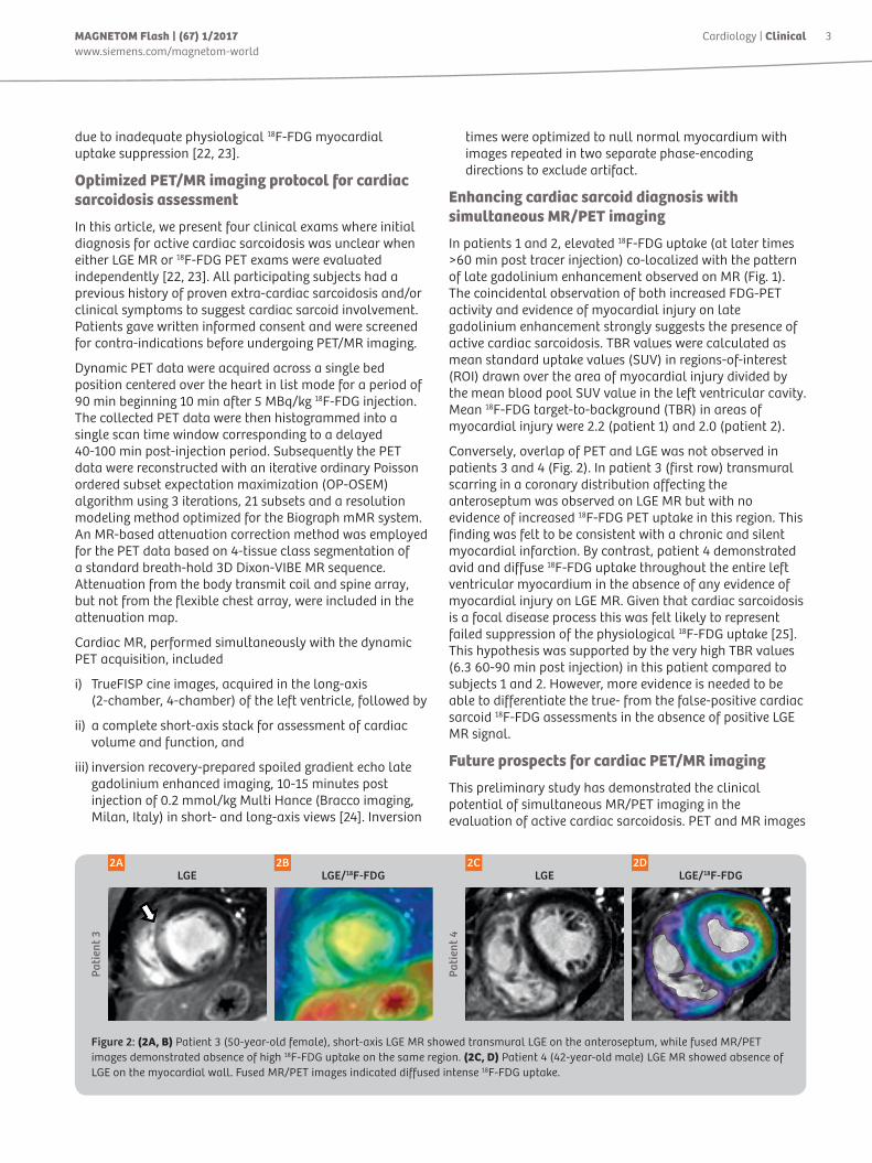
Conversely, overlap of PET and LGE was not observed in patients 3 and 4 (Fig. 2). In patient 3 (first row) transmural scarring in a coronary distribution affecting the anteroseptum was observed on LGE MR but with no evidence of increased 18F-FDG PET uptake in this region. This finding was felt to be consistent with a chronic and silent myocardial infarction. By contrast, patient 4 demonstrated avid and diffuse 18F-FDG uptake throughout the entire left ventricular myocardium in the absence of any evidence of myocardial injury on LGE MR. Given that cardiac sarcoidosis is a focal disease process this was felt likely to represent failed suppression of the physiological 18F-FDG uptake [25]. This hypothesis was supported by the very high TBR values (6.3 60-90 min post injection) in this patient compared to subjects 1 and 2. However, more evidence is needed to be able to differentiate the true- from the false-positive cardiac sarcoid 18F-FDG assessments in the absence of positive LGE MR signal.
Future prospects for cardiac PET/MR imaging
This preliminary study has demonstrated the clinical potential of simultaneous MR/PET imaging in the evaluation of active cardiac sarcoidosis. PET and MR images
LGE LGE/18F-FDG LGE LGE/18F-FDG2A 2D2B 2C
Figure 2: (2A, B) Patient 3 (50-year-old female), short-axis LGE MR showed transmural LGE on the anteroseptum, while fused MR/PET images demonstrated absence of high 18F-FDG uptake on the same region. (2C, D) Patient 4 (42-year-old male) LGE MR showed absence of LGE on the myocardial wall. Fused MR/PET images indicated diffused intense 18F-FDG uptake.
Pati
ent 4
Pati
ent 3
3MAGNETOM Flash | (67) 1/2017www.siemens.com/magnetom-world
Cardiology | Clinical

can be accurately aligned allowing a diagnosis of active cardiac sarcoidosis to be made with confidence when increased 18F-FDG uptake co-localizes with the pattern of injury on late gadolinium enhancement MR. Moreover this approach can help differentiate this pattern from non-active cardiac sarcoid LGE signal or false positive 18F-FDG uptake due to failed myocardial suppression.
References
1 Baughman, R.P., Teirstein, A.S., Judson, M.A., Rossman, M.D., Yeager Jr, H., Bresnitz, E.A., De Palo, L, Hunninghake, G., Iannuzzi, M.C., Johns, C.J. and McLennan, G. 2001. Clinical characteristics of patients in a case control study of sarcoidosis. Am J Resp Crit Care Med, 164(10), pp.1885-1889.
2 Kim, J.S., Judson, M.A., Donnino, R., Gold, M., Cooper, L.T., Prystowsky, E.N. and Prystowsky, S., 2009. Cardiac sarcoidosis. Am Heart J, 157(1), pp.9-21.
3 Iannuzzi, M.C. and Fontana, J.R., 2011. Sarcoidosis: clinical presentation, immunopathogenesis, and therapeutics. Jama, 305(4), pp.391-399.
4 Silverman, K.J., Hutchins, G.M. and Bulkley, B.H., 1978. Cardiac sarcoid: a clinicopathologic study of 84 unselected patients with systemic sarcoidosis.Circulation, 58(6), pp.1204-1211.
5 Hunninghake, G.W., Costabel, U., Ando, M., Baughman, R., Cordier, J.F., Du Bois, R., Eklund, A., Kitaichi, M., Lynch, J., Rizzato, G. and Rose, C., 1999. ATS/ERS/WASOG statement on sarcoidosis. J WASOG/World Assoc Sarc Other Granul Disord, 16(2), p.149.
6 Morgenthau, A.S. and Iannuzzi, M.C., 2011. Recent advances in sarcoidosis. CHEST, 139(1), pp.174-182.
7 Uemura, A., Morimoto, S.I., Hiramitsu, S., Kato, Y., Ito, T. and Hishida, H., 1999. Histologic diagnostic rate of cardiac sarcoidosis: evaluation of endomyocardial biopsies. Am Heart J, 138(2), pp.299-302.
8 Frank, H. and Globits, S., 1999. Magnetic resonance imaging evaluation of myocardial and pericardial disease. J Mag Res Imag, 10(5), pp.617-626.
9 Yamagishi, H., Shirai, N., Takagi, M., Yoshiyama, M., Akioka, K., Takeuchi, K. and Yoshikawa, J., 2003. Identification of cardiac sarcoidosis with 13N-NH3/18F-FDG PET. J Nucl Med, 44(7), pp.1030-1036.
10 Patel, M.R., Cawley, P.J., Heitner, J.F., Klem, I., Parker, M.A., Jaroudi, W.A., Meine, T.J., White, J.B., Elliott, M.D., Kim, H.W. and Judd, R.M., 2009. Detection of myocardial damage in patients with sarcoidosis.Circulation, 120(20), pp.1969-1977.
11 Tadamura, E., Yamamuro, M., Kubo, S., Kanao, S., Saga, T., Harada, M., Ohba, M., Hosokawa, R., Kimura, T., Kita, T. and Togashi, K., 2005. Effectiveness of delayed enhanced MRI for identification of cardiac sarcoidosis: comparison with radionuclide imaging. Am J Roentgen, 185(1), pp.110-115.
12 Youssef, G., Leung, E., Mylonas, I., Nery, P., Williams, K., Wisenberg, G., Gulenchyn, K.Y., DaSilva, J., Birnie, D., Wells, G.A. and Beanlands, R.S., 2012. The use of 18F-FDG PET in the diagnosis of cardiac sarcoidosis: a systematic review and metaanalysis including the Ontario experience.J Nucl Med, 53(2), pp.241-248.
13 Soussan, M., Augier, A., Brillet, P.Y., Weinmann, P. and Valeyre, D., 2014. Functional imaging in extrapulmonary sarcoidosis: FDG-PET/CT and MR features. Clin Nucl Med, 39(2), pp.e146-e159.
14 Manabe, O., Yoshinaga, K., Ohira, H. and Oyama-Manabe, N., 2016. Usefulness of 18F-FDG PET in Diagnosing Cardiac Sarcoidosis. InPerspectives on Nuclear Medicine for Molecular Diagnosis and Integrated Therapy (pp. 209-216). Springer Japan.
15 Tang, R., Wang, J.T.Y., Wang, L., Le, K., Huang, Y., Hickey, A.J. and Emmett, L., 2016. Impact of patient preparation on the diagnostic performance of 18F-FDG PET in cardiac sarcoidosis: A systematic review and meta-analysis. Clin Nucl Med, 41(7), pp.e327-e339.
16 Hulten, E., Aslam, S., Osborne, M., Abbasi, S., Bittencourt, M.S. and Blankstein, R., 2016. Cardiac sarcoidosis—state of the art review.Cardiov Diagn Therapy, 6(1), p.50.
17 Pichler, B.J., Kolb, A., Nägele, T. and Schlemmer, H.P., 2010. PET/MRI: paving the way for the next generation of clinical multimodality imaging applications. J Nucl Med, 51(3), pp.333-336
18 Ratib, O. and Nkoulou, R., 2014. Potential applications of PET/MR imaging in cardiology. J Nucl Med, 55(Supplement 2), pp.40S-46S.
19 Ohira, H., Birnie, D.H., Pena, E., Bernick, J., Mc Ardle, B., Leung, E., Wells, G.A., Yoshinaga, K., Tsujino, I., Sato, T. and Manabe, O., 2016. Comparison of 18F-fluorodeoxyglucose positron emission tomography (FDG PET) and cardiac magnetic resonance (CMR) in corticosteroid-naive patients with conduction system disease due to cardiac sarcoidosis. Eur J Nucl Med Mol Imag, 43(2), pp.259-269
20 Delso, G., Fürst, S., Jakoby, B., Ladebeck, R., Ganter, C., Nekolla, S.G., Schwaiger, M. and Ziegler, S.I., 2011. Performance measurements of the Siemens mMR integrated whole-body PET/MR scanner. J Nucl Med, 52(12), pp.1914-1922
21 Schneider, S., Batrice, A., Rischpler, C., Eiber, M., Ibrahim, T. and Nekolla, S.G., 2014. Utility of multimodal cardiac imaging with PET/MRI in cardiac sarcoidosis: implications for diagnosis, monitoring and treatment. Eur Heart J, 35(5), pp.312-312.
22 Abgral, R., Dweck, M.R., Robson, P.M., Trivieri, M.G., Karakatsanis, N.A., Sanz, J., Contreras, J., Fuster, V., Padilla, M., Kovacic, J.C. and Fayad, Z.A., 2016. Usefulness of combined FDG-PET/MR to diagnose active cardiac sarcoidosis. J Nucl Med, 57(suppl 2), pp.1668-1668.
23 Abgral, R., Dweck, M.R., Robson, P.M., Trivieri, M.G., Karakatsanis, N.A., Mani, V., Padilla, M., Miller, M., Lala, A., .Sanz, J., Narula, J., Fuster, V., Contreras, J., Kovacic, J.C. and Fayad, Z.A., 2016. Clinical Utility of Combined FDG-PET/MR to Assess Myocardial Disease J Am Coll Cardiol Img (in press) doi: 10.1016/j.jcmg.2016.02.029
24 Kellman, P. and Arai, A.E., 2012. Cardiac imaging techniques for physicians: late enhancement. J Mag Res Imag, 36(3), pp.529-542
25 Williams, G. and Kolodny, G.M., 2008. Suppression of myocardial 18F-FDG uptake by preparing patients with a high-fat, low-carbohydrate diet. Am J Roentg, 190(2), pp.W151-W156
Zahi A. Fayad, Ph.D, FAHA, FACC, FISMRM Icahn School of Medicine at Mount Sinai Mount Sinai Endowed Chair in Medical Imaging and Bioengineering Professor of Radiology and Medicine (Cardiology) Director, Translational and Molecular Imaging Institute Director, Cardiovascular Imaging Research Vice-Chair for Research, Department of Radiology One Gustave L. Levy Place Box 1234 New York, NY 10029-6574 USA Phone: +1 212 824 8452 Fax: +1 240 368 8096 [email protected]
Contact
Zahi A. Fayad
4 Clinical | Cardiology MAGNETOM Flash | (67) 1/2017 www.siemens.com/magnetom-world

HIGHLIGHTS OF PRESCRIBING INFORMATION
These highlights do not include all the information needed to use Fludeoxyglucose F 18 Injection safely and effectively. See full prescribing information for Fludeoxyglucose F 18 Injection. Fludeoxyglucose F 18 Injection, USP For intravenous use Initial U.S. Approval: 2005
RECENT MAJOR CHANGESWarnings and Precautions (5.1, 5.2) 7/2010 Adverse Reactions (6) 7/2010
INDICATIONS AND USAGEFludeoxyglucose F18 Injection is indicated for positron emission tomography (PET) imaging in the following settings: • Oncology: For assessment of abnormal
glucose metabolism to assist in the evaluation of malignancy in patients with known or suspected abnormalities found by other testing modalities, or in patients with an existing diagnosis of cancer.
• Cardiology: For the identification of left ventricular myocardium with residual glucose metabolism and reversible loss of systolic function in patients with coronary artery disease and left ventricular dysfunction, when used together with myocardial perfusion imaging.
• Neurology: For the identification of regions of abnormal glucose metabolism associated with foci of epileptic seizures (1).
DOSAGE AND ADMINISTRATIONFludeoxyglucose F 18 Injection emits radiation. Use procedures to minimize radiation exposure. Screen for blood glucose abnormalities. • In the oncology and neurology settings,
instruct patients to fast for 4 to 6 hours prior to the drug’s injection. Consider medical therapy and laboratory testing to assure at least two days of normoglycemia prior to the drug’s administration (5.2).
• In the cardiology setting, administration of glucose-containing food or liquids (e.g., 50 to 75 grams) prior to the drug’s injection facilitates localization of cardiac ischemia (2.3).
Aseptically withdraw Fludeoxyglucose F 18 Injection from its container and administer by intravenous injection (2).
The recommended dose: • for adults is 5 to 10 mCi (185 to 370 MBq),
in all indicated clinical settings (2.1). • for pediatric patients is 2.6 mCi in the
neurology setting (2.2).Initiate imaging within 40 minutes following drug injection; acquire static emission images 30 to 100 minutes from time of injection (2).
DOSAGE FORMS AND STRENGTHSMulti-dose 30mL and 50mL glass vial containing 0.74 to 7.40 GBq/mL (20 to 200 mCi/mL) Fludeoxyglucose F 18 Injection and 4.5mg of sodium chloride with 0.1 to 0.5% w/w ethanol as a stabilizer (approximately 15 to 50 mL volume) for intravenous administration (3).
CONTRAINDICATIONSNone
WARNINGS AND PRECAUTIONS• Radiation risks: use smallest dose necessary
for imaging (5.1). • Blood glucose adnormalities: may cause
suboptimal imaging (5.2).
ADVERSE REACTIONSHypersensitivity reactions have occurred; have emergency resuscitation equipment and personnel immediately available (6). To report SUSPECTED ADVERSE REACTIONS, contact PETNET Solutions, Inc. at 877-473-8638 or FDA at 1-800-FDA-1088 or www.fda.gov/medwatch.
USE IN SPECIFIC POPULATIONSPregnancy Category C: No human or animal data. Consider alternative diagnostics; use only if clearly needed (8.1). • Nursing mothers: Use alternatives to breast
feeding (e.g., stored breast milk or infant formula) for at least 10 half-lives of radioactive decay, if Fludeoxyglucose F 18 Injection is administered to a woman who is breast-feeding (8.3).
• Pediatric Use: Safety and effectiveness in pediatric patients have not been established in the oncology and cardiology settings (8.4).
See 17 for PATIENT COUNSELING INFORMATION
Revised: 1/2011
1.2 Cardiology For the identification of left ventricular myocardium with residual glucose metabolism and reversible loss of systolic function in patients with coronary artery disease and left ventricular dysfunction, when used together with myocardial perfusion imaging.
1.3 Neurology For the identification of regions of abnormal glucose metabolism associated with foci of epileptic seizures.
2 DOSAGE AND ADMINISTRATION Fludeoxyglucose F 18 Injection emits radiation. Use procedures to minimize radiation exposure. Calculate the final dose from the end of synthesis (EOS) time using proper radioactive decay factors. Assay the final dose in a properly calibrated dose calibrator before administration to the patient [see Description (11.2)].
2.1 Recommended Dose for Adults Within the oncology, cardiology and neurology settings, the recommended dose for adults is 5 to 10 mCi (185 to 370 MBq) as an intravenous injection.
2.2 Recommended Dose for Pediatric PatientsWithin the neurology setting, the recommended dose for pediatric patients is 2.6 mCi, as an intravenous injection. The optimal dose adjustment on the basis of body size or weight has not been determined [see Use in Special Populations (8.4)].
2.3 Patient Preparation• To minimize the radiation absorbed dose to the bladder, encourage adequate hydration.
Encourage the patient to drink water or other fluids (as tolerated) in the 4 hours before their PET study.
• Encourage the patient to void as soon as the imaging study is completed and as often as possible thereafter for at least one hour.
• Screen patients for clinically significant blood glucose abnormalities by obtaining a history and/or laboratory tests [see Warnings and Precautions (5.2)]. Prior to Fludeoxyglucose F 18 PET imaging in the oncology and neurology settings, instruct patient to fast for 4 to 6 hours prior to the drug’s injection.
• In the cardiology setting, administration of glucose-containing food or liquids (e.g., 50 to 75 grams) prior to Fludeoxyglucose F18 Injection facilitates localization of cardiac ischemia
2.4 Radiation DosimetryThe estimated human absorbed radiation doses (rem/mCi) to a newborn (3.4 kg), 1-year old (9.8 kg), 5-year old (19 kg), 10-year old (32 kg), 15-year old (57 kg), and adult (70 kg) from intravenous administration of Fludeoxyglucose F 18 Injection are shown in Table 1. These estimates were calculated based on human2 data and using the data published by the International Commission on Radiological Protection4 for Fludeoxyglucose 18 F. The dosimetry data show that there are slight variations in absorbed radiation dose for various organs in each of the age groups. These dissimilarities in absorbed radiation dose are due to developmental age variations (e.g., organ size, location, and overall metabolic rate for each age group). The identified critical organs (in descending order) across all age groups evaluated are the urinary bladder, heart, pancreas, spleen, and lungs.
Table 1. Estimated Absorbed Radiation Doses (rem/mCi) After Intravenous Administration of Fludeoxyglucose F-18 Injectiona
Organ
Newborn 1-year old 5-year old 10-year old
15-year old Adult
(3.4 kg) (9.8 kg) (19 kg) (32 kg) (57 kg) (70 kg)
Bladder wallb 4.3 1.7 0.93 0.60 0.40 0.32
Heart wall 2.4 1.2 0.70 0.44 0.29 0.22
Pancreas 2.2 0.68 0.33 0.25 0.13 0.096
Spleen 2.2 0.84 0.46 0.29 0.19 0.14
Lungs 0.96 0.38 0.20 0.13 0.092 0.064
Kidneys 0.81 0.34 0.19 0.13 0.089 0.074
Ovaries 0.80 0.8 0.19 0.11 0.058 0.053
Uterus 0.79 0.35 0.19 0.12 0.076 0.062
LLI wall * 0.69 0.28 0.15 0.097 0.060 0.051
Liver 0.69 0.31 0.17 0.11 0.076 0.058
Gallbladder wall 0.69 0.26 0.14 0.093 0.059 0.049
Small intestine 0.68 0.29 0.15 0.096 0.060 0.047
ULI wall ** 0.67 0.27 0.15 0.090 0.057 0.046
Stomach wall 0.65 0.27 0.14 0.089 0.057 0.047
Adrenals 0.65 0.28 0.15 0.095 0.061 0.048
Testes 0.64 0.27 0.14 0.085 0.052 0.041
Red marrow 0.62 0.26 0.14 0.089 0.057 0.047
Thymus 0.61 0.26 0.14 0.086 0.056 0.044
Thyroid 0.61 0.26 0.13 0.080 0.049 0.039
Muscle 0.58 0.25 0.13 0.078 0.049 0.039
Bone surface 0.57 0.24 0.12 0.079 0.052 0.041
Breast 0.54 0.22 0.11 0.068 0.043 0.034
Skin 0.49 0.20 0.10 0.060 0.037 0.030
Brain 0.29 0.13 0.09 0.078 0.072 0.070
Other tissues 0.59 0.25 0.13 0.083 0.052 0.042
a MIRDOSE 2 software was used to calculate the radiation absorbed dose. Assumptions on the biodistribution based on data from Gallagher et al.1 and Jones et al.2
b The dynamic bladder model with a uniform voiding frequency of 1.5 hours was used. *LLI = lower large intestine; **ULI = upper large intestine
1 INDICATIONS AND USAGE Fludeoxyglucose F 18 Injection is indicated for positron emission tomography (PET) imaging in the following settings:
1.1 Oncology For assessment of abnormal glucose metabolism to assist in the evaluation of malignancy in patients with known or suspected abnormalities found by other testing modalities, or in patients with an existing diagnosis of cancer.
FULL PRESCRIBING INFORMATION: CONTENTS*
1 INDICATIONS AND USAGE 1.1 Oncology 1.2 Cardiology 1.3 Neurology2 DOSAGE AND ADMINISTRATION
2.1 Recommended Dose for Adults2.2 Recommended Dose for Pediatric Patients2.3 Patient Preparation2.4 Radiation Dosimetry2.5 Radiation Safety – Drug Handling2.6 Drug Preparation and Administration2.7 Imaging Guidelines
3 DOSAGE FORMS AND STRENGTHS4 CONTRAINDICATIONS5 WARNINGS AND PRECAUTIONS 5.1 Radiation Risks 5.2 Blood Glucose Abnormalities6 ADVERSE REACTIONS7 DRUG INTERACTIONS
8 USE IN SPECIFIC POPULATIONS 8.1 Pregnancy 8.3 Nursing Mothers 8.4 Pediatric Use11 DESCRIPTION 11.1 Chemical Characteristics 11.2 Physical Characteristics12 CLINICAL PHARMACOLOGY 12.1 Mechanism of Action 12.2 Pharmacodynamics 12.3 Pharmacokinetics13 NONCLINICAL TOXICOLOGY 13.1 Carcinogenesis, Muta-genesis, Impairment of Fertility14 CLINICAL STUDIES 14.1 Oncology 14.2 Cardiology 14.3 Neurology15 REFERENCES16 HOW SUPPLIED/STORAGE AND DRUG HANDLING17 PATIENT COUNSELING INFORMATION
* Sections or subsections omitted from the full prescribing information are not listed.
FULL PRESCRIBING INFORMATION
5Fludeoxyglucose F 18 Injection, USPMAGNETOM Flash | (67) 1/2017www.siemens.com/magnetom-world

2.5 Radiation Safety – Drug Handling• Use waterproof gloves, effective radiation shielding, and appropriate safety measures when
handling Fludeoxyglucose F 18 Injection to avoid unnecessary radiation exposure to the patient, occupational workers, clinical personnel and other persons.
• Radiopharmaceuticals should be used by or under the control of physicians who are qualified by specific training and experience in the safe use and handling of radionuclides, and whose experience and training have been approved by the appropriate governmental agency authorized to license the use of radionuclides.
• Calculate the final dose from the end of synthesis (EOS) time using proper radioactive decay factors. Assay the final dose in a properly calibrated dose calibrator before administration to the patient [see Description (11.2)].
• The dose of Fludeoxyglucose F 18 used in a given patient should be minimized consistent with the objectives of the procedure, and the nature of the radiation detection devices employed.
2.6 Drug Preparation and AdministrationCalculate the necessary volume to administer based on calibration time and dose. Aseptically withdraw Fludeoxyglucose F 18 Injection from its container. Inspect Fludeoxyglucose F 18 Injection visually for particulate matter and discoloration before administration, whenever solution and container permit. Do not administer the drug if it contains particulate matter or discoloration; dispose of these unacceptable or unused preparations in a safe manner, in compliance with applicable regulations. Use Fludeoxyglucose F 18 Injection within 12 hours from the EOS.
2.7 Imaging GuidelinesInitiate imaging within 40 minutes following Fludeoxyglucose F 18 Injection administration.Acquire static emission images 30 to 100 minutes from the time of injection.
3 DOSAGE FORMS AND STRENGTHS Multiple-dose 30 mL and 50 mL glass vial containing 0.74 to 7.40 GBq/mL (20 to 200 mCi/mL) of Fludeoxyglucose F 18 Injection and 4.5 mg of sodium chloride with 0.1 to 0.5% w/w ethanol as a stabilizer (approximately 15 to 50 mL volume) for intravenous administration.
4 CONTRAINDICATIONS None
5 WARNINGS AND PRECAUTIONS 5.1 Radiation Risks
Radiation-emitting products, including Fludeoxyglucose F 18 Injection, may increase the risk for cancer, especially in pediatric patients. Use the smallest dose necessary for imaging and ensure safe handling to protect the patient and health care worker [see Dosage and Administration (2.5)].
5.2 Blood Glucose Abnormalities In the oncology and neurology setting, suboptimal imaging may occur in patients with inadequately regulated blood glucose levels. In these patients, consider medical therapy and laboratory testing to assure at least two days of normoglycemia prior to Fludeoxyglucose F 18 Injection administration.
6 ADVERSE REACTIONS Hypersensitivity reactions with pruritus, edema and rash have been reported in the post-marketing setting. Have emergency resuscitation equipment and personnel immediately available.
7 DRUG INTERACTIONS The possibility of interactions of Fludeoxyglucose F 18 Injection with other drugs taken by patients undergoing PET imaging has not been studied.
8 USE IN SPECIFIC POPULATIONS8.1 Pregnancy
Pregnancy Category C Animal reproduction studies have not been conducted with Fludeoxyglucose F 18 Injection. It is also not known whether Fludeoxyglucose F 18 Injection can cause fetal harm when administered to a pregnant woman or can affect reproduction capacity. Consider alternative diagnostic tests in a pregnant woman; administer Fludeoxyglucose F 18 Injection only if clearly needed.
8.3 Nursing MothersIt is not known whether Fludeoxyglucose F 18 Injection is excreted in human milk. Consider alternative diagnostic tests in women who are breast-feeding. Use alternatives to breast feeding (e.g., stored breast milk or infant formula) for at least 10 half-lives of radioactive decay, if Fludeoxyglucose F 18 Injection is administered to a woman who is breast-feeding.
8.4 Pediatric Use The safety and effectiveness of Fludeoxyglucose F 18 Injection in pediatric patients with epilepsy is established on the basis of studies in adult and pediatric patients. In pediatric patients with epilepsy, the recommended dose is 2.6 mCi. The optimal dose adjustment on the basis of body size or weight has not been determined. In the oncology or cardiology settings, the safety and effectiveness of Fludeoxyglucose F 18 Injection have not been established in pediatric patients.
11 DESCRIPTION 11.1 Chemical Characteristics
Fludeoxyglucose F 18 Injection is a positron emitting radiopharmaceutical that is used for diagnostic purposes in conjunction with positron emission tomography (PET) imaging. The active ingredient 2-deoxy-2-[18F]fluoro-D-glucose has the molecular formula of C6H1118FO5 with a molecular weight of 181.26, and has the following chemical structure:
Fludeoxyglucose F 18 Injection is provided as a ready to use sterile, pyrogen free, clear, colorless solution. Each mL contains between 0.740 to 7.40GBq (20.0 to 200 mCi) of 2-deoxy-2-[18F]fluoro-D-glucose at the EOS, 4.5 mg of sodium chloride and 0.1 to 0.5% w/w ethanol as a stabilizer. The pH of the solution is between 4.5 and 7.5. The solution is packaged in a multiple-dose glass vial and does not contain any preservative.
11.2 Physical Characteristics Fluorine F 18 decays by emitting positron to Oxygen O 16 (stable) and has a physical half-life of 109.7 minutes. The principal photons useful for imaging are the dual 511 keV gamma photons, that are produced and emitted simultaneously in opposite direction when the positron interacts with an electron (Table 2).
Table 2. Pricipal Radiation Emission Data for Fluorine F18
Radiation/Emission % Per Disintegration Mean Energy
Positron (b+) 96.73 249.8 keV
Gamma (±)* 193.46 511.0 keV
*Produced by positron annihilation From: Kocher, D.C. Radioactive Decay Tables DOE/TIC-I 1026, 89 (1981)The specific gamma ray constant (point source air kerma coefficient) for fluorine F 18 is 5.7 R/hr/mCi (1.35 x 10⁻⁶ Gy/hr/kBq) at 1 cm. The half-value layer (HVL) for the 511 keV photons is 4 mm lead (Pb). The range of attenuation coefficients for this radionuclide as a function of lead shield thickness is shown in Table 3. For example, the interposition of an 8 mm thickness of Pb, with a coefficient of attenuation of 0.25, will decrease the external radiation by 75%.
Table 3. Radiation Attenuation of 511 keV Photons by lead (Pb) shielding
Shield thickness (Pb) mm Coefficient of attenuation
0 0.00
4 0.50
8 0.25
13 0.10
26 0.01
39 0.001
52 0.0001
For use in correcting for physical decay of this radionuclide, the fractions remaining at selected intervals after calibration are shown in Table 4.
Table 4. Physical Decay Chart for Fluorine F18
Minutes Fraction Remaining
0* 1.000
15 0.909
30 0.826
60 0.683
110 0.500
220 0.250
*calibration time
12 CLINICAL PHARMACOLOGY 12.1 Mechanism of Action
Fludeoxyglucose F 18 is a glucose analog that concentrates in cells that rely upon glucose as an energy source, or in cells whose dependence on glucose increases under pathophysiological conditions. Fludeoxyglucose F 18 is transported through the cell membrane by facilitative glucose transporter proteins and is phosphorylated within the cell to [18F] FDG-6-phosphate by the enzyme hexokinase. Once phosphorylated it cannot exit until it is dephosphorylated by glucose-6-phosphatase. Therefore, within a given tissue or pathophysiological process, the retention and clearance of Fludeoxyglucose F 18 reflect a balance involving glucose transporter, hexokinase and glucose-6-phosphatase activities. When allowance is made for the kinetic differences between glucose and Fludeoxyglucose F 18 transport and phosphorylation (expressed as the ‚‘lumped constant‘‘ ratio), Fludeoxyglucose F 18 is used to assess glucose metabolism. In comparison to background activity of the specific organ or tissue type, regions of decreased or absent uptake of Fludeoxyglucose F 18 reflect the decrease or absence of glucose metabolism. Regions of increased uptake of Fludeoxyglucose F 18 reflect greater than normal rates of glucose metabolism.
12.2 Pharmacodynamics Fludeoxyglucose F 18 Injection is rapidly distributed to all organs of the body after intravenous administration. After background clearance of Fludeoxyglucose F 18 Injection, optimal PET imaging is generally achieved between 30 to 40 minutes after administration. In cancer, the cells are generally characterized by enhanced glucose metabolism partially due to (1) an increase in activity of glucose transporters, (2) an increased rate of phosphorylation activity, (3) a reduction of phosphatase activity or, (4) a dynamic alteration in the balance among all these processes. However, glucose metabolism of cancer as reflected by Fludeoxyglucose F 18 accumulation shows considerable variability. Depending on tumor type, stage, and location, Fludeoxyglucose F 18 accumulation may be increased, normal, or decreased. Also, inflammatory cells can have the same variability of uptake of Fludeoxyglucose F 18. In the heart, under normal aerobic conditions, the myocardium meets the bulk of its energy requirements by oxidizing free fatty acids. Most of the exogenous glucose taken up by the myocyte is converted into glycogen. However, under ischemic conditions, the oxidation of free fatty acids decreases, exogenous glucose becomes the preferred myocardial sub strate, glycolysis is stimulated, and glucose taken up by the myocyte is metabolized immediately instead of being converted into glycogen. Under these conditions, phosphorylated Fludeoxyglucose F 18 accumulates in the myocyte and can be detected with PET imaging. In the brain, cells normally rely on aerobic metabolism. In epilepsy, the glucose metabolism varies. Generally, during a seizure, glucose metabolism increases. Interictally, the seizure focus tends to be hypometabolic.
12.3 Pharmacokinetics Distribution: In four healthy male volunteers, receiving an intravenous administration of 30 seconds in duration, the arterial blood level profile for Fludeoxyglucose F 18 decayed triexponentially. The effective half-life ranges of the three phases were 0.2 to 0.3 minutes, 10 to 13 minutes with a mean and standard deviation (STD) of 11.6 (±) 1.1 min, and 80 to 95 minutes with a mean and STD of 88 (±) 4 min. Plasma protein binding of Fludeoxyglucose F 18 has not been studied. Metabolism: Fludeoxyglucose F 18 is transported into cells and phosphorylated to [18F]-FDG-6- phosphate at a rate proportional to the rate of glucose utilization within that tissue. [F18]-FDG-6-phosphate presumably is metabolized to 2-deoxy-2-[F18]fluoro-6-phospho-D-mannose([F 18]FDM-6-phosphate).
6 Fludeoxyglucose F 18 Injection, USP MAGNETOM Flash | (67) 1/2017 www.siemens.com/magnetom-world

Fludeoxyglucose F 18 Injection may contain several impurities (e.g., 2-deoxy-2-chloro-D-glucose (ClDG)). Biodistribution and metabolism of ClDG are presumed to be similar to Fludeoxyglucose F 18 and would be expected to result in intracellular formation of 2-deoxy-2-chloro-6-phospho-D-glucose (ClDG-6-phosphate) and 2-deoxy-2-chloro-6-phospho-D-mannose (ClDM-6-phosphate). The phosphorylated deoxyglucose compounds are dephosphorylated and the resulting compounds (FDG, FDM, ClDG, and ClDM) presumably leave cells by passive diffusion. Fludeoxyglucose F 18 and related compounds are cleared from non-cardiac tissues within 3 to 24 hours after administration. Clearance from the cardiac tissue may require more than 96 hours. Fludeoxyglucose F 18 that is not involved in glucose metabolism in any tissue is then excreted in the urine. Elimination: Fludeoxyglucose F 18 is cleared from most tissues within 24 hours and can be eliminated from the body unchanged in the urine. Three elimination phases have been identified in the reviewed literature. Within 33 minutes, a mean of 3.9% of the administrated radioactive dose was measured in the urine. The amount of radiation exposure of the urinary bladder at two hours post-administration suggests that 20.6% (mean) of the radioactive dose was present in the bladder. Special Populations: The pharmacokinetics of Fludeoxyglucose F 18 Injection have not been studied in renally-impaired, hepatically impaired or pediatric patients. Fludeoxyglucose F 18 is eliminated through the renal system. Avoid excessive radiation exposure to this organ system and adjacent tissues. The effects of fasting, varying blood sugar levels, conditions of glucose intolerance, and diabetes mellitus on Fludeoxyglucose F 18 distribution in humans have not been ascertained [see Warnings and Precautions (5.2)].
13 NONCLINICAL TOXICOLOGY 13.1 Carcinogenesis, Mutagenesis, Impairment of Fertility Animal studies have not been performed to evaluate the Fludeoxyglucose F 18 Injection carcinogenic potential, mutagenic potential or effects on fertility.14 CLINICAL STUDIES 14.1 Oncology
The efficacy of Fludeoxyglucose F 18 Injection in positron emission tomography cancer imaging was demonstrated in 16 independent studies. These studies prospectively evaluated the use of Fludeoxyglucose F 18 in patients with suspected or known malignancies, including non-small cell lung cancer, colo-rectal, pancreatic, breast, thyroid, melanoma, Hodgkin‘s and non-Hodgkin‘s lymphoma, and various types of metastatic cancers to lung, liver, bone, and axillary nodes. All these studies had at least 50 patients and used pathology as a standard of truth. The Fludeoxyglucose F 18 Injection doses in the studies ranged from 200 MBq to 740 MBq with a median and mean dose of 370 MBq. In the studies, the diagnostic performance of Fludeoxyglucose F 18 Injection varied with the type of cancer, size of cancer, and other clinical conditions. False negative and false positive scans were observed. Negative Fludeoxyglucose F 18 Injection PET scans do not exclude the diagnosis of cancer. Positive Fludeoxyglucose F 18 Injection PET scans can not replace pathology to establish a diagnosis of cancer. Non-malignant conditions such as fungal infections, inflammatory processes and benign tumors have patterns of increased glucose metabolism that may give rise to false-positive scans. The efficacy of Fludeoxyglucose F 18 Injection PET imaging in cancer screening was not studied.
14.2 Cardiology The efficacy of Fludeoxyglucose F 18 Injection for cardiac use was demonstrated in ten independent, prospective studies of patients with coronary artery disease and chronic left ventricular systolic dysfunction who were scheduled to undergo coronary revascularization. Before revascularization, patients underwent PET imaging with Fludeoxyglucose F 18 Injection (74 to 370 MBq, 2 to 10 mCi) and perfusion imaging with other diagnostic radiopharmaceuticals. Doses of Fludeoxyglucose F 18 Injection ranged from 74 to 370 MBq (2 to 10 mCi). Segmental, left ventricular, wall-motion assessments of asynergic areas made before revascularization were compared in a blinded manner to assessments made after successful revascularization to identify myocardial segments with functional recovery. Left ventricular myocardial segments were predicted to have reversible loss of systolic function if they showed Fludeoxyglucose F 18 accumulation and reduced perfusion (i.e., flow-metabolism mismatch). Conversely, myocardial segments were predicted to have irreversible loss of systolic function if they showed reductions in both Fludeoxyglucose F 18 accumulation and perfusion (i.e., matched defects). Findings of flow-metabolism mismatch in a myocardial segment may suggest that successful revascularization will restore myocardial function in that segment. However, false-positive tests occur regularly, and the decision to have a patient undergo revascularization should not be based on PET findings alone. Similarly, findings of a matched defect in a myocardial segment may suggest that myocardial function will not recover in that segment, even if it is successfully revascularized. However, false-negative tests occur regularly, and the decision to recommend against coronary revascularization, or to recommend a cardiac transplant, should not be based on PET findings alone. The reversibility of segmental dysfunction as predicted with Fludeoxyglucose F 18 PET imaging depends on successful coronary revascularization. Therefore, in patients with a low likelihood of successful revascularization, the diagnostic usefulness of PET imaging with Fludeoxyglucose F 18 Injection is more limited.
Indications
Fludeoxyglucose F18 Injection is indicated for positron emission tomography (PET) imaging in the following settings:
Oncology: For assessment of abnormal glucose metabolism to assist in the evaluation of malignancy in patients with known or suspected abnormalities found by other testing modalities, or in patients with an existing diagnosis of cancer.
Cardiology: For the identification of left ventricular myocardium with residual glucose metabolism and reversible loss of systolic function in patients with coronary artery disease and left ventricular dysfunction, when used together with myocardial perfusion imaging.
Neurology: For the identification of regions of abnormal glucose metabolism associated with foci of epileptic seizures.
Important Safety Information
Radiation Risks: Radiationemitting products, including Fludeoxyglucose F18 Injection, may increase the risk for cancer, especially in pediatric patients. Use the smallest dose necessary for imaging and ensure safe handling to protect the patient and healthcare worker.
Blood Glucose Abnormalities: In the oncology and neurology setting, suboptimal imaging may occur in patients with inadequately regulated blood glucose levels. In these patients, consider medical therapy and laboratory testing to assure at least two days of normoglycemia prior to Fludeoxyglucose F18 Injection administration.
Adverse Reactions: Hypersensitivity reactions with pruritus, edema and rash have been reported; have emergency resuscitation equipment and personnel immediately available.
14.3 Neurology In a prospective, open label trial, Fludeoxyglucose F 18 Injection was evaluated in 86 patients with epilepsy. Each patient received a dose of Fludeoxyglucose F 18 Injection in the range of 185 to 370 MBq (5 to 10 mCi). The mean age was 16.4 years (range: 4 months to 58 years; of these, 42 patients were less than 12 years and 16 patients were less than 2 years old). Patients had a known diagnosis of complex partial epilepsy and were under evaluation for surgical treatment of their seizure disorder. Seizure foci had been previously identified on ictal EEGs and sphenoidal EEGs. Fludeoxyglucose F 18 Injection PET imaging confirmed previous diagnostic findings in 16% (14/87) of the patients; in 34% (30/87) of the patients, Fludeoxyglucose F 18 Injection PET images provided new findings. In 32% (27/87), imaging with Fludeoxyglucose F 18 Injection was inconclusive. The impact of these imaging findings on clinical outcomes is not known. Several other studies comparing imaging with Fludeoxyglucose F 18 Injection results to subsphenoidal EEG, MRI and/or surgical findings supported the concept that the degree of hypometabolism corresponds to areas of confirmed epileptogenic foci. The safety and effectiveness of Fludeoxyglucose F 18 Injection to distinguish idiopathic epileptogenic foci from tumors or other brain lesions that may cause seizures have not been established.
15 REFERENCES1. Gallagher B.M., Ansari A., Atkins H., Casella V., Christman D.R., Fowler J.S., Ido T.,
MacGregor R.R., Som P., Wan C.N., Wolf A.P., Kuhl D.E., and Reivich M. “Radiopharmaceuticals XXVII. 18F-labeled 2-deoxy-2-fluoro-d-glucose as a radiopharmaceutical for measuring regional myocardial glucose metabolism in vivo: tissue distribution and imaging studies in animals,” J Nucl Med, 1977; 18, 990-6.
2. Jones S.C., Alavi, A., Christman D., Montanez, I., Wolf, A.P., and Reivich M. “The radiation dosimetry of 2 [F-18] fluoro-2-deoxy-D-glucose in man,” J Nucl Med, 1982; 23, 613-617.
3. Kocher, D.C. “Radioactive Decay Tables: A handbook of decay data for application to radiation dosimetry and radiological assessments,” 1981, DOE/TIC-I 1026, 89.
4. ICRP Publication 53, Volume 18, No. l-4,1987, pages 75-76. 16 HOW SUPPLIED/STORAGE AND DRUG HANDLING
Fludeoxyglucose F 18 Injection is supplied in a multi-dose, capped 30 mL and 50 mL glass vial containing between 0.740 to 7.40 GBq/mL (20 to 200 mCi/mL), of no carrier added 2-deoxy-2-[F 18] fluoro-D-glucose, at end of synthesis, in approximately 15 to 50 mL. The contents of each vial are sterile, pyrogen-free and preservative-free. NDC 40028-511-30; 40028-511-50 Receipt, transfer, handling, possession, or use of this product is subject to the radioactive material regulations and licensing requirements of the U.S. Nuclear Regulatory Commission, Agreement States or Licensing States as appropriate.Store the Fludeoxyglucose F 18 Injection vial upright in a lead shielded container at 25°C (77°F); excursions permitted to 15-30°C (59-86°F). Store and dispose of Fludeoxyglucose F 18 Injection in accordance with the regulations and a general license, or its equivalent, of an Agreement State or a Licensing State. The expiration date and time are provided on the container label. Use Fludeoxyglucose F 18 Injection within 12 hours from the EOS time.
17 PATIENT COUNSELING INFORMATIONInstruct patients in procedures that increase renal clearance of radioactivity. Encourage patients to: • drink water or other fluids (as tolerated) in the 4 hours before their PET study. • void as soon as the imaging study is completed and as often as possible thereafter
for at least one hour.
Manufactured by: PETNET Solutions Inc. 810 Innovation Drive Knoxville, TN 37932Distributed by: PETNET Solutions Inc. 810 Innovation Drive Knoxville, TN 37932
PN0002262 Rev. A
March 1, 2011
7Fludeoxyglucose F 18 Injection, USPMAGNETOM Flash | (67) 1/2017www.siemens.com/magnetom-world