combination surgery and nonviral interleukin 2 gene therapy for
TRANSCRIPT
Combination Surgery and Nonviral Interleukin 2 Gene Therapy forHead and Neck Cancer
Daqing Li, Wen Jiang, Jeffery S. Bishop,Robert Ralston, and Bert W. O’Malley, Jr.1
Johns Hopkins University, Baltimore, Maryland 21287 [D. L., W. J.,B. W. O.] and GeneMedicine, Inc., The Woodlands, Texas 77381[J. S. B., R. R.]
ABSTRACTWe have developed a novel nonviralinterleukin 2 (IL-2)
gene therapy that demonstrates significant treatment-spe-cific, antitumor efficacy in combination with subtotal surgi-cal resection in a head and neck cancer murine model.Treatment of established head and neck tumors in immu-nocompetent mice was performed via direct injection with acationic liposome composed of DOTMA and cholesterol for-mulation carrying DNA plasmid for human IL-2 ( hIL-2)gene expression. ELISA assays of tumor extracts 24 h aftertreatment of hIL-2 gene therapy revealed increased localhIL-2 production as well as a formulation-specific secondaryinduction of murine IFN- g and IL-12. We hypothesize thatthe paracrine production of multiple cytokines after IL-2single gene transfer is important for generating a therapeu-tic effect, and that this strategy will be well tolerated andeffective in combination with surgery for head and neckcancer. In animal experiments where surgery was per-formed in conjunction with an operative site injection ofhIL-2 plasmid formulation, no pre-, intra-, or postoperativetoxicity or compromise to wound healing was identified. Inmurine experiments combining partial surgical resectionwith the nonviral gene therapy, significant antitumor effi-cacy was demonstrated in the hIL-2 plasmid formulationgroup compared with empty plasmid formulation and lac-tose-injected controls. In a separate experiment usingsmaller tumor sizes, we also demonstrated that treatmentoutcomes were dependent on the technical aspect of theactual treatment injection as well as visualization with sur-gical access. The hIL-2 plasmid formulation gene therapyinduces local expression of multiple cytokines, results intreatment-specific antitumor effects, and circumvents manyof the concerns and toxicity encountered with viral genetransfer. These data support the need for continued preclin-
ical investigation and the consideration of human clinicaltrials for combination nonviral hIL-2 gene therapy andsurgery for head and neck cancer.
INTRODUCTIONHead and neck cancer afflicts 50,000 new patients each
year in the United States and.500,000 worldwide. The 3–5-year survival rate of patients suffering from advanced T3, T4
squamous cell carcinoma of the head and neck has remainedpoor, ranging from 20 to 30% (1). This historically poor survivalremains despite innovative surgery and improvements in radia-tion and chemotherapeutic strategies over the past 30 years.
It has been postulated for many years that the host immunesystem plays a major role in the recognition and destruction oftumor cells. The absence of an effectivein vivo immune re-sponse to a tumor has been postulated to be largely due to afailure in activation of T lymphocytes and local production oflymphokines that generate an antitumor response (2). IL-22 isnaturally produced by T cells and serves as an important growthand activation factor for CTLs, macrophages, natural killer cells,and B lymphocytes (3). Treatment with IL-2 and IL-2-activatedperipheral blood lymphoid cells, or even with IL-2 alone, hasproduced definite tumor regression in patients with advancedcancer such as renal cell carcinoma, melanoma, and colorectalcancer (4, 5). However, systemic administration of lymphokinesat pharmacological doses produces high concentrations of lym-phokines in the vasculature and often suboptimal levels at thelocal tumor site. This large differential in IL-2 concentrationresults in limited antitumor effects but moderate to severe tox-icity such as fever, chills, headaches, and capillary leak syn-drome (6). Strategies that increase IL-2 expression in the localtumor environment while decreasing systemic exposure mightenhance antitumor responses and eliminate toxicity. Using genetransfer technology is one such strategy that provides sustainedlocal release of cytokines that stimulate antitumor immuneresponse without significant systemic toxicity (2, 7). Althoughthe pharmacokinetics of different cytokines vary tremendously,it is rare to detect greater than 1 ng/ml cytokine in the serum ofmice, even after injection of 13 107 transduced cells secretinghigh amounts of cytokine (2, 8). This hypothesis has beenvalidated by several groups working in a variety of murinetumor models including melanoma, metastatic colon carcinoma,and head and neck squamous cell cancer (9).
The focus of these previous investigations has been on theuse of gene transfer of cytokines to the local tumor environmentusing the replication defective retrovirus or adenovirus (7).Adenovirus-mediated gene therapy has recently become a majorarea of investigation and development in cancer research be-
Received 11/9/98; revised 3/1/99; accepted 3/1/99.The costs of publication of this article were defrayed in part by thepayment of page charges. This article must therefore be hereby markedadvertisementin accordance with 18 U.S.C. Section 1734 solely toindicate this fact.1 To whom requests for reprints should be addressed, at Otolaryngolo-gy-Head and Neck Surgery, The University of Maryland School ofMedicine, 16 South Eutaw Street, Suite 500, Baltimore, MD 21201.Phone: (410) 328-5828; E-mail: [email protected]. W. O. is a consultant and holds equity interest in GeneMedicine, Inc.
2 The abbreviations used are: IL, interleukin; hIL, human IL; mIL,murine IL; CMV, cytomegalovirus.
1551Vol. 5, 1551–1556, June 1999 Clinical Cancer Research
Research. on March 26, 2018. © 1999 American Association for Cancerclincancerres.aacrjournals.org Downloaded from

cause of the inherent benefits over retroviral-based strategies(10–12). The recombinant adenovirus is much more efficient intransferring genes into most target tissues and does not requireactive cell division for gene uptake and expression. The majordisadvantage of adenoviral vectors, however, is that they induceantiviral immune responses and may generate toxicity fromsystemic dissemination. Attempts at rechallenging an immuno-competent host with an adenoviral vector have resulted in di-minished transgene expression (13). Given the limitations andpotential pitfalls including immunogenicity, risks of geneticrecombination, toxicity, and cytopathic effects of viral genetherapy in general, nonviral systems have been studied as analternative method of gene deliveryin vivo (14).
The major focus of nonviral gene therapy has been on thedevelopment of adequate vehicles to deliver therapeutic genes totarget tissuesin vivo. The most commonly investigated vehicleis the synthetic liposome, which is a positively charged lipid thatcomplexes with DNA. These liposomal complexes facilitatefusion with the negatively charged cell membrane, resulting intransfer of DNA into cells (13). The major criticism and limitingfactor of nonviral systems, however, has classically been thelow efficiency of in vivo gene transfer compared with viralstrategies. The objective of our study is to test the efficacy of anovel hIL-2 cationic lipid formulation gene therapy in combi-nation with surgery for head and neck cancer in an immuno-competent murine model. This novel lipid formulation has beendesigned to induce the local secondary production of IFN-g andIL-12 that augments the immune response generated byIL-2transgene expression.
MATERIALS AND METHODShIL-2 Plasmid. The hIL-2 plasmid used in this study is
derived from pUC19, in which the selectable marker for ampi-cillin resistance was replaced with a gene for kanamycin resist-ance. The expression cassette for hIL-2 used a HCMV MIEpromoter, a 56 nucleotide synthetic 59 untranslated region basedon b-globin, and the cDNA for human IL-2. The plasmid forexpression of human IL-2 (pIL0697) used the poly(A) signaland site from human growth hormone. An “empty” plasmid(pVC0612), identical to pIL0697 while only lacking the hIL-2cDNA sequence, was used as a control for analysis ofhIL-2gene-specific effects. Plasmids were propagated inEscherichiacoli strain DH 5aand purified using alkaline lysis and columnchromatography. The resulting plasmid preparations were testedfor contamination by endotoxin using aLimulus amebocytelysate assay (BioWhittaker, Walkersville, MD). Only plasmidpreparations containing,50 IU/mg DNA were used for addi-tional experiments.
Formulations. The formulation selected used DOTMAas the cationic lipid and cholesterol as the colipid to optimize thedelivery of CMV promoter-driven chloramphenicol acetyltrans-ferase expression plasmid for hIL-2. Small unilamellar vesicles(cationic liposomes) composed of the DOTMA and neutral lipidcholesterol in a 1:1M ratio were prepared by microfluidization.The resulting cationic liposomes were mixed with purified plas-mid at a DNA:lipid charge ratio of 1:0.52/1 (;1:1 w/w) undercontrolled conditions in a solution containing 10% lactose as an
isotonic agent. The DNA concentration of the final formulationwas 0.6 mg/ml.
Animal Model. Animal experiments, including designa-tions for survival outcomes, were approved by the Johns Hop-kins University Animal Care and Use Committee. A syngeneicorthotopic murine model for squamous cell carcinoma of thehead and neck that we previous developed and described wasused for these experiments (15). Floor of the mouth tumors wereestablished in C3H/HeJ mice by percutaneous injection of 53105 SCC VII (squamous carcinoma) cells using sterile tech-niques under a laminar flow hood. The animals were maintainedin standard housing conditions until an appropriate tumor sizewas observed by using neck exposure. Tumors were measuredin three dimensions using calipers following surgical exposure.Smaller tumors (30–40 mm3 were achieved 5 days after initialimplantation of SCC VII cells, and they were used in theexperiment that demonstrated the importance of delivery tech-niques. Larger tumors (150–200 mm3 and 180–240 mm3) wereachieved 7 days after implantation and were used for the initialassessment of tolerability and later partial surgical resection incombination with hIL-2 treatment. The designed treatmentswere then applied according to assigned experimental groups.Initial treatment were performed under direct visualization withsurgical exposure and subsequent treatments when performedwere via a percutaneous route.
Measurement of Primary and Secondary Cytokines.Posttreatment residual tumor masses or local-regional lymphnodes were harvested and minced. A consistent 63 6 3 6 mm3
(0.216 ml) volume of tissue was placed in culture in 3.8-cm2
wells containing 1 ml of DMEM plus 10% FCS. After 24 h,conditioned medium was harvested from the explant cultures,and the presence of cytokines was measured using commerciallyavailable monoclonal antibody ELISAs (from Genzyme, hIL-2;from R&D Systems, mIL-2, mIFN-g, and IL-12 p40).
PCR. Posttreatment local-regional lymph nodes wereharvested, and DNA material was extracted from the tissueusing standard methods. The hIL-2 plasmid-specific sequencewas amplified by PCR using forward and reverse primers(CMV78/IL206 from GeneMedicine). A 10-ml reaction con-sisted of 1.5mg of purified DNA, 1.5 mM deoxynucleotidetriphosphates, 0.5mM of each primer, 1X PCR buffer, and 0.5unit of Taq. When the OmniGene PCR machine was used, theamplification conditions were: 95°C for 3 min. (1 cycle); dena-turation at 95°C for 30 s, annealing at 65°C for 30 s, andextension at 72°C for 30 s (30 cycles); and 72°C for 10 min (1cycle). The size of this PCR product is 265 bp.
Statistical Analysis. The significance of differences be-tween treatment groups was determined by Mann-Whitney anal-ysis.
RESULTSTolerability and Limitation of hIL-2 Plasmid Formula-
tion. Forty C3H/HeJ mice with established floor of the mouthtumors ranging from 150 to 200 mm3 were assigned to twoexperimental groups, preoperative and intraoperative injection.The preoperative group of animals received the first hIL-2plasmid formulation treatment 24 h before the surgical accessprocedure, whereas the intraoperative group received the first
1552Surgery andIL-2 Gene Therapy for Head and Neck Cancer
Research. on March 26, 2018. © 1999 American Association for Cancerclincancerres.aacrjournals.org Downloaded from

treatment during the procedure. The two groups were subse-quently divided into subgroups and received up to four sequen-tial direct percutaneous injections of hIL-2 plasmid formulationinto palpable tumors every 48 h. All of the animals wereinspected daily with special attention to wound healing, assess-ing the presence of incisional ulcers, wound dehiscence, or frankinfection with purulence or seroma. Photographs were taken foran objective evaluation of wound conditions. At 2 weeks post-treatment, animals were sacrificed, and level of wound healingwas assessed by gross inspection, manual evaluation of tensilestrength, and microscopic analysis as compared with untreatedcontrols. Three-dimensional caliper measurements of the tumormass were obtained. No clinical wound healing complicationssuch as incisional ulcers, seroma, or frank infection were ob-served in any of the treatment groups. Manual tensile strengthwas normal in all groups. The surgical incisions were com-pletely healed in 5–7 days, and repeated needle injections didnot appear to affect normal wound healing. H&E staining ofsurrounding normal tissue (muscle, blood vessels, and salivaryglands) demonstrated no necrosis or significant inflammatoryinfiltrate. Microscopic analysis showing no histological changesin distant organs such as lung, liver, bowel, and kidney (data notshown) established relative clinical tolerability of the hIL-2plasmid formulation. Although all of the animals tolerated thegene therapy administration, there were no significant antitumoreffects in the treatment groupsversusthe controls (Fig. 1).
hIL-2 Gene Therapy in Smaller Tumors and the Im-portance of Delivery Technique. On the basis of the previouslack of efficacy upon treatment of a very large tumor burden for
this murine model, the following experiment was designed todetermine whether gross tumor burden was a limiting factor.This experiment was also designed to evaluate the importance ofdelivery techniques of the nonviral gene therapy strategy ontreatment outcome. A total of 15 C3H/HeJ mice with establishedFOM tumors ranging from 30 to 40 mm3 were divided into threeexperimental groups that received injections following a neckexposure operation: (a) hIL-2 standard injection with no leakageof formulation from the injection site; (b) hIL-2 injection withvisible leakage#30% of injected volume; and (c) standardinjection using 10% lactose solution as control. All animalsreceived a second percutaneous injection 4 days after the mocksurgical operation. Animals were observed daily with specialattention to changes in tumor size. They were sacrificed 8 daysafter the first injection, and three-dimensional caliper measure-ments of the tumor mass were obtained. Standard injection ofhIL-2 plasmid formulation was significantly more effective in
Fig. 1 Lack of significant antitumor response in large (150–200 mm3)floor of mouth murine squamous cell tumors. Forty mice were assignedto experimental groups that received up to five sequential intratumoralinjections of IL-2 plasmid formulation. The first injection was per-formed either 24 h before or during a surgical access procedure. Sub-sequent injections were via a direct percutaneous route. There is nosignificant difference in treatment groupsversusa control group thatreceived five PBS injections.Bars,SD.
Fig. 2 A, demonstration of dependency of therapeutic effect on deliv-ery technique. The hIL-2 plasmid formulation was administered usingeither standard technique without visible leakage or injections withvisible leakage#30% of total volume. The control was a standardinjection of 10% lactose (the base solution for the plasmid formula-tions). Standard injection was significantly more effective (P5 0.01 to0.009) in delaying tumor progression than either leaky or control.Bars,SD.B, diminished cytolytic T lymphocyte activity using leaky injectiontechnique compared with standard injection of hIL-2 plasmid formula-tion. M, IL-2 standard injection;E, IL-2 leaked injection.
1553Clinical Cancer Research
Research. on March 26, 2018. © 1999 American Association for Cancerclincancerres.aacrjournals.org Downloaded from
delaying tumor progression than leaked injection (P5 0.01) orcontrol lactose injection (P 5 0.009; Fig. 2A). When CTLactivity was assessed using block antibody, it was observed thattumors treated with hIL-2 plasmid formulation using the leakedinjection method had diminished CTL activity compared withthe standard injection technique (Fig. 2B).
hIL-2 Plasmid Formulation Therapy in Combinationwith Partial Surgical Resection. In the preceding experi-ments, we demonstrated a lack of antitumor efficacy in largetumors (150–200 mm3 and a significant antitumor benefit insmaller tumors (30–40 mm3). This suggests that a threshold oftumor size may limit efficacy of this nonviralhIL-2 gene ther-apy. Thus, surgical resection of large tumors is likely to increasethe efficacy of the gene therapy by reducing tumor burden whilealso providing direct visualization and access to the tumor itself.The following experiment was designed to test the hypothesisthat hIL-2 plasmid formulation can be effectively combinedwith partial surgery for large tumors.
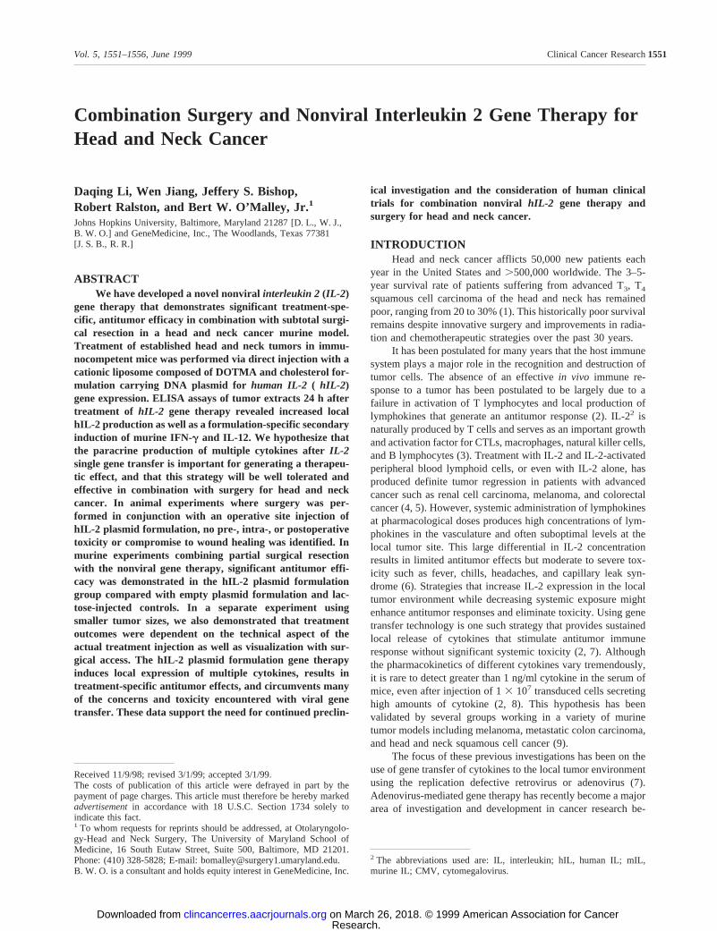
Established floor of the mouth tumors were allowed togrow to sizes ranging from 180 to 240 mm3, and then partialsurgical resections were performed, leaving residual tumors inthe mylohyoid muscle measuring;18–24 mm3. Exposed re-sidual tumors were then injected with 50ml of one of thefollowing four solutions according to assigned experimentalgroups: (a) hIL-2 formulation; (b) formulated empty plasmidformulation; (c) 10% lactose; and (d) PBS control. The surgicalwound was then closed, and treatment was repeated on postop-erative day 4 using percutaneous injections according to as-signed experimental groups. Animals were sacrificed 8 daysafter their first injection, and tumor sizes were determined usingthree-dimensional caliper measurements. Statistical significancewas achieved for hIL-2-treated tumors compared with all re-maining groups (P5 0.01–0.002; Fig. 3). Although the hIL-2plasmid formulation-treated tumor demonstrated the greatestlevel of antitumor efficacy, the empty control plasmid in lipid
formulation was also effective in delaying tumor progressionversuseither 10% lactose or PBS controls (P 5 0.02 and 0.004,respectively).
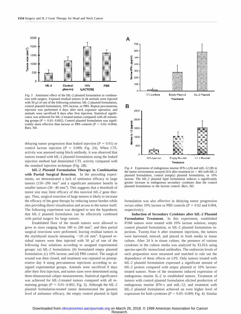
Induction of Secondary Cytokines after hIL-2 PlasmidFormulation Treatment. In this experiment, establishedFOM tumors were treated with 10% lactose solution, emptycontrol plasmid formulation, or hIL-2 plasmid formulation in-jections. Twenty-four h after treatment injections, the tumorswere harvested, minced, and placed in fresh media in tissueculture. After 24 h in tissue culture, the presence of variouscytokines in the culture media was analyzed by ELISA usingspecies-specific monoclonal antibodies. The endotoxin levels ofeach preparation were measured and matched to rule out thedependence of these effects on LPS. Only tumors treated withhIL-2 plasmid formulation expressed a significant amount ofhIL-2 protein compared with empty plasmid or 10% lactose-treated tumors. None of the treatments induced expression ofendogenous murine IL-2 in established tumors. Treatment oftumors with control plasmid formulation elicited production ofendogenous murine IFN-g and mIL-12; and treatment withhIL-2 plasmid formulation achieved an even higher level ofexpression for both cytokines (P 5 0.05–0.009; Fig. 4). Similar
Fig. 3 Antitumor effect of the hIL-2 plasmid formulation in combina-tion with surgery. Exposed residual tumors in 44 animals were injectedwith 50ml of one of the following solutions: hIL-2 plasmid formulation,control plasmid formulation, 10% lactose, or PBS. Repeat percutaneousinjection was performed 4 days after neck exposure operation, andanimals were sacrificed 8 days after first injection. Statistical signifi-cance was achieved for hIL-2-treated tumors compared with all remain-ing groups (P 5 0.01–0.002). Control plasmid formulation was signif-icantly more effective than lactose or PBS controls (P 5 0.02–0.004).Bars, SD.
Fig. 4 Expression of endogenous murine IFN-g (A) and mIL-12 (B) inthe tumor environment assayed 24 h after treatment (n 5 40) with hIL-2plasmid formulation, control (empty) plasmid formulation, or 10%lactose. The hIL-2 plasmid lipid formulation induces a significantlygreater increase in endogenous secondary cytokines than the controlplasmid formulation or the lactose control.Bars,SD.
1554Surgery andIL-2 Gene Therapy for Head and Neck Cancer
Research. on March 26, 2018. © 1999 American Association for Cancerclincancerres.aacrjournals.org Downloaded from

secondary induction of murine IFN-g and mIL-12 was alsoobserved in local-regional cervical draining lymph nodes afterinitial intratumoral injections of either hIL-2 plasmid or controlplasmid formulations (Fig. 5), and the presence of the hIL-2plasmid DNA in the lymph nodes after intratumoral injectionsof hIL-2 plasmid formulation was verified by PCR (Fig. 6).
DISCUSSIONAlthough gene therapy has great potential as a new treat-
ment strategy for head and neck cancers and other solid malig-nancies, it may find its most promising present application incombination with standard treatment interventions. It is essentialto establish the tolerability of our novel nonviral system incombination with surgical procedures. Our murine model ex-periments demonstrated thathIL-2 single gene delivery via aspecifically designed cationic lipid formulation could be welltolerated with head and neck surgical procedures. With respectto the surgical site, there were no clinical wound healing com-
plications in either treatment or control groups. Furthermore,there were no identifiable gross or microscope adverse changesin the incision site, the surrounding normal tissue, or distantorgans. The lack of serious clinical morbidity associated withhIL-2 plasmid formulation in animal experiments is encourag-ing; however, more complete systemic safety and biodistribu-tion analyses are required prior to considering human clinicaltrials.
We demonstrated a lack of antitumor efficacy in largetumors treated with hIL-2 plasmid formulation alone. This ispartially due to the historically lower efficiency of gene transferseen in nonviral delivery systems. Also, the limited diffusioncapacity of the nonviral formulation after directin vivo injectionmight explain the decreased therapeutic efficacy in treatinglarge tumors. We hypothesize that this limitation may be over-come or lessened when the gene therapy is combined withsurgery. Surgical resection will not only reduce tumor burdenbut will provide direct access to the tumor site. Surgical accessensures that the formulation is delivered to the specified tumorsite and maximizes the formulated plasmid to tumor cell ratio.After the initial intraoperative treatment, subsequent percutane-ous injections appear to be the only clinically feasible alterna-tive at this time. The need to control leakage of formulation andissues of inaccurate delivery emphasize the general limitationsof current gene transfer strategies. There is clearly a need for thedevelopment of improved delivery techniques such as image-guided direct delivery in the postsurgical setting or more im-portantly, the development of novel systemically deliverablegene therapies.
Despite the fact that the nonviral strategy of gene deliveryis a low efficiency system, we achieved significant antitumorefficacy with the hIL-2 plasmid formulation. This can be ex-plained by the paracrine production of endogenous murineIFN-g and mIL-12 elicited by bothhIL-2 gene product and theplasmid formulation itself. The production of secondary cyto-kines after delivery of the control plasmid formulation demon-
Fig. 5 hIL-2 plasmid formulation treatment effect on secondary cyto-kine production (murine IFN-g and mIL-12) in local-regional cervicallymph nodes. Draining lymph nodes from 40 animals were harvested24 h after direct injection of established floor of mouth tumors. Nodalextracts were assayed for secondary cytokine production.A, lymph nodeproduction of murine IFN-gwas significantly increased in the hIL-2-treated tumorsversuscontrols.B, lymph node production of mIL-12 wasequally significant for both the control (empty) plasmid and hIL-2formulation.Bars,SD.
Fig. 6 Presence of hIL-2 plasmid in local-regional cervical draininglymph nodes after initial intratumoral injection of hIL-2 plasmid for-mulation as verified by PCR amplification of plasmid containing thehIL-2 cDNA sequence.Lane M,marker;Lane C,positive control forhIL-2 plasmid;Lanes 1–7,PCR products from draining cervical lymphnodes of seven different animals, all positive for the plasmid sequencecontaining hIL-2 cDNA;Lane W,water;Lane B,buffer.
1555Clinical Cancer Research
Research. on March 26, 2018. © 1999 American Association for Cancerclincancerres.aacrjournals.org Downloaded from

strated that both cationic lipid and bacterial plasmids play a rolein this cytokine induction process. Our results are consistentwith previous nontumor studies that show mIFN-g and mIL-12production by normal mouse lung in response to intratrachealadministration of plasmid DNA formulated in cationic lipid(16). This secondary cytokine response was not observed afteradministration of formulated methylated plasmid DNA or for-mulated eukaryotic DNA. Neither cationic lipid alone nor plas-mid formulation in saline in our tumor model produced compa-rable levels of mIFN-gand mIL-12 (data not shown). DNA ofbacterial origin has been shown to elicit the production ofseveral Th-1 cytokines (IFN-g, IL-12, and IL-6) and increasednatural killer cell activityin vitro (17, 18). Induction of thesecytokines also can be elicited by oligonucleotides containing acentral unmethylated CpG dinucleotide (17, 18), suggesting thatthis is an innate immune response to a molecular structurecharacteristic of bacteria. Moreover, combining the oligonucleo-tide with Lipofectin can further enhance the production ofIFN-g and mIL-12 by mouse splenocytes treated with sucholigonucleotidesin vitro. In a separate experiment, we alsodemonstrated secondary induction of murine IFN-g and mIL-12in local-regional cervical draining lymph nodes. This may playan important role in the overall antitumor effects of the hIL-2plasmid formulation by augmenting the immune system andmay prove beneficial against microscopic regional nodal ordistant metastasis.
Although we demonstrated that direct injection of the cat-ionic-lipid hIL-2 plasmid formulation can significantly delaytumor progression in an orthotopic head and neck murine cancermodel, there is a lack of complete cure, despite a treatment-specific induction of hIL-2 and secondary cytokines. Futurestudies will address the issues of long-term survival advantageof the hIL-2 plasmid formulation and will evaluate the role ofhIL-2 in combination with other therapeutic genes using thenonviral cationic lipid delivery system to improve efficacy ofgene therapy treatment in combination with surgery. Our exper-iments in the murine model demonstrate that this novel nonviralhIL-2 plasmid formulation can be well tolerated and used ef-fectively in combination with surgery, delaying tumor progres-sion by treatment-specific immune stimulation. The significantdelay in tumor progression associated with hIL-2 formulation incombination with subtotal surgical resection establishes a rolefor this strategy as applied to large tumors. Regarding thepotential clinical application, this nonviral hIL-2 formulationmay prove useful as an adjuvant therapy combined with surgerywhen gross or microscopic residual disease is present.
ACKNOWLEDGMENTSWe thank Drs. Norman Hardman and Eric Tomlinson for scientific
support.
REFERENCES1. Vokes, E. E., Weichselbaum, R. R., Lippmann, S. M., and Hong,W. K. Head and neck cancer. N. Engl. J. Med.,328: 184–191, 1993.2. Fearon, E. R., Pardoll, D. M., Itaya, T., Golumbek, P., Levitsky,H. I., Simons, J. W., Karasuyama H., Vogelstein, B., and Frost, P.
Interleukin-2 production by tumor cells bypasses T helper function inthe generation of an antitumor response. Cell,60: 397–403, 1990.
3. Henny, C. S., Kuribayashi, K., Kern, D. E., and Gillis, S. Interleu-kin-2 augments natural killer cell activity. Nature (Lond.),291: 335–338, 1981.
4. Murphy, G. P., Lawrence, W., Jr., and Lenhard, R. E., Jr. Basis formajor current therapies for cancer. American Cancer Society Textbookof Clinical Oncology, pp. 96–129. Atlanta: American Cancer Society,1995.
5. Rosenberg, S. A., Lotze, M. T., Muul, L. M., Chang, A. E., Avis,F. P., Leitman, S., Linehan, W. M., Robertson, C. N., Lee, R. E., Rubin,J. T., Seipp, C. A., Simpson, C. G., and White, D. E. A progress reporton the treatment of 157 patients with advanced cancer using lympho-kine-activated killer cells and interleukin-2 or high-dose interleukin-2alone. N. Engl. J. Med.,316: 889–897, 1987.
6. West, W. H., Tauer, K. W., Yanelli, J. R., Marshall, G. D., Orr,D. W., Thurman, G. B., and Oldham, R. K. Constant infusion recom-binant interleukin-2 in adoptive immunotherapy of advanced cancer.N. Engl. J. Med.,316: 898–905, 1987.
7. Gansbacher, B., Zier, K., Daniels, B., Cronin, K., Bannerji, R., andGilboa, E. Interleukin-2 gene transfer into tumor cells abrogates tumor-igenicity and induces protective immunity. J. Exp. Med.,172: 1217–1224, 1990.
8. Golumbek, P. T., Lazenby, A. J., Levitsky, H. I., Jaffee, L. M.,Karasuyama, H., Baker, M., and Pardoll, D. M. Treatment of establishedrenal cancer by tumor cells engineered to secrete interleukin-4. Science(Washington DC),254: 713–716, 1991.9. O’Malley, B. W., Jr., Sewell, D. A., Li, D., Kosai, K., Chen, S., Woo,S. L. C., and Duan, L. The role of interleukin-2 in combination adeno-virus gene therapy for head and neck cancer. Mol. Endocrinol.,11:667–673, 1997.10. Boucher, R. C., Knowles, M. R., Johnson, L. G., and Olsen, J. C.Gene therapy for cystic fibrosis using E1-deleted adenovirus: a phase Itrial in the nasal cavity. Hum. Gene Ther.,5: 615–639, 1994.11. Rosenfeld, M. A., Yoshimurea, K., Trapnell, B. C., Yoneyama, K.,Rosenthal, E. R., Dalemans, W., Bargon, J., Stier, L. E., Stratfor-Perricaudet, L. D., Perricaudet, M., Guggino, W. B., Pavirani, A.,Lecocq, J. P., and Crystal, R. G.In vivo transfer of the human cysticfibrosis transmembrane conductance regulator gene to the airway epi-thelium. Cell,68: 143–155, 1992.12. Rich, D. P., Couture, M., Carodoza, L. M., Guiggio, V. M.,Armantano, D., Espino, P. C., Hehir, K., Welsh, M. J., Smith, A. E., andGregory, R. J. Development and analysis of recombinant adenovirusesfor gene therapy of cystic fibrosis. Hum. Gene Ther.,4: 461–476, 1993.13. Weichselbaum, R. R., and Kufe, D. Gene therapy of cancer. Lancet,349 (Suppl. II): 10–12, 1997.14. Parmiani, G., Colombo, M. P., Melani, C., and Arienti, F. Cytokinegene transduction in the immunotherapy of cancer. Adv. Pharmacol.,40: 259–307, 1997.15. O’Malley, B. W., Jr., Cope, K. A., Johnson, C. S., and Schwartz,M. R. A new immunocompetent murine model for oral cancer. Arch.Otolaryngol. Head Neck Surg.,123: 20–24, 1997.16. Yamamoto, S., Yamamoto, T., Shimada, S., Kuramoto, E., Yano,O., Kataoka, T., and Tokunaga, T. DNA from bacteria, but not fromvertebrates, induces interferon, activates natural killer cells and inhibitstumor growth. Microbiol. Immunol.,36: 983–997, 1992.17. Klinman, D. M., Yi, A. K., Beucage, S. L., Conover, J., and Krieg,A. M. CpG motifs present in bacterial DNA rapidly induce lymphocytesto secrete interleukin 6, interleukin 12, and interferong. Proc. Natl.Acad. Sci. USA,93: 2879–2883, 1996.18. Yamamoto, S., Yamamoto, T., Kataoka, T., Kuramoto, E., Yano,O., and Tokunaga, T. Unique palindromic sequences in synthetic oli-gonucleotides are required to induce IFN and augment IFN-mediatednatural killer activity. J. Immunol.,148: 4072–4076, 1992.
1556Surgery andIL-2 Gene Therapy for Head and Neck Cancer
Research. on March 26, 2018. © 1999 American Association for Cancerclincancerres.aacrjournals.org Downloaded from

1999;5:1551-1556. Clin Cancer Res Daqing Li, Wen Jiang, Jeffery S. Bishop, et al. for Head and Neck CancerCombination Surgery and Nonviral Interleukin 2 Gene Therapy
Updated version
http://clincancerres.aacrjournals.org/content/5/6/1551
Access the most recent version of this article at:
Cited articles
http://clincancerres.aacrjournals.org/content/5/6/1551.full#ref-list-1
This article cites 15 articles, 4 of which you can access for free at:
Citing articles
http://clincancerres.aacrjournals.org/content/5/6/1551.full#related-urls
This article has been cited by 2 HighWire-hosted articles. Access the articles at:
E-mail alerts related to this article or journal.Sign up to receive free email-alerts
Subscriptions
Reprints and
To order reprints of this article or to subscribe to the journal, contact the AACR Publications
Permissions
Rightslink site. Click on "Request Permissions" which will take you to the Copyright Clearance Center's (CCC)
.http://clincancerres.aacrjournals.org/content/5/6/1551To request permission to re-use all or part of this article, use this link
Research. on March 26, 2018. © 1999 American Association for Cancerclincancerres.aacrjournals.org Downloaded from