colorimetric determination of · pdf filecolorimetric determination of urea by reginald m....
TRANSCRIPT

COLORIMETRIC DETERMINATION OF UREA
BY REGINALD M. ARCHIBALD
WITH THE TECHNICAL ASSISTANCE OF P. ORTIZ, E. STROH, AND J. BRONNER
(From the Hospital of The Rockefeller Institute for Medical Research, New York)
(Received for publication, November 9, 1944)
The use of a-isonitrosopropiophenone, CS& *CO * CNOH. CH3, as a re- agent for the calorimetric determination of urea has been mentioned in a previous communication (1). The present report outlines a simple method for the calorimetric determination of urea in blood filtrates and urine by means of this reagent and compares the values obtained by this method with those given by the urease ((2), (3) p. 372) methods.
Apparatus
Xpectrophotometer or calorimeter. Reaction tubes. These are test-tubes of Pyrex glass, 20 by 150 mm. Each
tube is fitted with a l-hole rubber stopper through which is passed a short heavy walled glass capillary tube of 0.5 to 1.0 mm. bore. Such tubes have been illustrated by Van Slyke and Hawkins (4). These tubes are used to hold the blood filtrate and other urea solutions during the chromogenic reaction with cr-isonitrosopropiophenone in the hot water bath.
Reagents
Sulfuric-phosphoric acid mixture (1). 1 volume of concentrated sulfuric acid, 3 volumes of syrupy phosphoric acid, and 1 volume of water.
cr-Isonitrosopropiophenone.l 4 gm. in 100 cc. of alcohol. Stock standard solution of urea. 10.7 mg. of urea in 100 cc. of water.
Stored in the ice box; prepared fresh monthly. Working standard solution. 0.0107 mg. of urea per cc., containing 0.005
mg. of urea N per cc. To prepare the working standard, 1 cc. of stock standard is diluted to 10 cc. with water.
Acetate bu$er. 10 gm. of sodium acetate NaC2H302.3Hz0 and 10 cc. of glacial acetic acid made up to 1 liter with water.
Procedure
Preparation of Blood Filtrates-Filtrates prepared according to the method of Somogyi (5), Fujita and Iwatake (6), or Miller and Van Slyke (7)
1 Obtained from the Eastman Kodak Company, or from Anachemia, Ltd., Mon- treal, Canada, and 70 East 45th Street, New York 17.
507
by guest on May 2, 2018
http://ww
w.jbc.org/
Dow
nloaded from

508 COLORIMETRIC DETERMINllTION OF UREA
are suitable.2 Dialysates prepared according to the technique of Hamilton and Archibald (9) are equally satisfactory. 2 cc. aliquots of a 1: 10 filtrate or dialysate are used when the blood urea level is within normal limits. For uremic blood greater dilution than 1:lO is used. Each 2 cc. portion is measured into a reaction tube and mixed with exactly 5 cc. of water.
Preparation of Urine-Urine of adults is diluted according to the volume excreted per minute, as shown in Table I. For children the observed urine flow is corrected for the size of the patient, according to the directions of McIntosh, Moller, and Van Slyke (lo), and the corrected volume per min- ute is applied to Table I. The urines can be diluted in large graduated cylinders with as great an accuracy as the rate of urine flow is ordinarily measured.
Urines containing more than 0.5 per cent protein should be cleared of protein before dilution. Urine containing 0.5 per cent protein after dilution to 1:500 contains 0.01 mg. per cc. This amount of protein causes an error of fl per cent. To free urine of protein, add to 5 cc. an equal volume of acetate buffer. The mixture then has a pH between 4 and 5. The urine is brought to 100” and the precipitated protein is removed by centrifugation or filtration. The protein-free solution is then diluted, as indicated in the last column of Table I, to give the urine dilution indicated by the middle column.
Of the diluted urine 2 cc. are measured into a reaction tube and mixed with 5 cc. of water.
Thymol should not be used as a urine preservative, because if more than 0.001 gm. is present in the final solution analyzed it causes turbidity.
Preparation of Reagent Blank and Standards-Reagent blank and stand- ards are prepared by adding to reaction tubes 0, 2, 4, and 6 cc. portions of working standard and 7, 5, 3, and 1 cc., respectively, of water.
Development of Color-To the 7 cc. of fluid in each tube of blood filtrate, dilute urine, blank, or standard, add 5 cc. of the sulfuric-phosphoric acid mixture and 0.40 cc. of the alcoholic solution of cr-isonitrosopropiophenone. The contents of each tube are mixed thoroughly. The tubes are then closed with their perforated rubber stoppers and are placed in a boiling water bath from which light is excluded. The water in the bath should come just above the level of liquid in the tubes. After exactly 1 hour of heating the
2 The amount of urea in the Folin-Wu (8) filtrate has been shown by urease meas- urement to be no less than in the other filtrates. Nevertheless when Folin-Wu (8) filtrates of dog whole blood are compared with standards to which no sodium tungstate or sodium sulfate is added, the values obtained for urea by the calorimetric method average 10 per cent lower than values obtained with the other filtrates or with the dialysate. For this reason Folin-Wu filtrates are not recommended for use in the calorimetric determination of urea.
by guest on May 2, 2018
http://ww
w.jbc.org/
Dow
nloaded from

R. M. ARCHIBALD 509
tubes are set in a covered kettle containing water at room temperature. A large kitchen pot, the lid and jnside of which have been painted black, serves well. After 15 minutes, readings are taken in a calorimeter or in a spectro- photometer set at wave-length 540 rnp with the optical density of the re- agent blank adjusted to zero. When a visual calorimeter is used, it is important to select a standard which has a color depth almost equal to that of the sample.
Ccdculations
When the photometer is used, a curve is constructed by plotting the mg. of urea in the standards against the corresponding optical densities. From
TABLE I
Relation of Uri,ne Flow to Dilution Required
Corrected urine flow
‘G. fier min.
<0.2 0.2- 0.5 0.5- 1.0 l.o- 2.0 2.0- 4.0 4.0- Q.@ 9.0-15.0
D = dilutions for urines containing Dilutions for urine filtrate after less than 0.5 per cent protein coagulation of protein
1:500 1:375 1:250 I:150 1:lOO 1:60 1:25
1:lOOO I:750 I:500 1:300 1:200 1:120 1:50
the optical densities of the samples the mg. of urea (P) in each are read off from the curve.
Mg. urea N per 100 cc. blood or urine = 5ODP
where D = the number of volumes to which 1 volume of blood or untreated urine was diluted.
When a visual calorimeter is used
Mg. urea N in sample = C X s U
where X = the reading of the standard; U = the reading of the unknown; and C = the number of mg. of urea N in the standard.
Mg. urea N per 100 cc. blood or urine = 5oDC X s u
where D = the number of volumes to which 1 volume of blood or untreated urine was diluted.
by guest on May 2, 2018
http://ww
w.jbc.org/
Dow
nloaded from

510 COLORIMETRIC DETERMINATION OF UREA
In the case of normal blood D = 10 and C = 0.03. Hence
Mg. urea N per 100 cc. blood = 15 _s u
Mg. of urea N are converted to mg. of urea by the use of the factor 2.143.
Discussion of Method
The color produced by reaction of urea with cr-isonitrosopropiophenone in acid is red. The structure of the product is unknown. As is the case in the diacetyl-carbamido reaction (l), the color obtained is photolabile. Hence it is necessary to keep the tubes in the dark during and after heating until they are read. The color obtained under the prescribed conditions is stable in the dark for more than 24 hours. After 24 hours, less than 0.5 per cent of the color is faded.
The advantages of cr-isonitrosopropiophenone over diacetylmonoxime for determination of urea are (1) the reagent is less volatile; (2) the product has a red rather than a yellow color and is more readily compared by the eye; (3) the reagent is relatively less sensitive to citrulline and other urea derivatives than is diacetylmonoxime; therefore it is more specific for urea.
For some purposes it may be desirable to reduce the time of heating. When large concentrations of allantoin are present (as in the case of urine other than human), the specificity of the method can be increased by reduc- ing the heating period to 20 minutes. Under these conditions, although only 27 per cent as much color is obtained with urea, only 4 per cent as much color is obtained with allantoin (see Fig. 3) as when the heating is for 60 minutes. The specificity of the reaction in the presence of allantoin can be further increased by reducing the concentration of sulfuric acid in the acid mixture (1). Lower dilutions of urine or blood will be required when these alternative conditions are employed.
Results
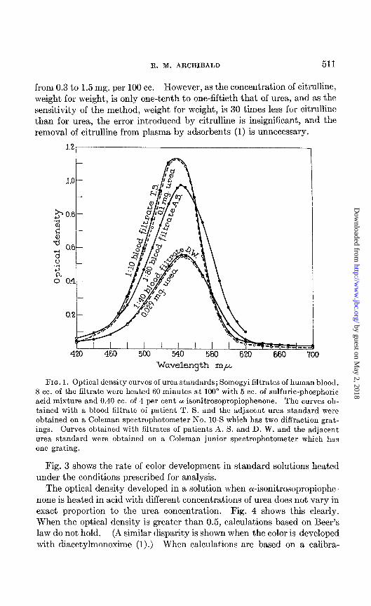
The optical density curves of the colored products obtained with urea and blood filtrate under the conditions of analysis outlined above are given in Fig. 1. It will be noted that the curves yielded by blood filtrates are practi- cally identical with curves yielded by pure urea solutions of similar urea content,. This close approximation to identity is evidence in favor of the probability that no significant part of the optical density in the utilized part of the curve obtained with blood filtrate is due to substances other than the colored product of urea.
The optical density curves of the products obtained with citrulline and a number of other compounds are indicated in Fig. 2. As already reported (l), citrulline is present in normal blood plasma in concentrations ranging
by guest on May 2, 2018
http://ww
w.jbc.org/
Dow
nloaded from
R. M. ARCHIBALD 511
from 0.3 to 1.5 mg. per 100 cc. However, as the concentration of citrulline, weight for weight, is only one-tenth to one-fiftieth that of urea, and as the sensitivity of the method, weight for weight, is 30 times less for citrulline than for urea, the error introduced by citrulline is insignificant, and the removal of citrulline from nlasma bv adsorbents (1) is unnecessary.
Wavelength rnp
FIG. 1. Optical density curves of urea standards; Somogyi filtrates of human blood. 8 cc. of the filtrate were heated 60 minutes at 100” with 5 cc. of sulfuric-phosphoric acid mixture and 0.40 cc. of 4 per cent or-isonitrosopropiophenone. The curves ob- tained with a blood filtrate of patient T. S. and the adjacent urea standard were obtained on a Coleman spectrophotometer No. 10-S which has two diffraction grat- ings. Curves obtained with filtrates of patients A. S. and D. W. and the adjacent urea standard were obtained on a Coleman junior spectrophotometer which has one grating.
Fig. 3 shows the rate of color development in standard solutions heated under the conditions prescribed for analysis.
The optical density developed in a solution when or-isonitrosopropiophe- none is heated in acid with different concentrations of urea does not vary in exact proportion to the urea concentration. Fig. 4 shows this clearly. When the optical density is greater than 0.5, calculations based on Beer’s law do not hold. (A similar disparity is shown when the color is developed with diacetylmonoxime (I).) When calculations are based on a calibra-
by guest on May 2, 2018
http://ww
w.jbc.org/
Dow
nloaded from

512 COLORIMETRIC DETERMINATION OF UREA
tion curve, determinations of blood and urine urea agree well with figures obtained by specific m-ease methods.
0.7
0.6
x c, .-I 2 0.5 4
2 *y 0.4 c, E
0.3
Wavelength rnp
FIG. 2. Optical densities of products obtained with substances giving color on heating in acid with a-isonitrosopropiophenone under the conditions prescribed for measurement of urea. The weights indicate the amount of substance present in 6.20 cc. of the mixture heated 60 minutes at 100”. The volume of mixture heated in this case is half that prescribed for routine determination of urea.
Table II compares the blood and urine urea levels found by this method with values obtained by the manometric urease method (2, 3).
Specijicity of the Method
No color formation results when ammonia, urethane, thiourea, benzimida- xole, proline, ornithine, glutamine, asparagine, gktamic acid, ammonium
by guest on May 2, 2018
http://ww
w.jbc.org/
Dow
nloaded from

R. M. ARCHIBALD 513
pyrrolidonecarboxylate, glutathione, ergothioneine, cafleine, adenine, hy- dantoin, acetamide glycocyamine, or creatine is heated under the conditions prescribed for urea determination.
Alloxantin and uric acid in relatively high concentrations give a trace of red. Parabanic acid, alloxan, alloxan treated with KCN, b&ret, allantoin, and protein give a strong color resembling the red obtained with urea. Phenylurea produces relatively little color. Methylurea reacts to yield a strong red color with an absorption maximum (525 rnp) lower than that
1.2, I
0 Minutes heated
FIG. 3. Rate of formation of color at 100” in reactions of substances with the same concentrations of or-isonitrosopropiophenone and sulfuric-phosphoric acid mixture prescribed for the measurement of urea. The weights indicate the amount of sub- stance heated in the 6.20 cc. of mixture. This volume of mixture is half that pre- scribed above for routine determination of urea.
obtained with urea (540 mp). 10 mg. each of N-benzyl-N-methylurea,3 N-ethyl-N-o-ethylphenyl-N’-dibenzoylurea,3 and N-ethyl-N-&&dimethyl- phenyl-N’-ethylurea yield no color with oc-isonitrosopropiophenone when heated under the prescribed conditions for 1 hour. Under the same condi- tions 10 mg. of N-hydroxyethyl-N-phenylureaa and N-d-methyl-&bromo- phenyl-N’-ethylurea give only a trace of color, similar in shade to that ob- tained with urea.
3 These urea derivatives were obtained through the courtesy of Dr. Richard Baltzly of the Wellcome Research Laboratories, Tuckahoe 7, New York.
by guest on May 2, 2018
http://ww
w.jbc.org/
Dow
nloaded from

514 COLORIMETRIC DETERMINATION OF UREA
Thymol, which in the diacetyl reaction yields a color resembling that obtained with citrulline (1, 11)) gives no color with or-isonitrosopropiophe- none but causes the development of turbidity if the sample contains more than 0.001 gm.
Barbituric acid reacts to give a yellow color with an absorption maximum near 450 rnp. Except with very high concentrations the product causes insignificant absorption at the wave-length used for measuring urea.
.04 .08 .12 .16 .20 .24 .28 32
My. urea heated. Total volume of solution 12.4 cc.
FIG. 4. Relation between the optical density at wave-length 540 rnp and the amount of urea heated, The conditions were as prescribed in the procedure for routine analysis for urea. The volume of mixture heated was twice that used in the cases illustrated in Fig. 2 or Fig. 3.
As indicated in a previous communication (1)) alloxan is absent from nor- mal dog and human plasma and urine; indeed it is rapidly destroyed in these media. This conclusion has been reached by the application of several methods for the determination of alloxan to be published shortly.
Nature of Chromogenic Reaction
Although the mechanism of the reactSion involved in the formation of color is unknown, some clues as to its nature can be obtained from the study
by guest on May 2, 2018
http://ww
w.jbc.org/
Dow
nloaded from

R. M. ARCHIBALD 515
of the structure of the diketo derivatives and the urea derivatives which react. Lang (12) pointed out that, in the case of color formation with guanadine derivatives in alkaline solution, the two carbonyl groups of the diketo compound must be adjacent. The same may be said of the carbamido reaction with or-isonitrosopropiophenone. Benz& CcHs. CO CO. CoHs, does not react to yield a color. Lang (12) stated that at least
TABLE II
Comparison of Whole Blood Urea N, Urine Urea N, and Urea Clearances by Calorimetric and Manometric Urease Methods
Subject Diagnosis
G. P.
M. B.
A. C.
C. T.
A. Si.
J. A.
L. R.
A. St.
Chronic nephritis
“ ‘<
“ “
Healed “
“ “
“ nephrosis
Latent nephritis
‘L nephrosis
/v1
I
Jhole blood urea N’
12.2
33.3
10.1
10.2
10.9
ic urease method
%J ::
27.0
11.9
33.7
9.7
10.2
10.4
T-
Colori- metric method
a .o
2
1
2 3 1 2 3 1 2 1 2 3 1 2 1 2 3 1 2 3 1 2
7% 2: 76% !cY
516 478 173 173 239 232
1111 1040 1296 1218
791 776 310 307 269 267 152 146 107 107 134 118 668 660 287 292 127 123 107 108 387 363 131 122
86 86.7 303 290
1132 1147 879 870
Urine urea N*
danomet ic urea31 method
- I
-I-
1
I 0
Urea clearance -I-
Colori- metric nethod
her Gent j3er cent f lZ*r?nll of normal
42.1 43.0 43.0 44.0 37.9 37.0
133 129 134 131 108 104
31.1 30.4 32.5 31.9
128 128 148 152 98 98
91.1 88.0 65.4 66.0 85.3 81.0
158 156 127 134 128 128
vlanomet- ric urease method
* These figures can be converted to urea by the factor 2.143. t These values were obtained on Somogyi filtrates.
one NH2 of the guanidine derivative must be free. This is not strictly true in the case of the carbamido reaction. A compound of the structure R1NHCONHR2 has been listed above as yielding a small amount of color with or-isonitrosopropiophenone. It yields much more color on 10 minutes heating in the diacetyl-carbamido reaction.4
4 R. M. Archibald, unpublished results.
by guest on May 2, 2018
http://ww
w.jbc.org/
Dow
nloaded from

516 COLORIMETRIC DETERMINATION OF UREA
Amides with the group R.CONH, (acetamide, asparagine, glutamine) do not yield color in acid with either diacetylmonoxime or cr-isonitrosopropio- phenone. Mono-substituted urea derivatives, R.NHCONI& (methylurea or phenylurea or citrulline), react, giving much more color with diacetyl- monoxime than with cr-isonitrosopropiophenonc. Asymmetrically disub- stituted ureas, RiRzNCONHz, yield insignificant amounts of color with either reagent. Compounds of the structure RiR2NCONHR3 or RIRZ- NCON(R& yield no color.
As indicated previously (1) diacetyl and its mono- or dioxime yield the same shade of color in the carbamido reaction. The intensity of color obtained, however, is slightly greater when the monoxime is used. Ben- zoylacetyl and its monoxime, a-isonitrosopropiophenone, likewise yield the same color. It is apparent therefore that the isonitroso group is not essen- tial to the reaction.
Lang (12) observed that benzoylacetyl in alkaline solution could replace diacetyl as a reagent for the determination of guanidine derivatives. In- deed he developed a calorimetric method for creatinine and arginine which involved use of benzoylacetyl. As far as the author is aware, this reagent or its monoxime has not been used in acid solution heretofore for the deter- mination of carbamido compounds.
Use of Method for Urea Clearance
Van Slyke and Cope (( 13)) (3) p. 935) described a calorimetric method for the determimation of urea clearance. This involves dilution of urine to such an extent that, if the clearance is average normal, the urea concentra- tion in the diluted urine equals the concentration in the blood. The urea contents of the diluted urine are then directly compared in a calorimeter; the ratio of the readings indicates trhe clearance value. This procedure has, over one requiring separate determinations of blood and urine urea contents, two simplifying advantages: (1) only one calorimetric reading is required for each clearance value instead of two; (2) no standard solutions are required. Van Slyke and Cope decomposed this urea with urease and used the nessler- ized solutions for calorimetry. The procedure of Van Slyke and Cope is rendered still simpler if the present calorimetric method is used.
Accuracy of the Method
There is present in human urine and blood material other than urea, which reacts with cY-isonitrosopropiophenone (and to an even greater extent with diacetyl). Probably part of this material is allantoin.
In blood the amount of this material is slight compared with the amount of urea. It is only enough to raise the apparent urea nitrogen determined by t’he calorimetric method by 0.0 to 0.8 mg. per 100 cc. above the value determined with urease, as exemplified by the results in Table II.
by guest on May 2, 2018
http://ww
w.jbc.org/
Dow
nloaded from

R. M. ARCHIBALD 517
Urine urea values (Table II) obtained by the calorimetric method average 2.8 per cent higher than those obtained by the manometric urease method. The standard deviation from this mean difference for twenty-two specimens (eight patients) is ~3.0 per cent. Because urea clearance values are calcu- lated from the rat’io of the urea concentration in blood to that in urine, the small positive errors in the concentrat,ions determined calorimetrically in urine and blood approximately compensate each other. The positive errors in the urine are slightly larger than in the corresponding bloods, and urea clearances determined calorimetrically (Table II) average 0.8 per cent higher than those determined by the gasometric urease method. The standard deviation from this mean difference is f2.6 per cent. The agree- ment with the clearances determined by the manometric urease method is somewhat better than that obtained by Van Slyke and Cope (13) using calorimetry based on nesslerization.
The calorimetric method at present cannot be recommended for the deter- mination of urea when a high degree of accuracy is required. However, the magnitude of t,he error in the calorimetric determination of urine urea is usually much smaller than that involved in the measurement of the rate of urine flow. Incomplete emptying of the bladder during urea clearance tests, especially with small urine flow, leads to errors much larger than those involved in measurement of urea concentration. Therefore the method, as it stands, will be sufficiently accurate for most clinical purposes.
It is hoped that at some fut,ure date there will be opportunity to investi- gate more extensively the cause, frequency, and degree of deviation of the calorimetric urea results so that a modification of the method may be used for the accurate det,ermination of urea.
The author is indebted to Dr. F. C. Uhle for the suggestion that or-isoni- trosopropiophenone might be a less volatile substitute for diacetylmonox- ime in the calorimetric determination of urea.
SUMMARY
1. A simple calorimetric method for the determination of urea has been outlined. It is based on the red color formed when urea is heated in acid with ol-isonitrosopropiophenone.
2. Neither ammonia nor products ordinarily encountered in human blood filtrates interfere appreciably with the analysis.
3. When applied to urine, the method gives urea values averaging 2.8 per cent higher than the concentrations indicated by the urease method. Hence application to urine is limited to cases in which approximate values suffice; it is not recommended for exact determination of the nitrogen distri- bution among urinary constituents.
4. The method lends itself well to determination of urea clearances by
by guest on May 2, 2018
http://ww
w.jbc.org/
Dow
nloaded from

518 COLORIMETRIC DETERMINATION OF UREA
direct calorimetric comparison of diluted urine with blood filtrate, as de- scribed by Van Slyke and Cope. It is more convenient than the nessleri- zation procedure which these authors used, and yields results agreeing more closely with those obtained by the exact manometric urease method.
BIBLIOGRAPHY
1. Archibald, R. M., J. Biol. Chem., 156, 121 (1944). 2. Van Slyke, D. D., J. Biol. Chem., 73, 697 (1927). 3. Peters, J. P., and Van Slyke, D. D., Quantitative clinical chemistry, Methods,
Baltimore, 372 (1932, 1943). 4. Van Slyke, D. D., and Hawkins, J. A., J. Biol. Chem., 79, 742 (1928). 5. Somogyi, M., J. Biol. Chem., 87, 339 (1930). 6. Fujita, A., and Iwatake, D., Biochem. Z., 242, 43 (1931). 7. Miller, B. F., and Van Slyke, D. D., J. Biol. Chem., 114, 583 (1936). 8. Folin, O., and Wu, H., J. Biol. Chem., 38, 81 (1919). 9. Hamilton, P. B., and Archibald, R. M., 1nd. and Eng. Chem., Anal. Ed., 16, 136
(1944). 10. McIntosh, J. F., Mplller, E., and Van Slyke, D. D., J. CZin. Invest., 6,467 (1928). 11. Barker, S. B., J. BioZ. Chem., 162, 453 (1944). 12. Lang, K., Z. physiol. Chem., 208,273 (1932). 13. Van Slyke, D. D., and Cope, C. L., Proc. Sot. Exp. Biol. and Med., 29, 1169 (1932j..
by guest on May 2, 2018
http://ww
w.jbc.org/
Dow
nloaded from
assistance of P. Ortiz, E. Stroh, and J. BronnerReginald M. Archibald and With the technical
UREACOLORIMETRIC DETERMINATION OF
1945, 157:507-518.J. Biol. Chem.
http://www.jbc.org/content/157/2/507.citation
Access the most updated version of this article at
Alerts:
When a correction for this article is posted•
When this article is cited•
alerts to choose from all of JBC's e-mailClick here
tml#ref-list-1
http://www.jbc.org/content/157/2/507.citation.full.haccessed free atThis article cites 0 references, 0 of which can be
by guest on May 2, 2018
http://ww
w.jbc.org/
Dow
nloaded from