color atlas of otoscopy - the eye atlas of otoscopy... · color atlas of otoscopy from diagnosis to...
TRANSCRIPT


Color Atlas of Otoscopy From Diagnosis to Surgery
Mario Sanna, MD Professor of Otolaryngology Department of Head and Neck Surgery University of Chieti Chieti, Italy Istituto Scientifico Ospedale San Raffaele Rome, Italy Gruppo Otologico Piacenza, Italy
Alessandra Russo, MD Gruppo Otologico Piacenza, Italy
Giuseppe De Donato, MD Gruppo Otologico Piacenza, Italy
with the collaboration of
Essam Saleh, Abdelkader Taibah, Maurizio Falcioni, Fernando Mancini
464 illustrations, most in color
Thieme Stuttgart • New York 1999

IV
Library of Congress Cataloging-in-Publication Data Sanna, M. Color atlas of otoscopy: from diagnosis to surgery / Mario Sanna, Alessandra Russo, Giuseppe De Donato; with the collaboration of Essam Saleh...[et al.].
p. cm. Includes bibliographical references and index. ISBN 3-13-111491-6 (hardcover) 1. Otoscopy-Atlases. 2. Ear-Diseases-Atlases. 3. Ear-Surgery-Atlases. I. Russo, Alessandra. II. Donato, Giuseppe De. III. Title.
[DNLM: 1. Ear Diseases-diagnosis atlases. 2. Otoscopes. 3. Ear Diseases-surgery atlases. WV 17S228c 1998] RF 123. S26 1998 617.8'07545-dc21 DNLM/DLC for Library of Congress 98-35434
CIP
Mario Sanna, MD Professor of Otolaryngology, Head and Neck Surgery University of Chieti, Chieti, Italy Gruppo Otologico Piacenza, Italy
Alessandra Russo, MD Abdelkader Taibah, MD Giuseppe De Donato, MD Maurizio Falcioni, MD Fernando Mancini, MD Gruppo Otologico Piacenza, Italy
All rights reserved. This book, including all parts thereof, is legally protected by copyright. Any use, exploitation or commercialization outside the narrow limits set by copyright legislation, without the publisher's consent, is illegal and liable to prosecution. This applies in particular to photostat or mechanical reproduction, copying, or duplication of any kind, translating, preparation of microfilms, and electronic data processing and storage.
Cover design by Renate Stockinger, Stuttgart
© 1999 Georg Thieme Verlag, RiidigerstraBe 14, D-70469 Stuttgart, Germany Thieme New York, 333 Seventh Avenue, New York, NY 10001 USA.
Typesetting and Photolitho: BEFORE S.r.l., Grottammare (AP), Italy
Printed in Germany by Staudigl Druck, Donauworth
ISBN 3-13-111491-6 GTV ISBN 0-86577-721-7 TNY 1 2 3 4 5 6
Essam Saleh, MD Department of Otolaryngology, Head and Neck Surgery University of Alexandria, Egypt
Important Note: Medicine is an ever-changing science. Research and clinical experience are continually expanding our knowledge, in particular our knowledge of proper treatment and drug therapy. Insofar as this book mentions any dosage or application, readers may rest assured that the authors, editors, and publishers have made every effort to ensure that such references are in accordance with the state of knowledge at the time of production of the book.
Nevertheless, this does not involve, imply, or express any guarantee or responsibility on the part of the publishers with respect to any dosage instructions and forms of application stated in the book. Every user is requested to examine carefully the manufacturer's leaflets accompanying each drug and to check, if necessary in consultation with a physician or specialist, whether the dosage schedules mentioned therein or the contraindications stated by the manufacturers differ from the statements made in the present book. Such examination is particularly important with drugs that are either rarely used or have been newly released on the market. Every dosage schedule or every form of application used is entirely at the user's risk and responsibility. The authors and publishers request every user to report to the publishers any discrepancies or inaccuracies noticed.
Any reference to or mention of manufacturers or specific brand names should not be interpreted as an endorsement or advertisement for any company or product. Some of the product names, patents, and registered designs referred to in this book are in fact registered trademarks or proprietary names, even though specific reference to this fact is not always made in the text. Therefore, the appearance of a name without designation as proprietary is not to be construed as a representation by the publisher that it is in the public domain.
Foreword
The good fortune of otology resides in the fact that in most cases a diagnosis can be established through careful otoscopic examination: the tympanic membrane is the window to the middle ear.
Otoscopy constitutes the first phase in the examination of the patient. The initiation of the young otologist begins with this basic step. Colleagues of my generation will recall the long months of training which were necessary to understand and identify something in the depths of a narrow, tortuous, and sensitive external canal, often obstructed by physiologic or pathologic secretions. It was difficult to find good textbook illustrations. There were only drawings and lengthy pages of description not worthy of comparison with the unparalleled iconography of Politzer or Toynbee in the last century... Photographs were either absent; or when included, were of such mediocer quality, that they were of limited interest. We experienced a feeling of frustration in that era of the electron microscope and of space probes bringing back photos of the earth taken from the moon...
Modern optical systems, in particular the binocular microscope, have permitted an unfettered approach and the detailed observation of the tympanic membrane under optimal conditions of lighting and magnification. The addition of observer tubes and video cameras have helped to further familiarize ourselves with the various pathologic conditions. However, the tympanic membrane has long defended itself from photographic intrusion. Inclined in relation to the three spatial planes, and of a diameter of 1 cm (while the normal canal accepts only a 4 mm speculum), it is only through progressive scanning that we view the totality of the surface. Our brain reconstructs the virtual image. Thus, otoscopic photography faces a formidable challenge: to reproduce not what one sees but what one imagines. The solution came with the introduction of the Hopkins optical system, which provides wide angle capability through a narrow diameter endoscope, affording an enlarged field of vision and greater depth of field with increased light transmission. The principle is simple; however, utilization of the equipment necessitates a certain degree of experience to obtain quality pictures with regularity. Through my father, to whom I am indebted, I acquired a passion for photography, permitting me to acquire
the necessary experience and subsequently to share it. This is the reason for which I feel honored, as friend and colleague, to preface this remarkable volume.
Having perfectly mastered the technical problems, we note with real pleasure that Dr. Sanna and his collaborators offer us more than an "Atlas of Otoscopy", as the title of the volume modestly suggests. It is truly a "Manual of Otology" in that it covers all aspects of inflammatory, infectious, and tumor pathology of the ear, as seen through modifications of the otoscopic image.
The reader, initially attracted by a book of pictures, will be further captivated by a concise text, where, with style and precision, the principal pathologic conditions are described: definition, nature, pathogenesis, and classification accompanied by diagrams. The text indicates as well the complementary examinations indispensable for diagnosis and available therapeutic options. Thus, radiographic images (CT scan, MRI) are juxtaposed with the otoscopic view when deemed appropriate. All pertinent information conforms to the most recently available sources and reflects the consensus of the scientific community.
A particularly interesting and original aspect is represented by the last chapters which deal with the pathology of the skull base: cholesteatoma of the pe-trosa, glomus tumors, meningoencephalic herniations, areas in which Dr. Sanna has special experience which he shares with us.
The resident or practitioner desirous of an initiation into otology will find a presentation of auricular pathology which is both general and detailed. Such a structure is thoroughly complementary to the knowledge acquired during his or her medical training. The well-informed otorhinolaryngologist will find an update of the most recent clinical, radiologic, and therapeutic acquisitions in a field which is in constant evolution.
We thank and warmly congratulate the author and his collaborators for this exceptional work which reflects the level of their talent and experience. It clearly represents a significant advance in the field of Otology.
Dr. C. Deguine Lille, France
VI
Preface
Despite advances in diagnostic techniques and imaging modalities, otoscopy remains the cornerstone in the diagnosis of otologic diseases. Every otolaryngologist, pediatrician, or even general practitioner dealing with ear diseases should have a good knowledge of otoscopy.
This atlas is based on 15 years of experience in the Gruppo Otologico in the treatment of otologic and neurotologic disorders. It presents a vast collection of otoscopic views of a variety of lesions that can affect the ear and temporal bone. Many examples are given for each disease so that the reader becomes acquainted with the variable presentations each pathology can have.
While otoscopy alone can establish the diagnosis in some cases, parameters such as history, or audiolog-ical and neuroradiological evaluation are required in others. An important aspect of this atlas is that it juxtaposes, when appropriate, the clinical picture, radiological diagnosis, and intraoperative findings with the otoscopic findings of the patient. Needless to say, every patient should be considered as a whole and in some particular cases, the otoscopic findings might only be the "tip of the iceberg." Otalgia, otorrhea, and granulations in the external auditory canal are manifestations of otitis externa, but when they persist, particularly in the elderly, they should arouse suspicion of malignancy. Otitis media with effusion can be a simple disease when seen in children, whereas unilateral persistent otitis media with effusion in an adult may be the only sign of a nasopharyngeal carcinoma. A small attic perforation in the presence of facial nerve paralysis and sensorineural hearing loss may be all that is
seen in a giant petrous bone cholesteatoma. The manifestation of an aural polyp can vary from a mucosal polyp associated with chronic suppurative otitis media to the much less common but more dangerous glomus jugulare tumor. A small retrotympanic mass may represent an anomalous anatomy such as a high jugular bulb or an aberrant carotid artery. It may also represent frank pathology such as facial nerve neuroma, congenital cholesteatoma, or even en-plaque meningioma.
In each chapter, a surgical summary that lists the different approaches for the management of the pathology dealt with is provided. Throughout the book, emphasis is on how the otoscopic view and the clinical picture may affect the choice of treatment and the surgical technique.
At the end of this atlas, a chapter on postsurgical conditions is presented. The presence of previous surgery poses special difficulties because of the distorted anatomy. Moreover, the otologist should be able to distinguish between what is considered to be normal postsurgical healing and complications that need further intervention.
The authors would like to thank Dr. Clifford Bergman, medical editor at Georg Thieme Verlag, for his excellent cooperation and help. Thanks also go to Paolo Piazza, neuroradiologist, for his continuous cooperation and to Maurizio Guida for the illustrations included in the book.
Mario Sanna, MD Alessandra Russo, MD
Giuseppe De Donato, MD

Contents
1 Methods of Otoscopy.
2 The Normal Tympanic Membrane 4
Anatomy 4 Histology 5
3 Diseases Affecting the External Auditory Canal
Exostosis and Osteoma 7 Pathologies Extending to the External Furunculosis 10 Auditory Canal 17 Myringitis and Meatal Stenosis 10 Carcinoid Tumors 17 Otomycosis 14 Histiocytosis X 19 Eczema 15 Other Pathologies 20 Cholesteatoma of the External Auditory Canal 15 Carcinoma of the External Auditory Canal. . .
4 Secretory Otitis Media (Otitis Media wi th Effusion) 26
5 Cholesterol Granuloma 34
6 Atelectasis, Adhesive Otitis Media 38
7 Non-Cholesteatomatous Chronic Otitis Media 46
General Characteristics of Tympanic Perforations Complicated by or Associated Membrane Perforations 46 with Other Pathologies 54 Posterior Perforations 47 Tympanosclerosis 56 Anterior Perforations 49 Tympanosclerosis Associated with Perforation. 57 Subtotal and Total Perforations 51 Tympanosclerosis with Intact Tympanic Posttraumatic Perforations 53 Membrane
8 Chronic Suppurative Otitis Media wi th Cholesteatoma 59
Epitympanic Retraction Pocket 60 Cholesteatoma Associated with Atelectasis... 68 Epitympanic Cholesteatoma 61 Cholesteatoma Associated with Complications 70 Mesotympanic Cholesteatoma 66
9 Congenital Cholesteatoma of the Middle Ear 73
10 Petrous Bone Cholesteatoma 75
11 Glomus Tumors (Chemodectomas) 83
Differential Diagnosis with Other Retrotympanic Masses 98

VIII
12 Meningoencephalic Herniation 109
13 Postsurgical Conditions 115
Myringotomy and Insertion of a Ventilation Myringoplasty Tube 115 Tympanoplasty
References 142
Index 145

1
1 Methods of Otoscopy
A preliminary examination is carried out using a head mirror or an otoscope.
For proper otoscopy, the external auditory canal should be cleaned. Few instruments are used for this step, namely, aural speculi of different sizes, a Billeau
ear loop, Hartman auricular forceps, and suction tips (Fig. 1.1). In cases with a history of recurrent otitis, we prefer to clean the ear with the aid of a microscope (Fig. 1.2).
Fig. 1.1
Fig. 1.2
2 1 Methods of Otoscopy
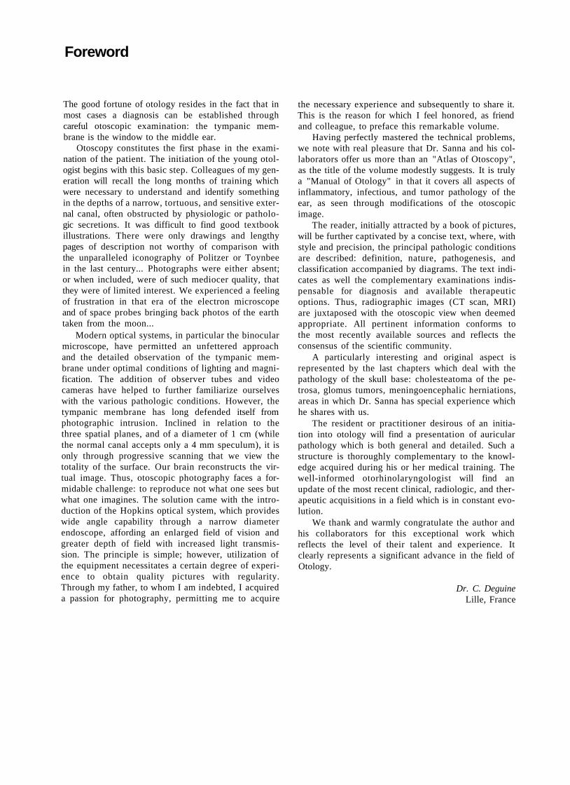
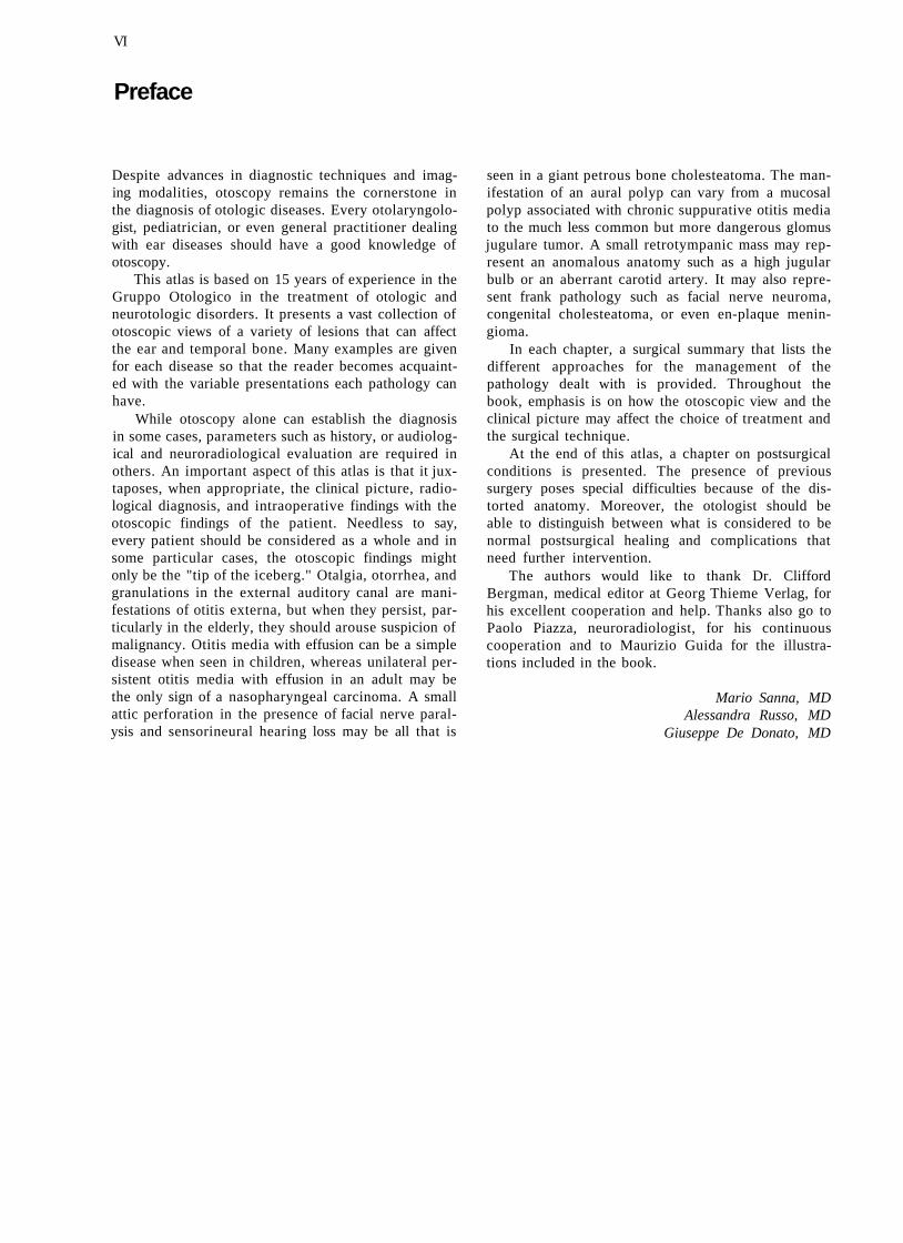
The use of a rigid 0° 6-cm endoscope (1215AA-Storz, Fig. 1.3) connected to a video system enables the patient to see the pathology involving his/her ear (Figs. 1.4 and 1.5 show the Endovision Telecam SL 20212001 and the Xenon Light Source 615-Storz). With the help of a video printer connected to the monitor, instant photos of the pathology can be obtained. The rigid 30° endoscope allows evaluation of attic retraction pockets, the extent of which cannot always be determined using the microscope or the 0° endoscope (Fig. 1.6 shows a series of rigid endoscopes -Storz).
During the last few years, instant photography has also been used in the operating room. A copy of the important steps of the operation is given to the patient while another copy is kept in the patient's chart. The patient is also photographed during the follow-up visit. Thus, for each patient pre-, intra-, and postoperative photographic documentation is obtained.
All the photos in this book were obtained with an Olympus OM 40 camera mounted to the endoscope with a Storz 593-T2 objective. The focus is adjusted to infinity and the diaphragm to 140. We use the TTL-Computer-Flash-Unit Model 600 BA Storz (Fig. 1.7). The film used is a Kodak Ektachrome 64T Professional Film (Tungsten).

Methods of Otoscopy
Fig. 1.6 In all the cases, the examiner sits to the side of the
patient whose head is slightly tilted towards the contralateral side. The examiner holds the camera attached to the endoscope with his right hand. With the ring and middle finger of the left hand, the examiner pulls the patient's auricle backwards and outwards to straighten the external auditory canal. The endoscope is advanced over the index finger of the examiner's left hand into the patient's external auditory canal. In this manner, any undue injury to the external auditory canal is prevented (Fig. 1.8).
Fig. 1.8
4
2 The Normal Tympanic Membrane
• Anatomy
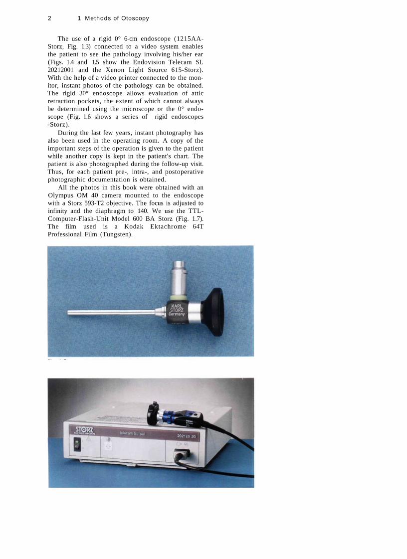
The tympanic membrane forms the major part of the lateral wall of the middle ear (see Figs. 2.1-2.3). It is thin, resistant, semitransparent, has a pearly gray color, and is cone-like. The apex of the membrane lies at the umbo, which corresponds to the lowest part of the han
dle of the malleus. Most of the membrane circumference is thickened to form a fibrocartilaginous ring, the tympanic annulus, which sits in a groove in the tympanic bone called the tympanic sulcus. The fibrocartilaginous ring is deficient superiorly. This deficiency is known as the notch of Rivinus. The anterior and posterior malleolar folds extend from the short process of
Figure 2.1 Right ear. Normal tympanic membrane. 1 = pars flaccida; 2 = short process of the malleus; 3 = handle of the malleus; 4 = umbo; 5 = supratubal recess; 6 = tubal orifice; 7 = hypotympanic air cells; 8 = stapedius tendon; c = chorda tympani; I = incus; P = promontory; o = oval window; R = round window; T = tensor tympani; A = annulus.
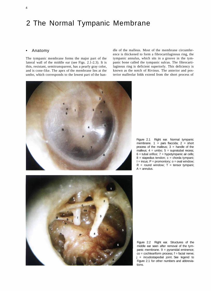
Figure 2.2 Right ear. Structures of the middle ear seen after removal of the tympanic membrane. 9 = pyramidal eminence; co = cochleariform process; f = facial nerve; j = incudostapedial joint. See legend to Figure 2.1 for other numbers and abbreviations.

Normal Otoscopy
Normal Otoscopy
Figure 2.3 Right ear. Division of the tympanic membrane into four quadrants: A.S. = anterosuperior; A.I. = anteroinferior; P.S. = posterosuperior; P.I. = posteroinferior. This division facilitates the description of different pathologic affections of the tympanic membrane.
the malleus to the tympanic sulcus, thus forming the inferior limit of the pars flaccida of Sharpnell's membrane. The membrane forms an obtuse angle with the posterior wall of the external auditory canal. It also forms an acute angle with the anterior wall of the canal. It is important to respect this acute angulation in the myringoplasty operation to maintain as much as possible the vibratory mechanism of the tympanic membrane and hence ensure maximum hearing improvement.
The external surface of the tympanic membrane is innervated by the auriculotemporal nerve and the auricular branch of the vagus nerve, whereas the inner surface is supplied by Jacobson's nerve, a branch of the glossopharyngeal nerve.
The blood supply is derived from the deep auricular and anterior tympanic arteries. Both are branches of the maxillary artery.
• Histology
The tympanic membrane consists of three layers: an outer epithelial layer continuous with the skin of the external auditory canal, a middle fibrous layer or lamina propria, and an inner mucosal layer continuous with the lining of the tympanic cavity.
The epidermis or outer layer is divided into the stratum corneum, the stratum granulosum, the stratum spinosum, and the stratum basale, which is the deepest layer that rests on the basement membrane.
The lamina propria is characterized by the presence of collagen fibers. In the pars tensa, these fibers are arranged in two basic layers: an outer radial layer that originates from the inferior part of the handle of the malleus and inserts in the annulus, and an inner circular layer that originates primarily from the short process of the malleus. Such a distinct arrangement, however, is absent in the pars flaccida.
The mucosal layer is formed mainly of a simple cuboidal or columnar epithelium. The free surface of the cells possesses numerous microvilli.
Figure 2.4 Left ear. Normal tympanic membrane. Note the acute angle formed between the tympanic membrane and the anterior wall of the external auditory canal. The pars tensa with the short process of the handle of the malleus, the umbo, the cone of light, the annulus, and the pars flaccida are seen. Note also the presence of early exostosis in the superior wall of the external auditory canal.
Figure 2.5 Right ear. Normal tympanic membrane. In this case, the drum is very thin and transparent. The handle and short process of the malleus as well as the umbo and cone of light are well visualized. Through the transparent tympanic membrane, the region of the oval window, the long process of the incus, the posterior arc of the stapes, the incudostape-dial joint, the round window, and the promontory can be distinguished. Anteriorly, at the region of the eustachian tube, the tensor tympani canal and the supratubaric recess can be observed.
6 2 The Normal Tympanic Membrane
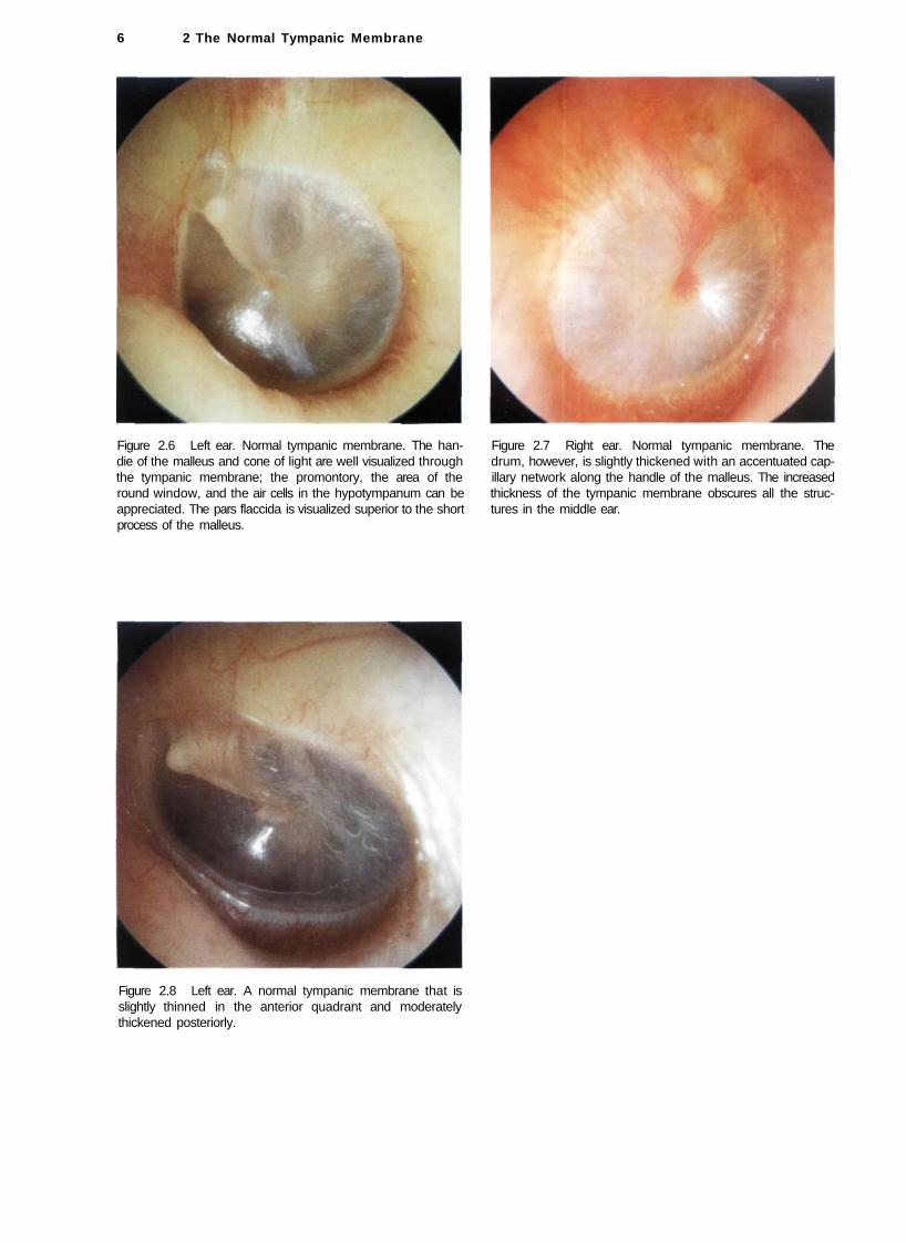
Figure 2.6 Left ear. Normal tympanic membrane. The han- Figure 2.7 Right ear. Normal tympanic membrane. The die of the malleus and cone of light are well visualized through drum, however, is slightly thickened with an accentuated cap-the tympanic membrane; the promontory, the area of the illary network along the handle of the malleus. The increased round window, and the air cells in the hypotympanum can be thickness of the tympanic membrane obscures all the struc-appreciated. The pars flaccida is visualized superior to the short tures in the middle ear. process of the malleus.
Figure 2.8 Left ear. A normal tympanic membrane that is slightly thinned in the anterior quadrant and moderately thickened posteriorly.

7
3 Diseases Affecting the External Auditory Canal
• Exostosis and Osteoma
Exostoses are defined as new bony growths in the osseous portion of the external auditory canal. They are usually multiple, bilateral, and are commonly sessile. They vary in shape, being either round, ovoid, or oblong. The condition is caused by periostitis secondary to exposure to cold water. This explains the high incidence of exostoses among divers and cold-water bathers. Histologically, they are formed from parallel layers of newly-formed bone. It is postulated that the periosteum stimulates an osteogenic reaction with each exposure to cold water, thus causing this stratification.
When exostoses are small they are asymptomatic. Large lesions, however, can occlude the external auditory canal and lead to conductive hearing loss or reten
tion of wax and debris with subsequent otitis externa. In such cases, and in cases in which a hearing aid is to be fitted, surgical removal of exostoses is indicated. In some cases, surgery is technically difficult and special care is taken to preserve the skin of the external auditory canal. Other structures at risk are the tympanic membrane and ossicular chain medially, the temporomandibular joint anteriorly, and the third segment of the facial nerve posteroinferiorly. A postauricular incision is preferred because it allows good exposure and proper replacement of the skin of the external auditory canal to prevent postoperative scarring and stenosis.
Osteoma is a true benign neoplasm of the bone of the external auditory canal, usually unilateral and pedunculated. Histologically, it can be differentiated from exostosis by the absence of the laminated growth pattern.
Figure 3.1 Right ear. Small exostosis originating from the superior wall of the external auditory canal. Anterosuperiorly, another exostosis is seen in the early phase of formation.
Figure 3.2 Right ear. A small asymptomatic exostosis of the superior wall of the external auditory canal is observed. A hump of the anterior wall precludes adequate visualization of the entire tympanic membrane.

3 Diseases Affecting the External Auditory Canal
Figure 3.3 Right ear. Osseous neoplasm of the external auditory canal. In this case, given the pedunculated narrow base, an osteoma is a more probable diagnosis. This was confirmed by pathological examination of the removed specimen. Ample bone removal is performed in such cases to avoid recurrence.
Figure 3.4 Exostosis of the superior wall of the left external auditory canal. The lesion prevents complete visualization of the tympanic membrane.
Figure 3.5 Same patient, right ear. Two exostoses are present in the superior wall of the external auditory canal. In addition, the anterosuperior wall shows an additional exostosis. The lesions allow only a limited view of the central part of the tympanic membrane. In this case, a regular follow-up and evaluation is necessary because further growth of the lesion could lead to accumulation of debris and cerumen, necessitating surgical intervention.
Figure 3.6 Right ear. Exostosis of the posterior superior wall of the external auditory canal that precludes visualization of the pars flaccida. A bony hump is also present in the anterior wall of the canal. In such a case, it is useful to photograph the ear for further follow-up within 1-2 years.

Figure 3.7a Left ear. Obstructing exostosis that causes subtotal occlusion of the external auditory canal. The patient complains of hearing loss and frequent episodes of otitis externa secondary to retention of water and debris inside the canal. A canalplasty under local anesthesia is indicated to restore the size of the external canal.
Figure 3.8 Obstructing exostosis of the external auditory canal resulting in otitis externa due to accumulation of squamous debris inside the canal. Surgery is essential both to avoid the formation of cholesteatoma and to improve hearing.
Exostosis and Osteoma 9
Figure 3.7b Computed tomography (CT) of the same case. The bony external canal is particularly narrowed.
Summary
Surgery in cases of exostosis is indicated only in cases with obstructing stenosis with or without hearing loss but with frequent otitis externa due to retention of debris. Surgery can be performed under local anesthesia, preferably using a postauricular incision. This approach allows excellent exposure of the whole meatus, thus minimizing the risk of injury to the tympanic membrane. In addition, it enables the surgeon to preserve the canal skin, thereby avoiding postoperative cicatricial stenosis. After dissecting the posterior limb, the flap is retained by the prongs of the self-retaining retractor. The skin of the anterior wall is incised medial to the tragus and is dissected in a lateral-to-medial direction. While drilling the exostosis, the skin of the canal is protected using an aluminum sheet (the cover of surgical sutures). Osteoma can be removed by using a curette. In case of recurrence, a wide drilling of the bone around its base is also indicated.

3 Diseases Affecting the External Auditory Canal 10
• Furunculosis
Furunculosis is pustular folliculitis caused by staphylococcal infection of a hair follicle. Infection occurs as a result of microabrasion or of decreased immunity, as in diabetics. It is characterized by severe pain. A tender swelling is seen in the cartilaginous part of the external auditory canal which may have a central necrotic part.
Figure 3.9 A furuncle almost totally occluding the meatus. Pain is caused by distention of the richly innervated skin. A central necrotic part is seen.
• Myringitis and Meatal Stenosis
Myringitis is an inflammatory process that affects the tympanic membrane. Three forms are recognized: acute myringitis, bullous myringitis, and myringitis granulomatosa.
Acute myringitis is usually seen in association with infection of the external ear (otitis externa) or middle ear (otitis media). It is characterized by hyperemia and thickening of the tympanic membrane, as well as the presence of purulent secretions (Fig. 3.10). Therapy consists of administration of general and/or local antibiotics and local steroids.
Figure 3.10 Left ear. The tympanic membrane is characterized by thickening and hyperemia. In this case, the skin of the external auditory canal is also hyperemic. The tympanic membrane seems lateralized.

Myringitis and Meatal Stenosis 11
Bullous myringitis is commonly associated with viral upper respiratory tract infection. It is characterized by the presence of bullae filled with sero-sanguineous fluid. The bullae are located between the outer and middle layers of the tympanic membrane. The patient complains of otalgia and hearing loss. Therapy consists of antibiotics and steroids (Figs. 3.11, 3.12).
In granulomatous myringitis, the outer epidermic layer of the tympanic membrane as well as the adjacent skin of the external auditory canal are replaced by granulation tissue. It is generally seen in patients suffering from frequent episodes of otitis externa. In some cases, it may ultimately lead to stenosis of the most medial part of the external auditory canal. It can usually be cured, however, by removing the granulations in the outpatient clinic using the microscope. This is followed by the administration of local steroid drops for nearly 1 month. In refractory cases, however, surgery in the form of canalplasty with free skin graft is necessary.
Figure 3.11 Left tympanic membrane with a large bulla anterior to the malleus and a smaller one posterior to it.
Figure 3.12 Right tympanic membrane with a large bulla occupying the entire surface of the membrane. The malleus is not visible.
Figure 3.13 Granulomatous myringitis. The granulomatous tissue has replaced the external skin layer of the tympanic membrane and part of the anterior wall of the external canal. This case was treated by removal of the granulation tissue under local anesthesia in the outpatient clinic. Local steroid drops were then administered for 1 month.

12 3 Diseases Affect ing the External Audi tory Canal
Figure 3.14 Postinflammatory stenosis of the right external auditory canal of a 68-year-old woman. The patient complained of bilateral continuous otorrhea and hearing loss of 3 years' duration. The otorrhea in the left ear stopped 2 months before presentation. The granulations over the tympanic membrane were removed in the outpatient clinic. A cellophane sheet was inserted into the external auditory canal to avoid the reformation of stenosis. Local steroid drops were
administered for 1 month. On follow-up, stenosis was already resolved and the granulation tissue in the external auditory canal was completely replaced by healthy skin.
Figure 3.15 CT of the same case. The bony walls of the external auditory canal are intact. The pathologic skin occupies the lumen of the external auditory canal.
Figure 3.16 Same patient, left ear (see also CT in Fig. 3.18). A canalplasty was performed on this side. After having removed the granulation tissue, myringoplasty and canalplasty were performed. Next, the meatal flaps were repositioned.
Figure 3.17 This CT scan demonstrates a similar lesion on the contralateral side.
Myringitis and Meatal Stenosis 13
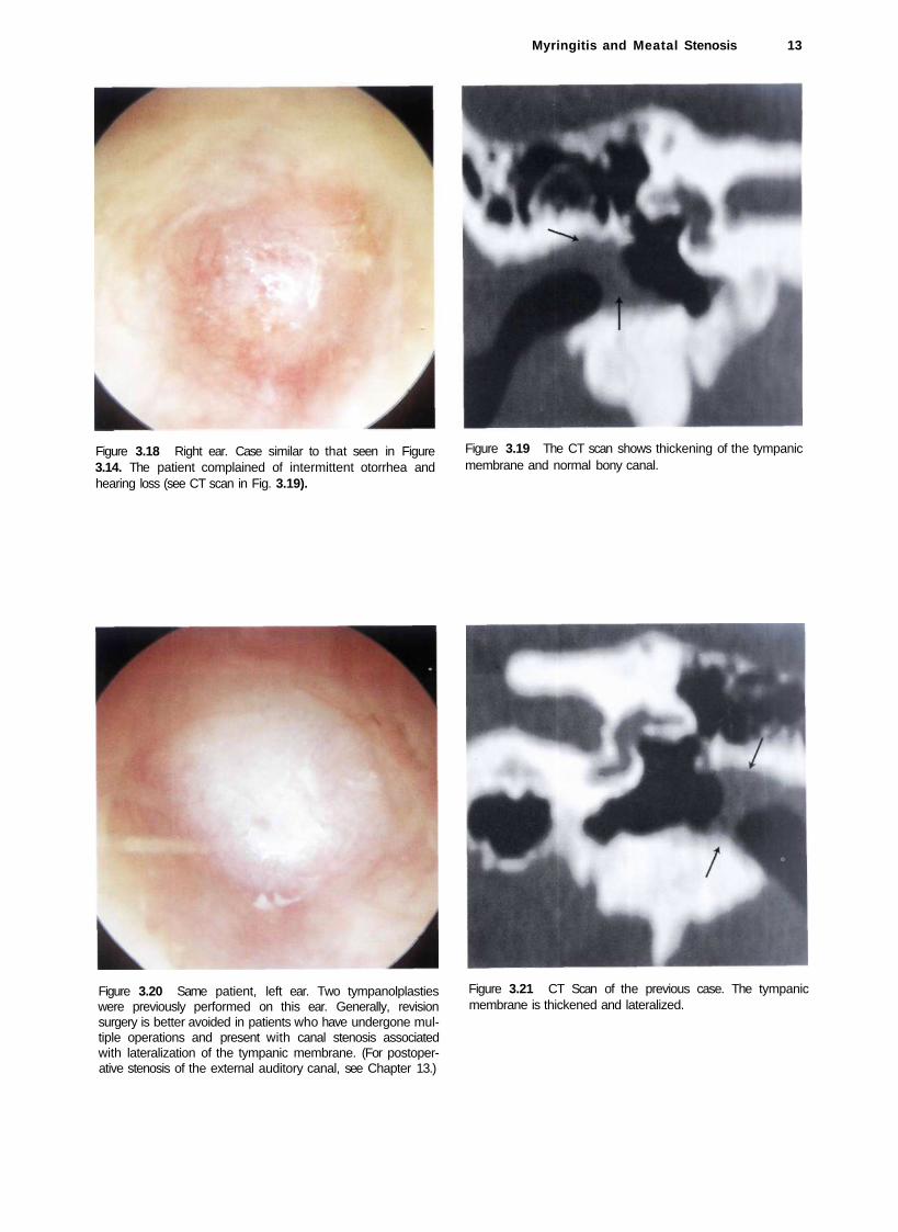
Figure 3.18 Right ear. Case similar to that seen in Figure 3.14. The patient complained of intermittent otorrhea and hearing loss (see CT scan in Fig. 3.19).
Figure 3.19 The CT scan shows thickening of the tympanic membrane and normal bony canal.
Figure 3.20 Same patient, left ear. Two tympanolplasties were previously performed on this ear. Generally, revision surgery is better avoided in patients who have undergone multiple operations and present with canal stenosis associated with lateralization of the tympanic membrane. (For postoperative stenosis of the external auditory canal, see Chapter 13.)
Figure 3.21 CT Scan of the previous case. The tympanic membrane is thickened and lateralized.
14 3 Diseases Affecting the External Auditory Canal
Summary
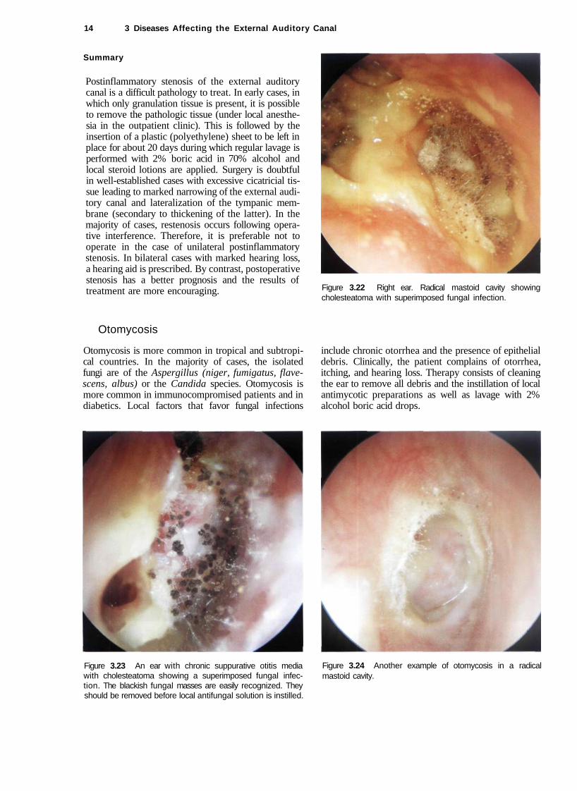
Postinflammatory stenosis of the external auditory canal is a difficult pathology to treat. In early cases, in which only granulation tissue is present, it is possible to remove the pathologic tissue (under local anesthesia in the outpatient clinic). This is followed by the insertion of a plastic (polyethylene) sheet to be left in place for about 20 days during which regular lavage is performed with 2% boric acid in 70% alcohol and local steroid lotions are applied. Surgery is doubtful in well-established cases with excessive cicatricial tissue leading to marked narrowing of the external auditory canal and lateralization of the tympanic membrane (secondary to thickening of the latter). In the majority of cases, restenosis occurs following operative interference. Therefore, it is preferable not to operate in the case of unilateral postinflammatory stenosis. In bilateral cases with marked hearing loss, a hearing aid is prescribed. By contrast, postoperative stenosis has a better prognosis and the results of treatment are more encouraging. Figure 3.22 Right ear. Radical mastoid cavity showing
cholesteatoma with superimposed fungal infection.
Otomycosis
Otomycosis is more common in tropical and subtropical countries. In the majority of cases, the isolated fungi are of the Aspergillus (niger, fumigatus, flave-scens, albus) or the Candida species. Otomycosis is more common in immunocompromised patients and in diabetics. Local factors that favor fungal infections
include chronic otorrhea and the presence of epithelial debris. Clinically, the patient complains of otorrhea, itching, and hearing loss. Therapy consists of cleaning the ear to remove all debris and the instillation of local antimycotic preparations as well as lavage with 2% alcohol boric acid drops.
Figure 3.23 An ear with chronic suppurative otitis media with cholesteatoma showing a superimposed fungal infection. The blackish fungal masses are easily recognized. They should be removed before local antifungal solution is instilled.
Figure 3.24 Another example of otomycosis in a radical mastoid cavity.
Cholesteatoma of the External Auditory Canal 15
Eczema
Eczema is a dermo-epidermal process of reactive nature resulting from local or general factors. Local factors include allergy, topical medical preparations, or cosmetics, whereas general factors include hepatic or gastrointestinal dysfunction. It manifests by itching, a bur-ning sensation, vesication, and sometimes serous otorrhea. Treatment consists of discontinuation the suspected causative irritant, correction of the systemic disturbances, as well as lavage with boric acid with alcohol and steroid lotion.
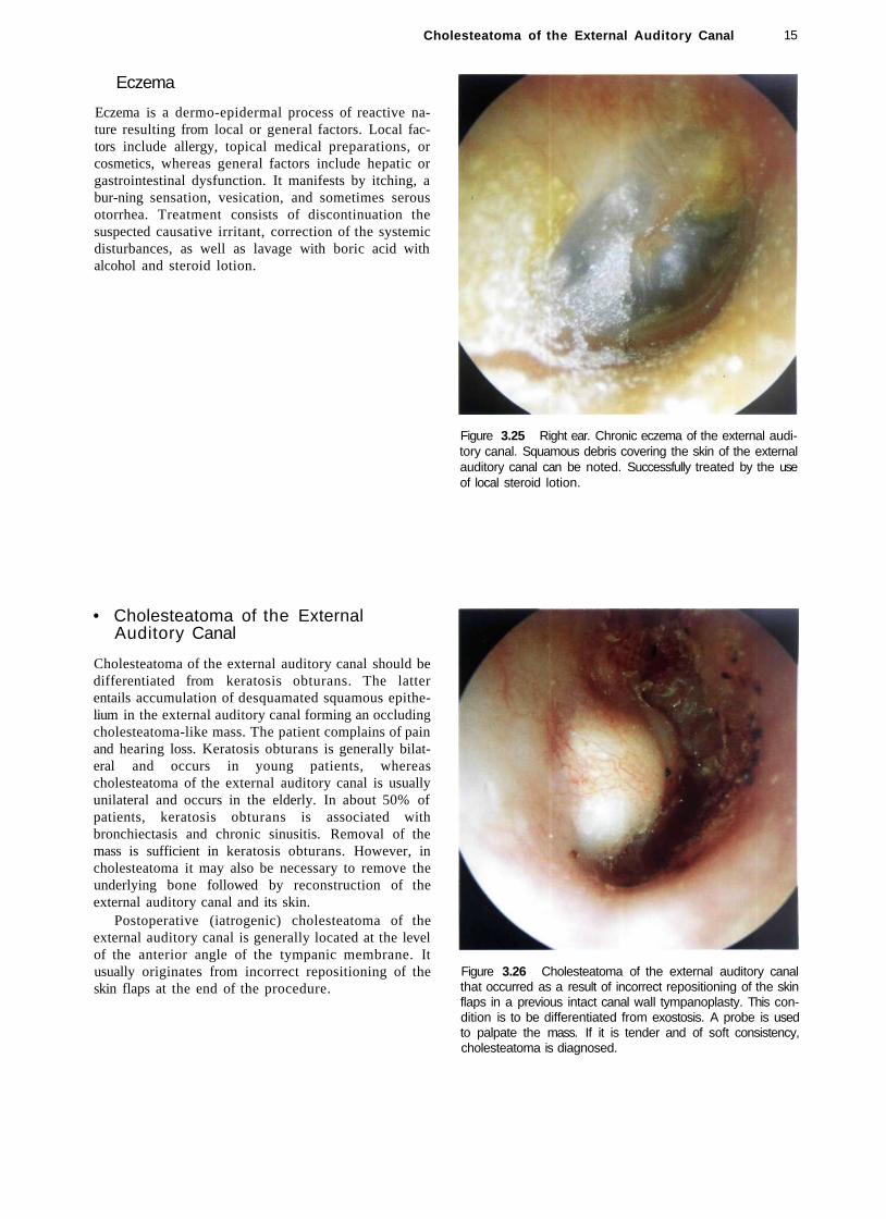
Figure 3.25 Right ear. Chronic eczema of the external auditory canal. Squamous debris covering the skin of the external auditory canal can be noted. Successfully treated by the use of local steroid lotion.
• Cholesteatoma of the External Auditory Canal
Cholesteatoma of the external auditory canal should be differentiated from keratosis obturans. The latter entails accumulation of desquamated squamous epithelium in the external auditory canal forming an occluding cholesteatoma-like mass. The patient complains of pain and hearing loss. Keratosis obturans is generally bilateral and occurs in young patients, whereas cholesteatoma of the external auditory canal is usually unilateral and occurs in the elderly. In about 50% of patients, keratosis obturans is associated with bronchiectasis and chronic sinusitis. Removal of the mass is sufficient in keratosis obturans. However, in cholesteatoma it may also be necessary to remove the underlying bone followed by reconstruction of the external auditory canal and its skin.
Postoperative (iatrogenic) cholesteatoma of the external auditory canal is generally located at the level of the anterior angle of the tympanic membrane. It usually originates from incorrect repositioning of the skin flaps at the end of the procedure.
Figure 3.26 Cholesteatoma of the external auditory canal that occurred as a result of incorrect repositioning of the skin flaps in a previous intact canal wall tympanoplasty. This condition is to be differentiated from exostosis. A probe is used to palpate the mass. If it is tender and of soft consistency, cholesteatoma is diagnosed.

16 3 Diseases Affecting the External Auditory
Figure 3.27 A case similar to that in Figure 3.26. The mass originating from the posterior canal wall inhibits the normal process of epithelial migration towards the outside.
Figure 3.29 Same patient, a few months later. Note the bone erosion caused by the cholesteatoma.
Canal
Figure 3.28 Cholesteatoma of the inferior wall of the left external auditory canal being removed in the outpatient clinic. In this case, the squamous debris led to erosion of the underlying bone.
Figure 3.30 A case similar to the that in Figure 3.28. The cholesteatoma occupies more than half of the external auditory canal and is in contact with the tympanic membrane. The CT scan (Fig. 3.31) demonstrates partial erosion of the underlying bone.

Carcinoid Tumors 17
Figure 3.31 CT scan of the same case, coronal view. The cholesteatoma is clearly seen in the anteroinferior portion of the external auditory canal with partial erosion of the underlying bone.
Summary
Postoperative (iatrogenic) cholesteatoma can almost always be removed in the outpatient clinic under local anesthesia using an endomeatal approach. The sac is opened and the cholesteatoma is aspirated. It is advisable to insert a plastic sheet in the external auditory canal for about 3 weeks to prevent the formation of adhesions that could lead to reformation of the cholesteatoma pearl. Cholesteatoma of the external auditory canal should be surgically removed using a postauricular approach. A wide drilling of the floor of the canal is mandatory to avoid recurrences.
Pathologies Extending to the External Auditory Canal
Some middle ear pathologies can extend into the external auditory canal (e.g., cholesteatomas, glomus tumors, meningiomas, carcinoid tumors, and histiocytosis X). These cases are discussed here to underline the importance of their inclusion in the differential diagnosis of "polypi" in the external auditory canal. Moreover, taking a biopsy of these polypi in the outpatient clinic without proper radiological study is sometimes hazardous. For a detailed discussion of these pathologies, the reader is referred to the relevant chapters.
Carcinoid Tumors
A carcinoid tumor is an adenomatous neuroendocrinal tumor of ectodermal origin. It has the same histologic and histochemical characteristics as other carcinoid tumors that involve different parts of the body. A carcinoid tumor is suspected whenever an adenomatous tumor of the middle ear has acinic or trabecular histologic features. The diagnosis is confirmed by electron microscopy and immunohistochemistry to demonstrate the presence of serotonin and argyrophilic granules. Surgical removal is indicated. To avoid recurrence, removal of the whole tumor together with the attached ossicular chain is essential.
Figure 3.32 This patient complained of hearing loss in the left ear and otalgia of 3 months' duration. Otoscopy revealed a mass occupying the external auditory canal and originating from its anterosuperior region. The inferior part of the tympanic membrane, which is the only visible part, appears whitish due to the presence of a mass in the middle ear. The audiogram (Fig. 3.33) revealed the presence of an ipsilateral conductive hearing loss. The tympanogram was type B. CT scan (Figs. 3.34, 3.35) demonstrated the presence of an iso-intense soft-tissue mass occupying the middle ear and mastoid with extension into the external auditory canal. No erosion of the ossicular chain, nor of the intercellular septae of the mastoid air cells, was noted. Intraoperatively, a glandular-like tissue was found and a frozen section obtained. The biopsy, confirmed by immmunohistochemical and electron microscopic studies, proved the presence of a carcinoid tumor. A tympanoplasty was performed with total removal of the pathology and the involved malleus and incus.

18 3 Diseases Affecting the External Auditory Canal
125 250 500 1K 2K 4K 8K 16KHZ
Figure 3.33 The audiogram shows the presence of significant ipsilateral conductive hearing loss.
Figure 3.34 The CT scan demonstrates a soft-tissue mass occupying the middle ear with extrusion through the tympanic membrane.
Summary
Carcinoid tumors of the middle ear are very rare. They are considered a subgroup of the adenomatous tumors of the middle ear. Clinically, they manifest as hearing loss, tinnitus, aural fullness, facial nerve paresis, vertigo, and otalgia. These tumors require a functional surgery that entails removal of the tympanic membrane and ossicular chain together with the mass. The tympanic membrane is grafted at the same stage, whereas the ossicular chain is reconstructed at a second stage. This strategy ensures that the condition is completely cured.
Figure 3.35 CT scan, axial view. Presence of glue in the mastoid cells without erosion of the intercellular septae.
• Histiocytosis X
Histiocytosis X 19
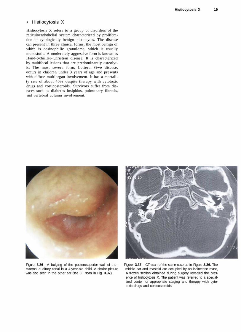
Histiocytosis X refers to a group of disorders of the reticuloendothelial system characterized by proliferation of cytologically benign histiocytes. The disease can present in three clinical forms, the most benign of which is eosinophilic granuloma, which is usually monostotic. A moderately aggressive form is known as Hand-Schiiller-Christian disease. It is characterized by multifocal lesions that are predominantly osteolytic. The most severe form, Letterer-Siwe disease, occurs in children under 3 years of age and presents with diffuse multiorgan involvement. It has a mortality rate of about 40% despite therapy with cytotoxic drugs and corticosteroids. Survivors suffer from diseases such as diabetes insipidus, pulmonary fibrosis, and vertebral column involvement.
Figure 3.36 A bulging of the posterosuperior wall of the Figure 3.37 CT scan of the same case as in Figure 3.36. The external auditory canal in a 4-year-old child. A similar picture middle ear and mastoid are occupied by an isointense mass, was also seen in the other ear (see CT scan in Fig. 3.37). A frozen section obtained during surgery revealed the pres
ence of histiocytosis X. The patient was referred to a specialized center for appropriate staging and therapy with cytotoxic drugs and corticosteroids.

20 3 Diseases Affecting the External Auditory Canal
Other Pathologies
Figure 3.38 Polyp in the external canal in a child presenting with continuous otorrhea and hearing loss. A CT scan (Fig. 3.39) shows the presence of a soft-tissue mass eroding the intercellular septae of the mastoid and the ossicular chain, suggestive of cholesteatoma. This was confirmed during surgery.
Figure 3.39 CT scan, axial view. The entire mastoid is occupied by a soft-tissue mass. The intercellular septae of the mastoid and the ossicular chain are absent.
Figure 3.40 Another example of chronic suppurative otitis media with cholesteatoma that manifests with an aural polyp. Though cholesteatoma presents frequently in this manner, it is absolutely essential to abstain from taking a biopsy of the polyp in the outpatient clinic without performing a CT scan of the temporal bone (see Fig. 3.41).
Figure 3.41 The otoscopic view is very similar to that in Figure 3.40. In this case, however, the diagnosis is that of an en-plaque supratentorial meningioma. An outpatient polypectomy in this case might lead to excessive bleeding (see MRI, Figs. 3.42 and 3.43).

Other Pathologies 21
Figure 3.42 MRI with gadolinium enhancement, axial view. The tumor (arrows) is located in the temporal fossa and reaches the area of the petrous apex and Meckel's cavity.
Figure 3.43 MRI with gadolinium, coronal view. The meningioma displaces the temporal lobe upwards (arrows); pathognomonic tails of the dura are visible.
Figure 3.44 Left ear. Glomus jugulare tumor with extension into the external auditory canal. A biopsy of this lesion might lead to severe and often difficult-to-control hemorrhage.
Figure 3.45 Left ear. Another example of a glomus tumor.

22 3 Diseases Affecting the External Auditory Canal
Figure 3.46 Pulsating neoplasm in the external auditory canal. MR I (Fig. 3.47) revealed the presence of a glomus jugu-lare tumor involving the vertical internal carotid artery.
Figure 3.47 MRI of the same case. A glomus jugulare tumor engulfing the vertical portion of the internal carotid artery is clearly visible.
• Carcinoma of the External Auditory Canal
Basal cell carcinoma is more frequent in the auricle, particularly in subjects with long exposure to the sun. On the other hand, squamous cell carcinoma accounts for about three quarters of invasive tumors of the external auditory canal and the middle ear. In about 11% of cases, cervical lymph node metastases are present at the time of diagnosis. The most common symptoms include otorrhea, otalgia, hearing loss, facial nerve paralysis, and vertigo. An accurate microscopic examination is important for proper evaluation of the lesion extension. Frequently, an exfoliative lesion is noted, whereas an ulcer is present in other cases. Carcinoma should be suspected in the case of a persistent otitis externa characterized by pain and otorrhea that does not resolve adequately with medical treatment. A biopsy of the lesion will clear any doubts. It is important to perform an accurate examination of the upper deep cervical, postauricu-lar, and parotid lymph nodes (anterior extension of the tumor). The cranial nerves are also evaluated. The facial nerve is the most frequently involved. Involvement of the mandibular nerve indicates tumor extension towards the glenoid fossa. A high-resolution CT scan (bone window) is the most important radiological investigation as it permits the evaluation of bone erosion at the level of the external auditory canal and middle ear. MRI with gadolinium allows the evaluation of tumor extension into the soft tissues.
The tumor should be considered to be T3 or T4 if there is infiltration of the posterior or middle cranial fossae, or invasion of the jugular foramen or glenoid fossa. In such cases, whatever the modality of treatment, the prognosis is almost always poor.
Surgery consists of en-bloc removal of the tumor and a trial to include a safety margin of the surrounding healthy tissue in the specimen. Postoperative radiotherapy should be subsequently performed.

Carcinoma of the External Audi tory Canal 23
Figure 3.48 An exfoliative neoplasm that occupies the external auditory canal. The patient complained of otalgia and attacks of bloody otorrhea of 1-month duration. A biopsy was taken and pathologic examination revealed the presence of squamous cell carcinoma. A CT scan (Fig. 3.49) demonstrated erosion of the external auditory canal, particularly its anteroinferior wall, without breaking into the glenoid fossa. En-bloc removal of the tumor was performed, together with a superficial parotidectomy. Radiotherapy was performed postoperatively.
Figure 3.50 Squamous cell carcinoma protruding through the external auditory canal with extension into the glenoid fossa and infiltration of the middle fossa dura (see CT scan, Fig. 3.51 and MRI, Fig. 3.52). Palliative surgery was performed followed by radiotherapy.
Figure 3.49 CT scan demonstrates erosion of the anteroinferior wall of the external auditory canal. The glenoid fossa is not invaded.
Figure 3.51 CT scan. The carcinoma occupies all of the middle ear and the mastoid. The glenoid fossa and the middle fossa plate are eroded.

24 3 Diseases Affecting the External Auditory Canal
Figure 3.52 MRI shows marked anterior extension of the tumor into the infratemporal fossa.
Figure 3.53 Squamous cell carcinoma with posterior extension. The mass ifiltrates the skin of the posterior wall of the external auditory canal (see CT scan, Fig. 3.54) as a result of which en-bloc resection and subsequent radiotherapy were performed.
Figure 3.54 CT scan, axial view. The tumor has eroded the most lateral portion of the posterior bony wall.
Figure 3.55 Nasopharyngeal carcinoma extending into the middle ear and external auditory canal. A polypoid mass infiltrates the tympanic membrane and partially fills the external auditory canal (see CT scan, Fig. 3.56 and MRI, Fig. 3.57). The patient was considered inoperable and was referred to radiotherapy.

Carcinoma of the External Auditory Canal 25
Figure 3.56 The CT scan demonstrates marked infiltration of the nasopharynx, the pterygoid muscles, and the petrous apex.
Figure 3.57 MRI with gadolinium confirms the infiltration.
Summary
A carcinoma arising from the external auditory canal is frequently confused with suppurative otitis. Because of the high incidence of otitis externa and media and because these pathologies are frequently chronic, the diagnosis of carcinoma of the external auditory canal is almost always late. Diagnosis is made by biopsy. A high-resolution CT scan and MRI are necessary for proper evaluation. A high-resolution CT scan determines the osseous erosion caused by the tumor, whereas MRI is superior to CT for the evaluation of soft tissues. MRI shows the presence of dural invasion, intracranial extension, as well as extracranial soft-tissue involvement. Until now there has been no universally accepted system of staging, which is the basis for planning therapy and proper treatment evaluation. Therapy for carcinoma of the external auditory canal is almost always surgical. Various degrees of resection are utilized according to the extent of the pathology. Very small lesions can be managed by excision biopsy with a safety margin and curettage of the underlying bone. Lateral en-bloc petrosectomy is the treatment of choice in the majority of carcinomas of the external auditory canal. It entails excision of the external auditory canal (bone and soft tissues), tympanic membrane, and ossicular chain with preservation of the facial nerve. Anteriorly, bone removal extends up
to the level of the temporomandibular joint. The cavity can be exteriorized or obliterated with abdominal fat and the external auditory canal closed as cul-de-sac. When indicated, the resection can include a superficial parotidectomy, resection of the mandibular condyle, and/or neck dissection. When the tumor has a deeper extension towards the middle ear, en-bloc subtotal resection of the temporal bone is indicated. In such cases, a middle and posterior fossa craniotomy is necessary. Bone removal is performed up to the level of the medial third of the petrous apex and the internal carotid artery. The facial nerve and inner ear are sacrificed. A more extended procedure is total en-bloc resection of the temporal bone entailing, in addition, the sacrifice of the internal carotid artery, closure of the sigmoid sinus and jugular bulb, and in some cases a total parotidectomy and neck dissection.

26
4 Secretory Otitis Media (Otitis Media wi th Effusion
Secretory otitis media is characterized by the presence of middle ear effusion composed of a transudate/exudate of the mucosa of the middle ear cleft that is formed behind an intact tympanic membrane. Classically, the tympanic membrane is retracted, immobile, dark yellowish or bluish, and thickened. At times, it may be transparent with a hairline (liquid level) or air bubbles visible through it.
The causes are generally: eustachian tube obstruction secondary to mucosal edema due to infection (sinusitis, nasopharyngitis) or allergy; extrinsic pressure on the cartilaginous portion of the eustachian tube due to hyperplasia of glandular or lymphoid tissue or, rarely, due to tumors; malfunction of the tubal muscles as in children with cleft palate, or malformation of the tube itself as in Down's syndrome. Other factors that may contribute include: bacteriologic, immunologic, genetic, socioeconomic status, seasonal variation, as well as lack of transmission of specific immunoglobulins in non-breast-fed infants. All these factors cause tubal dysfunction or occlusion leading to negative middle ear pressure due to oxygen absorption by the mucosa of the middle ear cleft. Normally, the tendency of the tubal walls to collapse at the level of the isthmus can be overcome by an increase in the nasopharyngeal pressure. A negative middle ear pressure up to -25 mm Hg can be thus corrected. On the other hand, with edema of the tubal mucosa, the same increase in the nasopharyngeal pressure cannot overcome a negative middle ear pressure less than -5 mm Hg.
In children, hyperplasia of the adenoid tissue is the most common predisposing factor, and nasopharyngitis is the most frequent cause of secretory otitis media. In adults, the condition is much less common and the presence of persistent unilateral otitis media with effusion can be due to a nasopharyngeal tumor that occludes the tubal opening, or a neoplasm that compresses or infiltrates the tube along its course.
In cases that do not resolve despite proper medical treatment (nasal and systemic decongestants, mucolytics, and antibiotics) or in cases with persistent conductive hearing loss (see Figs. 4.1 and 4.2), the insertion of a ventilation tube is indicated. In children, adenoidectomy is also performed. Surgery aims at alleviating the conductive hearing loss avoiding the sequelae of otitis media with effusion. Sequelae include recurrent otitis media, tympanosclerosis, adhesive otitis media, retraction pockets with eventual cholesteatoma formation, and in some long-standing cases the formation of cholesterol granuloma (see Chapter 5). In this chapter, some typical cases of otitis media with effusion will be shown. For the surgical
treatment (myringotomy and ventilation tube insertion), the reader is referred to Chapter 13 on postsurgical conditions.
Figure 4.1 Conductive hearing loss. Bone conduction is normal. Air conduction is on an average of 35 dB.
-200 -100 0 +100 +200
Figure 4.2 Tympanogram type B, typical of middle ear effusion.

Secretory Otisis Media (Otisis Media with Effusion) 27
Figure 4.3 Right ear. Secretory otitis media. Air bubbles can be seen anterior to the handle of the malleus and also in the posteroinferior quadrant.
Figure 4.4 Left ear. Secretory otitis media. Middle ear effusion having a reddish color inferiorly and a yellowish color superiorly. In this case, the differential diagnosis includes glomus tympanicum. If doubts still exist after microscopic examination, medical treatment is administered for several weeks and the patient is reexamined.
Figure 4.5 Left ear. Secretory otitis media with an apparently dense transudate that gives the tympanic membrane the characteristic dark yellow color. An air-fluid level can be appreciated at the posterosuperior quadrant. The tympanic membrane is diffusely hyperemic. If the condition is not resolved by medical treatment, a ventilation tube should be inserted.
Figure 4.6 Right ear. The presence of glue in the middle ear leads to bulging of the tympanic membrane. In the posterior quadrant, a thinned area of the drum is visualized through which the yellowish color of the effusion is visible. This area would probably be the site of a future perforation.

28 4 Secretory Otitis Media (Otitis Media with Effusion)
Figure 4.7 Right ear. Seromucoid effusion in the middle ear. Air bubbles can be seen in the anterior quadrants of the tympanic membrane. The patient is a 53-year-old woman who presented with a signs of right otitis media with effusion causing conductive hearing loss and ipsilateral paraesthesia of the maxillary and mandibular nerves, followed by episodes of trigeminal neuralgia and diplopia in the last few months. Computed tomography (CT) scan and magnetic resonance imaging (MRI) with gadolinium (see following figures) revealed
the presence of a tumor (later proven to be a trigeminal neurinoma) with an intra- and extracranial extension. The tumor compressed the eustachian tube and resulted in the middle ear effusion. Total removal of the tumor was performed in a single-stage operation using an infratemporal type B approach with orbitozygomatic extension (Fig. 4.10).
Figure 4.8 MRI, axial view, showing the extension of the giant trigeminal neurinoma.
Figure 4.9 MRI, sagittal view, confirms the intra-extracranial extension of the tumor.
and Figure 4.10 Trigeminal neurinoma removal using an infratemporal type B approach with orbitozygomatic extension.
/

Secretory Otisis Media (Otisis Media with Effusion) 29
Figure 4.11 removal.
Postoperative CT scan showing total tumor Figure 4.12 A different case similar to the one in Figure 4.7. This 64-year-old woman complained of right nasal obstruction and a sensation of right ear fullness of 1 year duration. One month before presentation the patient began to suffer from neuralgic pain in the region of the maxillary nerve. The tympanic membrane looks yellowish due to the presence of middle ear effusion (see following figures).
Figure 4.13 Right nasal cavity, same case. A mass is visualized in the middle meatus. A biopsy proved it to be a neurinoma.
30 4 Secretory Otitis Media (Otitis Media with Effusion)
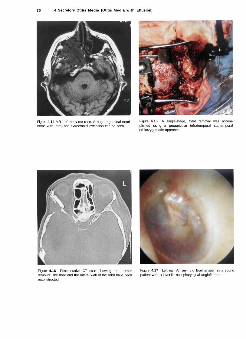
Figure 4.14 MR I of the same case. A huge trigeminal neurinoma with intra- and extracranial extension can be seen.
Figure 4.15 A single-stage, total removal was accomplished using a preauricular infratemporal subtemporal orbitozygomatic approach.
Figure 4.16 Postoperative CT scan showing total tumor removal. The floor and the lateral wall of the orbit have been reconstructed.
Figure 4.17 Left ear. An air-fluid level is seen in a young patient with a juvenile nasopharyngeal angiofibroma.

Secretory Otisis Media (Otisis Media with Effusion) 31
Figure 4.18 MRI of the same case. The angiofibroma occupies the nasopharynx, pterygopalatine fossa, and infratemporal fossa on the left side. Removal was accomplished via an infratemporal fossa approach type C according to Fisch.
Figure 4.19 Left ear. Secretory otitis media. The tympanic membrane is thickened. Catarrhal fluid can be seen through the relatively thinner anteroinferior quadrant.
Figure 4.20 Right ear. Secretory otitis media. The effusion is visible through two thinned areas of the tympanic membrane lying anterior and posterior to the handle of the malleus.
Figure 4.21 Right ear. Secretory otitis media with tympanosclerosis and epitympanic erosion. The tympanic membrane shows areas of tympanosclerosis alternating with areas of atrophy. Glue is present in the middle ear.

32 4 Secretory Otitis Media (Otitis Media
Figure 4.22 Left ear. Otitis media with effusion and a whitish retrotympanic mass in the posterior quadrant at 3 o'clock can be observed. The presence of congenital cholesteatoma was considered in the differential diagnosis. Exploratory tympanotomy showed only "glue" in the middle ear that was particularly dense in the posterior mesotympa-num.
Figure 4.24 MRI of the same case showing a schwannoma of the lower cranial nerves (T).
Effusion)
Figure 4.23 Left ear showing a pulsating air-fluid level in a patient operated 1 year previously to remove a lower cranial nerve neurinoma using a petro-occipital trans-sigmoid approach (POTS) (see preoperative MRI, Fig. 4.24 and postoperative CT scan, Fig. 4.25). The patient complained of a sensation of ear blockage and watery rhinorrhea on leaning forwards. The middle ear is full of cerebrospinal fluid (CSF) passing through open hypotympanic air cells that communicate with the subarachnoid space. The CSF rhinorrhea was treated by obliterating the eustachian tube and middle ear with the temporalis muscle and by closure of the external auditory canal as cul-de-sac.
Figure 4.25 Postoperative CT scan shows the petro-occipital craniotomy and the surgical cavity with preservation of the inner ear.
Secretory Otisis Media (Otisis Media with Effusion)
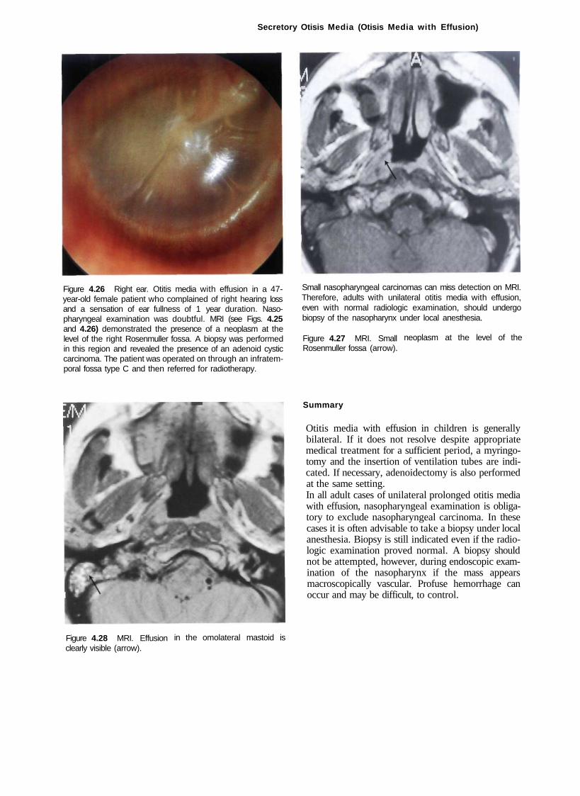
Figure 4.26 Right ear. Otitis media with effusion in a 47-year-old female patient who complained of right hearing loss and a sensation of ear fullness of 1 year duration. Nasopharyngeal examination was doubtful. MRI (see Figs. 4.25 and 4.26) demonstrated the presence of a neoplasm at the level of the right Rosenmuller fossa. A biopsy was performed in this region and revealed the presence of an adenoid cystic carcinoma. The patient was operated on through an infratemporal fossa type C and then referred for radiotherapy.
Small nasopharyngeal carcinomas can miss detection on MRI. Therefore, adults with unilateral otitis media with effusion, even with normal radiologic examination, should undergo biopsy of the nasopharynx under local anesthesia.
Figure 4.27 MRI. Small Rosenmuller fossa (arrow).
neoplasm at the level of the
Summary
Otitis media with effusion in children is generally bilateral. If it does not resolve despite appropriate medical treatment for a sufficient period, a myringotomy and the insertion of ventilation tubes are indicated. If necessary, adenoidectomy is also performed at the same setting. In all adult cases of unilateral prolonged otitis media with effusion, nasopharyngeal examination is obligatory to exclude nasopharyngeal carcinoma. In these cases it is often advisable to take a biopsy under local anesthesia. Biopsy is still indicated even if the radiologic examination proved normal. A biopsy should not be attempted, however, during endoscopic examination of the nasopharynx if the mass appears macroscopically vascular. Profuse hemorrhage can occur and may be difficult, to control.
Figure 4.28 MRI. Effusion clearly visible (arrow).
in the omolateral mastoid is

34
5 Cholesterol Granuloma
Cholesterol granuloma is a histologic term used to describe a foreign body, giant cell reaction to cholesterol crystals, and hemosiderin derived from ruptured erythrocytes. Cholesterol granuloma is thought to arise from obstructed drainage and insufficient aeration of cellular compartments of the temporal bone. This leads to reabsorption of air, negative pressure, mucosal edema, and hemorrhage. It can be present in the middle ear, mastoid, or petrous apex. Generally, patients with tympanomastoid cholesterol granuloma have a long history of recurrent otitis media or otitis media with effusion. They also complain of conductive hearing loss, and on otoscopy the tympanic membrane appears bluish in color. In some cases, where granulation tissue is more prevalent, cholesterol granuloma can present as a retrotympanic reddish-brown mass that may cause bulging of the tympanic membrane, thus mimicking a glomus tumor. In these cases, a computed tomography (CT) scan is sufficient to clear any doubts. A cholesterol granuloma rarely causes bone erosion. On the contrary, bone erosion is characteristic
Figure 5.1 Right ear. Typical blue tympanum caused by cholesterol granuloma. The blue color is due to hemosiderin crystals. The granuloma involves not only the middle ear but generally extends into the mastoid air cells.
of glomus tumors causing destruction of the jugular hypotympanic septum with an irregular "moth-eaten" contour.
In the initial phases, before cholesterol granuloma is formed, it might be sufficient to insert a ventilation tube, thus preventing further development of the granuloma. When the granuloma has already been formed, it is necessary to perform a tympanoplasty with mastoidectomy that opens the intercellular septae with subsequent aeration of the middle ear and mastoid.
Figure 5.2 Blue tympanum caused by cholesterol granuloma. An epitympanic retraction due to eustachian tube dysfunction is also present.
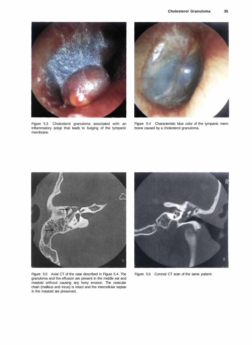
Figure 5.3 Cholesterol granuloma associated with an inflammatory polyp that leads to bulging of the tympanic membrane.
Figure 5.5 Axial CT of the case described in Figure 5.4. The granuloma and the effusion are present in the middle ear and mastoid without causing any bony erosion. The ossicular chain (malleus and incus) is intact and the intercellular septae in the mastoid are preserved.
Cholesterol Granuloma 35
Figure 5.6 Coronal CT scan of the same patient.
Figure 5.4 Characteristic blue color of the tympanic membrane caused by a cholesterol granuloma.

36 5 Cholesterol Granuloma
Figure 5.7 Left ear. A 17-year-old male patient complained of conductive hearing loss of 1 year duration accompanied by left nasal obstruction. Otoscopy revealed the presence of a left cholesterol granuloma. Rhinoscopy showed the presence of a nasopharyngeal swelling that extended into the left nasal cavity. The swelling was suggestive of a juvenile nasopharyngeal angiofibroma.
Figure 5.9 Magnetic resonance imaging (MRI) of the same case, coronal view, showing the extension of the angiofibroma.
Figure 5.8 CT, coronal view. Involvement of the nasopharynx and the sphenoidal sinus.
Figure 5.10 MRI of the same case, sagittal view, showing the extension of the tumor from the ethmoid to the rhinopharynx pushing the soft palate.
Cholesterol Granuloma 37
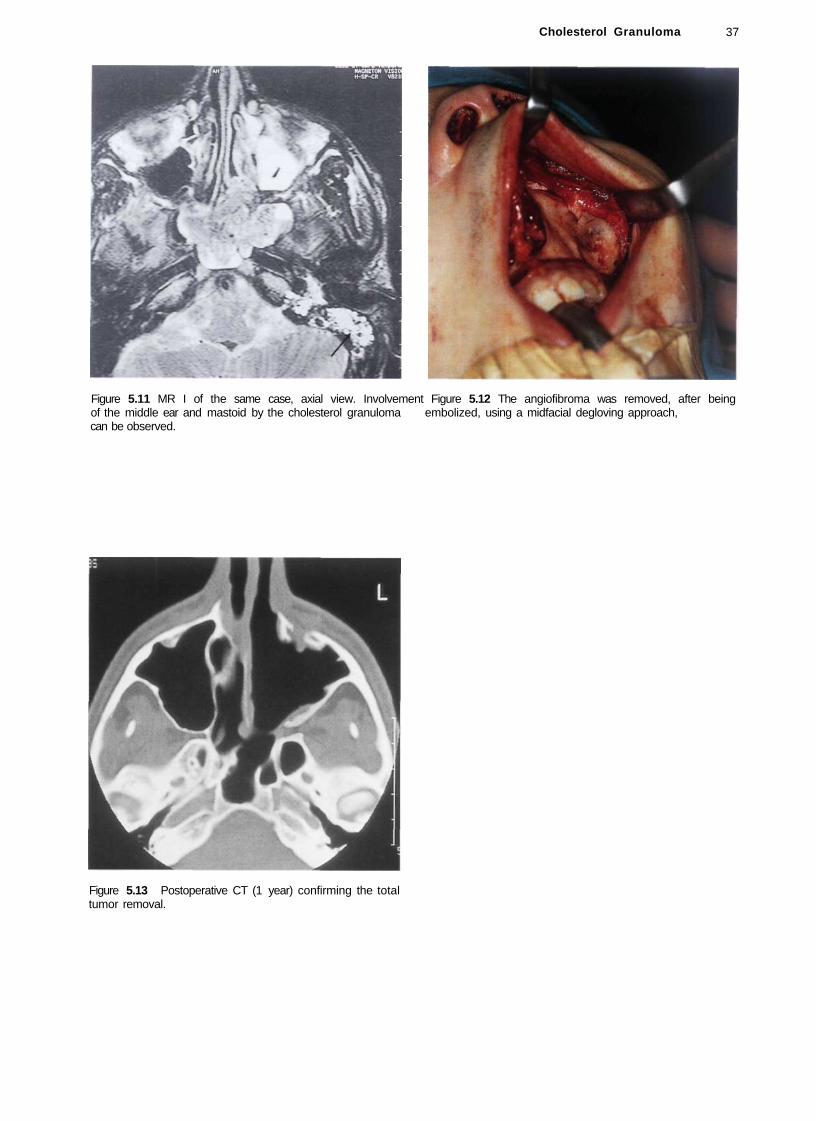
Figure 5.11 MR I of the same case, axial view. Involvement Figure 5.12 The angiofibroma was removed, after being of the middle ear and mastoid by the cholesterol granuloma embolized, using a midfacial degloving approach, can be observed.
Figure 5.13 Postoperative CT (1 year) confirming the total tumor removal.
38
6 Atelectasis, Adhesive Otitis Media
Adhesive otitis media is characterized by complete or partial adhesions between the thin retracted and atrophic pars tensa and the medial wall of the middle ear. Necrosis of the long process of the incus or the stapes' suprastructure can also occur with a resultant natural myringostapedopexy. It should be differentiated from atelectasis and from simple drum retraction in which the tympanic membrane is mobile with the Valsalva or Toynbee maneuvers.
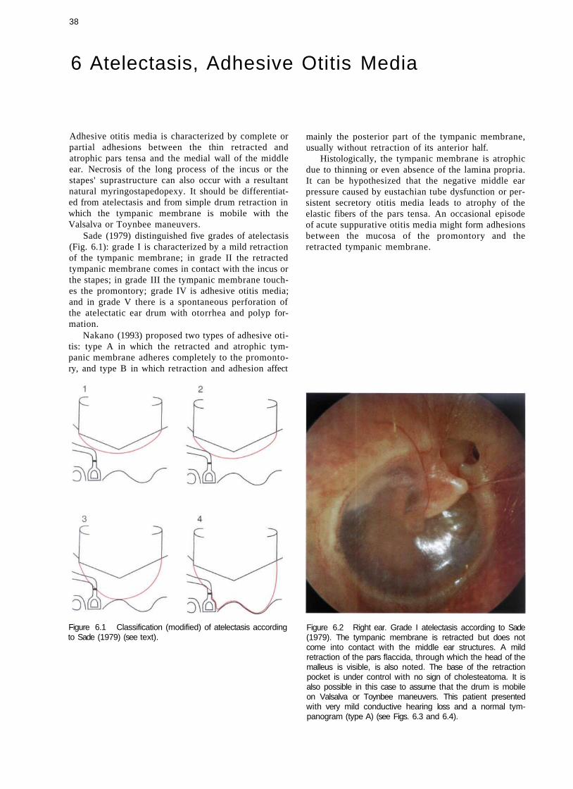
Sade (1979) distinguished five grades of atelectasis (Fig. 6.1): grade I is characterized by a mild retraction of the tympanic membrane; in grade II the retracted tympanic membrane comes in contact with the incus or the stapes; in grade III the tympanic membrane touches the promontory; grade IV is adhesive otitis media; and in grade V there is a spontaneous perforation of the atelectatic ear drum with otorrhea and polyp formation.
Nakano (1993) proposed two types of adhesive otitis: type A in which the retracted and atrophic tympanic membrane adheres completely to the promontory, and type B in which retraction and adhesion affect
Figure 6.1 Classification (modified) of atelectasis according to Sade (1979) (see text).
mainly the posterior part of the tympanic membrane, usually without retraction of its anterior half.
Histologically, the tympanic membrane is atrophic due to thinning or even absence of the lamina propria. It can be hypothesized that the negative middle ear pressure caused by eustachian tube dysfunction or persistent secretory otitis media leads to atrophy of the elastic fibers of the pars tensa. An occasional episode of acute suppurative otitis media might form adhesions between the mucosa of the promontory and the retracted tympanic membrane.
Figure 6.2 Right ear. Grade I atelectasis according to Sade (1979). The tympanic membrane is retracted but does not come into contact with the middle ear structures. A mild retraction of the pars flaccida, through which the head of the malleus is visible, is also noted. The base of the retraction pocket is under control with no sign of cholesteatoma. It is also possible in this case to assume that the drum is mobile on Valsalva or Toynbee maneuvers. This patient presented with very mild conductive hearing loss and a normal tym-panogram (type A) (see Figs. 6.3 and 6.4).
Atelectasis, Adhesive Otitis Media 39
120 125 250 500 1K 2K 4K 8K 16KHz
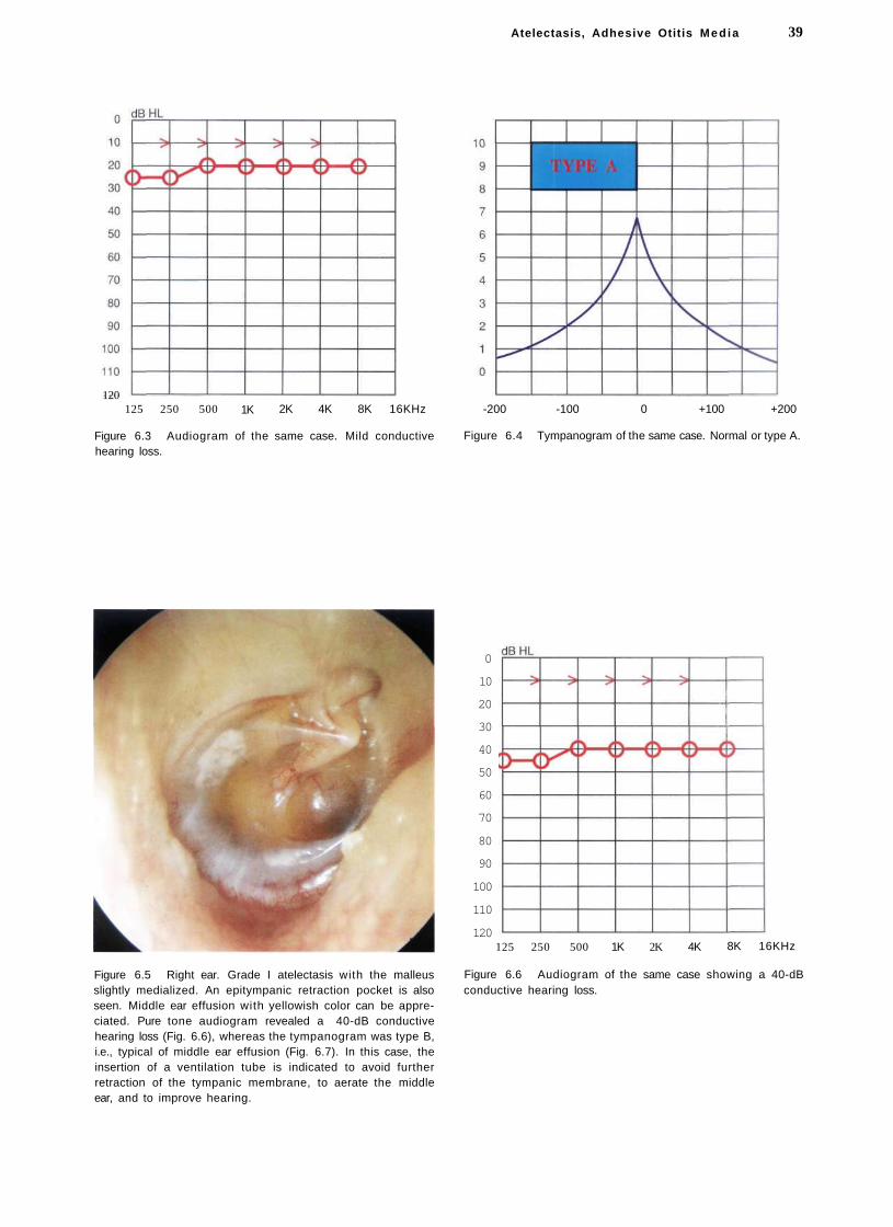
Figure 6.3 Audiogram of the same case. Mild conductive hearing loss.
-200 -100 0 +100 +200
Figure 6.4 Tympanogram of the same case. Normal or type A.
Figure 6.5 Right ear. Grade I atelectasis with the malleus slightly medialized. An epitympanic retraction pocket is also seen. Middle ear effusion with yellowish color can be appreciated. Pure tone audiogram revealed a 40-dB conductive hearing loss (Fig. 6.6), whereas the tympanogram was type B, i.e., typical of middle ear effusion (Fig. 6.7). In this case, the insertion of a ventilation tube is indicated to avoid further retraction of the tympanic membrane, to aerate the middle ear, and to improve hearing.
0
10
20
30
40
50
60
70
80
90
100
110
120 125 250 500 1K 2K 4K 8K 16KHz
Figure 6.6 Audiogram of the same case showing a 40-dB conductive hearing loss.

40 6 Atelectasis, Adhesive Otitis Media
-200 -100 0 +100 +200
Figure 6.7 Tympanogram type B of the same case, typical of middle ear effusion.
Figure 6.8 Right ear. Grade I atelectasis. The tympanic membrane is markedly thinned due to partial resorption of the lamina propria. The incus is seen in transparency. Pure tone audiogram is normal (Fig. 6.9), whereas the tympanogram has a very high compliance (Fig. 6.10). As the tympanic membrane is mobile with the Valsalva maneuver, insertion of a ventilation tube is not indicated.
10 dBHL
10
20
30
40
50
60
70
80
90
100
110
120 125 250 500 1K 2K 4K 8K 16KHz
Figure 6.9 Audiogram of the same case (see text).
10
9
8
7
6
5
4
3
2
1
0
-200 -100 + 100 +200
Figure 6.10 Tympanogram of the same case, type AD according to the classification of Liden-Jerger, 1976 (see text).
10
9
8
7
6
5
4
3
2
1
0
0

Atelectasis, Adhesive Otitis Media 41
Figure 6.11 Left ear. Grade II atelectasis with marked epi-tympanic retraction. The tympanic membrane touches the incus. The malleus is medialized. Air-fluid levels are seen in the anteroinferior quadrant. The insertion of a ventilation tube is necessary to restore normal conditions.
Figure 6.13 Right ear. Grade II atelectasis. The tympanic membrane is very thin due to absence of the fibrous layer. The membrane adheres to the incudostapedial joint and the tensor tympani tendon. Insertion of a ventilation tube is indicated.
Figure 6.12 Right ear. Grade II atelectasis. A condition similar to the previous case but with the onset of thickening of the tympanic membrane.
Figure 6.14 Left ear. Grade III atelectasis. The tympanic membrane touches the promontory and the incus. An air-fluid level and a tympanosclerotic plaque can be seen in the anterior quadrant.

42 6 Atelectasis, Adhesive Otitis Media
Figure 6.15 Left ear. Grade III atelectasis. The thin and atrophic tympanic membrane is in contact with the promontory. Middle ear effusion is seen. A tympanosclerotic plaque is present in the anterosuperior quadrant. The head of the malleus is visible through an epitympanic retraction pocket. The insertion of a ventilation tube is indicated.
Figure 6.17 Left ear. Grade IV atelectasis. The malleus is medialized and adherent to the promontory. The tympanic membrane is atrophic. The epidermal layer of the membrane is adherent to the incudostapedial joint, the promontory, and the round window. A retraction pocket corresponding to the eustachian tube orifice is also seen. Middle ear effusion is present. Insertion of a ventilation tube is indicated.
Figure 6.18 Left ear. Adhesive otitis media. This case represents the long-term sequela of persistent secretory otitis
Figure 6.16 Right ear. Adhesive otitis media or grade IV atelectasis associated with a mild epitympanic retraction pocket. The thin and atrophic tympanic membrane completely covers the promontory. The tympanic membrane retraction has caused erosion of the long process of the incus with a subsequent spontaneous myringostapedopexy As the patient does not complain of hearing loss, surgery is not indicated.
media with chronic eustachian tube dysfunction. The fibrous and mucosal layers of the tympanic membrane were resorbed, whereas the epidermal layer is completely adherent to the medial wall of the middle ear. The promontory, round and oval windows, as well as residues of the ossicular chain are all visible. The handle of the malleus is completely medialized and partially eroded. The long process of the incus is eroded, whereas the stapes suprastructure is completely absent. As the patient does not suffer from otorrhea, surgery is not advised.
Atelectasis, Adhesive Otitis Media 43
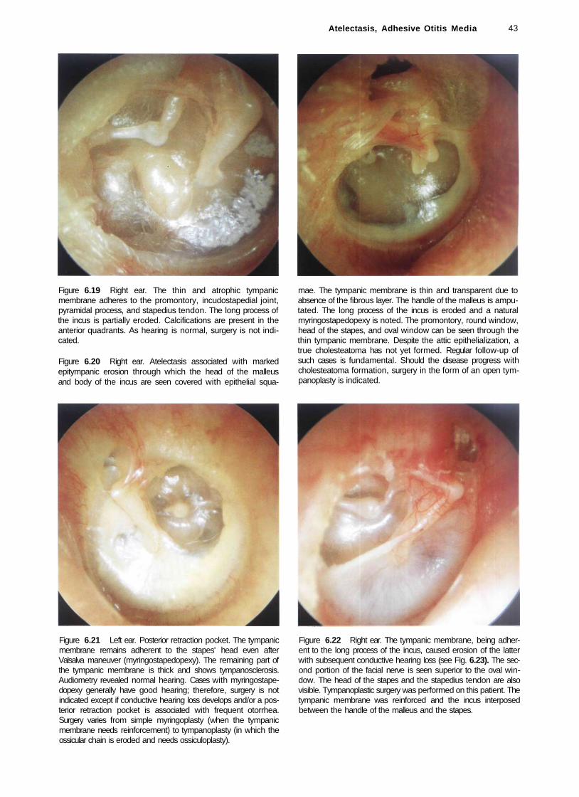
Figure 6.19 Right ear. The thin and atrophic tympanic membrane adheres to the promontory, incudostapedial joint, pyramidal process, and stapedius tendon. The long process of the incus is partially eroded. Calcifications are present in the anterior quadrants. As hearing is normal, surgery is not indicated.
Figure 6.20 Right ear. Atelectasis associated with marked epitympanic erosion through which the head of the malleus and body of the incus are seen covered with epithelial squa
mae. The tympanic membrane is thin and transparent due to absence of the fibrous layer. The handle of the malleus is amputated. The long process of the incus is eroded and a natural myringostapedopexy is noted. The promontory, round window, head of the stapes, and oval window can be seen through the thin tympanic membrane. Despite the attic epithelialization, a true cholesteatoma has not yet formed. Regular follow-up of such cases is fundamental. Should the disease progress with cholesteatoma formation, surgery in the form of an open tympanoplasty is indicated.
Figure 6.21 Left ear. Posterior retraction pocket. The tympanic membrane remains adherent to the stapes' head even after Valsalva maneuver (myringostapedopexy). The remaining part of the tympanic membrane is thick and shows tympanosclerosis. Audiometry revealed normal hearing. Cases with myringostapedopexy generally have good hearing; therefore, surgery is not indicated except if conductive hearing loss develops and/or a posterior retraction pocket is associated with frequent otorrhea. Surgery varies from simple myringoplasty (when the tympanic membrane needs reinforcement) to tympanoplasty (in which the ossicular chain is eroded and needs ossiculoplasty).
Figure 6.22 Right ear. The tympanic membrane, being adherent to the long process of the incus, caused erosion of the latter with subsequent conductive hearing loss (see Fig. 6.23). The second portion of the facial nerve is seen superior to the oval window. The head of the stapes and the stapedius tendon are also visible. Tympanoplastic surgery was performed on this patient. The tympanic membrane was reinforced and the incus interposed between the handle of the malleus and the stapes.

44 6 Atelectasis, Adhesive Otitis Media
0
10
20
30
40
50
60
70
80
90
100
110
120 125 250 500 1K 2K 4K 8K 16KHz
Figure 6.23 Audiogram of the same case showing conductive hearing loss.
Figure 6.24 Left ear. Meso- and epitympanic retraction pockets that adhere to the head of the malleus, the partially eroded long process of the incus, and the incudostapedial joint. A ventilation tube has been inserted in the anterior quadrant to avoid further retraction that might lead to cholesteatoma.
Figure 6.25 Right ear. Grade IV atelectasis. All of the middle ear structures can be seen in transparency. Starting from the malleus and moving in a clockwise direction, we can distinguish the tubal opening, the hypotympanum, the promontory, the round window, the stapedius tendon, and the incudostapedial joint.
Figure 6.26 Right ear. Large mesotympanic retraction pocket that caused erosion of the incus and stapes suprastructure. The second portion of the facial nerve passing superior to the oval window, the promontory, and the round window can all be seen in transparency. In cases with good social hearing and no otorrhea, surgery is not indicated.

Figure 6.27 Right ear. Posterior retraction pocket. The tympanic membrane adheres to the promontory, the round window, the partially eroded long process of the incus, the head of the stapes, and the stapedius tendon. The processus cochleariformis is clearly visible between the malleus and the long process of the incus. Middle ear effusion can be observed anterior to the malleus and in the region of the oval window. In this case, ventilation tube insertion is indicated in an attempt to prevent further erosion of the ossicular chain and the formation of mesotympanic cholesteatoma.
Atelectasis, Adhesive Otitis Media 45
Summary
In grade I, II, and III atelectasis, a long-term ventilation tube is usually inserted to prevent further retraction of the tympanic membrane. However, in cases with marked conductive hearing loss that denotes erosion of the incus or the superstructure of the stapes, ossiculoplasty is performed after extraction and sculpturing of the eroded incus or using a homologous incus. A large disk of tragal cartilage is used to reinforce the tympanic membrane. Indications for surgery in adhesive otitis media include cases with tympanic membrane perforation (grade V according to Sade 1979), with or without polypi, granulation or otorrhea, those cases with a large infected retraction pocket causing frequent otorrhea, or those with conductive hearing loss due to ossicular chain erosion. In all these cases a tympanoplasty is performed using a postauricular incision. A disk of tragal cartilage is used with the perichondrium adherent to its lateral surface. If the handle of the malleus is present, it is incorporated into the cartilaginous disk after creating a triangular defect for its accommodation. This technique has the advantage of preventing retraction and adhesions between the tympanic membrane and the promontory. At the same time, it enables repair of the tympanic membrane perforation with the tragal perichondrium. It can be concluded that there is no single treatment for the atelectatic ear. The milder the degree of atelectasis, the more conservative the treatment is. It should be noted, however, that in the long term conservative treatment (e.g., ventilation tube) was not found to modify further evolution of atelectasis. As atelectasis results from eustachian tube dysfunction, the ideal solution would be correction of this defect. At present, there is no acceptable "functional" surgery for the eustachian tube. Individual treatment should be administered according to the consequences of this dysfunction in each case. Such a strategy, however, requires a high mental elasticity and versatile surgical techniques.

46
7 Non-Cholesteatomatous Chronic Otitis Media
The difference between acute and chronic otitis media is not the duration of the disease but rather the anatomo-pathological characteristics. Untreated acute otitis media persisting for months is still a process that tends essentially to return to normality. On the other hand, a chronic otitis, even if the ear stops discharging, has anatomopathological sequelae of clinical importance.
The most commonly encountered forms are active chronic suppurative otitis media characterized by otorrhea and inactive chronic suppurative otitis media in which the ear is dry. Naturally, the active form may become quiescent either spontaneously or following treatment. The ear becomes dry and the condition is designated inactive. A dry perforation, however, may be infected, leading to ear discharge. In this latter case, the mucosa may be hyperplastic and thick due to interstitial edema, fibrosis, or cellular infiltration. In some cases, polypi are formed which may be large enough to occupy the external auditory canal. In other cases, persistence of suppuration can lead to ulceration of the mucosa, formation of granulation tissue, and even bone resorption. The anatomical sequelae of chronic otitis media vary. They may be in the form of a simple central tympanic membrane perforation, erosion of the ossicular chain, or formation of tympanosclerosis. Both the active and inactive forms cause functional alterations such as conductive or mixed hearing loss (very rarely sensorineural). The absence of squamous epithelium in the middle ear has led to the designation of this form as a "safe type" of otitis media. This is to distinguish it from cholesteatoma, which is considered "unsafe" due to the potential complications that may arise from the presence of keratinized squamous epithelium in the middle ear.
• General Characteristics of Tympanic Membrane Perforations
Tympanic membrane perforations are usually present at the pars tensa. Pars flaccida perforations are generally associated with epitympanic cholesteatoma.
If a tympanic membrane perforation does not heal spontaneously, the epithelial and mucosal layers creep and meet along the borders of the perforation. This pathological communication between the middle and external ear can be considered a true "air fistula." In the presence of a tympanic membrane perforation, the patient is subject to recurrent infections and ear discharge.
Whenever tympanic membrane perforations are diagnosed, the following three considerations must be fulfilled: 1) At the level of the perforation the site, size, and state of the remainder of the tympanic membrane
around the perforation should be determined. 2) At the level of the middle ear, the state of the mucosa, the condition of the ossicular chain (if possible), and the presence or absence of epithelialization should be evaluated. 3) The otoscopic examination has to be complemented with the pure tone audiometry to have a better understanding of the ossicular chain (possible erosion of the incus, fixity of the chain).
Pars tensa perforations can be either central or marginal. Marginal perforations lie at the periphery of the tympanic membrane with absence of the fibrous annulus. Marginal perforations are considered "unsafe" because the skin of the external auditory canal, in the absence of the annulus, can easily advance towards the middle ear, giving rise to cholesteatoma.
Otoscopic examination can often define the junction between the skin and mucosa at the borders of the tympanic membrane perforation. At this junction the squamous epithelium has a "velvety" appearance. The presence of a red de-epithelialized ring along the perforation rim indicates the evagination of the mucosa towards the external surface of the tympanic membrane residue. However, invagination of the skin towards the inner surface of the tympanic membrane residue is more difficult to diagnose. This inward skin migration is favored by the atrophy of the mucosa which occurs as a result of the perforation. At the time of myringoplasty, freshening of the edge of the perforation not only favors the attachment of the graft but also greatly reduces the risk of leaving entrapped skin on the undersurface of the drum, which may lead to iatrogenic cholesteatoma.
Conductive hearing loss caused by tympanic membrane perforation has two main causes: 1) Reduction of the tympanic membrane surface area on which the acoustic pressure exerts its action. 2) Reduction of the vibratory movements of the cochlear fluids because sound reaches both windows at nearly the same time without the dampening and phase-changing effect of the intact tympanic membrane.
The site of the perforation cannot be correlated to a particular audiometric pattern. However, it is generally observed that hearing loss occurs more in the low frequencies and that for perforations of the same size, hearing loss occurs more in posterior perforations than in anterior ones.
The majority of posttraumatic and postotitic perforations heal spontaneously. When large portions of the tympanic membrane are lost or when chronic or recurrent infections occur, the perforation may become permanent. In these cases, the tympanic membrane must be repaired (myringoplasty) to restore the normal physiology of the ear.

Posterior Perforations 47
Posterior Perforations
Figure 7.1 Left ear. The tympanic membrane is very thin due to atrophy of the fibrous layer. A posterosuperior marginal perforation is seen. This perforation is risky because the skin of the external auditory canal can easily advance into the middle ear, forming a cholesteatoma. In this case, a myringoplasty using an endomeatal approach is indicated.
Figure 7.2 Right ear. Marginal posterosuperior perforation through which the intact incudostapedial joint, the stapedius tendon, and the pyramidal process can be seen.
Figure 7.3 Left ear. Perforation of the posterosuperior quadrant of the tympanic membrane. Visualized through the perforation are the incudostapedial joint, the stapes, the stapedius tendon, the pyramidal process, the promontory, and the round window. The residue of the tympanic membrane is very thin due to absence of the fibrous layer. Tympanosclerosis can be seen in the marginal part of the drum residue. From the surgical point of view, posterior perforations are the easiest to repair especially when partial reconstruction of the tympanic membrane is all that is required. When the residue of the tympanic membrane is transformed into a rigid tympanosclerotic
plaque, it is advisable to remove it, conserving the epidermal layer to be laid over the graft.
Figure 7.4 Right ear. Large perforation of the posterior quadrants. Normal middle ear mucosa. The incudostapedial joint is intact. The oval window with the annular ligament surrounding the footplate can be seen. The pyramidal eminence, the stapedius tendon, the round window, and Jacobson's nerve running on the promontory are also visible. The remaining anterior quadrants of the tympanic membrane are tympanosclerotic and rigid, blocking the mobility of the malleus.

48 7 Non-Cholesteatomatous Chronic Otitis
Figure 7.5 Right ear. Presence of chronic otitis media. Dry perforation of the posterior quadrants of the tympanic membrane through which the head of the stapes and the round window are visible. The long process of the incus is necrosed. The middle ear mucosa is normal. The tympanic membrane residue shows tympanosclerosis with alternating areas of calcification and areas of thinned membrane due to atrophy of the fibrous layer. The operation, performed through a post-auricular incision, will also include the reconstruction of the ossicular chain using the autologous incus.
Figure 7.7 Right ear. Presence of simple chronic otitis media; a posteroinferior drum perforation. The middle ear mucosa is normal. The round window and Jacobson's nerve running on the promontory are seen. The incus can also be appreciated posterior to a retromalleolar tympanosderotic plaque. The tympanic membrane residue shows areas of atrophy alternating with areas of tympanosclerosis.
Figure 7.6 Left ear. Posterior nonmarginal perforation. The incudostapedial joint, the promontory, and the round window are all discernible.
Figure 7.8 Left ear. Perforation of the posterior quadrants of the tympanic membrane. The skin advances along the pos-terosuperior border of the perforation towards the incudostapedial joint. The middle ear mucosa appears hypertrophic. Mucoid discharge is also present. A tympanosderotic plaque can be seen in the residue of the tympanic membrane.

Anterior Perforations 49
Figure 7.9 Right ear. Marked posterior marginal perforation through which the skin penetrates into the middle ear. The ossicular chain is not identifiable.
Anterior Perforations
Figure 7.10 Left ear. Anterior perforation of the tympanic membrane through which the tubal orifice is visible. A white mass is present behind the anterosuperior margin of the perforation. This mass can be either a cholesteatoma or a tym-panosclerotic plaque. The consistency of the mass can be tested using an instrument under the microscope; the cholesteatoma is soft and will break, whereas tympanosclerosis is generally hard.
Figure 7.11 Right ear. Anterior perforation in a patient with anterior and posterior humps of the external auditory canal as well as exostosis of the superior canal wall. In this case, canalplasty should be performed at the same time as myringoplasty.
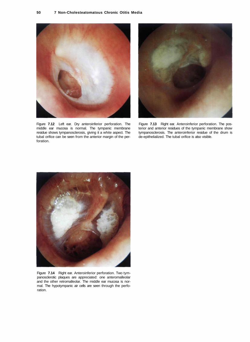
50 7 Non-Cholesteatomatous Chronic Otitis Media
Figure 7.12 Left ear. Dry anteroinferior perforation. The middle ear mucosa is normal. The tympanic membrane residue shows tympanosclerosis, giving it a white aspect. The tubal orifice can be seen from the anterior margin of the perforation.
Figure 7.13 Right ear. Anteroinferior perforation. The posterior and anterior residues of the tympanic membrane show tympanosclerosis. The anteroinferior residue of the drum is de-epithelialized. The tubal orifice is also visible.
Figure 7.14 Right ear. Anteroinferior perforation. Two tym-panosclerotic plaques are appreciated: one anteromalleolar and the other retromalleolar. The middle ear mucosa is normal. The hypotympanic air cells are seen through the perforation.

Subtotal and Total Perforations 51
• Subtotal and Total Perforations
Figure 7.15 Right ear. Large tympanic membrane perforation. The tubal orifice, the hypotympanic air cells, the promontory, the round and oval windows, and the intact stapes can be viewed. An onset of necrosis of the incus can be distinguished.
Figure 7.17 Right ear. Similar case. The promontory and the round window are visible. A tympanosclerotic plaque that engulfs the ossicular chain is seen at the level of the postero-superior border of the perforation.
Figure 7.16 Right ear. Perforation of the inferior quadrants of the tympanic membrane. All the tympanic membrane residue shows dense tympanosclerosis. Removal of these sclerotic plaques during myringoplasty assures an adequate vascularity to the graft, and thus a high success rate for the operation.
Figure 7.18 Left ear. Subtotal perforation. The annulus as well as a fibrous rim are visualized along the inferior border of the perforation. The handle of the malleus is medialized. The tubal orifice, the hypotympanic air cells covered with mucosa, Jacobson's nerve on the promontory, and the long process of the incus are visible. The residue of the tympanic membrane is thickened. In cases in which only a small anterior residue of the tympanic membrane is found, an overlay technique in which the graft is put over the annulus is used, thus preventing detachment of the anterior part of the graft leading to reperf oration.

52 7 Non-Cholesteatomatous Chronic Otitis
Figure 7.19 Left ear. Total perforation of the tympanic membrane through which evolving tympanosclerotic plaques are visible. The stapes and the stapedius tendon are visible. The long process of the incus is partially eroded. The handle of the malleus is medialized and adherent to the promontory. The tubal orifice and the hypotympanic air cells are also noted.
Figure 7.20 Left ear. Subtotal perforation of the tympanic membrane. The middle ear mucosa is normal. The tympanic membrane residue is de-epithelialized. The incudo-stapedial joint, the medialized handle of the malleus, and the hypotympanic air cells are visible.
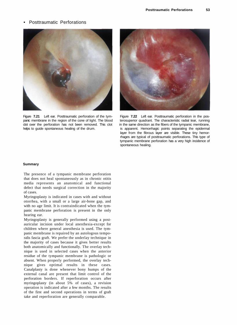
Posttraumatic Perforations 53
• Posttraumatic Perforations
Figure 7.21 Left ear. Posttraumatic perforation of the tym- Figure 7.22 Left ear. Posttraumatic perforation in the pos-panic membrane in the region of the cone of light. The blood terosuperior quadrant. The characteristic radial tear, running clot over the perforation has not been removed. This clot in the same direction as the fibers of the tympanic membrane, helps to guide spontaneous healing of the drum. is apparent. Hemorrhagic points separating the epidermal
layer from the fibrous layer are visible. These tiny hemorrhages are typical of posttraumatic perforations. This type of tympanic membrane perforation has a very high incidence of spontaneous healing.
Summary
The presence of a tympanic membrane perforation that does not heal spontaneously as in chronic otitis media represents an anatomical and functional defect that needs surgical correction in the majority of cases. Myringoplasty is indicated in cases with and without otorrhea, with a small or a large air-bone gap, and with no age limit. It is contraindicated when the tympanic membrane perforation is present in the only hearing ear. Myringoplasty is generally performed using a post-auricular incision under local anesthesia-except for children where general anesthesia is used. The tympanic membrane is repaired by an autologous temporalis fascia graft. We prefer the underlay technique in the majority of cases because it gives better results both anatomically and functionally. The overlay technique is used in selected cases when the anterior residue of the tympanic membrane is pathologic or absent. When properly performed, the overlay technique gives optimal results in these cases. Canalplasty is done whenever bony humps of the external canal are present that limit control of the perforation borders. If reperforation occurs after myringoplasty (in about 5% of cases), a revision operation is indicated after a few months. The results of the first and second operations in terms of graft take and reperforation are generally comparable.

54 7 Non-Cholesteatomatous Chronic Otitis
• Perforations Complicated or Associated with Other Pathologies
Figure 7.23 Right ear. Total perforation. Epidermization is present in the regions of the mesotympanum and the ossicular chain. The round window, hypotympanic air cells with thickened mucosa, Jacobson's nerve running on the promontory, and the tubal orifice are well visualized. This case is an example of chronic otitis media complicated by the presence of skin in the middle ear. Tympanoplasty should be staged. In the first stage, the skin is removed without traumatizing the ossicular chain, and the tympanic membrane is reconstructed.
Figure 7.25 Right ear. Perforation of the inferior quadrants of the tympanic membrane, the residues of which show tympanosclerosis. Epidermization is evident over the promontory. Since epidermization is limited in this case, a single-stage tympanoplasty can be performed.
In the second stage, the middle ear is checked for any residual skin, and the ossicular chain is reconstructed.
Figure 7.24 Left ear. Large perforation with diffuse epidermization of the middle ear associated with purulent otorrhea. In these cases, even if the ossicular chain proves intact, mastoid exploration should be done. A second stage is performed 1 year after the first operation to check for any skin residues.
Figure 7.26 Right ear. Another example of chronic otitis media complicated with diffuse epidermization of the middle ear. Surgery follows the same rules as for cholesteatoma.

Perforations Complicated or Associated with Other Pathologies 55
Figure 7.27 Right ear. Large tympanic membrane perforation. The anterior drum residue shows tympanosclerosis. The ossicular chain is difficult to identify because of the presence of epidermization at this level. The round window is visible. A staged tympanoplasty is also indicated in this case.
Figure 7.28 Right ear. Granulomatous otitis media. A roundish mass fills the middle ear. Serous otorrhea is present.
Figure 7.29 Right ear. Small perforation of the inferior quadrants of the tympanic membrane with eversion of the mucosa onto the outer layer of the membrane. Tympanosclerosis, both antero- and posteromalleolar, can be noted.
Figure 7.30 Right ear. Case similar to that in Figure 7.29. The mucosa has replaced the epithelial layer. Ear discharge is also present. During myringoplasty, curettage of the everted mucosa is necessary until the fibrous layer of the tympanic membrane is reached.

56 7 Non-Cholesteatomatous Chronic Otitis Media
Figure 7.31 Left ear. Perforation of the anterior quadrants. Skin envelopes the handle of the malleus. During myringoplasty, curettage of the skin is necessary before reconstruction.
Figure 7.32 Right ear. Posterior perforation. The residues of the tympanic membrane appear whitish and bulging. During surgery, the middle ear was occupied by granulomatous tissue that proved to be tuberculosis (TB) on histopathological examination. This patient had a past history of pulmonary TB. Tuberculous otitis media should be suspected in cases of pulmonary TB presenting with otorrhea.
• Tympanosclerosis
Tympanosclerosis is characterized by fibroblastic invasion of the submucosal spaces of the middle ear followed by thickening, hyalinization, and fusion of collagen fibers into a homogenous mass with calcium deposits and phosphate crystals. Though the pathogenesis is not yet clear, it seems that chronic otitis media is a predisposing factor. Two distinct forms are recognized:
Tympanosclerosis with Intact tympanic membrane. This is characterized by calcareous plaques (chalk patches) in the fibrous layer of the tympanic membrane. The antero- and posteromalleolar regions are usually involved. The periannular region of the inferior quadrants is also affected, forming a horseshoe pattern. The pars tensa is rigid, thick, and loses its elasticity, assuming a whitish aspect. Atrophic and thinned areas can also occur. Infrequently, in very advanced cases, the tympanosclerotic plaques occupy all the middle ear spaces, attic, and aditus and completely block the ossicular chain. The tympanic membrane in these cases is very thick or even replaced by the plaques.
Tympanosclerosis associated with tympanic membrane perforation. The perforation is frequently central or subtotal and the annulus, infiltrated by calcium deposits, is well visualized. Frequently, submucous nodular deposits are encountered in the middle ear. Ossicular fixation or erosion due to devitalization as a
result of loss of blood supply can also occur. The middle ear mucosa is very thin with reduced vascularity. In some cases, tympanosclerotic plaques are seen extruding from the mucosa to present as white middle ear masses.

• Tympanosclerosis Associated with Perforation
Tympanosclerosis Associated with Perforation 57
Figure 7.33 Right ear. Tympanosclerosis associated with perforation. The tympanic membrane residues and the middle ear (promontory and hypotympanum) show the characteristic plaques. The malleus is blocked by tympanosclerosis.
Figure 7.35 Right ear. Perforations of the inferior quadrants with tympanosclerosis involving the residues of the tympanic membrane and the middle ear.
Figure 7.34 Right ear. Tympanosclerosis with perforation. A large tympanosclerotic plaque is noted in the anterior residue of the tympanic membrane. The middle ear is also involved. The promontory, oval window, stapes footplate, and round window can be appreciated.
Figure 7.36 Right ear. Tympanosclerosis with perforation. The tympanosclerotic process involves the anterior residues of the tympanic membrane and the mucosa of the promontory reaching to the posterior mesotympanum. At this level, ossification of the stapedius tendon is seen. The tympanic segment of the fallopian canal is covered by a sclerotic plaque. The long process of the incus is eroded.
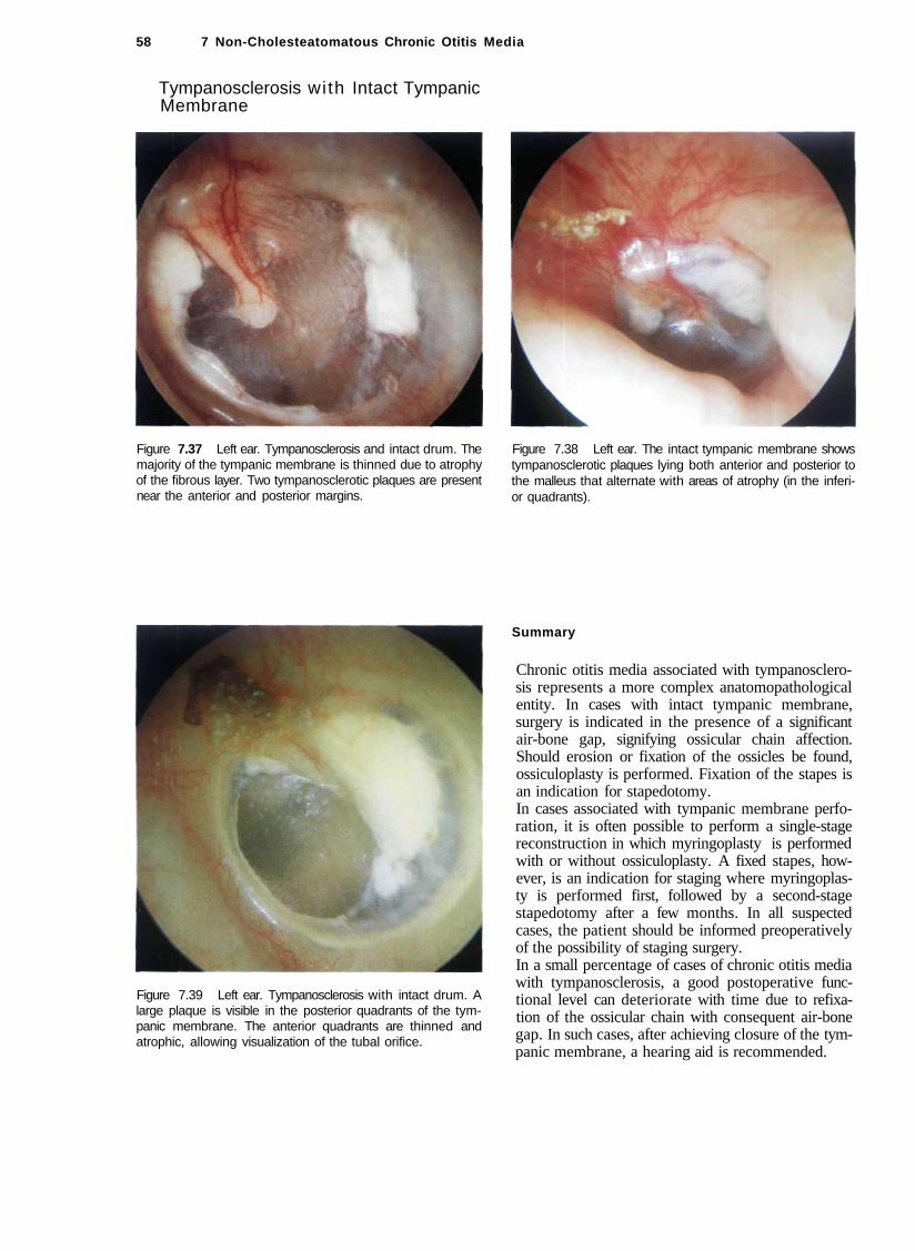
58 7 Non-Cholesteatomatous Chronic Otitis Media
Tympanosclerosis with Intact Tympanic Membrane
Figure 7.37 Left ear. Tympanosclerosis and intact drum. The majority of the tympanic membrane is thinned due to atrophy of the fibrous layer. Two tympanosclerotic plaques are present near the anterior and posterior margins.
Figure 7.38 Left ear. The intact tympanic membrane shows tympanosclerotic plaques lying both anterior and posterior to the malleus that alternate with areas of atrophy (in the inferior quadrants).
Figure 7.39 Left ear. Tympanosclerosis with intact drum. A large plaque is visible in the posterior quadrants of the tympanic membrane. The anterior quadrants are thinned and atrophic, allowing visualization of the tubal orifice.
Summary
Chronic otitis media associated with tympanosclerosis represents a more complex anatomopathological entity. In cases with intact tympanic membrane, surgery is indicated in the presence of a significant air-bone gap, signifying ossicular chain affection. Should erosion or fixation of the ossicles be found, ossiculoplasty is performed. Fixation of the stapes is an indication for stapedotomy. In cases associated with tympanic membrane perforation, it is often possible to perform a single-stage reconstruction in which myringoplasty is performed with or without ossiculoplasty. A fixed stapes, however, is an indication for staging where myringoplasty is performed first, followed by a second-stage stapedotomy after a few months. In all suspected cases, the patient should be informed preoperatively of the possibility of staging surgery. In a small percentage of cases of chronic otitis media with tympanosclerosis, a good postoperative functional level can deteriorate with time due to refixa-tion of the ossicular chain with consequent air-bone gap. In such cases, after achieving closure of the tympanic membrane, a hearing aid is recommended.

59
8 Chronic Suppurative Otitis Media wi th Cholesteatoma
Cholesteatoma is an epidermal inclusion cyst localized in the middle ear, whose capsule and matrix is formed from stratified squamous epithelium. The desquamating debris includes pearly white lamellae of keratin that accumulate concentrically, forming the cholestea-tomatous mass.
The term cholesteatoma is actually a misnomer. It is derived from the Greek "cole" or bile, "steatos" or fat, and "oma" or tumor. There is no relation between cholesteatoma and bile or fat. The suffix "oma" (tumor), however, is more appropriate because cholesteatoma can be considered an epidermal inclusion cyst.
Cholesteatoma can be divided into congenital (middle ear or petrous bone) and acquired (middle ear or petrous bone). Congenital cholesteatoma is derived from entrapped ectodermal cellular debris during embryonic development. When it involves the middle ear, it appears as a whitish retrotympanic mass that may be localized either anterior or posterior to the malleus (see Chapter 9). When it involves the petrous part of the temporal bone, it is termed congenital petrous bone cholesteatoma and in the majority of cases it is localized in the petrous apex (see Chapter 10). In this chapter we will deal exclusively with cholesteatoma involving the middle ear. Petrous bone cholesteatoma is dealt with in a later chapter.
Acquired cholesteatoma of the middle ear can be caused by invasion of the skin of the external auditory canal into the middle ear through a marginal perforation. It can also originate from a epitympanic retraction pocket that becomes so deep that keratin debris can no longer be expelled, leading to their accumulation and subsequent cholesteatoma formation. Such retraction pockets can remain asymptomatic until they become infected, resulting in otorrhea and hearing loss. In other cases, the only symptom might be progressive hearing loss due to erosion of the ossicular chain by the developing cholesteatoma.
Because it is not always easy to establish a clear distinction between epitympanic or posterosuperior retraction pockets and cholesteatoma, we prefer to follow up these patients with otomicroscopy and endoscopy. In cases in which the retraction pocket becomes deep, giving rise to a cholesteatoma, a tympanoplasty is indicated. Because of the early stage of the disease, surgery can be done in a single stage.
Fetid otorrhea and hearing loss are the main complaints in cholesteatoma. In addition, complicated cases can manifest with vertigo and/or facial nerve paralysis. Vertigo occurs as a result of labyrinthine fistula, which is most commonly located in the lateral semicircular canal. Facial paralysis can be caused by pressure of the cholesteatoma sac or neuritis.
In rare cases, the cholesteatoma can invade the labyrinth, cochlea, posterior and middle fossa durae, the internal auditory canal, and the petrous apex, forming a petrous bone cholesteatoma (see Chapter 10).
Treatment of cholesteatoma is exclusively surgical. Early this century, radical mastoidectomy, a destructive procedure for the middle ear, was performed with the only goal being eradication of infection to save the ear.
In the early 1950s, the concept of tympanoplasty was introduced. Tympanoplasty was aimed at eradication of infection as well as reconstruction of the tym-pano-ossicular system. Today, two types of tympanoplasty are employed: closed tympanoplasty in which the posterior canal wall is preserved, and open tympanoplasty in which the posterior canal wall is drilled. Both techniques, when performed appropriately and with the proper indications, can produce excellent results in terms of eradication of cholesteatoma and restoration of hearing. In children, the closed technique is preferred, performed in two stages, in the majority of cases due to their highly cellular mastoids and in an attempt to preserve the anatomy of the ear as much as possible. In adults, particularly in epitympanic cholesteatoma with marked erosion of the scutum, in cases with sclerotic mastoids, or when middle ear atelectasis is present, an open tympanoplasty is performed (see also Chapter 13).

60 8 Chronic Suppurative Otitis Media with Cholesteatoma
Epitympanic Retraction Pocket
Figure 8.1 Right ear. Early epitympanic retraction pocket. The tympanic membrane shows grade I atelectasis. Middle ear effusion with characteristic yellowish coloration of the drum is seen. In the anterosuperior quadrant, the tubal orifice is visible in transparency, whereas the long process of the incus is evident in the posterosuperior quadrant. In the area of the cone of light, an atrophic part of the tympanic membrane due to a previous myringotomy can be appreciated.
Figure 8.2 Right ear. Epitympanic retraction pocket with the onset of tympanosclerosis of the pars tensa of the tympanic membrane.
Figure 8.3 Right ear, similar case. The anterior quadrants of the pars tensa are retracted and thickened.
Figure 8.4 Right ear. A large controllable epitympanic retraction pocket with erosion of the scutum. The head of the malleus is seen. Middle ear effusion gives the tympanic membrane the characteristic yellowish coloration. To prevent progression of the retraction pocket and the formation of adhesions, myringotomy, ventilation tube insertion, and regular follow-up are indicated. These cases frequently represent the
transition from a simple retraction pocket to an initial attic cholesteatoma. The distinction between the two is sometimes difficult. In suspected cases, a high-resolution computed tomography (CT) scan (bone window) is beneficial for better evaluation of the extension of the retraction pocket. In cases where the condition remains stable with regular follow-up and where hearing is normal, no surgery is required. If the pocket extends deeper, giving rise to a frank cholesteatoma, surgery is indicated. If hearing is normal, an open tympanoplasty (modified Bondy technique) is performed in a single stage.
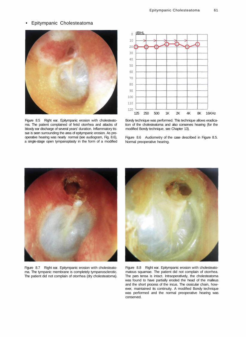
• Epitympanic Cholesteatoma
Figure 8.5 Right ear. Epitympanic erosion with cholesteatoma. The patient complained of fetid otorrhea and attacks of bloody ear discharge of several years' duration. Inflammatory tissue is seen surrounding the area of epitympanic erosion. As preoperative hearing was nearly normal (see audiogram, Fig. 8.6), a single-stage open tympanoplasty in the form of a modified
Figure 8.7 Right ear. Epitympanic erosion with cholesteatoma. The tympanic membrane is completely tympanosclerotic. The patient did not complain of otorrhea (dry cholesteatoma).
Epitympanic Cholesteatoma 61
dBHL
125 250 500 1K 2K 4K 8K 16KHz
Bondy technique was performed. This technique allows eradication of the cholesteatoma and also conserves hearing (for the modified Bondy technique, see Chapter 13).
Figure 8.6 Audiometry of the case described in Figure 8.5. Normal preoperative hearing.
Figure 8.8 Right ear. Epitympanic erosion with cholesteato-matous squamae. The patient did not complain of otorrhea. The pars tensa is intact. Intraoperatively, the cholesteatoma was found to have partially eroded the head of the malleus and the short process of the incus. The ossicular chain, however, maintained its continuity. A modified Bondy technique was performed and the normal preoperative hearing was conserved.
0
10
20
30
40
50
60
70
80
90
100
110
120
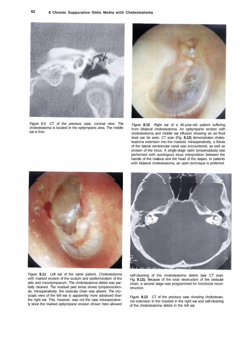
62 8 Chronic Suppurative Otitis Media with Cholesteatoma
Figure 8.9 CT of the previous case, coronal view. The cholesteatoma is located in the epitympanic area. The middle ear is free.
Figure 8.11 Left ear of the same patient. Cholesteatoma with marked erosion of the scutum and epidermization of the attic and mesotympanum. The cholesteatoma debris was partially cleaned. The residual pars tensa shows tympanosclerosis. Intraoperatively, the ossicular chain was absent. The oto-scopic view of the left ear is apparently more advanced than the right ear. This, however, was not the case intraoperatively since the marked epitympanic erosion shown here allowed
Figure 8.10 Right ear of a 46-year-old patient suffering from bilateral cholesteatoma. An epitympanic erosion with cholesteatoma and middle ear effusion showing an air-fluid level can be seen. CT scan (Fig. 8.12) demonstrates cholesteatoma extension into the mastoid. Intraoperatively, a fistula of the lateral semicircular canal was encountered, as well as erosion of the incus. A single-stage open tympanoplasty was performed with autologous incus interposition between the handle of the malleus and the head of the stapes. In patients with bilateral cholesteatoma, an open technique is preferred.
self-cleaning of the cholesteatoma debris (see CT scan, Fig. 8.12). Because of the total destruction of the ossicular chain, a second stage was programmed for functional reconstruction.
Figure 8.12 CT of the previous case showing cholesteatoma extension in the mastoid in the right ear and self-cleaning of the cholesteatoma debris in the left ear.

Epitympanic Cholesteatoma 63
Figure 8.13 Left ear. Small epitympanic erosion with cholesteatoma. The skin surrounding the erosion is hyperemic and everted.
Figure 8.15 Right ear. Large epitympanic erosion with cholesteatoma. This 18-year-old patient did not complain of otorrhea. Ipsilateral hearing was normal, whereas the contralateral side showed severe sensorineural hearing loss secondary to a previous surgery of radical mastoidectomy. Given the intact ossicular chain, an open tympanoplasty (modified Bondy technique) was performed. According to our strategy, cholesteatoma in the only hearing ear is one of the absolute indications for performing an open technique. The reason is
Figure 8.14 Left ear. Large epitympanic erosion with cholesteatoma and fetid otorrhea. The head of the malleus and body of the incus are eroded.
that this technique, if properly performed, ensures complete eradication of the pathology and better long-term follow-up, thus minimizing the risk of recurrence. Further surgical interventions, with their potential risk even in the most experienced hands, are therefore avoided.
Figure 8.16 Right ear. Large epitympanic erosion with cholesteatoma and polypoid tissue that covers the head of the malleus. The pars tensa is intact.
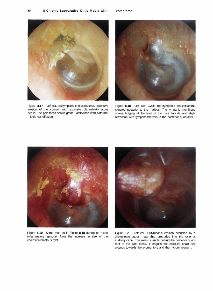
64 8 Chronic Suppurative Otitis Media with
Figure 8.17 Left ear. Epitympanic cholesteatoma. Extensive erosion of the scutum with excessive cholesteatomatous debris. The pars tensa shows grade I atelectasis with catarrhal middle ear effusion.
Figure 8.19 Same case as in Figure 8.18 during an acute inflammatory episode. Note the increase in size of the cholesteatomatous cyst.
esteatoma
Figure 8.18 Left ear. Cystic retrotympanic cholesteatoma situated posterior to the malleus. The tympanic membrane shows bulging at the level of the pars flaccida and slight retraction with tympanosclerosis in the posterior quadrants.
Figure 8.20 Left ear. Epitympanic erosion occupied by a cholesteatomatous mass that protrudes into the external auditory canal. The mass is visible behind the posterior quadrant of the pars tensa. It engulfs the ossicular chain and extends towards the promontory and the hypotympanum.

Epitympanic Cholesteatoma 65
Figure 8.21 Left ear. A large epitympanic erosion is seen with epidermization of the attic and posterior mesotympa-num. The cholesteatoma, visible in transparency, causes bulging of the tympanic membrane in the posterior inferior quadrants. Resorption of the incus and head of the malleus is discernible.
Figure 8.23 Left ear. Epitympanic erosion with cholesteatoma. Extension of the cholesteatoma into the mesotympa-num (visible through the transparent pars tensa).
Figure 8.22 Right ear. Epitympanic erosion with cholesteatoma. Extension of the cholesteatoma into the mesotympa-num is seen through the bulging posterior quadrants of the tympanic membrane.
Figure 8.24 Left ear. Epitympanic erosion with cholesteatoma. Epidermization of the posterior mesotympanum is seen through a posterior perforation of the tympanic membrane. The tympanic membrane residue has a whitish color. This can be either due to tympanosclerosis or to epidermization of the medial surface of the tympanic membrane. Examination under the microscope can, in many cases, determine the exact cause.

66 8 Chronic Suppurative Otitis Media with Cholesteatoma
Summary
An epitympanic retraction pocket should be regularly checked with otomicroscopy. The 30° rigid endoscope allows visualization of the extent of the retraction pocket that can be difficult with the microscope. When progression of the epithelium into the epitym-panum cannot be controlled, the presence of cholesteatoma is considered. In such cases, surgery should be performed. Whenever a minor epitympanic erosion is present, we adopt a closed technique with reconstruction of the attic using cartilage and bone pate. This technique is valid particularly in children where the mastoid is usually very pneumatized. Frequently, surgery is staged in these cases. When a marked attic erosion is present, especially in adults, we perform an open technique to avoid cholesteatoma recurrence that can occur due to absorption of the material used for reconstruction of the attic defect. When preoperative hearing is normal in the presence of attic cholesteatoma with large bony erosion, we perform an open tympanoplasty in the form of a modified Bondy technique. This technique allows single-stage eradication of the disease with conservation of the normal preoperative hearing.
il Mesotympanic Cholesteatoma
Figure 8.25 Right ear. Mesotympanic cholesteatoma. The Figure 8.26 Right ear. Posterior mesotympanic cholesteato-epithelial squamae can be seen through the retromalleolar ma associated with a polyp are seen at the level of the oval perforation. Anterior to the malleus, the cholesteatomatous window. There is evidence of discharge, mass causes bulging and whitish coloration of the tympanic membrane without perforating it. The entire middle ear is filled with cholesteatoma in this case.
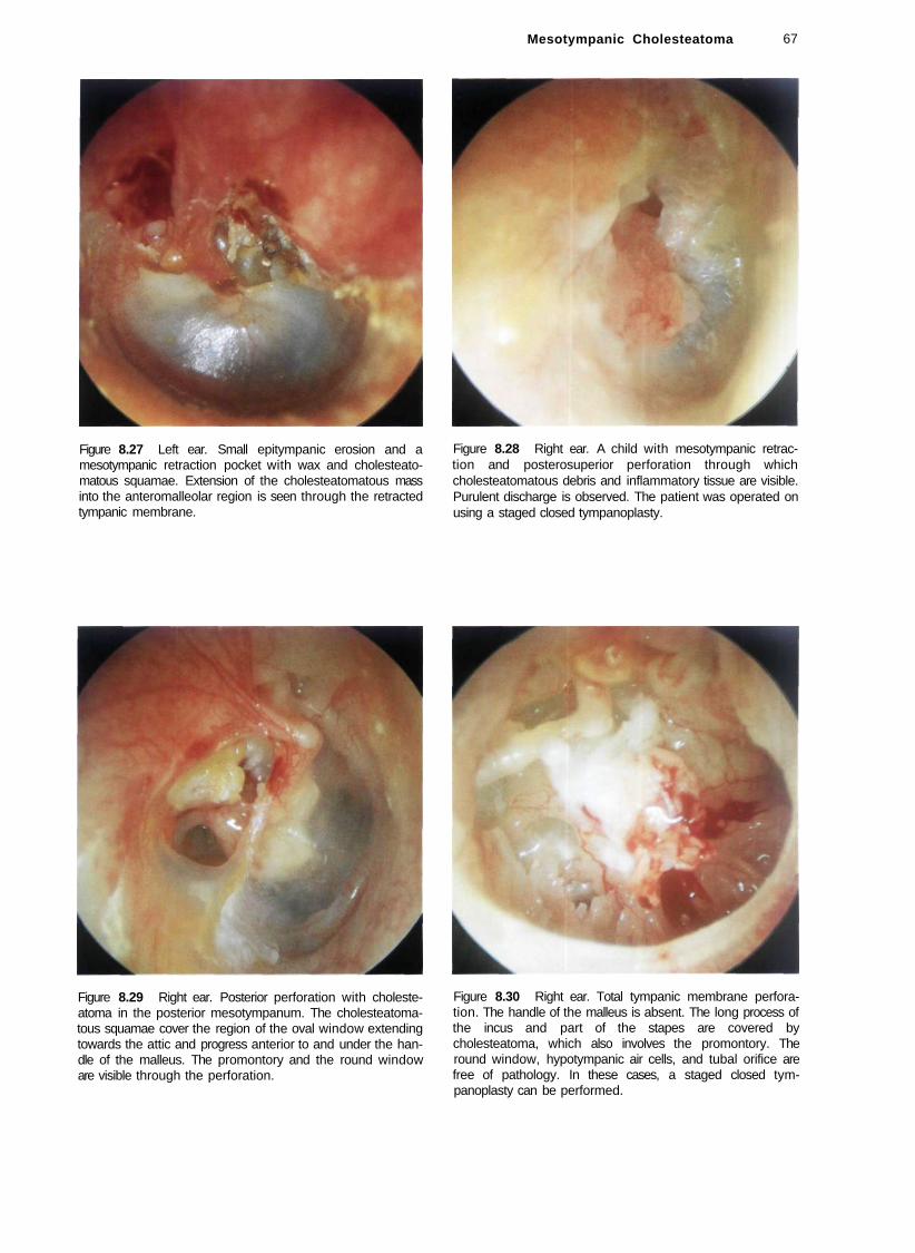
Mesotympanic Cholesteatoma 67
Figure 8.27 Left ear. Small epitympanic erosion and a mesotympanic retraction pocket with wax and cholesteato-matous squamae. Extension of the cholesteatomatous mass into the anteromalleolar region is seen through the retracted tympanic membrane.
Figure 8.28 Right ear. A child with mesotympanic retraction and posterosuperior perforation through which cholesteatomatous debris and inflammatory tissue are visible. Purulent discharge is observed. The patient was operated on using a staged closed tympanoplasty.
Figure 8.29 Right ear. Posterior perforation with cholesteatoma in the posterior mesotympanum. The cholesteatomatous squamae cover the region of the oval window extending towards the attic and progress anterior to and under the handle of the malleus. The promontory and the round window are visible through the perforation.
Figure 8.30 Right ear. Total tympanic membrane perforation. The handle of the malleus is absent. The long process of the incus and part of the stapes are covered by cholesteatoma, which also involves the promontory. The round window, hypotympanic air cells, and tubal orifice are free of pathology. In these cases, a staged closed tympanoplasty can be performed.
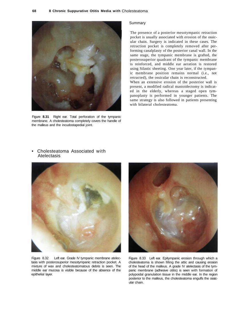
68 8 Chronic Suppurative Otitis Media with
Figure 8.31 Right ear. Total perforation of the tympanic membrane. A cholesteatoma completely covers the handle of the malleus and the incudostapedial joint.
• Cholesteatoma Associated with Atelectasis
Figure 8.32 Left ear. Grade IV tympanic membrane atelectasis with posterosuperior mesotympanic retraction pocket. A mixture of wax and cholesteatomatous debris is seen. The middle ear mucosa is visible because of the absence of the epithelial layer.
Cholesteatoma
Summary
The presence of a posterior mesotympanic retraction pocket is usually associated with erosion of the ossicular chain. Surgery is indicated in these cases. The retraction pocket is completely removed after performing canalplasty of the posterior canal wall. In the same stage, the tympanic membrane is grafted, the posterosuperior quadrant of the tympanic membrane is reinforced, and middle ear aeration is restored using Silastic sheeting. One year later, if the tympanic membrane position remains normal (i.e., not retracted), the ossicular chain is reconstructed. When an extensive erosion of the posterior wall is present, a modified radical mastoidectomy is indicated in the elderly, whereas a staged open tympanoplasty is performed in younger patients. The same strategy is also followed in patients presenting with bilateral cholesteatoma.
Figure 8.33 Left ear. Epitympanic erosion through which a cholesteatoma is shown filling the attic and causing erosion of the head of the malleus. A grade IV atelectasis of the tympanic membrane (adhesive otitis) is seen with formation of polypoidal granulation tissue in the middle ear. In the region posterior to the malleus, the cholesteatoma engulfs the ossicular chain.

Figure 8.34 Left ear. Epitympanic erosion with cholesteatoma associated with atelectasis of the tympanic membrane. The incus is absent. A natural myringostapedopexy has been created. The second portion of the facial nerve is seen superior to the stapes; inferiorly the round window is noted. The anterior part of the tympanic membrane is affected with tympanosclerosis. In these cases, as hearing loss is mild (< 30 dB), a modified radical mastoidectomy is indicated to maintain the normal preoperative hearing level obtained as a result of the spontaneous myringostapedopexy.
Figure 8.36 CT scan of the previous case. An epitympanic cholesteatoma is found. Adhesions between the tympanic membrane and the promontory can be observed. This 45-year-old woman underwent a modified radical mastoidectomy with no interference in the middle ear.
Cholesteatoma Associated with Atelectasis 69
Figure 8.35 Right ear. Epitympanic cholesteatoma associated with complete atelectasis of the tympanic membrane (see CTscan, Fig. 8.36).
Summary
In adult patients with extended epitympanic erosion or with bilateral cholesteatoma we prefer to perform an open technique. In all cases in which a spontaneous tympanostapedopexy with normal preoperative hearing or elderly patients with normal contralateral hearing, we prefer to leave the atelectatic tympanic membrane untouched after having verified the absence of any middle ear cholesteatoma. In the presence of mesotympanic cholesteatoma, staging is indicated. In the first operation a closed tympanoplasty is performed with reconstruction of the tympanic membrane, and a Silastic sheet is positioned in the middle ear. Silastic favors regeneration of the middle ear mucosa and prevents the formation of adhesions. In the second stage, performed 6 to 8 months later, the middle ear is checked for the presence of any residual cholesteatoma. The ossicular chain is then reconstructed using, preferably, an autologous incus. In children we always try to perform a staged closed tympanoplasty. If a recurrent cholesteatoma (epitympanic retraction pocket) is encountered in the second stage, we do not hesitate to transform it into an open technique.

70 8 Chronic Suppurative Otitis Media with
• Cholesteatoma Associated with Complications
Figure 8.37 Left ear. Large epitympanic perforation with pars tensa perforation. Cholesteatomatous squamae are present in the attic, whereas the middle ear is completely free. The handle of the malleus is present. The promontory, round window, and hypotympanic air cells are covered with normal mucosa. The tympanic annulus is intact. During surgery, a fistula of the lateral semicircular canal was encountered
Figure 8.39 Left ear. Large polyp obstructing the external auditory canal. The patient complained of fetid otorrhea, hearing loss, and vertigo. A high resolution CT scan of the temporal bone was ordered (see Fig. 8.40). A CT scan of the temporal bone should always be ordered in patients with chronic suppurative otitis media suffering from vertigo and/or instability.
(see Fig. 8.38). In such cases, because of the presence of marked epitympanic erosion and of the fistula, an open tympanoplasty is preferred.
Figure 8.38 Intraoperative view of the previous case. A fistula of the lateral semicircular canal is clearly seen.
Figure 8.40 CT scan of the previous case. A huge cholesteatoma causing a fistula of the lateral semicircular canal and erosion of the tegmen can be seen.
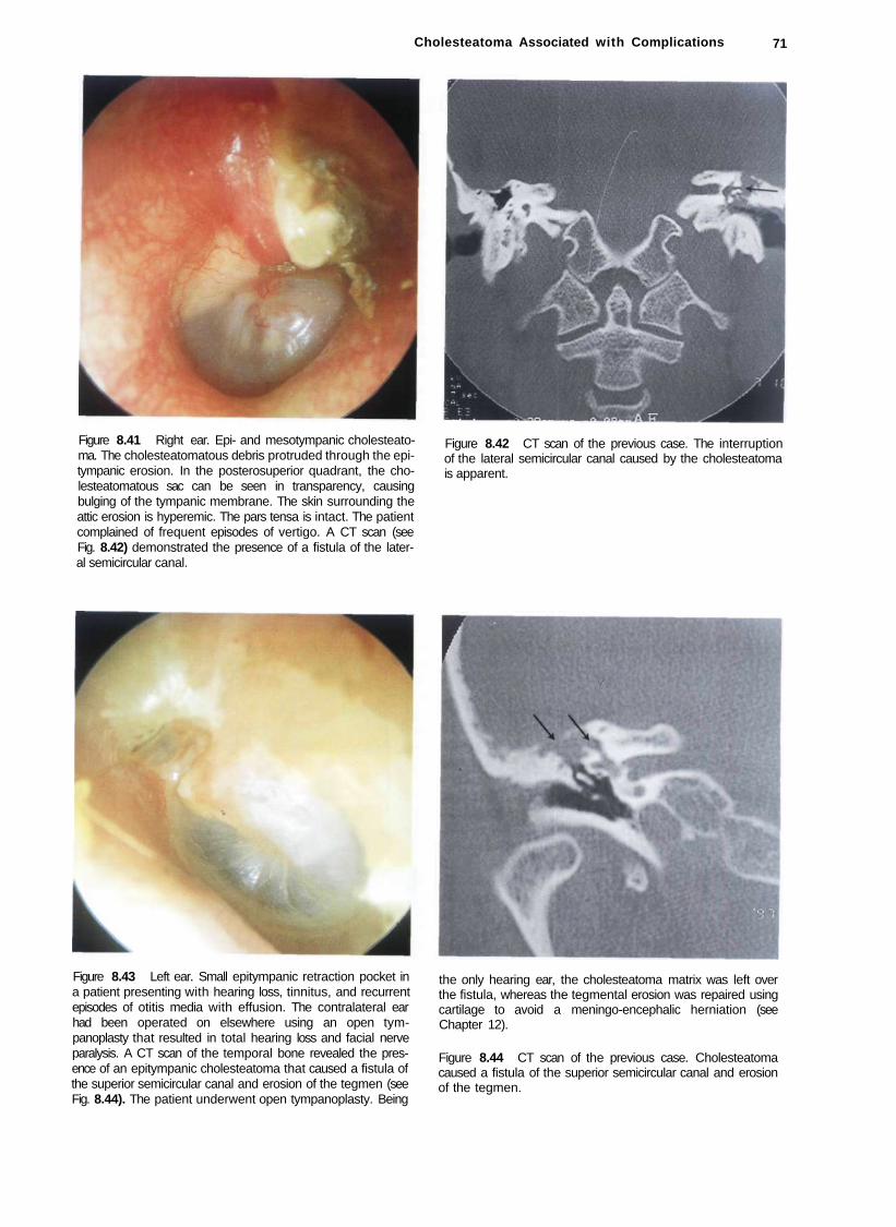
Cholesteatoma Associated with Complications 71
Figure 8.41 Right ear. Epi- and mesotympanic cholesteatoma. The cholesteatomatous debris protruded through the epi-tympanic erosion. In the posterosuperior quadrant, the cholesteatomatous sac can be seen in transparency, causing bulging of the tympanic membrane. The skin surrounding the attic erosion is hyperemic. The pars tensa is intact. The patient complained of frequent episodes of vertigo. A CT scan (see Fig. 8.42) demonstrated the presence of a fistula of the lateral semicircular canal.
Figure 8.42 CT scan of the previous case. The interruption of the lateral semicircular canal caused by the cholesteatoma is apparent.
Figure 8.43 Left ear. Small epitympanic retraction pocket in a patient presenting with hearing loss, tinnitus, and recurrent episodes of otitis media with effusion. The contralateral ear had been operated on elsewhere using an open tympanoplasty that resulted in total hearing loss and facial nerve paralysis. A CT scan of the temporal bone revealed the presence of an epitympanic cholesteatoma that caused a fistula of the superior semicircular canal and erosion of the tegmen (see Fig. 8.44). The patient underwent open tympanoplasty. Being
the only hearing ear, the cholesteatoma matrix was left over the fistula, whereas the tegmental erosion was repaired using cartilage to avoid a meningo-encephalic herniation (see Chapter 12).
Figure 8.44 CT scan of the previous case. Cholesteatoma caused a fistula of the superior semicircular canal and erosion of the tegmen.

72 8 Chronic Suppurative Otitis Media with Cholesteatoma
Figure 8.45 Left ear. This patient had already undergone bilateral radical mastoidectomy elsewhere. He presented with profound bilateral hearing loss and fetid otorrhea from his left ear. During revision surgery, a cholesteatoma causing a cochlear fistula was found. This patient suffered profound hearing loss in the other ear, thus the cholesteatoma matrix was left over the fistula to avoid deaf ear.
Figure 8.46 Polyp in the external auditory canal with purulent discharge. A cholesteatoma is frequently found behind such a polyp. In such cases, biopsy is not indicated as a CT scan is often used to differentiate cholesteatoma from other pathologies (glomus, carcinoid, or carcinoma). A tympanoplasty revealed the presence of a large cholesteatoma occupying the attic and mesotympanum.
Summary
At present, with the diagnostic methods at hand and increased medical care, it is very rare to find a cholesteatoma with intracranial complications (e.g., meningitis, brain abscess, lateral sinus thrombophlebitis, etc.). However, cases of cholesteatoma with massive bone destruction, labyrinthine fistulae, severe sensorineural hearing loss resulting in deaf ear, and facial nerve paralysis are not infrequently encountered. In general, it is not necessary to order a CT scan to diagnose a cholesteatoma. However, in the presence of headache, vertigo, facial nerve paralysis, severe sensorineural hearing loss, or sudden deafness, a high-resolution CT scan of the temporal bone becomes highly important. Axial and coronal cuts without contrast are required. When intracranial complications are suspected, contrast injection is also needed. A labyrinthine fistula is found in less than 10% of cases. The lateral semicircular canal, being the most superficial, is the most commonly involved. Treatment of a labyrinthine fistula depends on the type (bony or membranous) and size of the fistula. A tegmental erosion can be repaired using cartilage and bone pate. Facial nerve paralysis is either due to infection of the exposed nerve or secondary to compression by the cholesteatoma. In the majority of cases, removing the
cholesteatoma and clearing the infection are sufficient for the paralysis to resolve. It is very rare to find fibrosis or thinning of the nerve. In these cases, facial nerve reconstruction varies from rerouting and end-to-end anastomosis to nerve grafting, according to the degree of injury and length of the injured segment.
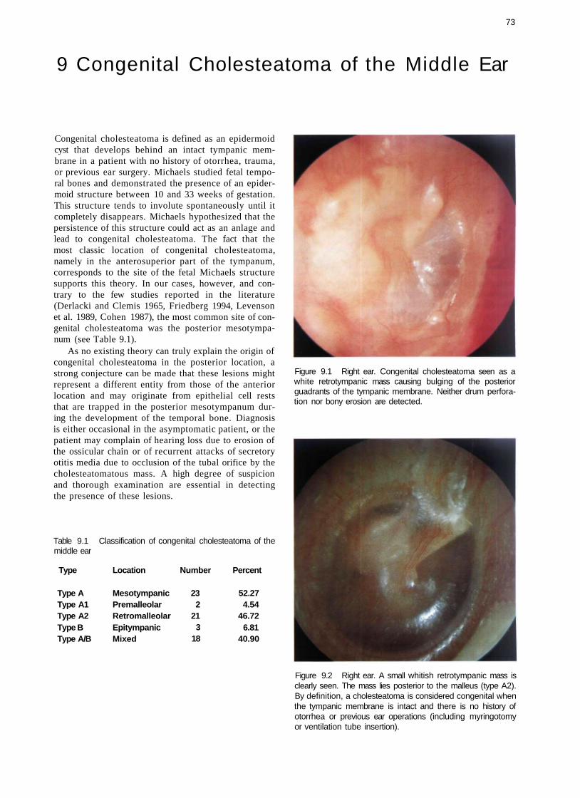
73
9 Congenital Cholesteatoma of the Middle Ear
Congenital cholesteatoma is defined as an epidermoid cyst that develops behind an intact tympanic membrane in a patient with no history of otorrhea, trauma, or previous ear surgery. Michaels studied fetal temporal bones and demonstrated the presence of an epidermoid structure between 10 and 33 weeks of gestation. This structure tends to involute spontaneously until it completely disappears. Michaels hypothesized that the persistence of this structure could act as an anlage and lead to congenital cholesteatoma. The fact that the most classic location of congenital cholesteatoma, namely in the anterosuperior part of the tympanum, corresponds to the site of the fetal Michaels structure supports this theory. In our cases, however, and contrary to the few studies reported in the literature (Derlacki and Clemis 1965, Friedberg 1994, Levenson et al. 1989, Cohen 1987), the most common site of congenital cholesteatoma was the posterior mesotympa-num (see Table 9.1).
As no existing theory can truly explain the origin of congenital cholesteatoma in the posterior location, a strong conjecture can be made that these lesions might represent a different entity from those of the anterior location and may originate from epithelial cell rests that are trapped in the posterior mesotympanum during the development of the temporal bone. Diagnosis is either occasional in the asymptomatic patient, or the patient may complain of hearing loss due to erosion of the ossicular chain or of recurrent attacks of secretory otitis media due to occlusion of the tubal orifice by the cholesteatomatous mass. A high degree of suspicion and thorough examination are essential in detecting the presence of these lesions.
Table 9.1 Classification of congenital cholesteatoma of the middle ear
Type Location Number Percent
Type A Type A1 Type A2 Type B Type A/B
Mesotympanic Premalleolar Retromalleolar Epitympanic Mixed
23 2
21 3
18
52.27 4.54
46.72 6.81
40.90
Figure 9.1 Right ear. Congenital cholesteatoma seen as a white retrotympanic mass causing bulging of the posterior guadrants of the tympanic membrane. Neither drum perforation nor bony erosion are detected.
Figure 9.2 Right ear. A small whitish retrotympanic mass is clearly seen. The mass lies posterior to the malleus (type A2). By definition, a cholesteatoma is considered congenital when the tympanic membrane is intact and there is no history of otorrhea or previous ear operations (including myringotomy or ventilation tube insertion).
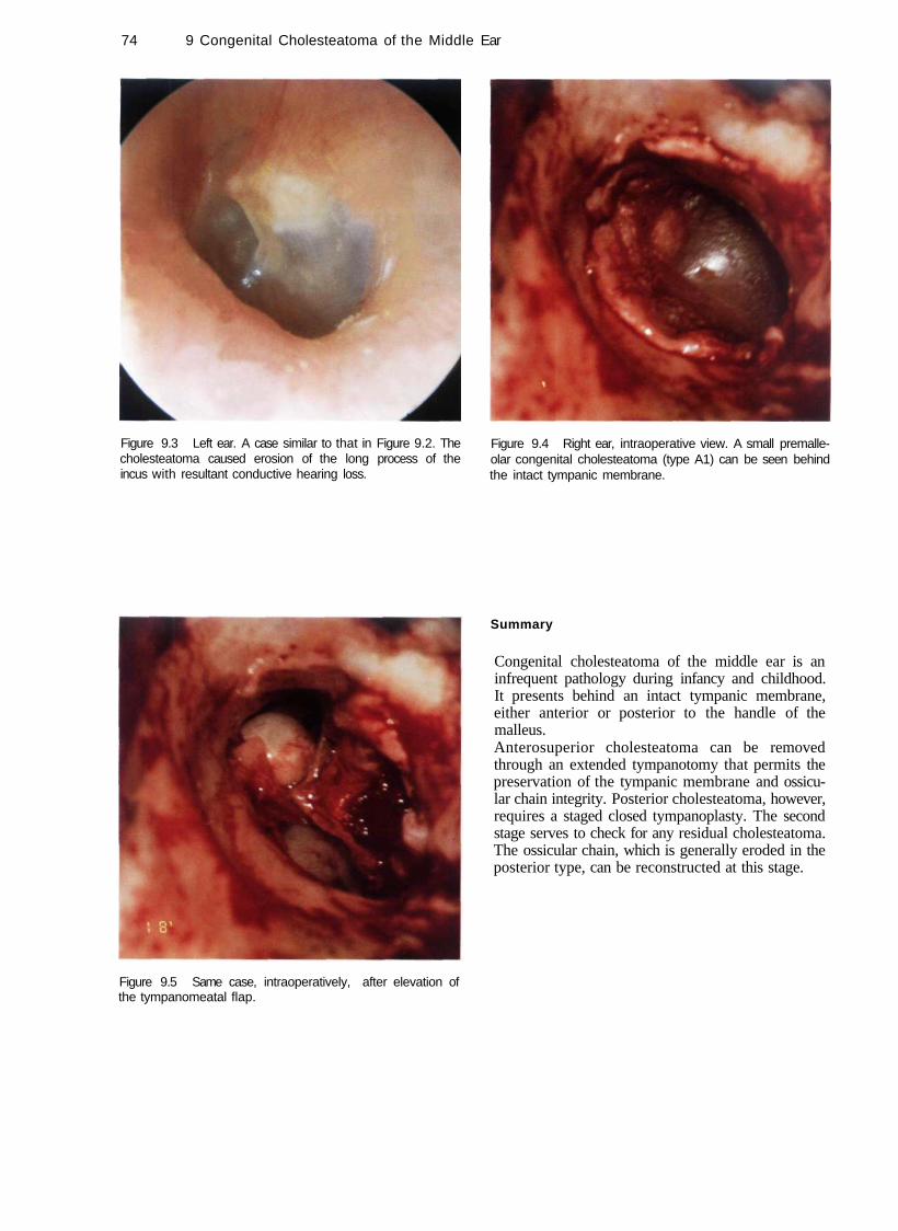
74 9 Congenital Cholesteatoma of the Middle Ear
Figure 9.3 Left ear. A case similar to that in Figure 9.2. The cholesteatoma caused erosion of the long process of the incus with resultant conductive hearing loss.
Figure 9.5 Same case, intraoperatively, after elevation of the tympanomeatal flap.
Figure 9.4 Right ear, intraoperative view. A small premalle-olar congenital cholesteatoma (type A1) can be seen behind the intact tympanic membrane.
Summary
Congenital cholesteatoma of the middle ear is an infrequent pathology during infancy and childhood. It presents behind an intact tympanic membrane, either anterior or posterior to the handle of the malleus. Anterosuperior cholesteatoma can be removed through an extended tympanotomy that permits the preservation of the tympanic membrane and ossicular chain integrity. Posterior cholesteatoma, however, requires a staged closed tympanoplasty. The second stage serves to check for any residual cholesteatoma. The ossicular chain, which is generally eroded in the posterior type, can be reconstructed at this stage.
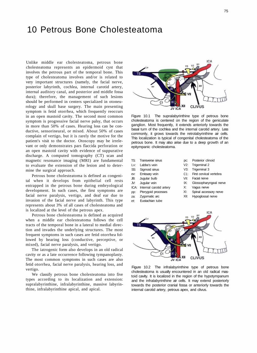
75
10 Petrous Bone Cholesteatoma
Unlike middle ear cholesteatoma, petrous bone cholesteatoma represents an epidermoid cyst that involves the petrous part of the temporal bone. This type of cholesteatoma involves and/or is related to very important structures (namely, the facial nerve, posterior labyrinth, cochlea, internal carotid artery, internal auditory canal, and posterior and middle fossa dura); therefore, the management of such lesions should be performed in centers specialized in otoneurology and skull base surgery. The main presenting symptom is fetid otorrhea, which frequently reoccurs in an open mastoid cavity. The second most common symptom is progressive facial nerve palsy, that occurs in more than 50% of cases. Hearing loss can be conductive, sensorineural, or mixed. About 50% of cases complain of vertigo, but it is rarely the motive for the patient's visit to the doctor. Otoscopy may be irrelevant or only demonstrates pars flaccida perforation or an open mastoid cavity with evidence of suppurative discharge. A computed tomography (CT) scan and magnetic resonance imaging (MRI) are fundamental to evaluate the extension of the lesion and to determine the surgical approach.
Petrous bone cholesteatoma is defined as congenital when it develops from epithelial cell rests entrapped in the petrous bone during embryological development. In such cases, the first symptoms are facial nerve paralysis, vertigo, and deaf ear due to invasion of the facial nerve and labyrinth. This type represents about 3% of all cases of cholesteatoma and is localized at the level of the petrous apex.
Petrous bone cholesteatoma is defined as acquired when a middle ear cholesteatoma follows the cell tracts of the temporal bone in a lateral to medial direction and invades the underlying structures. The most frequent symptoms in such cases are fetid otorrhea followed by hearing loss (conductive, perceptive, or mixed), facial nerve paralysis, and vertigo.
The iatrogenic form also develops in an old radical cavity or as a late occurrence following tympanoplasty. The most common symptoms in such cases are also fetid otorrhea, facial nerve paralysis, hearing loss, and vertigo.
We classify petrous bone cholesteatoma into five types according to its localization and extension: supralabyrinthine, infralabyrinthine, massive labyrinthine, infralabyrinthine apical, and apical.
Figure 10.1 The supralabyrinthine type of petrous bone cholesteatoma is centered on the region of the geniculate ganglion. Most frequently, it extends anteriorly towards the basal turn of the cochlea and the internal carotid artery. Less commonly, it grows towards the retrolabyrinthine air cells. This localization is typical of congenital cholesteatoma of the petrous bone. It may also arise due to a deep growth of an epitympanic cholesteatoma.
TS: Lv: SS: ev: JB: JV: ICA: pp: za: et:
Transverse sinus Labbe's vein Sigmoid sinus Emissary vein Jugular bulb Jugular vein Internal carotid artery Pterygoid processes Zygomatic arc Eustachian tube
pc: V2: V3: C1: VII: IX: X: XI: XII:
Posterior clinoid Trigeminal 2 Trigeminal 3 First cervical vertebra Facial nerve Glossopharyngeal nerve Vagus nerve Spinal accessory nerve Hypoglossal nerve
Figure 10.2 The infralabyrinthine type of petrous bone cholesteatoma is usually encountered in an old radical mas-toid cavity. It is localized in the region of the hypotympanum and the infralabyrinthine air cells. It may extend posteriorly towards the posterior cranial fossa or anteriorly towards the internal carotid artery, petrous apex, and clivus.

76 10 Pet rous B o n e C h o l e s t e a t o m a
ICA ICA
JVICA CLIVUS
Figure 10.3 The massive labyrinthine type of petrous bone cholesteatoma largely involves the posterior labyrinth and the cochlea. It may extend anteriorly towards the internal carotid artery, medially towards the internal auditory canal, posteriorly towards the posterior fossa, or interiorly towards the infra-labyrinthine compartment. Abbreviations are given in Figure 10.1.
JV ICA CLIVUS
Figure 10.4 The infralabyrinthine apical type of petrous bone cholesteatoma originates from the infralabyrinthine or apical compartments. When it originates from the former, it extends into the petrous apex. In some cases it may grow towards the sphenoid sinus or the horizontal portion of the internal carotid artery.
ICA
JV ICA CLIVUS
Figure 10.5 The apical type of petrous bone cholesteatoma is a rare congenital lesion. It may solely involve the apical com-partment, causing erosion of it. It may involve the trigeminal nerve or more posteriorly the posterior cranial fossa. It may also engulf the horizontal portion of the internal carotid artery.

Figure 10.6 Left acquired or iatrogenic supralabyrinthine petrous bone cholesteatoma in a radical cavity. A whitish retrotympanic mass is seen at the level of the second portion of the facial nerve. The patient presented with progressive facial nerve paralysis and total hearing loss. A correct diagnosis depends not only on otoscopy but also on the symptomatology (facial paralysis, anacusis) and a high-resolution CT scan.
Petrous Bone Cholesteatoma 77
Figure 10.7 CT scan of the case presented in Figure 10.6, axial section. Involvement of the lateral semicircular canal and the vestibule is well visualized. The cholesteatoma invades the cochlea anteriorly, while medially it reaches the fundus of the internal auditory canal. The posterior semicircular canal is not invaded.
Figure 10.8 CT scan of the case presented in Figure 10.6, coronal section. The medial extension of the cholesteatoma can be appreciated.
Figure 10.9 Postoperative CT scan. A transcochlear approach was performed and the operative cavity was obliterated with abdominal fat.
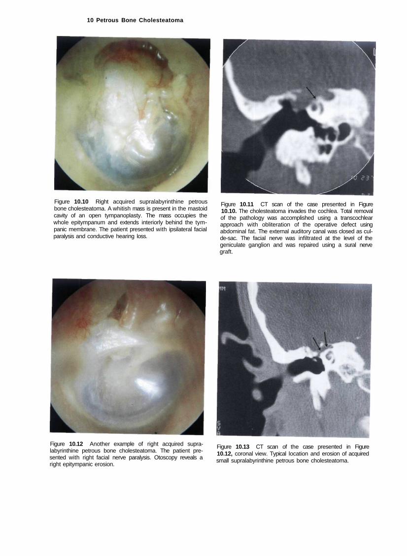
10 Petrous Bone Cholesteatoma
Figure 10.10 Right acquired supralabyrinthine petrous bone cholesteatoma. A whitish mass is present in the mastoid cavity of an open tympanoplasty. The mass occupies the whole epitympanum and extends interiorly behind the tympanic membrane. The patient presented with ipsilateral facial paralysis and conductive hearing loss.
Figure 10.12 Another example of right acquired supra-labyrinthine petrous bone cholesteatoma. The patient presented with right facial nerve paralysis. Otoscopy reveals a right epitympanic erosion.
Figure 10.11 CT scan of the case presented in Figure 10.10. The cholesteatoma invades the cochlea. Total removal of the pathology was accomplished using a transcochlear approach with obliteration of the operative defect using abdominal fat. The external auditory canal was closed as cul-de-sac. The facial nerve was infiltrated at the level of the geniculate ganglion and was repaired using a sural nerve graft.
Figure 10.13 CT scan of the case presented in Figure 10.12, coronal view. Typical location and erosion of acquired small supralabyrinthine petrous bone cholesteatoma.
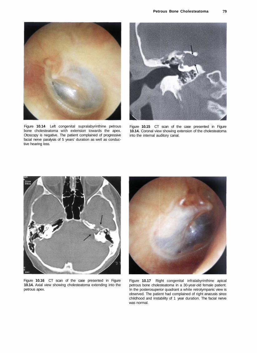
Petrous Bone Cholesteatoma 79
Figure 10.14 Left congenital supralabyrinthine petrous bone cholesteatoma with extension towards the apex. Otoscopy is negative. The patient complained of progressive facial nerve paralysis of 5 years' duration as well as conductive hearing loss.
Figure 10.15 CT scan of the case presented in Figure 10.14. Coronal view showing extension of the cholesteatoma into the internal auditory canal.
Figure 10.16 CT scan of the case presented in Figure 10.14. Axial view showing cholesteatoma extending into the petrous apex.
Figure 10.17 Right congenital infralabyrinthine apical petrous bone cholesteatoma in a 30-year-old female patient. In the posterosuperior quadrant a white retrotympanic view is observed. The patient had complained of right anacusis since childhood and instability of 1 year duration. The facial nerve was normal.
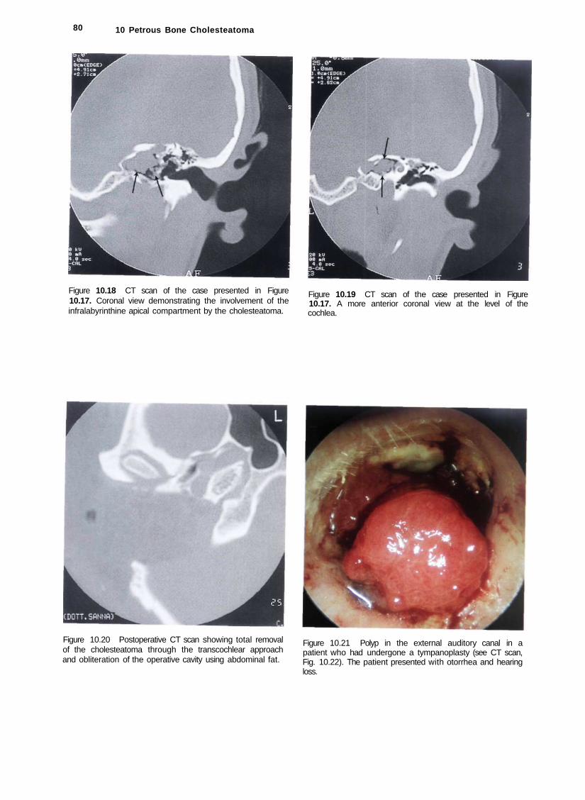
80 10 Petrous Bone Cholesteatoma
Figure 10.18 CT scan of the case presented in Figure 10.17. Coronal view demonstrating the involvement of the infralabyrinthine apical compartment by the cholesteatoma.
Figure 10.19 CT scan of the case presented in Figure 10.17. A more anterior coronal view at the level of the cochlea.
Figure 10.20 Postoperative CT scan showing total removal of the cholesteatoma through the transcochlear approach and obliteration of the operative cavity using abdominal fat.
Figure 10.21 Polyp in the external auditory canal in a patient who had undergone a tympanoplasty (see CT scan, Fig. 10.22). The patient presented with otorrhea and hearing loss.

Petrous Bone Cholesteatoma 81
Figure 10.22 CT scan of the case presented in Figure 10.21. A large infralabyrinthine apical petrous bone cholesteatoma extending to the cavernous sinus and to the sphenoid sinus can be seen. Total removal was achieved using an infratemporal fossa approach type B.
Figure 10.23 Postoperative CT scan showing total removal
Figure 10.24 Left acquired petrous bone cholesteatoma of the massive type. The patient had complained of fetid otorrhea and hearing loss since early childhood. Six months before presentation, he started to experience facial nerve paralysis. A radical mastoidectomy was performed in another center with partial removal of the pathology. The second and third portions of the facial nerve can be observed in the mastoid cavity. The patient underwent surgery using a transcochlear approach to obliterate of the cavity with abdominal fat.
Figure 10.25 CT scan of the case presented in Figure 10.24 demonstrating cholesteatoma invading the labyrinth.
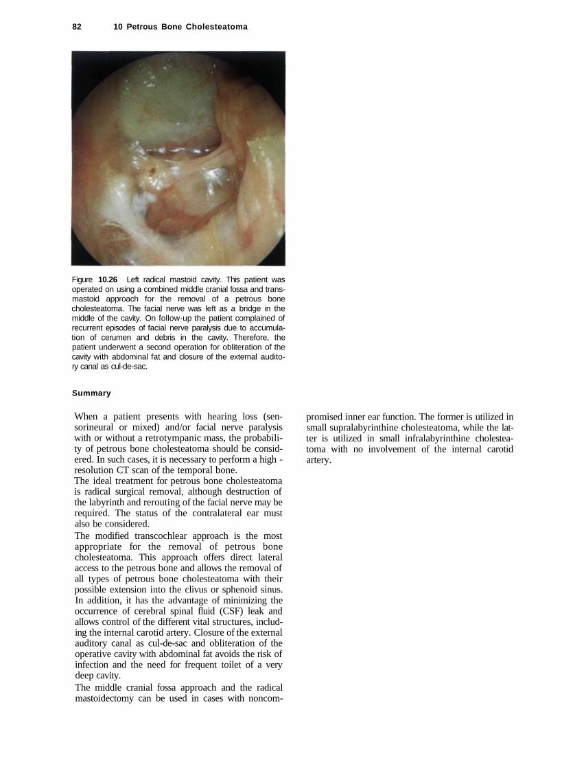
82 10 Petrous Bone Cholesteatoma
Figure 10.26 Left radical mastoid cavity. This patient was operated on using a combined middle cranial fossa and trans-mastoid approach for the removal of a petrous bone cholesteatoma. The facial nerve was left as a bridge in the middle of the cavity. On follow-up the patient complained of recurrent episodes of facial nerve paralysis due to accumulation of cerumen and debris in the cavity. Therefore, the patient underwent a second operation for obliteration of the cavity with abdominal fat and closure of the external auditory canal as cul-de-sac.
Summary
When a patient presents with hearing loss (sensorineural or mixed) and/or facial nerve paralysis with or without a retrotympanic mass, the probability of petrous bone cholesteatoma should be considered. In such cases, it is necessary to perform a high -resolution CT scan of the temporal bone. The ideal treatment for petrous bone cholesteatoma is radical surgical removal, although destruction of the labyrinth and rerouting of the facial nerve may be required. The status of the contralateral ear must also be considered. The modified transcochlear approach is the most appropriate for the removal of petrous bone cholesteatoma. This approach offers direct lateral access to the petrous bone and allows the removal of all types of petrous bone cholesteatoma with their possible extension into the clivus or sphenoid sinus. In addition, it has the advantage of minimizing the occurrence of cerebral spinal fluid (CSF) leak and allows control of the different vital structures, including the internal carotid artery. Closure of the external auditory canal as cul-de-sac and obliteration of the operative cavity with abdominal fat avoids the risk of infection and the need for frequent toilet of a very deep cavity. The middle cranial fossa approach and the radical mastoidectomy can be used in cases with noncom-
promised inner ear function. The former is utilized in small supralabyrinthine cholesteatoma, while the latter is utilized in small infralabyrinthine cholesteatoma with no involvement of the internal carotid artery.

83
11 Glomus Tumors (Chemodectomas)
The glomus body was first described by Guild in 1941 as a small highly vascular mass of epithelioid cells located in the region of the adventitia of the jugular bulb. In 1953, Guild described glomus formations along the tympanic branches of the glossopharyngeal and vagus nerves (Jacobson's and Arnold's nerves, respectively).
Glomus bodies are mainly found in the tympanic region, jugular bulb, at the carotid bifurcation, and related to the vagus nerve. They are classified as paraganglia that are derived from the neural crest. While the carotid and vagal bodies function as chemorecep-tors stimulated by the changes in the oxygen tension, tympanic and jugular bulb paraganglia do not exhibit this function.
The term glomus tympanicum is reserved for tumors that originate from the mesotympanum while the term glomus jugulare is attributed to those cases that arise from the jugular bulb or the hypotympanum with secondary invasion of the bulb. These tumors are highly vascular and they derive the blood supply mainly from the ascending pharyngeal artery. It is claimed that they have a hereditary transmission as autosomal dominant traits with penetrance that increases with age.
In the majority of cases, the initial symptoms are hearing loss (conductive, sensorineural, or mixed) and pulsatile tinnitus synchronous with pulse. The tumor can extend into the labyrinth, causing vertigo of peripheral origin; towards the jugular foramen, leading to deficits of one or more of the lower cranial nerves (IX-XI); or towards the occipital condyle, leading to hypoglossal nerve paralysis. Patients suffering from preoperative affection of the lower cranial nerves run a better postoperative course as compensation of the contralateral side would have already started. The contralateral vocal cord compensates by crossing the midline to meet the paralyzed cord, thereby markedly reducing the risk of aspiration pneumonia. On the other hand, patients with preoperative intact lower cranial nerves in whom the nerves are sacrificed during the operation suffer from deglutition problems in the postoperative course. Nasogastric feeding is used in such cases and oral feeding is resumed only when compensation from the contralateral side occurs. A useful alternative is vocal cord medialization either by Teflon injection or by medialization thyroplasty using cartilage or silicon.
The tumor can also extend into the petrous apex, leading to paralysis of the abducent nerve and trigeminal neuralgia, or invade the mastoid, resulting in facial nerve paralysis. Further extension can also occur in the external auditory canal. Tumors occupying the exter
nal auditory canal can lead to serous or purulent otorrhea due to irritation of the skin and retention of squamae and epithelial debris. Hemorrhagic discharge rarely occurs.
Fisch classified glomus tumors into four classes based on location and extension seen on high-resolution CT scans (see Table 11.1 and Figs. 11.1-11.6).
On otoscopy, a retrotympanic pulsatile mass is usually seen in the inferior quadrants. The mass is red or bordeaux red. In some cases the mass may have a reddish-blue color due to the presence of middle ear effusion secondary to eustachian tube blockage. The tumor may be seen as a polyp in the external auditory canal either due to erosion of the floor of the canal or to the tumor breaking through the tympanic membrane.
The diagnosis can be made clinically (history and otoscopic findings). Computed tomography (CT) with contrast and magnetic resonance imaging (MRI) with gadolinium allow exact definition of the tumor extension. Radiology also helps to differentiate between glomus tumors and other lesions such as aberrant carotid artery, high jugular bulb, cholesterol granuloma, or meningioma extending into the middle ear. Carotid and vertebral angiography allows identification of the arteries supplying the tumor; and they should be embolized before surgery to avoid excessive intraoperative bleeding.
In cases in which the horizontal carotid artery is engulfed by the tumor, the balloon occlusion test is indispensable for studying the perfusion by the contralateral carotid artery as well as for the safety of the closure of the involved carotid.
Table 11.1 Classification of glomus tumors according to Fisch (1978)
ICA = internal carotid artery; FL = anterior foramen lacerum
Class A: Glomus tympanicum Class B: Tympanomastoid Class C: Glomus jugulare
CI: Carotid foramen C2: Vertical ICA until genu C3: Horizontal ICA C4: ICA + FL
Class D: Intracranial extension De (1-2): Intracranial extradural Di (1-2): Intracranial intradural
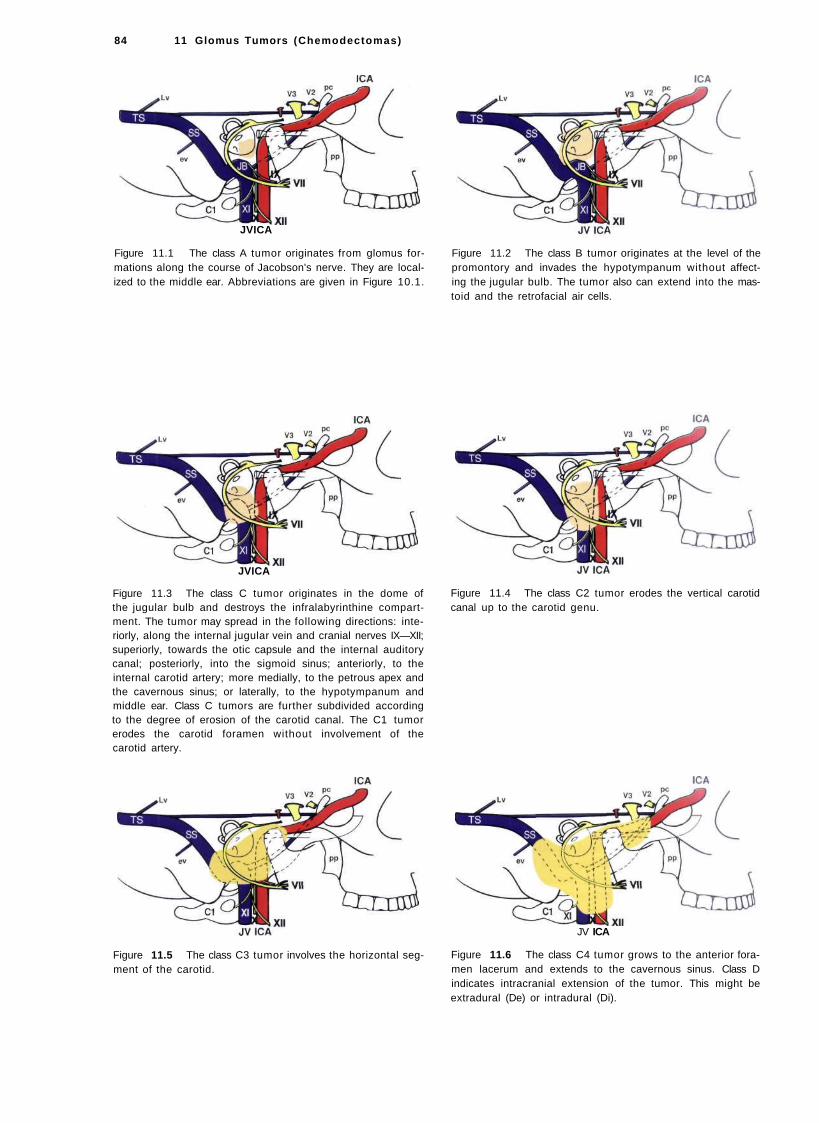
84 11 Glomus Tumors (Chemodectomas)
JVICA
Figure 11.1 The class A tumor originates from glomus formations along the course of Jacobson's nerve. They are localized to the middle ear. Abbreviations are given in Figure 10.1.
JVICA
Figure 11.3 The class C tumor originates in the dome of the jugular bulb and destroys the infralabyrinthine compartment. The tumor may spread in the following directions: interiorly, along the internal jugular vein and cranial nerves IX—XII; superiorly, towards the otic capsule and the internal auditory canal; posteriorly, into the sigmoid sinus; anteriorly, to the internal carotid artery; more medially, to the petrous apex and the cavernous sinus; or laterally, to the hypotympanum and middle ear. Class C tumors are further subdivided according to the degree of erosion of the carotid canal. The C1 tumor erodes the carotid foramen without involvement of the carotid artery.
Figure 11.5 The class C3 tumor involves the horizontal segment of the carotid.
Figure 11.2 The class B tumor originates at the level of the promontory and invades the hypotympanum without affecting the jugular bulb. The tumor also can extend into the mastoid and the retrofacial air cells.
Figure 11.4 The class C2 tumor erodes the vertical carotid canal up to the carotid genu.
JV ICA
Figure 11.6 The class C4 tumor grows to the anterior foramen lacerum and extends to the cavernous sinus. Class D indicates intracranial extension of the tumor. This might be extradural (De) or intradural (Di).

Glomus Tumors (Chemodectomas) 85
Figure 11.7 Left ear. Glomus tympanicum or class A tumor. The small red mass behind the anteroinferior quadrant is localized on the promontory and does not extend towards the hypotympanum (see Fig. 11.7).
Figure 11.8 CT scan of the case presented in Figure 11.7. The lesion is limited to the region of the promontory. There are no visible signs of bone erosion.
Figure 11.9 Left ear. Class A glomus tumor. The tumor is again limited to the promontory (see Figs. 11.10 and 11.11).
Figure 11.10 CT scan of the case described in Figure 11.9.
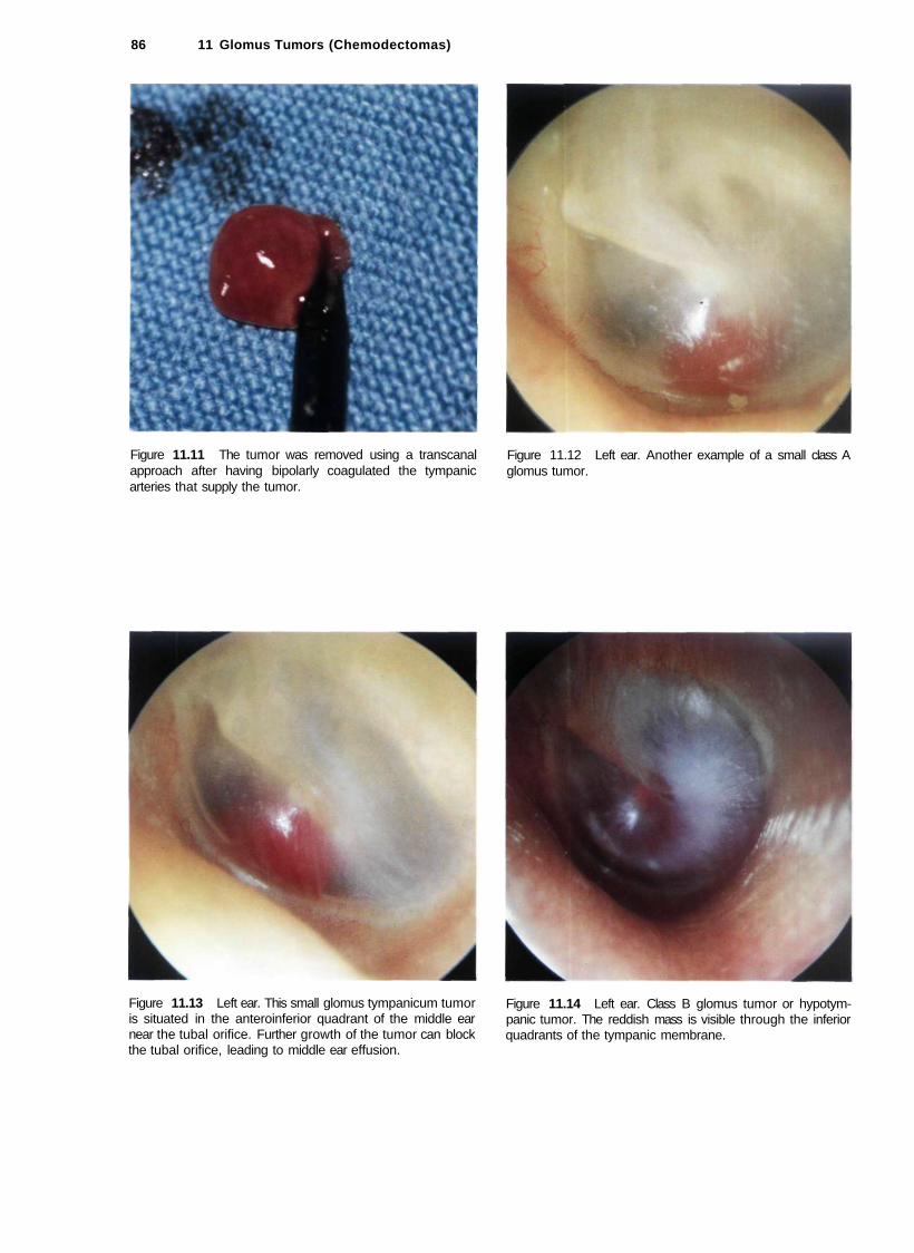
86 11 Glomus Tumors (Chemodectomas)
Figure 11.11 The tumor was removed using a transcanal approach after having bipolarly coagulated the tympanic arteries that supply the tumor.
Figure 11.13 Left ear. This small glomus tympanicum tumor is situated in the anteroinferior quadrant of the middle ear near the tubal orifice. Further growth of the tumor can block the tubal orifice, leading to middle ear effusion.
Figure 11.12 Left ear. Another example of a small class A glomus tumor.
Figure 11.14 Left ear. Class B glomus tumor or hypotym-panic tumor. The reddish mass is visible through the inferior quadrants of the tympanic membrane.

Glomus Tumors (Chemodectomas) 87
Figure 11.15 CT of the case presented in Figure 11.14. Tumor extension towards the hypotympanum is observed. There is no erosion of the bony plate covering the jugular bulb.
Figure 11.16 Right ear. Class B glomus tumor. The highly vascular red tumor mass pushes the tympanic membrane laterally. A middle ear effusion is present.
Figure 11.17 Right ear. Class B glomus tumor. An air-fluid level due to middle ear effusion is seen together with the tumor. A tympanoplasty removed all of the tumor while conserving the excellent preoperative hearing.
Figure 11.18 Left ear. Type B glomus tumor. The tumor causes bulging of the posterior quadrants of the tympanic membrane (see CT scan, Fig. 11.19).

88 11 Glomus Tumors (Chemodectomas)
Figure 11.19 CT scan of the case in Figure 11.18. An axial section demonstrates the presence of effusion in the mastoid due to retention.
Figure 11.21 Right ear. Reddish mass protruding from the inferior wall of the external auditory canal.
Figure 11.20 CT scan of the case in Figure 11.18. The tumor extends to the hypotympanum but does not erode the bone overlying the dome of the jugular bulb.
Figure 11.22 CT scan of the previous case. Axial view demonstrating the erosion caused by the tumor of the bone overlying the jugular bulb. This tumor can be considered an intermediate class between B and C. The tumor is localized in the hypotympanum and extends to the jugular bulb but does not invade it.
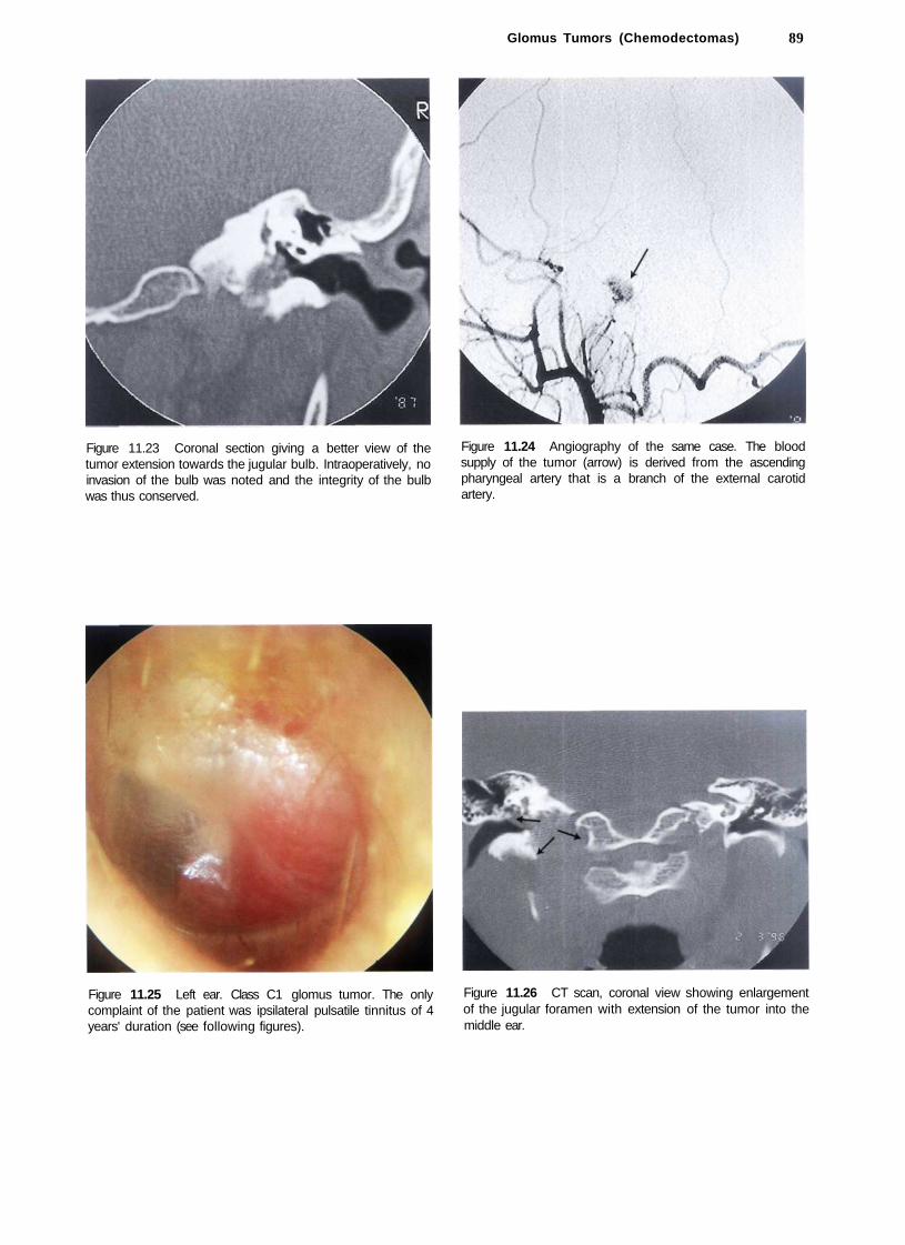
Glomus Tumors (Chemodectomas) 89
Figure 11.23 Coronal section giving a better view of the tumor extension towards the jugular bulb. Intraoperatively, no invasion of the bulb was noted and the integrity of the bulb was thus conserved.
Figure 11.24 Angiography of the same case. The blood supply of the tumor (arrow) is derived from the ascending pharyngeal artery that is a branch of the external carotid artery.
Figure 11.25 Left ear. Class C1 glomus tumor. The only complaint of the patient was ipsilateral pulsatile tinnitus of 4 years' duration (see following figures).
Figure 11.26 CT scan, coronal view showing enlargement of the jugular foramen with extension of the tumor into the middle ear.

90 11 Glomus Tumors (Chemodectomas)
Figure 11.27 CT scan, axial view. The jugular foramen is enlarged. Irregular erosion of the borders of the jugular foramen can be observed (differential diagnosis with lower cranial nerves' schwannoma).
Figure 11.28 Axial view demonstrates that the horizontal segment of the internal carotid artery is free of tumor.
Figure 11.29 Angiography demonstrating that the blood supply of the tumor comes from the ascending pharyngeal, the occipital, and the posterior auricular arteries.
Figure 11.30 MRI with gadolinium. The tumor is enhancing except for some flow-void zones corresponding to large vascular spaces. This picture is pathognomonic of glomus tumors.

Figure 11.31 Class C2 De2 glomus jugulare tumor of the left ear. The patient complained of pulsatile tinnitus, hearing loss, and 2 months before presentation started to suffer from dysphonia, dysphagia, and hypoglossal paresis. The affection of the lower cranial nerves was progressive in nature. It resulted from compression by the slowly growing tumor.
Figure 11.33 MRI demonstrating tumor in contact with the medial aspect of the horizontal carotid artery and the posterior fossa dura without infiltrating it.
Glomus Tumors (Chemodectomas) 91
Figure 11.32 CT scan of the case presented in Figure 11.31. The marked erosion of the jugular foramen and the vertical portion of the carotid canal can be appreciated.
Figure 11.34 Postoperative CT scan demonstrating tumor removal using an infratemporal fossa approach type A.

92 11 Glomus Tumors (Chemodectomas)
Figure 11.35 Right ear. Class C3 Di2 glomus jugulare tumor. The patient complained of pulsatile tinnitus and mixed hearing loss of 12 months' duration.
Figure 11.37 MRI, coronal view after first-stage removal of the extradural component of the tumor using an infratemporal fossa approach type A. The fat (F) obliterating the operative cavity can be seen. The intradural tumor residue (T) is also observed. Staging is necessary in such cases to avoid communication between the subarachnoid space and the wide open neck spaces.
Figure 11.36 MRI, sagittal view demonstrating intradural extension of the tumor.
Figure 11.38 Postoperative CT scan after the second-stage removal of the tumor through a petro-occipital trans-sigmoid approach (POTS).
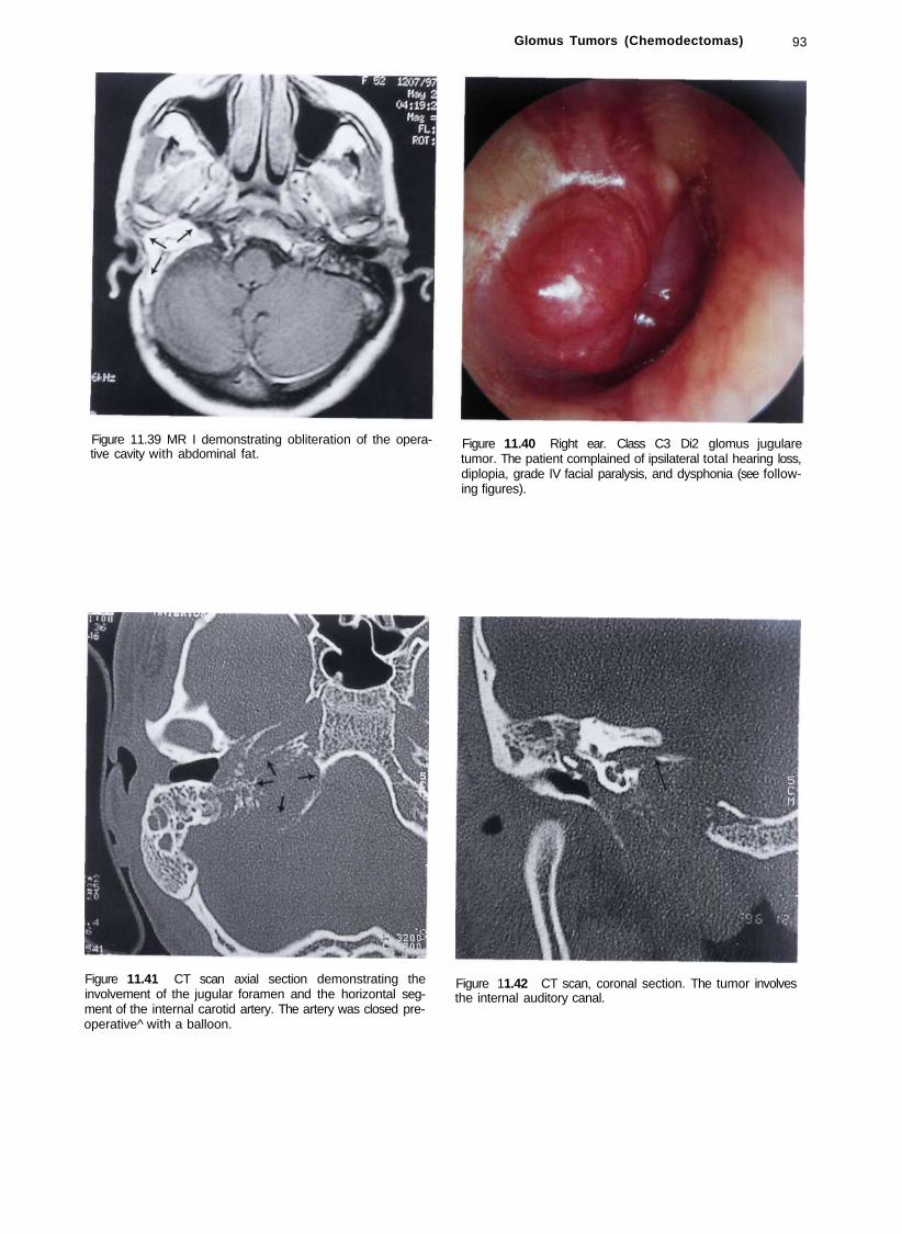
Glomus Tumors (Chemodectomas) 93
Figure 11.39 MR I demonstrating obliteration of the operative cavity with abdominal fat.
Figure 11.40 Right ear. Class C3 Di2 glomus jugulare tumor. The patient complained of ipsilateral total hearing loss, diplopia, grade IV facial paralysis, and dysphonia (see following figures).
Figure 11.41 CT scan axial section demonstrating the involvement of the jugular foramen and the horizontal segment of the internal carotid artery. The artery was closed preoperative^ with a balloon.
Figure 11.42 CT scan, coronal section. The tumor involves the internal auditory canal.

94 11 Glomus Tumors (Chemodectomas)
Figure 11.43 MRI, axial view giving a global idea of the Figure 11.44 MRI, sagittal view, extra- and intradural extension of the tumor.
Figure 11.45 Angiography before embolization. Figure 11.46 Angiography showing marked reduction of the tumor vascularity following embolization.

Figure 11.49 MRI, sagittal view demonstrating the intradural extension of the tumor as well as the inferior extension towards C1 and C2.
Glomus Tumors (Chemodectomas) 95
Figure 11.50 Preoperative CT scan. The jugular foramen is enlarged, with involvement of the foramen magnum.
Figure 11.47 CT scan performed after first-stage removal of the extradural part of the tumor using an infratemporal fossa approach type A. Staging is necessary to avoid communication between the subarachnoid spaces and the neck spaces. The balloon used for the closure of the internal carotid artery can be seen (arrow).
Figure 11.48 Left ear. Class C2 Di2 glomus jugulare tumor. The patient complained of hearing loss and pulsatile tinnitus of 2 years' duration. He also complained of dysphonia, dysphagia, paralysis of the left half of the tongue, and paresis of the lower face.

96 11 Glomus Tumors (Chemodectomas)
Figure 11.51 MRI with gadolinium after removal of the extradural part using an infratemporal fossa approach type A. Fat is seen obliterating the operative cavity (F). The intradural tumor residue at the level of the foramen magnum is noted (T).
Figure 11.53 CT scan following the second-stage removal of the intradural portion of the tumor. The removal of a large part of the left occipital condyle is also shown.
Figure 11.52 CT scan following the second-stage removal of the intradural portion of the tumor using an extreme lateral approach. The balloon used to close the vertebral artery is visible.
Figure 11.54 Another example of a large class C3 Di2 glomus tumor.

Figure 11.55 MRI of the case in Figure 11.54 (T= tumor).
Summary
Because of the complex anatomy of the temporal bone and the structures at the base of the skull, as well as the invasiveness, rich vascularity, and aggressive behavior of glomus tumors, surgery for these difficult lesions is problematic. Glomus tumors generally present with hearing loss and pulsatile tinnitus. When the lower cranial nerves are invaded, a jugular foramen syndrome becomes manifest. Otoscopy usually reveals a reddish retrotympanic mass. A definitive diagnosis is obtained after neuro-radiological studies are performed. These include a high-resolution CT scan with bony window, MRI with and without gadolinium, and digital subtraction angiography. Radiological studies are essential not only to confirm the diagnosis and define the exact tumor class, but also to properly evaluate these tumors. The neuroradiologist should be able to inform the surgeon about the following: • Details of the osseous lesion • Involvement of the jugular bulb and foramen • Exact involvement of the temporal bone • The presence of inner ear invasion • The relationship between the fallopian canal and the
tumor • Carotid canal erosion and exact involvement of the
internal carotid artery
Glomus Tumors (Chemodectomas) 97
• Invasion of the petrous apex and clivus • Details regarding the relationship between the tumor
and surrounding soft tissues, e.g.: - degree of neck extension - infratemporal fossa involvement - intracranial and intradural extension
Radiology also helps to determine the superior and inferior extension of the tumor, the possibility of other associated lesions (e.g., contralateral glomus or carotid body tumor), as well as the patency of the contralateral sigmoid sinus and internal jugular vein. In class C and D tumors, selective digital subtraction angiography is essential. Arteriography is performed for both ipsilateral and contralateral internal and external carotids and for the vertebrobasilar system. A study of the venous phase is also of great importance. Arteriography of the external carotid artery defines the exact feeding vessels for further embolization. In all tumors of class C and D, embolization is fundamental. Arteriography of the internal carotid artery shows vascularization from the caroticotympanic artery and from the cavernous branches of the artery as well as the exact status of arterial invasion by the tumor. Study of the vertebrobasilar system demonstrates the vascularization of intracranial extension of the tumor from muscular, meningeal, and parenchymal (PICA, AICA) branches. Arterial supply from these latter branches indicates a definite intradural extension of the tumor. This study also provides indications for the possibility of embolizing muscular or meningeal branches. When arteriography shows clear involvement of the internal carotid artery in its horizontal segment (C3 and C4 tumors), a balloon occlusion test to evaluate the collateral circulation and the possibility of sacrificing the artery is necessary. In some selected cases, when the temporary balloon occlusion test is negative, it might be necessary to perform a permanent closure of the artery 30 to 40 days before operation. In 1978, Fisch classified these lesions into four types: A, B, C, and D. He introduced the type A infratemporal fossa approach for the management of tumors localized in the jugular foramen that were considered inoperable at that time due to the presence of the facial nerve in the middle of the operative field and the inaccessibility of the internal carotid artery and petrous apex. To overcome these obstacles, Fisch proposed anterior rerouting of the facial nerve, giving direct access to the whole intratemporal course of the internal carotid artery as well as an excellent control of the large venous sinuses. Hearing loss is the only permanent postoperative deficit in this approach and is the result of obliteration of the middle ear. The type A infratemporal fossa approach is generally used for the removal of class C and D glomus tumors of the temporal bone according to the Fisch classification. In cases with intradural extension
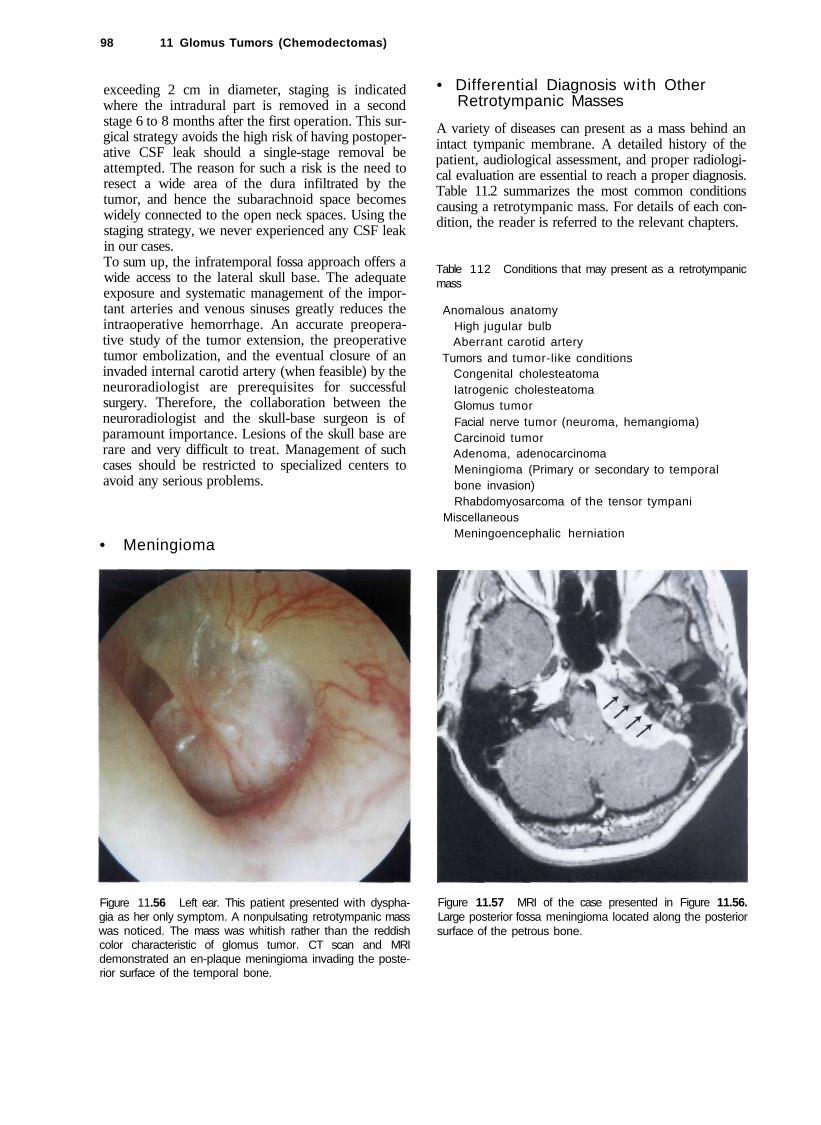
98 11 Glomus Tumors (Chemodectomas)
exceeding 2 cm in diameter, staging is indicated where the intradural part is removed in a second stage 6 to 8 months after the first operation. This surgical strategy avoids the high risk of having postoperative CSF leak should a single-stage removal be attempted. The reason for such a risk is the need to resect a wide area of the dura infiltrated by the tumor, and hence the subarachnoid space becomes widely connected to the open neck spaces. Using the staging strategy, we never experienced any CSF leak in our cases. To sum up, the infratemporal fossa approach offers a wide access to the lateral skull base. The adequate exposure and systematic management of the important arteries and venous sinuses greatly reduces the intraoperative hemorrhage. An accurate preoperative study of the tumor extension, the preoperative tumor embolization, and the eventual closure of an invaded internal carotid artery (when feasible) by the neuroradiologist are prerequisites for successful surgery. Therefore, the collaboration between the neuroradiologist and the skull-base surgeon is of paramount importance. Lesions of the skull base are rare and very difficult to treat. Management of such cases should be restricted to specialized centers to avoid any serious problems.
• Meningioma
Figure 11.56 Left ear. This patient presented with dysphagia as her only symptom. A nonpulsating retrotympanic mass was noticed. The mass was whitish rather than the reddish color characteristic of glomus tumor. CT scan and MRI demonstrated an en-plaque meningioma invading the posterior surface of the temporal bone.
• Differential Diagnosis with Other Retrotympanic Masses
A variety of diseases can present as a mass behind an intact tympanic membrane. A detailed history of the patient, audiological assessment, and proper radiological evaluation are essential to reach a proper diagnosis. Table 11.2 summarizes the most common conditions causing a retrotympanic mass. For details of each condition, the reader is referred to the relevant chapters.
Table 112 Conditions that may present as a retrotympanic mass
Anomalous anatomy High jugular bulb Aberrant carotid artery
Tumors and tumor-like conditions Congenital cholesteatoma Iatrogenic cholesteatoma Glomus tumor Facial nerve tumor (neuroma, hemangioma) Carcinoid tumor Adenoma, adenocarcinoma Meningioma (Primary or secondary to temporal bone invasion) Rhabdomyosarcoma of the tensor tympani
Miscellaneous Meningoencephalic herniation
Figure 11.57 MRI of the case presented in Figure 11.56. Large posterior fossa meningioma located along the posterior surface of the petrous bone.

Figure 11.58 Postoperative CT scan of the case described in Figure 11.56. The tumor was removed using a modified transcochlear approach. The surgical cavity was obliterated using abdominal fat.
Figure 11.60 CT scan, axial view, of the case presented in Figure 11.59. The tumor is centered on the left iuqular foramen.
Glomus Tumors (Differential Diagnosis)
• Facial Nerve Neurinoma
Figure 11.61 CT scan, coronal view. The mass eroded the bony plate over the jugular bulb extending into the hypotym-panum.
99
Figure 11.59 Left ear. Otoscopic view similar to the previous case. A whitish retrotympanic mass is seen causing bulging of the posterior quadrants of the tympanic membrane. A small reddish mass is visible in the posterior inferior regions of the external auditory canal (i.e., lateral to the annu-lus). The patient complained of left hearing loss and non-pulsating tinnitus of 2 years' duration. In the last 3 months before presentation, left facial nerve paresis started to appear (see following figures).

100 11 Glomus Tumors (Chemodectomas)
Figure 11.62 MRI, axial view, shows a mass centered on the jugular foramen (T= tumor).
Figure 11.64 Angiography did not show the characteristic tumor blush of glomus tumors. During surgery, the tumor proved to be a facial nerve neurinoma, as confirmed later by histopathological examination. The tumor was arising from the mastoid segment of the nerve and extended to the jugular bulb.
Figure 11.63 MRI, sagittal view, of the case in Figure 11.59.
Figure 11.65 Left ear. The patient complained of left facial twitches of 8 months' duration, sensation of ear fullness associated with pulsating tinnitus of 6 months' duration, and progressive conductive hearing loss of 3 months' duration.
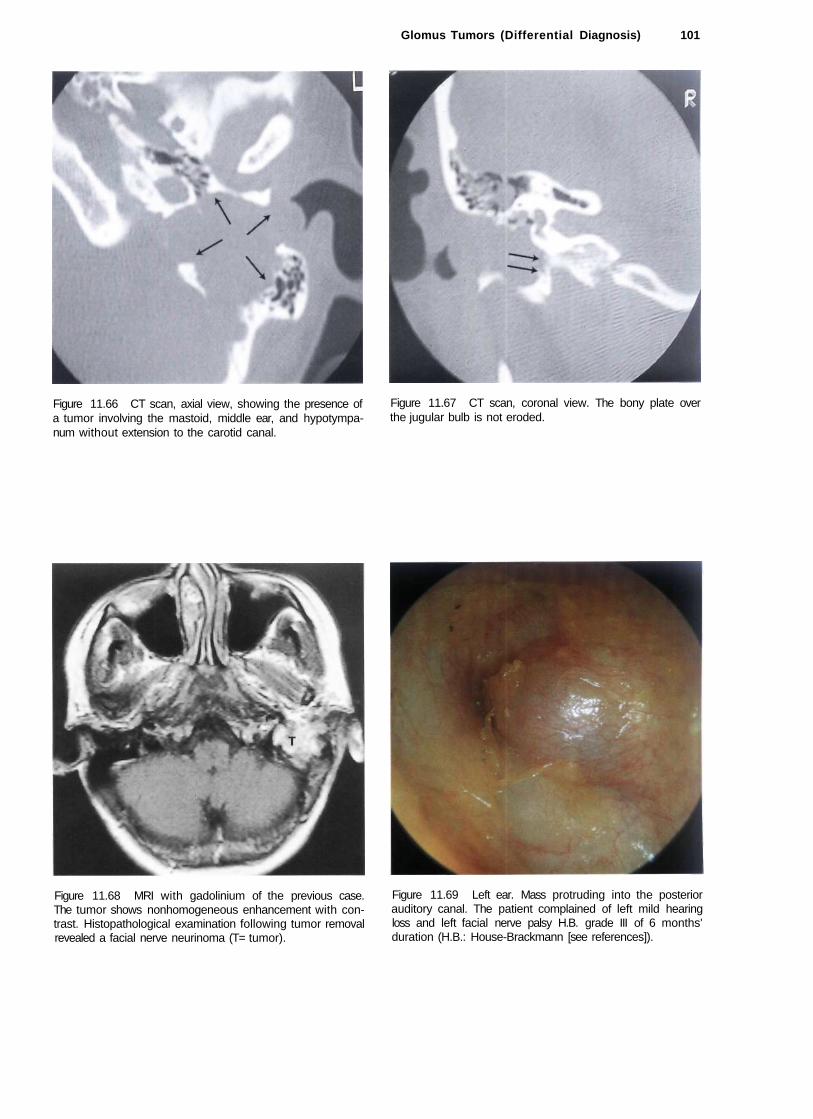
Glomus Tumors (Differential Diagnosis) 101
Figure 11.66 CT scan, axial view, showing the presence of a tumor involving the mastoid, middle ear, and hypotympa-num without extension to the carotid canal.
Figure 11.67 CT scan, coronal view. The bony plate over the jugular bulb is not eroded.
Figure 11.68 MRI with gadolinium of the previous case. The tumor shows nonhomogeneous enhancement with contrast. Histopathological examination following tumor removal revealed a facial nerve neurinoma (T= tumor).
Figure 11.69 Left ear. Mass protruding into the posterior auditory canal. The patient complained of left mild hearing loss and left facial nerve palsy H.B. grade III of 6 months' duration (H.B.: House-Brackmann [see references]).
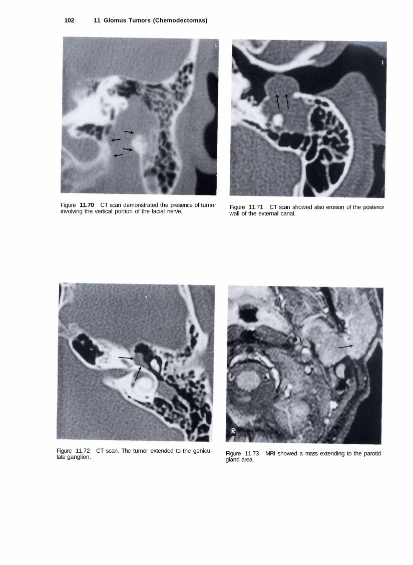
102 11 Glomus Tumors (Chemodectomas)
Figure 11.70 CT scan demonstrated the presence of tumor involving the vertical portion of the facial nerve.
Figure 11.71 CT scan showed also erosion of the posterior wall of the external canal.
Figure 11.72 CT scan. The tumor extended to the geniculate ganglion. Figure 11.73 MRI showed a mass extending to the parotid
gland area.

Glomus Tumors (Differential Diagnosis) 103
Ectopic Internal Carotid Artery
Figure 11.74 Another MR I of the same case. A combined middle fossa-transmastoid approach with parotid extension was performed. During surgery the tumor (T) proved to be a facial nerve neurinoma extended from the parotid to the intralabyrinthine segment of the facial nerve. The nerve was reconstructed with sural graft.
Figure 11.75 Left ear. A small pulsating reddish area in the anteroinferior quadrant of the tympanic membrane. This picture may be confused with a glomus tympanicum tumor.
High Jugular Bulb
Figure 11.76 A high-resolution CT scan established the diagnosis of an ectopic internal carotid artery.
Figure 11.77 Left ear. Tympanosclerosis involving the whole tympanic membrane. An epitympanic erosion with cholesteatoma is also visible. At the level of the posteroinferior quadrant, a bluish mass is observed. A CT scan (see Fig. 11.78) proved this mass to be a high jugular bulb.

104 11 Glomus Tumors (Chemodectomas)
Figure 11.78 CT scan of the previous case. The uncovered jugular bulb is seen protruding into the middle ear.
Figure 11.80 CT scan, axial view. The jugular bulb protrudes into the middle ear.
Figure 11.79 Right ear. Another example of a high jugular bulb covered by a thin bony shell in a young male patient with a skull-base malformation (see following figures).
Figure 11.81 CT scan, coronal view. The high jugular bulb can be observed.

Glomus Tumors (Differential Diagnosis) 105
Figure 11.82 Left ear. A high and uncovered jugular bulb reaching up to the level of the round window is visible through a posterior tympanic membrane perforation.
Figure 11.83 CT scan of the case in Figure 11.82.
• Polypoidal Pulsating Mass
Figure 11.84 Left ear. A polypoidal pulsating red mass is seen in the external auditory canal. This example has been included to emphasize the fact that biopsy of external auditory canal polypi should never be taken without radiological investigations.
Figure 11.85 CT scan, in this case, demonstrated the presence of a glomus tumor eroding the surrounding bone in an irregular way giving a moth-eaten appearance.

106 11 Glomus Tumors (Chemodectomas)
Figure 11.86 MRI demonstrates the presence of fluid voids typical of large intratumoral vessels.
Figure 11.88 CT scan of the case presented in Figure 11.87 demonstrating occlusion of the aneurysm with the coils.
• Internal Carotid Artery Aneurysm
Figure 11.87 Guglielmi coils used to occlude an intrape-trous internal carotid artery aneurysm.

Summary—Meningioma
Posterior fossa meningiomas are the second most common tumor of the cerebellopontine angle. These tumors are characterized by a higher morbidity and mortality than acoustic neurinoma. Surgical removal of these lesions poses many problems because of the deep location, the involvement of vital neurovascular structures, and the large sizes these tumors usually attain before diagnosis. Moreover, they have an aggressive behavior with frequent involvement of the dura and bone. Total removal is fundamental to avoid recurrence and is better achieved in the first operation. Total removal with minimal morbidity can be obtained utilizing an array of approaches that must be adapted to each individual case. In general, an ideal approach is that which allows total removal with minimal or no brain retraction. The site of the tumor is the most important factor for the choice of the surgical approach. The size of the tumor, the patient's age and general medical condition, and the preoperative status of the cranial nerves are other factors to consider. Tumors localized posterior to the internal auditory canal in young patients with good preoperative hearing can be removed using a retrosigmoid approach. In the elderly, however, a translabyrinthine approach is preferred to avoid cerebellar retraction. In cases of involvement of the jugular foramen, a POTS approach is adopted. In small tumors lying anterior to the internal auditory canal, the middle fossa transpetrous approach is utilized. In large petroclival lesions, which pose more difficulties due to their deep location, the intimate relation with the brain stem, and the involvement of vital neurovascular structures, the modified transcochlear approach should be used, irrespective of the preoperative hearing. This approach permits a wide and direct exposure, and a flat angle of vision with no cerebellar or brain stem retraction. Moreover, it allows the removal of any infiltrated dura or bone. Though total removal can be obtained in the majority of petroclival meningiomas, it is not always necessary or even safe. Subtotal removal is decided on in the absence of an arachnoid plane of cleavage between the tumor and the brain stem or when the perforating arteries are at risk of interruption during total tumor removal. Neuroradiologic evaluation is fundamental to plan surgery. A CT scan with contrast to evaluate the bone, MRI with gadolinium, and in some cases, digital subtraction angiography are of paramount importance in each case. The neuroradiologist should provide the surgeon with information on the following: • Anatomical relations of the tumor • Tumor consistency • Vascularity • Peritumoral edema
Glomus Tumors (Differential Diagnosis) 107
• Tumor-brain stem interface • Invasion of the dura and bone • Relationship between the tumor and the vertebrobasi
lar and carotid systems • Necessity of eventual embolization The main blood supply of these tumors comes from large dural arteries. However, significant contributions may also come from pial arteries or from dural branches of the internal carotid and vertebral arteries. The angiographic data helps the neuroradiologist and the skull-base surgeon to determine the need for embolization. When indicated, it should be performed a few days before surgery. It not only decreases the intraoperative bleeding, but also produces a certain amount of tumor necrosis, rendering some cases easier to remove. Close cooperation between the neuroradiologist and the skull-base surgeon offers optimal chances for successful management of these challenging tumors.

108 11 Meningioma—Facial Nerve Neurinoma
Summary—Facial Nerve Neurinoma
Tumor involvement of the facial nerve has been estimated to be the cause of facial palsy in 5% of cases. Though uncommon, facial neuromas should be considered in the differential diagnosis of facial nerve dysfunction. Unfortunately, the rarity of facial neuromas and the diversity of their clinical picture, together with the fact that their presentation may mimic other more common pathologies, renders the diagnosis of these tumors difficult. Facial nerve dysfunction is the most common symptom. It can vary from the classic progressive palsy to sudden or recurrent facial palsy or hemifacial spasm. In limited cases the function of the nerve is normal. Therefore, all patients with progressive facial palsy must be considered to have a tumor until proved otherwise. Moreover, all patients with Bell's palsy persisting for more than 4 weeks and with recurrent facial paralysis should be investigated for the presence of a tumor. The second most common complaint is hearing loss. Conductive hearing loss is usually associated with tumor involvement of the middle ear with subsequent interference with the ossicular chain. Sensorineural hearing loss is attributable to inner ear erosion or extension of the tumor into the internal auditory canal. Most diagnosed tumors are of large size. One reason is that the facial nerve can accommodate tumor expansion to some extent before significant pressure, with subsequent dysfunction, can occur. Another reason is the relatively long duration of symptoms before diagnosis is made. Because of the absence of classic symptomatology in such cases, a higher index of suspicion is needed for early diagnosis. Diagnostic work-up includes audiometric testing, vestibular testing, and auditory brain-stem evoked response. Electrophysiologic testing of facial nerve function in such cases is of little or no benefit. The usefulness of these tests in the diagnosis of facial neuromas has been challenged by other authors (Dort and Fish 1991, Neely and Alford 1974). Advances in radiologic techniques have aided greatly in the diagnosis of these lesions. The characteristic appearance on CT is that of an enhancing soft tissue mass, usually in the perigeniculate region, with sharp bony erosion and enlargement of the fallopian canal. High resolution CT scan is the best method to assess middle and inner ear involvement by tumor. However, MRI with gadolinium is the best available method for the preoperative assessment of tumor extension, especially of those involving the internal auditory canal, cerebellopontine angle, and/or the parotid region. Both methods are believed to be complementary for the preoperative assessment and the choice of the most suitable surgical approach for removal of these tumors. However, because these tumors show intraneural spread, it is still doubtful whether MRI with gadolinium can show the full extent of the lesion. Therefore, the surgeon should be prepared to expose the whole length of the facial nerve.
Differential diagnosis of these lesions includes acoustic neuroma, congenital cholesteatoma, chemodectoma, facial nerve hemangioma, and parotid tumors. Introdural facial nerve neuromas pose a major diagnostic difficulty, usually being mistaken for acoustic neuromas. Apart from the few cases in which tumor extension to the geniculate ganglion could establish the diagnosis, most of these cases were actually diagnosed intraoperatively. Congenital cholesteatomas of the petrous bone are uncommon lesions that usually present with hearing loss and facial weakness or paralysis and, therefore, can be mistaken for facial neuromas. Moreover, these lesions appear on CT as smoothly marginated expansile lesions, and on MRI as hypo/isointense on Tl and hyperintense on T2 images. Unlike facial neuromas, however, cholesteatomas do not show enhancement following contrast administration, a fact that helps to differentiate between the two lesions. Treatment generally aims at total removal of the tumor, restoration or preservation of facial nerve function, and conservation of hearing. The surgical approach depends on the extent of the lesion and the preoperative hearing level. There is general agreement that surgical removal is the treatment of choice. There is some controversy, however, regarding facial neuromas and absence of or mild preoperative facial nerve paresis. Some surgeons prefer to delay surgery because the patient is faced with the inevitable postoperative paralysis followed by some degree of recovery that will never be better than H.B. grade III. Patient counseling is important in these cases. The age at presentation is another factor to be considered. If the patient is young, early surgical resection should be done because these tumors grow inexorably with subsequent intracranial or extra temporal extension, making the approach more difficult and postoperative complications more likely. Moreover, tumor growth causes progressive degeneration and regeneration of facial nerve fibers, leading to collag-enization of the distal part of the nerve with consequent poor recovery of facial function following reconstruction. Another reason is that these tumors are potentially invasive: otic capsule erosion may be present in about 20% of the cases. On the other hand, in an elderly patient with an absence of or mild facial nerve paresis, facial nerve decompression may suffice if surgery is to be performed. When total tumor removal involves resection of a long segment of the nerve, a cable graft is usually needed for reconstruction of the facial nerve. The length of the graft and whether it is from the sural or great auricular nerve has no effect on the eventual recovery of facial function. In summary, facial nerve neuromas are uncommon tumors requiring a high degree of suspicion for their diagnosis. Recent advances in radiological techniques are the cornerstone for the diagnosis and preoperative assessment of these cases, and early surgical resection gives the best prognosis.

12 Meningoencephalic Herniation
Meningoencephalic herniation is the herniation of meningeal and/or encephalic tissue in the middle ear or mastoid. It occurs in connection with infection, previous surgery, head trauma, or congenital tegmental defects. A patient with meningoencephalic herniation has a high risk of developing meningitis and epilepsy due to epileptogenic focus in the herniating tissues. The patient may present with a pulsatile retrotympan-ic mass, cerebrospinal fluid (CSF) leakage, and aphasia. However, the most common manifestation is that of a conductive or mixed hearing loss with a draining ear or serous otitis media.
Figure 12.1 Left meningoencephalic herniation in a patient who had previously undergone open tympanoplasty. The hernia protrudes into the attic through a small tegmental defect and appears otoscopically as a pulsatile retrotympanic mass.
Figure 12.2 CT scan of the case described in Figure 12.1, coronal view. The osseous defect with the herniating tissue can be clearly visualized.
Figure 12.3 MRI of the previous case. The protrusion of the cerebral tissue into the middle ear is visible.
109

110 12 Meningoencephalic Herniation
Figure 12.4 Postoperative CT scan. The hernia was managed using a middle fossa approach. The bony defect was repaired using cartilage. The temporal craniotomy (arrow) and the cartilage (arrowhead) are clearly visible.
Figure 12.5 Left meningoencephalic hernia. The superior wall of the external auditory canal is dehiscent. A soft, reducible, nonpulsating mass is observed. The patient had a history of head trauma with transverse fracture of the temporal bone that occurred 3 years before presentation. He complained of left hearing loss and the sensation of ear fullness.
Figure 12.6 Preoperative CT scan of the case in Figure 12.5 demonstrating the herniation of cerebral tissue into the middle ear.
Figure 12.7 CT scan of the previous case 1 year postoperatively. The hernia was managed using a middle fossa approach, placing a cartilaginous plate to reconstruct the bony defect after having sectioned the neck of the herniating tissue. The cerebral tissue, which is left in the ear during the operation, is resorbed with time as seen in the CT scan

Merfingoencephalic Herniation 111
Figure 12.8 Left meningoencephalic herniation in a patient who had previously undergone multiple ear surgeries. The only manifestation was conductive hearing loss.
Figure 12.9 CT scan of the case presented in Figure 12.8.
Figure 12.10 Another example of a right meningoencephalic herniation in a patient who had undergone open tympanoplasty. Otoscopically, a large pulsatile mass is visible in the attic.
Figure 12.11 CT scan of the case presented in Figure 12.10, coronal view. The tegmen antri is absent and the herniation of the temporal lobe in the mastoid cavity and external auditory canal is demonstrated.

112 12 Meningoencephalic Herniation
Figure 12.12 A patient with a history of left open tympanoplasty presenting with conductive hearing loss. Otoscopy demonstrates a badly performed cavity with high facial ridge, secretions, granulations in the posterior wall of the cavity and an attic defect through which a soft-tissue mass protrudes into the middle ear. A CT scan was performed that confirmed the presence of a meningoencephalic hernia (see following figures).
Figure 12.14 CT scan, axial view. Arrows show the herniating cerebral tissue.
Figure 12.13 CT scan, coronal view, soft-tissue window of the case presented in Figure 12.12 demonstrating the herniating cerebral tissue into the cavity.
Figure 12.15 CT scan, coronal view, bone window.

Meningoencephalic Herniation 113
Figure 12.16 CT scan of a patient with a congenital tegmental defect. This patient has a higher risk of meningitis following an episode of otitis.
Figure 12.17 Right ear. Meningoencephalic herniation in a plurioperated patient. The otoscopy shows a new tympanic membrane lateralized by a retrotympanic whitish mass. The patient complained of right ear anacusis and H.B. grade III facial nerve palsy of 1 year duration.
Figure 12.19 MRI also demonstrated the presence of meningoencephalic herniation (arrows). During surgery, the cholesteatoma was confirmed together with a large encephalic herniation.
Figure 12.18 CT scan revealed the presence of a mass occupying the surgical cavity with erosion of the cochlea and absence of the tegmen.
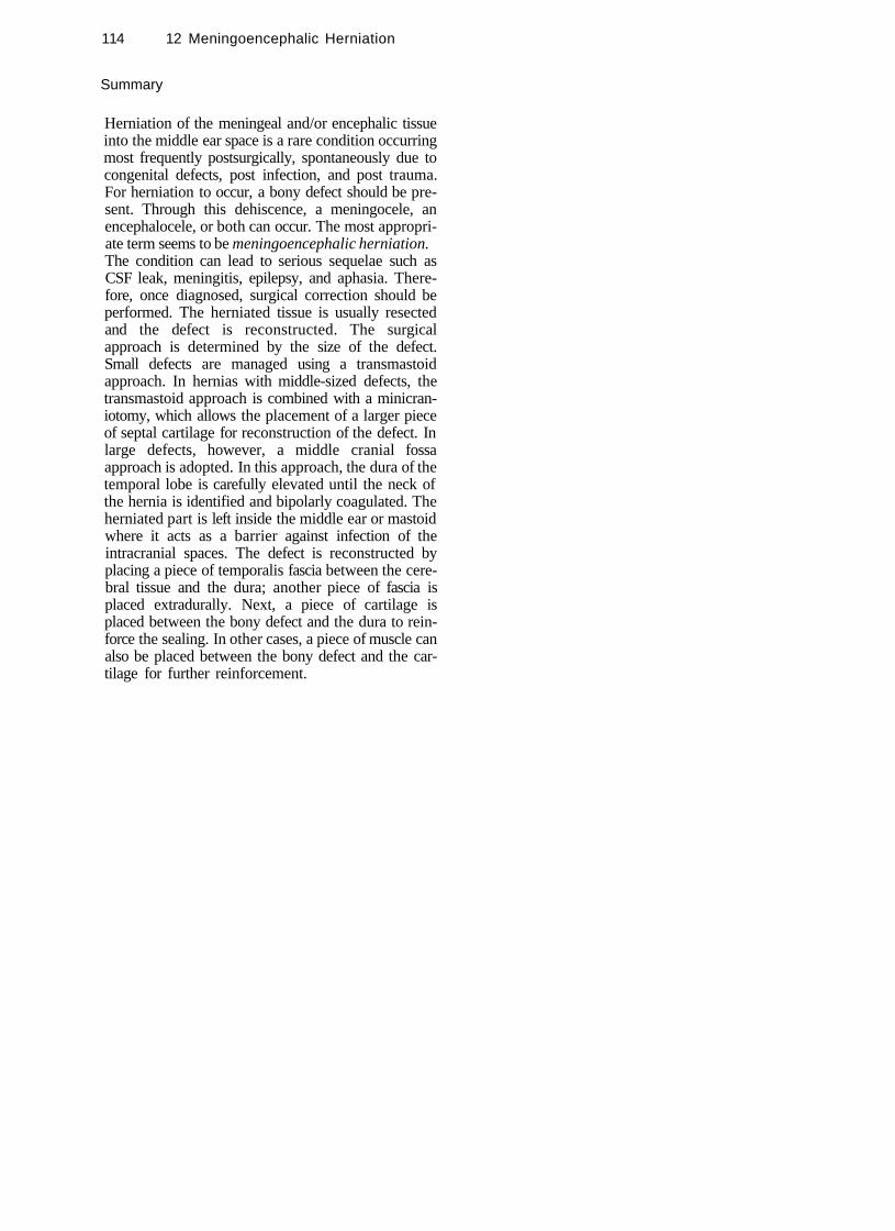
114 12 Meningoencephalic Herniation
Summary
Herniation of the meningeal and/or encephalic tissue into the middle ear space is a rare condition occurring most frequently postsurgically, spontaneously due to congenital defects, post infection, and post trauma. For herniation to occur, a bony defect should be present. Through this dehiscence, a meningocele, an encephalocele, or both can occur. The most appropriate term seems to be meningoencephalic herniation. The condition can lead to serious sequelae such as CSF leak, meningitis, epilepsy, and aphasia. Therefore, once diagnosed, surgical correction should be performed. The herniated tissue is usually resected and the defect is reconstructed. The surgical approach is determined by the size of the defect. Small defects are managed using a transmastoid approach. In hernias with middle-sized defects, the transmastoid approach is combined with a minicran-iotomy, which allows the placement of a larger piece of septal cartilage for reconstruction of the defect. In large defects, however, a middle cranial fossa approach is adopted. In this approach, the dura of the temporal lobe is carefully elevated until the neck of the hernia is identified and bipolarly coagulated. The herniated part is left inside the middle ear or mastoid where it acts as a barrier against infection of the intracranial spaces. The defect is reconstructed by placing a piece of temporalis fascia between the cerebral tissue and the dura; another piece of fascia is placed extradurally. Next, a piece of cartilage is placed between the bony defect and the dura to reinforce the sealing. In other cases, a piece of muscle can also be placed between the bony defect and the cartilage for further reinforcement.
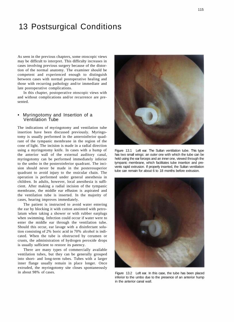
115
13 Postsurgical Conditions
As seen in the previous chapters, some otoscopic views may be difficult to interpret. This difficulty increases in cases involving previous surgery because of the distortion of the normal anatomy. The examiner should be competent and experienced enough to distinguish between cases with normal postoperative healing and those with recurring pathology and/or immediate and late postoperative complications.
In this chapter, postoperative otoscopic views with and without complications and/or recurrence are presented.
• Myringotomy and Insertion of a Ventilation Tube
The indications of myringotomy and ventilation tube insertion have been discussed previously. Myringotomy is usually performed in the anteroinferior quadrant of the tympanic membrane in the region of the cone of light. The incision is made in a radial direction using a myringotomy knife. In cases with a hump of the anterior wall of the external auditory canal, myringotomy can be performed immediately inferior to the umbo in the posteroinferior quadrant. The incision should never be made in the posterosuperior quadrant to avoid injury to the ossicular chain. The operation is performed under general anesthesia in children. In adults, however, local anesthesia is sufficient. After making a radial incision of the tympanic membrane, the middle ear effusion is aspirated and the ventilation tube is inserted. In the majority of cases, hearing improves immediately.
The patient is instructed to avoid water entering the ear by blocking it with cotton anointed with petrolatum when taking a shower or with rubber earplugs when swimming. Infection could occur if water were to enter the middle ear through the ventilation tube. Should this occur, ear lavage with a disinfectant solution consisting of 2% boric acid in 70% alcohol is indicated. When the tube is obstructed by cerumen or crusts, the administration of hydrogen peroxide drops is usually sufficient to restore its patency.
There are many types of commercially available ventilation tubes, but they can be generally grouped into short- and long-term tubes. Tubes with a larger inner flange usually remain in place longer. Once extruded, the myringotomy site closes spontaneously in about 98% of cases.
Figure 13.1 Left ear. The Sultan ventilation tube. This type has two small wings: an outer one with which the tube can be held using the ear forceps and an inner one, viewed through the tympanic membrane, which facilitates tube insertion and prevents rapid extrusion. If properly inserted, the Sultan ventilation tube can remain for about 6 to 18 months before extrusion.
Figure 13.2 Left ear. In this case, the tube has been placed inferior to the umbo due to the presence of an anterior hump in the anterior canal wall.

116 13 Postsurgical Conditions
Figure 13.3 Right ear. The consequences of a misplaced ventilation tube is shown. A healed myringotomy is seen in the posterosuperior quadrant (at 9 o'clock). Two months later the tube was extruded. During tube insertion, however, dislocation of the incus occurred. The dislocated incus fell to the hypotympanum where its body and short process can be clearly seen. In the anteroinferior quadrant, immediately under the umbo, another healed myringotomy site (this time correctly placed) is visible. In the latter, tube extrusion occurred 1 year later.
Figure 13.4 Left ear. A long-term ventilation tube inserted 6 months after tympanoplasty because of an observed tendency for graft retraction. The graft is seen in an optimal condition with no evidence of retraction, indicating patency of the ventilation tube. This tube has been in situ for more than 10 years.
Figure 13.5 Left ear. Long-term ventilation tube. A large tympanosclerotic plaque that formed 1 year after the tube insertion can be clearly seen. Such plaques result from hemorrhagic infiltrate between the epidermal and fibrous layers of the tympanic membrane secondary to the myringotomy and are asymptomatic.
Figure 13.6 Left ear. Example of a long-term "T" tube inserted in the anteroinferior quadrant of the tympanic membrane. After its insertion the two wings of the tube open by virtue of their retained "memory," thereby preventing tube extrusion.

Myringoplasty 117
Figure 13.7 Left ear. A ventilation tube in the process of extrusion. It is preferable not to take out the tube but rather wait for self-extrusion to occur. Closure of the myringotomy site occurs in about 98% of cases.
• Myringoplasty
The aim of reconstructing a tympanic membrane perforation is twofold: first, to allow the patient to have a normal social life with no restrictions, even regarding water entry into the ear, and second, to correct the hearing loss resulting from the perforation.
There are essentially two techniques for myringoplasty. The underlay technique is utilized in the presence of an anterior residue (at least the annulus) of the tympanic membrane, under which the graft can be placed. In the absence of any anterior residue of the membrane, the overlay technique is used. In such cases, the graft is positioned against the anterior wall of the external auditory canal.
Normally, the tympanic membrane forms an acute angle with the anterior wall of the external auditory canal. While performing myringoplasty, it is generally possible to respect this angulation when the annulus is present anteriorly.
The myringoplasty operation is considered a success when the reconstructed tympanic membrane is intact, well epithelialized, and has normal angulation with the external auditory canal. These characteristics allow the patient to have a normal social life (hearing improvement and possibility of water entry into the ear). Reperforation is a frequent complication of myringoplasty that occurs in about 5 to 10% of cases in the best series. Reperforation occurs more commonly in the underlay technique, particularly in the anterior
quadrant where the graft is detached from the anterior residues of the tympanic membrane and falls into the middle ear. When an overlay technique is utilized, blunting of the anterior angle can occur with resultant conductive hearing loss. Lateralization, in which the graft is detached from the handle of the malleus, is another possible complication that leads to conductive hearing loss. It occurs mostly when the graft is placed lateral rather than medial to the handle of the malleus. Stenosis of the external auditory canal, due either to inflammatory reaction or as a result of bad repositioning of the meatal flaps, can also occur.
Figure 13.8 Left ear. Normal aspect of the reconstructed tympanic membrane. The posterior quadrant is slightly elevated. In this case, a posterior perforation was grafted with temporalis fascia using an underlay technique.
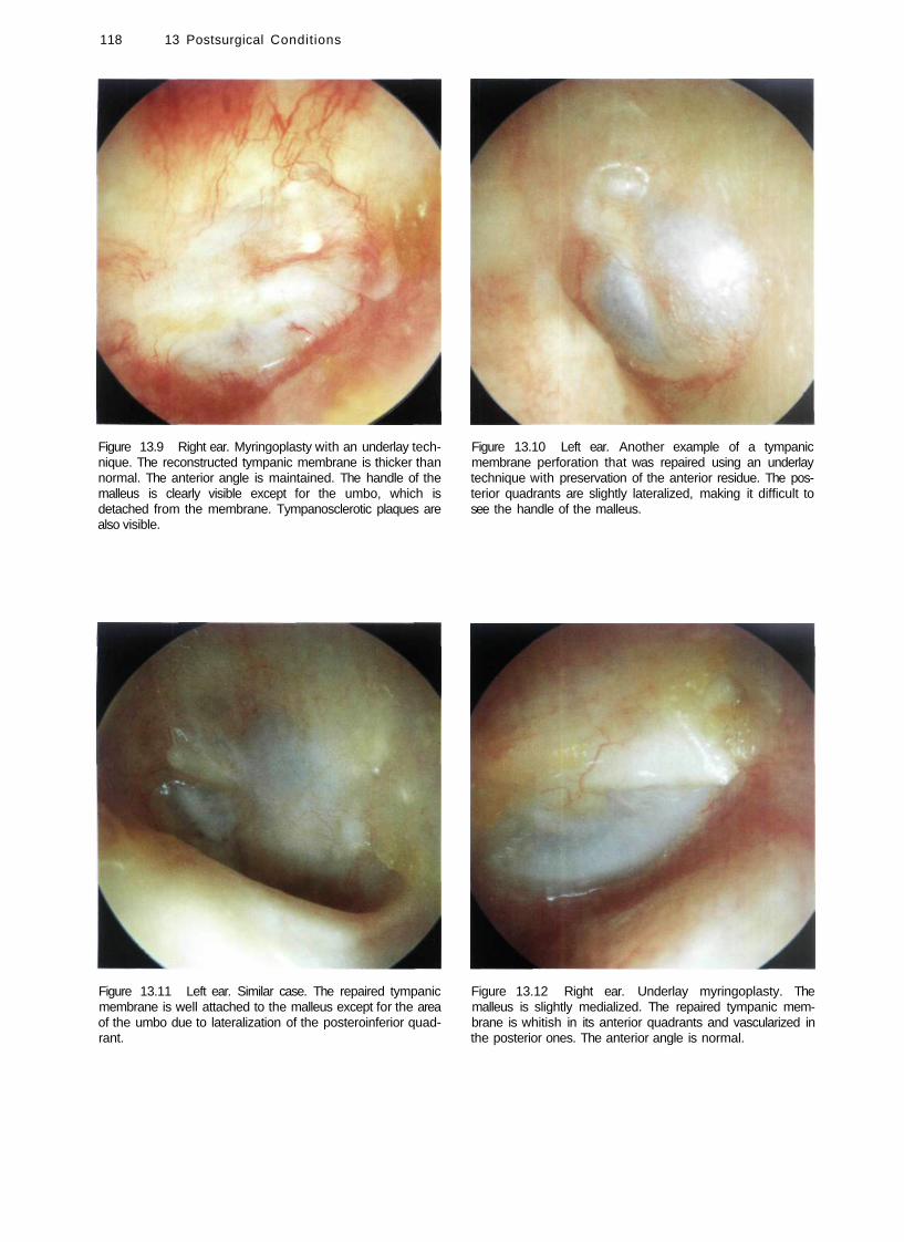
118 13 Postsurgical Conditions
Figure 13.9 Right ear. Myringoplasty with an underlay technique. The reconstructed tympanic membrane is thicker than normal. The anterior angle is maintained. The handle of the malleus is clearly visible except for the umbo, which is detached from the membrane. Tympanosclerotic plaques are also visible.
Figure 13.10 Left ear. Another example of a tympanic membrane perforation that was repaired using an underlay technique with preservation of the anterior residue. The posterior quadrants are slightly lateralized, making it difficult to see the handle of the malleus.
Figure 13.11 Left ear. Similar case. The repaired tympanic membrane is well attached to the malleus except for the area of the umbo due to lateralization of the posteroinferior quadrant.
Figure 13.12 Right ear. Underlay myringoplasty. The malleus is slightly medialized. The repaired tympanic membrane is whitish in its anterior quadrants and vascularized in the posterior ones. The anterior angle is normal.

Figure 13.13 Left ear. The repaired tympanic membrane retains a normal anterior angle and is well vascularized, though thicker than normal. A small cholesteatomatous pearl is observed. This pearl can be easily removed in the outpatient clinic under the microscope.
Figure 13.15 Left ear. Another example of a graft that is detached from the handle of the malleus using an underlay technique.
Myringoplasty 119
Figure 13.16 A lateralized reconstructed tympanic membrane with blunting of the anterior angle following an overlay technique. Both complications lead to altered mobility of the tympanic membrane with consequent conductive hearing loss.
Figure 13.14 Right ear. The repaired tympanic membrane has normal thickness. The short process of the malleus can be observed, although the handle is not visible due to lateralization.

120 13 Postsurgical Conditions
Figure 13.17 The external auditory canal is wide but the repaired tympanic membrane is lateralized and shows blunting.
Figure 13.18 Similar case. The reconstructed tympanic membrane is lateralized with marked blunting of the anterior angle.
Figure 13.19 Postoperative myringitis. The tympanic membrane is hyperemic, thickened, and lateralized following a tympanoplasty. The epidermal layer is substituted by granulation tissue. Myringitis is a rare complication that usually resolves with local steroid applications. In very rare cases, reoperation is necessary. The pathological tympanic membrane is removed followed by grafting.
Figure 13.20 A patient who has undergone quadruple myringoplasty. In these cases, myringitis and canal stenosis are frequent; therefore, it is necessary to remove the pathological tissues, perform canalplasty, and use free skin flaps.
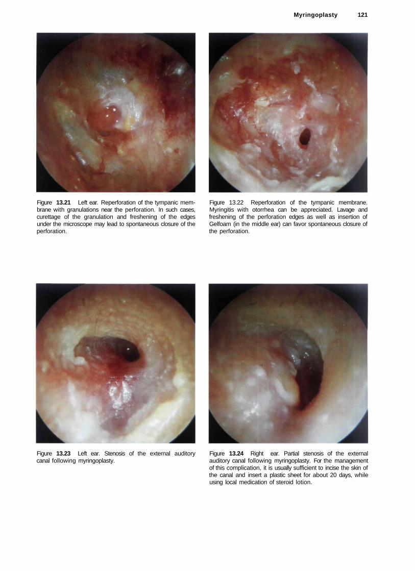
Figure 13.21 Left ear. Reperforation of the tympanic membrane with granulations near the perforation. In such cases, curettage of the granulation and freshening of the edges under the microscope may lead to spontaneous closure of the perforation.
Figure 13.23 Left ear. Stenosis of the external auditory canal following myringoplasty.
Myringoplasty 121
Figure 13.22 Reperforation of the tympanic membrane. Myringitis with otorrhea can be appreciated. Lavage and freshening of the perforation edges as well as insertion of Gelfoam (in the middle ear) can favor spontaneous closure of the perforation.
Figure 13.24 Right ear. Partial stenosis of the external auditory canal following myringoplasty. For the management of this complication, it is usually sufficient to incise the skin of the canal and insert a plastic sheet for about 20 days, while using local medication of steroid lotion.

122 13 Postsurgical Conditions
Figure 13.25 Retrotympanic cholesteatoma following myringoplasty. This iatrogenic cholesteatoma can be explained by the entrapment of epidermal residues in the middle ear or malpositioning of the meatal flap at the level of the anterior angle. It can be managed by incision of the cholesteatoma sac, aspiration of its contents, and insertion of a plastic sheet in the external auditory canal for about 20 days to favor healing.
• Tympanoplasty
Tympanoplasty operations can be classified into those without mastoidectomy, performed with chronic otitis media in which the tympanic membrane perforation is associated with necrosis of the ossicular chain, and those with mastoidectomy, performed in chronic suppurative otitis media with cholesteatoma. As mentioned previously, tympanoplasty with mastoidectomy can be either closed or open.
In closed tympanoplasty, the posterior wall of the external auditory canal is kept intact. This technique is employed in children and in patients with very pneu-matized mastoids to avoid having a large cavity. Regular otoscopic follow-up is essential to identify the formation of a retraction pocket or a recurrent cholesteatoma. Should these occur, there should be no hesitation in switching to an open technique.
In open tympanoplasty, the posterior wall of the external auditory canal is removed. The indications of this technique in the treatment of cholesteatoma include: a wide erosion of the posterosuperior wall, cholesteatoma in the only hearing ear, bilateral cholesteatoma, cholesteatoma in patients with Down's syndrome, the presence of a contracted mastoid, a large labyrinthine fistula, and recurrent cholesteatoma following a closed tympanoplasty. Because the posterior canal wall is removed, the mastoid cavity is exteriorized and on otoscopy the external auditory canal and the mastoid appear as one communicating cavity. If properly performed, the cavity appears rounded in shape, dry, and well epithelialized. On the other hand, a badly performed cavity may appear wet, irregular, and be lined with granulation tissue in addition to accumulated debris. There may also be the possibility of a residual cholesteatoma.
In cases of tympanoplasty, it is usually possible to see the reconstructed ossicular chain through the tympanic membrane. We generally prefer to utilize an autologous or homologous incus for reconstruction. In our experience (more than 1000 tympanoplasties) we never encountered any case of extrusion when the incus was used. In contrast, variable rates of extrusion were noticed when biological materials (e.g., plasti-pore, ceramics, hydroxyapatite) were utilized. Although the use of homologous ossicles has never been proven to transmit slow viruses (e.g., Creutzfeldt-Jakob disease), the theoretical risk makes it more prudent to use predominantly autologous tissue or biomaterial of better characteristics that might appear in the future.
Later on in this chapter, some otoscopic views of cases managed by the modified Bondy technique are shown. This is an open technique indicated in epitym-panic cholesteatoma with a good preoperative hearing in which the tympanic membrane and the ossicular chain are intact. Some cases of radical mastoidectomy are also shown. This technique is used mainly in elderly patients with sensorineural hearing loss in which the only goal of surgery is to have a dry and safe ear.
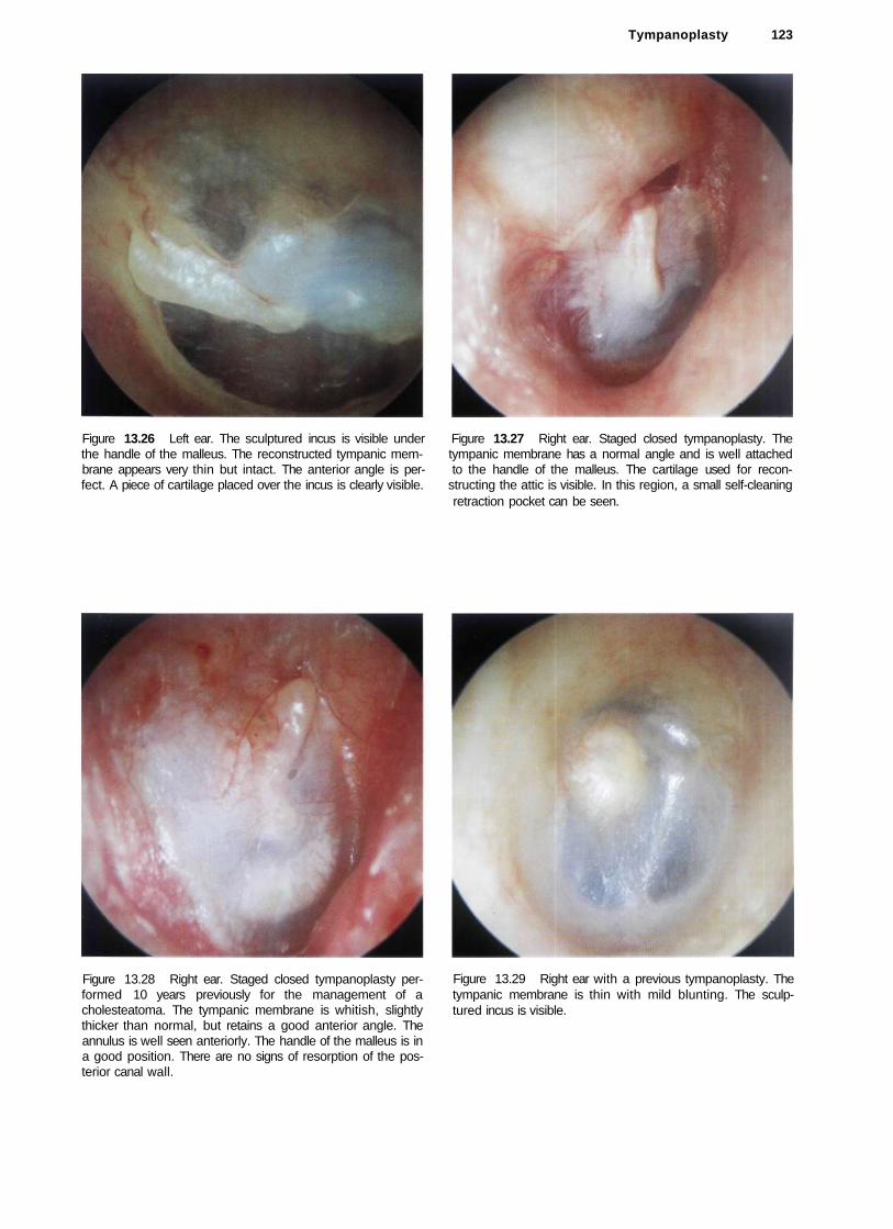
Tympanoplasty 123
Figure 13.26 Left ear. The sculptured incus is visible under Figure 13.27 Right ear. Staged closed tympanoplasty. The the handle of the malleus. The reconstructed tympanic mem- tympanic membrane has a normal angle and is well attached brane appears very thin but intact. The anterior angle is per- to the handle of the malleus. The cartilage used for recon-fect. A piece of cartilage placed over the incus is clearly visible. structing the attic is visible. In this region, a small self-cleaning
retraction pocket can be seen.
Figure 13.28 Right ear. Staged closed tympanoplasty performed 10 years previously for the management of a cholesteatoma. The tympanic membrane is whitish, slightly thicker than normal, but retains a good anterior angle. The annulus is well seen anteriorly. The handle of the malleus is in a good position. There are no signs of resorption of the posterior canal wall.
Figure 13.29 Right ear with a previous tympanoplasty. The tympanic membrane is thin with mild blunting. The sculptured incus is visible.

124 13 Postsurgical Conditions
Figure 13.30 Right ear. Otoscopic view after a second-stage tympanoplasty in which the incus was used for ossiculoplasty. The tympanic membrane and the handle of the malleus are excellently positioned.
Figure 13.32 Left ear. Another example of the incus positioned under the handle of the malleus.
Figure 13.31 Left ear. Perfect reconstructed tympanic membrane with optimal thickness and no blunting. The sculptured incus is in contact with the handle of the malleus: It is slightly elevated with respect to the level of the tympanic membrane.
Figure 13.33 Left ear. Ossiculoplasty. The tympanic membrane is retracted and the malleus is medialized. The sculptured incus is displaced posteriorly and is adherent to the posterior mesotympanum. Two tympanosclerotic plaques are noted anteriorly and interiorly.
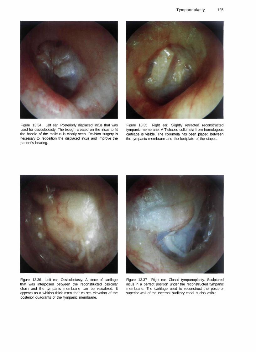
Figure 13.34 Left ear. Posteriorly displaced incus that was used for ossiculoplasty. The trough created on the incus to fit the handle of the malleus is clearly seen. Revision surgery is necessary to reposition the displaced incus and improve the patient's hearing.
Figure 13.36 Left ear. Ossiculoplasty. A piece of cartilage that was interposed between the reconstructed ossicular chain and the tympanic membrane can be visualized. It appears as a whitish thick mass that causes elevation of the posterior quadrants of the tympanic membrane.
Tympanoplasty 125
Figure 13.35 Right ear. Slightly retracted reconstructed tympanic membrane. A T-shaped collumela from homologous cartilage is visible. The collumela has been placed between the tympanic membrane and the footplate of the stapes.
Figure 13.37 Right ear. Closed tympanoplasty. Sculptured incus in a perfect position under the reconstructed tympanic membrane. The cartilage used to reconstruct the postero-superior wall of the external auditory canal is also visible.

126 13 Postsurgical Conditions
Figure 13.38 Right ear. Post tympanoplasty. Good position of the tympanic membrane. In this case, it is difficult to identify the type of ossicular chain reconstruction due to the thickness of the tympanic membrane, particularly noted at its posterior quadrants.
Figure 13.40 Right ear. Another example of a TORP that is visible through the tympanic membrane. The overlying cartilage, which is whitish in color, has been displaced into the posteroinferior quadrant. There are no signs of extrusion.
Figure 13.39 Left ear. In the posterosuperior quadrant a TORP (total ossicular replacement prosthesis) with its circular head is noted. The overlying cartilage is partially resorbed. There are no signs of extrusion.
Figure 13.41 Left ear. Posterosuperior perforation of the reconstructed tympanic membrane with extrusion of the TORP. The shaft of the prosthesis has caused an erosion of the footplate of the stapes (which appears through the perforation as a rounded dark area).
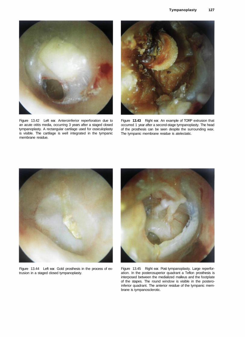
Tympanoplasty 127
Figure 13.42 Left ear. Anteroinferior reperforation due to an acute otitis media, occurring 3 years after a staged closed tympanoplasty. A rectangular cartilage used for ossiculoplasty is visible. The cartilage is well integrated in the tympanic membrane residue.
Figure 13.44 Left ear. Gold prosthesis in the process of extrusion in a staged closed tympanoplasty.
Figure 13.43 Right ear. An example of TORP extrusion that occurred 1 year after a second-stage tympanoplasty. The head of the prosthesis can be seen despite the surrounding wax. The tympanic membrane residue is atelectatic.
Figure 13.45 Right ear. Post tympanoplasty. Large reperforation. In the posterosuperior quadrant a Teflon prosthesis is interposed between the medialized malleus and the footplate of the stapes. The round window is visible in the postero-inferior quadrant. The anterior residue of the tympanic membrane is tympanosclerotic.

128 13 Postsurgical Conditions
Figure 13.46 Right ear. Post stapedectomy. The atticotomy is seen in the posterosuperior quadrant. The preserved chorda tympani is well appreciated.
Figure 13.48 Right ear. A cholesteatomatous pearl in the external auditory canal in a patient who had previously undergone a stapedectomy. The tympanomeatal flap was not correctly repositioned. This skin was thus folded in on itself and the entrapped epithelium gave rise to this pearl. This complication was easily resolved in the outpatient clinic by incising the skin (see Fig. 13.49) and removing the cholesteatomatous cyst (see Fig. 13.50).
Figure 13.47 Left ear. A rare case of extrusion of a stapes prosthesis. The metallic ring is seen extruding through a microperforation covered with epidermal squames. The Teflon shaft of the prosthesis can be visualized through the tympanic membrane.
Figure 13.49 Incision of the skin over the cyst.

Figure 13.50 Removal of the cholesteatomatous cyst.
Figure 13.52 Right ear. Post tympanoplasty. A white retrotympanic mass (cholesteatoma of the anterior angle) is noted causing bulging of the tympanic membrane. The cholesteatoma is probably the result of inadequate removal of the epithelium in an overlay technique. The entrapped skin led to the formation of the cholesteatoma.
Tympanoplasty 129
Figure 13.51 Left ear. Silastic sheet in extrusion through a posterosuperior perforation. The handle of the malleus is clearly visible anteriorly. In general, Silastic is inserted in first-stage tympanoplasty. This material is usually placed in the middle ear to favor the restoration of the normal mucosal lining of the middle ear and to avoid the formation of adhesions in the meantime. It is removed during the second-stage tympanoplasty, except in cases showing a tendency towards atelectasis.
Figure 13.53 Left ear. Good anterior angle of the reconstructed tympanic membrane. An anteromalleolar cholesteatomatous cyst is seen. An epitympanic retraction pocket that is adherent to the head of the malleus and body of the incus is also observed.

130 13 Postsurgical Conditions
Figure 13.54 Left ear. In the posterosuperior quadrant, the sculptured incus with the short process pointing anteriorly is seen through the retracted tympanic membrane. In cases with hearing loss, repeat surgery is indicated to reinforce the tympanic membrane and improve the hearing. Surgery entails dissection of the retraction pocket from the incus, and the placement of cartilage between the sculptured incus and the tympanic membrane. This cartilage prevents (or delays) the reformation of a retraction pocket and corrects the hearing deficit.
Figure 13.56 Right ear. Sculptured incus seen through the tympanic membrane in a case of closed tympanoplasty. An epitympanic retraction pocket is seen. This pocket should be followed up regularly to guard against the formation of a recurrent cholesteatoma. Should this occur, the closed technique must be transformed into an open one to avoid further recurrence of the cholesteatoma.
Figure 13.55 Left ear. Another example of retraction of the tympanic membrane leading to inclination of the sculptured incus. The dislocated incus becomes fixed to the posterior mesotympanum, resulting in hearing loss.
Figure 13.57 Right ear. Recurrent epitympanic cholesteatoma following closed tympanoplasty. The reconstructed tympanic membrane (pars tensa) shows an optimal anterior angle and is perfectly attached to the handle of the malleus. In this case, transformation to an open technique is indicated while conserving the tympanic membrane and ossicular chain if there is no hearing loss.

Figure 13.58 Left ear. Another example of a staged closed tympanoplasty 6 years after the second stage. A large resorption of the posterosuperior wall of the external auditory canal associated with recurrent cholesteatoma is observed. In the posterosuperior quadrant, the cartilage used for ossiculoplasty is seen. Revision surgery was performed with transformation into an open technique.
Figure 13.60 Right ear. Partial resorption of the posterior wall of the external auditory canal about 7 to 8 mm from the annulus following a closed tympanoplasty. The atrophic area appears bluish due to lack of underlying bone. No cutaneous retraction is seen. However, due to the lack of bone, the skin can invaginate into the mastoid cavity giving rise to recurrent cholesteatoma. In such cases, regular long-term follow-up is indicated.
Tympanoplasty 131
:
Figure 13.59 Left ear. A small epitympanic retraction pocket is observed. Though smail and shallow, this pocket should be followed up regularly as it may become deeper with time, leading to the formation of a recurrent cholesteatoma.
Figure 13.61 Left ear. Total resorption of the posterior wall of the external auditory canal 3 years after a closed tympanoplasty. The otoscopic view is similar to that observed after an open tympanoplasty. Repeat surgery was necessary. The facial ridge was lowered and all bony irregularities were smoothed to avoid the retention of squamous debris with subsequent otorrhea. An adequate meatoplasty was also performed.

132 13 Postsurgical Conditions
Figure 13.62 Right ear. An example of a successful closed tympanoplasty 11 years postoperatively. The tympanic membrane is in perfect position and angulation. The posterior canal wall is intact and there is no evidence of recurrent cholesteatoma (see previous cases in Figs. 13.60 and 13.61).
Figure 13.63 Left ear. Another example of a closed tympanoplasty 2 years postoperatively. The attic was reconstructed using cartilage and bone pate and shows no signs of erosion. A small cholesteatomatous pearl is seen in the postero-superior quadrant. It can be easily removed in the outpatient clinic under microscopic control.
Figure 13.64 Right ear. A well performed open tympanoplasty. The cavity is epithelialized and the facial ridge is adequately lowered. In the attic region, the material used for obliteration can be noted.
Figure 13.65 Left ear. Open tympanoplasty. Attic obliteration with autologous bone.
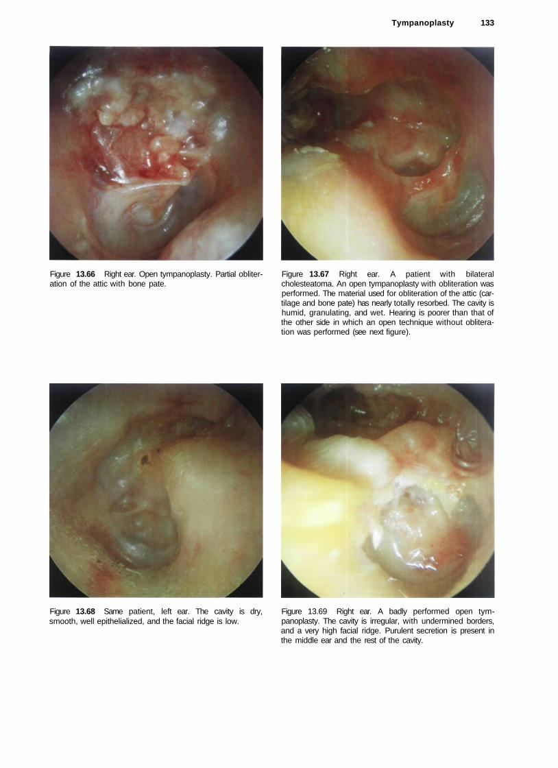
Tympanoplasty 133
Figure 13.66 Right ear. Open tympanoplasty. Partial obliteration of the attic with bone pate.
Figure 13.67 Right ear. A patient with bilateral cholesteatoma. An open tympanoplasty with obliteration was performed. The material used for obliteration of the attic (cartilage and bone pate) has nearly totally resorbed. The cavity is humid, granulating, and wet. Hearing is poorer than that of the other side in which an open technique without obliteration was performed (see next figure).
Figure 13.68 Same patient, left ear. The cavity is dry, smooth, well epithelialized, and the facial ridge is low.
Figure 13.69 Right ear. A badly performed open tympanoplasty. The cavity is irregular, with undermined borders, and a very high facial ridge. Purulent secretion is present in the middle ear and the rest of the cavity.

134 13 Postsurgical Conditions
Figure 13.70 Right ear. Another example of a badly performed open tympanoplasty. Purulent secretions and a high facial ridge are observed.
Figure 13.72 Right ear. Open tympanoplasty. The facial ridge has not been sufficiently lowered in this case. This leads to accumulation of cerumen and cellular debris in the cavity with subsequent infection, secretion, and maceration of the skin lining the cavity.
Figure 13.71 Left ear. Open tympanoplasty. A large perforation of the reconstructed tympanic membrane is seen. Cholesteatomatous pearls are observed in the attic.'
Figure 13.73 Left ear. TORP in extrusion following a second-stage open tympanoplasty. In the first stage, a cholesteatoma involving the attic and mesotympanum and causing erosion of the ossicular chain was removed. In the second stage, a TORP was used for reconstruction. It was placed between the footplate of the stapes and the tympanic membrane. One year postoperatively, early extrusion of the prosthesis is observed. To avoid this complication, a tragal cartilage has to be placed between the prosthesis and the tympanic membrane.

Tympanoplasty 135
Figure 13.74 Left ear. An example of a correctly performed open tympanoplasty. The cavity shows perfect epithelializa-tion. The facial ridge is low. The tympanic membrane is well positioned with excellent contact with the handle of the malleus.
Figure 13.76 Right ear. Open tympanoplasty. The cartilage used for obliteration of the attic is seen in the superior part.
Figure 13.75 Left ear. Open tympanoplasty with a well epithelialized cavity. The tympanic membrane shows a tym-panosclerotic plaque anteriorly; posteriorly, the ossicular chain reconstruction is observed.
Figure 13.77 Right ear. Open tympanoplasty. The tympanic bone was drilled in this case because it was involved with the cholesteatoma. The inferior annulus is visible. Superiorly, the chorda tympani is observed close to the incus used for reconstruction of the ossicular chain.

136 13 Postsurgical Conditions
Figure 13.78 Left ear. Example of a modified Bondy technique. In this case, the preoperative pure tone average was 20 dB. The patient conserved his preoperative hearing. The modified Bondy technique is indicated in epitympanic cholesteatoma with an intact tympanic membrane and ossicular chain. It is an open technique in which the attic and the mastoid are exteriorized (Fig. 13.79) and the facial ridge is lowered until the level of the annulus. The ossicular chain and the tympanic membrane are left in situ (Fig. 13.80). If necessary, the attic is obliterated with a piece of cartilage; this procedure helps to reduce the risk of retractions around the ossicles (Fig. 13.81). Fascia is then inserted with two anterior tongues; one is positioned under the incus body, the other between the handle of the malleus and the long process of the incus (Figs. 13.82, 13.83). A meatoplasty according to the size of the cavity is performed at the end of the procedure.
Figure 13.79 Same case, intraoperative view. Exteriorization of the attic and the mastoid, lowering the facial ridge until the level of the annulus.
Figure 13.80 The ossicular chain and the tympanic membrane are left in situ.
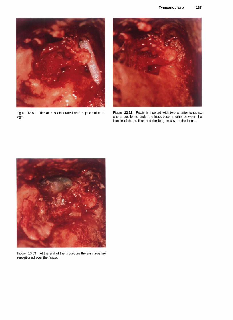
Tympanoplasty 137
Figure 13.81 The attic is obliterated with a piece of cartilage.
Figure 13.82 Fascia is inserted with two anterior tongues: one is positioned under the incus body, another between the handle of the malleus and the long process of the incus.
Figure 13.83 At the end of the procedure the skin flaps are repositioned over the fascia.

138 13 Postsurgical Conditions
Figure 13.84 Left ear. Another case of the modified Bondy technique. Note the intact ossicular chain.
Figure 13.85 Right ear. The modified Bondy technique. A ventilation tube was inserted because of the presence of middle ear effusion that did not respond to medical treatment.
Figure 13.86 Left ear. The modified Bondy technique. Although an attic retraction is noted recurrent cholesteatoma is uncommon with this technique. The tympanic membrane is retracted and middle ear effusion is noted. In this case, the insertion of a ventilation tube is indicated.
Figure 13.87 Right ear. The modified Bondy technique. The attic is obliterated with cartilage.

Tympanoplasty 139
Figure 13.88 Left ear. Open tympanoplasty. The ossicular chain was reconstructed using an autologous cartilage that was not extruded despite the presence of atelectasis of the tympanic membrane.
Figure 13.89 Left ear. Another case of the modified Bondy technique. As the incus was slightly eroded, a piece of cartilage was placed between it and the malleus. The attic was obliterated with cartilage.
Figure 13.90 Right ear. The modified Bondy technique. The tympanic membrane is normal and the cavity is dry and perfectly epithelialized.
Figure 13.91 Right ear. In this case of a modified Bondy technique, incus erosion occurred 3 years postoperatively due to the presence of a significant retraction pocket. The middle ear shows a catarrhal effusion.

140 13 Postsurgical Conditions
Figure 13.92 Right ear. A modified Bondy technique. Two cholesteatomatous pearls are present in the cavity. They are easily removed in the outpatient clinic. The attic, antrum, and mastoid were exteriorized. The ossicular chain was left in situ.
Figure 13.94 Same patient after removal of the pearl in the outpatient clinic.
Figure 13.93 Left ear. A cholesteatomatous pearl seen in the attic following a modified Bondy technique.
Figure 13.95 Radical mastoidectomy. A mucosal cyst causes complete obstruction of the external auditory canal.

Tympanoplasty 141
Figure 13.96 Right ear. Radical mastoidectomy. The second portion of the facial nerve is uncovered. Scars around the nerve produced an H.B. grade III palsy.
Figure 13.98 Another example of a meatoplasty performed in a 10-year-old boy who underwent surgery for bilateral epi-tympanic cholesteatoma using a modified Bondy technique.
Figure 13.97 Example of a well performed meatoplasty in an open tympanoplasty. The performance of an adequate meatoplasty that suits the dimension of the cavity is fundamental to assure proper aeration and prevent accumulation of epithelial debris and cerumen in the cavity.
Figure 13.99 Example of a meatoplasty that shows mild stenosis.

142 References
References
Aristegui M, Cokkeser Y, Saleh E, Naguib M, Landolfi M, Taibah A, et al. Surgical anatomy of the extended middle cranial fossa approach. Skull Base Surg 1994;4:188-95.
Aristegui M, Falcioni M, Saleh E, Taibah A, Russo A, Landolfi M et al. Meingoencephalic herniation into the middle ear: a report of 27 cases. Laryngoscope 1995;105:513-18.
Austin DF. The significance of the retraction pockets in the treatment of cholesteatoma. In: McCabe BF, Sade J, Abramson M, eds. Cholesteatoma: First International Conference. Birmingham, AL: Aesculapius, 1977;379-83.
Brackmann DE, Shelton C, Arriaga MA. Otologic Surgery. Philadelphia: WB Saunders Company, 1994.
Caparosa R. An Atlas of Surgical Anatomy and Techniques of the Temporal Bone. Springfield: Thomas, 1972.
Celikkanat S, Saleh E, Khashaba A, Taibah A, Russo A, Mazzoni A, et al. Cerebrospinal fluid leak after translabyrinthine acoustic neuroma surgery. Otolaryngol Head Neck Surg 1995;112:654-58.
Charachon R. La tympanoplastie. Grenoble: Presses Universitaires, 1990.
Charachon R, Roulleau P, Bremond G et al. Les ossiculoplasties: etat actuel. Paris: Arnette, 1987.
Chole RA. Petrous apicitis: surgical anatomy. Ann Otol Rhinol Laryngol\985;94:251-57.
Cody DTR, Taylor W. Mastoidectomy for acquired cholesteatoma: long-term results. In: McCabe BF, Sade J, Abramson M, eds. Cholesteatoma: First International Conference. Birmingham, AL: Aesculapius, 1977;337-51.
Cohen D. Location of primary cholesteatoma. Am J Otol 1987;8:61-5
Coker NJ, Jenkins HA, Fisch U. Obliteration of the middle ear and mastoid cleft in subtotal petrosectomy: indications, technique and results. Ann Otol Rhinol Laryngol 1986;95:5-11.
Cokkeser Y, Aristegui M, Naguib M, Saleh E, Sanna M. Surgical anatomy of the vertebral artery at the craniovertebral junction. In: Mazzoni A, Sanna M, eds. Skull Base Surgery Update, vol. 1. Amsterdam: Kugler, 1995;43-8.
Deguine C. Long-term results in cholesteatoma surgery. Clin Otolaryngol 1978;3:301-10.
De la Cruz A. The transcochlear approach to meningiomas and cholesteatoma of the cerebellopontine angle. In: Brackmann DE, ed. Neurological Surgery of the Ear and Skull Base. New York: Raven Press, 1982;353-60.
Derlacki EL, Clemis JD. Congenital cholesteatoma of the middle ear and mastoid. Ann Otol Rhinol Laryngol 1965;74:706-27.
Di Chiro G, Fisher RL, Nelson KB. The jugular foramen. J Neurosurg 1964;21:447-60.
Donaldson A, Duckert LG, Lambert PM, Rubel EW. Anson and Donaldson Surgical Anatomy of the Temporal Bone. New York: Raven Press, 1992.
Dort JC, Fish U. Facial nerve schwannomas. Skull Base Surgery 1991;1:51-6.
Farrior JB. Anterior facial nerve decompression. Otolaryngol Head Neck Surg 1985;93:765-68.
Farrior JB. The canal wall in tympanoplasty and mastoidectomy. Arch Otolaryngol 1969;90:706-14.
Farrior JB. Systematized approach to surgery for cholesteatoma. Arch Otolaryngol 1973;97:188-90.
Fisch U. Infratemporal fossa approach to tumors of the temporal bone and base of the skull../ Laryngol Otol 1978;92:949-67.
Fisch U. Infratemporal fossa approach for glomus tumors of the temporal bone. Ann Otol Rhinol Laryngol 1982;91:474-79.
Fisch U. The infratemporal fossa approach for nasopharyngeal tumors. Laryngoscope 1983;93:36-44.
Fisch U. Tympanoplasty, Mastoidectomy, and Stapes Surgery. Stuttgart: Thieme, 1994.
Fisch U, Esslen E. Total intratemporal exposure of the facial nerve. Arch Otolaryngol 1972;95:335-41.
Fisch U, Mattox D. Microsurgery of the Skull Base. New York: Thieme, 1988.
Fisch U, Fagan P, Valavanis A. The infratemporal fossa approach for the lateral skull base. Otolaryngol Clin North Am 1984;17:513-22.
Flood LM, Kemink JL. Surgery in lesions of the petrous apex. Otolaryngol Clin North Am 1984;7:565-75.
Friedberg J. Congenital cholesteatoma. Laryngoscope 1994:104 (Suppl):l-24.
Gacek R. Surgical landmarks for the facial nerve in the epitym-panum. Ann Otol Rhinol Laryngol 1980;89:249-50.
Gantz B, Fisch U. Modified transotic approach to the cerebellopontine angle. Arch Otolaryngol 1983;109:252-56.
Glasscock ME. Surgical technique for open mastoid procedures: how I did it. Laryngoscope 1982;92:1140-42.
Glasscock ME, Harris FH. Glomus tumors: diagnosis and treatment. Laryngoscope 1974;84:2006-32.
Glasscock ME, Miller GW. Intract canal wall tympanoplasty in the management of cholesteatoma. Laryngoscope 1976;86:1639-57.
Glasscock ME, Shambaugh GE. Surgery of the Ear, 4th ed. Philadelphia: Saunders, 1990.
Goodhill V. Tragal perichondrium and cartilage in tympanoplasty. Arch Otolaryngol 1967;85:480-91.
Guild SR. A hitherto unrecognized structure, the glomus jugu-laris, in man. Anat Rec (Suppl) 1941;79:28.
Guild SR. The glomus jugulare, a nonchromaffin paraganglion, in man. Ann Otol Rhinol Laryngol 1953;62:1045-71.
Hakuba A, Nishimura S, Jang BJ. A combined retro-auricolar and preauricolar transpetrosal-transtentorial approach to clivus meningiomas. Surg Neurol 1988;30:108-16.
Hoffman RA. Cerebrospinal fluid leak following acoustic neuroma removal. Laryngoscope 1994;104:40-58.
House WE Surgical exposure of the internal auditory canal and its contents through the middle cranial fossa. Laryngoscope 1961;71:1363-65.
House WF. Middle cranial fossa approach to the petrous pyramid: report of 50 cases. Arch Otolaryngol 1963;78:460-68.
House WF. Transtemporal bone microsurgical removal of acoustic neuromas. Arch Otolaryngol 1964;80:599-756.
House WF, Shechy JL. Functional restoration in tympanoplasty. Arch Otolaryngol 1963;78:304-9.
House WF, Glasscock ME III. Glomus tympanicum tumors. Arch Otolaryngol 1968;87:124-28.
House WF, Hitselberger WE. The transcochlear approach to the skull base. Arch Otolaryngol 1976;102:334-42.
House JL, Hitselberger WE, House WF. Wound closure and cerebrospinal fluid leak after translabyrinthine surgery. Am J Otol 1982;4:126-28.
House JW, Brackmann DE. Facial nerve grading system. Otolaryngol Head Neck Surg 1985;93:146-47.
Jackler RK. Overview of surgical neuro-otology. In: Jackler RK. Brackmann DE, eds. Neuro-otology. Baltimore: Mosby. 1993;651-84.
Jackson CG. Surgery of Skull Base Tumors. New York: Churchill Livingstone, 1991.

References 143
Jackson CG, Glasscock ME, McKennan KX, et al. The surgical treatment of skull base tumors with intracranial extension. Otolaryngol Head Neck Surg 1987;96:175.
Jackson CG. Cueva RA, Thedinger BA, Glasscok ME. Conservation surgery for glomus jugulare tumors: the value of early diagnosis. Laryngoscope 1990;100:10.
Jansen C. The combined approach for tympanoplasty. / Laryngol Otol 1968;82:771-93.
Jansen C. Posterior tympanotomy: experiences and surgical details. Otolaryngol Clin North Am 1972:5:79-96.
Jansen C. Intact canal wall tympanoplasty. In: Shambaugh G, Shea J, eds. Fifth International Workshop on Middle Ear Microsurgery. Huntsville: Strode, 1977:370-75.
Jenkins HA, Fisch U. Glomus tumors of the temporal region: technique of surgical resection. Arch Otolaryngol 1981;107:209-13.
Karmarkar S, Bhatia S, Saleh E, De Donato G, Taibah A, Russo A et al. Cholesteatoma surgery: the individualized technique. Ann Otol Rhinol Laryngol 1995;104:591-95.
Krmpotic-Nemanic J, Draf W, Helms J. Surgical Anatomy of the Head and Neck. Berlin: Springer, 1985.
Kveton JF, Cooper MH. Microsurgical anatomy of the jugular foramen region. Am J Otol 1988;9:109-12.
Landolfi M, Taibah A. Russo A, Szymanski M, Shaan M, Sanna M. Revalidation of the Bondy technique. In: Nakano Y, ed. Cholesteatoma and Mastoid Surgery. Amsterdam: Kugler, 1993:719-21.
Lang J. Topographical anatomy of the skull base and adjacent tissues. In: Scheunemann H, Schurmann K, Helms J, eds. Tumors of the Skull Base. Berlin: de Gruyter, 1986;3-28.
Lau T, Tos M. Treatment of sinus cholesteatoma: long-term results and recurrence rate. Arch Otolaryngol 1988a;9:456-64.
Lau T, Tos M. Sinus cholesteatomas: recurrences and observation time. Acta Otolaryngol 1988b;449(suppl):191-93.
Lau T, Tos M. Tensa retraction cholesteatoma: treatment and long-term results../ Laryngol Otol 1989;103:149-57.
Lempert J. Radical subcortical mastoidectomy. Ann Mai Oreille 1929;48:111-37.
Levenson JM, Michaels L, Parisier SC. Congenital cholesteatomas of the middle ear in children: origin and management. Otolaryngol Clin North Am 1989;22:941-54.
Liden-Jerger. Tympanoplasty—procedures, interpretation and variables (pp. 103-155). In: Feldman AS, Wilber LA, eds. Acoustic impedance and admittance—the measurement of middle ear function. Baltimore: Williams and Wilkins, 1976.
Magnan J, Bremond C. Les conditions de guerison de l'otite chronique cholesteatomateuse. Ann Otolaryngol Chir Cervicofac 1985;102:565-73.
Magnan J, Chays A, Gignac D, Bremond G. Reconstruction of posterior canal wall: long-term results. In: Charachon R, Garcia-Ibancz E, eds. Long-Term Results and Indications in Otology and Otoneurosurgery. Amsterdam: Kugler, 1991; 57-61.
Marquet J. Eradication of cholesteatoma. In: Tos M, Thomsen J, Peitersen E, eds. Cholesteatoma and Mastoid Surgery. Amsterdam: Kugler, 1989;811-16.
Martin CH. Prades JM. Removal of selected infralabyrinthine lesions without facial nerve mobilization. Skull Base Surg 1992;2:220-26.
May M. Total facial nerve exploration: transmastoid, extra-labyrinthine, and subtemporal indications and results. Laryngoscope 1979;89:906-17.
Mazzoni A. Internal auditory canal: arterial relations at the porus acusticus. Ann Otol Rhinol Laryngol 1969;78:794-814.
Mazzoni A. Internal auditory artery supply to the petrous bone. Ann Otol Rhinol Laryngol 1972;81:13-21.
Mazzoni A. Jugulo-petrosectomy. Arch Ital Otyol Rhinol Laring 1974;2:20-35.
Mazzoni A, Sanna M. The petro-occipital trans-sigmoid approach to the posterolateral skull base: results and indications [paper presented at the Third Annual Meeting of the
North American Skull Base Society, Acapulco, Mexico, 15-20 February 1992].
Michaels L. An epidermoid formation in the developing middle ear: possible source of cholesteatoma. J Otolaryngol 1986; 15:169-74.
Morimitsu T, Nagai T, Nagai M, et al. Pathogenesis of cholesteatoma based on clinical results of anterior tympanotomy. Auris Nasus Larynx 1989;16(suppl):9-14.
Nadol JB. Causes of failure of mastoidectomy for chronic otitis media. Laryngoscope 1985;95:410-13.
Nager GT Pathology of the Ear and Temporal Bone. Baltimore: Williams and Wilkins, 1993.
Naguib M, Aristcgui M. Saleh E. Cokkeser Y, Landolfi M, Sanna M. Surgical anatomy of the petrous apex as it relates to the enlarged middle cranial fossa approaches. Otolaryngol Head Neck Surg 1994;111:488-93.
Naguib M, Saleh E, Aristegui M, Cokkeser Y Management of epitympanic cholesteatoma with intact ossicular chain: the modified Bondy technique. Otolaryngol Head Neck Surg 1994;111:545-49.
Nakano Y Cholesteatoma surgery and mastoid obliteration. In: Nakano Y, ed. Cholesteatoma and Mastoid Surgery. Amsterdam: Kugler, 1993:769-73.
Neely GJ, Alford BR. Facial nerve neuromas. Arch Otolaryngol 1974;100:298-301.
Palva T. Reconstruction of ear canal and middle ear in chronic otitis. Acta Otolaryngol 1963;188(suppl):228-33.
Palva T, Makinen J. The meatally based musculoperiosteal flap in cavity obliteration. Arch Otolaryngol 1979;105:377-80.
Palva T, Palva A, Karja J. Cavity obliteration and ear canal size. Arch Otolaryngol 1970;92:366-71.
Paparella MM, Jung TTK. Intact bridge lympanomastoidecto-my (IBM): combined essential features of open vs. closed procedures. J Laryngol Otol 1983;97:575-85.
Paparella MM, Jung TTK. Intact bridge tympanomastoideclo-my. Otolaryngol Head Neck Surg 1984;92:334-38.
Paparella MM, Shumrick DA. Otolaryngology, 3 vols. Philadelphia: Saunders, 1988.
Pellet W. Cannoni M, Pech A. A widened transchoclear approach to jugular foramen tumors. J Neurosurg 1988;69:887-94.
Plester D. Tympanic membrane homografts in ear surgery. Acta Otolaryngol 1970;24:34-7.
Portmann M. The Ear and Temporal Bone. New York: Masson. 1979.
Proctor B. Surgical anatomy of the posterior tympanum. Ann Otol Rhinol Laryngol 1969;78:1026-41.
Proctor B. Surgical Anatomy of the Ear and Temporal Bone. Stuttgart: Thieme, 1989.
Rambo J H T A new obliteration to restore hearing in conductive deafness of chronic suppurative otitis. Arch Otolaryngol 1957;66:525-32.
Rambo JHT. Musculoplasty for restoration of hearing in chronic suppurative ears. Arch Otolaryngol 1969;89:184-90.
Rhoton AL, Buza R. Microsurgical anatomy of the jugular foramen. J Neurosurg 1975;42:541-50.
Rhoton AL, Pulec JL, Hall GM, Boyd A. Absence of bone over the geniculate ganglion. J Neurosurg 1968;28:48-53.
Sade J. Postoperative cholesteatoma recurrence. In: McCabe BF, Sade J. Abramson M, eds. Cholesteatoma: First International Conference. Birmingham, AL: Aesculapius, 1977;284-89.
Sade J. Secretory Otitis Media and its Sequelae. New York: Churchill Livingstone, 1979.
Saleh E, Miguel M, Taibah A, Mazzoni A, Sanna M. Management of the high jugular bulb in the translabyrinthine approach. Otolaryngol Head Neck Surg 1994a;l 10:397-99.
Saleh E, Taibah A, Achilli V, Aristegui M, Mazzoni A, Sanna M. Posterior fossa meningioma: surgical strategy. Skull Base Surg 1994b;4:209-19.
Saleh E, Achilli V, Naguib M, Taibah A, Russo A, Sanna M, et

144 References
al. Facial nerve neuromas: diagnosis and management. Am J Otol I 995a; 16:521-26.
Saleh E, Nagiub M, Aristegui M, Cokkeser Y, Sanna M. The lower skull base: anatomical study with surgical implications. Ann Otol Rhinol Laryngol 1995b;104:57-61.
Saleh E, Naguib M, Aristegui M, Cokkeser Y, Russo A, Sanna M. Surgical anatomy of the jugular foramen area. In: Mazzoni A, Sanna M, eds. Skull Base Surgery Update, vol. 1. Amsterdam: Kugler, 1995c;3-8.
Samii M, Draf W. Surgery of the Skull Base. Berlin: Springer, 1989.
Sanna M. Anatomy of the posterior mesotympanum. In: Zini C, Sheehy IL, Sanna M, eds. Microsurgery of Cholesteatoma of the Middle Ear. Milan: Ghedini, 1980;69-73.
Sanna M. Proceedings of the Fifth International Conference on Cholesteatoma and Mastoid Surgery, Alghero-Sardinia, Italy, 1-6 September 1996. CIC edizioni internazionali.
Sanna M, Mazzoni A. The modified transcochlear approach to the tumors of the petroclival area and prepontine cistern [paper presented at the Third Annual Meeting of the North American Skull Base Society, Acapulco, Mexico, 15-20 February 1992].
Sanna M, Zini C, Scandellari R, Jemmi G. Residual and recurrent cholesteatoma in closed tympanoplasty. Am J Otol 1984;5:277-82.
Sanna M, Gamoletti R, Bortesi G, Jemmi G, Zini C. Posterior canal wall atrophy after intact canal wall tympanoplasty. Am J Otol 1986;7:74-5.
Sanna M, Zini C, Gamoletti R, et al. Prevention of recurrent cholesteatoma in closed tympanoplasty. Ann Otol Rhinol Laryngol 1987a;96:273-75.
Sanna M, Zini C, Gamoletti R, et al. The surgical management of childhood cholesteatoma. / Laryngol Otol 1987b;101:1221-6.
Sanna M, Zini C, Gamoletti R, Delogu P, Russo A, Scandellari R, et al. Surgical treatment of cholesteatoma in children. Adv Otorhinolaryngol 1987c;37:110-16.
Sanna M, Zini C, Gamoletti R, Pasanisi E. Primary infratemporal tumors in the facial nerve: diagnosis and treatment. ./ Laringol Otol 1990;104:765-71.
Sanna M, Shea C, Gamoletti R, Russo A. Surgery of the "only hearing ear" with chronic ear disease. ./ Laryngol Otol 1992;106:793-98.
Sanna M, Zini C, Gamoletti R, Frau N, Taibah A, Russo A, et al. Petrous bone cholesteatoma. Skull Base Surg 1993;3:201-13.
Sanna M, Mazzoni A, Saleh E, Taibah A, Russo A. Lateral approaches to the median skull base through the petrous bone: the system of the modified transcochlear approach. J Laryngol Otol 1994;108:1035-43.
Sanna M, Mazzoni A, Taibah A, Saleh E, Russo A, Khashaba A. The modified transcochlear approaches to the skull base: results and indications. In: Mazzoni A, Sanna M, eds. Skull Base Surgery Update, vol. 1. Amsterdam: Kugler, 1995a;315-23.
Sanna M et al. Atlas of Temporal Bone and Lateral Skull Base Surgery. Stuttgart: Thieme, 1995b.
Sanna M et al. Atlas of Acoustic Neurinoma Microsurgery. Stuttgart: Thieme, 1998.
Saunders WH, Paparclla MM. Atlas of Ear Surgery. St. Louis: Mosby, 1971.
Schuknecht HF Pathology of the Ear, 2nd ed. Malvern: Lea & Febiger, 1993.
Schuknecht HF, Gylya J A. Anatomy of the Temporal Bone with Surgical Implications. Philadelphia: Lea and Febiger, 1986.
Shambaugh GE, Glasscock ME III. Surgery of the Ear, 3rd ed. Philadelphia: Saunders, 1980.
Shea JJ, Homsy CA. The use of Proplast in otologic surgery. Laryngoscope 1974;84:1835-45.
Shea MC, Gardner G Jr. Mastoid obliteration using homograft bone: preliminary report. Arch Otolaryngol 1970;92:358-65.
Sheehy JL. Surgery of chronic otitis media. In: Coates BM, Schenk HD, Miller MV, eds. Otolaryngology. Hagerstown: Prior, 1965.
Sheehy JL. Intact canal wall technique in management of aural cholesteatoma. Laryngoscope 1970a;84:l-31.
Sheehy JL. Tympanoplasty with mastoidectomy: a reevaluation. Laryngoscope 1970b;80:1212-30.
Sheehy JL. Surgery of chronic otitis media: In: English GM, ed. Otolaryngology, vol. 2. Hagerstown: Harper and Row. 1972; 1-86.
Sheehy JL. Cholesteatoma surgery: canal wall down procedures. Ann Otol Rhinol Laryngol 1988;97:30-5.
Sheehy JL. Surgery for chronic otitis media. In: English GM. ed. Otolaryngology, 2d ed., vol. 1. Philadelphia: Lippincott, 1990;l-86.
Sheehy JL, Patterson MF Intact canal wall tympanoplasty with mastoidectomy: a review of eight year's experience. Laryngoscope 1967;77:1502-42.
Sheehy JL, Brackmann D, Graham M. Cholesteatoma surgery: residual and recurrent disease. A review of 1024 cases. Ann Otol Rhino Laryngol 1977;86:451-62.
Smyth GDL. Preliminary report of the technique in tympanoplasty designed to eliminate the cavity problems. Laryngoscope 1962;76:460-63.
Smyth GDL. Combined approach tympanoplasty. Arch Otolaryngol 1969;89:250-51.
Smyth GD, Dowe AC. Cartilage canalplasty. Laringoscope 1971;81:786-92.
Takahashi S, Nakano Y. Tympanoplasty with mastoid obliteration using hydroxyapatite granules. In: Yanagihara N, Suzucki Y, eds. Transplants and Implants in Otology. Amsterdam: Kugler, 1992; 159-63.
Tos M. Obliteration of the cavity in mastoidectomy. Acta Otolaryngol 1969;67:516-20.
Tos M. Pathogenesis and pathology of chronic secretory otitis. Ann Otol 1980;89:91-7. Tos M. Modification of combined approach tympanoplasty in
attic cholesteatoma. Arch Otolaryngol 1982;108:772-78. Tos M. Manual of Middle Ear Surgery, vol. 1: Approaches.
Myringoplasty. Ossiculoplasty. Tympanoplasty. Stuttgart: Thieme, 1993.
Tos M. Manual of Middle Ear Surgery, vol. 2: Mastoid Surgery and Reconstructive Procedures. Stuttgart: Thieme, 1995.
Tos M. Manual of Middle Ear Surgery, vol. 3: Surgery of the External Auditory Canal. Stuttgart: Thieme, 1997.
Tos M. Lau T. Attic cholesteatoma: recurrence rate related to observation time. Am J Otol 1988;9:456-64.
Tos M, Stangerup SE. The causes of asymmetry of the mastoid air cell system. Acta Otolaryngol 1985:99:564-70.
Tos M, Stangerup SE, Andreassen UK. Size of the mastoid air cells and otitis media. Ann Otol Rhinol Laryngol 1985;94:386-92.
Wigand ME, Trillsch K. Surgical anatomy of the sinus epitympa-ni. Ann Otol Rhinol Laryngol 1973;82:378-84.
Wullstein H. The restoration of the function of the middle ear in chronic otitis media. Ann Otol 1956;65:1020-41.
Wullstein HL, Wullstein SR. Tympanoplasty: Osteoplastic Epitympanotomy. Stuttgart: Thieme, 1990.
Wullstein SR. Osteoplastic epitympanotomy. Ann Otol Rhinol Laryngol 1974;83:663-69.
Yanagihara N, Gyo K, Sasaki Y, Hinohira Y. Prevention of recurrence of cholesteatoma in intact canal wall tympanoplasty. Am J Otol 1993;14:590-94.
Zini C. Homotransplantation de dent en tympanoplastie. Rev Laryngol 1970; 91:258-61
Zini C, Sanna M, Bacciu S, Delogu P, Gamoletti R, Scandellari R. Molded tympanic heterograft: an eight-year experience. Am J Otol 1985;6:253-56.
Zini C, Sheehy JL, Sanna M. Microsurgery of Cholesteatoma of the Middle Ear. Milan: Ghedini, 1993.
Zollner C, Busing CM. How useful is tricalcium phosphate ceramic in middle ear surgery? Am J Otol 1986;7:289-93.
Zollner F Tympanoplasty. In: Coates G, Schenck HP, Miller MV. eds. Otolaryngology, vol. 1. Hagerstown: Prior, 1959.

Index
abducent nerve paralysis 83 adenoid cystic carcinoma 33 adenoidectomy 26 adenomatous tumors of middle ear 18 air fistula 46 anacusis, petrous bone cholesteatoma 77, 79 aneurysm, internal carotid artery 106 angiofibroma 36-7
nasopharyngeal 30 aphasia 109 Arnold's nerve 83 Aspergillus 14 atelectasis 38-45
cholesteatoma association 68-9 epitympanic cholesteatoma 64 epitympanic erosion 43, 69 grades 38 post-tympanoplasty 127
attic cholesteatomatous pearl 134,140 exteriorization 136 modified Bondy technique 136 obliteration 132, 133, 135-9 reconstruction 132
atticotomy 128 aural speculi 1 auricular artery 5, 90
B basal cell carcinoma of external auditory canal 22 Billeau ear loop 1 Bondy technique, modified 60, 61, 63, 66, 122, 136-41
cholesteatomatous pearls 140 epitympanic cholesteatoma 141 fascia insertion 136, 137 indications 136 tympanoplasty 136 ventilation tube 138
bone erosion carcinoma of external auditory canal 24 cholesteatoma 16-17 glomus tumor 34, 105
bullae, myringitis 11
canalplasty 11, 12,49,53 canal stenosis 120
Candida 14 carcinoid tumors 17-18 carcinoma
external auditory canal 22-5 imaging 25 infratemporal fossa 24 mastoid 23 middle ear 23 nasopharynx 24, 25 therapy 25
carotid artery ectopic 103
glomus tumors 83 carotid canal 84 cavernous sinus 84 cerebral tissue herniation 109, 110,112 cerebrospinal fluid see CSF chemodectoma see glomus tumors cholesteatoma
acquired 59 anterior angle 129 atelectasis association 68-9 attic 60, 66 bilateral 133 cochlear fistula 72 complications 70-2 congenital 59
middle ear 73-4 petrous bone 59, 79, 108
CT scan 72 cystic rctrotympanic 64 debris 67 diagnosis 49 dry 61 epitympanic 46, 61-6, 64, 71, 122, 136
modified Bondy technique 141 extension
into anteromalleolar region 67 into mastoid 62 into mesotympanum 65
external auditory canal 15-17 polyp 72
facial nerve paralysis 72 hearing loss 59 iatrogenic 15, 17, 46, 122 keratin debris 59 lateral semicircular canal interruption 71 mastoid 20 meningocephalic herniation 113 mesotympanic 45, 66-8, 71 mesotympanum epidermization 65 modified Bondy technique 61, 63, 66 open tympanoplasty 122 ossicular chain 68
erosion 59, 68 otorrhea 59 overlay technique of tympanoplasty 129 petrous bone 75-82
acquired 75, 77. 78, 81 apical 75, 76 congenital 59, 79, 108 iatrogenic 75, 77 infralabyrinthine 75. 76, 79-80, 81, 82 massive labyrinthine 75, 76 supralabyrinthine 75, 78, 82 surgical treatment 82
recurrent 69, 122, 131 epitympanic 130
retrotympanic 122 self-cleaning of debris 62 semicircular canal fistula 72 superimposed fungal infection 14 symptoms 72

146 Index
cholesteatomatous cyst 64,128,129 anteromalleolar 129
cholesteatomatous pearl 119, 128-9, 132 attic 134, 140 modified Bondy technique 140
cholesteatomatous squamae 67, 70 cholesterol granuloma 34-7 chorda tympani 128, 135 cochlea
fistula 72 petrous bone cholesteatoma 78
collumela 125 conchoplasty 131, 136, 141 cone of light 5. 6 CSF leak 109
glomus tumors 98 middle ear 32
cyst cholesteatomatous 64, 128, 129 external auditory canal obstruction 140
D diabetes, otomycosis 14 Down's syndrome 122
E
eczema 15 embolization of glomus tumors 94, 98 encephalocele 114 endoscope 2, 3 epidermoid cyst
congenital cholesteatoma of middle ear 73 petrous bone cholesteatoma 75
epitympanic erosion 66 epitympanic erosion with cholesteatoma 61-6
atelectasis association 69 ossicular chain destruction 62
eustachian tube dysfunction 26, 42, 45 extrinsic pressure 26
exostosis external auditory canal 7, 9 obstructing 9 surgical removal 7, 8, 9
external auditory canal carcinoma 22-5 cartilage reconstruction 125 cholesteatomatous mass 64 cholesteatomatous pearl 128-9 cleaning 1 closure as cul-de-sac 82 diseases 7-25 facial nerve neurinoma 102 meningocephalic herniation 111 middle ear pathology 17 mucosal cyst obstruction 140 occlusion 9 plastic sheet insertion 121, 122 polyp 70, 72. 80-1,105 stenosis 11, 12-14, 117, 120, 121 tympanoplasty 122 wall resorption 131
external carotid artery, glomus tumors 97
F
facial nerve carcinoma of external auditory canal 22 dysfunction 108 graft 108
neurinoma 100-3,108 reconstruction 108
facial nerve paralysis cholesteatoma 59. 72 facial nerve neurinoma 101 glomus tumors 83, 95 meningioma 99 petrous bone cholesteatoma 77, 79, 81. 82
facial ridge high 133, 134 low 133. 135 lowering 131,132, 133,136
fallopian canal 57 foramen lacerum, anterior 84 foramen magnum 95 furunculosis 10
G Gelfoam 121 geniculate ganglion 102 glomus jugulare 83
tumor 21-2, 91-2 glomus tumors 83-108
angiography 94 bone erosion 105 classification 83, 84 CSF leak 98 diagnosis 83 differential diagnosis 98-108 embolization 94, 98 intradural portion removal 96 MRI with gadolinium 90, 96 signs/symptoms 83 staging 95, 98 surgery 97-8
glomus tympanicum 83, 85, 86 glossopharyngeal nerve 5 granulation tissue, polypoid 68 granuloma see cholesterol granuloma Gugliemi coils 106
H Hand-Schuller-Christian disease 19 Hartman auricular forceps 1 hearing loss
carcinoid tumor 17, 18 cholesteatoma 59
petrous bone 75, 82 conductive 26
atelectasis 38, 39-42, 45 cholesterol granuloma 34, 36 congenital cholesteatoma of middle ear 74 incus erosion 43, 44 myringoplasty 119 tympanic membrane perforation 46
exostosis 7, 9 facial nerve neurinoma 100,108 glomus tumors 91, 92. 93, 95, 97 incus dislocation 130 meningioma 99 meningocephalic herniation 110, 111, 112 polyp in external auditory canal 70 sensorineural 122
hemosiderin, blue tympanum 34 histiocytosis X 19 hypotympanic air cells 32, 50, 51,52 hypotympanum 44
facial nerve neurinoma 101 glomus tumors 86-7, 88 meningioma 99

Index 147
meningitis 113 meningocele 114 mcningocephalic herniation 109-14
surgery 114 metastases, external auditory canal 22 middle ear
carcinoma 23 CSF 32 effusion
atelectasis 42 modified Bondy technique 138, 139 ventilation tube 138, 139
epidermization 54, 55 Gelfoam insertion 121 glue 27. 31, 32 mcningocephalic herniation 109 Silastic sheet 69 submucous nodular deposits 56
myringitis 10 acute 10 bullous 10, 11 granulomatous 10, 11 postoperative 120 reperforation of tympanic membrane 121
myringoplasty 43, 49, 53, 55 endomeatal approach 47 external auditory canal stenosis 120, 121 lateralization 117,120 overlay technique 117,119 perforation edge 46 postsurgical conditions 117-22 quadruple 120 reperforation 117, 121 retrotympanic cholesteatoma 122 skin curettage 56 techniques 117 tympanic membrane perforation with otitis media 58 underlay technique 117, 118, 119
myringostapedopexy natural 38, 43 spontaneous 69 see also Valsalva maneuver
myringotomy closure of site 117 epitympanic retraction pocket 60 postsurgical conditions 115-17
N nasopharyngeal carcinoma 24, 25 nasopharyngeal pressure 26 nasopharyngitis 26 nasopharynx, angiofibroma 30 neurinoma
facial nerve 100-3, 108 trigeminal 28-30
notch of Rivinus 4
o occipital artery 90 ossicular chain
cholesteatoma 68 destruction 62 erosion 45, 46, 59. 68. 74 prosthesis extrusion 127, 128 reconstruction 18, 68. 69. 74, 122, 125
open tympanoplasty 135 refixation 58 resporption 65
I incudostapedial joint 41, 44 incus
autologous 122 dislocated 125, 130 erosion 43, 44
modified Bondy technique 139 homologous 122 necrosis 38 sculpted 123, 124, 125, 130
infratemporal fossa, carcinoma 24 internal auditory canal
glomus tumors 93 petrous bone cholesteatoma 79
internal carotid artery glomus jugulare tumor 22 glomus tumors 93, 97 intrapetrous aneurysm 106
J Jacobson's nerve 5
glomus tumors 83, 84 jugular bulb
glomus tumors 84, 88 high 103-4, 105 meningioma 99
jugular foramen facial nerve neurinoma 100 glomus tumors 89-90, 93, 95, 97 syndrome 97
K keratosis obturans 15
L labyrinthine fistula 122 lamina propria 5 Letterer-Siwe disease 19
M malleolar folds 4 malleus 4, 5, 6
blockage by tympanosclerosis 57 handle 6 myringoplasty 118, 119
mandibular nerve 22 mastoid
carcinoma 23 cells 18 cholesteatoma 20 contracted 122 effusion 88 exteriorization 136 meningocephalic herniation 109, 111
mastoid cavity, radical 82 mastoidectomy
modified radical 68 radical 82, 140, 141
maxillary artery 5 meatal flap, malpositioning 122 meatal stenosis 10, 12-14 meningioma
en plaque 98 supratentorial 20-1
petroclival 107 posterior fossa 98-9, 107
148 Index
ossiculoplasty 58, 124,125 cartilage 131
osteoma external auditory canal 7-9 removal 9
otitis externa, exostosis 7, 9 otitis media
active chronic suppurative 46 adhesive 38-45, 68 chronic
epidermization of middle ear 54 suppurative with cholesteatoma 59-72 tympanic membrane perforation 48 tympanosclerosis 56, 58
dry perforation 46 granulomatous 55 non-cholesteatomatous chronic 46-58 recurrent 26 safe type 46 secretory 26-33 suppurative 20, 38 tuberculous 56 tympanic membrane
perforation 46-53 reperforation 127
unsafe 46 otomycosis 14 otorrhea
cholesteatoma 59 fetid 61,63,70,72
petrous bone cholesteatoma 75, 81
P
parotid gland, facial nerve neurinoma 102 pars flaccida 5, 6
perforation 46 pars tensa 5
marginal perforation 46 tympanic membrane perforation 46 tympanosclerosis 56
periostitis, cold water exposure 7 petro-occipital trans-sigmoid (POTS) approach 92 petrosectomy 25 pharyngeal artery, ascending 89, 90 photography 2, 3
exostoses 8 polyp/polypi 17, 20
active chronic suppurative otitis media 46 external auditory canal 70, 80-1, 105 inflammatory 35 mesotympanic cholesteatoma 66
polypoid tissue epitympanic erosion with cholesteatoma 63 granulation 68
postsurgical conditions myringoplasty 117-22 myringotomy 115-17 tympanoplasty 122-41 ventilation tube insertion 115-17
promontary, glomus tumors 85
R
retraction pocket adhesion prevention 60 epitympanic 44, 60, 66-7, 71. 129-31 infection 59 mesotympanic 44, 68 modified Bondy technique 139 posterior 43, 45
surgery 45 ventilation tube 44
rctromalleolar perforation 66 retrotympanic mass
conditions presenting as 98 differential diagnosis from glomus tumors 98-108 pulsatile 109
rhinorrhea, CSF 32 Rosenmiiller fossa 33
s scutum erosion 60, 64 semicircular canal
fistula 70, 71,72 petrous bone cholesteatoma 77
Silastic sheet 69, 129 skull base malformation, high jugular bulb 104 stapedectomy 128 stapedius tendon ossification 57 stapedotomy 58 stapes 5
erosion 126 fixed 58 necrosis 38 prosthesis extrusion 128
staphylococcal infection, furunculosis 10 suction tips 1
T tegmen erosion 70, 71, 72 tegmental defect, congenital 113 temporal bone
CT scan 70 en-bloc resection 25
tinnitus facial nerve neurinoma 100 glomus tumors 89, 91. 92. 95, 97 meningioma 99
tongue paralysis, glomus tumors 95 TORP (total ossicular replacement prosthesis) 126 trigeminal neuralgia 83 tuberculosis 56 tympanic annulus 4, 51
tympanosclerosis 56 tympanic arteries 5 tympanic membrane
anatomy 4-5 atrophy 42, 43, 47, 48, 58 blue 34, 35 blunting 120 calcareous plaques 56 epidermization 65 glomus tumors 87 graft 18 histology 5-6 intact 73-4 lateralization 117,120 layers 5 mucosa eversion 55 myringitis 10, 11 perforation 46-53
anterior 49-50 cholesteatoma 67, 68 complicated 54-6 epidermization of middle car 54 high jugular bulb 105 posterior 47-8 postsuperior 47 posttraumatic 53
Index 149
tympanic membrane perforation
reconstruction 117 retromalleolar 66 subtotal/total 51-2
positioning 135 postoperative myringitis 120 reconstructed 123, 124, 125 repair 46, 53 reperforation 117, 121
otitis media 127 retraction 38, 130 secretory otitis media 26. 27, 31 thickening 41. 51 thickness 119, 126
tympanic sulcus 4 tympanomeatal flap 128 tympanoplasty 43
adhesive otitis media 45 closed 59, 122, 125, 130, 131, 132
staged 123, 127, 131 epitympanic cholesteatoma 71 glomus tumors 87 with mastoidectomy 122 meningocephalic herniation 109, 111, 112 mcsotympanic cholesteatoma 72 open 43, 59, 60, 61, 70, 122, 132-5
conchoplasty 141 petrous bone cholesteatoma 75 postsurgical conditions 122-41 second stage 124 Silastic sheet 129 staged closed 67, 69 without mastoidectomy 122 see also Bondy technique, modified
tympanosclerosis 31, 47, 48, 50, 51, 54, 56-8
anterior drum 55 epitympanic erosion with cholesteatoma 61 formation 46 post-tympanoplasty 127 tympanic membrane
intact 56, 58 perforation 56. 57 whole 103
tympanosclerotic plaque 41, 42, 47, 48, 49, 50, 51 extrusion 56, 57 intact tympanic membrane 58 myringoplasty 118 open tympanoplasty 135 tympanosclerosis with perforation 57 ventilation tube 116
tympanostapcdopexy, spontaneous 69
V Valsalva maneuver 43
see also myringostapedopexy ventilation tube
atelectasis 41, 42, 45 cholesterol granuloma 34 epitympanic retraction pocket 60 extrusion 117 insertion
misplaced 116 postsurgical conditions 115-17
long-term 116 modified Bondy technique 138 retraction pockets 44, 45 secretory otitis media 26
vertigo 70, 71 cholesteatoma 59 petrous bone cholesteatoma 75