collecting cancer data: melanoma of skin...collecting cancer data: melanoma 5/3/12 naaccr...
TRANSCRIPT
Collecting Cancer Data: Melanoma 5/3/12
NAACCR 2011‐2012 Webinar Series 1
Collecting Cancer Data: Melanoma of Skin
NAACCR 2011‐2012 Webinar Series5/3/2012
Q&A• Please submit all questions concerning webinar content through the Q&A panel.
Reminder:• If you have participants watching this webinar at your site, please collect their names and emails.– We will be distributing a Q&A document in about one week. This document will fully answer questions asked during the webinar and will contain any corrections that we may discover after the webinar.
2
Fabulous Prizes

Collecting Cancer Data: Melanoma 5/3/12
NAACCR 2011‐2012 Webinar Series 2
Agenda• Overview• Collaborative Stage Data Collection System• Treatment• Review of Case Scenarios
4
OVERVIEW
The Numbers• Estimated new cases and deaths from melanoma in the United States in 2012– New cases: 76,250.– Deaths: 9,180.
• American Cancer Society, Facts and Figures 2012
• Estimated new cases and death from melanoma in Canada in 2011– New cases: 5,500– Deaths: 950
• Canadian Cancer Statistics 2011

Collecting Cancer Data: Melanoma 5/3/12
NAACCR 2011‐2012 Webinar Series 3
Facts and Figures• Melanoma is 10 times more common in whites than in blacks
• Age– Patients under 40‐more common in women– Patients over 40‐almost twice as common in men
• Incidence rates have been increasing over the last 30 years– A 3% annual increase among both men and women since 2004
• Death rates among whites under 50 decreased in 2004‐2008
• Death rates among whites 50 and over increased in 2004‐2008
American Cancer Society, Facts and Figures 2012
Risk Factors• Personal or family history of melanoma• Presence of atypical or numerous moles• Sun sensitive skin
– Sunburn easily– Difficulty tanning– Natural blond or red hair
• History of excessive sun exposure• Use of tanning booths• Diseases that suppress the immune system
American Cancer Society, Facts and Figures 2012
Melanoma • Melanoma is a malignant tumor of melanocytes, which are the cells that make the pigment melanin
• Most melanomas arise in the skin, they may also arise from mucosal surfaces or at other sites to which neural crest cells migrate– Eye– Mouth– Larynx– Lip– Sinus– Etc.

Collecting Cancer Data: Melanoma 5/3/12
NAACCR 2011‐2012 Webinar Series 4
Anatomy
© 2010 Terese Winslow, U.S. Govt. has certain rights
Anatomy• Laterality
– C44.1 Skin of eyelid– C44.2 Skin of external ear– C44.3 Skin of other and
unspecified parts of face– C44.5 Skin of trunk– C44.6 Skin of upper limb
and shoulder– C44.7 Skin of lower limb
and hip• C44.4 Skin of scalp and
neck is not considered a paired organ– Laterality may be coded for
this site
Right Left
Mitotic Rate
• Pathologist counts the number of cells actively dividing
• Mitotic rate is the second most powerful predictor of survival outcome (after tumor thickness).

Collecting Cancer Data: Melanoma 5/3/12
NAACCR 2011‐2012 Webinar Series 5
Ulceration
• Absence of an intact epidermis
• Survival rates are lower for patients with ulceration than for patients without ulceration and similar tumors.
© 2010 Terese Winslow, U.S. Govt. has certain rights
Growth Phases
• Radial Growth Phase (RGP)
• Vertical Growth Phase (VGP)
© 2010 Terese Winslow, U.S. Govt. has certain rights
Histology• Melanoma‐8270‐8290
– Superficial spreading melanoma – 70%• Grows horizontally first (RGP)
– Nodular melanoma – 15%• Most aggressive
– Lentigo maligna melanoma – 10%• Least aggressive
– Acral lentiginous melanoma – 5%• Most common in dark‐skinned people
– Desmoplastic melanoma ‐ rare• Characterized by non‐pigmented lesions• Lymph node metastasis is rare
15

Collecting Cancer Data: Melanoma 5/3/12
NAACCR 2011‐2012 Webinar Series 6
Histology
• Superficial spreading melanoma– 70% of all melanoma cases diagnosed in the US
– Often arise from a pigmented dysplastic nevus
– Radial growth phase often occurs before the vertical growth phase
Histology
• Nodular Melanoma– 10‐15% of all melanomas in the US
– Often are symmetrical and uniform
– Tend to be dark brown or black
– Short radial growth phase
Histology• Lentigo Maligna
– Confined to the epidermis– May remain non‐invasive
for years– Often occurs in sun
damaged skin• Lentigo Maligna
Melanoma– Invaded the dermis– May be raised– Accounts for 10‐15% of all
melanomas in the US

Collecting Cancer Data: Melanoma 5/3/12
NAACCR 2011‐2012 Webinar Series 7
Lentigo
• Small pigmented spot on the skin– Caused by hyperplasia of melanocytes
– Linear spread– Restricted to the cell layer above basement membrane of the epidermis
• Mole (melanocytic nevus)– Caused by nests of multi layered melanocytes
• Freckle (ephelis)– Normal amount of melanocytes
– Increased amount of melanin
Histology
• Acral lentiginous melanoma– Typically found
• Under the toenails or fingernails (subungual)
• On the soles of the feet, palm of the hands, or inside mucous membranes
– Starts as lentigo maligna– Accounts for about 3% of melanomas in the US
Histology• Meningeal Melanomatosis
– Malignant melanoma of the CNS that seems to arise directly from melanocytes within the leptomeninges
– May also be used to refer to metastasis from another primary

Collecting Cancer Data: Melanoma 5/3/12
NAACCR 2011‐2012 Webinar Series 8
Regression
• Melanoma regression does not refer to a specific histology– It is the size and physical appearance of the lesion– Shrinking in size is the immune system’s reaction to
the melanoma– It may indicate a poor prognosis
• Only code regressing melanoma (8723/3) if it is the final diagnosis
• Regression does not affect staging
Prognostic Factors
• Thickness of the tumor• Ulceration• Mitotic rate
• Clarks Level
© 2010 Terese Winslow, U.S. Govt. has certain rights
Prognostic Factors
• Number of positive lymph nodes
• Macro vs. Micro lymph node metastasis
© 2010 Terese Winslow, U.S. Govt. has certain rights

Collecting Cancer Data: Melanoma 5/3/12
NAACCR 2011‐2012 Webinar Series 9
Prognostic Factors
• Site of distant metastasis– M1a
• Skin• Subcutaneous tissue• Distant lymph nodes
– M1b• Lung
– M1c• All other sites
• Elevated serum lactate dehydrogenase (M1c)
© 2010 Terese Winslow, U.S. Govt. has certain rights
Unknown Primary Site
• Metastatic melanoma with not apparent primary should be coded to C44.9– Metastatic melanoma to the lymph nodes, skin, and subcutaneous tissue should be considered regional (stage III) if no sign of additional metastasis
© 2010 Terese Winslow, U.S. Govt. has certain i ht
Unknown Primary Site• Metastatic melanoma to
the lymph nodes should be considered regional (stage III)in the absence of additional metastasis
• Metastatic melanoma to the skin and subcutaneous tissue should be considered regional (stage III) if no sign of additional metastasis
© 2010 Terese Winslow, U.S. Govt. has certain rights

Collecting Cancer Data: Melanoma 5/3/12
NAACCR 2011‐2012 Webinar Series 10
Unknown Primary Site
• Metastatic melanoma to all other sites should be considered distant metastasis (stage IV)
MULTIPLE PRIMARY RULESMelanoma of the Skin
29
Multiple Primary Rules• Rule M1 Unknown if single or multiple melanoma’s– When it is not possible to determine if there is a single melanoma or multiple melanomas, opt for a single melanoma and abstract as a single primary.
• Rule M2 Single Tumor– A single melanoma is always a single primary.
30

Collecting Cancer Data: Melanoma 5/3/12
NAACCR 2011‐2012 Webinar Series 11
Multiple Primary Rules• Rule M3
– Melanomas in sites with ICD‐O‐3 topography codes that are different at the second (Cxxx), third (Cxxx) or fourth (C44x) character are multiple primaries.
• Rule M4– Melanomas with a different laterality are multiple primaries.
• A midline melanoma is a different laterality than right or left.
31
Multiple Primary Rules• Rule M5
– Melanomas with ICD‐O‐3 histology codes that are different at the first (xxxx), second (xxxx) or third number (xxxx) are multiple primaries.
• Rule M6– An invasive melanoma that occurs more than 60 days after an in situ melanoma is a multiple primary.
32
Multiple Primary Rules• Rule M7
– Melanomas diagnosed more than 60 days apart are multiple primaries.
• Rule M8– Melanomas that do not meet any of the above criteria are abstracted as a single primary.
33

Collecting Cancer Data: Melanoma 5/3/12
NAACCR 2011‐2012 Webinar Series 12
HISTOLOGY CODING RULESMelanoma of the Skin
34
Histology Coding• Rule H1
– Code the histology documented by the physician when there is no pathology/cytology specimen or the pathology/cytology report is not available.
• Rule H2– Code the histology from the metastatic site when there is no pathology/cytology specimen from the primary site.
• Rule H3– Code the histology when only one histologic type is identified.
35
Histology Rules• Rule H4
– Code the invasive histologic type when there are invasive and in situ components.
• Rule H5– Code the histologic type when the diagnosis is regressing melanoma and a histologic type.
• Rule H6– Code 8723 (Malignant melanoma, regressing) when the diagnosis is regressing melanoma.
36

Collecting Cancer Data: Melanoma 5/3/12
NAACCR 2011‐2012 Webinar Series 13
Histology Rules• Rule H7
– Code the histologic type when the diagnosis is lentigo maligna melanoma and a histologic type.
• Rule H8– Code 8742 (Lentigo maligna melanoma) when the diagnosis is lentigo maligna melanoma.
37
Histology Rules• Rule H9
– Code the most specific histologic term when the diagnosis is melanoma, NOS (8720) with a single specific type.
• Rule H10– Code the histology with the numerically higher ICD‐O‐3 code.
38
Question• A patient had two skin lesions removed at your facility.– Left upper lateral calf (C44.7)
• Superficial spreading melanoma (8743/3)
– Under the left toenail (44.7)• Acral lentiginous(8744/3)
• Is this one or two primaries and what rule did you use?
39

Collecting Cancer Data: Melanoma 5/3/12
NAACCR 2011‐2012 Webinar Series 14
Answer• Per Melanoma Rule M5, this is one primary.
– Code as acral lentiginous melanoma per Melanoma Histology Rule H10.
40
QUESTIONS?
41
COLLABORATIVE STAGE DATA COLLECTION SYSTEM (CS)
Malignant Melanoma of Skin, Vulva, Penis, Scrotum
Collecting Cancer Data: Melanoma 5/3/12
NAACCR 2011‐2012 Webinar Series 15
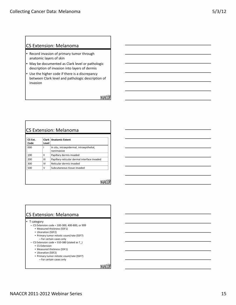
CS Extension: Melanoma• Record invasion of primary tumor through anatomic layers of skin
• May be documented as Clark level or pathologic description of invasion into layers of dermis
• Use the higher code if there is a discrepancy between Clark level and pathologic description of invasion
CS Extension: Melanoma
CS Ext. Code
Clark Level
Anatomic Extent
000 I In situ, intraepidermal, intraepithelial, noninvasive
100 II Papillary dermis invaded200 III Papillary‐reticular dermal interface invaded300 IV Reticular dermis invaded500 V Subcutaneous tissue invaded
CS Extension: Melanoma• T category
– CS Extension code = 100‐300, 400‐800, or 999• Measured thickness (SSF1)• Ulceration (SSF2)• Primary tumor mitotic count/rate (SSF7)
– For certain cases only– CS Extension code = 310‐380 (stated as T_)
• CS Extension• Measured thickness (SSF1)• Ulceration (SSF2)• Primary tumor mitotic count/rate (SSF7)
– For certain cases only

Collecting Cancer Data: Melanoma 5/3/12
NAACCR 2011‐2012 Webinar Series 16
Pop Quiz: CS Extension• Final pathologic diagnosis: Malignant melanoma, superficial spreading type, Clark level I with minute focus of microinvasion.
• What is the code for CS Extension?a. 000: In situ; Clark level Ib. 100: Papillary dermis invaded; Clark level IIc. 400: Skin/dermis NOS; localized NOSd. 999: Unknown
CS Lymph Nodes: Melanoma• Code isolated tumor cells (ITC) in regional lymph nodes as
regional node involvement in CS Lymph Nodes– Code 010: ITC only (v02.04)
• Code involvement of bilateral or contralateral nodes for head, neck, and trunk tumors in CS Lymph Nodes– Primary nodal basement
• Regional for AJCC and Summary Stage– Secondary nodal basement
• Regional for AJCC• May be distant for Summary Stage
CS Lymph Nodes: Melanoma• Stated as N_ with no other information on regional nodes (v02.04)– Code 121: Clinically N1– Codes 122‐124: Pathologically N1_– Code 125: N1; no information on clinical or pathologic evaluation
– Code 128: Clinically N2– Codes 152‐154: Pathologically N2_– Code 155: N2; no information on clinical or pathologic evaluation

Collecting Cancer Data: Melanoma 5/3/12
NAACCR 2011‐2012 Webinar Series 17
CS Lymph Nodes: Melanoma• Code satellite lesions or in‐transit metastasis in CS Lymph Nodes– Satellite lesions
• Clinical or microscopic presence of satellites around the primary melanoma
– In‐transit metastases• Metastases between the primary melanoma and regional lymph node basin
CS Lymph Nodes: Melanoma• Regional node involvement without satellite lesions or in‐transit metastases– Codes 100‐120 (v02.03)– Codes 100‐118 (v02.04)
• Satellite lesions or in‐transit metastases withoutregional node involvement– Codes 130‐150 (v02.03)– Codes 140‐151 (v02.04)
• Satellite lesions or in‐transit metastases and regional node involvement– Codes 200‐220 (v02.03)– Codes 200‐223 (v02.04)
Regional Nodes PositiveRegional Nodes Examined
• Do not count satellite lesions and in‐transit metastases in these fields
• Count nodes with ITCs in positive lymph node count for melanoma

Collecting Cancer Data: Melanoma 5/3/12
NAACCR 2011‐2012 Webinar Series 18
Pop Quiz: Lymph Nodes• Final diagnosis: Right forearm lesion, 2 cm, malignant melanoma, Clark level II, Breslow depth 2 mm; satellite nodule 1 cm from forearm lesion, malignant melanoma; sentinel node biopsy to right axillary nodes, 2 nodes removed, 1 with melanoma in isolated tumor cells.
Pop Quiz: Lymph Nodes• What is the code for CS Lymph Nodes?
a. 010: Isolated tumor cells onlyb. 100: Regional nodes by site – arm/shoulder –
axillaryc. 140: Satellite nodule(s) or in‐transit metastases less
than or equal to 2 cm from primary tumord. 200: Satellite nodule(s) or in‐transit metastases
WITH regional lymph nodes listed in code 100
Pop Quiz: Lymph Nodes• What is the code for Regional Nodes Positive?
a. 00b. 01c. 02d. 95: Positive or core biopsy of lymph node
• What is the code for Regional Nodes Examined?a. 02b. 03c. 95: No regional nodes removed but aspiration or core biopsy
of regional nodes performedd. 96: Regional lymph node removal documented as sampling
and number of nodes unknown/not stated

Collecting Cancer Data: Melanoma 5/3/12
NAACCR 2011‐2012 Webinar Series 19
CS Mets at DX: Melanoma• Do not code involvement of contiguous and bidirectional nodal basins in CS Mets at DX– Code such nodal involvement in CS Lymph Nodes
• M category– CS Mets at DX code = 05, 10, 42, 43, 52, 53, 55, 56, or 60
• Serum lactate dehydrogenase ‐ LDH (SSF4)
Pop Quiz: CS Mets at DX• Final diagnosis: Right forearm lesion, 2 cm, malignant melanoma, Clark level II, Breslow depth 2 mm; lymphadenectomy – 1/5 metastatic axillary nodes; 1/1 metastatic supraclavicular node. Chest x‐ray: normal.
• What is the code for CS Mets at DX?a. 00: No distant metastasisb. 10: Distant lymph nodes c. 60: Distant metastasis NOSd. 99: Unknown
SSF1:Measured Thickness (Depth) Breslow Measurement • Code measured thickness of primary melanoma in hundredths of mm– Breslow measurement
• Vertical measurement from the granular layer of the epidermis to the deepest point of invasion
– Code measurement labeled as thickness or depth• Next priority: Measurement described as taken from the cut surface of specimen
• Last priority: Third dimension in a statement of tumor size

Collecting Cancer Data: Melanoma 5/3/12
NAACCR 2011‐2012 Webinar Series 20
SSF1:Measured Thickness (Depth) Breslow Measurement • Assign code 999 (unknown) for melanoma in situ• Code maximum tumor thickness if there is biopsy followed by definitive excision– Do not add the measurements together
Pop Quiz: SSF1• Patient A
– Punch biopsy right upper back: Superficial spreading melanoma, Breslow depth 6.3 mm
– Wide excision right upper back: Superficial spreading melanoma, Breslow thickness 3.2 mm
• What is the code for SSF1?a. 320b. 630c. 950d. 999: Unknown
SSF2: Ulceration• Code the absence (000) or presence (010) of primary tumor ulceration as documented in path report– Ulceration: Absence of intact epidermis covering primary melanoma
– Assign code 000 (no ulceration present) if path report is available for review and there is no mention of ulceration

Collecting Cancer Data: Melanoma 5/3/12
NAACCR 2011‐2012 Webinar Series 21
Pop Quiz: SSF2• Final diagnosis: Right forearm lesion, 2 cm, malignant melanoma, Clark level II, Breslow depth 2 mm.
• What is the code for SSF2?a. 000: No ulceration presentb. 010: Ulceration presentc. 999: Unknown
SSF3: Clinical Status of Lymph Node Mets• Tumor burden of nodal metastases
– Micrometastasis• No clinical regional lymph node metastasis but nodes pathologically positive
– Macrometastasis• Regional lymph node metastasis confirmed pathologically by lymphadenectomy
SSF3: Clinical Status of Lymph Node Mets (v02.04)Code Description
000 OBSOLETE DATA RETAINED V0204005 Clinically negative lymph node metastasis AND
no pathologic examination performed Or unknown if pathologic exam performed Or nodes negative on pathologic examination
010 Clinically occult (microscopic) lymph node metastasis only020 OBSOLETE DATA RETAINED V0204
043 Clinically apparent nodal metastasis in 1 regional node045 Clinically apparent nodal metastasis in 2‐3 regional nodes048 Clinically apparent nodal metastasis in 4+ regional nodes050 Clinically apparent nodal metastasis in regional node(s) but number not
specified100 Clinically apparent in transit metastasis only150 Clinically apparent in transit metastasis and clinically apparent nodal
metastasis (at least one node)

Collecting Cancer Data: Melanoma 5/3/12
NAACCR 2011‐2012 Webinar Series 22
Pop Quiz: SSF3• Patient diagnosed elsewhere. Referred here for treatment. Wide excision of left leg lesion, melanoma Clark II Breslow 3mm; superficial inguinal lymphadenectomy, 1 of 5 lymph nodes with metastasis.
• What is the code for SSF3?a. 043: Clinically apparent nodal metastasis in 1 regional nodeb. 048: Clinically apparent nodal metastasis in 4+ regional
nodesc. 050: Clinically apparent nodal metastasis in regional nodes
but number not specifiedd. 999: Unknown or no information about clinical nodal
involvement
SSF4: Serum Lactate Dehydrogenase (LDH)Code Description000 Within normal limits010 Range 1: Less than 1.5 x upper limit of normal for LDH assay
Stated as elevated NOS020 Range 2: 1.5 – 10 x upper limit of normal for LDH assay030 Range 3: More than 10 x upper limit of normal for LDH assay988 Not applicable997 Test ordered, results not in chart998 Test not done999 Unknown
• Use information from same test used to code SSF5 and SSF6
SSF4: Serum Lactate Dehydrogenase (LDH)• Positive LDH results from 2 lab tests required to code as positive (v02.04 clarification)– Assign code 000 if 1st test positive and 2nd test negative
– Assign code 998 if 1st test positive and no 2nd test performed
– Assign code 999 if 1st test positive and no information about 2nd test
– Assign code 000 if only 1 test performed and it is within normal limits

Collecting Cancer Data: Melanoma 5/3/12
NAACCR 2011‐2012 Webinar Series 23
SSF5: LDH Lab Value• Record LDH lab value prior to treatment or within 6 weeks of diagnosis– Record exact value for values 001‐800– Record range for values 801 and greater– Use information from same test used to code SSF4 and SSF6
SSF6: LDH Upper Limits of Normal
• Record exact upper limit of normal for LDH as documented on lab report– Values vary by lab– Use information from same test used to code SSF4 and SSF5
Pop Quiz: SSF4, SSF5, and SSF6• 2/27/12: LDH lab value elevated at 300; lab range 150‐250 U/L
• 3/7/12: Punch biopsy left arm – melanoma • 3/7/12: LDH lab value 200; lab range 150‐250 U/L
• 3/21/12: Wide excision left arm – 0.5 cm melanoma, Breslow 3mm, Clark II

Collecting Cancer Data: Melanoma 5/3/12
NAACCR 2011‐2012 Webinar Series 24
Pop Quiz: SSF4 and SSF5• What is the code for SSF4?
a. 000: Within normal limitsb. 010: Range 1: Less than 1.5 x upper limit of normalc. 998: Test not doned. 999: Unknown
• What is the code for SSF5?a. 200b. 300c. 998: Test not doned. 999: Unknown
Pop Quiz: SSF6• What is the code for SSF6?
a. 150b. 250c. 998: Test not doned. 999: Unknown
SSF7: Primary Tumor Mitotic Count/Rate• Record the number of mitoses per square mm as documented in path report
• T category– CS Extension code = 100‐800 or 999; SSF1 (measured thickness) = 001‐100
• Ulceration (SSF2)• Primary tumor mitotic count/rate (SSF7)
• Code 996 (v02.04)– Mitotic rate described with denominator other than square mm

Collecting Cancer Data: Melanoma 5/3/12
NAACCR 2011‐2012 Webinar Series 25
Pop Quiz: SSF7• Final path diagnosis: Nodular melanoma, right ankle, Breslow 3 mm; Clark III; greater than 1 mitosis per square mm.
• What is the code for SSF7?a. 001b. 990: Stated as less than 1 mitosis/square mm; stated as
nonmitogenicc. 991: Stated as at least 1 mitosis/square mm; stated as
mitogenicd. 996: Mitotic rate described with denominator other
than square mm
SSF8: Primary Tumor Regression• Record the absence or presence of regression as documented in path report– Assign code 000 (regression absent) if regression is not identified
SSF9: Vertical Growth Phase (VGP)• Record the absence or presence of VGP as documented in path report– Assign code 000 (VGP absent) if VGP is not identified– Assign code 010 (VGP present) if VGP is identified OR if tumor is nodular melanoma

Collecting Cancer Data: Melanoma 5/3/12
NAACCR 2011‐2012 Webinar Series 26
Pop Quiz: SSF8 and SSF9• Final path diagnosis: Nodular melanoma, Breslow 1 mm; Clark level III.
• What is the code for SSF8?a. 000: Regression absentb. 010: Regression presentc. 999: Unknown
• What is the code for SSF9?a. 000: VGP absentb. 010: VGP presentc. 999: Unknown
Standard Setters SSF Requirements for Melanoma Skin • CoC, SEER
– SSF: 1‐7
• NPCR– SSF: 1‐4, 7
• Canadian Council of Cancer Registries– SSF: 1‐4, 5‐6*, 7, 8‐9**
– * Collect if readily available in clinical chart– ** Collect if in path report
TREATMENTMelanoma of the Skin

Collecting Cancer Data: Melanoma 5/3/12
NAACCR 2011‐2012 Webinar Series 27
DIAGNOSTIC BIOPSIES
Excisional Biopsy
• Suspicious pigmented lesions usually undergo an excisional biopsy– 1‐3mm margins– Elliptical shape– Should be done with future lymphatic mapping in mind
‐‐3mm
Punch Biopsy• For some sites a standard excisional biopsy may be inappropriate– Face, palmar surface of the hand, sole of the foot, distal digit, subungal (under a nail)
– Very large lesions • May be excisional or incisional

Collecting Cancer Data: Melanoma 5/3/12
NAACCR 2011‐2012 Webinar Series 28
Shave Biopsy• Superficial
– “shaves” off the epidermis and part of the dermis
– Not generally done for suspected invasive melanoma
• Deep– “Scoops” out the
suspicious lesion with sufficient depth to stage
• Least invasive type of biopsy– No stitches
Clinical Stage
• Pathology report– Breslow’s Depth– Ulceration– Mitotic rate– Deep and peripheral margin status
– Satellitosis
• Clinically positive lymph nodes
• In‐transit metastasis• Imaging if suspected distant mets
• LDH
Coding Surgical Procedures• Incisional biopsy
– Removal of the tumor with positive margins• Punch• Shave• Elliptical
– Code as a diagnostic staging procedure (02)• Excisional biopsy (27)
– Elliptical– Shave– Punch

Collecting Cancer Data: Melanoma 5/3/12
NAACCR 2011‐2012 Webinar Series 29
Coding Surgical Procedures
• Mohs Surgery– 34‐margins unknown– 35‐margins 1cm or less– 36‐margins 1cm or more
© 2010 Terese Winslow, U.S. Govt. has certain rights
Coding Surgical Procedures• Biopsy of primary tumor followed by gross excision of the lesion (codes 30‐33)– Incisional biopsy followed by gross excision– Excisional biopsy with margins less than 1cm– Does not have to be done under the same anesthesia
Coding Surgical Procedures
• Wide excision– Code 45 If the nearest involved margin is more than 1cm, but unknown how much more.
– Code 46 if the nearest involved margin is >1cm and < or = 2cm
– Code 47 if the nearest involved margin is > 2cm
‐‐1cm‐‐2cm
Code 46Code 47

Collecting Cancer Data: Melanoma 5/3/12
NAACCR 2011‐2012 Webinar Series 30
Wide excision• In situ melanoma
– .5cm
• Stage IA– 1cm margins
• Breslow’s depth of 1.01 to 2.0mm– 1‐2cm margins
• Breslow’s depth more than 2.1 mm – 2cm margins
Sentinel Lymph Node Biopsy
© 2010 Terese Winslow, U.S. Govt. has certain rights
Lymph Node Dissection• Clinically negative lymph nodes
– If sentinel lymph node is negative, regional node dissection is not required
– If sentinel lymph node is positive, dissection of the lymph node basin should be offered.
• Clinically positive lymph nodes– Lymph node dissection of the lymph node basin should be offered

Collecting Cancer Data: Melanoma 5/3/12
NAACCR 2011‐2012 Webinar Series 31
Adjuvant treatment• Stage III (lymph node positive)
– Interferon (BRM) • Low dose or intermediate dose• High‐dose or pegylated interferon
• Stage IV (distant metastasis)– Clinical Trial– Chemotherapy– Ipilimumab (BRM)– Excision of solitary metastatic lesions– Radiation
PUVA• Psoralens (P) and then exposing the skin to UVA (long wave ultraviolet radiation)– Code as Other treament
QUESTIONS?

Collecting Cancer Data: Melanoma 5/3/12
NAACCR 2011‐2012 Webinar Series 32
Coming up!• 6/14/12
– Using and Interpreting Data Quality Indicators
• 7/12/12– ICD‐10‐CM and Cancer Surveillance
94
And the winners of the fabulous prizes are….
Thank You!
95