coarctation of the aorta - learningradiology
TRANSCRIPT
Obstructive LesionsObstructive Lesions
In Slide Show mode, to advance slides, press spacebaror click left mouse button
William Herring, M.D. © 2004
All photos retain the rights of their original owners
Lesions ThatCause CHF
Lesions ThatCause CHF

CHF In NewbornImpede Return of Flow to Left Heart
Infantile coarctationCongenital aortic stenosisHypoplastic left heart syndromeCongenital mitral stenosisCor triatriatumObstruction to venous return from lungs
TAPVR from below diaphragm

Coarctation of the Aorta
Congenital Aortic Stenosis
Hypoplastic Left Heart
Congenital Mitral Stenosis
Cor Triatriatum
Obstruction to venous return from
lungs
Causes of CHF in the Newborn
© Netter
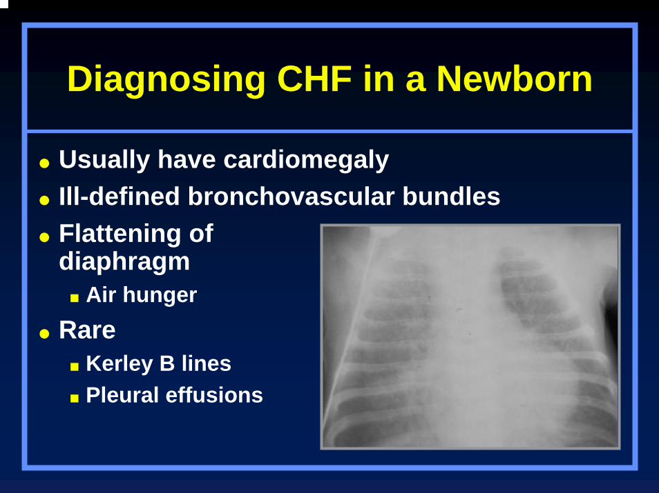
Diagnosing CHF in a Newborn
Usually have cardiomegalyIll-defined bronchovascular bundlesFlattening of diaphragm
Air hungerRare
Kerley B linesPleural effusions

CHF In Chronologic Sequence
CHF In Chronologic Sequence
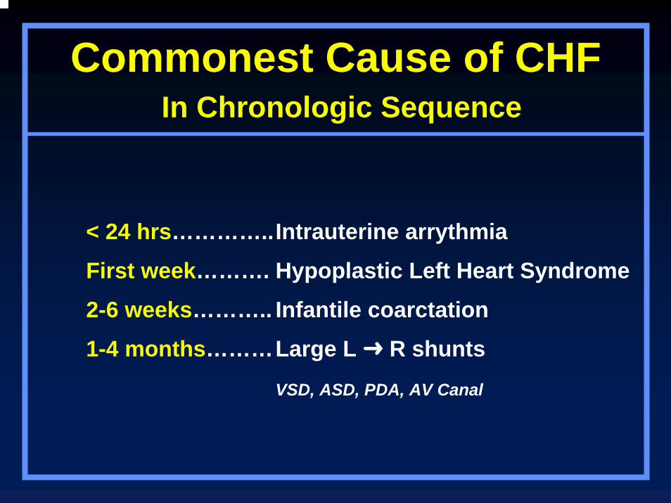
Commonest Cause of CHFIn Chronologic Sequence
< 24 hrs…………..Intrauterine arrythmia
First week………. Hypoplastic Left Heart Syndrome
2-6 weeks……….. Infantile coarctation
1-4 months………Large L R shunts
VSD, ASD, PDA, AV Canal
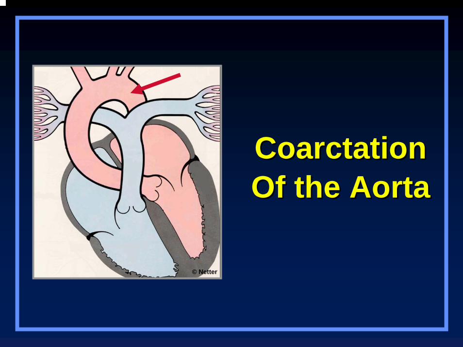
CoarctationCoarctationOf the AortaOf the Aorta
© Netter
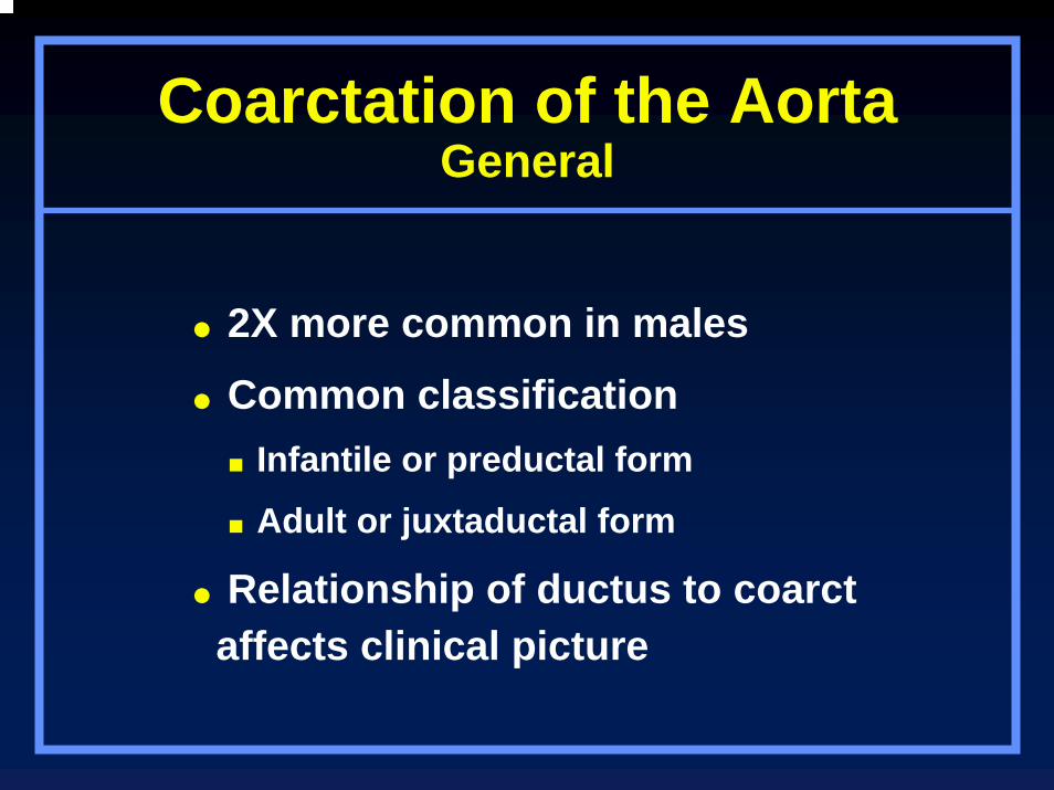
Coarctation of the AortaGeneral
2X more common in males
Common classification Infantile or preductal form
Adult or juxtaductal form
Relationship of ductus to coarct affects clinical picture
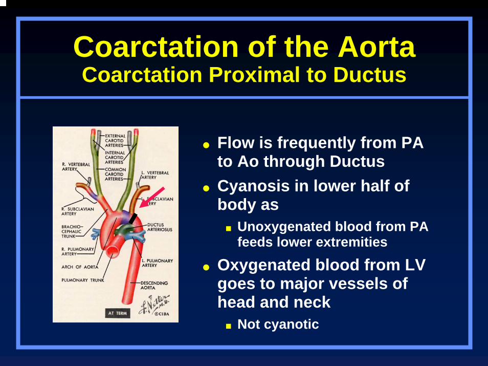
Coarctation of the AortaCoarctation Proximal to Ductus
Flow is frequently from PA to Ao through DuctusCyanosis in lower half of body as
Unoxygenated blood from PA feeds lower extremities
Oxygenated blood from LV goes to major vessels of head and neck
Not cyanotic
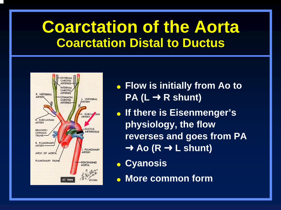
Coarctation of the AortaCoarctation Distal to Ductus
Flow is initially from Ao to PA (L R shunt)If there is Eisenmenger’s physiology, the flow reverses and goes from PA
Ao (R L shunt)CyanosisMore common form
Coarctation of the AortaOther Classifications
More complicated classifications take following into accountLocation and length of coarct
Patency of ductus arteriosis
Relationship of coarct to ductus
Coarctation of the AortaAdult Form
Adult or juxtaductal (postductal) form is more common than infantileUsually localizedArea of coarctation just beyond origin
of LSCA at level of ductus
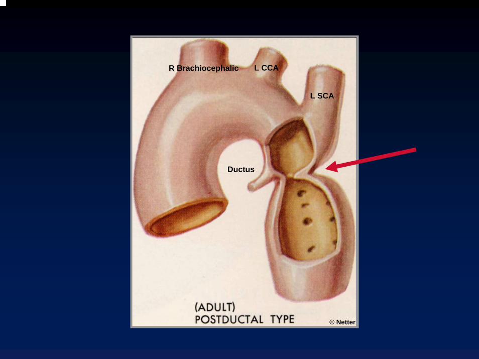
R Brachiocephalic L CCA
L SCA
Ductus
© Netter

Coarctation of the Aorta
Coarctation of the AortaInfantile Form
Infantile, preductal form = diffuse typeLong, tubular segment of narrowed aorta
From just distal to brachiocephalic artery to level of ductus
Intracardiac defects (VSD, ASD, deformed mitral valve) present in 50% of diffuse type
Also patent ductus arteriosis
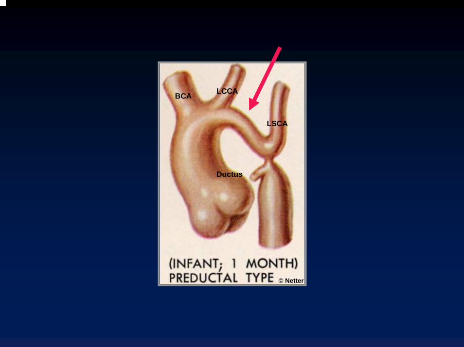
BCA LCCA
LSCA
Ductus
© Netter
Coarctation of the AortaAssociated Defects
Bicuspid aortic valve (most common associated defect seen in 50%)VSDASDTransposition25% of patients with Turner’s Syndrome
have coarctation of aorta
Coarctation of the AortaShone Syndrome
Coarctation of aortaAortic stenosisParachute mitral valveSupravalvular mitral ring
X-Ray FindingsRib Notching
Single best signOlder the person, more likely to have rib
notching (uncommon <6 yrs) Majority with coarcts display it >20 years
of age Rib notching occurs in high pressure
circuit
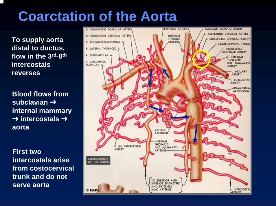
Coarctation of the AortaCoarctation of the AortaTo supply aorta distal to ductus, flow in the 3rd-8th
intercostals reverses
© Netter
Blood flows from subclavian internal mammary
intercostals aorta
First two intercostals arise from costocervicaltrunk and do not serve aorta
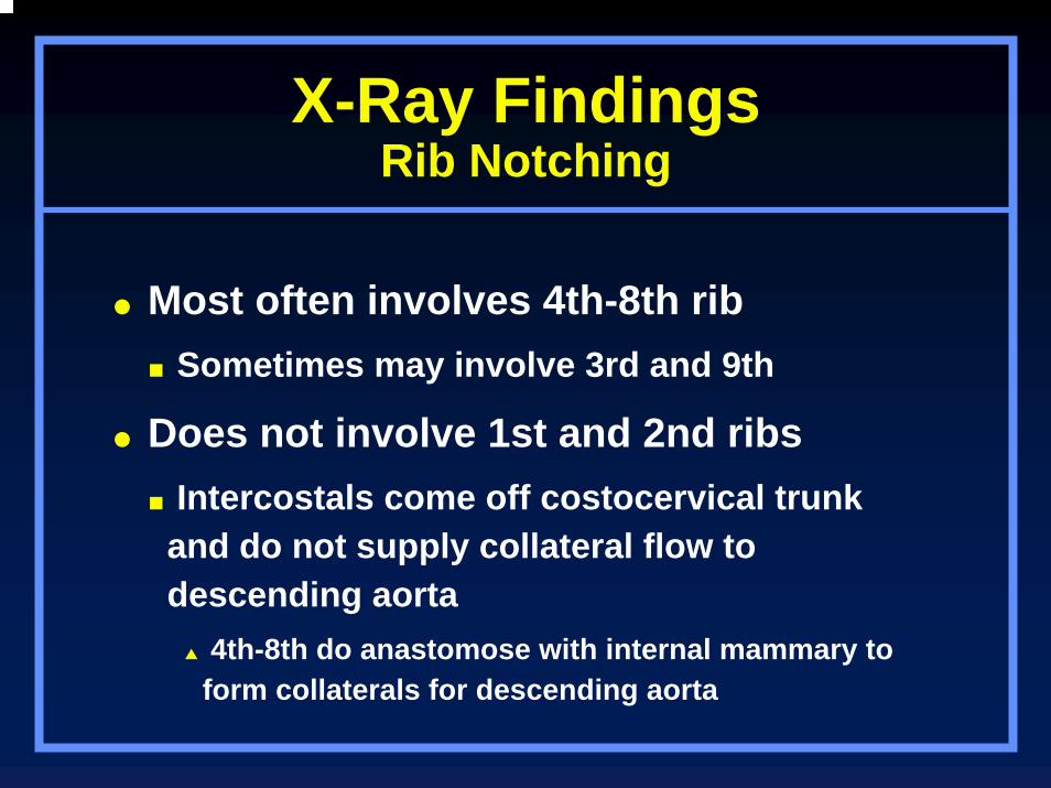
X-Ray FindingsRib Notching
Most often involves 4th-8th ribSometimes may involve 3rd and 9th
Does not involve 1st and 2nd ribsIntercostals come off costocervical trunk and do not supply collateral flow to descending aorta
4th-8th do anastomose with internal mammary to form collaterals for descending aorta
7
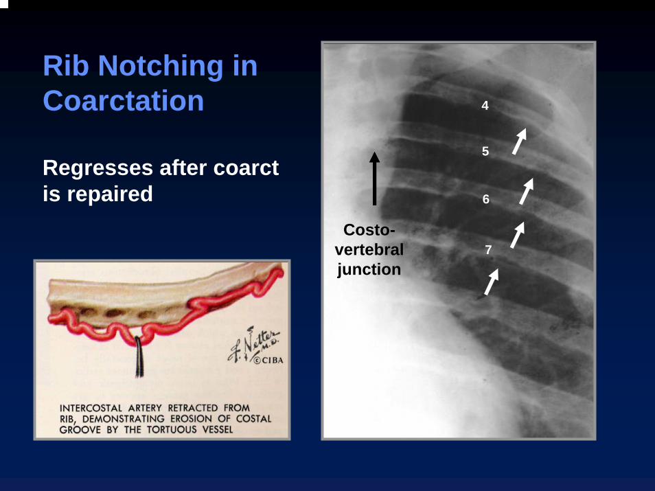
Rib Notching in Coarctation 4
5
6
Regresses after coarct is repaired
Costo-vertebral junction
X-Ray FindingsRib Notching–Unilateral
Rib notching occurs in the high pressure circuit
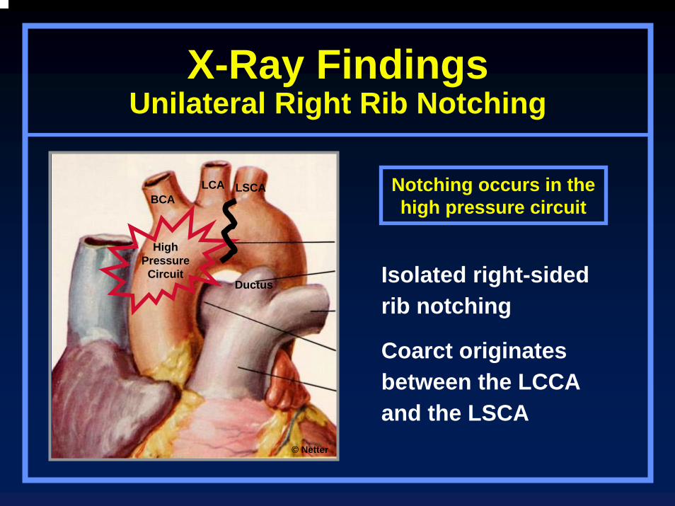
X-Ray FindingsUnilateral Right Rib Notching
Notching occurs in the high pressure circuit
HighPressure Circuit
BCALCA LSCA
Ductus
© Netter
Isolated right-sided rib notching
Coarct originates between the LCCA and the LSCA
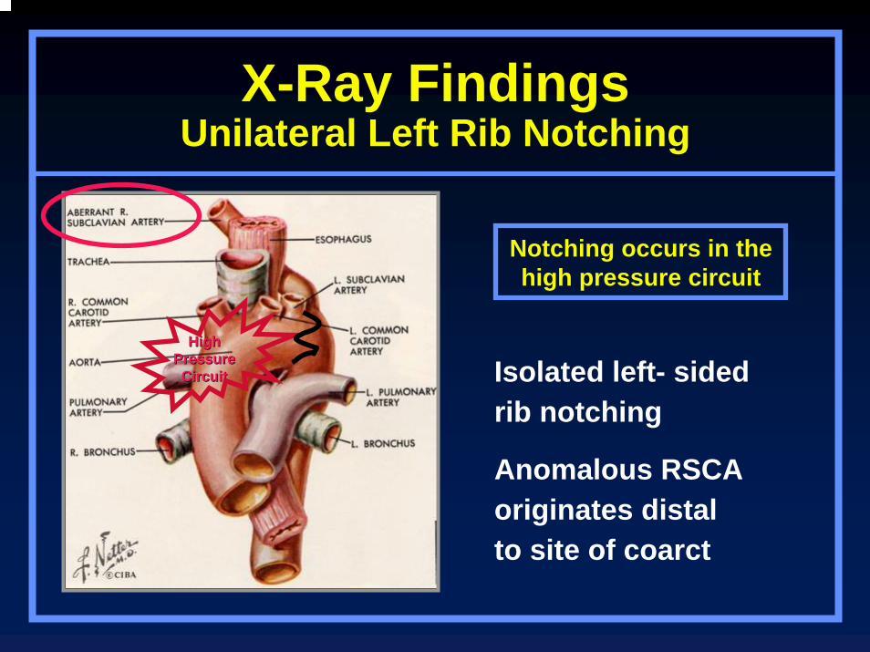
X-Ray FindingsUnilateral Left Rib Notching
HighHighPressure Pressure CircuitCircuit
Notching occurs in the high pressure circuit
Isolated left- sided rib notching
Anomalous RSCA originates distal to site of coarct

X-Ray FindingsFigure 3 Sign
Caused by (in order) Dilated LSCA or aortic knob“Tuck” of coarct itselfPoststenotic dilatation
Occurs in 1/3–1/2 of patients with coarctNot in children
Matched by “reverse 3” or “E” on barium-filled esophagus

© Netter
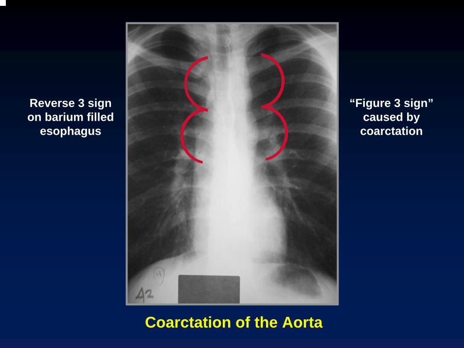
Reverse 3 sign on barium filled
esophagus
“Figure 3 sign”caused by coarctation
Coarctation of the Aorta
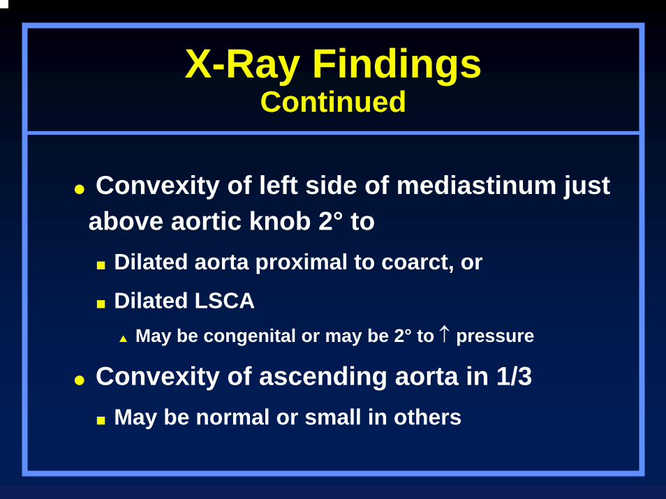
X-Ray FindingsContinued
Convexity of left side of mediastinum just above aortic knob 2° to
Dilated aorta proximal to coarct, or
Dilated LSCAMay be congenital or may be 2° to ↑ pressure
Convexity of ascending aorta in 1/3May be normal or small in others
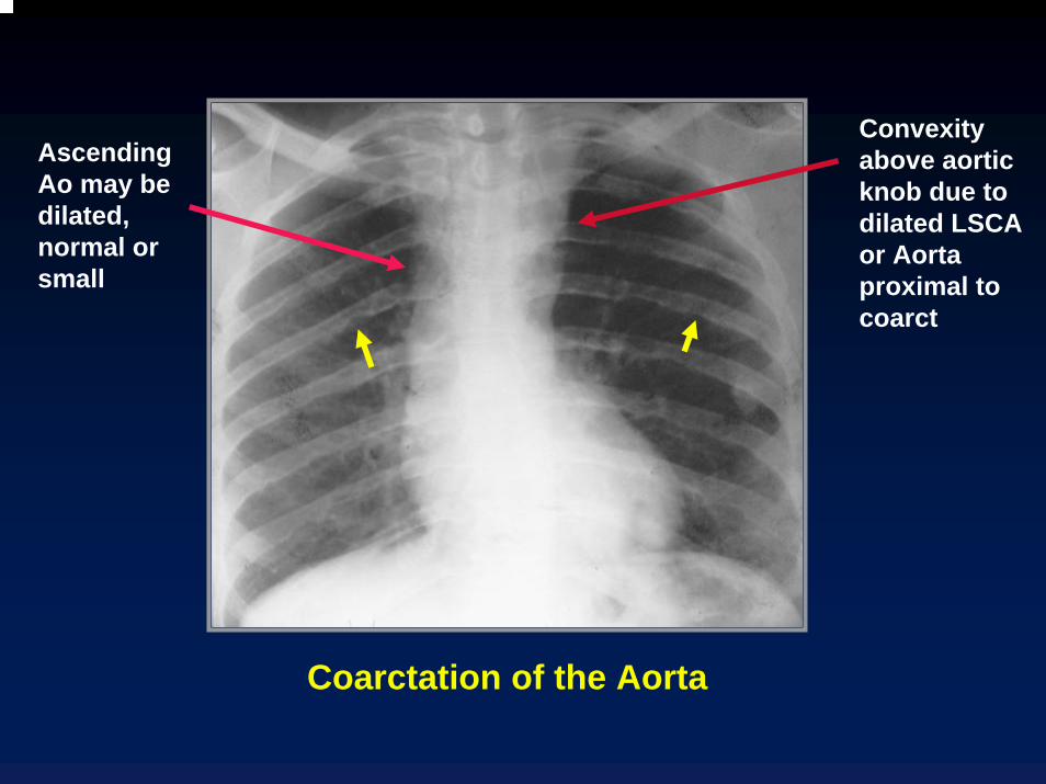
Convexity above aortic knob due to dilated LSCA or Aorta proximal to coarct
Ascending Ao may be dilated, normal or small
Coarctation of the Aorta
Coarctation of the AortaClinical Findings–Infancy
Severe CHF most common from 2nd to 6th week of life
Weak or absent leg pulses
Lower BP in the legs than in the arms
EKG: RV hypertrophy because RV assumes most of the cardiac output during fetal life in these patients
Coarctation of the AortaEchocardiographic Findings
In infants, 2D echo can demonstrate coarcts from suprasternal notch
Echo helpful in excluding associated hypoplastic left heart syndrome
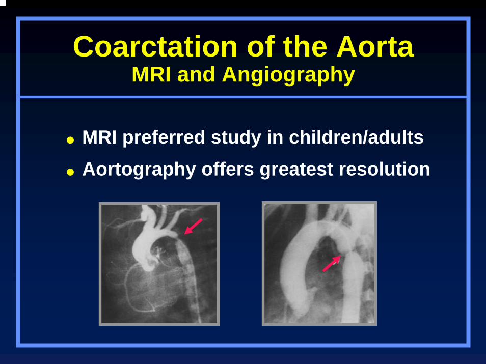
Coarctation of the AortaMRI and Angiography
MRI preferred study in children/adultsAortography offers greatest resolution

Amersham
Contrast enhanced MRA shows long segment coarctation of the aorta
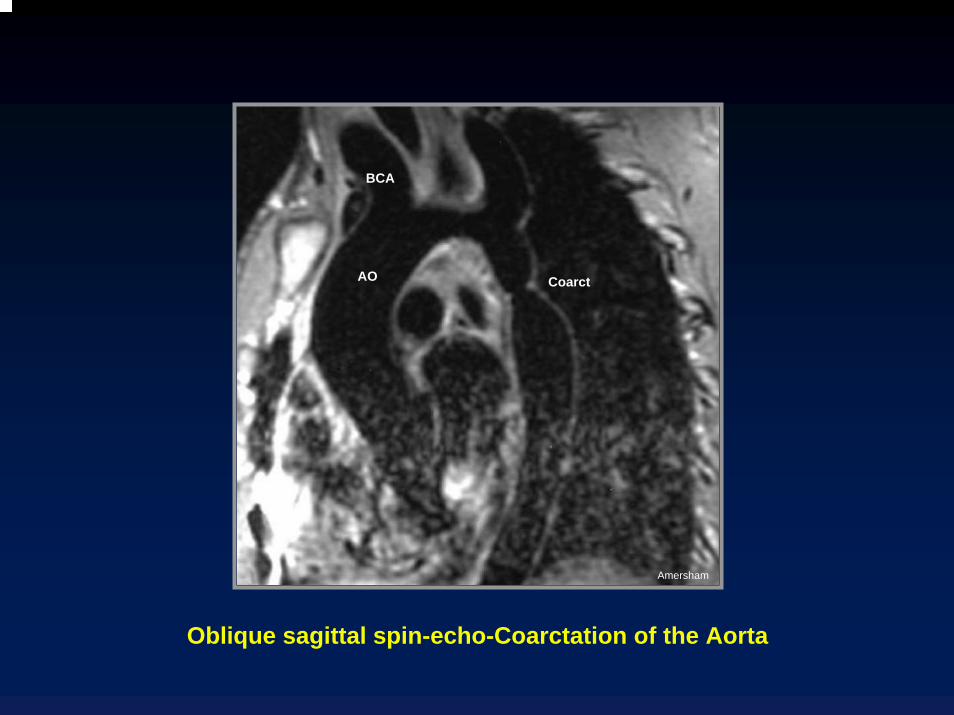
AO
BCA
Coarct
Amersham
Oblique sagittal spin-echo-Coarctation of the Aorta

Amersham
Axial spin-echo MRI-Coarctation of the Aorta
Coarctation of the AortaComplications
Heart failure in neonateSubarachnoid bleeds 2° ruptured Berry
aneurysmsDissection of aortaBacterial endocarditisMycotic aneurysm

Pseudocoarctation
Buckling of aorta resembles true coarctation
No pressure gradient (<30mmHg)
Figure 3 sign present
No rib notching
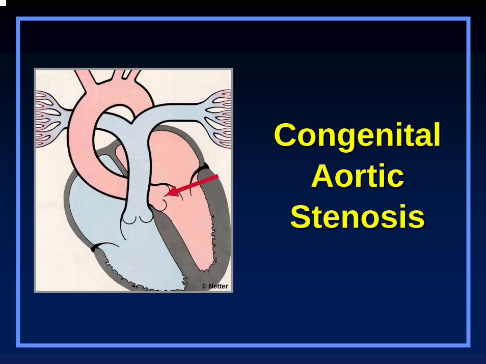
Congenital Congenital Aortic Aortic
StenosisStenosis
© Netter
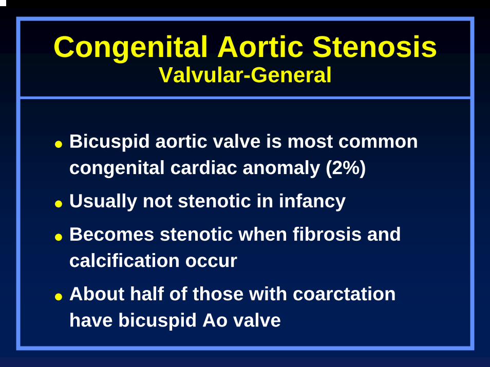
Congenital Aortic StenosisValvular-General
Bicuspid aortic valve is most common congenital cardiac anomaly (2%)
Usually not stenotic in infancy
Becomes stenotic when fibrosis and calcification occur
About half of those with coarctation have bicuspid Ao valve

Congenital Aortic StenosisAngiography
Domed and thickened leaflets in systole
Two leaflets and two sinuses of Valsalva
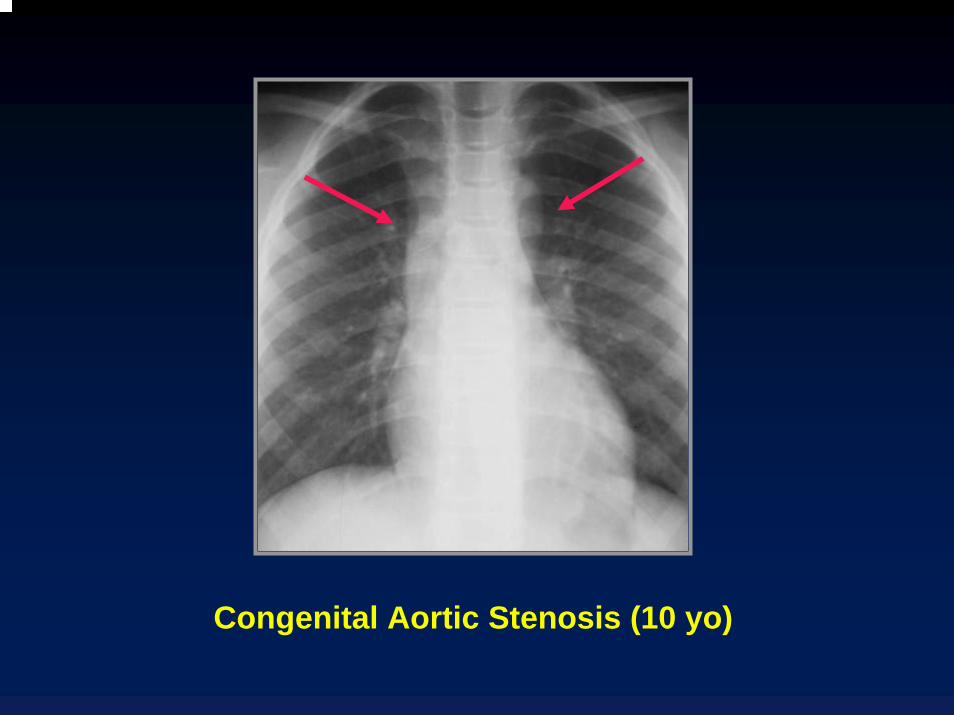
Congenital Aortic Stenosis (10 yo)
LV
Ao valve
Ao
Signal void
Amersham
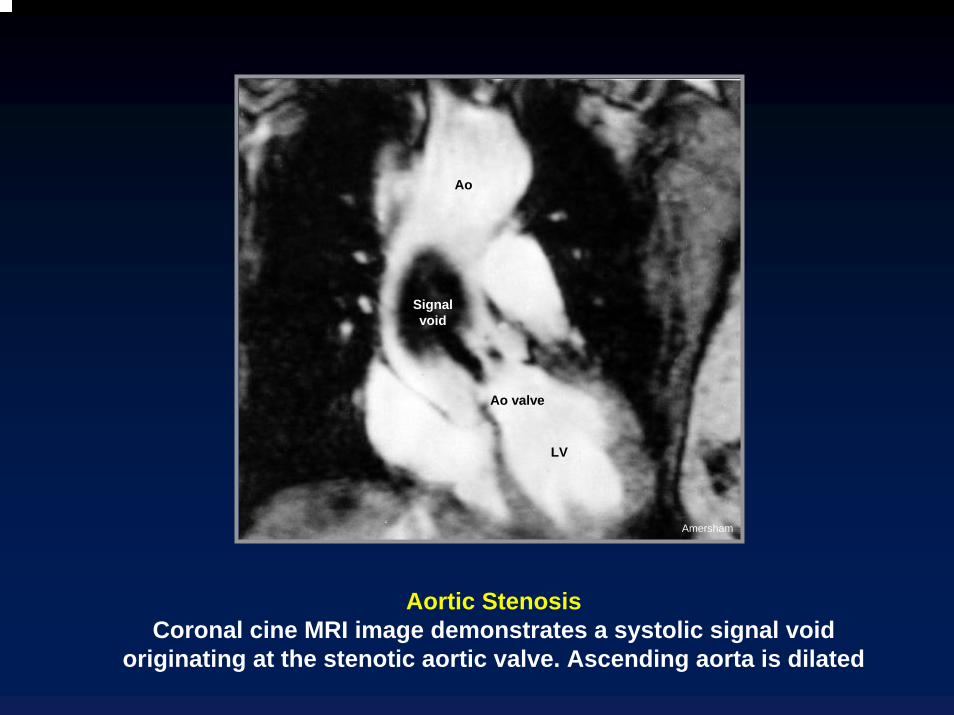
Aortic StenosisCoronal cine MRI image demonstrates a systolic signal void
originating at the stenotic aortic valve. Ascending aorta is dilated
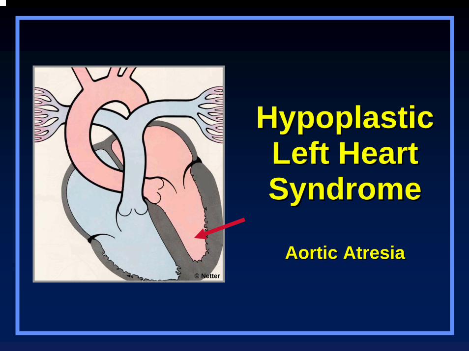
HypoplasticHypoplasticLeft HeartLeft HeartSyndromeSyndrome
Aortic AtresiaAortic Atresia© Netter

Hypoplastic Left Heart SyndromeGeneral
Most common cause of death from cardiac cause during first week of life
Common clinical expression of this lesion is CHF in first week of life
Usually cyanotic
Heart is enlarged in most
Hypoplastic Left Heart SyndromeGeneral
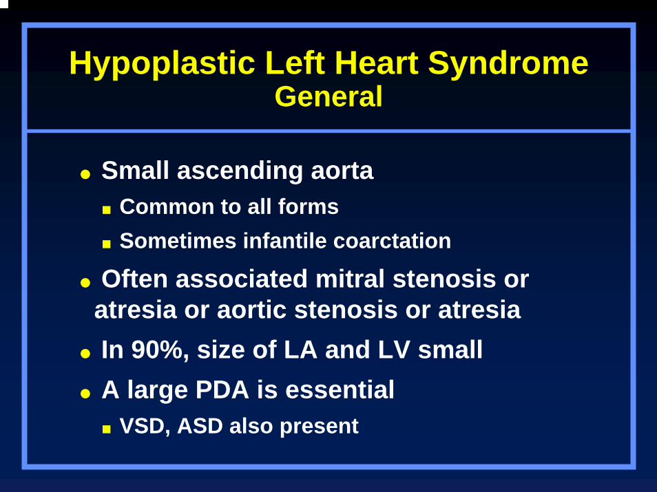
Small ascending aortaCommon to all formsSometimes infantile coarctation
Often associated mitral stenosis or atresia or aortic stenosis or atresia In 90%, size of LA and LV smallA large PDA is essential
VSD, ASD also present
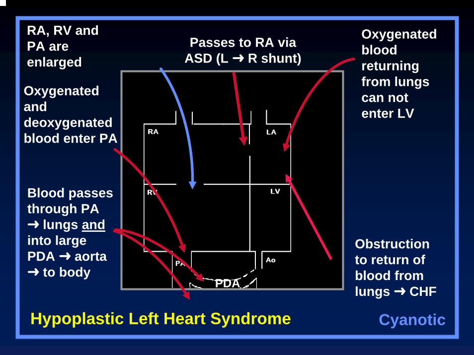
Oxygenated blood returning from lungs can not enter LV
Passes to RA via ASD (L R shunt)
RA, RV and PA are enlarged
Oxygenated and deoxygenated blood enter PA
Blood passes through PA
lungs andinto large PDA aorta
to body
Hypoplastic Left Heart Syndrome
Obstruction to return of blood from lungs CHFPDA
Cyanotic
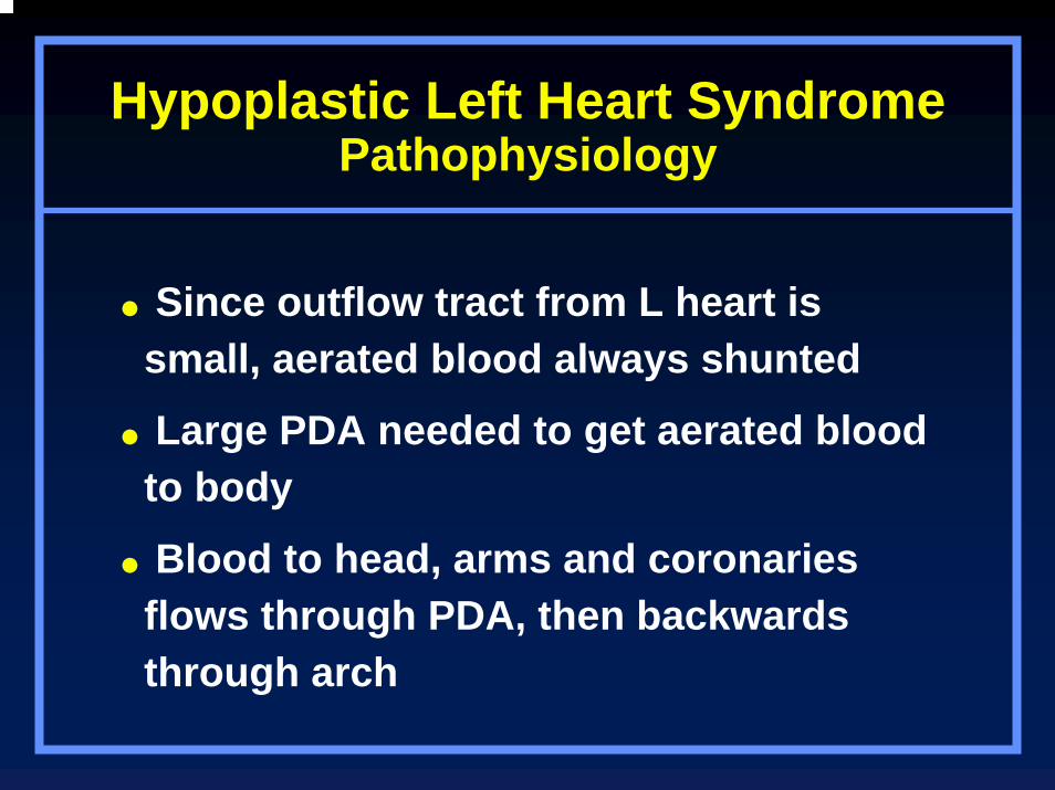
Hypoplastic Left Heart SyndromePathophysiology
Since outflow tract from L heart is small, aerated blood always shunted
Large PDA needed to get aerated blood to body
Blood to head, arms and coronaries flows through PDA, then backwards through arch
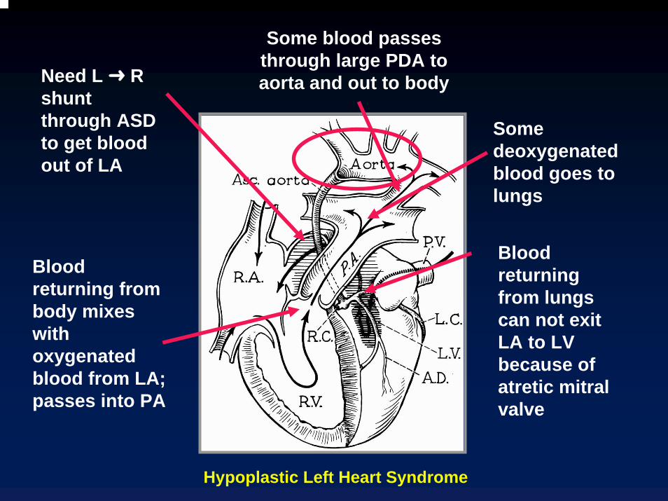
Blood returning from body mixes with oxygenated blood from LA; passes into PA
Some blood passes through large PDA to aorta and out to body
Some deoxygenated blood goes to lungs
Need L R shunt through ASD to get blood out of LA
Blood returning from lungs can not exit LA to LV because of atretic mitral valve
Hypoplastic Left Heart Syndrome
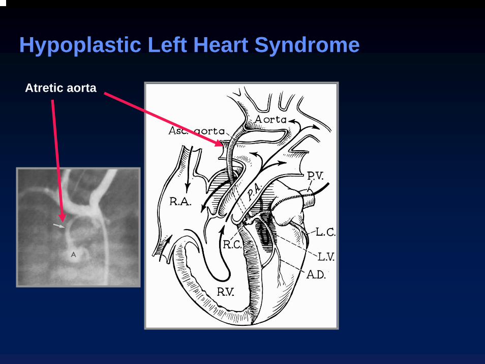
Hypoplastic Left Heart Syndrome
Atretic aorta
Hypoplastic Left Heart SyndromeAssociated Anomalies
Coarctation of the aortaInterruption of the aortic archAV communisAnomalies of the R subclavian arteryBicuspid aortic valve
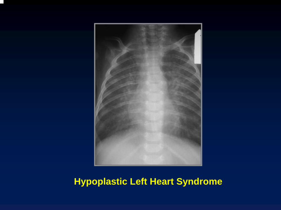
Hypoplastic Left Heart SyndromeX-ray Findings
Increased load on RV marked cardiomegaly at birth
Obstruction to return of blood from lungs CHF at birth
Most common cause of CHF in first two weeks of life
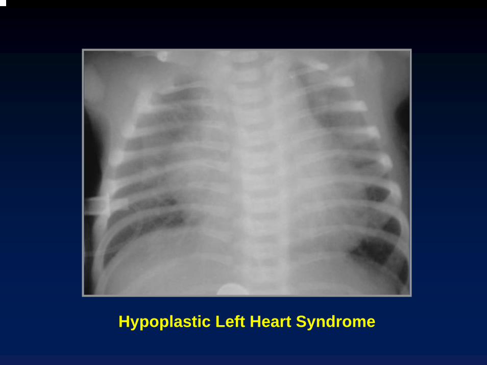
Hypoplastic Left Heart Syndrome
Hypoplastic Left Heart Syndrome

RA
MPA
Ao
LA
Amersham
Hypoplastic Left heart SyndromeGated spin echo at base of heart shows hypoplastic aorta
(arrow) posterior and right of main pulmonary artery
Hypoplastic Left Heart SyndromeDiagnosis
Diagnosis can be made by echoCatheterization may be hazardous
Spasm of PDA during cath can death
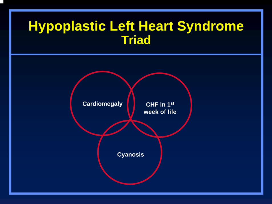
Hypoplastic Left Heart SyndromeTriad
Cardiomegaly CHF in 1st
week of life
Cyanosis

CongenitalCongenitalMitralMitral
StenosisStenosis
© Netter

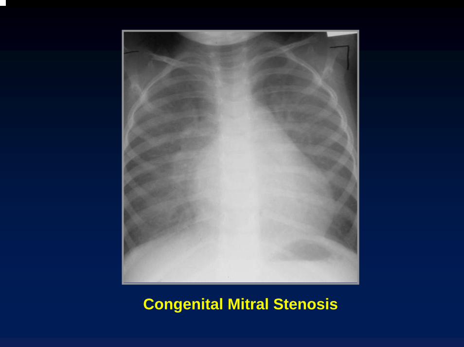
Congenital Mitral Stenosis
Exists as isolated abnormality 25% of time
Coexists with VSD 30% of time
Coexists with another form of left ventricular outflow obstruction 40% of time—SHONE’S Syndrome
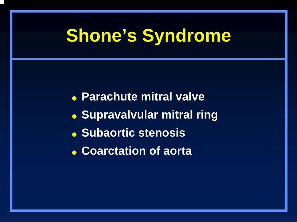
Shone’s Syndrome
Parachute mitral valveSupravalvular mitral ringSubaortic stenosisCoarctation of aorta
Congenital Mitral Stenosis
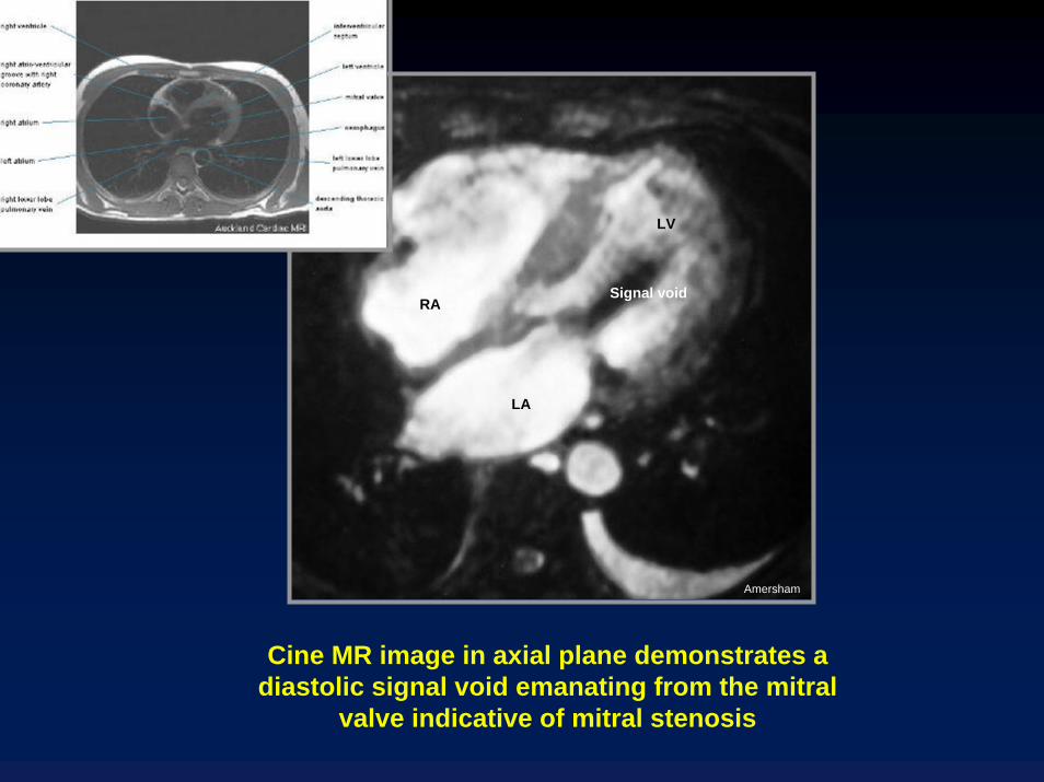
Cine MR image in axial plane demonstrates a diastolic signal void emanating from the mitral
valve indicative of mitral stenosis
RA
LA
LV
Signal void
Amersham
CorCorTriatriatumTriatriatum
© Netter
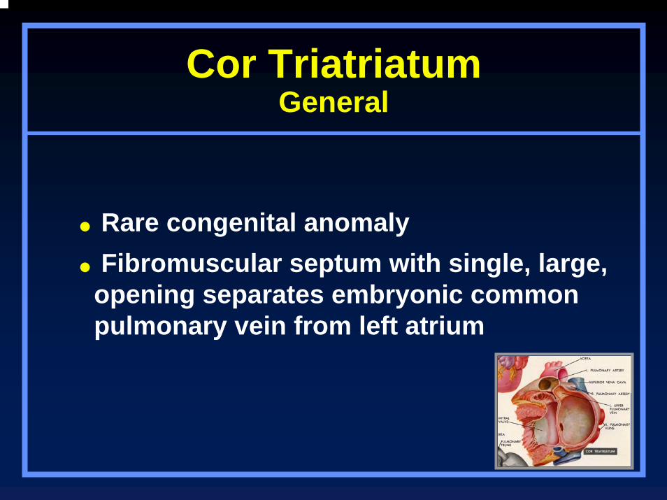
Cor TriatriatumGeneral
Rare congenital anomalyFibromuscular septum with single, large,
opening separates embryonic common pulmonary vein from left atrium
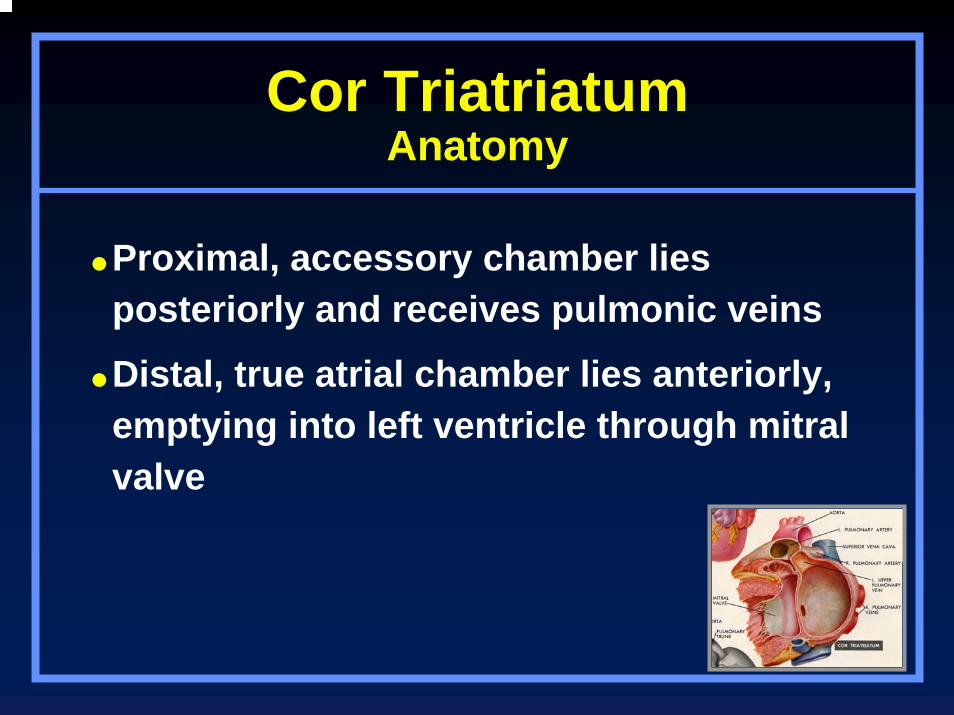
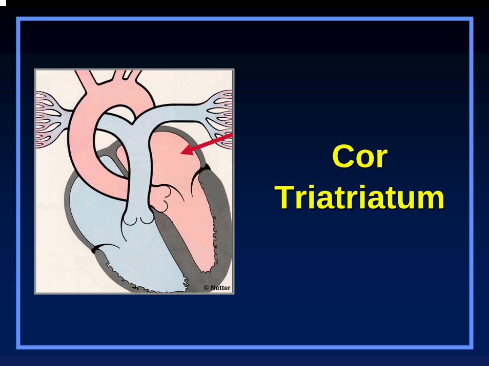
Cor TriatriatumAnatomy
Proximal, accessory chamber lies posteriorly and receives pulmonic veins
Distal, true atrial chamber lies anteriorly, emptying into left ventricle through mitral valve

Cor Triatriatum
Distal, true atrial chamber lies anteriorly and contains mitral valve
Proximal accessory chamber lies posterior and receives pulmonary veins
© Netter

Cor TriatriatumAssociations
ASDPDAAnomalous pulmonary venous drainageLeft SVCVSDTetralogy of Fallot
Cor TriatriatumClinical
Clinically similar to mitral stenosis
Dyspnea
Heart failure
Failure to thrive

Cor TriatriatumX-ray Findings
Pulmonary edemaEnlarged LA
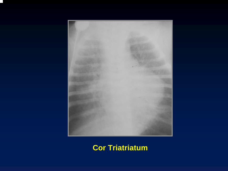
Cor Triatriatum
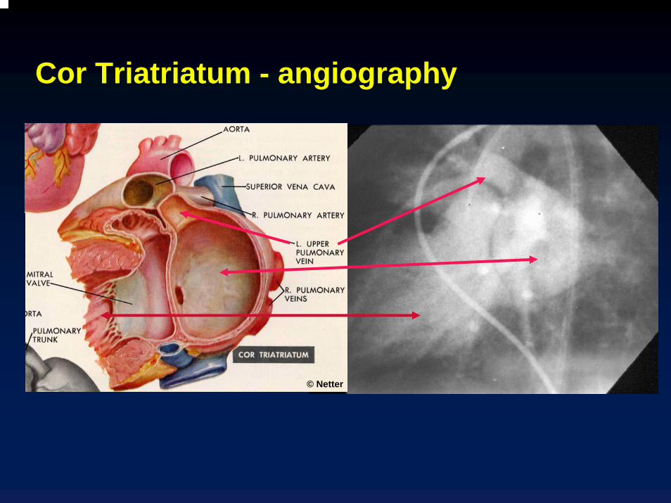
Cor Triatriatum - angiography
© Netter
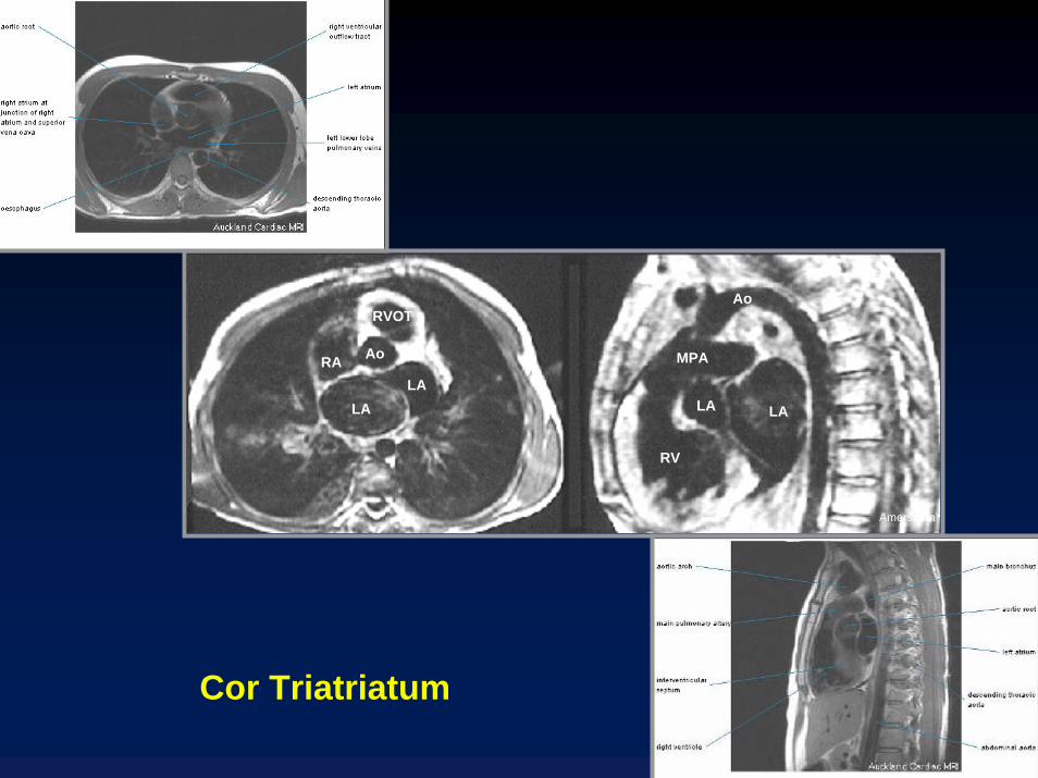
LALA
RVOT
RA Ao
RV
LALA
MPA
Ao
Amersham
Cor Triatriatum
Cor TriatriatumTreatment
Surgical excision of obstructing membrane
Cor TriatriatumPrognosis
Usually fatal in first 2 years of lifeAssociated abnormalities
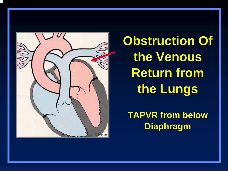
Obstruction Of Obstruction Of the Venous the Venous Return from Return from the Lungsthe Lungs
TAPVR from below TAPVR from below DiaphragmDiaphragm
© Netter
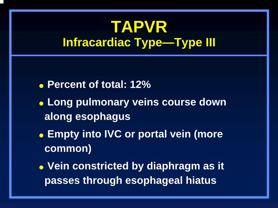
TAPVRInfracardiac Type—Type III
Percent of total: 12%Long pulmonary veins course down along esophagusEmpty into IVC or portal vein (more common)Vein constricted by diaphragm as it passes through esophageal hiatus
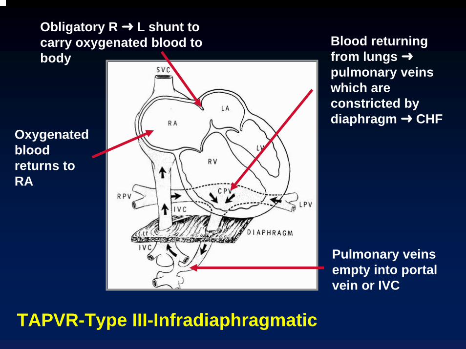
Blood returning from lungs pulmonary veins which are constricted by diaphragm CHF
Pulmonary veins empty into portal vein or IVC
Oxygenated blood returns to RA
Obligatory R L shunt to carry oxygenated blood to body
TAPVR-Type III-Infradiaphragmatic
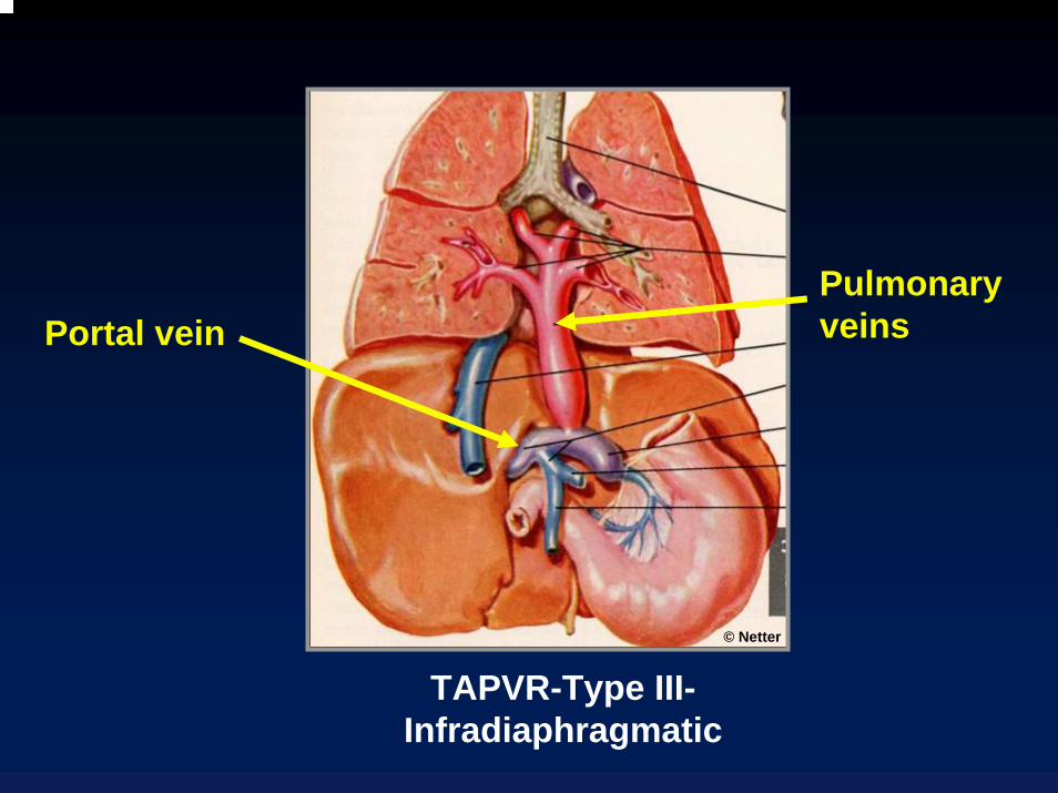
Pulmonary veinsPortal vein
© Netter
TAPVR-Type III-Infradiaphragmatic
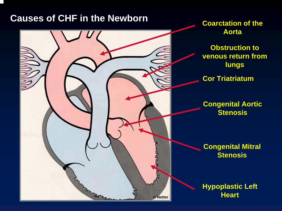
Coarctation of the Aorta
Congenital Aortic Stenosis
Hypoplastic Left Heart
Congenital Mitral Stenosis
Cor Triatriatum
Obstruction to venous return from
lungs
Causes of CHF in the Newborn
© Netter

The End