co-expression of putative pheromone receptors in the ... · precipitation followed by deae...
TRANSCRIPT
Co-Expression of Putative Pheromone Receptors in the SensoryNeurons of the Vomeronasal Organ
Sara Martini,1 Lucia Silvotti,1 Arild Shirazi,2 Nicholas J. P. Ryba,2 and Roberto Tirindelli1
1Istituto di Fisiologia Umana, Universita’ di Parma, I-43100 Parma, Italy and 2National Institute of Dental and CraniofacialResearch, National Institutes of Health, Bethesda, Maryland 20892
Two large and divergent families of G-protein-coupled recep-tors (V1Rs and V2Rs) are expressed in subsets of neurons inthe vomeronasal organ. These receptors are likely to mediatepheromone responses, but it appears that many V2R genesmay encode expressed pseudogenes rather than functionalproteins. Therefore we have raised antibodies to representativeV2Rs and show labeling of vomeronasal neurons demonstrat-ing that V2R genes encode expressed receptors. V2R immu-noreactivity was detected at the sensory surface of the vome-ronasal organ in dendritic terminals, indicating that thesereceptors are capable of directly interacting with pheromonesand mediating physiological responses. Immunohistochemistryconfirmed that three V2R receptors are expressed in small
subsets of sensory neurons. However, surprisingly we foundthat a subfamily of V2R genes is broadly expressed in theGoa-layer of the vomeronasal organ and are coexpressed in thesame cells as other V2Rs. This is in direct contrast to the mainolfactory epithelium where sensory neurons express only asingle receptor. Thus, our results suggest that different modesof the information processing may occur in the main and ac-cessory olfactory systems.
Key words: vomeronasal organ; pheromone receptors;G-protein-coupled receptors; immunohistochemistry; coex-pression; olfaction; pheromone; receptors and signal transduc-tion; chemosensory receptors; sensory coding
Mammalian pheromones are thought to be diverse chemical sig-nals that play a role in controlling interactions between individ-uals of a single species (Keverne, 1983; Novotny et al., 1986;Halpern, 1987; Tirindelli et al., 1998). For example, under specificconditions, pheromones evoke neuroendocrine responses amongconspecifics resulting in mating (Keverne, 1983). Thus subtlechanges in response to pheromones may provide an importantmode of speciation.
The vomeronasal organ (VNO) is a chemosensory organ lo-cated at the base of the nasal septum of most terrestrial verte-brates that plays a major role in pheromone responses in manymammalian species (Halpern, 1987). Molecular analysis of VNOneurons has revealed unexpected functional differences betweenthis organ and the main olfactory epithelium (Berghard et al.,1996; Wu et al., 1996). In the VNO, the G-proteins Gai2 and Gao
are expressed in distinct subsets of mature sensory neurons(Halpern et al., 1995; Berghard and Buck, 1996; Jia and Halpern,1996). Two large and unrelated families of G-protein-coupledreceptors, the V1Rs and V2Rs, are expressed in small subsets ofthe neurons containing Gai2 and Gao, respectively, and mayencode pheromone receptors (Dulac and Axel, 1995; Herradaand Dulac, 1997; Matsunami and Buck, 1997; Ryba and Tirin-delli, 1997). It has been demonstrated that a V1R (VN6) is foundin the sensory microvilli, supporting this idea (Takigami et al.,1999). Moreover, the pattern of cellular activation of VNO neu-rons in response to pheromones parallels the expression pattern
of receptors (Holy et al., 2000; Leinders-Zufall et al., 2000).However, there is no direct evidence that any of these moleculesare pheromone receptors.
The V2Rs are homologous to the extracellular Ca-sensingreceptors (Brown et al., 1993), metabotropic glutamate receptors(Nakanishi, 1992), taste receptors (Hoon et al., 1999), and afamily of fish receptors that also appear to play chemosensory role(Cao et al., 1998; Naito et al., 1998; Speca et al., 1999). All thesereceptors possess a large N-terminal extracellular domain that islikely to form the ligand-binding site (Nakanishi, 1992; Brown etal., 1993). The ligand specificity of one of these receptors (recep-tor 5.24) from goldfish olfactory epithelium was recently reported(Speca et al., 1999). This receptor is activated primarily by thebasic amino acids, arginine and lysine, which are odorants for thefish. One intriguing facet of the expression of receptor 5.24 is thatit is widely expressed in the olfactory epithelium. In contrast,many other receptors are expressed in small subsets of cells (Caoet al., 1998; Naito et al., 1998; Speca et al., 1999).
We raised antibodies to several V2Rs to examine expression ofthese proteins and to investigate their cellular distribution. Im-munohistochemistry demonstrates that V2R genes encode pro-teins that are expressed in the VNO and are likely to function aspheromone receptors. Surprisingly, we observed that V2R2 isexpressed at a lower level in almost all the VNO neurons of theGao-positive layer, suggesting that cellular responses to phero-mones may involve the interaction between receptors of the samefamily.
MATERIALS AND METHODSIsolation and expression of the VNO receptors. Escherichia coli expressionsystems were used to produce peptides encoding part of the extracellulardomain of V2Rs. A fragment of the mouse receptor, V2R2 (bases1053–1807), was subcloned in pET28 (Novagen, Madison, WI). Theequivalent region of the extracellular domains of other V2Rs: mouse
Received Sept. 6, 2000; revised Nov. 6, 2000; accepted Nov. 10, 2000.This research was supported by the Italian Ministero dell’Universita’ e della
Ricerca Scientifica e Tecnologica. New sequences of V2R2-subfamily members havebeen deposited in GenBank with accession numbers AF318939 and AF318940. Wethank Dr. R. Percudani for helpful discussion.
Correspondence should be addressed to Roberto Tirindelli, Istituto di FisiologiaUmana, Via Volturno, 39, I-43100, Parma, Italy. E-mail: [email protected] © 2001 Society for Neuroscience 0270-6474/01/210843-06$15.00/0
The Journal of Neuroscience, February 1, 2001, 21(3):843–848
V2R1 (991–1669), rat Go-VN1 (986–1738), rat Go-VN2 (975–1730), ratGo-VN3 (1577–2332) and rat Go-VN4 (1110–1868) (Herrada and Du-lac, 1997; Ryba and Tirindelli, 1997), were amplified from cDNA, se-quenced and subcloned in the plasmid pTrcHis2 (Invitrogen, San Diego,CA). The untranslated region of three distinct members of the V2R2subfamily were amplified and subcloned in pCRII. A fragment encodingthe rat homolog of the human receptor related to goldfish receptor 5.24was generated by degenerate PCR of genomic DNA using primerspreceding the first and sixth transmembrane helices. Peptide expressionwas induced with isopropylthiogalactoside according to standard meth-ods (Invitrogen; Novagen). Bacterial pellets were resuspended in 10 mMTris, 150 mM NaCl, and 1 mM PMSF and sonicated for 1 min. Aftercentrifugation, pellets were dissolved in 6 M guanidinum-HCl in resus-pension buffer. Purification of the peptide was performed by affinitychromatography onto a Talon metal affinity resin (Clontech, Palo Alto,CA) according to the manufacturer’s instruction. Approximately 2–4 mgwas obtained from 200 ml culture.
For Southern hybridization, a fragment of mouse V2R2 correspondingto a single extracellular exon (540–1388) and the 39 nontranslated regionof rat V2R2, V2R2a, and V2R2b were amplified by PCR. Southern blotswere washed at high stringency (1 hr at 65°C in 0.13 SSC for theextracellular probe and 20 min at 65°C in 0.53 SSC for the 39 nontrans-lated region probes). The rat cDNA library was screened at moderatestringency with a probe to V2R2 (filters were washed at 65°C in 13 SSC).
Antibody generation and Western blotting. The V2R extracellular do-main fragments were extensively dialyzed against PBS, and the precipi-tate that formed was used to immunize rabbits (500–1000 mg eachinjection). Antibody purification was performed by ammonium sulfateprecipitation followed by DEAE exclusion chromatography (Harlow andLane, 1988). Because the fusion proteins all contained hexahistidinetags, antibodies were preabsorped with a saturated solution of polyhisti-dine to reduce cross-reactivity. Antibodies were used at a concentrationof 20 ng/ml for Go-VN2, 45 ng/ml for Go-VN3, 4 ng/ml for Go-VN4,and 1–20 ng/ml for V2R2. Antibodies were assayed by Western blotanalysis of crude plasma membrane preparation from rat VNO andcontrol tissues (Tirindelli and Ryba, 1996).
In situ hybridization and immunohistochemistry. Tissue was obtainedfrom adult Wistar rats and C57BL/6 mice. Frozen sections were cut at 14mm and attached to silanized slides. Probe preparation and in situprocedures were essentially as described previously (Ryba and Tirindelli,1997). Riboprobes were labeled with digoxigenin, and signal was devel-oped using an alkaline phosphatase-conjugated antibody and chromo-genic substrate. For double-label fluorescent detection, probes werelabeled with fluorescein or with digoxigenin. An alkaline–phosphatase-conjugated anti-fluorescein antibody (Amersham Pharmacia Biotech,UK) and a horseradish-peroxidase conjugated anti-digoxigenin antibodywere used in combination with fast red and tyramide fluorogenic sub-strates (Boehringer Mannheim, Indianapolis, IN; New England Nuclear,Boston, MA). Confocal images were obtained with a Leica (Nussloch,
Germany) TSC confocal microscope using an argon–krypton laser; 1 mmoptical sections were recorded to ensure that any overlapping signaloriginated from single cells.
For immunohistochemistry, sections were prepared as for the in situhybridization, blocked in 1% albumin and 0.3% Triton X-100 (blockingsolution) for 20 min and incubated with the anti-V2R2 antibody inblocking solution. For double-label immunohistochemistry, anti-V2R2antibody was labeled with N-hydroxysuccinimide biotin (Sigma, St.Louis, MO) at a ratio of 5:1. Sections were first incubated with theanti-VN antibody and developed with an anti-rabbit IgG conjugated withAlexa-586. After blocking with normal rabbit serum, sections were incu-bated with 20 ng/ml biotinylated anti-V2R2 antibody (Harlow and Lane,1988) in presence of 10% normal rabbit serum, and immune complexeswere visualized with fluorescein avidin (Vector Laboratories, Burlin-game, CA). For preabsoption controls, 5 mg of anti-V2R2 antibody wasincubated with 10 mg of the polypeptide against which it was raised or amix of 10 mg of each of the other V2R polypeptides. Fluorescent imageswere obtained using a Zeiss fluorescent microscope and a Leica TSCconfocal microscope equipped with an argon–krypton laser.
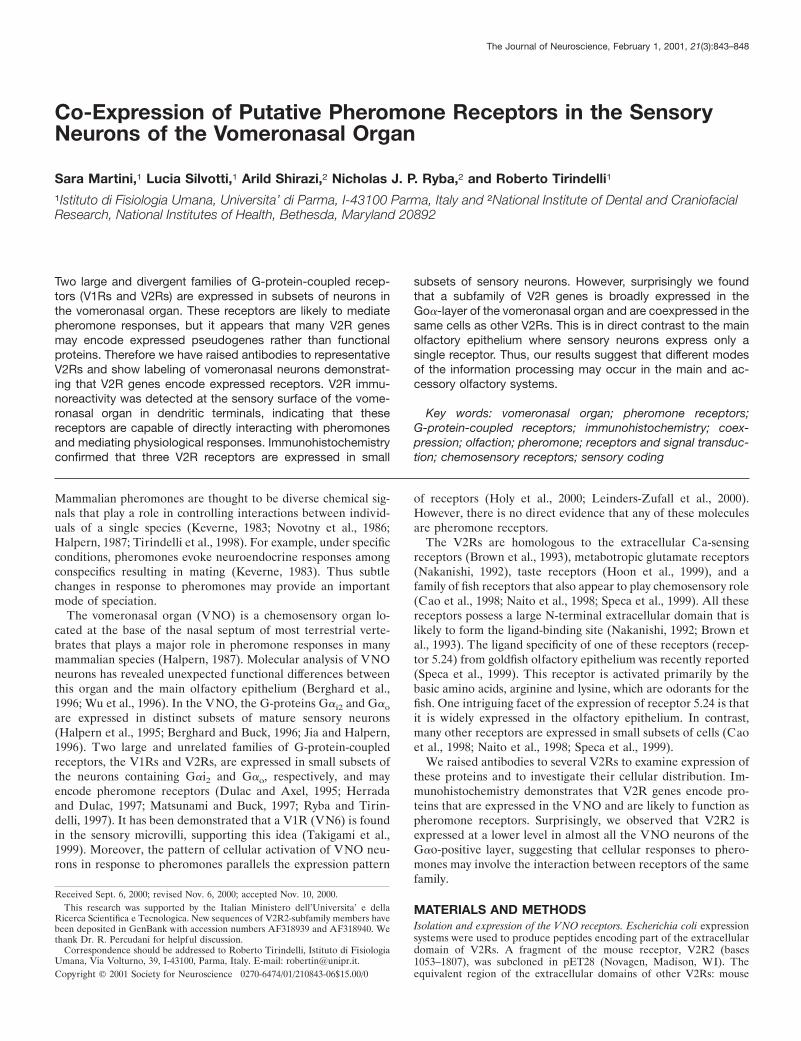
RESULTSWe grouped V2Rs according to their sequence conservation andimmunized rabbits with expressed extracellular domains of rep-resentative V2R genes that encode members of distinct subfam-ilies. Antibodies against the N-terminal extracellular domain ofthree V2R-family receptors: Go-VN2, Go-VN3, and Go-VN4labeled small subsets of VNO neurons (Fig. 1). For all the V2Rantibodies, strongest immunoreactivity was in the cell body ofVNO neurons, and as indicated in Figure 1d, expression ofreceptors was detectable in the sensory dendrites extending to thesurface of the epithelium. In contrast, no specific immunostainingwas observed in the axon bundles and accessory olfactory bulb ofeither adult or neonatal animals (data not shown).
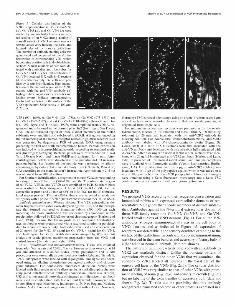
The pattern of immunoreactivity observed with an antibody toV2R2 was markedly distinct. Unlike the punctate pattern ofexpression observed for the other V2Rs that we examined, theantibody to V2R2 labeled all neurons in the basal half of thesensory cell layer of the VNO (Fig. 2a,b). The cellular distribu-tion of V2R2 was very similar to that of other V2Rs with prom-inent labeling of soma (Fig. 2a,b) and sensory microvilli (Fig. 2c)but no labeling of the axon bundles or accessory bulb (data notshown; Fig. 2d). To rule out the possibility that this antibodyrecognized a truncated receptor or other proteins expressed in a
Figure 1. Cellular distribution of theV2Rs. Representative rat V2Rs: Go-VN2(a), Go-VN3 (b), and Go-VN4 (c) werestudied by immunohistochemistry in coro-nal sections of rat VNO; strong staining ofa small subset of VNO neurons was ob-served; dotted lines indicate the basal andluminal edge of the sensory epithelium.The number of antibody staining cells wasquantitated and compared with in situ hy-bridization of corresponding V2R probesby counting positive cells in double-labeledsections. Similar numbers of cells were de-tected with antibodies and riboprobes forGo-VN2 and Go-VN3, but antibodies toGo-VN4 detected 4172 cells in 40 sections(4 rats), whereas only 1560 cells were pos-itive by in situ hybridization. High magni-fication of the luminal region of the VNOstained with the anti-VN4 antibody (d)highlights labeling of sensory dendrites andknobs; arrows indicate immunopositiveknobs and dendrites on the surface of theVNO epithelium. Scale bars: a–c, 100 mm;d, 25 mm.
844 J. Neurosci., February 1, 2001, 21(3):843–848 Martini et al. • Coexpression of V2R Pheromone Receptors
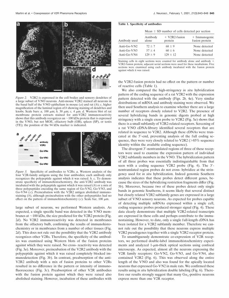
large subset of neurons, we performed Western analysis. Asexpected, a single specific band was detected in the VNO mem-branes at ;100 kDa, the size predicted for the V2R2 protein (Fig.2d). No V2R2 immunoreactivity was detected in membranesfrom the olfactory bulb, confirming the results of immunohisto-chemistry or in membranes from a number of other tissues (Fig.2d). This does not rule out the possibility that the V2R2 antibodyrecognizes other V2Rs. Therefore, the specificity of the antibod-ies was examined using Western blots of the fusion proteinsagainst which they were raised. No cross- reactivity was detected(Fig. 3a). Moreover, preincubation of the V2R2 antibody with thepolypeptide against which it was raised completely blocked im-munodetection (Fig. 3b). In contrast, preabsorption of the anti-V2R2 antibody with a mix of fusion proteins to other V2Rsresulted in no difference in the pattern or intensity of immuno-fluorescence (Fig. 3c). Preabsorption of other V2R antibodieswith the fusion protein against which they were raised alsoabolished staining. However, incubation of these antibodies with
the V2R2-fusion protein had no effect on the pattern or numberof reactive cells (Table 1).
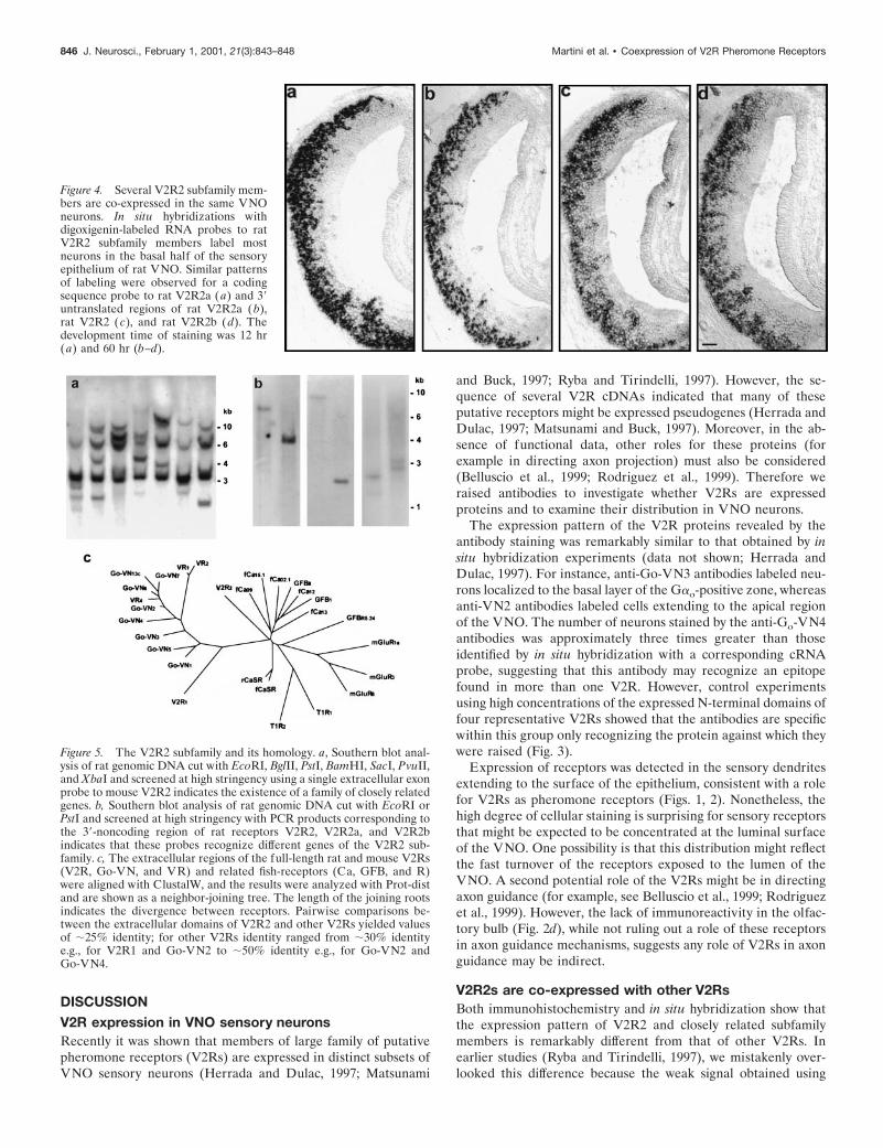
We also compared the high-stringency in situ hybridizationpattern of the coding sequence of a rat V2R2 with the expressionpattern detected with the antibody (Figs. 2b, 4a). Very similardistributions of mRNA and antibody staining were observed. Wethen used Southern analysis to examine whether there are a largenumber of receptors closely related to V2R2. The presence ofseveral hybridizing bands in genomic digests probed at highstringency with a single exon probe to V2R2 (Fig. 5a) shows thatthere is a small subfamily of V2R2-related receptors. Screening ofa rat VNO cDNA-library identified several receptors that arerelated in sequence to V2R2. Although these cDNAs were trun-cated at the 59-end, preventing analysis of the full coding se-quence, they were very closely related to V2R2 (.85% sequenceidentity within the available coding sequence).
The divergent 39-nontranslated regions of three of these recep-tors were used to examine the expression pattern of individualV2R2 subfamily members in the VNO. The hybridization patternof all three probes was essentially indistinguishable from thatseen with a coding sequence V2R2 probe (Fig. 4). The 39-nontranslated region probes do not cross- hybridize at the strin-gency used for in situ hybridization. Indeed genomic Southernanalysis indicates that these probes detect different genes, be-cause the sizes of the hybridizing restriction fragments differ (Fig.5b). Moreover, because two of these probes detect only singlebands in genomic Southerns, it seems likely that several distinctbut closely related V2R2 subfamily genes are expressed in a largesubset of VNO sensory neurons. As expected for probes capableof detecting multiple mRNAs expressed within a single cell,coding sequence probes produced stronger signal (Fig. 4). Thesedata clearly demonstrate that multiple V2R2-related transcriptsare expressed in these cells and perhaps contribute to the immu-nostaining. However, to date, only a single full-length cDNA hasbeen isolated for a V2R2 subfamily member. Therefore we can-not rule out the possibility that these neurons express multipleV2R2 pseudogenes together with a single V2R2 receptor protein.
To unambiguously demonstrate co-expression of V2R recep-tors, we performed double-label immunohistochemistry experi-ments and analyzed 1-mm-thick optical sections using confocalmicroscopy. As expected, almost all the neurons expressing theV2R family receptors: Go-VN2, Go-VN3, and Go-VN4, alsocontained V2R2 (Fig. 6). This was observed along the entirelength of the VNO and also was found for the apically locatedneurons that expressed Go-VN2 in male rats. We confirmed theseresults using in situ hybridization double labeling (Fig. 6). There-fore our results strongly suggest that many Gao-positive neuronsexpress more than one V2R receptor.
Figure 2. V2R2 is expressed in the cell bodies and sensory dendrites ofa large subset of VNO neurons. Anti-mouse V2R2 stained all neurons inthe basal half of the VNO epithelium in mouse (a) and rat (b); c, highermagnification of the luminal region of b showing staining of dendrites andknobs. Scale bars: a, 100 mm; b, 30 mm; c, 4 mm. d, Western blot of ratmembrane protein extracts stained for anti-V2R2 immunoreactivityshows that this antibody recognizes an ;100 kDa protein that is expressedin the VNO, but not MOE, olfactory bulb (OB), spleen (SP), or testis(TE); the position of the 94 kDa marker is indicated.
Figure 3. Specificity of antibodies to V2Rs. a, Western analysis of thefour V2R-family antigens using the four antibodies; each antibody onlyrecognizes the polypeptide against which it was raised. b, c, To demon-strate specificity of immunohistochemistry, the anti-V2R2 antibody wasincubated with the polypeptide against which it was raised (b) or a mix ofthree polypeptides encoding the same region of Go-VN2, Go-VN3, andGo-VN4 (c). Preincubation with the V2R2 antigen abolished immuno-staining (b), although preincubation with the other polypeptides had noeffect on the pattern of immunohistochemistry ( c). Scale bar, 100 mm.
Table 1. Specificity of antibodies
Antibody used
Mean 6 SD number of cells detected per section
Antibodyalone
1 V2R2-fusionprotein
1 Immunogenicfusion protein
Anti-Go-VN2 72 6 7 68 6 9 None detectedAnti-Go-VN3 57 6 4 60 6 6 None detectedAnti-Go-VN4 129 6 9 129 6 12 None detected
Staining cells in eight sections were counted for antibody alone and antibody 1V2R2-fusion protein; adjacent serial sections were used for these incubations. Fivesections were examined using each antibody incubated with the fusion proteinagainst which it was raised.
Martini et al. • Coexpression of V2R Pheromone Receptors J. Neurosci., February 1, 2001, 21(3):843–848 845
DISCUSSIONV2R expression in VNO sensory neuronsRecently it was shown that members of large family of putativepheromone receptors (V2Rs) are expressed in distinct subsets ofVNO sensory neurons (Herrada and Dulac, 1997; Matsunami
and Buck, 1997; Ryba and Tirindelli, 1997). However, the se-quence of several V2R cDNAs indicated that many of theseputative receptors might be expressed pseudogenes (Herrada andDulac, 1997; Matsunami and Buck, 1997). Moreover, in the ab-sence of functional data, other roles for these proteins (forexample in directing axon projection) must also be considered(Belluscio et al., 1999; Rodriguez et al., 1999). Therefore weraised antibodies to investigate whether V2Rs are expressedproteins and to examine their distribution in VNO neurons.
The expression pattern of the V2R proteins revealed by theantibody staining was remarkably similar to that obtained by insitu hybridization experiments (data not shown; Herrada andDulac, 1997). For instance, anti-Go-VN3 antibodies labeled neu-rons localized to the basal layer of the Gao-positive zone, whereasanti-VN2 antibodies labeled cells extending to the apical regionof the VNO. The number of neurons stained by the anti-Go-VN4antibodies was approximately three times greater than thoseidentified by in situ hybridization with a corresponding cRNAprobe, suggesting that this antibody may recognize an epitopefound in more than one V2R. However, control experimentsusing high concentrations of the expressed N-terminal domains offour representative V2Rs showed that the antibodies are specificwithin this group only recognizing the protein against which theywere raised (Fig. 3).
Expression of receptors was detected in the sensory dendritesextending to the surface of the epithelium, consistent with a rolefor V2Rs as pheromone receptors (Figs. 1, 2). Nonetheless, thehigh degree of cellular staining is surprising for sensory receptorsthat might be expected to be concentrated at the luminal surfaceof the VNO. One possibility is that this distribution might reflectthe fast turnover of the receptors exposed to the lumen of theVNO. A second potential role of the V2Rs might be in directingaxon guidance (for example, see Belluscio et al., 1999; Rodriguezet al., 1999). However, the lack of immunoreactivity in the olfac-tory bulb (Fig. 2d), while not ruling out a role of these receptorsin axon guidance mechanisms, suggests any role of V2Rs in axonguidance may be indirect.
V2R2s are co-expressed with other V2RsBoth immunohistochemistry and in situ hybridization show thatthe expression pattern of V2R2 and closely related subfamilymembers is remarkably different from that of other V2Rs. Inearlier studies (Ryba and Tirindelli, 1997), we mistakenly over-looked this difference because the weak signal obtained using
Figure 4. Several V2R2 subfamily mem-bers are co-expressed in the same VNOneurons. In situ hybridizations withdigoxigenin-labeled RNA probes to ratV2R2 subfamily members label mostneurons in the basal half of the sensoryepithelium of rat VNO. Similar patternsof labeling were observed for a codingsequence probe to rat V2R2a (a) and 39untranslated regions of rat V2R2a (b),rat V2R2 (c), and rat V2R2b (d). Thedevelopment time of staining was 12 hr(a) and 60 hr (b–d).
Figure 5. The V2R2 subfamily and its homology. a, Southern blot anal-ysis of rat genomic DNA cut with EcoRI, BglII, PstI, BamHI, SacI, PvuII,and XbaI and screened at high stringency using a single extracellular exonprobe to mouse V2R2 indicates the existence of a family of closely relatedgenes. b, Southern blot analysis of rat genomic DNA cut with EcoRI orPstI and screened at high stringency with PCR products corresponding tothe 39-noncoding region of rat receptors V2R2, V2R2a, and V2R2bindicates that these probes recognize different genes of the V2R2 sub-family. c, The extracellular regions of the full-length rat and mouse V2Rs(V2R, Go-VN, and VR) and related fish-receptors (Ca, GFB, and R)were aligned with ClustalW, and the results were analyzed with Prot-distand are shown as a neighbor-joining tree. The length of the joining rootsindicates the divergence between receptors. Pairwise comparisons be-tween the extracellular domains of V2R2 and other V2Rs yielded valuesof ;25% identity; for other V2Rs identity ranged from ;30% identitye.g., for V2R1 and Go-VN2 to ;50% identity e.g., for Go-VN2 andGo-VN4.
846 J. Neurosci., February 1, 2001, 21(3):843–848 Martini et al. • Coexpression of V2R Pheromone Receptors
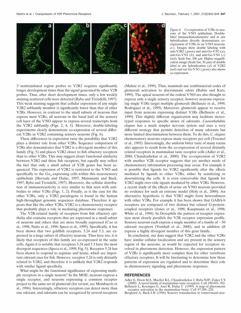
39-nontranslated region probes to V2R2 requires significantlylonger development times than the signal generated by other V2Rprobes. Thus, after short development times, only a few weaklystaining scattered cells were detected (Ryba and Tirindelli, 1997).This weak staining suggests that cellular expression of any singleV2R2 subfamily member is significantly lower than that of otherV2Rs. However, in contrast to the small subsets of neurons thatexpress most V2Rs, all neurons in the basal half of the sensorycell layer of the VNO appear to express several transcripts fromthe V2R2 subfamily (Figs. 2, 4, 5). Moreover, double-labelingexperiments clearly demonstrate co-expression of several differ-ent V2Rs in V2R2 containing sensory neurons (Fig. 6).
These differences in expression raise the possibility that V2R2plays a distinct role from other V2Rs. Sequence comparison ofV2Rs also demonstrates that V2R2 is a divergent member of thisfamily (Fig. 5) and places V2R2 closer to fish olfactory receptorsthan to other V2Rs. This may suggest closer functional similaritybetween V2R2 and these fish receptors, but equally may reflectthe fact that only a small subset of V2R genes has been se-quenced. The expression of V2R2 is restricted to the VNO andspecifically to the Gao-expressing cells within this neurosensoryepithelium (Herrada and Dulac, 1997; Matsunami and Buck,1997; Ryba and Tirindelli, 1997). Moreover, the cellular distribu-tion of immunoreactivity is very similar to that seen with anti-bodies to other V2Rs (Figs. 1, 2). Finally, as is the case for theother V2Rs, only a V2R2 pseudogene is found in the humanhigh-throughput genomic sequences database. Therefore it ap-pears that like the other V2Rs, V2R2 is a chemosensory receptorthat probably plays a role in mediating pheromone responses.
The V2R-related family of receptors from fish olfactory epi-thelia also contains receptors that are expressed in a small subsetof neurons and others that are more broadly expressed (Cao etal., 1998; Naito et al., 1998; Speca et al., 1999). Specifically, it hasbeen shown that two goldfish receptors, 5.24 and 5.3, are ex-pressed in a large subset of olfactory neurons. Thus here too, it islikely that receptors of this family are co-expressed in the samecells. Again it is notable that receptors 5.24 and 5.3 have the mostdivergent sequences (Speca et al., 1999; Fig. 5). Receptor 5.24 hasbeen shown to respond to arginine and lysine, which are impor-tant odorant cues for fish. However, receptor 5.24 is only distantlyrelated to V2R2, and therefore it is unlikely that V2R2 respondswith similar ligand specificity.
What might be the functional significance of expressing multi-ple receptors in a single neuron? In the MOE, neurons express asingle receptor, and neurons expressing a common receptorproject to the same set of glomeruli (for review, see Mombaerts etal., 1996). Interestingly, olfactory receptors can detect more thanone odorant, and odorants can be recognized by several receptors
(Malnic et al., 1999). Thus, mammals use combinatorial codes ofglomeruli activation to discriminate odors (Rubin and Katz,1999). The apical neurons of the rodent VNO are also thought toexpress only a single sensory receptor, however neurons express-ing single V1Rs target multiple glomeruli (Belluscio et al., 1999;Rodriguez et al., 1999). Moreover, glomeruli appear to receiveinput from neurons expressing distinct V1Rs (Belluscio et al.,1999). This slightly different organization may facilitate stereo-typed responses to specific mixes of odorants. Caenorhabditiselegans has a much simpler nervous system and uses a verydifferent strategy that permits detection of many odorants butmore limited discrimination between them. To do this, C. eleganschemosensory neurons express many receptors per cell (Troemelet al., 1995). Interestingly, the uniform bitter taste of many toxinsalso appears to result from the co-expression of several distantlyrelated receptors in mammalian taste receptor cells (Adler et al.,2000; Chandrashekar et al., 2000). The co-expression of V2R2with another V2R receptor suggests that yet another mode ofchemosensory information processing may occur. For example itis possible that ligands for V2R2 significantly alter the effectsmediated by ligands to other V2Rs, either by sensitizing ordesensitizing the cells. It is even conceivable that ligands forV2R2 might over-ride signals mediated by other V2Rs. However,a recent study of the effects of urine on VNO neurons providedno evidence for such an extreme model (Holy et al., 2000). Analternative hypothesis is that V2R2 might form heterodimerswith other V2Rs. For example it has been shown that GABA-breceptors are composed of two distinct but related G-protein-coupled receptors (Jones et al., 1998; Kaupmann et al., 1998;White et al., 1998). In Drosophila the pattern of receptor expres-sion most closely parallels the V2R receptor expression profile.Sensory neurons each express a single member of a family of ;50odorant receptors (Vosshall et al., 2000), and in addition allexpress a highly divergent member of this gene family.
In conclusion, our data suggest that V2R2 and the other V2Rshave similar cellular localization and are present in the sensoryregion of the neurons, as would be expected for receptors in-volved in pheromone detection. However, the expression patternof V2Rs is significantly more complex than for other vertebrateolfactory receptors. It will be fascinating to determine how thesepatterns of expression are regulated and to determine their rolein chemosensory signaling and pheromone responses.
REFERENCESAdler E, Hoon MA, Mueller KL, Chandrashekar J, Ryba NJP, Zuker CS
(2000) A novel family of mammalian taste receptors. Cell 100:693–702.Belluscio L, Koentges G, Axel R, Dulac C (1999) A map of pheromone
receptor activation in the mammalian brain. Cell 97:209–220.Berghard A, Buck LB (1996) Sensory transduction in vomeronasal neu-
Figure 6. Co-expression of V2Rs in neu-rons of the VNO epithelium. Double-label immunohistochemistry and in situhybridization directly demonstrates co-expression of V2Rs in rat VNO neurons.a–c, Images show double labeling withanti-V2R2 ( green) and anti-Go-VN2 (a),anti-Go-VN3 (b), and anti-Go-VN4 (c)(red). Scale bar, 100 mm. Higher magnifi-cation image (Scale bar, 50 mm) of doublelabel in situ hybridization (d) of V2R2(red) and rat Go-VN3 ( green) also showsco-expression.
Martini et al. • Coexpression of V2R Pheromone Receptors J. Neurosci., February 1, 2001, 21(3):843–848 847
rons: evidence for G alpha o, G alpha i2, and adenylyl cyclase II asmajor components of a pheromone signaling cascade. J Neurosci16:909–918.
Berghard A, Buck LB, Liman ER (1996) Evidence for distinct signalingmechanisms in two mammalian olfactory sense organs. Proc Natl AcadSci USA 93:2365–2369.
Brown EM, Gamba G, Riccardi D, Lombardi M, Butters R, Kifor O, SunA, Hediger MA, Lytton J, Hebert SC (1993) Cloning and character-ization of an extracellular Ca( 21)-sensing receptor from bovine para-thyroid. Nature 366:575–580.
Cao Y, Oh BC, Stryer L (1998) Cloning and localization of two multi-gene receptor families in goldfish olfactory epithelium. Proc Natl AcadSci USA 95:11987–11992.
Chandrashekar J, Mueller KL, Hoon MA, Adler E, Feng L, Guo W,Zuker CS, Ryba NJP (2000) T2Rs function as bitter taste receptors.Cell 100:703–711.
Dulac C, Axel R (1995) A novel family of genes encoding putativepheromone receptors in mammals. Cell 83:195–206.
Halpern M (1987) The organization and function of the vomeronasalsystem. Annu Rev Neurosci 10:325–362.
Halpern M, Shapiro LS, Jia C (1995) Differential localization of Gproteins in the opossum vomeronasal system. Brain Res 677:157–161.
Harlow E, Lane D (1988) Antibodies: a laboratory manual. Cold SpringHarbor, New York: Cold Spring Harbor.
Herrada G, Dulac C (1997) A novel family of putative pheromone re-ceptors in mammals with a topographically organized and sexuallydimorphic distribution. Cell 90:763–773.
Holy TE, Dulac C, Meister M (2000) Responses of vomeronasal neuronsto natural stimuli. Science 289:1569–1572.
Hoon MA, Adler E, Lindemeier J, Battey JF, Ryba NJP, Zuker CS(1999) Putative mammalian taste receptors: a class of taste-specificGPCRs with distinct topographic selectivity. Cell 96:541–551.
Jia C, Halpern M (1996) Subclasses of vomeronasal receptor neurons:differential expression of G proteins (Gi alpha 2 and G(o alpha)) andsegregated projections to the accessory olfactory bulb. Brain Res719:117–128.
Jones KA, Borowsky B, Tamm JA, Craig DA, Durkin MM, Dai M, YaoWJ, Johnson M, Gunwaldsen C, Huang LY, Tang C, Shen Q, Salon JA,Morse K, Laz T, Smith KE, Nagarathnam D, Noble SA, Branchek TA,Gerald C (1998) GABA(B) receptors function as a heteromeric as-sembly of the subunits GABA(B)R1 and GABA(B)R2. Nature396:674–679.
Kaupmann K, Malitschek B, Schuler V, Heid J, Froestl W, Beck P,Mosbacher J, Bischoff S, Kulik A, Shigemoto R, Karschin A, Bettler B(1998) GABA(B)-receptor subtypes assemble into functional hetero-meric complexes. Nature 396:683–687.
Keverne EB (1983) Pheromonal influences on the endocrine regulationof reproduction. Trends Neurosci 6:381–384.
Leinders-Zufall T, Lane AP, Puche AC, Ma W, Novotny MV, Shipley
MT, Zufall F (2000) Ultrasensitive pheromone detection by mamma-lian vomeronasal neurons. Nature 405:792–796.
Malnic B, Hirono J, Sato T, Buck LB (1999) Combinatorial receptorcodes for odors. Cell 96:713–723.
Matsunami H, Buck LB (1997) A multigene family encoding a diversearray of putative pheromone receptors in mammals. Cell 90:775–784.
Mombaerts P, Wang F, Dulac C, Vassar R, Chao SK, Nemes A, Men-delsohn M, Edmondson J, Axel R (1996) The molecular biology ofolfactory perception. Cold Spring Harb Symp Quant Biol 61:135–145.
Naito T, Saito Y, Yamamoto J, Nozaki Y, Tomura K, Hazama M,Nakanishi S, Brenner S (1998) Putative pheromone receptors relatedto the Ca 21-sensing receptor in Fugu. Proc Natl Acad Sci USA95:5178–5181.
Nakanishi S (1992) Molecular diversity of glutamate receptors and im-plications for brain function. Science 258:597–603.
Novotny M, Jemiolo B, Harvey S, Wiesler D, Marchlewska-Koj A (1986)Adrenal-mediated endogenous metabolites inhibit puberty in femalemice. Science 231:722–725.
Rodriguez I, Feinstein P, Mombaerts P (1999) Variable patterns of ax-onal projections of sensory neurons in the mouse vomeronasal system.Cell 97:199–208.
Rubin BD, Katz LC (1999) Optical imaging of odorant representationsin the mammalian olfactory bulb. Neuron 23:499–511.
Ryba NJP, Tirindelli R (1997) A new multigene family of putative pher-omone receptors. Neuron 19:371–379.
Speca DJ, Lin DM, Sorensen PW, Isacoff EY, Ngai J, Dittman AH (1999)Functional identification of a goldfish odorant receptor. Neuron23:487–498.
Takigami S, Osada T, Yoshida-Matsuoka J, Matsuoka M, Mori Y,Ichikawa M (1999) The expressed localization of rat putative phero-mone receptors. Neurosci Lett 272:115–118.
Tirindelli R, Ryba NJP (1996) The G-protein gamma-subunit G gamma8 is expressed in the developing axons of olfactory and vomeronasalneurons. Eur J Neurosci 8:2388–2398.
Tirindelli R, Mucignat-Caretta C, Ryba NJP (1998) Molecular aspectsof pheromonal communication via the vomeronasal organ of mammals.Trends Neurosci 21:482–486.
Troemel ER, Chou JH, Dwyer ND, Colbert HA, Bargmann CI (1995)Divergent seven transmembrane receptors are candidate chemosensoryreceptors in C. elegans. Cell 83:207–218.
Vosshall LB, Wong AM, Axel R (2000) An olfactory sensory map in thefly brain. Cell 102:147–159.
White JH, Wise A, Main MJ, Green A, Fraser NJ, Disney GH, BarnesAA, Emson P, Foord SM, Marshall FH (1998) Heterodimerization isrequired for the formation of a functional GABA(B) receptor. Nature396:679–682.
Wu Y, Tirindelli R, Ryba NJP (1996) Evidence for different chemosen-sory signal transduction pathways in olfactory and vomeronasal neu-rons. Biochem Biophys Res Commun 220:900–904.
848 J. Neurosci., February 1, 2001, 21(3):843–848 Martini et al. • Coexpression of V2R Pheromone Receptors