cns magnetic resonance spectroscopy (mrs) in … · peter b. barker, d.phil. ... cns magnetic...
TRANSCRIPT

Peter B. Barker, D.Phil.Russell H. Morgan Department of Radiology and Radiological Science, Johns Hopkins University School of Medicine, and the Kennedy Krieger Institute Baltimore, MD, USA
CNS magnetic resonance spectroscopy (MRS) in inborn errors of metabolism
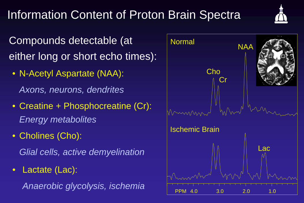
Information Content of Proton Brain Spectra
Compounds detectable (at either long or short echo times):• N-Acetyl Aspartate (NAA):
Axons, neurons, dendrites
• Creatine + Phosphocreatine (Cr): Energy metabolites
• Cholines (Cho):
Glial cells, active demyelination
PPM 4.0 3.0 2.0 1.0
Normal
ChoCr
NAA
Lac
Ischemic Brain
• Lactate (Lac):
Anaerobic glycolysis, ischemia

Compounds detectable at short echo times (< ~35 ms)• Myo-inositol (mI) - glial cells, demyelination, osmolyte (?)• Glutamate + Glutamine (Glx)• Lipids
PPM 4.0 3.0 2.0 1.0
mI“Glx”
Lipids
TE = 35 msecCho
NAA
Cr

• Other Compounds normally present (if you look closely!)• NAAG, Aspartate• Taurine, Scyllo-Inositol• Betaine, Ethanolamine• Purine Nucleotides• Histidine • Glucose (Glycogen?)
• Compounds observed using “Spectral Editing”• GABA• Ascorbic acid• Glutathione• ‘Macromolecules’
• Compounds which may be detectable under abnormal/pathological conditions• -Hydroxy-butyrate, acetone• Phenylalanine (PKU)• Galactitol, Ribitol, Arabitol• Succinate, pyruvate• Alanine• Glycine• Valine, leucine, isoleucine• Threonine?
• Exogenous Compounds• Propan-1,2-diol• Mannitol• Ethanol• MSM - methylsulfonylmethane

Human Brain MRS at 7 Tesla
STEAM, TE = 6 ms, TM = 32 ms, TR = 5 s, VOI = 8 ml, NT = 160, Tkác et al. MRM 46:451-6 (2001)

The Developing Brain: Kreis et al. MRM 30: 424-437 (1993)
Parietal White Matter Occipital Gray Matter

MR Spectroscopic Imaging (MRSI)
PPM 4.0 3.0 2.0 1.0
ChoCr
NAA
Parietal White Matter
Frontal White Matter
Centrum Semiovale
Cho Cr
NAAFrontal Gray Matter- Lateral
Frontal Gray Matter- Mesial
Posterior Gray Matter- Mesial
Posterior Gray Matter- Lateral
T1 Cho
Cr NAA
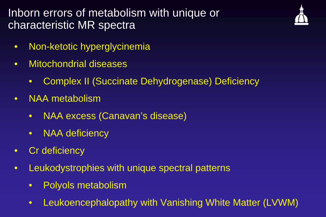
Inborn errors of metabolism with unique or characteristic MR spectra
• Non-ketotic hyperglycinemia
• Mitochondrial diseases
• Complex II (Succinate Dehydrogenase) Deficiency
• NAA metabolism
• NAA excess (Canavan’s disease)
• NAA deficiency
• Cr deficiency
• Leukodystrophies with unique spectral patterns
• Polyols metabolism
• Leukoencephalopathy with Vanishing White Matter (LVWM)

‘Non disease-specific’ metabolic patterns
• Reduced NAA – neuroaxonal loss or dysfunction
• Increased Cho – demyelination
• Increased mI – gliosis
• Lactate – non-oxidative glycolysis

Non-ketotic hyperglycinemia (NKH)
Choi et al., Korean J Radiol. 2001 Oct-Dec;2(4):239-242
12 day old male infantNormal brain MRISTEAM TR/TE 3000/166 msec

“Mitochondrial Diseases” - disorder of TCA cycle metabolism

Mitochondrial Encephalopathy with Lactic Acidosis and Stroke like Episodes (MELAS)
Lin, Crawford and Barker, AJNR 24; 33-41 (2003)

PPM 3.0 2.0 1.0
PPM 3.0 2.0 1.0
Complex II (Succinate Dehydrogenase) Deficiency
ChoCr
NAA
Lac
Succ
White Matter
Gray Matter
Succ
T2
Provided by Dr Alberto Bizzi, Istituto Nazionale Neurologico “Carlo Besta”, Milan, Italy
PPM 3.0 2.0 1.0
PPM 3.0 2.0 1.0

Disorders of N-Acetyl Aspartate Metabolism
NAA
Glucose, Acetate,Pyruvate, ...
Glycolysis, TCA cycle
Aspartate + Acetyl CoA
L-Aspartate N-acetyl transferase “ANAT”, EC 2.3.1.17
Acetate + Aspartate
“Aspartoacylase”EC 3.5.1.15
“NAALADase”EC 3.4.17.21
NAAG
Canavan Disease
PPM 4.0 3.0 2.0 1.0
Frontal W.M.TE=270
NAA

Disorders of N-Acetyl Aspartate Metabolism
NAA
Glucose, Acetate,Pyruvate, ...
Glycolysis, TCA cycle
Aspartate + Acetyl CoA
L-Aspartate N-acetyl transferase “ANAT”, EC 2.3.1.17
Acetate + Aspartate
“Aspartoacylase”EC 3.5.1.15
“NAALADase”EC 3.4.17.21
NAAG
?
Martin et al., Ann Neurol 2001;49:518-21
Control
Patient

Guanidinoacetate Methyltransferase (GAMT) Deficiency
Provided by Dr Alberto Bizzi, Istituto Nazionale Neurologico “Carlo Besta”, Milan, Italy
PPM 4.0 3.0 2.0 1.0
Cho
NAACr
NAA T2 MRI
Glycine
Guanidinoacetete
Creatine
Brain
Liver
GAMT
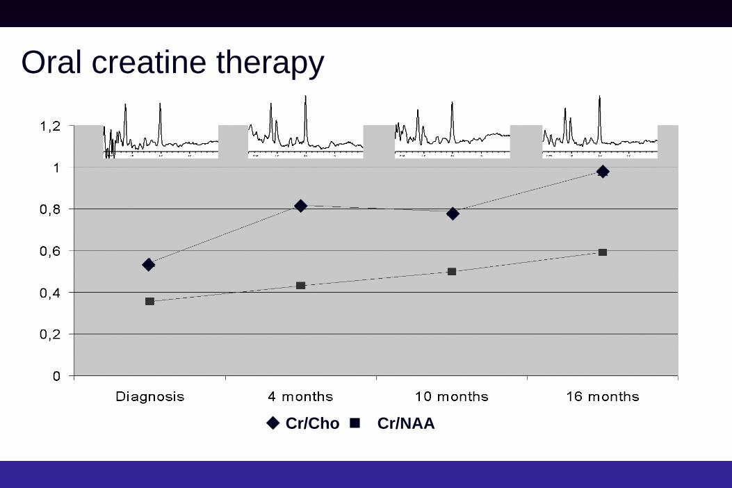
Oral creatine therapy
Cr/Cho
Cr/NAA

Leukoencephalopathy associated with a disturbance in the metabolism of polyols
van der Knaap et al.Ann Neurol 1999;46:925-928
Patient Control
WM
GM
WM
GM
Arabitol/Ribitol
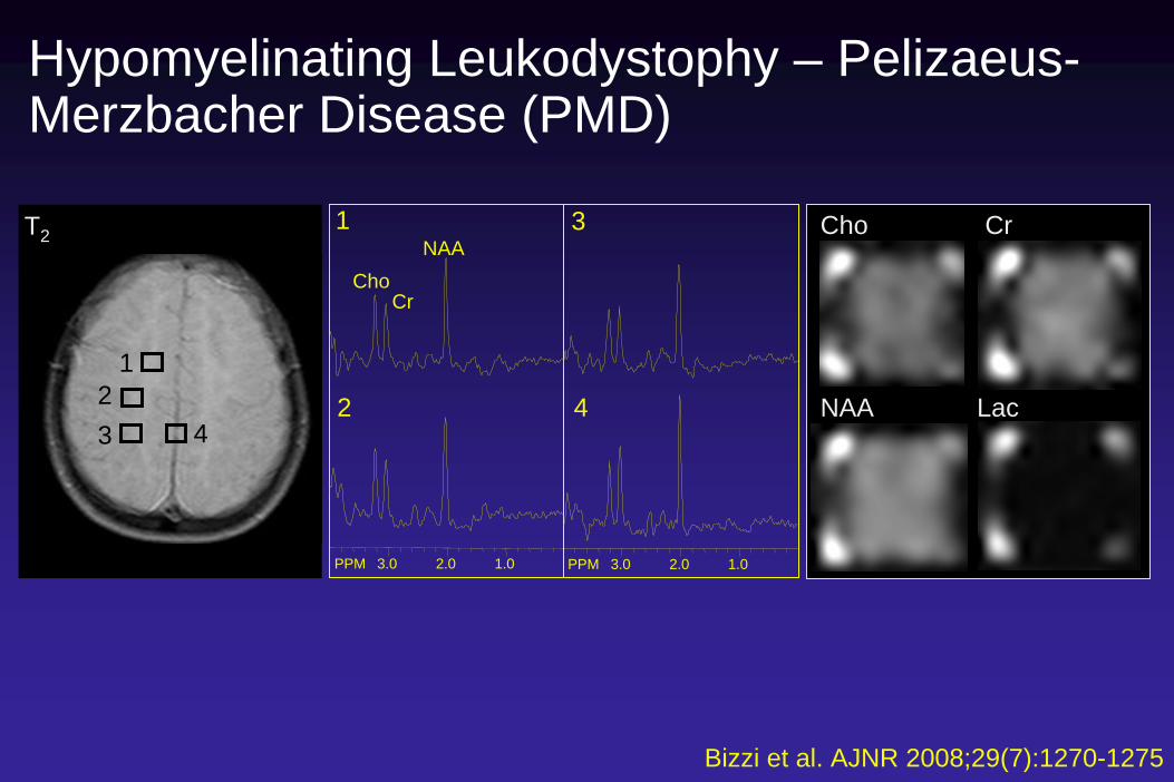
Hypomyelinating Leukodystophy – Pelizaeus- Merzbacher Disease (PMD)
Cho
NAA
Cr
Lac
123 4
PPM 3.0 2.0 1.0 PPM 3.0 2.0 1.0
1
2
3
4
ChoNAA
Cr
T2
Bizzi et al. AJNR 2008;29(7):1270-1275

White Matter Rarefaction: Leukodystrophy with Vanishing White Matter (LVWM)
Cho
NAA
Cr
Lac
12
3 4
PPM 3.0 2.0 1.0 PPM 3.0 2.0 1.0
1
2
3
4
ChoNAA
Cr
T2
Bizzi et al. AJNR 2008;29(7):1270-1275

Demyelinating Leukodystrophy: Metachromatic Leukodystrophy (MLD)
Cho
NAA
Cr
1
2
3 4
1
2
3
4
ChoNAA
Cr
T2
Lac
PPM 3.0 2.0 1.0 PPM 3.0 2.0 1.0
Bizzi et al. AJNR 2008;29(7):1270-1275

Demyelinating Leukodystrophy: Adrenoleukodystrophy (ALD)
T1 MRI Cho NAA Lac
PPM 4.0 3.0 2.0 1.0
13
2
13
2
PPM 4.0 3.0 2.0 1.0
Cho
CrNAA
Lac

Classification of Leukodystrophies
Bizzi et al. AJNR 2008;29(7):1270-1275
Intra-voxel metabolite ratio Metabolite values normalizedto gray matter
Group 1: Hypomyelination, Group 2: Rarefaction, Group 3: Demyelination

Classification of Leukodystrophies
33 of 44 cases correctlyClassified (75%)

Discussion - Misclassification
• Reasons for misclassification• Effects of age (e.g. 2 youngest hypomyelination
cases classified as rarefaction)• Regional variations in metabolite levels• Phenotypic and disease severity variability in
metabolite levels

Summary• Some rare diseases have specific, pathognomic
metabolic patterns• Canavan’s Disease• NKH• NAA, Cr deficiency• Complex II (succinate dehydrogenase) deficiency• PKU, Maple Syrup Urine Disease (not discussed)
• More commonly non-specific metabolic changes related to underlying tissue pathophysiology• NAA decreased – neuroaxonal loss/dysfunction• Cho elevation in active demyelination• Lactate increased in anaerobic glcolysis

Summary
• MRS may provide useful surrogate biomarkers in treatment trials, e.g. • Oral Cr in Cr deficiency: Biachi et al. Ann Neurol. 2000
Apr;47(4):511-3 (Creatine)• Dichloroacetate in mitochondrial disease: De Stefano
et al. NEUROLOGY 1995;45:1193-1198 (Lactate)• ‘Lorenzo’s oil’ in ALD: Moser et al. 2007; ASNR,
Chicago, IL. p 284. (NAA)

Acknowledgments
Johns Hopkins/Kennedy Krieger Inst. Tom CrawfordDoris LinHugo MoserMichael MoserSakkubai NaiduJerry Raymond
Istituto Carlo Besta, Milan, ItalyAlberto Bizzi


Sjögren-Larsson syndrome
Mano, Rizzo et al., AJNR Am J Neuroradiol 20:1671–1673, 1999
Lipids, fatty alcohol (hexadecanol, octadecanol) peaks at 0.9 and 1.3 ppm

Sjögren-Larsson syndrome
Domburg et al., Neurology, 52, 1345-52 (1999)

Salla Disease
Varho et al., Neurology, 52(8), 1668-92 (1999)

Leukodystrophy with vanishing white matter

Methylmalonic Acidemia (M 14 yrs, mut-)
Trinh, Melhem and Barker, AJNR Am J Neuroradiol 22:831–833, 2001
Isoleucine, valine, threonine
Proprionic acid
Methylmalonic acid
Succinate
Methylmalonyl CoA mutase,Adenosyl cobalamine

Methylmalonic Acidemia
Trinh, Melhem and Barker, AJNR Am J Neuroradiol 22:831–833, 2001
F 16 yrs, cobalamin synthesis defect

Leukoencephalopathy with Vanishing White Matter
Van der Knaap et al., Neurology, 1997 48, 845-55 (1997)
Proton Density T2 T1
Patient
Control