cme article clinics in diagnostic imaging (102) · pdf filepakistan m azeemuddin, mcps, fcps...
TRANSCRIPT

Singapore Med J 2005; 46(2) : 93M e d i c a l E d u c a t i o n
Department ofRadiology
Aga Khan UniversityHospital
PO Box 3500Karachi 74800Pakistan
M Azeemuddin,MCPS, FCPS
Assistant Professor
T UI-Haq, MCPS,FCPS, FRCR
Associate Professor
H Ahsan, MCPS, FCPSAssociate Professor
W A Memom, MCPS,FCPS
Senior Instructor
Correspondence to:Dr Muhammad
AzeemuddinTel: (92) 21 4859 2020Fax: (92) 21 493 4294Email: [email protected]
Clinics in diagnostic imaging (102)M Azeemuddin, T UI-Haq, H Ahsan, W A Memon
CME Article
CASE PRESENTATIONA 19-year-old man presented with cough andhaemoptysis of ten days duration. There was alsohistory of mild pain in right hypochondrium for lasttwo months. Patient was afebrile with no past historyof tuberculosis. He was a non-smoker. On clinicalexamination, decreased air entry was noted on theleft side of his chest. The liver was enlarged andpalpable below the costal margins. Chest radiographsand ultrasonography (US) of the abdomen wereperformed, followed by computed tomography(CT) of the chest and abdomen. What do the chestradiograph (Fig. 1) and CT (Fig. 2) show? What isyour diagnosis?Fig. 1 Chest radiograph (posteroanterior projection).
Fig. 2a Enhanced axial CT image of the thorax taken at the level oflower lung lobes.
Fig. 2b Enhanced axial CT images of the upper abdomen.

Singapore Med J 2005; 46(2) : 94
IMAGE INTERPRETATIONThe chest radiograph (Fig. 1) showed a rounded softtissue density opacity in the lower zone of the leftlung. A small air lucency was also seen in thesuperior portion of this opacity (air crescent sign).Axial CT of this lesion (Fig. 2a) showed this massto be of low density. A small amount of air was againnoted within it. There was also slight adjacentpleural reaction. CT images taken through liver(Fig. 2b) showed multiple cystic lesions in the liver.The largest of these was located in the right lobe andhad a daughter cyst in its lateral wall. No lymphnode enlargement was identified in the abdomenor mediastinum.
DIAGNOSISHydatid disease of the lung and liver.
CLINICAL COURSEThe patient was treated on Albendazole 400mg twicedaily, with resolution of his chest symptoms. Theliver lesions were followed-up using US. Because ofthe large size of the right lobe cyst and the associatedpain in right hypochondrium, a PAIR (percutaneousaspiration, instillation and reaspiration) procedure wasperformed. US done prior to the procedure showeddetachment of the cyst walls producing the “floatingmembrane sign”. The daughter cyst was intact(Fig. 3a). During the PAIR procedure, the daughtercyst was intentionally ruptured into the mother cyst,the contents of the cyst aspirated, and hypertonic salineinstilled. This was re-aspirated and finally, a smallquantity of absolute alcohol was injected (Fig. 3b).
Fig. 3a US image obtained prior to PAIR procedure showsdetachment of cyst membranes producing the “floating membranesign” in the larger hepatic hydatid cyst. The daughter cyst is intact.
Fig. 3b US image obtained during PAIR procedure. The daughtercyst is punctured by a needle, seen as parallel echogenic lines(arrow). The cyst is now partially collapsed.
Fig. 4 Complicated hydatid cyst. US images of the liver show a hydatid cyst with secondary infection. The cyst contents appear echogenicrather than hypoechoic.

Singapore Med J 2005; 46(2) : 95
DISCUSSIONHydatid is a parasitic disease cause by the larvae ofthe dog tapeworm, Echinococcus granulosis andE. alveolaris. This disease is endemic in many parts ofthe world but is most commonly found in the MiddleEast, Australia, Iceland and South America. Humansmay become intermediate hosts through contactwith a definitive host (usually a domesticated dog)or ingestion of contaminated water or vegetables(1,2).In man, the hydatid disease usually affects the liverand lungs, and typically demonstrates characteristicimaging findings.
The right lobe is the most frequently involvedportion of the liver. Imaging findings in hepatichydatid disease depend on the stage of cyst growthi.e. whether the cyst is unilocular, contains daughtervesicles, contains daughter cysts, is partially calcifiedor is completely calcified (dead)(3). Calcification isseen at radiography in 20%-30% of hydatid cysts,and usually manifests with a peripheral curvilinearor ring-like pattern. Complete calcification of thecysts is suggestive of death of the parasite(1,2).
The US appearances of hydatid cysts are typicalbut may vary according to the stage of evolution of
Fig. 5 Snowstorm sign. US images of the liver show hydatid sand producing the snowstorm sign. Mobile echogenic foci are demonstratedwhen the patient is rolled during the examination.
Fig. 6 Septations in hydatid cyst. US images of the liver show septations producing linear echogenic bands within the hydatid cyst.
Singapore Med J 2005; 46(2) : 96
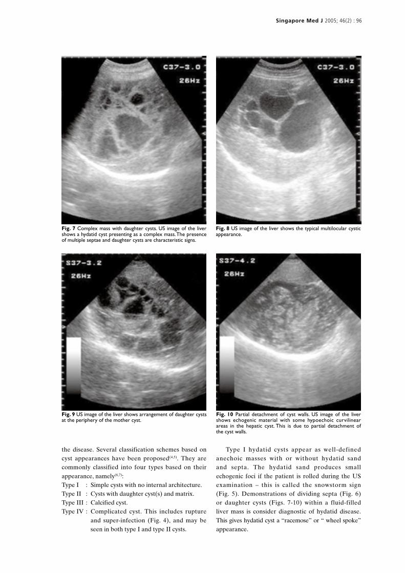
Fig. 7 Complex mass with daughter cysts. US image of the livershows a hydatid cyst presenting as a complex mass. The presenceof multiple septae and daughter cysts are characteristic signs.
Fig. 8 US image of the liver shows the typical multilocular cysticappearance.
the disease. Several classification schemes based oncyst appearances have been proposed(4,5). They arecommonly classified into four types based on theirappearance, namely(6,7):Type I : Simple cysts with no internal architecture.Type II : Cysts with daughter cyst(s) and matrix.Type III : Calcified cyst.Type IV : Complicated cyst. This includes rupture
and super-infection (Fig. 4), and may beseen in both type I and type II cysts.
Fig. 9 US image of the liver shows arrangement of daughter cystsat the periphery of the mother cyst.
Fig. 10 Partial detachment of cyst walls. US image of the livershows echogenic material with some hypoechoic curvilinearareas in the hepatic cyst. This is due to partial detachment ofthe cyst walls.
Type I hydatid cysts appear as well-definedanechoic masses with or without hydatid sandand septa. The hydatid sand produces smallechogenic foci if the patient is rolled during the USexamination – this is called the snowstorm sign(Fig. 5). Demonstrations of dividing septa (Fig. 6)or daughter cysts (Figs. 7-10) within a fluid-filledliver mass is consider diagnostic of hydatid disease.This gives hydatid cyst a “racemose” or “ wheel spoke”appearance.
Partial detachment of the capsule from thesurrounding liver parenchyma leads to a pericysticfluid collection. In complete detachment, the capsulefloats freely in the cyst giving the “floating membrane”sign(8) (Figs. 3a-b & 10). This is equivalent to theradiographical “water lily sign” of lung hydatiddisease. When a liver hydatid cyst does not containsepta or daughter cysts, demonstrating a capsulecan lead to a correct diagnosis. Showing the capsuleminimises the difficulty in differentiating an infectedhydatid cyst from tumour(9).
Besides the liver, hydatid disease can involvealmost every organ of the body. However, the basicappearances remain almost the same. In a series of275 patients(6), the sites of involvement (in decreasingorder of frequency) included the liver (74.8%),lungs (48.3%), peritoneum, kidney (Figs. 11-13),brain (Fig. 14), mediastinum, heart, bone, soft tissues,spinal cord, spleen, pleura, adrenal glands, bladder,
Singapore Med J 2005; 46(2) : 97
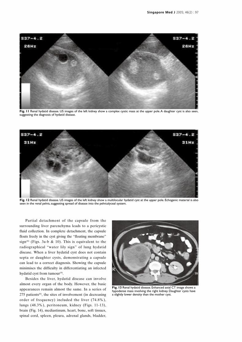
Fig. 11 Renal hydatid disease. US images of the left kidney show a complex cystic mass at the upper pole. A daughter cyst is also seen,suggesting the diagnosis of hydatid disease.
Fig. 12 Renal hydatid disease. US images of the left kidney show a multilocular hydatid cyst at the upper pole. Echogenic material is alsoseen in the renal pelvis, suggesting spread of disease into the pelvicalyceal system.
Fig. 13 Renal hydatid disease. Enhanced axial CT image shows ahypodense mass involving the right kidney. Daughter cysts havea slightly lower density than the mother cyst.
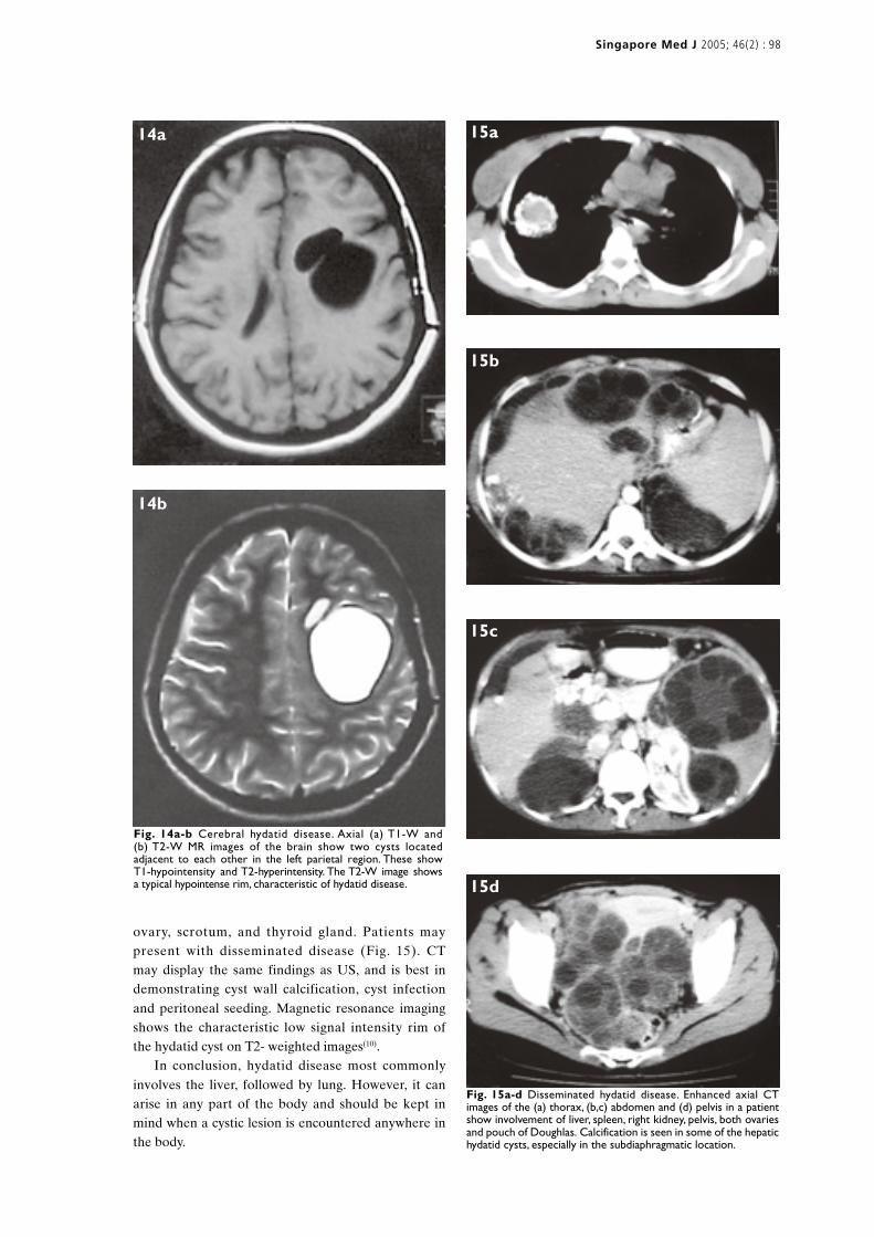
Fig. 14a-b Cerebral hydatid disease. Axial (a) T1-W and(b) T2-W MR images of the brain show two cysts locatedadjacent to each other in the left parietal region. These showT1-hypointensity and T2-hyperintensity. The T2-W image showsa typical hypointense rim, characteristic of hydatid disease.
Fig. 15a-d Disseminated hydatid disease. Enhanced axial CTimages of the (a) thorax, (b,c) abdomen and (d) pelvis in a patientshow involvement of liver, spleen, right kidney, pelvis, both ovariesand pouch of Doughlas. Calcification is seen in some of the hepatichydatid cysts, especially in the subdiaphragmatic location.
15a
15b
15c
15d
Singapore Med J 2005; 46(2) : 98
14a
14b
ovary, scrotum, and thyroid gland. Patients maypresent with disseminated disease (Fig. 15). CTmay display the same findings as US, and is best indemonstrating cyst wall calcification, cyst infectionand peritoneal seeding. Magnetic resonance imagingshows the characteristic low signal intensity rim ofthe hydatid cyst on T2- weighted images(10).
In conclusion, hydatid disease most commonlyinvolves the liver, followed by lung. However, it canarise in any part of the body and should be kept inmind when a cystic lesion is encountered anywhere inthe body.
ACKNOWLEDGEMENTWe thank Dr Rashid Ahmed, DABR, Head ofAdvanced Radiology Clinic, Karachi, Pakistan, forproviding some of the images.
ABSTRACT
A 19-year-old man presented with cough andhaemoptysis of ten days duration. He also had mildright hypochondrial pain. Chest radiograph andcomputed tomography (CT) showed a roundedsoft tissue density opacity with an air crescentsign. CT showed multiple cystic lesions in the liverwith a daughter cyst in its lateral wall. Diagnosisof hydatid disease of lung and liver was made.The contents of the liver cyst were aspirated,hypertonic saline instilled, re-aspirated, andabsolute alcohol injected. Hydatid disease isendemic in certain parts of the world. Althoughthe lungs and liver are most frequently affected,the disease can arise in any part of the bodyand should be kept in differential diagnosiswhenever a cystic lesion is encountered. Hydatidcysts typically demonstrate characteristic imagingfindings, however, the appearances may becomecomplicated due to cyst rupture or secondary
Singapore Med J 2005; 46(2) : 99
infection. Ultrasonography is the imaging modalityof choice particularly in hepatic disease. CTbest demonstrates cyst wall calcification andcyst infection.
Keywords: echinococcosis, hepatic hydatodosis,hydatid cyst, hydatid disease, liver disease
Singapore Med J 2005; 46(2):93-100
REFERENCES1. Beggs I. The radiology of hydatid disease. Am J Roentgenol 1985;
145:639-48.2. Lewall DB. Hydatid disease: biology, pathology, imaging and
classification. Clin Radiol 1998; 53:863-74.3. Pedrosa I, Saiz A, Arrazola J, Ferreiros J, Pedrosa CS. Hydatid disease:
radiologic and pathologic features and complications. Radiographics2000; 20:795-817.
4. Gharbi HA, Hassine W, Brauner MW, Dupuch K. Ultrasoundexamination of the hydatic liver. Radiology 1981; 139:459-63.
5. Lewall DB, MC Corkell SJ. Hepatic enchinococcal cyst: sonographicappearance and classification. Radiology 1985; 155:773-5.
6. Polat P, Kantarci M, Alper F, Suma S, Koruyucu M, Okur A. Hydatiddisease from head to toe. Radiographics 2003; 23:475-94.
7. Von Sinner W, Le Strake L, Clark D, Sharif H. MR Imaging in hydatiddisease. Am J Roentgenol 1991; 157:741-5.
8. Lewall DB, McCorkell SJ. Rupture of echinococcal cysts: diagnosis,classification, and clinical implications. Am J Roentgenol 1986;146:391-4.
9. Hussain S. Diagnostic criteria of hydatid disease of hepatic sonography.J Ultrasound Med 1985; 4:603-7.
10. Marani SA, Canossi GC, Nicoli FA, Alberti GP, Monni SG, Casolo PM.Hydatid disease: MR imaging study. Radiology 1990; 175:701-6.

SINGAPORE MEDICAL COUNCIL CATEGORY 3B CME PROGRAMMEMultiple Choice Questions (Code SMJ 200502B)
True False
Question 1. Regarding the “PAIR” procedure for the management of hydatid cysts:(a) It is the recommended technique for unilocular, non-calcified lung hydatid cysts. � �(b) It should not be performed if the cysts are super-infected. � �(c) It is contraindicated for the management of peritoneal hydatid disease. � �(d) Absolute alcohol is the only scolicidal agent recommended. � �
Question 2. Regarding hydatid infestation:(a) It is caused by the larvae of the dog tapeworm Echinococcus granulosis and E.alveolaris. � �(b) Humans may become definitive host through contact with a domesticated dog. � �(c) Humans can also get infected by ingestion of contaminated water or vegetables. � �(d) The left lobe of liver is most frequently involved. � �
Question 3. Regarding the presence of calcification within hydatid cysts:(a) Calcification is seen in > 30% cases of hydatid cysts. � �(b) Lung cysts show a similar incidence of calcification as hepatic hydatid cysts. � �(c) Demonstration of peripheral ring calcification implies inactive disease. � �(d) Completely calcified hydatid cysts in liver are easily differentiated from calcified,
healed amoebic liver abscess. � �
Question 4. Considering ultrasonography of hepatic hydatid cysts:(a) It is usually difficult to differentiate type 1 hydatid cysts from simple hepatic cysts. � �(b) The snowstorm sign is produced by detached membranes. � �(c) Hydatid cysts do not show a capsule unless calcified. � �(d) The floating membrane sign is produced when the cyst is completely ruptured. � �
Question 5. The following statements are correct regarding hydatid cysts:(a) CT is more sensitive than ultrasonography in showing membranes and septae within the cysts. � �(b) MR imaging shows a high signal intensity rim on T2-weighted images. � �(c) The peritoneum is the third most frequent organ involved. � �(d) Hydatid cysts have been reported in parathyroid glands. � �
Doctor’s particulars:
Name in full: _______________________________________________________________________________________
MCR number: ______________________________________ Specialty: ______________________________________
Email address: ______________________________________________________________________________________
Submission instructions:A. Using this answer form1. Photocopy this answer form.2. Indicate your responses by marking the “True” or “False” box �3. Fill in your professional particulars.4. Either post the answer form to the SMJ at 2 College Road, Singapore 169850 OR fax to SMJ at (65) 6224 7827.
B. Electronic submission1. Log on at the SMJ website: URL http://www.sma.org.sg/cme/smj2. Either download the answer form and submit to [email protected] OR download and print out the answer form for this
article and follow steps A. 2-4 (above) OR complete and submit the answer form online.
Deadline for submission: (February 2005 SMJ 3B CME programme): 12 noon, 25 March 2005Results:1. Answers will be published in the SMJ April 2005 issue.2. The MCR numbers of successful candidates will be posted online at http://www.sma.org.sg/cme/smj by 20 April 2005.3. Passing mark is 60%. No mark will be deducted for incorrect answers.4. The SMJ editorial office will submit the list of successful candidates to the Singapore Medical Council.
✓
Singapore Med J 2005 Vol 46(2) : 100