clinicalstudy - hindawi publishing...
TRANSCRIPT

Clinical StudyThe Efficiency of Anterior Repositioning Splints in theManagement of Pain Related to Temporomandibular JointDisc Displacement with Reduction
Malgorzata Pihut ,1 Malgorzata Gorecka,2 Piotr Ceranowicz ,3
and Mieszko Wieckiewicz 4
1Department of Prosthodontics, Consulting Room of Temporomandibular Joint Dysfunction, Jagiellonian University MedicalCollege, 4 Montelupich St., 31-155 Krakow, Poland2University Dental Clinic, Jagiellonian University Medical College, 4 Montelupich St., 31-155 Krakow, Poland3Department of Physiology, Jagiellonian University Medical College, 16 Grzegorzecka St., 31-531 Krakow, Poland4Department of Experimental Dentistry, Faculty of Dentistry, Wroclaw Medical University, 26 Krakowska St., 50-425Wroclaw, Poland
Correspondence should be addressed to Mieszko Wieckiewicz; [email protected]
Received 4 September 2017; Accepted 29 January 2018; Published 21 February 2018
Academic Editor: Jacob Ablin
Copyright © 2018 Malgorzata Pihut et al. ,is is an open access article distributed under the Creative Commons AttributionLicense, which permits unrestricted use, distribution, and reproduction in any medium, provided the original work isproperly cited.
Background and Objective. Intra-articular temporomandibular disorders are often related to pain in the area of the temporo-mandibular joint, ear, and temple. ,e aim of the study was to investigate the efficiency of anterior repositioning splints indecreasing pain related to temporomandibular joint disc displacement with reduction. Methods. ,e research material consistedof 112 patients, aged 24 to 45 years, of both genders, who reported for treatment at the Consulting Room of TemporomandibularJoint Dysfunctions at the Jagiellonian University in Cracow between 2014 and 2016 due to pain in the area of the temporo-mandibular joint(s) and noise(s) of temporomandibular joint(s) present during jaw movements with comorbid contracture ofmasticatory muscles. Subjects were examined according to the Diagnostic Criteria for Temporomandibular Disorders (DC/TMD)protocol and, after diagnosis of painful disc displacement with reduction and masticatory muscle contracture, they were assignedrandomly to either the study or control groups (56 patients in each). In the study group, we used an anterior repositioning splinton the full lower arch for about 20 hours usage over a 4-month period. In the control group, a noninvasive therapy was appliedusing a biostimulation laser over 12 sessions performed every second day on the area of both temporomandibular joints withmouth open and while performing muscle self-exercises with a dominant protrusive position of the mandible. Pain intensity wasevaluated using the Verbal Numerical Rating Scale (VNRS) immediately before the treatment and then after 4 and 16 weeks. ,eobtained data were analyzed using the Mann–Whitney U test (p≤ 0.005). Results. ,e VNRS values reported during the finalexamination for the study group were significantly lower than for the control group (p � 0.0004). Conclusions. ,e anteriorrepositioning splint is an efficient tool in decreasing pain related to disc displacement with reduction. ,is trial is registered withClinicaltrials.gov NCT03057262.
1. Introduction
Intra-articular temporomandibular disorders present asvarious pathological conditions, often concerning deran-gements of the condyle-disc complex [1, 2]. ,ey occurbecause the physiological relationship between the articular
disc and the condyle head changes. According to the Di-agnostic Criteria for Temporomandibular Disorders(DC/TMD), we can distinguish four basic types of articulardisc displacements: disc displacement with reduction, discdisplacement with reduction with intermittent locking,disc displacement without reduction with limited opening,
HindawiPain Research and ManagementVolume 2018, Article ID 9089286, 6 pageshttps://doi.org/10.1155/2018/9089286
and disc displacement without reduction without limitedopening [3].
Many studies confirm that disc displacement with re-duction (DDwR) is the most prevalent derangement of thecondyle-disc complex [4–6].,eDC/TMDdefines DDwR asan intracapsular biomechanical disorder when, in closedmouth position, the disc is in an anterior position relative tothe condylar head, and the disc reduces upon opening of themouth (medial and lateral displacement of the disc may alsobe present; clicking, popping, or snapping noises may occurwith disc reduction) [3]. DDwR could be the result of mas-ticatory muscle contracture. Acoustic symptoms such asclicking, popping, and snapping are evidence of disc dis-placementwith reduction.Clicking occurswhen the condylarhead of the mandible skips the rear edge of the displacedarticular disc during mouth opening and/or closing. After-wards, the disc does not return to the correct position inrelation to the condyle when the mandible is again, uponclosure, in the central position. Clicking may occur in theinitial, middle, and final phase of the mandible openingmovement. Different studies have reported that pain is acomorbid symptom of temporomandibular joint (TMJ) discdisplacement [7, 8].When the disc is anteriorly displaced, theligaments of the rear disc are elongated, stretched, anddamaged, and the bilaminar zone is compressed; and thus,pain can be generated. Sometimes acoustic symptoms areaccompanied by pain located in the area of the temporo-mandibular joints and surrounding tissues, intensifyingwhen the patient opens theirmouth and chews food.Over theyears, many studies have confirmed the efficacy of anteriorrepositioning splints (ARS) for themanagement of TMJ discsdisplacements, but the number of studies simultaneouslyevaluating the level of pain is limited [9, 10].
,erefore, the aim of the study was to investigate theefficiency of anterior repositioning splints in the alleviationof pain related to temporomandibular joint disc displace-ment with reduction.
2. Materials and Methods
,is is a prospective outcome study which consisted of 112subjects of both genders with unilateral or bilateral discdisplacement(s) with reduction and pain in the area ofTMJ. Patients were recruited from the Consulting Room ofTemporomandibular Joint Dysfunction at the JagiellonianUniversity in Cracow during the years 2014–2016 and wereincluded in the study if they met the following criteria: (1)unilateral or bilateral presence of clicking, popping, and/orsnapping noise(s) detected with palpation during opening orclosing or lateral or protrusive movements in TMJ(s); (2) inthe previous 30 days, any TMJ noise(s) present with jawmovement or function; (3) unilateral or bilateral pain in thearea of TMJ(s); (4) presence of masticatory muscle con-tracture during palpation; (5) full dentition or single toothloss; (6) good general health; (7) positive mandible pro-trusion test; (8) no contraindications for laser therapy; and(9) patient consent to be involved in the study. Other pa-tients representing TMJ intra-articular disorders who hadundergone treatment during the years 2014–2016 were
excluded because of partial tooth loss or edentulism, con-traindications for laser therapy as well as absence of ap-propriate symptoms and/or consent to be involved in thestudy.
Clinical assessment of temporomandibular joints andmasticatorymuscles was performed by one experienced, self-trained examiner according to DC/TMD recommendations[3]. TMJ noise(s) had persisted for between 3 and 18 monthsprior to the beginning of the study among included subjects.All patients reported pain in the area of TMJ(s) and positivemandible protrusion test, resulting in the disappearing of theclicking, popping, and/or snapping duringmouth opening orclosing or lateral movements from protrusive incisal posi-tioning of themandible.,is testwas a clue to use the anteriorrepositioning splint. Patients were randomly assigned to thestudy group or control group (56 patients in each). Bothgroups were informed about prevention for disc(s) dis-placement with reduction. In the study group, we useda typical acrylic anterior repositioning splint fabricated intete-a-tete (incisal) jaw position, covering the full lower teetharch to recapture a displaced disc and decrease the level ofpain. ARS was recommended for 20-hour use for fourmonths. In the control group, we used a biostimulation laser(Terapus 2, Accuro, Poland), wavelength 808 nm, power 32 J,over 12 sessions (the duration of each session was 3min 45 s),performed every other day, on the area of both temporo-mandibular joints (distance to the skinwas 1 cm)withopenedmouth and systematic performance of TMJ and masticatorymuscle self-exercises twice a day for 5 minutes for 16 weeks,according to the TMJ self-mobilization described by Shafferet al. [11].
Pain level was evaluated using the Verbal NumericalRating Scale (VNRS). VNRS comprises assessment that isbased on an 11-point numerical scale (0–10) in combinationwith a color-coded scale, in which an increase in the score isaccompanied by an increase in color intensity indicated onthe scale. Clinical examinations of TMJ noise(s) and painlevel evaluations were performed immediately before thetreatment (examination I) and then during follow-up visitsafter 4 weeks (examination II) and 16 weeks (examinationIII). At the beginning of the treatment, each ARS was ad-justed, and upon follow-up visits, occlusion was checkedusing clinical observations and marking paper to preventocclusal changes.
,e study protocol was conducted in accordance withthe Declaration of Helsinki and approved by the LocalBioethical Committee (no. 122.6120.43.2016). All patientsprovided written informed consent prior to participating inthe study.
,e obtained data were compared using the Mann–Whitney U test because of noncompliance with the normaldistribution in the tested groups (p≤ 0.005).
3. Results
A total of 112 subjects were included in the study: 83 womenand 29 men, aged 24–45 years (mean age: 31). ,e studygroup (SG) included 40 women and 16 men, and the controlgroup (CG) contained 43 women and 13 men. ,e clinical
2 Pain Research and Management

�ndings collected during follow-up visits in SG and CG andtheir speci�cation are presented in Tables 1 and 2. SGparticipants of both genders most often complained ofunilateral acute pain in the area of TMJ which correspondedto other clinical �ndings, for example, pain occurring duringchewing or jawmovements, pain during palpation, impairedmandible movement, referral of pain within the head, andunilateral clicking in TMJ. �e frequency of the abovesymptoms in SG decreased with the progress of the appliedintervention. CG female participants most often complainedof unilateral mild pain in the area of TMJ, and CG maleparticipants most often complained of unilateral acute painin the area of TMJ, which in both genders corresponded toother clinical �ndings, for example, pain occurring duringchewing or jawmovements, pain during palpation, impairedmandible movement, referral of pain within the head, andunilateral clicking in TMJ. �e frequency of the abovesymptoms in CG decreased with the progress of the appliedinterventions. �ere were no occlusal changes among par-ticipants during ARS therapy. Pain levels reported by pa-tients during follow-up visits are presented in Figure 1. �emean value of VNRS obtained during examination I in SGwas 5,589 points and in CG was 5,436. �ese results did notdi�er signi�cantly (p � 0.5015), which indicates that groups
are similar in terms of reported pain levels.�e VNRS valuescollected during examination II are lower across the twogroups in comparison to examination I (SG was 2,054, andCG was 2,571), and they di�er signi�cantly (p � 0.00048).�e values collected during examination III are much loweracross the two groups in comparison to examination I;namely, the mean value of reported VNRS for SG was 0.411,and for CG was 1.303. �ese mean values di�er signi�cantly(p � 0.0004). �e statistical analysis is presented in Table 3,
Table 1: �e results of clinical �ndings collected during follow-up visits within the study group.
Parameters of pain occurring in the area of TMJWomen Men
Examination ExaminationI II III I II III
PainUnilateral Acute 24 6 1 8 4 1
Mild 6 2 0 5 2 1
Bilateral Acute 2 0 2 1 0 0Mild 8 5 0 2 1 0
Pain that occurs during food chewing or jawmovements 28 11 1 14 10 1
Pain during palpation 20 16 1 13 16 0Impaired movement of the mandible 38 3 1 24 11 3Referral of pain within the head 29 12 0 15 7 2
Clicking in temporomandibular joint Unilateral 28 24 4 11 5 0Bilateral 12 8 2 5 2 1
Table 2: �e results of clinical �ndings collected during follow-up visits within the control group.
Parameters of pain occurring in the area of TMJWomen Men
Examination ExaminationI II III I II III
PainUnilateral Acute 12 6 1 7 5 3
Mild 26 12 6 3 3 2
Bilateral Acute 3 1 1 1 0 0Mild 2 1 1 2 1 0
Pain that occurs during food chewing or jawmovements 26 19 5 11 10 1
Pain during palpation 22 16 10 12 16 0Impaired movement of the mandible 21 23 11 10 9 4Referral of pain within the head 22 12 6 9 6 5
Clicking in temporomandibular joint Unilateral 32 4 9 10 6 4Bilateral 11 8 2 3 2 1
0
1
2
3
4
5
6
Examination I Examination II Examination III
VA
S an
d V
NRS
Study groupControl group
Figure 1: �e values of VNRS reported in examinations I, II, andIII.
Pain Research and Management 3
and it showed a significant decrease in reported VNRSvalues during follow-up visits. ,ese analyzes indicate thatanterior repositioning splints were an effective tool in painrelief in the group of patients with disc(s) displacement withreduction.
4. Discussion
Disc displacement with reduction is the most common de-rangements of the condyle-disc complex [12]. ,ere areseveral forms of these derangements, but anterior andanterior-medial displacements are themost frequent [12–14].Disc displacementsmay be partial or complete, depending ontheir extension. Trauma, anatomy of TMJ, bruxism, stress,masticatory muscle contracture, and abnormal dental oc-clusion may lead to elongation of the disc ligaments andindirectly to disc displacements and excessive load withintemporomandibular joints and retrodiscal tissues [7, 15–19].
A serious limitation of this study was the use of onlyclinical findings without magnetic resonance imaging (MRI)for ruling in a diagnosis of DDwR, because the sensitivity ofthis method without imaging is 0.34 [3]. According toDC/TMD, imaging is the reference standard for this di-agnosis to increase validity, but in Poland, it is not a standardprocedure to refer a patient for MRI without serious medicalindications; otherwise, he/she will be forced to cover the feefor any such imaging, because the national healthcare systemwill not cover these costs (the Polish private health insurancesector is underdeveloped). ,erefore, we have to base di-agnoses on clinical findings.
,e use of ARS in the treatment of disc displacementplays an important role, due to TMJ tissue unloading includ-ing retrodiscal tissues and insertions of selected masticatorymuscles to articular discs [10, 19, 20]. ,e occlusal splinttherapy which was carried out by the authors with the useof ARS was dependent on the results of a positive mandibleprotrusion test, resulting in the disappearance of the noise(s)during mouth opening from the protrusive incisal position ofthe mandible. ,ere is some dispute among researchers andclinicians concerning the efficiency of treatment with the useof repositioning splints, with some researchers reporting thatocclusal splints are not useful in the treatment of all de-rangements of the condyle-disc complex. Badel et al. per-formed a study concerning the effectiveness of Michigan-typesplints in displaced disc recapture [21]. ,e study showed
that this splint is not an efficient tool in disc recapture in caseswith disc displacement with or without reduction. Conti et al.proved that a positive result from a clinical procedure usingan occlusal splint is related to the splint design [22]. ,erefore,the practitioner has to correctly choose the design of theocclusal splint upon clinical manifestation of the tempo-romandibular disorders.
On the other hand, randomized clinical trials performedby Schiffman et al. and Haketa et al. showed that occlusalsplint therapy could be an efficient method in displaced discrecapture and also pain level alleviation [23, 24]. In bothclinical trials, the authors compared occlusal splint therapywith other treatment options, including conservative andsurgical approaches, and concluded that all of these areefficient in themanagement of intra-articular TMJ disorders.
,e assumption behind the control group design was touse noninvasive intervention with low risk of side effects toreduce TMJ(s) pain. ,erefore, laser biostimulation combinedwith TMJ and masticatory muscle self-exercises was a suitablesolution.,e setup of 12 laser sessions, performed every otherday on the area of both temporomandibular joints, is a stan-dard and valid procedure in the Consulting Room of Tempo-romandibular Joint Dysfunction at the Jagiellonian Universityfor TMJ(s) pain alleviation. Furthermore, regular performanceof exercises by the patient provides long-term biomechanicalstability to impaired TMJ(s) and masticatory muscle function.,e combination of noninvasive techniques could be a suf-ficient solution to reduce pain among patients with tempo-romandibular disorders [20].
Invasive treatment such as arthroscopies, arthrocentesis,and other surgical techniques are still considered after failureof conservative management [20, 25]. Invasive proceduresare always associated with risk, such as lesion of the articularstructures and facial nerve or perforation of the mandibularfossa. However, a few authors suggest the implementation ofarthrocentesis to both joints as a good solution for healingretrodiscal tissues [26, 27]. Pain relief in this group of patientsis one of the main goals of treatment and the most commoncause of patients reporting to the doctor. Evaluation of theefficacy of ARS for the treatment of DDwR showed a signif-icant reduction of pain in the area of the temporomandibularjoints. Mean VNRS values were much lower in SG treatedwith anterior repositioning splints than in CG.,erefore, wecan assume that this treatment option is effective uponpainful disc displacement with reduction. Management of
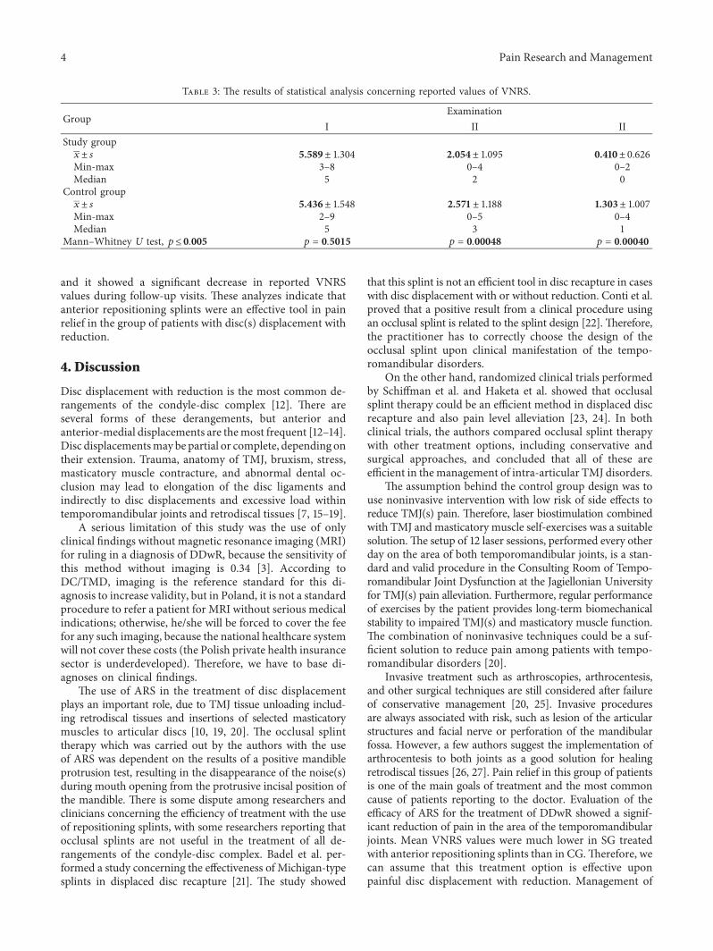
Table 3: ,e results of statistical analysis concerning reported values of VNRS.
GroupExamination
I II IIStudy group
x± s 5.589± 1.304 2.054± 1.095 0.410± 0.626Min-max 3–8 0–4 0–2Median 5 2 0
Control groupx± s 5.436± 1.548 2.571± 1.188 1.303± 1.007Min-max 2–9 0–5 0–4Median 5 3 1
Mann–Whitney U test, p≤ 0.005 p � 0.5015 p � 0.00048 p � 0.00040
4 Pain Research and Management
pain related to TMJ disc displacement using ARS has beenalso positively evaluated by other authors [28–30].
5. Conclusion
Considering the limitation of the study, we can state that theanterior repositioning splint is an efficient tool in decreasingpain related to disc displacement with reduction.
Abbreviations
ARS: Anterior repositioning splintCG: Control groupDC/TMD: Diagnostic Criteria for Temporomandibular
DisordersDDwR: Disc displacement with reductionSG: Study groupTMJ: Temporomandibular jointVNRS: Verbal Numerical Rating Scale.
Conflicts of Interest
,e authors declare that they have no conflicts of interest.
Authors’ Contributions
Malgorzata Pihut created research concept, performed oc-clusal splint therapy and laser therapy, collected and ana-lyzed the data, and wrote the manuscript. MalgorzataGorecka selected the references. Piotr Ceranowicz edited themanuscript. Mieszko Wieckiewicz analyzed the data, wroteand edited the manuscript, selected the references, and fi-nally revised it before submission. All authors read andapproved the final manuscript.
References
[1] M. Ahmad and E. L. Schiffman, “Temporomandibular jointdisorders and orofacial pain,” Dental Clinics of NorthAmerica, vol. 60, no. 1, pp. 105–124, 2016.
[2] J. P. Okeson, “Joint intracapsular disorders: diagnostic andnonsurgical management considerations,” Dental Clinics ofNorth America, vol. 51, no. 1, pp. 85–103, 2007.
[3] E. Schiffman, R. Ohrbach, E. Truelove et al., “Diagnosticcriteria for temporomandibular disorders (DC/TMD) forclinical and research applications: recommendations of theInternational RDC/TMD Consortium Network and OrofacialPain Special Interest Group,” Journal of Oral and Facial Painand Headache, vol. 28, no. 1, pp. 6–27, 2014.
[4] M. Wieckiewicz, N. Grychowska, K. Wojciechowski et al.,“Prevalence and correlation between TMD based onRDC/TMD diagnoses, oral parafunctions and psychoemo-tional stress in Polish university students,” Biomed ResearchInternational, vol. 2014, Article ID 472346, 7 pages, 2014.
[5] A. Al-Khotani, A. Naimi-Akbar, E. Albadawi, M. Ernberg,B. Hedenberg-Magnusson, and N. Christidis, “Prevalence ofdiagnosed temporomandibular disorders among Saudi Ara-bian children and adolescents,” Journal of Headache and Pain,vol. 17, no. 1, pp. 1–11, 2016.
[6] S. I. Kalaykova, F. Lobbezoo, and M. Naeije, “Risk factors foranterior disc displacement with reduction and intermittent
locking in adolescents,” Journal of Orofacial Pain, vol. 25,no. 2, pp. 153–160, 2011.
[7] Y. Kumazaki, S. Kawakami, A. Hirata, K. Oki, and S. Minagi,“Ipsilateral molar clenching induces less pain and discomfortthan contralateral molar clenching in patients with unilateralanterior disc displacement of the temporomandibular joint,”Journal of Oral and Facial Pain and Headache, vol. 30, no. 3,pp. 241–248, 2016.
[8] U. Karacayli, G. Mumcu, H. Cimilli, N. Sisman, H. Sur, andY. Gunaydin, “,e effects of chronic pain on oral healthrelated quality of life in patients with anterior disc dis-placement with reduction,” Community Dental Health,vol. 28, no. 3, pp. 211–215, 2011.
[9] J. P. Okeson, “Long-term treatment of disk-interferencedisorders of the temporomandibular joint with anteriorrepositioning occlusal splints,” Journal of Prosthetic Dentistry,vol. 60, no. 5, pp. 611–616, 1988.
[10] D. Eberhard, H. P. Bantleon, and W. Steger, “,e efficacy ofanterior repositioning splint therapy studied by magneticresonance imaging,” European Journal of Orthodontics,vol. 24, no. 4, pp. 343–352, 2002.
[11] S. M. Shaffer, J. M. Brismee, P. S. Sizer, and C. A. Courtney,“Temporomandibular disorders. Part 2: conservative man-agement,” Journal of Manual and Manipulative 8erapy,vol. 22, no. 1, pp. 13–23, 2014.
[12] X. Y. Cai, J. M. Jin, and C. Yang, “Changes in disc position,disc length, and condylar height in the temporomandibularjoint with anterior disc displacement a longitudinal retro-spective magnetic resonance imaging study,” Journal of Oraland Maxillofacial Surgery, vol. 69, no. 11, pp. e340–e346, 2011.
[13] S. Sato, H. Kawamura, H. Nagasaka, and K. Motegi, “,enatural course of anterior disc displacement without re-duction in the temporomandibular joint: follow-up at 6, 12,and 18 months,” Journal of Oral and Maxillofacial Surgery,vol. 55, no. 3, pp. 234–238, 1997.
[14] N. Tomura, T. Otani, K. Narita et al., “Visualization of an-terior disc displacement in temporomandibular disorders oncontrast-enhanced magnetic resonance imaging: comparisonwith T2-weighted, proton density–weighted, and precontrastT1-weighted imaging,” Oral Surgery, Oral Medicine, OralPathology, Oral Radiology and Endodontology, vol. 103, no. 2,pp. 260–266, 2007.
[15] Y. K. Hu, C. Yang, X. Y. Cai, and Q. Y. Xie, “Does condylarheight decrease more in temporomandibular joint non-reducing disc displacement than reducing disc displacement?:a magnetic resonance imaging retrospective study,”Medicine,vol. 95, no. 35, p. e4715, 2016.
[16] D. He, X. Yang, F. Wang, C. Yang, and M. Dong, “Acutetrauma induced disc displacement without reduction and itssequelae,” Scientific Reports, vol. 6, no. 32684, pp. 1–5, 2016.
[17] A. Jimenez-Silva, C. Peña-Duran, J. Tobar-Reyes, andR. Frugone-Zambra, “Sleep and awake bruxism in adults andits relationship with temporomandibular disorders: a sys-tematic review from 2003 to 2014,” Acta OdontologicaScandinavica, vol. 75, no. 1, pp. 36–58, 2017.
[18] W. S. Jung, H. Kim, D. M. Jeon, S. J. Mah, and S. J. Ahn,“Magnetic resonance imaging-verified temporomandibularjoint disk displacement in relation to sagittal and vertical jawdeformities,” International Journal of Oral and MaxillofacialSurgery, vol. 42, no. 9, pp. 1108–1115, 2013.
[19] M. Kuroda, M. Otonari-Yamamoto, T. Sano, M. Fujikura, andM. Wakoh, “Diagnosis of retrodiscal tissue in painful tempo-romandibular joint (TMJ) by fluid-attenuated inversion recovery
Pain Research and Management 5

(FLAIR) signal intensity,” Journal of Craniomandibular and SleepPractice, vol. 33, no. 4, pp. 272–276, 2015.
[20] M. Wieckiewicz, K. Boening, P. Wiland, Y. Y. Shiau, andA. Paradowska-Stolarz, “Reported concepts for the treatmentmodalities and pain management of temporomandibulardisorders,” Journal of Headache and Pain, vol. 16, no. 106,pp. 1–12, 2015.
[21] T. Badel, M.Marotti, J. Kern, andM. Laskarin, “A quantitativeanalysis of splint therapy of displaced temporomandibularjoint disc,” Annals of Anatomy, vol. 191, no. 3, pp. 280–287,2009.
[22] P. C. Conti, A. S. Correa, J. R. Lauris, and J. Stuginski-Barbosa,“Management of painful temporomandibular joint clickingwith different intraoral devices and counseling: a controlledstudy,” Journal of Applied Oral Sciences, vol. 23, no. 5,pp. 529–535, 2015.
[23] E. L. Schiffman, A. M. Velly, J. O. Look et al., “Effects of fourtreatment strategies for temporomandibular joint closedlock,” International Journal of Oral and Maxillofacial Surgery,vol. 43, no. 2, pp. 217–226, 2014.
[24] T. Haketa, K. Kino, M. Sugisaki, M. Takaoka, and T. Ohta,“Randomized clinical trial of treatment for TMJ disc dis-placement,” Journal of Dental Research, vol. 89, no. 11,pp. 1259–1263, 2010.
[25] R. F. C. P. De Freitas, M. A. F. Ferreira, G. A. S. Barbosa, andP. S. Calderon, “Counselling and self-management therapiesfor temporomandibular disorders: a systematic review,”Journal of Oral Rehabilitation, vol. 40, no. 11, pp. 864–874,2013.
[26] R. Emshoff, “Clinical factors affecting the outcome ofarthrocentesis and hydraulic distension of temporomandib-ular joint,” Oral Surgery, Oral Medicine, Oral Pathology, OralRadiology and Endodontology, vol. 100, no. 4, pp. 409–414,2005.
[27] M. Ethunandan and A. W. Wilson, “Temporomandibularjoint arthocentesis-more questions than answers?,” Journal ofOral and Maxillofacial Surgery, vol. 64, no. 6, pp. 952–955,2006.
[28] P. C. Conti, C. N. dos Santos, E. M. Kogawa, A. C. de CastroFerreira Conti, and R. de Araujo Cdos, “,e treatment ofpainful temporomandibular joint clicking with oral splints:a randomized clinical trial,” Journal of American DentalAssociation, vol. 137, no. 8, pp. 1108–1114, 2006.
[29] S. Tecco, S. Caputi, S. Tete, G. Orsini, and F. Festa, “Intra-articular and muscle symptoms and subjective relief duringTMJ internal derangement treatment with maxillary anteriorrepositioning splint or SVED and MORA splints: a compar-ison with untreated control subjects,” Journal of Cranio-mandibular and Sleep Practice, vol. 24, no. 2, pp. 119–129,2006.
[30] P. C. Conti, J. E. Miranda, A. C. Conti, L. F. Pegoraro, andR. Araujo Cdos, “Partial time use of anterior repositioningsplints in the management of TMJ pain and dysfunction:a one-year controlled study,” Journal of Applied Oral Sciences,vol. 13, no. 4, pp. 345–350, 2005.
6 Pain Research and Management

Stem Cells International
Hindawiwww.hindawi.com Volume 2018
Hindawiwww.hindawi.com Volume 2018
MEDIATORSINFLAMMATION
of
EndocrinologyInternational Journal of
Hindawiwww.hindawi.com Volume 2018
Hindawiwww.hindawi.com Volume 2018
Disease Markers
Hindawiwww.hindawi.com Volume 2018
BioMed Research International
OncologyJournal of
Hindawiwww.hindawi.com Volume 2013
Hindawiwww.hindawi.com Volume 2018
Oxidative Medicine and Cellular Longevity
Hindawiwww.hindawi.com Volume 2018
PPAR Research
Hindawi Publishing Corporation http://www.hindawi.com Volume 2013Hindawiwww.hindawi.com
The Scientific World Journal
Volume 2018
Immunology ResearchHindawiwww.hindawi.com Volume 2018
Journal of
ObesityJournal of
Hindawiwww.hindawi.com Volume 2018
Hindawiwww.hindawi.com Volume 2018
Computational and Mathematical Methods in Medicine
Hindawiwww.hindawi.com Volume 2018
Behavioural Neurology
OphthalmologyJournal of
Hindawiwww.hindawi.com Volume 2018
Diabetes ResearchJournal of
Hindawiwww.hindawi.com Volume 2018
Hindawiwww.hindawi.com Volume 2018
Research and TreatmentAIDS
Hindawiwww.hindawi.com Volume 2018
Gastroenterology Research and Practice
Hindawiwww.hindawi.com Volume 2018
Parkinson’s Disease
Evidence-Based Complementary andAlternative Medicine
Volume 2018Hindawiwww.hindawi.com
Submit your manuscripts atwww.hindawi.com