clinical validation of a noninvasive, raman spectroscopic method to assess carotenoid nutritional...
TRANSCRIPT

Original Research
Clinical Validation of a Noninvasive, RamanSpectroscopic Method to Assess Carotenoid NutritionalStatus in Humans
Jeffrey A. Zidichouski, PhD, Angela Mastaloudis, PhD, Stephen J. Poole, BS, James C. Reading, PhD, Carsten R. Smidt, PhD,
FACN
Pharmanex Research Institute, Provo (J.A.Z., A.M., S.J.P., C.R.S.), Department of Family and Preventive Medicine, University of
Utah School of Medicine, Salt Lake City (J.C.R.), Utah
Key words: carotenoids, Raman spectroscopy, nutritional status, skin, serum, antioxidants, photonics
Background: Carotenoids are an important group of phytonutrients that are abundant in fruits and
vegetables. Epidemiological and clinical intervention studies have implied the presence of protective qualities of
these nutrients against the development of a variety of chronic diseases. Previously, human carotenoid status has
been assessed in serum and tissue using high-performance liquid chromatography (HPLC) methodology.
Recently, a Raman spectroscopy (RS)–based photonic method has been developed to accurately and
noninvasively measure the carotenoid concentration in human skin.
Objectives: (1) To validate skin RS methodology against standard serum carotenoid measurements by
HPLC and (2) to establish and compare the reliability of the 2 methods.
Design: This study included 372 healthy adults who provided 3 blood samples and 3 RS skin carotenoid
measurements within an 8-day period; each day-matched blood sample and RS determination was spaced by
$48 hours.
Results: Consistent positive correlations were observed for each of 3 separate same-day correlation plots of
total serum versus RS skin carotenoids. Overall estimate of the line of best fit from analysis of covariance, using
all 3 samples (n 5 1116), yielded a Pearson correlation of R 5 0.81 (r2 5 0.66; p , 0.001). Based on analysis of
variance, RS skin carotenoid methodology exhibited 0.9% less variance over the 3 tests than serum carotenoids
by the HPLC method (p , 0.03).
Conclusions: RS accurately measures total carotenoids in human skin with less intra-individual variability
than measurement of serum carotenoids by HPLC analysis. RS technology is a valid and reliable noninvasive
method to rapidly assess carotenoid nutritional status in humans.
INTRODUCTION
There is strong scientific evidence that high intakes of
antioxidant-rich fruits and vegetables are protective against
certain types of cancer, cardiovascular disease, diabetes,
specific neurodegenerative diseases, and a variety of other
chronic conditions associated with oxidative stress [1]. Thus, a
diet high in antioxidant-containing fruits and vegetables may
help to tip the balance away from the deleterious effects of
oxidants in favor of the beneficial effects of antioxidants.
Address correspondence to: A. Mastaloudis, Pharmanex Research Institute, 75 W. Center Street, Provo, UT 84601. E-mail: [email protected]
Data presented here have been published previously in abstract form in Zidichouski JA, Poole SJ, Gellermann W, Smidt CR: Clinical validation of a novel Raman
spectroscopic technology to non-invasively assess carotenoid status in humans. J Am Coll Nutr 23:A468, 2004.
Dr. Zidichouski is now with the Institute for Nutrisciences and Health, National Research Council of Canada.
Dr. Reading is Professor Emeritus at the Department of Family and Preventive Medicine, University of Utah School of Medicine.
Dr. Smidt is now at Shaklee Corporation, Pleasanton, California.
This study was funded by the Pharmanex Research Institute, Pharmanex LLC, Provo, Utah.
Author Contributions: Jeffrey A. Zidichouski was responsible for study design, subject recruitment, data collection, and analysis and preparation and revision of the
manuscript; Angela Mastaloudis contributed to the statistical analyses and was responsible for the preparation and revision of the manuscript; Stephen J. Poole contributed
to the subject recruitment, data collection, and data analyses; James C. Reading contributed to the statistical analyses; Carsten R. Smidt contributed to the study design,
offered expertise in carotenoids and Raman spectroscopy, and contributed to manuscript revisions. A.M. and S.J.P. are currently employed by Pharmanex Research
Institute (Pharmanex, LLC). J.A.Z. and C.R.S. were employed by Pharmanex Research Institute (Pharmanex, LLC) at the time the study was conducted.
Journal of the American College of Nutrition, Vol. 28, No. 6, 687–693 (2009)
Published by the American College of Nutrition
687

Carotenoids are some of the most abundant antioxidant
phytonutrients present in fruits and vegetables. It has been
theorized that the same antioxidant mechanisms that confer
protection against photo-oxidative processes in plants are
responsible for the protective effects of a diet high in fruits and
vegetables in humans [2,3]. In fact, there is strong epidemio-
logical and clinical evidence that carotenoids may help to
defend against cardiovascular disease, certain cancers, and
specific eye diseases [3].
Carotenoids compose a family of some 600 nutrients made
up of a common polyisoprenoid structure containing 40 carbon
atoms and a system of conjugated double bonds [4]. Their
elongated structure enables them to both scavenge singlet
oxygen directly and quench peroxyl radicals [2,5]. In addition
to providing antioxidant protection, these important nutrients
are involved in intercellular communication [2,6] and some
serve as important precursors to vitamin A [6].
For decades, high-performance liquid chromatography
(HPLC) has been considered the gold standard for assessment
of carotenoid status in humans, based on the quantification of
carotenoid compounds extracted from serum [5,7–11] and
tissue samples or biopsies [12]. Limitations associated with
this methodology include that it is technically difficult,
expensive, requires specialized equipment, and necessitates
the collection of biologically invasive samples. Furthermore,
blood concentrations can be influenced by variability in daily
carotenoid intake. Consequently, accurate assessment of
carotenoid status is not easily determined, particularly in
large, population-based studies, and has therefore been limited
to research and medical institutions.
The present study utilized a highly specific resonance
Raman scattering spectroscopic technology (RS) that has been
under investigation as a valid, noninvasive alternative for
measuring carotenoid status in situ in human tissue [11,13–15].
RS technology takes advantage of the polyisoprenoid back-
bone common to all carotenoids. Upon exposure to blue light,
the vibrational energy of the conjugated double bonds of the
carotenoid backbone is increased. As a result, a portion of the
blue light is scattered inelastically in what is termed Raman
scattering [14], creating a spectral fingerprint that is unique to
carotenoids. The intensity of the Raman scattering is directly
proportional to the concentration of carotenoids present,
making it possible to quantify carotenoids in human skin.
Primary carotenoids detected in the skin are lycopene, b-
carotene, a-carotene, b-cryptoxanthin, lutein, zeaxanthin,
phytoene, and phytofluene, with lycopene present in the
highest amounts [14]. The RS measurement technique is fast,
painless, and cost effective, making it ideal for monitoring
large groups of people. Furthermore, carotenoid levels
measured in the skin by RS have been demonstrated to be
directly related to both self-reported fruit and vegetable
consumption and carotenoid supplementation [5].
The purpose of this study was to test the hypothesis that a
novel RS technology is a valid, quantitative, and reliable
technique for assessment of carotenoid status in a hetero-
geneous population of healthy men and women by comparing
it to the established HPLC method.
SUBJECTS AND METHODS
Subjects
Three hundred ninety-one people were recruited to
participate in this study. All participants provided written
informed consent as approved by and in accordance with the
ethical standards of the Western Institutional Review Board
(protocol #1053052). Subjects gave both written and verbal
confirmation that they had not dramatically changed their
dietary habits, lifestyle, or supplementation regimen during the
month prior to enrollment or for the short duration of the trial
(up to 8 days for each trial participant). The age and sex of
each subject were recorded as part of the screening process.
Exclusion criteria included age younger than 18 years and/or
pregnancy or suspected pregnancy.
Protocol
Over the course of 8 days, each subject visited the lab for
blood sample collection and RS determination 3 times, with at
least 48 hours between each sample collection. Each day, a 10-
ml blood sample was collected from an antecubital arm vein
into untreated glass vacutubes; all were fasting, morning blood
draws. Within 15 minutes of blood sampling, skin carotenoid
concentrations in the palm of the hand were determined using
RS technology. All data were obtained over a 3-month period
and without any changes to study personnel.
Serum Carotenoids by HPLC
Immediately after collection, each sample was permitted to
clot for 1 hour at room temperature. Serum was prepared by
centrifugation of the clotted sample at 3200 rpm for 5 minutes at
room temperature (Drucker, Model #6138), transferred to
polypropylene cryotubes, and stored at 280uC until time of
HPLC analysis. Frozen samples were collectively shipped on dry
ice to a specialized micronutrient analytical laboratory (Craft
Technologies, Wilson, NC) for analysis of total serum carote-
noids by HPLC. Serum carotenoid (b- and a-carotene, lutein,
zeaxanthin, lycopene, and b-cryptoxanthin) concentrations were
quantified according to methods described previously [16].
Raman Scattering Technology
Using RS technology (Biophotonic Scanner, Pharmanex,
LLC), each individual’s skin carotenoid level was determined
Noninvasive Assessment of Human Carotenoid Status
688 VOL. 28, NO. 6
by calculating the average height of the peak Raman
absorbance signal obtained and quantified from excitation of
skin carotenoids using a low-intensity blue (l 5 473 nm)
solid-state laser with green light (510 nm) detection [5]. Skin
carotenoids are reported as Raman intensity counts. The higher
the count, the higher the concentration of carotenoid molecules
detected at the site of measurement. All measurements were
performed by experienced technicians, according to the
manufacturer’s specifications and requirements.
Statistical Analysis
Descriptive statistics (means, medians, standard deviations,
etc.) were calculated and statistical tests were conducted to
compare and contrast the RS and HPLC methodologies of
determining carotenoid nutritional status. All data are pre-
sented as means 6 standard deviations.
Criterion validity was used to validate the RS methodology
by comparing it to serum carotenoid quantification by HPLC.
Based on individual data for each subject (n 5 372), scatter
plots, lines of best fit (with 95% confidence curves for the
fitted line), and Pearson correlation coefficients were gener-
ated for total serum carotenoid concentration versus the same-
day RS skin carotenoid determination for each of the follow-
ing 3 test periods: serum 1 vs. RS 1 (sample 1), serum 2 vs.
RS 2 (sample 2), and serum 3 vs. RS 3 (sample 3). In order to
generate an overall estimate of the line of best fit, in addition
tothe 3 (1 for each serum sample) above analyses, an analysis
of covariance (ANCOVA) was performed using all 3 samples
(1116 observations) and modeling subject ID as a main effect.
To evaluate the consistency between methods, the intra-
individual variability of both the RS and the HPLC methods
was determined. Pooled estimates of variance (from a 1-way
analysis of variance [ANOVA]) of the 3 RS and serum
measurements across individuals were calculated. Variance of
the pooled estimates was compared with an F-test. For each
subject, the standard deviation of each RS determination and
day-matched serum sample was calculated and the population
of standard deviations was used to perform a Wilcoxon test for
differences in medians between the RS count and serum HPLC
analysis. Statistics were calculated using JMP Statistical
Discovery Software (SAS Institute Inc, Cary, NC).
RESULTS
Of the 391 volunteers, 5 were excluded due to pregnancy or
breastfeeding and 14 were withheld from the final analysis due
to incomplete datasets. Data presented here are for the 372
subjects (95% of enrolled subjects) who completed the study,
providing complete skin and blood sample datasets. Of these,
199 were men and 173 were women; the average age was 33.4
6 10.0 years (range 18–68 years). There was no significant
difference in average age for men and women (men 34.3 6
10.4 years and women 32.4 6 9.5 years).
Validity
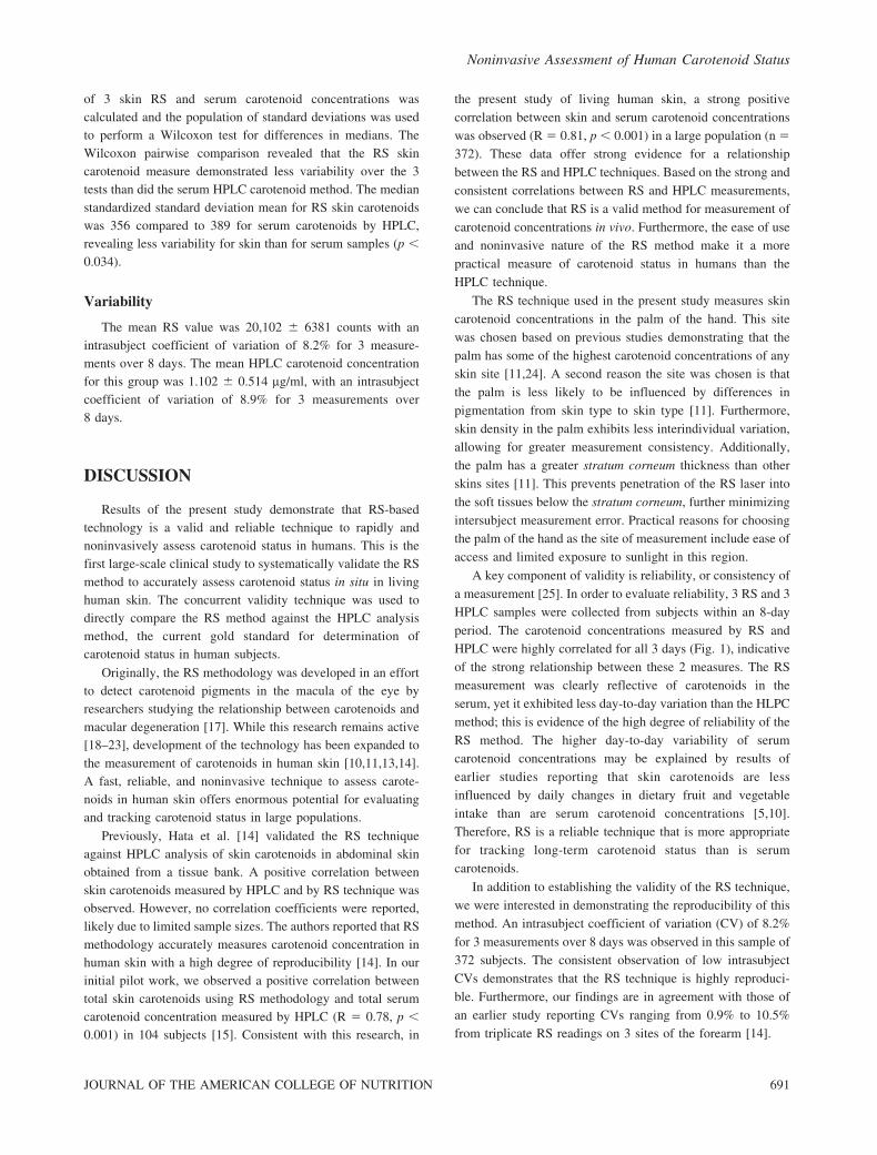
Correlation between Skin Carotenoids by RS and Total
Serum Carotenoid Concentrations by HPLC. For the first
measurement, the mean RS value was 20,095 6 6439 (range 9137–
49,666 counts) and the mean total serum carotenoid concentration
was 1.08 6 0.51 mg/ml (range 0.21–3.74 mg/ml). A positive linear
correlation between RS and total serum carotenoid concentration
was observed (R 5 0.82; r2 5 0.67; p , 0.001; Fig. 1A).
For the second sampling, mean RS skin carotenoid count
was 20,178 6 6367 (range 8624–48,441 counts) and mean
total serum carotenoid concentration was 1.08 6 0.50 (range
0.23–3.97 mg/ml). A direct correlation between RS and total
serum carotenoid concentration was observed (R 5 0.80, r2 5
0.64; p , 0.001; Fig. 1B).
In the third and final sampling, mean RS skin carotenoid
count was 20,034 6 6352 (range 7888–49,262 counts) and the
mean total serum carotenoid concentration was 1.14 6 0.52
(range 0.22–4.41 mg/ml). Consistent with samplings 1 and 2, a
strong positive correlation between the 2 measures was
observed (R 5 0.81, r2 5 0.65; p , 0.001; Fig. 1C).
A linear regression was performed with RS as the
dependent variable and serum concentration as the independent
variable. The results of the best fit lines for the 3 samples were
sample 1: slope 0.000066, intercept 20.24 (y 5 20.24 +0.000066x); sample 2: slope 0.000063, intercept 20.19 (y 5
20.19 + 0.000063x); and sample 3: slope 0.000066, intercept
20.19 (y 5 20.19 + 0.000066x), where x 5 Raman intensity
counts and y 5 serum carotenoid concentration.
The composite correlation based on the overall estimate of
the line of best fit from ANCOVA, using all 3 samples (n 5
1116) and modeling subject ID as a main effect, was: slope
0.000064 with intercept 20.09 (20.09 + 0.000064x) with a
Pearson correlation of R 5 0.81 (r2 5 0.66; p , 0.001).
Reliability
Pooled Estimate of Variance. Based on ANOVA, the RS
skin carotenoid pooled estimate of standard deviation was
0.095 compared to a pooled estimate of standard deviation of
0.104 for serum carotenoids by HPLC. Results demonstrate
that the RS skin carotenoid method resulted in less variance
over the 3 tests than did serum carotenoids quantified by the
HPLC method (skin carotenoid standard deviation 9.5% of the
mean compared to 10.4% for serum carotenoids; p , 0.03).
Wilcoxon Test. To further investigate the variance of the 2
methods, for each individual, the standard deviation of each set
Noninvasive Assessment of Human Carotenoid Status
JOURNAL OF THE AMERICAN COLLEGE OF NUTRITION 689
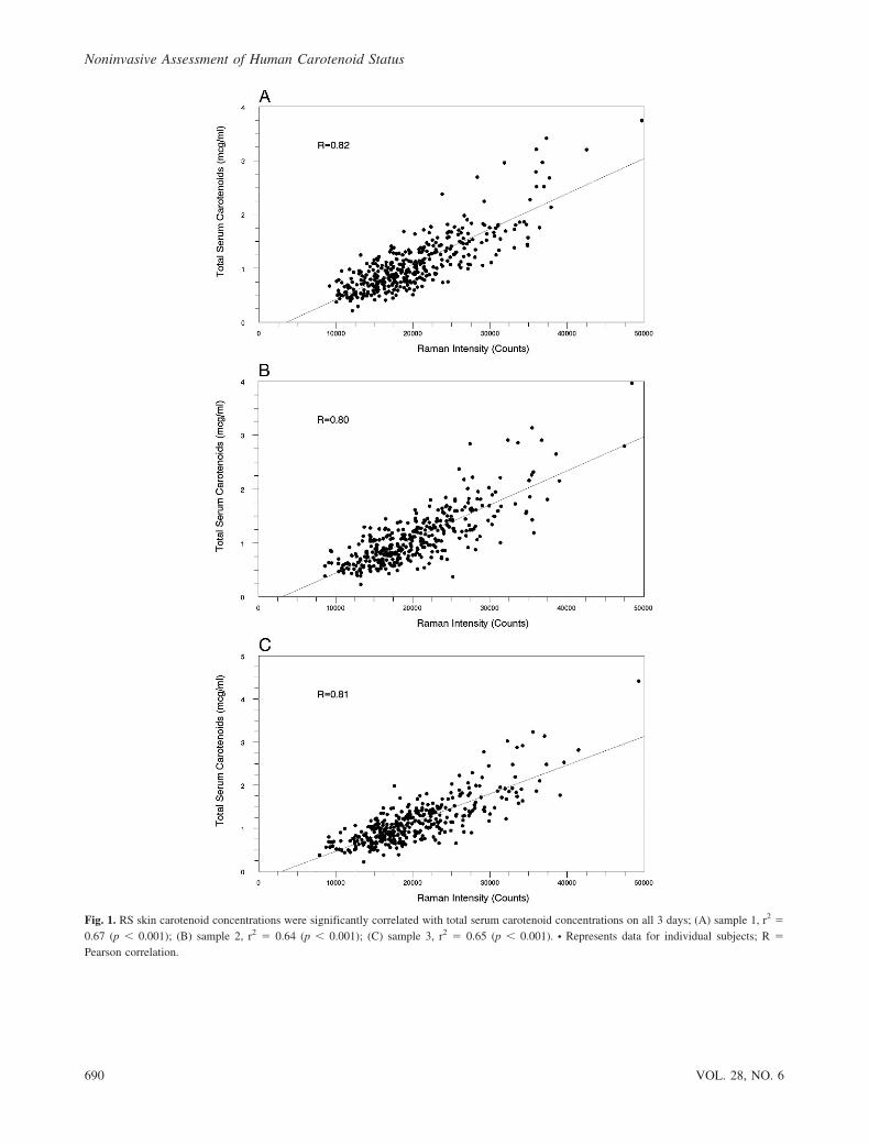
Fig. 1. RS skin carotenoid concentrations were significantly correlated with total serum carotenoid concentrations on all 3 days; (A) sample 1, r2 5
0.67 (p , 0.001); (B) sample 2, r2 5 0.64 (p , 0.001); (C) sample 3, r2 5 0.65 (p , 0.001). N Represents data for individual subjects; R 5
Pearson correlation.
Noninvasive Assessment of Human Carotenoid Status
690 VOL. 28, NO. 6
of 3 skin RS and serum carotenoid concentrations was
calculated and the population of standard deviations was used
to perform a Wilcoxon test for differences in medians. The
Wilcoxon pairwise comparison revealed that the RS skin
carotenoid measure demonstrated less variability over the 3
tests than did the serum HPLC carotenoid method. The median
standardized standard deviation mean for RS skin carotenoids
was 356 compared to 389 for serum carotenoids by HPLC,
revealing less variability for skin than for serum samples (p ,
0.034).
Variability
The mean RS value was 20,102 6 6381 counts with an
intrasubject coefficient of variation of 8.2% for 3 measure-
ments over 8 days. The mean HPLC carotenoid concentration
for this group was 1.102 6 0.514 mg/ml, with an intrasubject
coefficient of variation of 8.9% for 3 measurements over
8 days.
DISCUSSION
Results of the present study demonstrate that RS-based
technology is a valid and reliable technique to rapidly and
noninvasively assess carotenoid status in humans. This is the
first large-scale clinical study to systematically validate the RS
method to accurately assess carotenoid status in situ in living
human skin. The concurrent validity technique was used to
directly compare the RS method against the HPLC analysis
method, the current gold standard for determination of
carotenoid status in human subjects.
Originally, the RS methodology was developed in an effort
to detect carotenoid pigments in the macula of the eye by
researchers studying the relationship between carotenoids and
macular degeneration [17]. While this research remains active
[18–23], development of the technology has been expanded to
the measurement of carotenoids in human skin [10,11,13,14].
A fast, reliable, and noninvasive technique to assess carote-
noids in human skin offers enormous potential for evaluating
and tracking carotenoid status in large populations.
Previously, Hata et al. [14] validated the RS technique
against HPLC analysis of skin carotenoids in abdominal skin
obtained from a tissue bank. A positive correlation between
skin carotenoids measured by HPLC and by RS technique was
observed. However, no correlation coefficients were reported,
likely due to limited sample sizes. The authors reported that RS
methodology accurately measures carotenoid concentration in
human skin with a high degree of reproducibility [14]. In our
initial pilot work, we observed a positive correlation between
total skin carotenoids using RS methodology and total serum
carotenoid concentration measured by HPLC (R 5 0.78, p ,
0.001) in 104 subjects [15]. Consistent with this research, in
the present study of living human skin, a strong positive
correlation between skin and serum carotenoid concentrations
was observed (R 5 0.81, p , 0.001) in a large population (n 5
372). These data offer strong evidence for a relationship
between the RS and HPLC techniques. Based on the strong and
consistent correlations between RS and HPLC measurements,
we can conclude that RS is a valid method for measurement of
carotenoid concentrations in vivo. Furthermore, the ease of use
and noninvasive nature of the RS method make it a more
practical measure of carotenoid status in humans than the
HPLC technique.
The RS technique used in the present study measures skin
carotenoid concentrations in the palm of the hand. This site
was chosen based on previous studies demonstrating that the
palm has some of the highest carotenoid concentrations of any
skin site [11,24]. A second reason the site was chosen is that
the palm is less likely to be influenced by differences in
pigmentation from skin type to skin type [11]. Furthermore,
skin density in the palm exhibits less interindividual variation,
allowing for greater measurement consistency. Additionally,
the palm has a greater stratum corneum thickness than other
skins sites [11]. This prevents penetration of the RS laser into
the soft tissues below the stratum corneum, further minimizing
intersubject measurement error. Practical reasons for choosing
the palm of the hand as the site of measurement include ease of
access and limited exposure to sunlight in this region.
A key component of validity is reliability, or consistency of
a measurement [25]. In order to evaluate reliability, 3 RS and 3
HPLC samples were collected from subjects within an 8-day
period. The carotenoid concentrations measured by RS and
HPLC were highly correlated for all 3 days (Fig. 1), indicative
of the strong relationship between these 2 measures. The RS
measurement was clearly reflective of carotenoids in the
serum, yet it exhibited less day-to-day variation than the HLPC
method; this is evidence of the high degree of reliability of the
RS method. The higher day-to-day variability of serum
carotenoid concentrations may be explained by results of
earlier studies reporting that skin carotenoids are less
influenced by daily changes in dietary fruit and vegetable
intake than are serum carotenoid concentrations [5,10].
Therefore, RS is a reliable technique that is more appropriate
for tracking long-term carotenoid status than is serum
carotenoids.
In addition to establishing the validity of the RS technique,
we were interested in demonstrating the reproducibility of this
method. An intrasubject coefficient of variation (CV) of 8.2%
for 3 measurements over 8 days was observed in this sample of
372 subjects. The consistent observation of low intrasubject
CVs demonstrates that the RS technique is highly reproduci-
ble. Furthermore, our findings are in agreement with those of
an earlier study reporting CVs ranging from 0.9% to 10.5%
from triplicate RS readings on 3 sites of the forearm [14].
Noninvasive Assessment of Human Carotenoid Status
JOURNAL OF THE AMERICAN COLLEGE OF NUTRITION 691
We observed a wide range of RS scores across subjects
(7888–49,666 counts), with a mean of 20,102 6 6381 counts,
which is in excellent agreement with a previous study of 57
subjects who exhibited a mean of ,21,000 counts (range
10,000–49,000 counts) [11]. This large variability across
subjects is in line with studies of serum carotenoids assessed
by HPLC [26,27]. This large range in carotenoid status has
been attributed to differences in dietary habits [10,27],
cigarette smoking [10,13,27,28], and other lifestyle factors
[28].
Epidemiological evidence has demonstrated an inverse
association between the intake of fruits and vegetables and
the risk of certain chronic diseases including cardiovascular
disease, specific eye diseases, and various types of cancer
[6,29]. However, the tracking of dietary intake of fruits and
vegetables is extremely problematic. The most common
methods of dietary assessment—food diaries, 24-hour food
recalls, and food frequency questionnaires—all suffer from a
number of limitations. Such dietary assessment methods are
vulnerable to inaccurate reporting of portion sizes, under-
reporting consumption of ‘‘unhealthy’’ foods, and overreport-
ing intakes of foods perceived as healthful and therefore
unlikely to represent usual dietary intake [30]. Consequently,
estimates of carotenoid intakes from dietary questionnaires are
typically only moderately correlated with carotenoid concen-
trations in the blood [29,31]. For one, dietary estimates of
carotenoid intakes are dependent on both the accuracy of
reporting and the accuracy of the food composition databases
used for diet analyses [31]. Additionally, variations in the
bioavailability of individual carotenoids from different sources
contribute to any disconnection between intakes and blood
concentrations [31]. Due to the limitations of dietary assess-
ment techniques, blood carotenoid concentrations quantified
by HPLC have been used as an objective marker of fruit and
vegetable intake. Unfortunately, this technique is invasive,
expensive, and time-consuming, making it impractical for
monitoring large populations. In contrast, the RS method of
measuring skin carotenoids is fast, accurate, noninvasive, and
does not require a high degree of technical expertise. The
strong correlation between skin carotenoids measured by RS
and serum carotenoids measured by HPLC indicates that the
RS technique is an excellent method for quickly and accurately
assessing carotenoid status in humans. Ultimately, the RS
technique offers an objective measurement of fruit and
vegetable intake, making it an ideal tool for studying dietary
intake in large human populations.
The potential for RS methodology to study carotenoid
concentrations in humans is immense. Currently, it is being
used to measure carotenoid concentrations in the skin of
individuals who are at risk for or suffering from skin cancer.
So far, researchers have reported lower levels of carotenoids in
precancerous lesions and skin neoplasms compared to
carotenoid levels detected in healthy skin [14]. RS technology
is also being used to measure carotenoid levels in fruits and
vegetables in an effort to identify carotenoid-rich varieties
[32]. Ongoing studies are examining the relationship between
skin carotenoids and markers of oxidative stress, antioxidant
status, dietary intake of fruits and vegetables, and antioxidant
supplement use. Future studies will be able to use RS
methodology to track fruit and vegetable intake in large
populations, measure the effects of dietary supplements, and
investigate the relationships between skin carotenoids and
chronic diseases.
CONCLUSIONS
Based on the present study, the RS method is a valid and
reliable technique to measure carotenoids in human skin in
vivo. There is a strong correlation between skin and serum
carotenoid concentrations. Due to its noninvasive nature and
low variability, the RS-based method is an attractive tool for
assessing carotenoid status in humans and appears to be
superior to the costly, more invasive blood carotenoid HPLC
method.
ACKNOWLEDGMENTS
The authors are grateful to Neil Craft, PhD, and his staff at
Craft Technologies (Wilson, NC) for their expertise in HPLC
analysis of serum carotenoids.
REFERENCES
1. Liu CS, Glahn RP, Liu RH: Assessment of carotenoid bioavail-
ability of whole foods using a caco-2 cell culture model coupled
with an in vitro digestion. J Agric Food Chem 52:4330–4337,
2004.
2. Stahl W, Sies H: Antioxidant effects of carotenoids: implication in
photoprotection in humans. In Cadenas E, Packer L (eds):
‘‘Handbook of Antioxidants,’’ 2nd ed. New York: Marcel-Dekker,
pp 223–233, 2002.
3. Krinsky N, Johnson EJ: Carotenoid actions and their relation to
health and disease. Mol Aspects Med 26:459–516, 2005.
4. During A, Harrison E: Intestinal absorption and metabolism of
carotenoids: insights from cell culture. Arch Biochem Biophys
430:77–88, 2004.
5. Smidt CR, Burke DS: Nutritional significance and measurement of
carotenoids. Curr Topics Nutraceut Res 2:79–91, 2004.
6. Deming DM, Boileau TWM, Heintz KH, Atkinson CA, Erdman
JW Jr: Carotenoids: linking chemistry, absorption, and metabolism
to potential roles in human health and disease. In Cadenas E,
Packer L (eds): ‘‘Handbook of Antioxidants,’’ 2nd ed. New York:
Marcel-Dekker, pp 189–221, 2002.
Noninvasive Assessment of Human Carotenoid Status
692 VOL. 28, NO. 6
7. Broich CR, Gerber LE, Erdman Jr JW: Determination of lycopene,
alpha- and beta-carotene and retinyl esters in human serum by
reversed-phase high performance liquid chromatography. Lipids
18:253–258, 1983.
8. Nelis HJ, De Roose J, Vandenbaviere H, De Leenheer AP:
Nonaqueous reversed-phase liquid chromatography and fluorime-
try compared for determination of retinol in serum. Clin Chem
7:1431–1434, 1983.
9. Peng YM, Beaudry J, Alberts DS, Davis TP: High-performance
liquid chromatography of the provitamin A beta-carotene in
plasma. J Chromatogr 273:410–414, 1983.
10. Gellermann W, Ermakov IV, Scholz T, Bernstein PS: Noninvasive
laser Raman detection of carotenoid antioxidants in skin. Cosmetic
Dermatol 15:65–68, 2002.
11. Ermakov IV, Ermakova MR, McClane RW, Gellermann W:
Resonance Raman detection of carotenoid antioxidants in living
human tissues. Opt Lett 26:1179–1181, 2001.
12. Nierenberg DW, Nann SL: A method for determining concentra-
tions of retinol, tocopherol, and five carotenoids in human plasma
and tissue samples. Am J Clin Nutr 56:417–426, 1992.
13. Ermakov IV, Ermakova MR, Gellermann W: Noninvasive
selective detection of lycopene and beta-carotene in human skin
using Raman spectroscopy. J Biomed Opt 9:332–338, 2004.
14. Hata TR, Scholz TA, Ermakov IV, McClane RW, Khachik F,
Gellermann W, Pershing LK: Non-invasive Raman spectroscopic
detection of carotenoids in human skin. J Invest Dermatol
115:441–448, 2000.
15. Zidichouski JA, Poole SJ, Gellermann W, Smidt CR: Clinical
validation of a novel Raman spectroscopic technology to non-
invasively assess carotenoid status in humans. J Am Coll Nutr
23:A468, 2004.
16. Craft NE, Furr HC: Improved HPLC analysis of retinol, and retinyl
esters, tocopherols, and carotenoids in human serum for the
NHANES. FASEB J 18:A534, 2004.
17. Bernstein PS, Yoshida MD, Katz NB, McClane RW, Gellermann
W: Raman detection of macular carotenoid pigments in intact
human retina. Invest Ophthalmol Vis Sci 39:2003–2011, 1998.
18. Bernstein PS, Zhao DY, Wintch SW, Ermakov IV, McClane RW,
Gellermann W: Resonance Raman measurement of macular
carotenoids in normal subjects and in age-related macular
degeneration patients. Ophthalmology 109:1780–1787, 2002.
19. Bernstein PS, Gellermann W: Measurement of carotenoids in the
living primate eye using resonance Raman spectroscopy. In
Armstrong D (ed): ‘‘Oxidants and Antioxidants: Ultrastructure
and Molecular Biology Protocols.’’ Totowa, NJ: Humana Press,
Inc., pp 321–329, 2002.
20. Ermakov IV: Macular pigment Raman detector for clinical
applications. J Biomed Opt 9:139–148, 2004.
21. Gellermann W, Ermakov IV, Ermakova MR, McClane RW, Zhao
DY, Bernstein PS: In vivo resonant Raman measurement of
macular carotenoid pigments in the young and the aging human
retina. J Opt Soc Am 19:1172–1186, 2002.
22. Gellermann W, Bernstein PS: Noninvasive detection of macular
pigments in the human eye. J Biomed Opt 9:75–85, 2004.
23. Zhao DY: Resonance Raman measurement of macular carotenoids
in retinal, choroidal, and macular dystrophies. Arch Ophthalmol
121:967–972, 2003.
24. Stahl W, Heinrich U, Jungmann H, von Laar J, Schietzel M, Sies
H, Tronnier H: Increased dermal carotenoid levels assessed by
noninvasive reflection spectrophotometry correlate with serum
levels in women ingesting betatene. J Nutr 128:903–907, 1998.
25. Thomas JR, Nelson JK: ‘‘Research Methods in Physical
Activity,’’ 3rd ed. Champaign, IL: Human Kinetics, pp 213–231,
1996.
26. Svilaas A, Sakhi AK, Andersen LF, Svilaas T, Strom EC, Jacobs
DR Jr, Ose L, Blomhoff R: Intakes of antioxidants in coffee, wine,
and vegetables are correlated with plasma carotenoids in humans. J
Nutr 134:562–567, 2006.
27. Peng YM, Peng YS, Lin Y, Moon T, Roe DJ, Ritenbaugh C:
Concentrations and plasma-tissue-diet relationships of carotenoids,
retinoids, and tocopherols in humans. Nutr Cancer 23:233–246,
1995.
28. Gellermann W, Sharifzadeh M, Ermakova MR, Ermakov IV,
Bernstein PS: Resonant Raman detectors for noninvasive assess-
ment of carotenoid antioxidants in human tissue. In Vo-Dinh T,
Grundfest WS, Benaron DA, Cohn GE (eds): ‘‘Advanced
Biomedical and Clinical Diagnostic Systems,’’ Proc SPIE
4958:78–87, 2003.
29. Tucker KL, Chen H, Vogel S, Wilson PWF, Schaefer EJ, Lammi-
Keefe CJ: Carotenoid intakes, assessed by dietary questionnaire,
are associated with plasma carotenoid concentrations in an elderly
population. J Nutr 129:438–445, 1999.
30. Lee RD, Nieman DC: ‘‘Nutritional Assessment,’’ 2nd ed. Boston:
WCB/McGraw-Hill, pp 91–146, 1996.
31. Mayne ST: Antioxidant nutrients and chronic disease: use of
biomarkers of exposure and oxidative stress status in epidemio-
logic research. J Nutr 133:933S–940S, 2003.
32. Bhosale P, Ermakov IV, Ermakova MR, Gellermann W, Bernstein
PS: Resonance Raman quantification of nutritionally important
carotenoids in fruits, vegetables and their juices in comparison to
high-pressure liquid chromatography analysis. J Agric Food Chem
52:3281–3285, 2004.
Received April 7, 2008; revision accepted March 19, 2009.
Noninvasive Assessment of Human Carotenoid Status
JOURNAL OF THE AMERICAN COLLEGE OF NUTRITION 693