clinical utility of pet in a variety of tumor types - molecular imaging
TRANSCRIPT

JNCCN Volume 7 Supplement 2 Journal of the National Comprehensive Cancer Network
NCCN.org
S U P P L E M E N T
NCCN Task Force Report: Clinical Utility of PET in a Variety of Tumor TypesDonald A. Podoloff, MD; Douglas W. Ball, MD; Edgar Ben-Josef, MD; Al B. Benson III, MD; Steven J. Cohen, MD; R. Edward Coleman, MD; Dominique Delbeke, MD, PhD; Maria Ho, PhD; David H. Ilson, MD, PhD; Gregory P. Kalemkerian, MD; Richard J. Lee, MD, PhD; Jay S. Loeffler, MD; Homer A. Macapinlac, MD; Robert J. Morgan, Jr., MD; Barry Alan Siegel, MD; Seema Singhal, MD; Douglas S. Tyler, MD; and Richard J. Wong, MD
NCCN appreciates that supporting companies recognize NCCN’s need for autonomy in the development of the content of NCCN resources. All NCCN content is produced completely independently. The distribution of this task force report is supported by educational grants from the Academy of Molecular Imaging, Institute for Molecular Technologies, Society for Nuclear Medicine.
CE Provided by NCCN

Volume 7 Supplement 2 Journal of the National Comprehensive Cancer Network
JNCCNEditorial
Editor-in-Chief:Harold J. Burstein, MD, PhD
National Comprehensive Cancer Network
Director of NCCN Publications/Managing Editor:Kimberly A. Callan, MS, ELS
Assistant Managing Editor:Kerrin Robinson, MA
Editorial Associate:Genevieve Emberger Hartzman, MA
National Comprehensive Cancer Network
Chairman of the Board:Al B. Benson III, MD
Vice Chair of the Board: Thomas A. D’Amico, MD
Chief Executive Officer: William T. McGivney, PhD
Executive Vice President/Chief Operating Officer:
Patricia J. Goldsmith
Senior VP, Finance/Chief Financial Officer:Lisa Kimbro, CPA, MBA
Clinical Practice Guidelines
Senior VP, Clinical Information and Publications: Joan S. McClure, MS
VP, Clinical Information Operations:Kristina M. Gregory, RN, MSN, OCN
Associate Director, Clinical Information:Dorothy A. Shead, MS
Guidelines Coordinators: Nicole R. McMillian, MS Mary Dwyer Rosario, MS
Oncology Scientists/Sr. Medical Writers: Miranda Hughes, PhD Hema Sundar, PhD Susan J. Moench, PhD Rashmi Kumar, PhD Maria Ho, PhD
Administrative Coordinators:Mary Anne Bergman Jean Marie Dougherty
Business Development and Marketing
Sr. VP, Strategic Development:Alana L.K. Brody, MBA
Cold Spring Publishing
Executive Editor: Conor Lynch
Production Coordinator: Sarah McGullam
Director of Business Development:David Horowitz
President: Anthony Cutrone
Chairman/Publisher: John A. Gentile, Jr.
Masthead Postal and Contact Information
JNCCN (ISSN 1540-1405), the official journal of the National Comprehensive Cancer Network, is published 10 times annually by Cold Spring Publishing, 147 Main Street, Cold Spring Harbor, NY 11724.
Copyright © 2009 by the National Comprehensive Cancer Network. All rights reserved. No part of this publication may be reproduced or transmitted in any form or by any means now or hereafter known, electronic or mechanical, including photocopy, recording, or any information storage and retrieval system, without permission in writing from NCCN. Subscriptions: Prices for yearly subscriptions (10 issues plus supplements) are: Individual: Print only or online only, US $440; Can/Mex + Int’l $545; print and online, US $485; Can/Mex + Int’l $610. Institutional: Print only or online only, US $685; Can/Mex + Int’l $790; print and online, US $750; Can/Mex + Int’l $865. Single Copy: US $70.00; Can/Mex $85.00; Int’l $95.00. Subscription Inquiries should be directed to Sarah McGullam, Cold Spring Publishing, at: 631-692-0800 x317 or www.cspubs.com/jnccn.html. Online access is available to subscribers through IngentaConnect (www.ingentaconnect.com).Contact Information Editorial Office: Manuscripts, correspondence, and commentaries to be considered for publication should be sent to Kimberly Callan, Director of NCCN Publications, JNCCN, 275 Commerce Drive, Suite 300, Fort Washington, PA 19034; or e-mail [email protected]. Correspondence can also be faxed: 215-690-0283 (attn: JNCCN). Questions about requirements for publication or topic suitability can be directed as above or to Harold J. Burstein, MD, PhD, Editor-in-Chief, JNCCN, 275 Commerce Drive, Suite 300, Fort Washington, PA 19034; or e-mail [email protected].
Instructions for authors are published in JNCCN as space allows and can be found on-line at www.nccn.org/jnccn. They can also be requested by calling 215-690-0270 or e-mailing [email protected] purchase advertising space: Contact David Horowitz, Director of Business Development, Cold Spring Publishing, 147 Main Street, Cold Spring Harbor, NY 11724; phone 631-692-0800 x304; fax 631-692-0805; or e-mail [email protected] send film or digital ad materials: Ship to Cold Spring Publishing, Attn: Sarah McGullam, (JNCCN, Vol ___ Issue ___), 147 Main Street, Cold Spring Harbor, NY 11724; phone 631-692-0800 x317; fax 631-692-0805; or e-mail [email protected] send pre-printed inserts: Ship to Publishers Press, Inc., Attn: Jamie Baugh, 13487 South Preston Highway, Lebanon Junction, KY 40150.ProductionReprints: Reprints of individual articles are available. Orders must be for a minimum of 100 copies. Please contact David Horowitz, Director of Business Development, Cold Spring Publishing, 147 Main Street, Cold Spring Harbor, NY 11724; phone 631-692-0800 x304; fax 631-692-0805; or e-mail [email protected] information about photocopying, republishing, reprinting, or adapting material, please go online to www.nccn.org/about/permissions/default.asp.IndexingJNCCN is indexed by MEDLINE/PUBMED®, Chemical Abstracts, EMBASE, EmCare, and Scopus. This paper meets the requirements of ANSI/NISO Z39.48-1992 (Permanence of Paper) effective with Volume 1, Issue 1, 2003.JNCCN is a member of the Medscape Publisher’s Circle®, an alliance of leading medical publishers whose content is featured on Medscape (http://www.medscape.com). Medscape is part of the WebMD Medscape Health Network, a leading online healthcare resource for professionals and consumers.
DisclaimerThe treatment algorithms presented in JNCCN and its supplements are a statement of consensus of the authors regarding their views of currently accepted approaches to treatment. Any clinician seeking to apply or consult these guidelines is expected to use independent medical judgment in the context of individual circumstances to determine any patient’s care or treatment. The research articles, reviews, and other individually authored papers presented herein are the work of the authors listed. Furthermore, the reader is advised that, except where specifically stated, all of the ideas and opinions expressed in JNCCN are the authors’ own and do not necessarily reflect those of NCCN, the member organizations, the editor, or the publisher. Publication of an advertisement or other product mention in JNCCN should not be construed as an endorsement of the product or the manufacturer’s claims.
The information contained in JNCCN is presented for the purpose of educating our readership on cancer treatment and management. The information should not be relied on as complete or accurate, nor should it be relied on to suggest a course of treatment for a particular individual. It should not be used in place of a visit, call, consultation, or the advice of a licensed physician or other qualified health care provider. Patients with health care-related questions or concerns are advised to contact a physician or other qualified health care provider promptly.
Although every attempt has been made to verify that information presented within is complete and accurate, the information is provided “AS IS” without warranty, express or implied. NCCN hereby excludes all implied warranties of merchantability and fitness for a particular use or purpose with respect to the Information. Furthermore, NCCN makes no warranty as to the reliability, accuracy, timeliness, usefulness, adequacy, completeness, or suitability of the information.

Volume 7 Supplement 2 Journal of the National Comprehensive Cancer Network
JNCCNNCCN Member InstitutionsCity of Hope Comprehensive
Cancer Center Los Angeles, California
Dana-Farber/Brigham and Women’s Cancer Center| Massachusetts General Hospital Cancer Center Boston, Massachusetts
Duke Comprehensive Cancer CenterDurham, North Carolina
Fox Chase Cancer Center Philadelphia, Pennsylvania
Huntsman Cancer Institute at the University of Utah Salt Lake City, Utah
Fred Hutchinson Cancer Research Center/ Seattle Cancer Care Alliance Seattle, Washington
The Sidney Kimmel Comprehensive Cancer Center at Johns Hopkins Baltimore, Maryland
Robert H. Lurie Comprehensive Cancer Center of Northwestern University Chicago, Illinois
Memorial Sloan-Kettering Cancer Center New York, New York
H. Lee Moffitt Cancer Center & Research Institute Tampa, Florida
The Ohio State University Comprehensive Cancer Center – James Cancer Hospital and Solove Research Institute Columbus, Ohio
Roswell Park Cancer Institute Buffalo, New York
Siteman Cancer Center at Barnes-Jewish Hospital and Washington University School of Medicine St. Louis, Missouri
St. Jude Children’s Research Hospital/University of Tennessee Cancer Institute Memphis, Tennessee
Stanford Comprehensive Cancer Center Stanford, California
University of Alabama at Birmingham Comprehensive Cancer Center Birmingham, Alabama
UCSF Helen Diller Family Comprehensive Cancer Center San Francisco, California
University of Michigan Comprehensive Cancer Center Ann Arbor, Michigan
UNMC Eppley Cancer Center at The Nebraska Medical Center Omaha, Nebraska
The University of Texas M. D. Anderson Cancer Center Houston, Texas
Vanderbilt-Ingram Cancer Center Nashville, Tennessee
For more information, visit www.nccn.org
JNCCN is dedicated to improving the quality of cancer care locally, nationally, and internationally while enhancing the collaboration between academic medicine and the community physician. JNCCN is further committed to disseminating information across the cancer care continuum by publishing clinical practice guidelines and reporting rigorous outcomes data collected and analyzed by experts from the world’s leading care centers. JNCCN also provides a forum for original research and review papers focusing on clinical and translational research and applications of the NCCN Guidelines in everyday practice, as well as correspondence and commentary.
Mission Statement
The National Comprehensive Cancer Network (NCCN), a not-for-profit alliance of 21 of the world’s leading cancer centers, is dedicated to improving the quality and effectiveness of care provided to patients with cancer. Through the leadership and expertise of clinical professionals at NCCN Member Institutions, NCCN develops resources that present valuable information to the numerous stakeholders in the health care delivery system. As the arbiter of high-quality cancer care, NCCN promotes the importance of continuous quality improvement and recognizes the significance of creating clinical practice guidelines appropriate for use by patients, clinicians, and other health care decision-makers. The primary goal of all NCCN initiatives is to improve the quality, effectiveness, and efficiency of oncology practice so patients can live better lives. For more information, visit www.nccn.org.
About the NCCN
NCCN
275 Commerce Drive
Suite 300
Fort Washington, PA 19034
215–690–0300
www.nccn.org

Volume 7 Supplement 2 Journal of the National Comprehensive Cancer Network
JNCCN*Donald A. Podoloff, MD֧
The University of Texas M. D. Anderson Cancer Center
*Douglas W. Ball, MDðThe Sidney Kimmel Comprehensive Cancer Center at Johns Hopkins
Edgar Ben-Josef, MD§University of Michigan Comprehensive Cancer Center
Al B. Benson III, MD†Robert H. Lurie Comprehensive Cancer Center of Northwestern University
*Steven J. Cohen, MD†Fox Chase Cancer Center
R. Edward Coleman, MDΦDuke Comprehensive Cancer Center
*Dominique Delbeke, MD, PhDΦVanderbilt University Medical Center
*Maria Ho, PhDNational Comprehensive Cancer Network
*David H. Ilson, MD, PhD†Memorial Sloan-Kettering Cancer Center
*Gregory P. Kalemkerian, MD†University of Michigan Comprehensive Cancer Center
*Richard J. Lee, MD, PhD†Massachusetts General Hospital Cancer Center
*Jay S. Loeffler, MD§ΨMassachusetts General Hospital Cancer Center
Homer A. Macapinlac, MDΦThe University of Texas M. D. Anderson Cancer Center
*Robert J. Morgan, Jr., MD†‡City of Hope Comprehensive Cancer Center
*Barry Alan Siegel, MD֧Alvin J. Siteman Cancer Center/ Washington University School of Medicine
*Seema Singhal, MD‡
Robert H. Lurie Comprehensive
Cancer Center of
Northwestern University
*Douglas S. Tyler, MD¶
Duke University Medical Center
Richard J. Wong, MD¶
Memorial Sloan-Kettering Cancer
Center
KEY:
*Writing Committee Member
Specialties: ΦNuclear Medicine;
§Radiotherapy/Radiation Oncology;
ðEndocrinology; †Medical Oncology;
ΨNeuro-Oncology; ‡Hematology/
Hematology Oncology; ¶Surgery/
Surgical Oncology
NCCN Task Force: Clinical Utility of PET in a Variety of Tumor Types Panel Members
Disclosure of Affiliations and Significant RelationshipsDr. Podoloff has disclosed that he has financial interests, arrangements, or affiliations with the manufacturer of products and devices discussed in this report or who may financially support the educational activity. He has received research support from GE Healthcare and Siemens AG.
Dr. Ball has disclosed that he has financial interests, arrangements, or affiliations with the manufacturer of products and devices discussed in this report or who may financially support the educational activity. He is on the advisory board for Exelixis Inc.
Dr. Ben-Josef has disclosed that he has no financial interests, arrangements, or affiliations with the manufacturer of products and devices discussed in this report or who may financially support the educational activity.
Dr. Benson has disclosed that he has financial interests, arrangements, or affiliations with the manufacturer of products and devices discussed in this report or who may financially support the educational activity. He is a scientific advisor for GE Healthcare, and is a scientific advisor for and has received research support from the National Cancer Institute.
Dr. Cohen has disclosed that he has no financial interests, arrangements, or affiliations with the manufacturer of products and devices discussed in this report or who may financially support the educational activity.
Dr. Coleman has disclosed that he has financial interests, arrangements, or affiliations with the manufacturer of products and devices discussed in this report or who may financially support the educational activity. He has received research grant support from and is on the medical advisory board for GE Healthcare. He also has stock in and is on the medical advisory for Radiology Corporation of America.
Dr. Delbeke has disclosed that she has no financial interests, arrangements, or affiliations with the manufacturer of products and devices discussed in this report or who may financially support the educational activity.
Dr. Ho has disclosed that she has no financial interests, arrangements, or affiliations with the manufacturer of products and devices discussed in this report or who may financially support the educational activity. She is an employee of the National Comprehensive Cancer Network.
Dr. Ilson has disclosed that he has no financial interests, arrangements, or affiliations with the manufacturer of products and devices discussed in this report or who may financially support the educational activity.
Dr. Kalemkerian has disclosed that he has no financial interests, arrangements, or affiliations with the manufacturer of products and devices discussed in this report or who may financially support the educational activity.
Dr. Lee has disclosed that he has no financial interests, arrangements, or affiliations with the manufacturer of products and devices discussed in this report or who may financially support the educational activity.

Dr. Loeffler has disclosed that he has no financial interests, arrangements, or affiliations with the manufacturer of products and devices discussed in this report or who may financially support the educational activity.
Dr. Macapinlac has disclosed that he has financial interests, arrangements, or affiliations with the manufacturer of products and devices discussed in this report or who may financially support the educational activity. He has received grants from General Electric, and is on the advisory board and speakers’ bureau for GE and Siemens AG.
Dr. Morgan has disclosed that he has no financial interests, arrangements, or affiliations with the manufacturer of products and devices discussed in this report or who may financially support the educational activity.
Dr. Siegel has disclosed that he has financial interests, arrangements, or affiliations with the manufacturer of products and devices discussed in this report or who may financially support the educational activity. He is a co-chair of the National Oncologic PET Registry and a co-investigator of multiple oncologic PET research at Washington University and American College of Radiology Imaging Network. He is on the advisory board for and owns stock in Radiology Corporation of America and is a speaker for PETNET Pharmaceuticals, Inc.
Dr. Singhal has disclosed that she has no financial interests, arrangements, or affiliations with the manufacturer of products and devices discussed in this report or who may financially support the educational activity.
Dr. Tyler has disclosed that he has no financial interests, arrangements, or affiliations with the manufacturer of products and devices discussed in this report or who may financially support the educational activity.
Dr. Wong has disclosed that he has no financial interests, arrangements, or affiliations with the manufacturer of products and devices discussed in this report or who may financially support the educational activity.

Volume 7 Supplement 2 Journal of the National Comprehensive Cancer Network
JNCCNCME AcceditationThe National Comprehensive Cancer Network (NCCN) is accredited by the Accreditation Council for Continuing Medical Education (ACCME) to provide continuing medical education for physicians.
The NCCN designates this educational activity for a maximum of 1.0 AMA PRA Category 1 Credits™. Physicians should only claim credit commensurate with the extent of their participation on the activity.
This educational activity was planned and produced in accordance with ACCME Essential Areas and Policies.
The NCCN adheres to the ACCME Standards for Commercial Support of Continuing Medical Education.
This activity is approved for 1.0 contact hours. NCCN is an approved provider of continuing nursing education by the PA State Nurses Association, an accredited approver by the American Nurses Credentialing Center’s Commission on Accreditation.
Approval as a provider refers to recognition of educational activities only and does not imply ANCC Commission Accreditation of PA Nurses approval or endorsement of any product. Kristina M. Gregory, RN, MSN, OCN, is our nurse planner for this educational activity.
Continuing Education Information
Target AudienceThis educational program is designed to meet the needs of oncologists, advanced practice nurses, and other clinical professionals who treat and manage patients with cancer.
Educational Objectives After completion of this CME activity, participants should be able to:• Describe the various context-specific applications of PET scanning in oncology.• Identify important general and technical precautions to be observed when applying
the technology.• Make disease-specific recommendations on the use of PET scanning for initial or
subsequent treatment evaluations in patients with different types of cancer.• Summarize the strengths and limitations of data collected and analyzed by the
National Oncologic PET Registry.
The opinions expressed in this publication are those of the participating faculty and not those of the National Comprehensive Cancer Network, Academy of Molecular Imaging, Institute for Molecular Technologies, Society for Nuclear Medicine, or the manufacturers of any products mentioned herein.
This publication may include the discussion of products for indications not approved by the FDA.
Participants are encouraged to consult the package inserts for updated information and changes regarding indications, dosages, and contraindications. This recommendation is particularly important with new or infrequently used products.
Activity InstructionsParticipants will read all portions of this monograph, including all tables, figures, and references. A post-test and an evaluation form follow this activity, both of which require completion. To receive your continuing education certificate, you will need a score of at least 70% on the post-test. The post-test and evaluation form must be completed and returned by June 26, 2010. It should take approximately 1.0 hour to complete this activity as designed.
There are no registration fees for this activity. Certificates will be mailed within 3 to 4 weeks of receipt of the post-test.
Copyright 2009, National Comprehensive Cancer Network (NCCN). All rights reserved. No part of this publication may be reproduced or transmitted in any other form or by any means, electronic or mechanical, without first obtaining written permission from the NCCN.
SU70601 CMEInfo 060909.indd 5 6/23/09 11:20 AM

Supplement
© Journal of the National Comprehensive Cancer Network | Volume 7 Supplement 2 | June 2009
S-1
Key WordsPET, PET/CT, genitourinary cancers, gynecological cancers, pancre-atic cancer, liver cancer, biliary tract cancer, sarcoma, thyroid can-cer, brain cancer, small cell lung cancer, myeloma, gastric cancer, esophageal cancer
AbstractUse of PET is widespread and increasing in the United States, mainly for oncologic applications. In November 2006, the National Comprehensive Cancer Network (NCCN) gathered a panel of ex-perts to review the literature and develop clinical recommenda-tions for using PET scans in lymphoma and non–small cell lung, breast, and colorectal cancers. However, because its use is not restricted to these diseases, and evidence is accumulating for its application in other types of cancers, NCCN convened a second meeting in December 2008 to expand on the initial report. A mul-tidisciplinary panel met to discuss the current data on PET applica-tion for various tumor types, including genitourinary, gynecologic, pancreatic, hepatobiliary, thyroid, brain, small cell lung, gastric, and esophageal cancers, and sarcoma and myeloma. This report summarizes the proceedings of this meeting, including discussions of the background of PET, the role of PET in oncology, principles of PET use, emerging applications, and possible future developments. (JNCCN 2009;7[Suppl 2]:1–23)
physicians, medical oncologists, and general internists discussed the current data on PET use in various tumor types, including genitourinary, gynecologic, pancreatic, hepatobiliary, thyroid, brain, small cell lung, gastric, and esophageal cancers, sarcoma, and myeloma. This supplement summarizes the proceedings of this meeting.
In this report, the term PET refers to either PET or PET/CT, unless otherwise specified. In addition, the radio-pharmaceutical used for PET is fluorine-18–labeled fluoro-deoxyglucose (18F-FDG), unless otherwise specified.
What is PET and How Does it Work?Imaging can be broadly subdivided into anatomic and molecular methods. CT and MRI are anatomic imag-ing methods, whereas PET and some forms of MRI are considered molecular imaging methods. PET/CT, which is the fusion or “coregistration” of PET and CT images taken sequentially in the same scanning ses-sion, provides the advantage of combined anatomic and molecular images.
PET imaging is based on a unique physical process involving the interaction between an electron and a positron arising from the decay of a positron-emitting radioisotope. This process, known as annihilation, pro-duces two 511-KeV photons emitted at 180° that can be simultaneously detected (coincidence detection) by a PET scanner consisting of multiple stationary detectors encircling the patient. PET images are reconstructed from large numbers of detected coincident events and represent the radiotracer distribution in the body.
18F-FDG is a glucose analogue and the most com-mon tracer used clinically for PET. Because F-18 has a half-life of approximately 110 minutes, FDG can be transported easily to sites remote from its production.
Use of PET is widespread and increasing in the United States, mainly for oncologic applications, and espe-cially in lymphoma and non–small cell lung (NSCLC), breast, and colorectal cancers. In November 2006, the NCCN gathered a panel of experts to review the lit-erature and develop clinical recommendations for us-ing PET scans in these malignancies.1 However, because PET use is not restricted to these diseases and evidence is accumulating for its application in other types of can-cers, NCCN convened a second meeting in December 2008 to expand on the initial report. A panel of radiolo-gists, surgeons, radiation oncologists, nuclear medicine
NCCN Task Force: Clinical Utility of PET in a Variety of Tumor TypesDonald A. Podoloff, MD; Douglas W. Ball, MD; Edgar Ben-Josef, MD; Al B. Benson III, MD; Steven J. Cohen, MD; R. Edward Coleman, MD; Dominique Delbeke, MD, PhD; Maria Ho, PhD; David H. Ilson, MD, PhD; Gregory P. Kalemkerian, MD; Richard J. Lee, MD, PhD; Jay S. Loeffler, MD; Homer A. Macapinlac, MD; Robert J. Morgan, Jr., MD; Barry Alan Siegel, MD; Seema Singhal, MD; Douglas S. Tyler, MD; and Richard J. Wong, MD
Supplement
NCCN Task Force Report
© Journal of the National Comprehensive Cancer Network | Volume 7 Supplement 2 | June 2009
S-2
with the CT scan. However, PET/CT scans pro-vide more specific anatomic correlation than PET alone, and this technology has been widely adopted. Although studies directly comparing PET/CT with PET are still limited and much of the older literature centers on PET, clinicians generally feel comfortable in extrapolating PET findings to PET/CT. A rapid conversion to PET/CT has clearly occurred, and this technique has become the new standard. In specific clinical situations, PET/CT has been reported to be an improvement over PET alone.2–8 For example, a study of 260 patients with cancer showed that the accuracy of PET/CT in tumor staging (84%) was su-perior to side-by-side PET + CT, CT alone, or PET alone (76%, 63%, and 64%, respectively).9
Notably, the CT component of a PET/CT is often performed without contrast material administration and using lower-dose technique than conventional diagnostic CT. Hence, if a diagnostic CT is indicat-ed, patients often must undergo a separate scan. For example, patients who are potential candidates for liver resection will typically undergo an initial diag-nostic CT to evaluate the vascular anatomy of the liver, and then be referred for PET/CT to evaluate for extrahepatic metastases. In most current PET/CT scanners, the CT component is comparable to that in stand-alone CT devices and capable of providing high-quality diagnostic CT images. Therefore, in some institutions, when patients require a diagnostic CT concurrently with PET/CT, it can be performed as the CT component of the PET/CT examination or immediately after the PET/CT in the same scan-ner but using optimized diagnostic CT scan tech-nique and contrast material.
Role of PET in OncologyThe oncologic applications of PET scanning are based on increased FDG uptake by most malignant tumors. The Warburg effect, which is when cancer cells have abnormally accelerated rates of glycolysis in the presence of oxygen, was first observed in the 1930s.10 Glucose metabolism is the culmination of many different molecular pathways, and interrupting any of these components can result in glycolysis in-terruption and a change in the FDG-PET scan.11–13 Therefore, FDG can be viewed as a downstream biomarker. Glycolysis can be stimulated by several oncogenic biologic factors associated with tumor
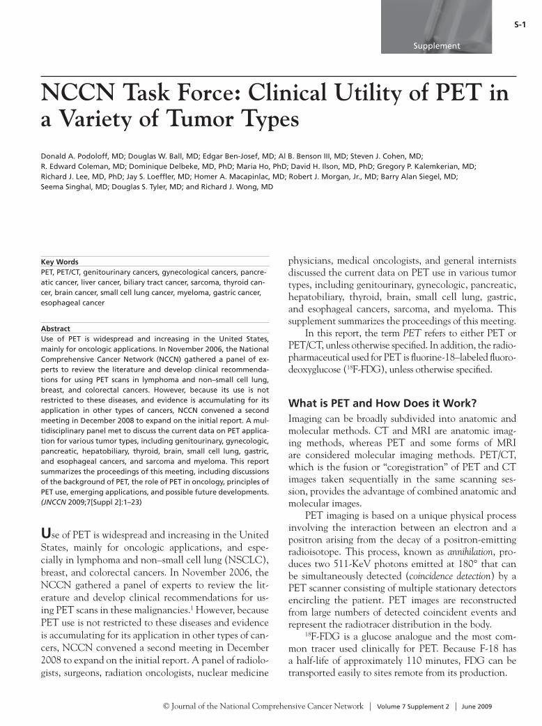
Transport of glucose and FDG from the blood-stream into the cell is mediated by facilitative glu-cose transporters (especially GLUT-1). FDG is phosphorylated into FDG-6-phosphate (FDG-6P) by hexokinase, paralleling the conversion of glucose into glucose-6-phosphate in the glycolytic path-way. However, the substitution of fluorine for the 2-hydroxyl group of glucose blocks further metabo-lism of FDG, leaving FDG-6P trapped in the cell. The level of FDG uptake reflects the rate of FDG-6P trapping (Figure 1). Like most other imaging tech-niques, PET is minimally invasive.
Standardized Uptake ValueA semiquantitative measure, the standardized up-take value (SUV), is most commonly used to assess the uptake of the tracer to control for variations in body weight. Because of controversy regarding the best methodology for assigning measurement regions in tumor images, the maximum SUV (SUVmax) is generally a better parameter than the average SUV. The SUV is calculated using the following formula:
Activity per unit volumeInjected activity/body weight
PET and PET/CTAn estimated 1800 PET and PET/CT scanners are currently available in the United States, with ap-proximately 80% of these PET/CT. The original im-petus for combining PET/CT scans was to improve attenuation correction and throughput associated
FDG-6P18
Vascular Cancer Cell
Angiogenesis, bFGF,PDGF-BB, EGFR
FDG1818FDG
GLUT-1
HIF1a(hypoxia)
K1
K2
Glycogen
18FDG-1-P
K3
K4
Hexokinase
Glucose-6-phosphatase
18FDG-6–phospho-glucono-lactone
HMPshunt
18F-fru-6-P
Glycolysis
Figure 1 FDG uptake in a cancer cell. Source: Podoloff DA, Advani RH, Allred C, et al. NCCN Task Force Report: Positron Emission Tomography (PET)/Computed Tomography (CT) Scanning in Cancer. J Natl Compr Canc Netw 2007;5(Suppl 1):S1–S22.

Supplement
Clinical Utility of PET
© Journal of the National Comprehensive Cancer Network | Volume 7 Supplement 2 | June 2009
S-3
progression or proliferation, such as the multifunc-tional Akt signaling pathway.14 Additionally, elevat-ed expression of GLUT proteins has been described in many cancers, which can further enhance FDG uptake.15 However, various benign pathologies, such as trauma, infection, noninfectious inflammatory diseases, and some benign tumors, can cause false-positive PET findings.
Evidence shows that elevated FDG uptake is associated with poor prognosis in various cancers with widely varying biology and treatment.16–19 For instance, a retrospective review of 400 patients with iodine-refractory thyroid cancer indicates that those with positive PET scans have significantly worse sur-vival than those with negative scans (P < .001).19
National Oncologic PET Registry and Research IssuesCoverage for clinical use of PET in oncology varies among third-party payers, but development of cover-age policies has been dominated historically by the Centers for Medicare & Medicaid Services (CMS) for the Medicare program. Starting from January 2005, PET scans were covered by Medicare (Table 1) for diagnosis, staging, and restaging for esophageal, head and neck, NSCLC, and colorectal cancers, and lymphoma and melanoma (excluding regional lymph node evaluation). Reimbursement for PET also was approved for specific indications in breast, cervical, and thyroid cancers. Coverage for all oth-er cancers and indications (except those explicitly non-covered) required participation in the Cover-age with Evidence Development (CED) program. In response to this CMS policy, the Academy of Mo-lecular Imaging in collaboration with the American College of Radiology Imaging Network developed a CED program known as the National Oncologic PET Registry (NOPR). Partly due to data gathered by the NOPR, in April 2009 CMS announced a new coverage framework for PET to combine diagnosis and staging into “initial treatment strategy,” and re-staging and treatment monitoring into “subsequent treatment strategy” (Table 1).20 This new national coverage determination expanded coverage to lift the CED requirement for initial treatment evalua-tion for nearly all tumors, while maintaining data collection for subsequent treatment evaluations for a range of solid tumors.
Open since May 2006, the NOPR is a nation-wide prospective medical registry designed to sys-
tematically collect clinical and demographic data on the usefulness and impact of PET in previously noncovered cancer types and indications.21 The main goal of the NOPR is to evaluate the impact of PET on physicians’ plans for patient management. Providers are required to submit data from pre- and post-PET physician questionnaires to the NOPR as a condition of reimbursement for the PET study.
At the end of its first year of operation, the NOPR published results from nearly 23,000 scans performed in more than 21,000 patients at 1178 cen-ters.22 Of these, 24% were for cancer diagnosis, 28% for initial staging, 24% for restaging after treatment, and 24% for evaluation of suspected recurrence. Studies performed for treatment monitoring during cancer therapy were excluded from this analysis. The investigators reported that PET resulted in a change in intended management (classified as treatment or nontreatment) in 36.5% of cases.
In a subsequent study, the NOPR investiga-tors reported on the impact of PET in patients with pathologically proven cancer of known origin to evaluate whether important differences were pres-ent as a function of cancer type. This study included results from nearly 41,000 scans performed in more than 34,000 patients at 1368 centers.23 Of these, 35% were for initial staging, 36% for restaging af-ter treatment, and 29% for evaluation of recurrence. The investigators reported that PET resulted in a change in intended management in 38.0% of cases overall; results were provided for 18 specific cancer types, and ranged from 31.4% for non-melanoma skin cancer to 48.7% for myeloma (Table 2). Most of these changes were from nontreatment to treatment (30%) rather than vice versa (8%), perhaps because of PET’s capacity to detect unsuspected lesions. In many cases, additional imaging such as CT or MRI was indicated as the initial management plan, and this may have caused overestimation of the impact of PET. To account for this, an imaging-adjusted im-pact was calculated by excluding these cases from the numerator but leaving them in the denominator (i.e., assuming no benefit from PET in these cases). This adjusted impact ranged from 9.6% for non-melanoma skin cancers to 16.2% for ovarian cancer (overall, 14.7%). The true impact is likely between the unadjusted and adjusted rates.
Notably, although the greatest number of scans was performed for prostate cancer, this is attributable

Supplement
NCCN Task Force Report
© Journal of the National Comprehensive Cancer Network | Volume 7 Supplement 2 | June 2009
S-4
their relevance amidst rapid advances in imaging technologies. Such rapidity and breadth is difficult to achieve in a prospective randomized trial. De-spite these strengths, however, several confounding factors and limitations are present.24 In contrast to randomized studies, a registry analysis is observa-tional by nature, with inevitable potential bias. For example, physicians who participated may have the preconception that PET will change their clinical decisions. Furthermore, no control group was present to compare the impact of PET with that of current standard tests. Because data were derived from self-completed questionnaires, accuracy will vary, and whether the intended change will result in an actual change in management remains unknown.
A recent series of Australian studies25–28 under-took a similar questionnaire format but with addi-tional follow-up of 12 months on 4 cancers currently reimbursed by Medicare (head and neck, lymphoma, colorectal, esophageal). They reported that treat-ment plans were implemented in more than 90% of cases. The NOPR investigators seek to adopt
to the high incidence (and prevalence) of the disease rather than a high frequency of PET use. Adjusting for disease rate, the use per incident cancer was only 3% for prostate cancer compared with 38% for ovar-ian. It is thought-provoking that the NOPR finds little variation in impact across cancer types despite apparent variation in clinical value. One possible explanation is that physicians are selective and only order PET when it is most likely to be useful. For example, prostate cancer is known to generally have low FDG avidity until it becomes castrate-refractory; thus, physicians may be using PET only in selected cases to help resolve clinical dilemmas. However, this lack of variation in impact may also reflect phy-sician overconfidence and misconception about the usefulness of PET.
The NOPR has an impressive population size (> 130,000 cases as of March 2009, with approxi-mately 88% consenting for research use of data) col-lected from a large fraction (approximately 80%) of PET facilities nationwide. Data were analyzed and reported in a timely manner, which heightened
Table 1 Medicare Coverage of PET in CancerPrevious Framework (as of January 2005) New Framework (as of April 2009)
Cancer Type Diagnosis Initial Staging
Restaging (and
Suspected
Recurrence)
Treatment
Monitoring
Initial Treatment
Strategy
Evaluation
Subsequent
Treatment
Strategy
Evaluation
Breast NC Covered* Covered Covered Covered† Covered
Cervix CED Covered‡/CED CED CED Covered‡/CED Covered
Colorectal Covered Covered Covered CED Covered Covered
Esophagus Covered Covered Covered CED Covered Covered
Head and neck Covered Covered Covered CED Covered Covered
Lymphoma Covered Covered Covered CED Covered Covered
Melanoma Covered Covered§ Covered CED Covered§ Covered
Myeloma CED CED CED CED Covered Covered
NSCLC Covered Covered Covered CED Covered Covered
Ovary CED CED CED CED Covered Covered
Prostate CED CED CED CED NC CED
Thyroid Covered Covered Covered¶ CED Covered Covered¶/CED
All other solid tumors CED CED CED CED Covered CED
Abbreviations: CED, coverage with evidence development; NC, non-covered; NSCLC, non–small cell lung cancer. *Non-covered for initial staging of axillary lymph nodes.†Non-covered for diagnosis and/or initial staging of axillary lymph nodes. Covered for staging of metastatic disease.‡Covered for initial staging with negative conventional imaging for extrapelvic metastasis. All other uses are CED.§Non-covered for initial staging of regional lymph nodes. Other uses for initial staging are covered.¶Covered for restaging of previously treated cancers of follicular cell origin with negative I-131 whole-body scintigraphy and rising thyroglobulin ( > 10 ng/mL).

Supplement
Clinical Utility of PET
© Journal of the National Comprehensive Cancer Network | Volume 7 Supplement 2 | June 2009
S-5
this approach and relate their findings to CMS bill-ing records to assess the impact of PET on actual management change.
More importantly, even when management is changed, whether this change will benefit the patient remains to be elucidated. As Mol et al.29 pointed out, the practical value of a diagnostic test such as PET ultimately relies on how it affects health measures, including survival, quality of life, toxicity, and symptom relief, through its impact on treatment decisions. Most research has focused on assessing the test characteristics of PET (i.e., sensitivity, specific-ity, and accuracy). However, the clinical context can undermine the usefulness of even a highly ac-curate scan. For example, therapeutic options may be limited for some advanced cancers, and sensitive or early detection of residual disease will not result in improved outcome; it may even cause unnecessary or prolonged anxiety in some patients. Although the NOPR looked beyond test characteristics, an impact on intended or actual change in management may not always translate to clinical advantage, particu-larly when consensus is lacking on the optimal man-agement of the disease. The difficulty in assessing the indirect impact of any diagnostic test on outcome is a general problem in oncology given the complex nature of cancer and the individualized factors that can contribute to treatment response.
Prospective trials randomizing patients to under-go or skip PET are still the most direct ways to jus-tify the clinical role of this technique. Admittedly, rigorous research data usually are not available even for existing conventional tests. Nonetheless, an in-creasing number of randomized studies have recently shown the clinical impact of PET imaging.
One established area is the use of PET in dese-lecting patients with suspected NSCLC for curative surgery that was reportedly unsuccessful in up to 50% cases. Van Tinteren et al.30 randomized 188 patients to conventional workup with or without PET before surgery. The PET arm showed a much lower rate of futile thoracotomy (21%) compared with the con-trol (41%). Another recently completed random-ized study on 337 patients echoed these findings.31 In another trial on NSCLC that randomly assigned 465 patients to either traditional workup or up-front PET,32 initial PET resulted in fewer invasive proce-dures without compromising staging accuracy or rais-ing costs.
Ideally, more sufficiently powered trials will be conducted for other cancer types and to address questions other than those pertaining to the surgi-cal setting. For example, a French group randomized 130 patients who had undergone curative therapy for colorectal cancer to either conventional or PET-based follow-up.33 They reported that follow-up with PET allowed earlier detection of recurrence (12.1 vs. 15.4 months; P = .01) and improved cure after surgery compared with conventional follow-up (10 vs. 2 patients). However, randomized studies such as these may be difficult to undertake for an imag-ing technique because of potential ethical issues and physician preconceptions.
Alternatively, PET and treatment can be per-formed on all patients, and PET findings compared with the treatment outcome. This design has been incorporated within a Dutch multicenter random-ized study on surgery with or without preoperative neoadjuvant chemoradiation in esophageal cancer.34 In the neoadjuvant arm, PET and CT will be per-formed before and during chemoradiotherapy. All patients complete therapy and surgery regardless of results. Subsequent analysis will then seek to com-pare the capacity of PET and CT for predicting non-response to chemoradiation. Furthermore, survival
Table 2 Impact of PET on Intended Management of the Top 10 Cancers in the National Oncologic PET Registry
CancerNo. of Scans
% Change in Intended Treatment
% Imaging-Adjusted Impact
Prostate 5309 35.1 15.0
Ovary 4509 41.4 16.2
Bladder 3578 37.9 15.4
Pancreas 3314 39.0 14.8
Stomach 3025 36.9 14.5
Small cell lung 2983 41.2 13.1
Kidney 2877 35.8 16.0
Uterus 2869 36.5 15.1
Myeloma 1784 48.7 11.5
Connective tissue
1350 36.4 13.6
Adapted from Hillner BE, Siegel BA, Shields AF, et al. Relationship between cancer type and impact of PET and PET/CT on intended management: findings of the national oncologic PET registry. J Nucl Med 2008;49:1928–1935.

Supplement
NCCN Task Force Report
© Journal of the National Comprehensive Cancer Network | Volume 7 Supplement 2 | June 2009
S-6
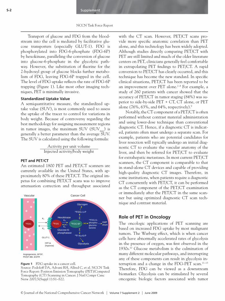
sues. Patients must fast for 4 to 6 hours (Figure 2, top panel) and avoid strenuous exercise for 24 hours to reduce uptake in skeletal muscle. They should also be adequately hydrated to facilitate clearance of ex-creted FDG from the urinary tract. Because various benign features (e.g., some benign tumors, inflamma-tory and infectious lesions) and normal tissues (e.g., brain, gastrointestinal, and genitourinary tracts) can also accumulate FDG, physicians must take these into account when analyzing imaging results.
Patient history is equally important. For exam-ple, scanning conditions and medications may need to be adjusted for patients with diabetes undergoing PET; both hyperglycemia and insulin effects can lead to reduced tumor FDG uptake. Physicians must note other concurrent medications, such as granulocyte colony-stimulating factor, hormonal therapy, and chemotherapy, that may also influence scan findings.
Physicians ordering a PET/CT must be aware that it does not replace a diagnostic CT scan (Figure 2, bottom panel). The CT component of a PET/CT adds anatomic accuracy to molecular imaging com-pared with PET alone, but the PET component of a PET/CT does not make it superior to a diagnostic CT. Compared with a diagnostic CT, very small le-sions may be missed on PET/CT because of the omis-sion of contrast material or lack of full inspiration of the CT component. For example, micronodular me-tastasis to the lungs, common in patients with thy-roid cancer, may only be detectable with a breath-holding diagnostic CT with full inspiration. PET/CT and diagnostic CT serve different purposes and indications cannot be applied interchangeably.
Panelists expressed concerns about the potential overuse of PET. Physicians should avoid ordering scans routinely if results are not likely to influence management. For example, in patients with wide-spread metastasis, finding additional scattered sites of disease using whole-body PET usually will have no impact on treatment decisions; thus, PET should not be performed simply to refine the assessment of disease extent. However, PET may be appropriate in these cases for a different purpose, such as establish-ing a baseline for treatment monitoring before start-ing an expensive therapy.
The NOPR reported a surprisingly high rate of cancellation of planned biopsy (75%) after PET.22 Physicians also expressed that PET results allowed them to avoid additional procedures or tests in 77%
and costs associated with PET or CT will be com-pared with those for patients who had no imaging prediction. This trial design may be more helpful in validating PET use in oncology than one that’s truly randomized.
Issues and Concerns in Clinical UsefulnessPET is a noninvasive and sensitive imaging method for detecting metabolic changes in cancer. However, it is also expensive and has limitations, such as false-positive results from tracer uptake in normal tissues and benign lesions. As with any other advanced technology, challenges and concerns inevitably arise with the ever-increasing use of PET in the clinical setting. In particular, protocols of PET imaging have not yet been standardized, and both the method of performing PET and interpretation of the findings vary among cancer centers and clinical sites. The panel agreed that health care professionals consid-ering PET must be alert to several important issues applicable to all types of cancers (Table 3).
Optimal and appropriate use of PET requires meticulous attention to technique. Proper patient preparation is essential, because PET is a sensitive measure of real-time metabolism of the body.35 Care should be taken to minimize tracer uptake in normal tissues while maintaining uptake in target tumor tis-
Table 3 Principles of PET Interpretation and Use in Oncology• PET/CT has an additive anatomic component and
is generally considered more informative than PET alone.
• PET/CT and diagnostic CT are tests that serve different purposes. Patients must undergo separate scans where indicated; however, these may be performed on the same PET/CT scanner with the diagnostic CT performed as the CT component of PET/CT or immediately after PET/CT.
• PET is best used as an adjunct in diagnosis and has not been shown to be a definitive test by itself. PET should not be performed if results are not anticipated to change management.
• PET must not be used in lieu of a biopsy to establish a diagnosis of cancer.
• Extrapolation of evidence from the advanced setting to early-stage disease should be avoided.
• No patient should be denied potentially curative therapy based on PET or any other imaging findings alone. Patients with stable disease should not undergo therapy based on PET findings alone.
• PET as a surveillance tool should only be used in clinical trials.
Supplement
Clinical Utility of PET
© Journal of the National Comprehensive Cancer Network | Volume 7 Supplement 2 | June 2009
S-7
of the cases. Although these observations may poten-tially reflect a positive impact in avoiding the risks and costs of biopsies and other procedures, they can also indicate overconfidence in PET findings among the general medical community, and that many phy-sicians see PET as the final arbiter that completes patient evaluation and decides treatment. Likewise, the lack of variation in apparent impact on manage-ment decisions across various different tumors can be interpreted as either high selectivity on the physi-cians’ part in applying PET scans or overestimation of the significance of PET.
However, although PET is an established tech-nology past the experimental stage, its clinical role in many cancers is still evolving and its usefulness can vary widely among different types of cancer. Most evidence indicates that PET is best used as an adjunctive imaging technique to conventional tests. Biopsy remains the gold standard in confirming tumor presence and must not be conveniently avoided or replaced by PET. Decisions of treatment or nontreat-ment should always be based on the combination of test results and the patient’s overall situation, rather than PET findings alone. Specifically, PET results should not be the sole reason for deciding against potentially curative therapy. Similarly, a single posi-tive PET finding is not sufficient to initiate therapy if patients seem to have stable disease otherwise.
Physicians also should be aware that data sup-porting a definitive role for PET in disease surveil-lance are still lacking, and therefore exploratory use should be restricted to well-designed clinical trials. Care should also be taken to distinguish evidence supporting its use in late- versus early-stage disease. A prime example is breast cancer, for which PET is sensitive in detecting recurrent and metastatic dis-ease but insufficient to replace surgical staging of the axilla in early-stage disease.36 Physicians should therefore avoid inappropriate extrapolation of data that may result in overuse of PET.
Undeniably, PET is emerging as a very useful test that can improve decision-making in oncology. However, potential abuse or misuse can also arise with its increasingly widespread use. In this respect, large registries like the NOPR provide timely data that allow monitoring of general conception and practice patterns in the medical community. Physi-cian education regarding the appropriate use of PET is critical to maximize the value of this technology.
Brain CancerDespite early exploration of PET imaging in brain cancers, the literature still reflects a paucity of defini-tive data on its clinical efficacy. Historically, PET has been used in grading and prognosis, with high FDG uptake generally correlating with higher grade and shorter survival.37–39 Most brain tumors can be effec-tively visualized with MRI, but PET may be useful in nonenhancing tumors. In a study of 28 patients with low-grade gliomas, increasing FDG avidity, as opposed to low avidity, indicated anaplastic transfor-mation and predicted poor outcome (2-year survival, 33% vs. 100%).38
Figure 2 Potential misinterpretation and limitation of PET. (Top) PET scan on a patient is dramatically different after eat-ing (left, note avidity of muscles) from that after proper fasting (right). (Bottom) The mid-inspiration CT component of PET/CT (left) provides less detail than a diagnostic CT with full inspiration (right). Contrast is used for both images.
Supplement
NCCN Task Force Report
© Journal of the National Comprehensive Cancer Network | Volume 7 Supplement 2 | June 2009
S-8
agnostic PET imaging in gastric cancer, with as much as half of primary tumors being FDG-negative.53–56 Nonetheless, in patients with FDG-avid tumors, PET may detect metastatic disease not identified by other imaging modalities. In contrast, 95% of prima-ry esophageal tumors were FDG-avid.57 PET is much more sensitive than CT and endoscopic ultrasonog-raphy in detecting stage IV disease (74% vs. 47%) with distant lymph node involvement. Meta-analy-ses attributed to PET a 67% pooled sensitivity, 97% specificity, and small added value after conventional staging in detecting distant metastasis.58,59 Based on its efficacy, PET is approved by Medicare for both initial and subsequent treatment strategy evaluation of esophageal cancer.
Recent research has generated strong interest in the ability of PET to assess response and predict out-come to neoadjuvant therapy.60 In 2 studies involv-ing 36 and 39 patients with esophageal cancer, re-sponse to preoperative chemoradiation as defined by PET was strongly correlated with prognosis.61,62 Re-sponse assessment seems most valuable for induction chemotherapy in patients eligible for potentially cu-rative resection. Among patients with gastric cancer undergoing preoperative neoadjuvant chemothera-py, Ott et al.63 showed a superior 90% 2-year survival in those experiencing PET-defined response (> 35% decline in SUV) compared with 25% in those ex-periencing no response. Response can be predicted with PET as early as 14 days into treatment. Because more than 60% of patients have unresponsive dis-ease, they may be spared further unnecessary toxic therapy after early assessment with PET.
The same group reported similar findings in esophageal cancer, with PET unresponsiveness cor-relating to shorter time to progression and overall survival.64 They further investigated 119 patients in the MUNICON trial to assess a PET response–guid-ed therapeutic algorithm.65 Patients for whom PET showed no response to platinum- and fluorouracil-based induction chemotherapy at day 14 were sent for immediate surgery, whereas those who did show response completed 3 months of therapy before resec-tion. Again, a median follow-up of 2.3 years showed dramatic survival benefit for responders (hazard ratio, 2.13; P < .015). Notably, in patients experiencing no response, this trial showed that stopping chemother-apy early did not seem to reduce long-term survival compared with continuing treatment in the previous
The treatment paradigm has recently shifted to-ward concomitant use of radiation and temozolomide for glioblastoma.40 Pseudo-progression and radiation injury can occur with concurrent radiation + temo-zolomide use, which can obscure MRI findings and hinder response assessment.41 FDG-PET has a rela-tively high sensitivity (80%–86%) for distinguishing radiation injury from high-grade tumor recurrence; the specificity ranges from 80% to 100% in 7 stud-ies totaling 241 patients, although in 2 smaller studies of 19 and 20 patients, respectively, it was only in the 40% to 63% range.42 Correlation with MRI findings is critical for optimal interpretation of PET images.43–45 Causes of false-negative PET studies include recent radiation therapy, low histologic grade, and small tumor volume. False-positive PET findings may oc-cur in inflammatory processes and subclinical seizure activity. Technical improvements have shown some success, such as delaying time to imaging,46 MRI cor-relation, and the development of amino acid tracers (which currently are only for investigational use).47
PET also has an emerging role in radiotherapy; it has been used to delineate tumor activity and target volume for radiation planning in gliomas.48,49 Latest research interest is turning toward PET verification of dose distribution in the growing field of proton beam therapy (Figure 3).50–52
ConclusionsMRI is still the gold standard for diagnosing and staging brain cancers, but PET may be useful in identifying nonenhancing, low-grade gliomas under-going malignant conversion. A negative PET scan is helpful in excluding recurrent anaplastic astro-cytoma and glioblastoma multiforme. PET is useful for differentiating radiation effect from tumor recur-rence, is a good predictor of survival in high-grade recurrent gliomas, and can guide biopsy to the site of maximum uptake. PET shows promise in aiding radiation planning and dose confirmation. With the rapid expansion of proton beam treatment centers, PET may become more commonly used as an in vivo dosimetric tool for radiation therapy.
Gastric/Esophageal CancerAlthough the incidence of gastric cancer is declin-ing, that of esophageal adenocarcinoma is increasing, particularly for tumors of the distal esophagus and gastroesophageal junction. Results are mixed on di-

Supplement
Clinical Utility of PET
© Journal of the National Comprehensive Cancer Network | Volume 7 Supplement 2 | June 2009
S-9
trial. Additionally, early PET-defined response corre-sponded with a high rate of histologic remission. An ongoing phase II study66 is adopting a similar PET-based approach in predicting the efficacy of induction chemotherapy followed by chemoradiation before sur-gery on potentially resectable esophageal and gastro-esophageal junction cancers.
ConclusionsThe role of PET in the primary imaging of gastric cancer remains to be established, but it is valuable in detecting advanced disease for gastric and esopha-geal cancer. Recent evidence shows that PET pro-vides an exciting opportunity to accurately predict early which patients for whom induction therapy is likely to fail, thereby sparing them from futile, toxic treatment and directing them to potentially helpful salvage therapies.
Genitourinary CancersA major challenge for PET in genitourinary oncol-ogy is the physiologic urinary excretion of FDG,
which can significantly mask detection of localized prostate and bladder cancers. Urinary activity, how-ever, can be minimized by good hydration, use of di-uretics, and bladder catheterization. Another prob-lem is the variable FDG uptake among genitourinary malignancies (e.g., low glycolysis of prostate tumor cells). Other tracers are being investigated to over-come these problems.
Diagnosis and Initial StagingBecause studies have not shown FDG to be reliable for diagnosing or initial staging of prostate cancers, Medicare has recently determined that PET is not covered for these purposes. Reports show significant overlap between benign prostatic hyperplasia, ma-lignant tumor, local recurrence, and postoperative scarring.67,68 Sensitivity may be as low as 4% because of urinary excretion of FDG.69 Similarly, urinary ex-cretion limits diagnostic use of FDG in bladder and kidney cancers, unless diuretics and/or bladder cath-eterization are used to minimize physiologic activity. Although FDG-PET exhibited equally high specific-ity (100%) as abdominal CT in kidney cancer, it had much lower sensitivity than CT (60% vs. 92%).70
Figure 3 PET verification of radiation dose distribution in brain tumor. The PET/CT measurement (left) is comparable to the planned dose (right).

Supplement
NCCN Task Force Report
© Journal of the National Comprehensive Cancer Network | Volume 7 Supplement 2 | June 2009
S-10
ventional imaging techniques (i.e., MRI/magnetic resonance spectroscopy, CT, and bone scintigraphy). Although 11C-choline is generally considered more reliable than FDG in restaging localized disease in prostate and bladder cancers, it has limited broader application because of its short half-life and investiga-tional status (see section on “Emerging Applications and Future Direction”). Because of its high specificity but low sensitivity, PET may be most useful in resolv-ing diagnostic dilemmas in advanced disease.
Gynecologic CancersResearch on PET usefulness in detecting gynecologic malignancies has not been extensive, with most data involving cervical cancer and the fewest involving uterine endometrial cancer. The role of PET in ovar-ian, cervical, and uterine cancers differs because of the varying nature and course of these diseases.
Diagnosis and Initial StagingBased on findings that PET is superior in evaluat-ing lymph nodes, in 2005 CMS approved coverage of PET for initial staging in patients with cervical cancer for whom conventional imaging methods (CT or MRI) showed no evidence of extrapelvic metastasis. In a larger study of 135 patients with lo-cally advanced or recurrent cervical cancer, PET has greater sensitivity than MRI/CT for detecting pelvic (88% vs. 75%) and para-aortic (95% vs. 72%) lymph node involvement.82 Lin et al.83 performed PET on 50 patients with negative abdominal CT scans and found that 12 had para-aortic lymph nodal metas-tasis, confirmed with histology. Additionally, PET has prognostic value in cervical cancer; patients with high pretreatment tumor SUV have worse disease-free survival rates.84
Diagnostic use of PET in suspected ovarian cancer has been investigated in a prospective study by Risum et al.85 In this study, PET scans were per-formed within 2 weeks before standard debulking surgery in 101 patients with a suspicious pelvic mass. The authors reported a high diagnostic sensitivity and specificity of 100% and 93%, respectively, al-though 7 PET-negative borderline ovarian cancers were categorized as benign. However, unlike cervical cancer, the usual late presentation of ovarian cancer generally limits the practical value of PET in initial evaluation. More than 80% of the cases are found at stage III or IV, with patients typically presenting
Other potentially more helpful tracers are being studied. For example, a small study of 18 patients showed 11C-choline uptake in all primary bladder tumors tested.71
Restaging and Metastasis DetectionFDG-PET has limited usefulness in detecting pros-tate cancer metastasis, except in castration-resistant disease, in which several panelists report from their experience at large cancer institutions that FDG-PET has high sensitivity for detecting distant me-tastases.72 Furthermore, in this subset of patients, FDG-PET has prognostic significance and may alter treatment intensity and duration. Although FDG-PET is possibly more useful in the distant metastatic setting,73 with a low false-positive rate, a negative scan does not exclude metastatic disease. For ex-ample, a study of 24 patients showed that FDG-PET had higher specificity but lower sensitivity than bone scintigraphy.74
Few studies are available on the usefulness of PET in restaging bladder cancer recurrence, but evi-dence suggests a role in detecting metastasis. In a study of 27 patients using histopathologic findings as reference, 11C-choline was more accurate than CT in detecting lymph node metastasis.75 In another study, FDG-PET was complementary to CT in finding positive lymph nodes in invasive bladder cancer.76 Use of diuretics was reported to improve detection of locally recurrent disease.77 However, the sensitiv-ity of FDG-PET may decline in patients who have undergone chemotherapy.78
Studies in kidney cancer have been focused on FDG. FDG-PET was used by Safaei et al.79 to cor-rectly restage 32 of 36 patients (89%) with advanced disease. FDG-PET was also more specific in visual-izing distant metastasis than bone scintigraphy and chest CT in trials, including 18 to 66 patients, al-though sensitivity was variable (64%–100%).70,80,81
ConclusionsEvidence supporting a routine role for FDG-PET in genitourinary cancers is lacking. However, FDG-PET may be indicated in castration-resistant metastatic prostate cancer, in which it has been reported to have high sensitivity for detecting distant metastasis. FDG-PET may also be considered in detecting metastasis in kidney cancer and muscle-invasive bladder cancer. Currently, FDG-PET should only be considered as an adjunct to, and not a replacement for, other con-

Supplement
Clinical Utility of PET
© Journal of the National Comprehensive Cancer Network | Volume 7 Supplement 2 | June 2009
S-11
with extensive symptoms. In the remaining 15% to 20% of patients with clinical stage I or II disease, upstaging after surgical exploration primarily detects small volume involvement (< 1 cm) in para-aortic nodes, which is undetectable using current PET technology. Most patients require up-front debulk-ing surgery, which minimizes the value of imaging for diagnostic and staging purposes.
Although uterine cancer is usually diagnosed at an early stage, it has the same tendency as ovarian cancer to spread as small nodal deposits for which PET has very low sensitivity. In a study of 30 pa-tients, Suzuki et al.86 found that preoperative FDG-PET detected none of 5 cases of lymph node involve-ment of 0.6 cm or less. PET was more sensitive than CT or MRI in visualizing non-nodal extrauterine lesions or the primary lesion86,87 but, similar to ovar-ian cancer, the problem is that up-front surgery is in-dicated for staging and treatment of uterine cancer. PET imaging does not currently preclude the need for surgical staging.
RecurrencePET has significant value in diagnosing recurrent cervical cancer and restaging after chemoradia-tion.88 In contrast to patients with ovarian or uterine cancers, those with cervical cancer experiencing a pelvic recurrence without extrapelvic disease have nearly a one third chance of long-term disease-free survival with pelvic exenteration. Because this is a highly invasive procedure, determining the presence of extrapelvic metastasis is important before making a clinical decision. In studies examining recurrence detection, the sensitivity and specificity of PET ranged from 76% to 100% and 57% to 100%, respec-tively.89 Alteration in treatment plans based on PET results has been reported. Yen et al.90 found that of the 55 patients whose recurrences were initially con-sidered potentially curable, 36 (66%) experienced treatment modifications after PET, with 27 undergo-ing palliative therapy instead of aggressive surgical treatment, which would not be beneficial in patients with distant metastatic disease (Figure 4). In a more recent prospective study of 20 patients with recur-rence, PET was 100% sensitive and 73% specific in detecting extrapelvic metastasis, which would obvi-ate recommendations for pelvic exenteration.91
Several trials have shown PET’s ability to detect recurrence in ovarian cancer. One larger study of 90 patients showed that FDG-PET was superior to con-
ventional imaging in verifying recurrence in patients, followed by CA-125.92 In combination, PET and CA-125 have 98% sensitivity. Similar results were found in a small trial of 22 patients with negative or indeterminate CT scans, in whom PET has very high overall sensitivity and specificity (95% and 100%, re-spectively) for assessment.93 The use of PET seems to have a significant impact on clinical decision-making, resulting in changes in management strategies for 44% to 58% of patients.94,95 The question remains as to whether these changes are beneficial in the recur-rence setting, given the lack of consensus on the best management. CMS recently approved coverage of PET for both initial and subsequent treatment strat-egy evaluation of ovarian cancer.
Data are scarce for uterine cancer. In one study of 90 women, Kitajima et al.5 found that PET had better sensitivity and specificity than CT for assessing re-currence of uterine cancer. A change of management based on PET findings was also reported for 42% of the patients. Another study reported that 70% of the 33 PET scans performed after recurrence/salvage therapy had a positive impact on management.87
ConclusionsAmong gynecologic malignancies, PET efficacy is best supported for initial staging of patients with cervical cancer who are to undergo chemoradiation. Approximately 7% are upstaged to stage IVb because PET detects unsuspected metastases in supraclavicu-lar lymph nodes. PET also delineates involved pelvic and para-aortic nodes, which is essential for proper radiation therapy planning. PET is also useful in the recurrent setting, in which some patients may ben-efit by avoiding unnecessary invasive surgery.
The technology assessment performed for CMS by the University of Alberta Evidence-Based Prac-tice Center showed substantial usefulness of PET in ovarian cancer.96 In uterine cancer, the reported impact of PET on disease management is not as substantial. Although PET has been shown to have improved sensitivity and specificity compared with conventional imaging, it does not preclude the stan-dard recommendation for initial surgery. PET may be helpful in confirming recurrence of ovarian can-cer in patients with elevated CA-125 levels, when the information would change subsequent diagnos-tic evaluation and/or management. Additionally, current PET technology is inadequate in detecting small nodal metastasis common in these patients.

Supplement
NCCN Task Force Report
© Journal of the National Comprehensive Cancer Network | Volume 7 Supplement 2 | June 2009
S-12
MyelomaIn multiple myeloma, imaging studies are critical to identify lytic bone lesions that may indicate active disease requiring treatment, but established tech-niques such as radiography (skeletal survey) and MRI have their limitations. Recently, increasing in-terest has been shown in exploring the diagnostic/staging value of PET compared with conventional imaging. PET was consistently found to be more sensitive than radiography in finding bone lesions. Additional lesions were reported in 23% to 57% of patients examined, frequently resulting in upstaging and change in management.97–100 In other studies in-volving 16 to 33 patients, MRI was able to detect much spinal disease not seen on PET, but the much larger field of view allowed PET to visualize lesions in other areas undetectable with MRI.100–102 Clearly, PET would be most useful when used in combination with other imaging tests, especially MRI.
A prognostic value has also been attributed to PET. Durie et al.103 reported a consistent correlation
of PET negativity to indolent plasma cell disease. PET also showed extramedullary uptake in 23% of patients experiencing relapse, which is associated with a poor prognosis. However, whether PET adds independently to prognosis when powerful prognos-tic factors such as cytogenetic abnormalities are also considered remains unknown.
ConclusionsPET is an informative test that has a potential complementary role to conventional imaging in the diagnosis and staging of multiple myeloma. CMS recently approved coverage of PET for both initial and subsequent treatment strategy evaluation of my-eloma. One key area for research is to investigate the correlation of FDG avidity with local disease activity using biopsy.
Pancreatic and Hepatobiliary CancersPET use is evolving in pancreatic and hepatobiliary malignancies. These cancers generally have a poor prognosis, with surgery the only potentially curative treatment. However, only a minority of patients are eligible for resection, and recurrence is common and typically incurable. These stark realities guide evalu-ation of PET efficacy in these diseases.
Diagnosis and Initial StagingIn pancreatic cancer, studies of 34 to 106 patients have consistently shown diagnostic accuracy for PET, which surpasses that of CT.2,96,104,105 In particu-lar, PET can differentiate malignant tumors from benign cysts or pancreatitis with 84% to 94% accu-racy.105,106 Because a diagnostic biopsy is performed for most patients, the clinical efficacy of FDG-PET/CT for diagnosis is questionable. However, although biopsy may provide a tissue diagnosis, this technique is associated with significant sampling error.107,108 FDG-PET/CT may represent a useful add-on diag-nostic tool in the evaluation of patients with sus-pected pancreatic cancer, especially when CT and biopsy results are inconclusive.109
PET may be more useful in staging. A recent study of 82 patients showed an improved detection sensitivity for metastases when combining PET/CT with standard CT (combined, 87%; PET/CT, 61%; CT, 57%).110 Detection of additional metastases resulted in a management change in 11% to 16% of patients.110,111
Figure 4 Efficacy of PET in recurrent cervical cancer. (Left) Three months after chemoradiation, a left neck mass was palpated in this patient. MRI and CT showed no definite ab-normal findings, but PET suggested nodal metastases confirmed by histopathology. She received palliation treatment instead of salvage radiation. (Right) Six months after chemoradiation, elevated tumor marker level was noted in this patient. Again, MRI and CT were negative, whereas PET disclosed lung metas-tasis. She received pneumonectomy and was well for 1 year. Source: Yen TC, See LC, Chang TC, et al. Defining the prior-ity of using 18F-FDG PET for recurrent cervical cancer. J Nucl Med 2004;45:1632–1639.

Supplement
Clinical Utility of PET
© Journal of the National Comprehensive Cancer Network | Volume 7 Supplement 2 | June 2009
S-13
In the fewer studies available for the less-com-mon biliary tract (gallbladder and bile duct) ma-lignancies, PET was generally found to be effective in visualizing primary tumors and distant metasta-sis,112–115 although false-positives were concerning for patients with cholangitis or biliary stents.116 Similar to pancreatic cancer, PET findings caused a change in primary treatment in 17% to 30% of patients be-cause it detected unsuspected metastases.112,114,116,117
The clinical picture may be different for hepa-tocellular cancer. The primary tumor generally has lower, more variable avidity for FDG, although PET is still effective in detecting 86% of metastatic le-sions.118,119 PET may have an increasing role in as-sessing the impact of liver-directed therapies, which are notoriously difficult to judge with conventional CT imaging. Wudel et al.119 reported that FDG-PET added clinically significant information in 26 of 91 patients (28%) as a result of metastasis detection and response assessment of hepatic-directed therapy.
Recurrence and Other ApplicationsSeveral studies have investigated PET for recurrence detection, but its usefulness may be limited because few options are available when these cancers recur. Thus, early detection of an incurable recurrent ma-lignancy with limited treatment options is unlikely to impact patient management. A study of 31 pa-tients with pancreatic cancer showed that PET was more sensitive than CT/MRI for local recurrence but significantly less sensitive for liver metastases.120 In a study of patients with biliary cancer, PET identified recurrence in 86% of patients but altered treatment in only 9%.112 For hepatocellular carcinoma, Chen et al.121 reported a 73% PET sensitivity in detecting recurrence in patients with rising alpha-fetoprotein but otherwise normal conventional examinations. Thus, PET may have higher value in assessing re-currence or persistent disease in patients with hepa-tocellular cancer, because additional liver-directed treatments may be considered.
PET probably has prognostic significance. Seo et al.117 reported a lower disease-free survival for pa-tients with cholangiocarcinoma cancer undergoing resection with high versus low SUV. Similarly in hepatocellular cancer, PET positivity was associated with shorter survival after liver transplantation122 or resection.123 Smaller reports suggest a potential role for PET in response monitoring,104,124 particularly for liver-directed therapy in patients with hepatocellu-
lar cancer,125–127 but more study and more-effective treatments are required to show clear benefit.
ConclusionsPET is most promising as an adjunct to standard staging tests for maximum metastasis detection to prevent unnecessary surgery. Although most primary pancreatic and biliary tract tumors are FDG-avid, hepatocellular cancers are not as much. In hepato-cellular cancer, PET may have an expanded role in recurrence assessment and evaluation of response to liver-directed therapy, because additional treatment options may be available for localized disease. FDG-PET/CT imaging may represent a useful adjunctive diagnostic tool for evaluating patients with sus-pected pancreatic cancer, especially when CT and biopsy results are inconclusive. Improved detection of recurrence in pancreatic and biliary cancers is less likely to be of clinical benefit. Similarly, PET is un-likely to be used for response assessment in pancre-atic and biliary cancers, given the limited efficacy of available treatments.
SarcomaData on the use of PET to distinguish between be-nign masses and sarcomas are variable, depending on the definition of malignancy and the type of sar-coma examined.128,129 With respect to staging, FDG-PET is clearly inadequate (sensitivity, 50%–87%) in detecting lung involvement compared with chest CT (75%–100%), but whole-body PET is useful in detecting extrapulmonary metastasis.130,131 PET is particularly helpful in Ewing’s sarcoma. FDG-PET alone detected osseous metastasis at a much higher sensitivity (100%) than conventional bone scin-tigraphy (68%).132 The hybrid PET/CT technique seems to further improve sensitivity in the staging of Ewing’s sarcoma.4
Most sarcomas respond poorly to therapy. In re-cent years, however, targeted agents such as imatinib and sunitinib have shown dramatic, albeit often temporary, tumor control for gastrointestinal stromal tumors (GISTs).133,134 The assessment of response to these agents is where PET proved to be a valuable tool. Figure 5 illustrates the rapid change in tumor metabolism shown by PET, without a corresponding change in lesion size on CT.135 FDG uptake signifi-cantly decreased in responsive tumors as early as 24 hours after the first dose of imatinib,133 and PET was

Supplement
NCCN Task Force Report
© Journal of the National Comprehensive Cancer Network | Volume 7 Supplement 2 | June 2009
S-14
much more accurate than CT in diagnosing response by 1 month (85% vs. 44%).136 Likewise, FDG-PET sensitively showed on and off tumor response to sunitinib as patients with imatinib-resistant GISTs underwent treatment cycles.137
Additionally, Prior et al.138 reported a significant correlation between SUV at week 4 and progression-free survival in a study of 23 patients on sunitinib. For borderline resectable tumors, timely treatment response assessment can be the key to choosing between a less-invasive local excision and major surgery associated with high morbidity. This issue is critical when the tumor is located in the gastro-esophageal junction, periampullary regions, and rectal/prostate areas.
ConclusionsPET may help complement conventional imaging in clarifying the disease stage of sarcomas, particularly in Ewing’s sarcoma. PET imaging has an exciting role in monitoring response of GISTs to targeted agents. Rapid assessment using PET may allow a valuable window of opportunity for important surgical deci-sions, especially for borderline resectable tumors in specific locations.
Small Cell Lung CancerEvidence of benefit from PET use in small cell lung cancer (SCLC) mainly came from small studies fo-cused on staging, which often included heteroge-neous populations and used suboptimal convention-al imaging algorithms for comparison. These studies included 18 to 120 patients with a cumulative stag-ing concordance of 87% (range, 73%–100%) be-tween PET and conventional imaging.139–147 Based on PET findings, approximately 15% of patients were upstaged from limited to extensive disease and 5% were downstaged. Because SCLC is an aggressive disease, it generally has high FDG uptake, leading to a sensitivity of nearly 100% for primary tumors. For most metastatic sites, PET was superior to standard imaging techniques with sensitivity of 97% to 100% and specificity of 78% to 96%. However, PET was inadequate in detecting brain metastases (sensitivity, approximately 45%) compared with cranial MRI or CT.139,140,143,147 NSCLC is more frequently associated with hypermetabolic metastatic brain lesions than SCLC (80% vs. 26.7%).148
Changes in management based on PET staging
were reported in 16% to 38% of patients in 3 studies (vs. 41% reported by NOPR23), primarily because of alterations in the radiotherapy field.139,143,147
Data on other applications such as prognosis and response monitoring are based on small num-bers of patients. One retrospective analysis showed that patients with positive PET findings had worse 2-year survival rates than those with negative find-ings (23% vs. 67%; P = .01) and that SUVmax in-versely correlated with survival.149 This information, however, is unlikely to have a significant impact on disease management.
ConclusionsRigorous prospective research is still needed to de-termine the overall efficacy of PET in SCLC. PET seems to improve staging accuracy, although patho-logic confirmation is still required for lesions that are upstaged. PET also seems to improve detection of intrathoracic sites of disease that can impact ra-diation planning in patients with limited-stage dis-ease. However, PET is not adequate for detecting brain metastases.
Thyroid CancerThe incidence of thyroid cancer has risen 2.4-fold over the past 30 years due to an increased detection of small papillary thyroid cancers, which represent 87% of all cases.150,151 These variants, together with the follicular and Hurtle cell subtypes, are classified as differentiated thyroid cancer (DTC), which has been the main research focus of PET in thyroid cancer. Medullary thyroid cancer (MTC), encompassing ap-proximately 3% of cases, has a less than well-defined role for PET than DTC.
Diagnosis and Initial StagingEmerging evidence shows PET provides effective diagnostic imaging for Hurtle cell thyroid cancer, which generally has low avidity to I-131.152,153 More generally, incidental focally high thyroid FDG up-take has been well documented in large studies, reporting 1.1% to 2.9% incidence in 1330 to 8800 individuals.154 A significant number of these—14% to 47%, depending on the fraction of suspected le-sions subjected to tissue biopsy—were confirmed to be thyroid cancer. In comparison, conventional imaging detected a higher rate of incidental nodules but lower rate of malignancy.155 Hypermetabolic thy-

Supplement
Clinical Utility of PET
© Journal of the National Comprehensive Cancer Network | Volume 7 Supplement 2 | June 2009
S-15
roid “incidentalomas” identified with PET should be further evaluated using thyroid ultrasonography and fine needle aspiration. However, because of lower cost and higher sensitivity, thyroid ultrasonography is the preferred modality for the initial evaluation of a thyroid nodule.
PET is less useful in MTC, but the sensitivity is 70% when calcitonin levels are greater than 1000 pg/mL.156 A larger study of 55 patients showed that PET was inferior to either ultrasonography, CT, or MRI for detecting disease at individual common metastatic sites, including neck, lung, mediastinum, liver, and bone.157
Recurrence and PrognosisFDG-PET sensitivity for recurrent DTC varied wide-ly, from 45% to 100%, improving at higher serum thyroglobulin (Tg) levels and in patients with more extensive metastatic disease.158 Because Tg is already a good marker for recurrence, the value of PET in this setting is generally restricted to localizing the residual disease, especially when I-131 scintigraphy is negative. Stimulation of thyroid stimulating hor-mone moderately increases the sensitivity for detect-ing FDG-positive lesions.159
In the posttreatment setting, PET has profound prognostic impact based on the fact that PET-posi-tive lesions are probably most metabolically active and aggressive. In a retrospective review of 400 pa-tients who underwent PET (50% with metastasis), Robbins et al.19 found that only old age and posi-tive PET findings continue to be strong predictors of short survival after multivariate analysis. Similarly, Wang et al.160 report volume of FDG-avid disease to be the single strongest prognostic factor. Moreover, of the 58 patients with distant metastasis, those who had positive PET findings had a significantly shorter survival than the group with negative findings.
Notably, FDG avidity and concomitant loss of I-131 uptake is known to be associated with dediffer-entiation,161 and several studies have used PET to de-tect metastasis in patients with negative I-131 scin-tigraphy and elevated Tg (Figure 6).162,163 Together, these findings favor PET use in selecting patients within this group who may require more vigilant follow-up or systemic therapy beyond radioactive io-dine. For poorly DTC or anaplastic thyroid cancer (ATC), FDG-PET may improve metastasis detection and alter therapy.164 Because of the generally poor prognosis of patients with ATC, no correlations be-
tween PET findings and prognosis are available. PET received CMS approval of coverage in 2003 for re-staging patients with previously treated thyroid can-cer of follicular cell origin, elevated Tg (> 10 ng/mL), and negative whole-body I-131 scintigraphy.
ConclusionsThe role of PET is limited for MTC, but expanding in DTC. Incidental discovery of focal FDG avidity may be helpful in identifying potentially malignant thy-roid nodules for diagnosis, but cost and sensitivity pre-clude its use as a frontline diagnostic tool. Research data best support using FDG-PET in established inter-mediate- and high-risk patients with DTC and those
Figure 5 PET can provide rapid evaluation of tumor response to targeted agents in sarcoma. Comparing scans of a patient before (A) and 1 month after (B) imatinib therapy, PET/axial PET slices showed dramatic shrinkage of the tumor mass, whereas the corresponding axial CTs (inset panels) showed no apparent treatment effect. Source: Van den Abbeele AD. The lessons of GIST—PET and PET/CT: a new paradigm for imaging. Oncologist 2008;13(Sup-pl 2):8–13.

Supplement
NCCN Task Force Report
© Journal of the National Comprehensive Cancer Network | Volume 7 Supplement 2 | June 2009
S-16
with ATC. PET is a powerful prognostic indicator in the metastatic setting. One emerging use is in assess-ing patients with high Tg and negative I-131 imaging, where a positive PET may be indicative of dedifferen-tiation associated with tumor aggressiveness, calling for modification in treatment plans.
ConclusionsTable 4 summarizes the panel conclusions on PET use in different cancers based on a review of the lit-erature. These are based on lower-level evidence and panel consensus was reached, corresponding to 2A category of NCCN recommendations. PET is gen-erally useful as an adjunctive imaging technique in detecting unsuspected metastasis. This information is especially valuable in deselecting patients from fu-tile, invasive treatment, such as pelvic exenteration in cervical cancer. PET also shows promise in assess-ing treatment response in gastric/esophageal cancer and GIST. Notably, however, PET has been found to be inadequate for disease detection in certain set-tings (e.g., brain metastasis of SCLC).
In some cases, panelists found the practical value of PET to be limited by the clinical circumstance.
For example, although PET can sensitively detect primary and recurrent ovarian tumors, its use is low in practice because initial debulking surgery is rec-ommended for most patients and opinions differ on the best management for recurrent disease. However, the technology assessment performed for CMS by the University of Alberta Evidence-Based Practice Center showed substantial efficacy of PET in ovarian cancer.96 The results of this technology report indi-cate that FDG-PET, especially when combined with CT, is a potentially useful tool for detecting recurrent ovarian tumors. A negative CA-125 and FDG-PET/CT is considered to rule out recurrent ovarian cancer and, in this clinical setting, can substitute for CT.
The essential message is that although the evi-dence is imperfect (and definitely indicates limited efficacy for certain cancers), the technology is now mature enough and its general use in cancer under-stood sufficiently for physicians to be empowered to use it as they think best for individual patients. More intelligent use will occur over time as more evidence for PET use in all types of cancer is developed. The continued development of new evidence by the NOPR should help accomplish this goal.
Emerging Applications and Future DirectionsInterest in measuring metabolic change to evaluate therapeutic success has recently increased. PET is an area of active reseach, as an early surrogate bio-marker based on the fact that alterations in glucose metabolism and thus FDG uptake in cancer tissues may sensitively reflect response to treatment before a gross reduction in tumor measurements. This can be especially useful in avoiding substantial side ef-fects of futile therapy or allowing a timely switch to another potentially more effective treatment. Esophageal cancer and GIST are 2 examples dis-cussed earlier, with several current trials using PET for response assessment. Similar trials are ongoing in a vast array of other cancers, including brain, breast, cervical, colorectal, head and neck, kidney, lung, and nasopharyngeal cancers and lymphoma and sarcoma. More research effort is needed to correlate PET find-ings to patient outcome for this application. Stan-dardized protocols and evaluation criteria for specific diseases, such as those developed for lymphoma,165 are also necessary to ensure reporting of quality data.
Figure 6 131I-negative thyroid cancer metastases are often FDG-avid. In contrast to 131I scan (left), FDG-PET scan (right) revealed extensive metastasis in this patient. Source: Feine U, Lietzenmayer R, Hanke JP, et al. Fluorine-18-FDG and iodine-131-iodide uptake in thyroid cancer. J Nucl Med 1996;37:1468–1472.

Supplement
Clinical Utility of PET
© Journal of the National Comprehensive Cancer Network | Volume 7 Supplement 2 | June 2009
S-17
Determining whether PET-guided adaptive treat-ment paradigms will lead to improved patient out-comes will likely require prospective, randomized, controlled trials.
A supplemental NOPR report discussed data collected on 10,497 PET scans performed for treat-ment monitoring, mainly for chemotherapy alone (82%).166 Overall, PET led to an intended change of
Table 4 Role of PET in Various Types of Cancer*Diagnosis/Staging Restaging/Recurrence
• Brain: may identify anaplastic transformation in nonenhancing, low-grade gliomas
• Gastric/esophageal: ! Gastric: not for diagnosis; potential use for metastasis
detection ! Esophageal: detection of advanced disease
• Genitourinary: FDG not for diagnosis; potential for adjunctive detection of metastasis
• Gynecologic: ! Cervical: detect nodal involvement ! Ovarian/uterine: limited use
• Myeloma: potential adjunct to MRI for detecting extraspinal lesions
• Pancreatic/hepatobiliary: ! Pancreatic/biliary tract: for diagnosis when other
imaging and biopsy are nondiagnostic, and adjunct in metastasis detection
! Liver: adjunct in metastasis detection, not for primary diagnosis
• Sarcoma: ! Ewing’s sarcoma: adjunct in staging ! Others: detecting extrapulmonary metastasis, not for
lung involvement• SCLC: potential adjunct in nodal/distant metastasis
detection, but not for brain metastasis• Thyroid:
! DTC: incidental discovery of suspicious nodes ! MTC: limited use
• Brain: differentiation of recurrence from radiation necrosis
• Gastric/esophageal: ! Gastric: unclear ! Esophageal: distant lymph node detection
• Genitourinary: limited use for local recurrence, possible use in detecting metastasis
• Gynecologic: ! Cervical: restaging to detect residual disease after
chemoradiation, presurgical detection of extra-pelvic disease (deselection for surgery)
! Ovarian: restage when CA-125 is elevated and CT normal
! Uterine: unclear• Myeloma: potential adjunct to MRI for detecting
extraspinal lesions• Pancreatic/hepatobiliary:
! Pancreatic/biliary tract: limited use ! Liver: potential use in assessing recurrent/persistent
disease• Sarcoma: unclear• SCLC: unclear• Thyroid:
! DTC: detection of suspected recurrence when Tg is elevated and whole-body I-131 imaging is negative
! MTC: restage when calcitonin > 1000 pg/mL
Prognosis Treatment Planning and Response Monitoring
• Brain: possible negative correlation with survival• Gastric/esophageal: negative correlation with
chemoradiation/radiation outcome• Genitourinary: unclear• Gynecologic:
! Cervical: negative correlation with survival• Myeloma: possible negative correlation with survival• Pancreatic/hepatobiliary: negative correlation with
survival• Sarcoma:
! GIST: negative correlation with targeted therapy outcome
• SCLC: unclear• Thyroid:
! DTC: negative correlation with survival ! MTC: unclear
• Brain: potential use in radiation planning and dose verification
• Gastric/esophageal: response assessment for preoperative induction therapy
• Genitourinary: unclear• Gynecologic: unclear• Myeloma: unclear• Pancreatic/hepatobiliary:
! Pancreatic/biliary tract: limited use ! Liver: potential response assessment to liver-directed
therapies• Sarcoma:
! GIST: response assessment for targeted therapy• SCLC: may modify radiation field• Thyroid: unclear
Abbreviations: DTC, differentiated thyroid cancer; GIST, gastrointestinal stromal tumor; MTC, medullary thyroid cancer; SCLC, small cell lung cancer. *Based on lower-level evidence (lack of randomized studies) with panel consensus, corresponding to the 2A category of NCCN recommendations.

Supplement
NCCN Task Force Report
© Journal of the National Comprehensive Cancer Network | Volume 7 Supplement 2 | June 2009
S-18
therapy or modification in current treatment scheme (dose or duration) for 43% of patients. An intended change was recorded more often when PET findings suggested a worse or unchanged prognosis (78% vs. 40%). The highest percentage use of PET in this data set was for ovarian cancer (14% of all cases), which further shows its popular use for this cancer among the community and indicates the need for better research validation.
The role of PET in aiding other procedures is emerging. The latest evolution of image-guided ra-diation therapy involves the incorporation of PET scans in a 3-dimensional radiation planning process to maximize target dose while minimizing effects on surrounding tissue. For example, Esthappan et al.167 described the use of PET to accurately guide inten-sity-modulated radiotherapy to positive para-aortic lymph nodes in patients with cervical cancer. PET has also been suggested as a tool to guide biopsy to the most biologically significant tumor area, based on the fact that aggressive cancer cells are often metabolically active and hence highly FDG-avid. This is currently under investigation for cancers with high tissue heterogeneity, such as lymphoma and sar-coma. Another application of PET with FDG and 11C-methionine is in selecting the best biopsy site for diagnosis and PET-guided therapy.168
Research is ongoing on new tracers that reflect other molecular processes, with 18F-fluorothymidine (FLT) being a common example.169 18F-FLT is a thy-midine analog and a marker for cell proliferation. Studies using FLT to monitor therapeutic response are showing encouraging results.
Another promising tracer is 18F-misonidazole (FMISO) for detecting tumor hypoxia, a key mecha-nism in radioresistance. In a substudy of a larger trial randomizing patients with head and neck can-cer to standard radiochemotherapy with or without the hypoxia-induced drug tirapazamine, 45 subjects underwent FMISO-PET before and during treat-ment.170 The authors reported that hypoxia as shown by FMISO-PET in the no-tirapazamine arm is associ-ated with an increased risk for locoregional failure.
The American College of Radiology Imaging Network is initiating 2 trials that use PET to image hypoxia. These and other studies may further expand potential application of PET in oncology. Another example, 11C-choline, is a tracer that has low urinary excretion and has been found to be more sensitive
than FDG for detecting prostate and urinary tract cancers. Its uptake in malignant cells largely reflects the increased cell membrane synthesis in proliferat-ing tumors. However, it has the significant limita-tion of a short half-life of 20 minutes, which limits its usefulness in centers with cyclotrons. Similar tracers labeled with 18F are under development.
References 1. Podoloff DA, Advani RH, Allred C, et al. NCCN task force report:
positron emission tomography (PET)/computed tomography (CT) scanning in cancer. J Natl Compr Canc Netw 2007;5(Suppl 1):S1–22; quiz S23–22.
2. Casneuf V, Delrue L, Kelles A, et al. Is combined 18F-fluorodeoxyglucose-positron emission tomography/computed tomography superior to positron emission tomography or computed tomography alone for diagnosis, staging and restaging of pancreatic lesions? Acta Gastroenterol Belg 2007;70:331–338.
3. Dirisamer A, Halpern BS, Flory D, et al. Performance of integrated FDG-PET/contrast-enhanced CT in the staging and restaging of colorectal cancer: comparison with PET and enhanced CT. Eur J Radiol 2009; in press.
4. Gerth HU, Juergens KU, Dirksen U, et al. Significant benefit of multimodal imaging: PET/CT compared with PET alone in staging and follow-up of patients with Ewing tumors. J Nucl Med 2007;48:1932–1939.
5. Kitajima K, Murakami K, Yamasaki E, et al. Performance of integrated FDG-PET/contrast-enhanced CT in the diagnosis of recurrent uterine cancer: comparison with PET and enhanced CT. Eur J Nucl Med Mol Imaging 2009; in press.
6. Lardinois D, Weder W, Hany TF, et al. Staging of non-small-cell lung cancer with integrated positron-emission tomography and computed tomography. N Engl J Med 2003;348:2500–2507.
7. Strobel K, Heinrich S, Bhure U, et al. Contrast-enhanced 18F-FDG PET/CT: 1-stop-shop imaging for assessing the resectability of pancreatic cancer. J Nucl Med 2008;49:1408–1413.
8. Tateishi U, Yamaguchi U, Seki K, et al. Bone and soft-tissue sarcoma: preoperative staging with fluorine 18 fluorodeoxyglucose PET/CT and conventional imaging. Radiology 2007;245:839–847.
9. Antoch G, Saoudi N, Kuehl H, et al. Accuracy of whole-body dual-modality fluorine-18-2-fluoro-2-deoxy-D-glucose positron emission tomography and computed tomography (FDG-PET/CT) for tumor staging in solid tumors: comparison with CT and PET. J Clin Oncol 2004;22:4357–4368.
10. Warburg O. The Metabolism of Tumors. New York: Richard R. Smith; 1931.
11. Gatenby RA, Gillies RJ. Why do cancers have high aerobic glycolysis? Nat Rev Cancer 2004;4:891–899.
12. Kelloff GJ, Hoffman JM, Johnson B, et al. Progress and promise of FDG-PET imaging for cancer patient management and oncologic drug development. Clin Cancer Res 2005;11:2785–2808.
13. Mankoff DA, Eary JF, Link JM, et al. Tumor-specific positron emission tomography imaging in patients: [18F] fluorodeoxyglucose and beyond. Clin Cancer Res 2007;13:3460–3469.
14. Thompson JE, Thompson CB. Putting the rap on Akt. J Clin
Supplement
Clinical Utility of PET
© Journal of the National Comprehensive Cancer Network | Volume 7 Supplement 2 | June 2009
S-19
Oncol 2004;22:4217–4226. 15. Macheda ML, Rogers S, Best JD. Molecular and cellular regulation
of glucose transporter (GLUT) proteins in cancer. J Cell Physiol 2005;202:654–662.
16. Eary JF, O’Sullivan F, Powitan Y, et al. Sarcoma tumor FDG uptake measured by PET and patient outcome: a retrospective analysis. Eur J Nucl Med Mol Imaging 2002;29:1149–1154.
17. Oshida M, Uno K, Suzuki M, et al. Predicting the prognoses of breast carcinoma patients with positron emission tomography using 2-deoxy-2-fluoro[18F]-D-glucose. Cancer 1998;82:2227–2234.
18. Vansteenkiste J, Fischer BM, Dooms C, Mortensen J. Positron-emission tomography in prognostic and therapeutic assessment of lung cancer: systematic review. Lancet Oncol 2004;5:531–540.
19. Robbins RJ, Wan Q, Grewal RK, et al. Real-time prognosis for metastatic thyroid carcinoma based on 2-[18F]fluoro-2-deoxy-D-glucose-positron emission tomography scanning. J Clin Endocrinol Metab 2006;91:498–505.
20. CMS final decision memo: Available at: https://www.cms.hhs.gov/mcd/viewdecisionmemo.asp?from2=viewdecisionmemo.asp&id=218&. Accessed April 7, 2009.
21. Hillner BE, Liu D, Coleman RE, et al. The National Oncologic PET Registry (NOPR): design and analysis plan. J Nucl Med 2007;48:1901–1908.
22. Hillner BE, Siegel BA, Liu D, et al. Impact of positron emission tomography/computed tomography and positron emission tomography (PET) alone on expected management of patients with cancer: initial results from the National Oncologic PET Registry. J Clin Oncol 2008;26:2155–2161.
23. Hillner BE, Siegel BA, Shields AF, et al. Relationship between cancer type and impact of PET and PET/CT on intended management: findings of the national oncologic PET registry. J Nucl Med 2008;49:1928–1935.
24. Levine MN, Julian JA. Registries that show efficacy: good, but not good enough. J Clin Oncol 2008;26:5316–5319.
25. Chatterton BE, Ho Shon I, Baldey A, et al. Positron emission tomography changes management and prognostic stratification in patients with oesophageal cancer: results of a multicentre prospective study. Eur J Nucl Med Mol Imaging 2009;36:354–361.
26. Scott AM, Gunawardana DH, Bartholomeusz D, et al. PET changes management and improves prognostic stratification in patients with head and neck cancer: results of a multicenter prospective study. J Nucl Med 2008;49:1593–1600.
27. Scott AM, Gunawardana DH, Kelley B, et al. PET changes management and improves prognostic stratification in patients with recurrent colorectal cancer: results of a multicenter prospective study. J Nucl Med 2008;49:1451–1457.
28. Scott AM, Gunawardana DH, Wong J, et al. Positron emission tomography changes management, improves prognostic stratification and is superior to gallium scintigraphy in patients with low-grade lymphoma: results of a multicentre prospective study. Eur J Nucl Med Mol Imaging 2009;36:347–353.
29. Mol BW, Lijmer JG, Evers JL, Bossuyt PM. Characteristics of good diagnostic studies. Semin Reprod Med 2003;21:17–25.
30. van Tinteren H, Hoekstra OS, Smit EF, et al. Effectiveness of positron emission tomography in the preoperative assessment of patients with suspected non-small-cell lung cancer: the PLUS multicentre randomised trial. Lancet 2002;359:1388–1393.
31. Maziak D, Darling GE, Inculet RI, et al. A randomized controlled trial (RCT) of 18F-fluorodeoxyglucose (FDG) positron emission
tomography (PET) versus conventional imaging (CI) in staging potentially resectable non-small cell lung cancer (NSCLC) [abstract]. J Clin Oncol 2008;26(Suppl 1):Abstract 7502.
32. Herder GJ, Kramer H, Hoekstra OS, et al. Traditional versus up-front [18F] fluorodeoxyglucose-positron emission tomography staging of non-small-cell lung cancer: a Dutch cooperative randomized study. J Clin Oncol 2006;24:1800–1806.
33. Sobhani I, Tiret E, Lebtahi R, et al. Early detection of recurrence by 18FDG-PET in the follow-up of patients with colorectal cancer. Br J Cancer 2008;98:875–880.
34. van Heijl M, Omloo JM, van Berge Henegouwen MI, et al. NEOadjuvant therapy monitoring with PET and CT in Esophageal Cancer (NEOPEC-trial). BMC Med Phys 2008;8:3.
35. Shankar LK, Hoffman JM, Bacharach S, et al. Consensus recommendations for the use of 18F-FDG PET as an indicator of therapeutic response in patients in National Cancer Institute Trials. J Nucl Med 2006;47:1059–1066.
36. Hodgson NC, Gulenchyn KY. Is there a role for positron emission tomography in breast cancer staging? J Clin Oncol 2008;26:712–720.
37. Alavi JB, Alavi A, Chawluk J, et al. Positron emission tomography in patients with glioma. A predictor of prognosis. Cancer 1988;62:1074–1078.
38. De Witte O, Levivier M, Violon P, et al. Prognostic value positron emission tomography with [18F]fluoro-2-deoxy-D-glucose in the low-grade glioma. Neurosurgery 1996;39:470–476; discussion 476–477.
39. Padma MV, Said S, Jacobs M, et al. Prediction of pathology and survival by FDG PET in gliomas. J Neurooncol 2003;64:227–237.
40. Stupp R, Mason WP, van den Bent MJ, et al. Radiotherapy plus concomitant and adjuvant temozolomide for glioblastoma. N Engl J Med 2005;352:987–996.
41. Brandsma D, Stalpers L, Taal W, et al. Clinical features, mechanisms, and management of pseudoprogression in malignant gliomas. Lancet Oncol 2008;9:453–461.
42. Langleben DD, Segall GM. PET in differentiation of recurrent brain tumor from radiation injury. J Nucl Med 2000;41:1861–1867.
43. Chao ST, Suh JH, Raja S, et al. The sensitivity and specificity of FDG PET in distinguishing recurrent brain tumor from radionecrosis in patients treated with stereotactic radiosurgery. Int J Cancer 2001;96:191–197.
44. Chen W, Silverman DH. Advances in evaluation of primary brain tumors. Semin Nucl Med 2008;38:240–250.
45. Hustinx R, Pourdehnad M, Kaschten B, Alavi A. PET imaging for differentiating recurrent brain tumor from radiation necrosis. Radiol Clin North Am 2005;43:35–47.
46. Spence AM, Muzi M, Mankoff DA, et al. 18F-FDG PET of gliomas at delayed intervals: improved distinction between tumor and normal gray matter. J Nucl Med 2004;45:1653–1659.
47. Tsuyuguchi N, Sunada I, Iwai Y, et al. Methionine positron emission tomography of recurrent metastatic brain tumor and radiation necrosis after stereotactic radiosurgery: is a differential diagnosis possible? J Neurosurg 2003;98:1056–1064.
48. Grosu AL, Weber WA, Franz M, et al. Reirradiation of recurrent high-grade gliomas using amino acid PET (SPECT)/CT/MRI image fusion to determine gross tumor volume for stereotactic fractionated radiotherapy. Int J Radiat Oncol Biol Phys 2005;63:511–519.
49. Tralins KS, Douglas JG, Stelzer KJ, et al. Volumetric analysis of

Supplement
NCCN Task Force Report
© Journal of the National Comprehensive Cancer Network | Volume 7 Supplement 2 | June 2009
S-20
18F-FDG PET in glioblastoma multiforme: prognostic information and possible role in definition of target volumes in radiation dose escalation. J Nucl Med 2002;43:1667–1673.
50. Attanasi F, Belcari N, Camarda M, et al. Experimental validation of the filtering approach for dose monitoring in proton therapy at low energy. Phys Med 2008;24:102–106.
51. Knopf A, Parodi K, Paganetti H, et al. Quantitative assessment of the physical potential of proton beam range verification with PET/CT. Phys Med Biol 2008;53:4137–4151.
52. Parodi K, Ferrari A, Sommerer F, Paganetti H. Clinical CT-based calculations of dose and positron emitter distributions in proton therapy using the FLUKA Monte Carlo code. Phys Med Biol 2007;52:3369–3387.
53. Chen J, Cheong JH, Yun MJ, et al. Improvement in preoperative staging of gastric adenocarcinoma with positron emission tomography. Cancer 2005;103:2383–2390.
54. Shah MA, Yeung H, Trocola R, et al. The characteristics and utility of FDG-PET/CT scans in patients with localized gastric cancer (GC) [abstract]. Presented at the 2007 Gastrointestinal Cancers Symposium; January 19–21, 2007; Orlando, Florida. Abstract 2.
55. Yang QM, Kawamura T, Itoh H, et al. Is PET-CT suitable for predicting lymph node status for gastric cancer? Hepatogastroenterology 2008;55:782–785.
56. Yun M, Lim JS, Noh SH, et al. Lymph node staging of gastric cancer using (18)F-FDG PET: a comparison study with CT. J Nucl Med 2005;46:1582–1588.
57. Flamen P, Lerut A, Van Cutsem E, et al. Utility of positron emission tomography for the staging of patients with potentially operable esophageal carcinoma. J Clin Oncol 2000;18:3202–3210.
58. van Westreenen HL, Westerterp M, Bossuyt PM, et al. Systematic review of the staging performance of 18F-fluorodeoxyglucose positron emission tomography in esophageal cancer. J Clin Oncol 2004;22:3805–3812.
59. van Westreenen HL, Westerterp M, Sloof GW, et al. Limited additional value of positron emission tomography in staging oesophageal cancer. Br J Surg 2007;94:1515–1520.
60. Westerterp M, van Westreenen HL, Reitsma JB, et al. Esophageal cancer: CT, endoscopic US, and FDG PET for assessment of response to neoadjuvant therapy—systematic review. Radiology 2005;236:841–851.
61. Downey RJ, Akhurst T, Ilson D, et al. Whole body 18FDG-PET and the response of esophageal cancer to induction therapy: results of a prospective trial. J Clin Oncol 2003;21:428–432.
62. Flamen P, Van Cutsem E, Lerut A, et al. Positron emission tomography for assessment of the response to induction radiochemotherapy in locally advanced oesophageal cancer. Ann Oncol 2002;13:361–368.
63. Ott K, Fink U, Becker K, et al. Prediction of response to preoperative chemotherapy in gastric carcinoma by metabolic imaging: results of a prospective trial. J Clin Oncol 2003;21:460–4610.
64. Weber WA, Ott K, Becker K, et al. Prediction of response to preoperative chemotherapy in adenocarcinomas of the esophagogastric junction by metabolic imaging. J Clin Oncol 2001;19:3058–3065.
65. Lordick F, Ott K, Krause BJ, et al. PET to assess early metabolic response and to guide treatment of adenocarcinoma of the oesophagogastric junction: the MUNICON phase II trial. Lancet Oncol 2007;8:797–805.
66. Clinicaltrials.gov. A phase II trial of preoperative irinotecan, cisplatin and radiation in esophageal cancer. Available at: http://clinicaltrials.gov/ct/show/NCT00316862. Accessed February 9, 2009.
67. Effert PJ, Bares R, Handt S, et al. Metabolic imaging of untreated prostate cancer by positron emission tomography with 18fluorine-labeled deoxyglucose. J Urol 1996;155:994–998.
68. Hofer C, Laubenbacher C, Block T, et al. Fluorine-18-fluorodeoxyglucose positron emission tomography is useless for the detection of local recurrence after radical prostatectomy. Eur Urol 1999;36:31–35.
69. Liu IJ, Zafar MB, Lai YH, et al. Fluorodeoxyglucose positron emission tomography studies in diagnosis and staging of clinically organ-confined prostate cancer. Urology 2001;57:108–111.
70. Kang DE, White RL Jr, Zuger JH, et al. Clinical use of fluorodeoxyglucose F 18 positron emission tomography for detection of renal cell carcinoma. J Urol 2004;171:1806–1809.
71. Gofrit ON, Mishani E, Orevi M, et al. Contribution of 11C-choline positron emission tomography/computerized tomography to preoperative staging of advanced transitional cell carcinoma. J Urol 2006;176:940–944; discussion 944.
72. Morris MJ, Akhurst T, Osman I, et al. Fluorinated deoxyglucose positron emission tomography imaging in progressive metastatic prostate cancer. Urology 2002;59:913–918.
73. Fricke E, Machtens S, Hofmann M, et al. Positron emission tomography with 11C-acetate and 18F-FDG in prostate cancer patients. Eur J Nucl Med Mol Imaging 2003;30:607–611.
74. Kao CH, Hsieh JF, Tsai SC, et al. Comparison and discrepancy of 18F-2-deoxyglucose positron emission tomography and Tc-99m MDP bone scan to detect bone metastases. Anticancer Res 2000;20:2189–2192.
75. Picchio M, Treiber U, Beer AJ, et al. Value of 11C-choline PET and contrast-enhanced CT for staging of bladder cancer: correlation with histopathologic findings. J Nucl Med 2006;47:938–944.
76. Drieskens O, Oyen R, Van Poppel H, et al. FDG-PET for preoperative staging of bladder cancer. Eur J Nucl Med Mol Imaging 2005;32:1412–1417.
77. Anjos DA, Etchebehere EC, Ramos CD, et al. 18F-FDG PET/CT delayed images after diuretic for restaging invasive bladder cancer. J Nucl Med 2007;48:764–770.
78. Liu IJ, Lai YH, Espiritu JI, et al. Evaluation of fluorodeoxyglucose positron emission tomography imaging in metastatic transitional cell carcinoma with and without prior chemotherapy. Urol Int 2006;77:69–75.
79. Safaei A, Figlin R, Hoh CK, et al. The usefulness of F-18 deoxyglucose whole-body positron emission tomography (PET) for re-staging of renal cell cancer. Clin Nephrol 2002;57:56–62.
80. Wu HC, Yen RF, Shen YY, et al. Comparing whole body 18F-2-deoxyglucose positron emission tomography and technetium-99m methylene diphosphate bone scan to detect bone metastases in patients with renal cell carcinomas—a preliminary report. J Cancer Res Clin Oncol 2002;128:503–506.
81. Majhail NS, Urbain JL, Albani JM, et al. F-18 fluorodeoxyglucose positron emission tomography in the evaluation of distant metastases from renal cell carcinoma. J Clin Oncol 2003;21:3995–4000.
82. Yen TC, Ng KK, Ma SY, et al. Value of dual-phase 2-fluoro-2-deoxy-d-glucose positron emission tomography in cervical cancer. J Clin Oncol 2003;21:3651–3658.
83. Lin WC, Hung YC, Yeh LS, et al. Usefulness of (18)

Supplement
Clinical Utility of PET
© Journal of the National Comprehensive Cancer Network | Volume 7 Supplement 2 | June 2009
S-21
F-fluorodeoxyglucose positron emission tomography to detect para-aortic lymph nodal metastasis in advanced cervical cancer with negative computed tomography findings. Gynecol Oncol 2003;89:73–76.
84. Xue F, Lin LL, Dehdashti F, et al. F-18 fluorodeoxyglucose uptake in primary cervical cancer as an indicator of prognosis after radiation therapy. Gynecol Oncol 2006;101:147–151.
85. Risum S, Hogdall C, Loft A, et al. The diagnostic value of PET/CT for primary ovarian cancer--a prospective study. Gynecol Oncol 2007;105:145–149.
86. Suzuki R, Miyagi E, Takahashi N, et al. Validity of positron emission tomography using fluoro-2-deoxyglucose for the preoperative evaluation of endometrial cancer. Int J Gynecol Cancer 2007;17:890–896.
87. Chao A, Chang TC, Ng KK, et al. 18F-FDG PET in the management of endometrial cancer. Eur J Nucl Med Mol Imaging 2006;33:36–44.
88. Schwarz JK, Siegel BA, Dehdashti F, Grigsby PW. Association of posttherapy positron emission tomography with tumor response and survival in cervical carcinoma. JAMA 2007;298:2289–2295.
89. Gold MA. PET in cervical cancer—implications for ‘staging,’ treatment planning, assessment of prognosis, and prediction of response. J Natl Compr Canc Netw 2008;6:37–45.
90. Yen TC, See LC, Chang TC, et al. Defining the priority of using 18F-FDG PET for recurrent cervical cancer. J Nucl Med 2004;45:1632–1639.
91. Husain A, Akhurst T, Larson S, et al. A prospective study of the accuracy of 18Fluorodeoxyglucose positron emission tomography (18FDG PET) in identifying sites of metastasis prior to pelvic exenteration. Gynecol Oncol 2007;106:177–180.
92. Murakami M, Miyamoto T, Iida T, et al. Whole-body positron emission tomography and tumor marker CA125 for detection of recurrence in epithelial ovarian cancer. Int J Gynecol Cancer 2006;16(Suppl 1):99–107.
93. Thrall MM, DeLoia JA, Gallion H, Avril N. Clinical use of combined positron emission tomography and computed tomography (FDG-PET/CT) in recurrent ovarian cancer. Gynecol Oncol 2007;105:17–22.
94. Simcock B, Neesham D, Quinn M, et al. The impact of PET/CT in the management of recurrent ovarian cancer. Gynecol Oncol 2006;103:271–276.
95. Mangili G, Picchio M, Sironi S, et al. Integrated PET/CT as a first-line re-staging modality in patients with suspected recurrence of ovarian cancer. Eur J Nucl Med Mol Imaging 2007;34:658–666.
96. Positron emission tomography for nine cancers (bladder, brain, cervical, kidney, ovarian, pancreatic, prostate, small cell lung, testicular). Technology assessment report prepared by UAEPC for AHRQ. Available at: http://www.cms.hhs.gov/determinationprocess/downloads/id54TA.pdf. Accessed February 24, 2009.
97. Schirrmeister H, Bommer M, Buck AK, et al. Initial results in the assessment of multiple myeloma using 18F-FDG PET. Eur J Nucl Med Mol Imaging 2002;29:361–366.
98. Schirrmeister H, Buck AK, Bergmann L, et al. Positron emission tomography (PET) for staging of solitary plasmacytoma. Cancer Biother Radiopharm 2003;18:841–845.
99. Mileshkin L, Blum R, Seymour JF, et al. A comparison of fluorine-18 fluoro-deoxyglucose PET and technetium-99m sestamibi in assessing patients with multiple myeloma. Eur J Haematol 2004;72:32–37.
100. Nanni C, Zamagni E, Farsad M, et al. Role of 18F-FDG PET/CT in the assessment of bone involvement in newly diagnosed multiple myeloma: preliminary results. Eur J Nucl Med Mol Imaging 2006;33:525–531.
101. Breyer RJ III, Mulligan ME, Smith SE, et al. Comparison of imaging with FDG PET/CT with other imaging modalities in myeloma. Skeletal Radiol 2006;35:632–640.
102. Fonti R, Salvatore B, Quarantelli M, et al. 18F-FDG PET/CT, 99mTc-MIBI, and MRI in evaluation of patients with multiple myeloma. J Nucl Med 2008;49:195–200.
103. Durie BG, Waxman AD, D’Agnolo A, Williams CM. Whole-body (18)F-FDG PET identifies high-risk myeloma. J Nucl Med 2002;43:1457–1463.
104. Bang S, Chung HW, Park SW, et al. The clinical usefulness of 18-fluorodeoxyglucose positron emission tomography in the differential diagnosis, staging, and response evaluation after concurrent chemoradiotherapy for pancreatic cancer. J Clin Gastroenterol 2006;40:923–929.
105. Zimny M, Bares R, Fass J, et al. Fluorine-18 fluorodeoxyglucose positron emission tomography in the differential diagnosis of pancreatic carcinoma: a report of 106 cases. Eur J Nucl Med 1997;24:678–682.
106. Sperti C, Pasquali C, Decet G, et al. F-18-fluorodeoxyglucose positron emission tomography in differentiating malignant from benign pancreatic cysts: a prospective study. J Gastrointest Surg 2005;9:22–28; discussion 28–29.
107. Brandt KR, Charboneau JW, Stephens DH, et al. CT- and US-guided biopsy of the pancreas. Radiology 1993;187:99–104.
108. Chang KJ, Nguyen P, Erickson RA, et al. The clinical utility of endoscopic ultrasound-guided fine-needle aspiration in the diagnosis and staging of pancreatic carcinoma. Gastrointest Endosc 1997;45:387–393.
109. Mertz HR, Sechopoulos P, Delbeke D, Leach SD. EUS, PET, and CT scanning for evaluation of pancreatic adenocarcinoma. Gastrointest Endosc 2000;52:367–371.
110. Farma JM, Santillan AA, Melis M, et al. PET/CT fusion scan enhances CT staging in patients with pancreatic neoplasms. Ann Surg Oncol 2008;15:2465–2471.
111. Heinrich S, Goerres GW, Schafer M, et al. Positron emission tomography/computed tomography influences on the management of resectable pancreatic cancer and its cost-effectiveness. Ann Surg 2005;242:235–243.
112. Corvera CU, Blumgart LH, Akhurst T, et al. 18F-fluorodeoxyglucose positron emission tomography influences management decisions in patients with biliary cancer. J Am Coll Surg 2008;206:57–65.
113. Kim JY, Kim MH, Lee TY, et al. Clinical role of 18F-FDG PET-CT in suspected and potentially operable cholangiocarcinoma: a prospective study compared with conventional imaging. Am J Gastroenterol 2008;103:1145–1151.
114. Petrowsky H, Wildbrett P, Husarik DB, et al. Impact of integrated positron emission tomography and computed tomography on staging and management of gallbladder cancer and cholangiocarcinoma. J Hepatol 2006;45:43–50.
115. Rodriguez-Fernandez A, Gomez-Rio M, Llamas-Elvira JM, et al. Positron-emission tomography with fluorine-18-fluoro-2-deoxy-D-glucose for gallbladder cancer diagnosis. Am J Surg 2004;188:171–175.
116. Anderson CD, Rice MH, Pinson CW, et al. Fluorodeoxyglucose PET imaging in the evaluation of gallbladder carcinoma and cholangiocarcinoma. J Gastrointest Surg 2004;8:90–97.

Supplement
NCCN Task Force Report
© Journal of the National Comprehensive Cancer Network | Volume 7 Supplement 2 | June 2009
S-22
117. Seo S, Hatano E, Higashi T, et al. Fluorine-18 fluorodeoxyglucose positron emission tomography predicts lymph node metastasis, P-glycoprotein expression, and recurrence after resection in mass-forming intrahepatic cholangiocarcinoma. Surgery 2008;143:769–777.
118. Park JW, Kim JH, Kim SK, et al. A prospective evaluation of 18F-FDG and 11C-acetate PET/CT for detection of primary and metastatic hepatocellular carcinoma. J Nucl Med 2008;49:1912–1921.
119. Wudel LJ Jr, Delbeke D, Morris D, et al. The role of [18F]fluorodeoxyglucose positron emission tomography imaging in the evaluation of hepatocellular carcinoma. Am Surg 2003;69:117–124; discussion 124–116.
120. Ruf J, Lopez Hanninen E, Oettle H, et al. Detection of recurrent pancreatic cancer: comparison of FDG-PET with CT/MRI. Pancreatology 2005;5:266–272.
121. Chen YK, Hsieh DS, Liao CS, et al. Utility of FDG-PET for investigating unexplained serum AFP elevation in patients with suspected hepatocellular carcinoma recurrence. Anticancer Res 2005;25:4719–4725.
122. Yang SH, Suh KS, Lee HW, et al. The role of (18)F-FDG-PET imaging for the selection of liver transplantation candidates among hepatocellular carcinoma patients. Liver Transpl 2006;12:1655–1660.
123. Seo S, Hatano E, Higashi T, et al. Fluorine-18 fluorodeoxyglucose positron emission tomography predicts tumor differentiation, P-glycoprotein expression, and outcome after resection in hepatocellular carcinoma. Clin Cancer Res 2007;13:427–433.
124. Chikamoto A, Tsuji T, Takamori H, et al. The diagnostic efficacy of FDG-PET in the local recurrence of hilar bile duct cancer. J Hepatobiliary Pancreat Surg 2006;13:403–408.
125. Anderson GS, Brinkmann F, Soulen MC, et al. FDG positron emission tomography in the surveillance of hepatic tumors treated with radiofrequency ablation. Clin Nucl Med 2003;28:192–197.
126. Paudyal B, Oriuchi N, Paudyal P, et al. Early diagnosis of recurrent hepatocellular carcinoma with 18F-FDG PET after radiofrequency ablation therapy. Oncol Rep 2007;18:1469–1473.
127. Zhao M, Wu PH, Zeng YX, et al. [Evaluating efficacy of transcatheter arterial chemo-embolization combined with radiofrequency ablation on patients with hepatocellular carcinoma by 18FDG-PET/CT]. Ai Zheng 2005;24:1118–1123 [in Chinese].
128. Nieweg OE, Pruim J, van Ginkel RJ, et al. Fluorine-18-fluorodeoxyglucose PET imaging of soft-tissue sarcoma. J Nucl Med 1996;37:257–261.
129. Schulte M, Brecht-Krauss D, Heymer B, et al. Grading of tumors and tumorlike lesions of bone: evaluation by FDG PET. J Nucl Med 2000;41:1695–1701.
130. Franzius C, Daldrup-Link HE, Sciuk J, et al. FDG-PET for detection of pulmonary metastases from malignant primary bone tumors: comparison with spiral CT. Ann Oncol 2001;12:479–486.
131. Lucas JD, O’Doherty MJ, Wong JC, et al. Evaluation of fluorodeoxyglucose positron emission tomography in the management of soft-tissue sarcomas. J Bone Joint Surg Br 1998;80:441–447.
132. Franzius C, Sciuk J, Daldrup-Link HE, et al. FDG-PET for detection of osseous metastases from malignant primary bone tumours: comparison with bone scintigraphy. Eur J Nucl Med 2000;27:1305–1311.
133. Demetri GD, von Mehren M, Blanke CD, et al. Efficacy and safety of imatinib mesylate in advanced gastrointestinal stromal tumors.
N Engl J Med 2002;347:472–480. 134. Demetri GD, van Oosterom AT, Garrett CR, et al. Efficacy and
safety of sunitinib in patients with advanced gastrointestinal stromal tumour after failure of imatinib: a randomised controlled trial. Lancet 2006;368:1329–1338.
135. Van den Abbeele AD. The lessons of GIST—PET and PET/CT: a new paradigm for imaging. Oncologist 2008;13(Suppl 2):8–13.
136. Antoch G, Kanja J, Bauer S, et al. Comparison of PET, CT, and dual-modality PET/CT imaging for monitoring of imatinib (STI571) therapy in patients with gastrointestinal stromal tumors. J Nucl Med 2004;45:357–365.
137. van den Abbeele A, Melenevsky Y, de Vries D, et al. Imaging kinase target inhibition with SU11248 by FDG-PET in patients (pts) with imatinib-resistant gastrointestinal stromal tumors (I-R GIST) [abstract]. J Clin Oncol 2005;23(Suppl 1):Abstract 9006.
138. Prior JO, Montemurro M, Orcurto MV, et al. Early prediction of response to sunitinib after imatinib failure by 18F-fluorodeoxyglucose positron emission tomography in patients with gastrointestinal stromal tumor. J Clin Oncol 2009;27:439–445.
139. Bradley JD, Dehdashti F, Mintun MA, et al. Positron emission tomography in limited-stage small-cell lung cancer: a prospective study. J Clin Oncol 2004;22:3248–3254.
140. Brink I, Schumacher T, Mix M, et al. Impact of [18F]FDG-PET on the primary staging of small-cell lung cancer. Eur J Nucl Med Mol Imaging 2004;31:1614–1620.
141. Chin R Jr, McCain TW, Miller AA, et al. Whole body FDG-PET for the evaluation and staging of small cell lung cancer: a preliminary study. Lung Cancer 2002;37:1–6.
142. Hauber HP, Bohuslavizki KH, Lund CH, et al. Positron emission tomography in the staging of small-cell lung cancer: a preliminary study. Chest 2001;119:950–954.
143. Kamel EM, Zwahlen D, Wyss MT, et al. Whole-body (18)F-FDG PET improves the management of patients with small cell lung cancer. J Nucl Med 2003;44:1911–1917.
144. Kut V, Spies W, Spies S, et al. Staging and monitoring of small cell lung cancer using [18F]fluoro-2-deoxy-D-glucose-positron emission tomography (FDG-PET). Am J Clin Oncol 2007;30:45–50.
145. Schumacher T, Brink I, Mix M, et al. FDG-PET imaging for the staging and follow-up of small cell lung cancer. Eur J Nucl Med 2001;28:483–488.
146. Shen YY, Shiau YC, Wang JJ, et al. Whole-body 18F-2-deoxyglucose positron emission tomography in primary staging small cell lung cancer. Anticancer Res 2002;22:1257–1264.
147. Vinjamuri M, Craig M, Campbell-Fontaine A, et al. Can positron emission tomography be used as a staging tool for small-cell lung cancer? Clin Lung Cancer 2008;9:30–34.
148. Lee HY, Chung JK, Jeong JM, et al. Comparison of FDG-PET findings of brain metastasis from non-small-cell lung cancer and small-cell lung cancer. Ann Nucl Med 2008;22:281–286.
149. Pandit N, Gonen M, Krug L, Larson SM. Prognostic value of [18F]FDG-PET imaging in small cell lung cancer. Eur J Nucl Med Mol Imaging 2003;30:78–84.
150. Davies L, Welch HG. Increasing incidence of thyroid cancer in the United States, 1973–2002. JAMA 2006;295:2164–2167.
151. Albores-Saavedra J, Henson DE, Glazer E, Schwartz AM. Changing patterns in the incidence and survival of thyroid cancer with follicular phenotype—papillary, follicular, and anaplastic: a morphological and epidemiological study. Endocr Pathol

Supplement
Clinical Utility of PET
© Journal of the National Comprehensive Cancer Network | Volume 7 Supplement 2 | June 2009
S-23
2007;18:1–7. 152. Pryma DA, Schoder H, Gonen M, et al. Diagnostic accuracy and
prognostic value of 18F-FDG PET in Hurthle cell thyroid cancer patients. J Nucl Med 2006;47:1260–1266.
153. Lowe VJ, Mullan BP, Hay ID, et al. 18F-FDG PET of patients with Hurthle cell carcinoma. J Nucl Med 2003;44:1402–1406.
154. Katz SC, Shaha A. PET-associated incidental neoplasms of the thyroid. J Am Coll Surg 2008;207:259–264.
155. Gough J, Scott-Coombes D, Fausto Palazzo F. Thyroid incidentaloma: an evidence-based assessment of management strategy. World J Surg 2008;32:1264–1268.
156. Ong SC, Schoder H, Patel SG, et al. Diagnostic accuracy of 18F-FDG PET in restaging patients with medullary thyroid carcinoma and elevated calcitonin levels. J Nucl Med 2007;48:501–507.
157. Giraudet AL, Vanel D, Leboulleux S, et al. Imaging medullary thyroid carcinoma with persistent elevated calcitonin levels. J Clin Endocrinol Metab 2007;92:4185–4190.
158. Leboulleux S, Schroeder PR, Schlumberger M, Ladenson PW. The role of PET in follow-up of patients treated for differentiated epithelial thyroid cancers. Nat Clin Pract Endocrinol Metab 2007;3:112–121.
159. Leboulleux S, Schroeder PR, Busaidy NL, et al. Assessment of the incremental value of recombinant TSH stimulation before FDG PET/CT imaging to localize residual differentiated thyroid cancer. J Clin Endocrinol Metab 2009;94:1310–1316.
160. Wang W, Larson SM, Fazzari M, et al. Prognostic value of [18F]fluorodeoxyglucose positron emission tomographic scanning in patients with thyroid cancer. J Clin Endocrinol Metab 2000;85:1107–1113.
161. Feine U, Lietzenmayer R, Hanke JP, et al. Fluorine-18-FDG and iodine-131-iodide uptake in thyroid cancer. J Nucl Med 1996;37:1468–1472.
162. Hung MC, Wu HS, Kao CH, et al. F18-fluorodeoxyglucose positron emission tomography in detecting metastatic papillary thyroid
carcinoma with elevated human serum thyroglobulin levels but negative I-131 whole body scan. Endocr Res 2003;29:169–175.
163. Schluter B, Bohuslavizki KH, Beyer W, et al. Impact of FDG PET on patients with differentiated thyroid cancer who present with elevated thyroglobulin and negative 131I scan. J Nucl Med 2001;42:71–76.
164. Bogsrud TV, Karantanis D, Nathan MA, et al. 18F-FDG PET in the management of patients with anaplastic thyroid carcinoma. Thyroid 2008;18:713–719.
165. Juweid ME, Stroobants S, Hoekstra OS, et al. Use of positron emission tomography for response assessment of lymphoma: consensus of the Imaging Subcommittee of International Harmonization Project in Lymphoma. J Clin Oncol 2007;25:571–578.
166. Hillner BE, Siegel BA, Shields AF, et al. The impact of positron emission tomography (PET) on expected management during cancer treatment: findings of the National Oncologic PET Registry. Cancer 2009;115:410–418.
167. Esthappan J, Chaudhari S, Santanam L, et al. Prospective clinical trial of positron emission tomography/computed tomography image-guided intensity-modulated radiation therapy for cervical carcinoma with positive para-aortic lymph nodes. Int J Radiat Oncol Biol Phys 2008;72:1134–1139.
168. Pirotte B, Goldman S, Massager N, et al. Combined use of 18F-fluorodeoxyglucose and 11C-methionine in 45 positron emission tomography-guided stereotactic brain biopsies. J Neurosurg 2004;101:476–483.
169. Bading JR, Shields AF. Imaging of cell proliferation: status and prospects. J Nucl Med 2008;49(Suppl 2):64S–80S.
170. Rischin D, Hicks RJ, Fisher R, et al. Prognostic significance of [18F]-misonidazole positron emission tomography-detected tumor hypoxia in patients with advanced head and neck cancer randomly assigned to chemoradiation with or without tirapazamine: a substudy of Trans-Tasman Radiation Oncology Group Study 98.02. J Clin Oncol 2006;24:2098–2104.

© Journal of the National Comprehensive Cancer Network | Volume 7 Supplement 2 | June 2009
S-24
1. Which of the following statements is TRUE regarding PET/CT scans?a. PET/CT is not used as widely as PET alone.b. PET/CT imaging improves the sensitivity of
PET scans.c. A diagnostic CT scan is always included in
a PET/CT scan.d. PET/CT requires a contrast medium.
2. Which of the following statements is/are TRUE regarding the NOPR?a. The NOPR is a medical registry that
collects data on the impact of PET, as part of the Coverage with Evidence Development program of CMS.
b. PET resulted in a change in intended management for 38% of cases in the NOPR.
c. The NOPR does not directly measure the impact of PET on clinical outcome.
d. All are true.
3. Which of the following is/are TRUE regarding the correct use of PET?a. Meticulous patient preparation is essential.b. PET should not be performed if results
will have no impact on subsequent management.
c. A PET/CT scan does not replace a diagnostic CT scan.
d. All are true.
4. 18F-FDG is a reliable tracer for visualization of tumors in the genitourinary tract.a. Trueb. False
5. Current applications of PET in gynecologic cancers include:a. diagnosing ovarian cancer to avoid up-front
debulking surgery.b. identifying small nodal deposits in patients
with uterine cancer.c. detecting extra-pelvic metastasis to deselect
patients from pelvic exenteration.
6. For patients with myeloma, PET is sensitive in detecting bone lesions, and MRI is sensitive in detecting spinal involvement.a. Trueb. False
7. Which of the following statements is FALSE regarding PET in pancreatic/hepatobiliary cancers?a. PET shows high sensitivity in detection of
metastases for pancreatic cancer.b. False positive results can occur for patients
with biliary stents or cholangitis.c. Hepatocellular tumors are more FDG-avid
than pancreatic tumors.d. Detection of recurrence by PET may be
useful in hepatocellular cancer as additional therapeutic options are available.
8. Which of the following is NOT an application of PET in thyroid cancer? a. PET performed for other purposes that
leads to incidental discovery of potentially malignant thyroid nodules
b. Detection of metastatic MTCc. Restaging of previously treated patients
with elevated Tg but negative I-131d. Diagnosis of Hurthle cell cancer
9. PET is a promising tool for treatment monitoring in:a. GISTs.b. glioma.c. myeloma.d. uterine cancer.
10. Which of the following statements is/are TRUE regarding future developments of PET?a. 18F-FDG may achieve widespread use for
diagnosis of prostate cancer.b. 11C-choline will likely replace 18F-FDG in
detecting 131I-positive recurrent thyroid cancer.
c. 18F-FLT may be used to detect proliferation.d. All are true.
Post-test Please circle the correct answer on the enclosed answer sheet.

© Journal of the National Comprehensive Cancer Network | Volume 7 Supplement 2 | June 2009
S-25
Post-Test Answer SheetPlease circle one answer per question. A score of at least 70% on the post-test is required.
1. a b c d 6. a b
2. a b c d 7. a b c d
3. a b c d 8. a b c d
4. a b 9. a b c d
5. a b c 10. a b c d
The activity content helped me to achieve the following objectives:(1 = Strongly disagree; 3 = Not sure; 5 = Strongly agree)
Describe the various context-specific applications of PET scanning in oncology.
1 2 3 4 5
Identify important general and technical precautions to be observed when applying the technology.
1 2 3 4 5
Make disease-specific recommendations on the use of PET scanning for initial or subsequent treatment evalua-tions in patients with different types of cancer.
1 2 3 4 5
Summarize the strengths and limitations of data collected and analyzed by the National Oncologic PET Registry.
1 2 3 4 5
Please indicate the extent to which you agree or disagree with the following statements:
You were satisfied with the overall quality of this activity.
Strongly agree Agree Undecided Disagree Strongly disagree
Participation in this activity changed your knowledge/attitudes
Strongly agree Agree Undecided Disagree Strongly disagree
You will make a change in your practice as s result of participation in this activity.
Strongly agree Agree Undecided Disagree Strongly disagree
The activity presented scientifically rigorous, unbiased, and balanced information.
Strongly agree Agree Undecided Disagree Strongly disagree
Individual presentations were free of commercial bias.
Strongly agree Agree Undecided Disagree Strongly disagree

© Journal of the National Comprehensive Cancer Network | Volume 7 Supplement 2 | June 2009
S-26
NCCN Task Force Report: Clinical Utility of PET in a
Variety of Tumor Types
Release Date: June 26, 2009Expiration Date: June 26, 2010
Registration for Credit
To receive credit, please complete this page, the post-test, and the evaluation, and mail to the following address:
Continuing Education DepartmentNCCN275 Commerce Drive, Suite 300Fort Washington, PA 19034
There is no fee for participating in this activity.
Please print clearly.
Name Degree
Title/Position
Affiliation (University or Hospital)
Business Address
City State Zip
Business Telephone Business Fax
Email Address
I am claiming credits (maximum 1.0)
I certify that I have participated in this activity as designed.
Signature Date
TO RECEIVE CREDIT, YOU MUST SUBMIT THIS FORM BY JUNE 26, 2010.
Comments and suggestions:
