clinical study high prevalence of antinuclear antibodies
TRANSCRIPT

Clinical StudyHigh Prevalence of Antinuclear Antibodies in Children withThyroid Autoimmunity
Maria Segni,1 Ida Pucarelli,1 Simona Truglia,2 Ilaria Turriziani,1
Chiara Serafinelli,1 and Fabrizio Conti2
1 Department of Pediatrics, Endocrinology Unit, “Sapienza” University of Rome, Via Regina Elena 324, 00161 Rome, Italy2 Department of Internal Medicine and Medical Specialties, Rheumatology, “Sapienza” University of Rome, Rome, Italy
Correspondence should be addressed to Maria Segni; [email protected]
Received 8 December 2013; Accepted 8 January 2014; Published 24 February 2014
Academic Editor: Marvin J. Fritzler
Copyright © 2014 Maria Segni et al. This is an open access article distributed under the Creative Commons Attribution License,which permits unrestricted use, distribution, and reproduction in any medium, provided the original work is properly cited.
Background. Antinuclear antibodies (ANA) are a hallmark of many autoimmune diseases and can be detected many yearsbefore disease onset. Autoimmune thyroid diseases (AITD) are frequently associated with other organ- and non-organ-specificautoimmune disorders.Objectives. To assess the prevalence of ANA in pediatric patients with AITD and their clinical correlations.Methods. Ninety-three consecutive pediatric patients with AITD were enrolled (86 children with chronic lymphocytic thyroiditisand 7 with Graves’ disease). ANA, anti-double DNA (anti-dsDNA) antibodies, anti-extractable nuclear antigen (anti-ENA), anti-cyclic citrullinated peptide antibodies (anti-CCP), and rheumatoid factor (RF) was obtained. Signs and symptoms potentiallyrelated to rheumatic diseases in children were investigated by a questionnaire. Results. ANA positivity was found in 66/93 children(71%), anti-ENA in 4/93 (4.3%), anti-dsDNA in 1/93 (1.1%), RF in 3/93 (3.2%), and anti-CCP in none. No significant differenceswere found between the ANA-positive and ANA-negative groups with respect to age, sex, L-thyroxine treatment, or prevalence ofother autoimmune diseases. Overall, parental autoimmunity was found in 23%. Conclusions. ANA positivity was demonstrated in71% of children with AITD. ANA positivity was not related to overt immune-rheumatic diseases. However, because the positivityof ANA can occur even many years before the onset of systemic autoimmune diseases, prospective studies are warranted.
1. Introduction
Antinuclear antibodies (ANA) are a marker of severalautoimmune diseases including autoimmune thyroid disease(AITD) and Systemic Lupus Erythematosus (SLE) and theycan be detected many years before disease onset [1–5]. ANApositivity can also be found in patients with malignant orinfectious diseases as well as in healthy subjects [2–5].
The AITD, Graves’ disease (GD), and chronic lym-phocytic thyroiditis (CLT) are organ-specific autoimmunedisorders that are defined by lymphocytic infiltration ofthe thyroid [6] and autoantibodies against thyroid antigens[i.e., thyroid peroxidase antibody (TPOAb), thyroglobulinantibody (TgAb), and anti-TSH-receptor antibody (TRAb)][7]. AITD is frequently associated with other organ and non-organ-specific autoimmunedisorders [8–10].This associationhas been also reported in juvenile forms of chronic arthritis
and SLE [11, 12]. ANAprevalence up to 45%has been reportedin adult patients with AITD [13, 14]. To the best of ourknowledge, so far, only one study analyzed ANA positivityin children with AITD [15]. We investigated the prevalenceof serum ANA in pediatric patients with AITD and theirassociation with signs and symptoms related to immune-rheumatic diseases.
2. Patients and Methods
We studied 93 consecutive children (86 patients with CLTand 7 with GD, 76 females) referred for AITD to thePediatric Endocrinology Unit of “Sapienza” University inRome. CLT was defined on the basis of the presence ofthyroid autoantibodies more than two times the uppernormal value (nv) ([TPOAb nv < 20 IU/mL] and/or TgAb
Hindawi Publishing CorporationJournal of Immunology ResearchVolume 2014, Article ID 150239, 6 pageshttp://dx.doi.org/10.1155/2014/150239

2 Journal of Immunology Research
[nv< 50 IU/mL]) and thyroid ultrasound evaluation showingreduced echogenicity compatible with thyroiditis, regardlessof thyroid function. GDwas defined by clinical and biochem-ical hyperthyroidism and by positivity for TRAb.
Written informed consent was obtained from parents.This study was approved by the ethical committee of ourinstitution.
The enrolled patients underwent a complete physicalexamination and the clinical and laboratory data were col-lected in a standardized form, which includes demographics,past medical history with date of diagnosis, comorbidities,and previous and concomitant treatments. All children andparents were interviewed according to a questionnaire seek-ing signs and symptoms related to rheumatic diseases inchildren. The questionnaire took into account the follow-ing signs and symptoms: joint pain, joint swelling, backpain, morning stiffness, asthenia, Raynaud’s phenomenon,xerostomia, xerophthalmia, pleuritis, and pericarditis. Chil-dren with signs and/or symptoms suggestive of immune-rheumatic diseases were examined by a rheumatologist (FC).
Patients underwent peripheral blood sample collectionand sera were stored at −20∘C until being assayed.
Free T3, free T4, TSH, TPOAb, and TgAb serum levelswere determined in all patients andTRAbwere determined inpresence of hyperthyroidism. FT3 and FT4 were determinedby RIA and TSH (upper normal value 3.5 𝜇U/mL) wasdetermined by immunoradiometric assay (all by Byk SangtecDiagnostica, Dietzbach, Germany). TRAb were detected byradioreceptor assay (Radim, Angleur, Belgium) and TPOAband TgAb by immunoradiometric assay (ICN Pharmaceuti-cal Inc., Costa Mesa, CA).
ANA were detected by indirect immunofluorescenceassay (IIFA) on HEp-2 cells (ANA Nova LIte TM HEp-2, INOVA Diagnostics Inc., San Diego, CA 92131, USA).Anti-double DNA antibodies (anti-dsDNA) were investi-gated by Crithidia luciliae immunofluorescence test (CLIFT)(A. Menarini Diagnostics, 50131 Florence, Italy). ANA wereconsidered positive at a titer ≥ 1 : 80, anti-dsDNA at a titer ≥1 : 10. Immunofluorscence intensity ranged from + to ++++.Serum antibodies against extractable nuclear antigen (anti-ENA screening and in the case of ENA positivity: Sm,RNP, SS-A, SS-B, Scl-70, Jo-1) were determined by ELISA(QUANTA Lite ENA 6, INOVA Diagnostics Inc., San Diego,CA 92131, USA). In addition, anti-cyclic citrullinated peptideantibodies (anti-CCP) and rheumatoid factor (RF) weredetermined. Anti-CCP were determined by fluoroenzyme-immunoassay (EliACCP), using immunoCAP 100 analyzer(Phadia AB, 75002 Uppsala, Sweden) and RFwas determinedby nephelometry using BN2 automate (N Latex RF kit,Siemens Healthcare Diagnostics Products, Marburg 35037,Germany).
3. Statistics
Statistical calculations were performed using SPSS for Win-dows Version 17 (SPSS, Chicago, IL, USA). Data wereexpressed as mean and standard deviation for continuousdata. Chi-square or Fisher’s exact test was carried out when
appropriate. 𝑃 values < 0.05 were considered statisticallysignificant.
4. Results
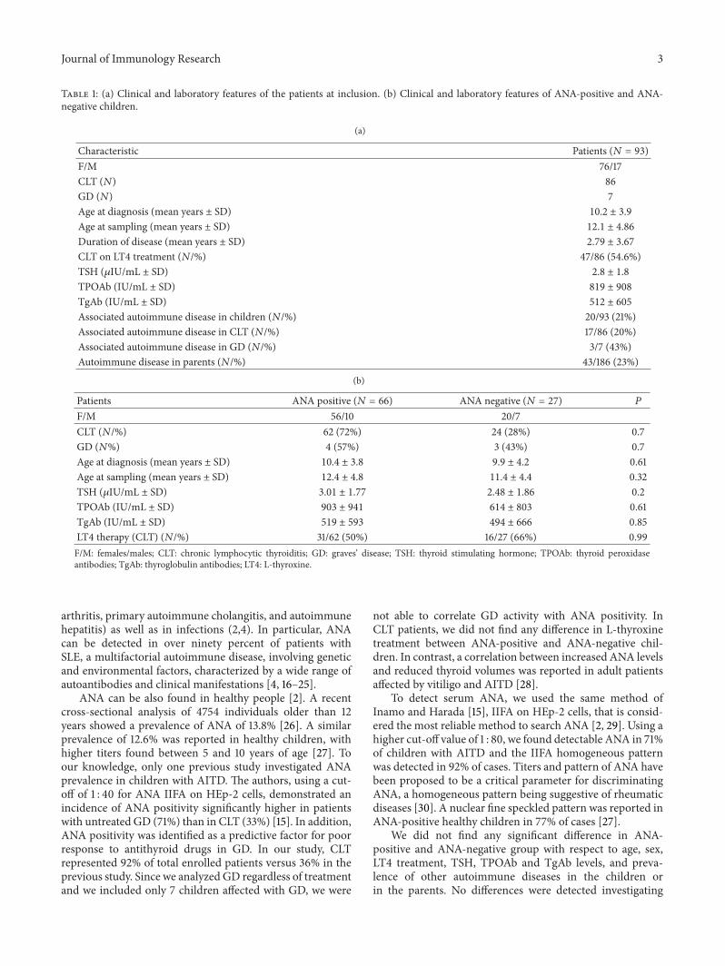
Clinical and laboratory features of the enrolled patientsare reported in Table 1(a). ANA positivity was found in66/93 children (71%), anti-ENA was found in 4/93 (4.3%),anti-dsDNA antibodies were found in 1/93 (1.1%), RF wasdetectable in 3/93 (3.2%), and anti-CCP were found in none.ENA-specific autoantibodies were determined in 3 out of 4anti-ENA-positive patients, 1 had anti-RNP positive, 1 hadanti-Jo-1, and 1 was negative for specific autoantibodies. TheANA pattern was homogeneous in 61/66 (92.4%), coarse/finespeckled in 4/66 (6%), and nucleolar in 2/66 (3%). The IIFAintensity was +++ in 23/66 (34.8%), ++ in 24/66 (36.3%), and+ in 19/66 (28.7%) cases.
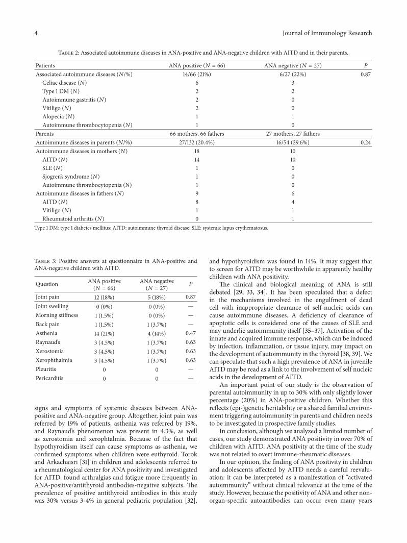
Among the 93 children with AITD, 20 (21%) had at leastone other autoimmune disease: 9 with celiac disease (CD), 2with autoimmune gastritis (AG), 4 with type 1 diabetes melli-tus (type 1 DM), 2 with alopecia, 2 with vitiligo, and 1 withautoimmune thrombocytopenia. We found that 3/7 (43%)of children with GD presented with one other autoimmunedisease (1 CD, 1 type DM, and 1 AG), versus 17/86 (20%)with CLT (8 CD, 1 AG, 3 type 1 DM, 2 alopecia, 2 vitiligo,and 1 autoimmune thrombocytopenia). The difference in thefrequency of autoimmune disease associated with CLT versusGD was not significant.
Clinical and laboratory features of ANA-positive andANA-negative patients are compared in Table 1(b). No signif-icant differences were found between the ANA-positive andANA-negative groups with respect to age, sex, L-thyroxinetreatment, TSH, TPOAb, and TgAb levels, or presence ofother autoimmune diseases in them and in their parents.Associated autoimmune diseases in ANA-positive and ANA-negative children and their parents are reported in Table 2.Hereby ANA-positive children did not differ for the fre-quency of additional autoimmune disease, but there wasa higher percentage of parental autoimmunity in ANA-negative patients, however, without significance.
Signs and symptoms as investigated by questionnaire aredetailed in Table 3. No significant differences were foundbetween ANA-positive and ANA-negative children withAITD.
Children with persistent joint pain were referred to arheumatologist (FC). Children with Raynaud’s phenomenonunderwent capillaroscopy. No evidence of SLE, RA, or othersystemic autoimmune diseases was found.
5. Discussion
AITD is a common autoimmune disease and it is frequentlyassociated with other organ and non-organ-specific autoim-mune disorders [8–10]. A variable ANAprevalence up to 45%has been reported in AITD adult patients [13, 14].
ANA can also be detected in different autoimmunedisorders (i.e., SLE, Sjogren’s syndrome, progressive systemicsclerosis, mixed connective-tissue disease, juvenile idiopathic
Journal of Immunology Research 3
Table 1: (a) Clinical and laboratory features of the patients at inclusion. (b) Clinical and laboratory features of ANA-positive and ANA-negative children.
(a)
Characteristic Patients (𝑁 = 93)F/M 76/17CLT (𝑁) 86GD (𝑁) 7Age at diagnosis (mean years ± SD) 10.2 ± 3.9
Age at sampling (mean years ± SD) 12.1 ± 4.86
Duration of disease (mean years ± SD) 2.79 ± 3.67
CLT on LT4 treatment (𝑁/%) 47/86 (54.6%)TSH (𝜇IU/mL ± SD) 2.8 ± 1.8
TPOAb (IU/mL ± SD) 819 ± 908
TgAb (IU/mL ± SD) 512 ± 605
Associated autoimmune disease in children (𝑁/%) 20/93 (21%)Associated autoimmune disease in CLT (𝑁/%) 17/86 (20%)Associated autoimmune disease in GD (𝑁/%) 3/7 (43%)Autoimmune disease in parents (𝑁/%) 43/186 (23%)
(b)
Patients ANA positive (𝑁 = 66) ANA negative (𝑁 = 27) 𝑃
F/M 56/10 20/7CLT (𝑁/%) 62 (72%) 24 (28%) 0.7GD (𝑁%) 4 (57%) 3 (43%) 0.7Age at diagnosis (mean years ± SD) 10.4 ± 3.8 9.9 ± 4.2 0.61Age at sampling (mean years ± SD) 12.4 ± 4.8 11.4 ± 4.4 0.32TSH (𝜇IU/mL ± SD) 3.01 ± 1.77 2.48 ± 1.86 0.2TPOAb (IU/mL ± SD) 903 ± 941 614 ± 803 0.61TgAb (IU/mL ± SD) 519 ± 593 494 ± 666 0.85LT4 therapy (CLT) (𝑁/%) 31/62 (50%) 16/27 (66%) 0.99F/M: females/males; CLT: chronic lymphocytic thyroiditis; GD: graves’ disease; TSH: thyroid stimulating hormone; TPOAb: thyroid peroxidaseantibodies; TgAb: thyroglobulin antibodies; LT4: L-thyroxine.
arthritis, primary autoimmune cholangitis, and autoimmunehepatitis) as well as in infections (2,4). In particular, ANAcan be detected in over ninety percent of patients withSLE, a multifactorial autoimmune disease, involving geneticand environmental factors, characterized by a wide range ofautoantibodies and clinical manifestations [4, 16–25].
ANA can be also found in healthy people [2]. A recentcross-sectional analysis of 4754 individuals older than 12years showed a prevalence of ANA of 13.8% [26]. A similarprevalence of 12.6% was reported in healthy children, withhigher titers found between 5 and 10 years of age [27]. Toour knowledge, only one previous study investigated ANAprevalence in children with AITD. The authors, using a cut-off of 1 : 40 for ANA IIFA on HEp-2 cells, demonstrated anincidence of ANA positivity significantly higher in patientswith untreated GD (71%) than in CLT (33%) [15]. In addition,ANA positivity was identified as a predictive factor for poorresponse to antithyroid drugs in GD. In our study, CLTrepresented 92% of total enrolled patients versus 36% in theprevious study. Since we analyzed GD regardless of treatmentand we included only 7 children affected with GD, we were
not able to correlate GD activity with ANA positivity. InCLT patients, we did not find any difference in L-thyroxinetreatment between ANA-positive and ANA-negative chil-dren. In contrast, a correlation between increased ANA levelsand reduced thyroid volumes was reported in adult patientsaffected by vitiligo and AITD [28].
To detect serum ANA, we used the same method ofInamo and Harada [15], IIFA on HEp-2 cells, that is consid-ered the most reliable method to search ANA [2, 29]. Using ahigher cut-off value of 1 : 80, we found detectable ANA in 71%of children with AITD and the IIFA homogeneous patternwas detected in 92% of cases. Titers and pattern of ANA havebeen proposed to be a critical parameter for discriminatingANA, a homogeneous pattern being suggestive of rheumaticdiseases [30]. A nuclear fine speckled pattern was reported inANA-positive healthy children in 77% of cases [27].
We did not find any significant difference in ANA-positive and ANA-negative group with respect to age, sex,LT4 treatment, TSH, TPOAb and TgAb levels, and preva-lence of other autoimmune diseases in the children orin the parents. No differences were detected investigating
4 Journal of Immunology Research
Table 2: Associated autoimmune diseases in ANA-positive and ANA-negative children with AITD and in their parents.
Patients ANA positive (𝑁 = 66) ANA negative (𝑁 = 27) 𝑃
Associated autoimmune diseases (𝑁/%) 14/66 (21%) 6/27 (22%) 0.87Celiac disease (𝑁) 6 3Type 1 DM (𝑁) 2 2Autoimmune gastritis (𝑁) 2 0Vitiligo (𝑁) 2 0Alopecia (𝑁) 1 1Autoimmune thrombocytopenia (𝑁) 1 0
Parents 66 mothers, 66 fathers 27 mothers, 27 fathersAutoimmune diseases in parents (𝑁/%) 27/132 (20.4%) 16/54 (29.6%) 0.24Autoimmune diseases in mothers (𝑁) 18 10
AITD (𝑁) 14 10SLE (𝑁) 1 0Sjogren’s syndrome (𝑁) 1 0Autoimmune thrombocytopenia (N) 1 0
Autoimmune diseases in fathers (𝑁) 9 6AITD (𝑁) 8 4Vitiligo (𝑁) 1 1Rheumatoid arthritis (𝑁) 0 1
Type 1 DM: type 1 diabetes mellitus; AITD: autoimmune thyroid disease; SLE: systemic lupus erythematosus.
Table 3: Positive answers at questionnaire in ANA-positive andANA-negative children with AITD.
Question ANA positive(𝑁 = 66)
ANA negative(𝑁 = 27) 𝑃
Joint pain 12 (18%) 5 (18%) 0.87Joint swelling 0 (0%) 0 (0%) —Morning stiffness 1 (1.5%) 0 (0%) —Back pain 1 (1.5%) 1 (3.7%) —Asthenia 14 (21%) 4 (14%) 0.47Raynaud’s 3 (4.5%) 1 (3.7%) 0.63Xerostomia 3 (4.5%) 1 (3.7%) 0.63Xerophthalmia 3 (4.5%) 1 (3.7%) 0.63Pleuritis 0 0 —Pericarditis 0 0 —
signs and symptoms of systemic diseases between ANA-positive and ANA-negative group. Altogether, joint pain wasreferred by 19% of patients, asthenia was referred by 19%,and Raynaud’s phenomenon was present in 4.3%, as wellas xerostomia and xerophtalmia. Because of the fact thathypothyroidism itself can cause symptoms as asthenia, weconfirmed symptoms when children were euthyroid. Torokand Arkachaisri [31] in children and adolescents referred toa rheumatological center for ANA positivity and investigatedfor AITD, found arthralgias and fatigue more frequently inANA-positive/antithyroid antibodies-negative subjects. Theprevalence of positive antithyroid antibodies in this studywas 30% versus 3-4% in general pediatric population [32],
and hypothyroidism was found in 14%. It may suggest thatto screen for AITD may be worthwhile in apparently healthychildren with ANA positivity.
The clinical and biological meaning of ANA is stilldebated [29, 33, 34]. It has been speculated that a defectin the mechanisms involved in the engulfment of deadcell with inappropriate clearance of self-nucleic acids cancause autoimmune diseases. A deficiency of clearance ofapoptotic cells is considered one of the causes of SLE andmay underlie autoimmunity itself [35–37]. Activation of theinnate and acquired immune response, which can be inducedby infection, inflammation, or tissue injury, may impact onthe development of autoimmunity in the thyroid [38, 39]. Wecan speculate that such a high prevalence of ANA in juvenileAITDmay be read as a link to the involvement of self nucleicacids in the development of AITD.
An important point of our study is the observation ofparental autoimmunity in up to 30% with only slightly lowerpercentage (20%) in ANA-positive children. Whether thisreflects (epi-)genetic heritability or a shared familial environ-ment triggering autoimmunity in parents and children needsto be investigated in prospective family studies.
In conclusion, although we analyzed a limited number ofcases, our study demonstrated ANA positivity in over 70% ofchildren with AITD. ANA positivity at the time of the studywas not related to overt immune-rheumatic diseases.
In our opinion, the finding of ANA positivity in childrenand adolescents affected by AITD needs a careful reevalu-ation: it can be interpreted as a manifestation of “activatedautoimmunity” without clinical relevance at the time of thestudy.However, because the positivity of ANAand other non-organ-specific autoantibodies can occur even many years

Journal of Immunology Research 5
before the onset of systemic autoimmune diseases [5, 40, 41],we think that prospective studies are warranted, especially insubjects positive for anti-dsDNA and anti-ENA.
Conflict of Interests
The authors declare that there is no conflict of interestsregarding the publication of this paper.
References
[1] R. H. White and D. L. Robbins, “Clinical significance andinterpretation of antinuclear antibodies,” Western Journal ofMedicine, vol. 147, no. 2, pp. 210–213, 1987.
[2] N. Agmon-Levin, J. Damoiseaux, C. Kallenberg et al., “Interna-tional recommendations for the assessment of autoantibodies tocellular antigens referred to as anti-nuclear antibodies,” Annalsof the Rheumatic Diseases, vol. 73, pp. 17–23, 2013.
[3] A. P. Weetman, “Non-thyroid autoantibodies inauitoimmunethyroid disease,” Best Practice and Research, vol. 19, no. 1, pp.17–32, 2005.
[4] F. Conti, C. Alessandri, D. Bompane et al., “Autoantibodyprofile in systemic lupus erythematosus with psychiatric man-ifestations: a role for anti-endothelial-cell antibodies,” ArthritisResearch &Therapy, vol. 6, no. 4, pp. R366–R372, 2004.
[5] M. R. Arbuckle, M. T. McClain, M. V. Rubertone et al., “Devel-opment of autoantibodies before the clinical onset of systemiclupus erythematosus,” New England Journal of Medicine, vol.349, no. 16, pp. 1526–1533, 2003.
[6] F. G. A. Delemarre, P. J. Simons, and H. A. Drexhage,“Histomorphological aspects of the development of thyroidautoimmune diseases: consequences for our understanding ofendocrine ophthalmopathy,”Thyroid, vol. 6, no. 4, pp. 369–377,1996.
[7] B. Rapoport and S. M. McLachlan, “Thyroid autoimmunity,”Journal of Clinical Investigation, vol. 108, no. 9, pp. 1253–1259,2001.
[8] G. S. Eisenbarth and P. A. Gottlieb, “Medical progress: autoim-mune polyendocrine syndromes,” New England Journal ofMedicine, vol. 350, no. 20, pp. 2068–2079, 2004.
[9] K. Boelaert, P. R. Newby, M. J. Simmonds et al., “Prevalenceand relative risk of other autoimmune diseases in subjects withautoimmune thyroid disease,” American Journal of Medicine,vol. 123, no. 2, pp. 183.e1–183.e9, 2010.
[10] I. Lazurova and K. Benhatchi, “Autoimmune Thyroid diseasesand nonorgan-specific autoimmunity,” Polskie ArchiwumMedy-cyny Wewnętrznej, vol. 122, supplement 1, pp. 55–59, 2012.
[11] D. Mihailova, R. Grigorova, B. Vassileva et al., “Autoimmunethyroid disorders in juvenile chronic arthritis and systemiclupus erythematosus,” Advances in Experimental Medicine andBiology, vol. 455, pp. 55–60, 1999.
[12] S. Stagi, T. Giani, G. Simonini, and F. Falcini, “Thyroid function,autoimmune thyroiditis and coeliac disease in juvenile idio-pathic arthritis,”Rheumatology, vol. 44, no. 4, pp. 517–520, 2005.
[13] M. G. Tektonidou, M. Anapliotou, P. Vlachoyiannopoulos, andH. M. Moutsopoulos, “Presence of systemic autoimmune dis-orders in patients with autoimmune thyroid diseases,” Annalsof the Rheumatic Diseases, vol. 63, no. 9, pp. 1159–1161, 2004.
[14] I. Lazurova, K. Benhatchi, J. Rovensky et al., “Autoimmunethyroid disease and autoimmune rheumatic disorders: a two-sided analysis,”Annals of the New York Academy of Sciences, vol.1173, pp. 211–216, 2009.
[15] Y. Inamo and K. Harada, “Antinuclear antibody positivity inpediatric patients with autoimmune thyroid disease,” Journal ofRheumatology, vol. 24, no. 3, pp. 576–578, 1997.
[16] R. Priori, E. Medda, F. Conti et al., “Familial autoimmunity asa risk factor for systemic lupus erythematosus and vice versa: acase-control study,” Lupus, vol. 12, no. 10, pp. 735–740, 2003.
[17] G. Valesini, C. Alessandri, D. Celestino, and F. Conti, “Anti-endothelial antibodies and neuropsychiatric systemic lupuserythematosus,” Annals of the New York Academy of Sciences,vol. 1069, pp. 118–128, 2006.
[18] T. Colasanti, A. Maselli, F. Conti et al., “Autoantibodies toestrogen receptor 𝛼 interfere with T lymphocyte homeostasisand are associated with disease activity in systemic lupuserythematosus,” Arthritis and Rheumatism, vol. 64, no. 3, pp.778–787, 2012.
[19] P. Margutti, M. Sorice, F. Conti et al., “Screening of an endothe-lial cDNA library identifies the C-terminal region of Nedd5as a novel autoantigen in systemic lupus erythematosus withpsychiatric manifestations,” Arthritis Research & Therapy, vol.7, no. 4, pp. R896–R903, 2005.
[20] G. C. Tsokos, “Mechanisms of disease: systemic lupus erythe-matosus,”New England Journal of Medicine, vol. 365, no. 22, pp.2110–2121, 2011.
[21] F. Conti, F. R. Spinelli, C. Alessandri, and G. Valesini, “Toll-likereceptors and lupus nephritis,” Clinical Reviews in Allergy andImmunology, vol. 40, no. 3, pp. 192–198, 2011.
[22] C. Perricone, C. Ciccacci, F. Ceccarelli et al., “TRAF3IP2 geneand systemic lupus erythematosus: association with diseasesusceptibility and pericarditis development,” Immunogenetics,vol. 65, pp. 703–709, 2013.
[23] F. Conti, C. Alessandri, C. Perricone et al., “Neurocognitivedysfunction in systemic lupus erythematosus: association withantiphospholipid antibodies, disease activity and chronic dam-age,” PLoS ONE, vol. 7, no. 3, Article ID e33824, 2012.
[24] F. Conti, F. Ceccarelli, L. Massaro et al., “Evaluation of thepatient acceptable symptom state (PASS) in Italian patientsaffected by systemic lupus erythematosus: association withdisease activity indices,” PLoS ONE, vol. 8, Article ID e73517,2013.
[25] C. Iannuccelli, F. R. Spinelli, M. P. Guzzo et al., “Fatigueand widespread pain in Systemic Lupus Erythematosus andSjogren’s Syndrome: symptoms of the inflammatory disease orassociated Fibromyalgia?” Clinical and Experimental Rheuma-tology, vol. 30, pp. S117–S121, 2012.
[26] M. Satoh, E. K. L. Chan, L. A. Ho et al., “Prevalenceand sociodemographic correlates of antinuclear antibodies inUnited States,” Arthritis & Rheumatism, vol. 64, pp. 2319–2327,2012.
[27] M. O. Esteves Hilario, C. A. Len, S. Campos Roja, M. T.Terreri, G. Almeida, and L. E. Coelho Andrade, “Frequencyof antinuclear antibodies in healthy children and adolescents,”Clinical Pediatrics, vol. 43, no. 7, pp. 637–642, 2004.
[28] G. Zettinig, A. Tanew, G. Fischer, W. Mayr, R. Dudczak, andM. Weissel, “Autoimmune diseases in vitiligo: do anti-nuclearantibodies decrease thyroid volume?”Clinical andExperimentalImmunology, vol. 131, no. 2, pp. 347–354, 2003.
[29] D. H. Solomon, A. J. Kavanaugh, P. H. Schur et al., “Evidence-based guidelines for the use of immunologic tests: antinuclear

6 Journal of Immunology Research
antibody testing,” Arthritis and Rheumatism, vol. 47, no. 4, pp.434–444, 2002.
[30] H. A. Mariz, E. I. Sato, S. H. Barbosa, S. H. Rodrigues, A.Dellavance, and L. E. C. Andrade, “Pattern on the antinuclearantibody-HEp-2 test is a critical parameter for discriminatingantinuclear antibody-positive healthy individuals and patientswith autoimmune rheumatic diseases,” Arthritis and Rheuma-tism, vol. 63, no. 1, pp. 191–200, 2011.
[31] K. S. Torok and T. Arkachaisri, “Autoimmune thyroiditis inantinuclear antibody positive children without rheumatologicdisease,” Pediatric Rheumatology, vol. 8, article 15, 2010.
[32] A. Loviselli, L. Grasso, M. Songini et al., “The sardinian autoim-munity study: 3. Studies on circulating antithyroid antibodies insardinian schoolchildren: relationship to goiter prevalence andthyroid function,”Thyroid, vol. 11, no. 9, pp. 849–857, 2001.
[33] P. L. Meroni and P. H. Schur, “ANA screening: an old test withnew recommendations,” Annals of the Rheumatic Diseases, vol.69, no. 8, pp. 1420–1422, 2010.
[34] D. S. Pisetsky, “Antinuclear antibodies in healthy people: the tipof autoimmunity’s iceberg?”Arthritis Research andTherapy, vol.13, no. 2, article 109, 2011.
[35] T. Colasanti, C. Alessandri, A. Capozzi et al., “Autoantibodiesspecific to a peptide of 𝛽2-glycoprotein I cross-react with TLR4inducing a pro-inflammatory phenotype in endothelial cellsand monocytes,” Blood, vol. 120, pp. 3360–3370, 2012.
[36] S. Nagata, R. Hanayama, and K. Kawane, “Autoimmunity andthe clearance of dead cells,” Cell, vol. 140, no. 5, pp. 619–630,2010.
[37] U. S. Gaipl, A. Kuhn, A. Sheriff et al., “Clearance of apoptoticcells in human SLE,” Current Directions in Autoimmunity, vol.9, pp. 173–187, 2006.
[38] A. Kawashima, K. Tanigawa, T. Akama et al., “Fragmentsof cenomic DNA released by injured cells activate innateimmunity and suppress endocrine function in the thyroid,”Endocrinology, vol. 152, no. 4, pp. 1702–1712, 2011.
[39] A. Kawashima, K. Tanigawa, T. Akama, A. Yoshihara, N. Ishii,and K. Suzuki, “Innate immune activation and thyroid autoim-munity,” Journal of Clinical Endocrinology and Metabolism, vol.96, no. 12, pp. 3661–3671, 2011.
[40] H. Kokkonen, M. Mullazehi, E. Berglin et al., “Antibodies ofIgG, IgA and IgM isotypes against cyclic citrullinated peptideprecede the development of rheumatoid arthritis,” ArthritisResearch andTherapy, vol. 13, no. 1, article R13, 2011.
[41] P. Moinzadeh, S. I. Nihtyanova, K. Howell, V. H. Ong, andC. P. Denton, “Inpact of hallmark autoantibody reactivity onearly diagnosis in scleroderma,” Clinical Reviews in Allergy &Immunology, vol. 43, pp. 249–255, 2012.

Submit your manuscripts athttp://www.hindawi.com
Stem CellsInternational
Hindawi Publishing Corporationhttp://www.hindawi.com Volume 2014
Hindawi Publishing Corporationhttp://www.hindawi.com Volume 2014
MEDIATORSINFLAMMATION
of
Hindawi Publishing Corporationhttp://www.hindawi.com Volume 2014
Behavioural Neurology
EndocrinologyInternational Journal of
Hindawi Publishing Corporationhttp://www.hindawi.com Volume 2014
Hindawi Publishing Corporationhttp://www.hindawi.com Volume 2014
Disease Markers
Hindawi Publishing Corporationhttp://www.hindawi.com Volume 2014
BioMed Research International
OncologyJournal of
Hindawi Publishing Corporationhttp://www.hindawi.com Volume 2014
Hindawi Publishing Corporationhttp://www.hindawi.com Volume 2014
Oxidative Medicine and Cellular Longevity
Hindawi Publishing Corporationhttp://www.hindawi.com Volume 2014
PPAR Research
The Scientific World JournalHindawi Publishing Corporation http://www.hindawi.com Volume 2014
Immunology ResearchHindawi Publishing Corporationhttp://www.hindawi.com Volume 2014
Journal of
ObesityJournal of
Hindawi Publishing Corporationhttp://www.hindawi.com Volume 2014
Hindawi Publishing Corporationhttp://www.hindawi.com Volume 2014
Computational and Mathematical Methods in Medicine
OphthalmologyJournal of
Hindawi Publishing Corporationhttp://www.hindawi.com Volume 2014
Diabetes ResearchJournal of
Hindawi Publishing Corporationhttp://www.hindawi.com Volume 2014
Hindawi Publishing Corporationhttp://www.hindawi.com Volume 2014
Research and TreatmentAIDS
Hindawi Publishing Corporationhttp://www.hindawi.com Volume 2014
Gastroenterology Research and Practice
Hindawi Publishing Corporationhttp://www.hindawi.com Volume 2014
Parkinson’s Disease
Evidence-Based Complementary and Alternative Medicine
Volume 2014Hindawi Publishing Corporationhttp://www.hindawi.com