clinical study a minimally invasive endoscopic surgery for
TRANSCRIPT

Clinical StudyA Minimally Invasive Endoscopic Surgery for InfectiousSpondylodiscitis of the Thoracic and Upper Lumbar Spine inImmunocompromised Patients
Hsin-Chuan Chen,1 Teng-Le Huang,2,3,4 Yen-Jen Chen,5,6
Hsi-Kai Tsou,7,8 Wei-Ching Lin,6,9 Chih-Hung Hung,5,10 Chun-Hao Tsai,5,6
Horng-Chaung Hsu,5,6 and Hsien-Te Chen5,11
1 Department of Orthopedic Surgery, Show Chwan Memorial Hospital, No. 542, Section 1, Zhongshan Road, Changhua City,Changhua County 500, Taiwan
2 Department of Orthopedics, Tainan Municipal An-Nan Hospital-China Medical University, No. 66, Section 2, Changhe Road,Annan District, Tainan City 709, Taiwan
3 Department of Bioscience Technology, College of Science, Chung Yuan Christian University, No. 200 Chung Pei Road,Chung Li District, Taoyuan City 320, Taiwan
4 Department of Sports Medicine, College of Health Care, China Medical University, No. 91, Hsueh-Shih Road, North District,Taichung 404, Taiwan
5 Department of Orthopedic Surgery, China Medical University Hospital, No. 2 Yuhder Road, North District, Taichung 404, Taiwan6 School of Medicine, China Medical University, No. 91 Hsueh-Shih Road, North District, Taichung 404, Taiwan7 Functional Neurosurgery Division, Neurological Institute, Taichung Veterans General Hospital, 1650 Taiwan Boulevard Section 4,Taichung 407, Taiwan
8 Department of Early Childhood Care and Education, Jen-Teh Junior College of Medicine, Nursing and Management,No. 79-9 Sha-Luen Hu, Xi-Zhou Li, Hou-Loung Town, Miaoli County 356, Taiwan
9 Department of Radiology, China Medical University Hospital, No. 2 Yuhder Road, North District, Taichung 404, Taiwan10China Medical University, No. 91 Hsueh-Shih Road, North District, Taichung 404, Taiwan11School of Chinese Medicine, College of Chinese Medicine, China Medical University, No. 91 Hsueh-Shih Road,North District, Taichung 404, Taiwan
Correspondence should be addressed to Hsien-Te Chen; [email protected]
Received 2 January 2015; Accepted 20 April 2015
Academic Editor: Yi Liu
Copyright © 2015 Hsin-Chuan Chen et al. This is an open access article distributed under the Creative Commons AttributionLicense, which permits unrestricted use, distribution, and reproduction in any medium, provided the original work is properlycited.
This study evaluates the safety and effectiveness of computed tomography- (CT-) assisted endoscopic surgery in the treatmentof infectious spondylodiscitis of the thoracic and upper lumbar spine in immunocompromised patients. From October 2006to March 2014, a total of 41 patients with infectious spondylodiscitis underwent percutaneous endoscopic surgery under localanesthesia, and 13 lesions from 13 patients on the thoracic or upper lumbar spine were selected for evaluation. A CT-guided catheterwas placed before percutaneous endoscopic surgery as a guide to avoid injury to visceral organs, major vessels, and the spinalcord. All 13 patients had quick pain relief after endoscopic surgery without complications. The bacterial culture rate was 77%.Inflammatory parameters returned to normal after adequate antibiotic treatment. Postoperative radiographs showed no significantkyphotic deformity when compared with preoperative films. As of the last follow-up visit, no recurrent infections were noted.Traditional transthoracic or diaphragmatic surgery with or without posterior instrumentation is associated with high rates ofmorbidity and mortality, especially in elderly patients, patients with multiple comorbidities, or immunocompromised patients.Percutaneous endoscopic surgery assisted by a CT-guided catheter provides a safe and effective alternative treatment for infectiousspondylodiscitis of the thoracic and upper lumbar spine.
Hindawi Publishing CorporationBioMed Research InternationalVolume 2015, Article ID 780451, 8 pageshttp://dx.doi.org/10.1155/2015/780451

2 BioMed Research International
1. Introduction
In recent years, the incidence of infectious spondylodiscitishas increased due to vast improvements in medical careand prolonged life expectancies. The condition is associ-ated with advanced age, intravenous drug use, immuno-compromised status, and significant medical comorbidities[1]. Identifying the causative pathogen is the key to treat-ment. Computed tomography- (CT-) guided biopsy anddrainage are the standard procedure for identifying causativepathogens. However, the pathogen-identification rate variesamong studies [2–4]. Surgical intervention is indicated ifneurological deficit, progressive deformity, failure to respondto conservative treatment, or the need to obtain specimens toidentify causative pathogens is present. However, traditionalanterior debridement and reconstruction with or withoutposterior instrumentation are associated with high ratesof morbidity and mortality, especially in elderly immuno-compromised patients and patients with multiple comor-bidities. Percutaneous endoscopic discectomy, debridement,and drainage provide a minimally invasive surgical choicefor the treatment of infectious spondylodiscitis [4–6]. Thismethod provides adequate debridement and fast pain reliefand has a relatively high pathogen-identification rate [4,7, 8]. However, in the upper lumbar and thoracic spine,percutaneous endoscopy is associated with visceral organdamage, major vessel injury, and the spinal cord injury,which limits the use of this procedure in these areas[9].
A CT-guided catheter was placed before percutaneousendoscopic surgery as a guide to avoid injury to visceralorgans, major vessels, and the spinal cord. We analyzedthe clinical outcomes, inflammatory parameters, and radio-graphic findings for 13 lesions that occurred on thoracic orupper lumbar spine.
2. Materials and Methods
2.1. Patient Population. From October 2006 to March 2014, atotal of 41 patients with infective spondylodiscitis underwentpercutaneous endoscopic surgery with local anesthesia and13 patients’ lesions on the thoracic or upper lumbar spinewere selected for evaluation (Table 1). A CT-guided angio-graphic catheter was placed before percutaneous endoscopicsurgery as a guide to avoid injury to visceral organs, majorvessels, and the spinal cord. Of the 13 patients evaluated,5 were men and 8 were women. Their mean age was 65.6years (range, 49–84 years). The affected level ranged fromT11-T12 to L1-L2 in 11 cases, T8-T9 in 1 case, and T9-T10 in the other case. All patients underwent plain filmradiography and enhanced magnetic resonance imaging(MRI) of the involved spine, which revealed evidence ofinfectious spondylodiscitis (Figure 1). Most patients hadhigh inflammatory markers (C-reactive protein (CRP) anderythrocyte sedimentation rate (ESR)) and complained ofsevere back pain. They also had a variety of comorbidities,including renal failure, heart failure, rheumatic arthritis, livercirrhosis, polycystic liver posttransplantation, and diabetes(Table 1).
2.2. Surgical Procedures. The CT-guided biopsy and catheterplacement were performed by an experienced radiologist onthe day of or the day before the scheduled percutaneousendoscopic surgery (Figure 2). The patient was positionedprone, and local anesthesia with 2% lidocaine was injectedinto the area of needle insertion. A 6-in-long number 11G-wide multiple side hole bone puncture needle was insertedinto the lesion site with CT guidance. A J guidewire wasthen inserted via the bone biopsy needle. Finally, a number5 Fr C1 angiographic catheter (Cook, Bloomington, USA)was inserted along the J guidewire and left in the infectivearea after guidewire removal. The specimen obtained duringthe procedure was sent for bacterial, tuberculosis (TB), andfungal cultures and pathologic analysis.
Percutaneous endoscopic surgery was then performedafter placement of the CT-guided angiographic catheter. Thepatient was positioned prone on a spine-operating tablewith the abdomen hanging free. The patient was underintravenous pain control butwas kept awake during the endo-scopic surgery so that he or she could respond well when thedura or nerve roots were irritated. Local anesthesia was alsoperformed with 2% lidocaine around the area of endoscopicinsertion. A percutaneous endoscopic guidewire was inserteddirectly through the CT-guided number 5 Fr C1 angiographiccatheter and advanced slowly with the assistance of fluo-roscopy to ensure that the wire was targeting the infectivearea without penetrating the angiographic catheter wall andinjuring any related structures. After the guidewire was set inthe infected area, the number 5 Fr C1 angiographic catheterwas then removed. A dilator was inserted along the endo-scopic guidewire, and the position was again checked underfluoroscopy.The infected disc and vertebrawere harvested forbacterial, fungal, and TB cultures by using endoscopicmicro-rongeurs and microscissors before starting irrigation. Afteradequate tissue sampling for culture, radical debridement,sequestrectomy, and irrigation could be performed with theaid of direct endoscope vision andfluoroscopy.TheSurgitron,a high-voltage bipolar probe (Ellman Innovations, New York,USA), was used for thermocoagulation of infected tissueand bleeders. All the operating instruments and endoscopicsystems were supplied by Richard and Wolf (Knittlingen,Germany). The high-resolution endoscope has a diameter of8mm with a 4.1mm intraendoscopic working channel. Theangle of vision is 25∘.Theworking sheath has an 8.0mmouterdiameter and a beveled opening, both of which enable thecreation of visual and working fields in an area without aclear, anatomically preformed cavity. More than 4 L normalsaline with 1 g cefazolin in each liter was used for pressuredirrigation and drainage of infected materials and pus. A 1/4in drainage tube was left in the infected area at the end ofsurgery for further drainage of infective materials, pus, andexudates (Figure 3).
2.3. Postoperative Care. The 1/4 in drainage tube was leftin place for at least 7 days until the daily drainage amountwas less than 5mL. Effective antibiotics were administeredintravenously for patients with known causative pathogensbefore surgery. For patients with unknown pathogens, empir-ical antibiotics were administered immediately after surgery.
BioMed Research International 3
Table 1: Patient demographic data.
Patient number Age Gender Level Neurological deficit Associated medical illness1 71 F L1-2 Frankle D Uremia, CHF, RHD2 60 M T11-12 Frankle D CAD, DM, CHF, asthma3 67 F T12 Nil Uremia, DM4 65 F L1-2 Nil HTN, RA5 55 F T11-12 Nil DM, HTN6 73 F T12-L1 Mild sensory deficit DM7 71 F T11-T12 Nil DM, liver cirrhosis, burst fracture T11 vertebra8 71 M L1 Frankle D None9 84 F T9-10 Frankle D HTN, CHF, PSVT10 63 M T12-L1-2 Mild sensory deficit Liver cirrhosis, asthma11 52 M L1-2 Nil BPH12 49 F L1-2 Nil Polycystic liver, kidney s/p liver transplantation13 72 M T8-9 Mild sensory deficit DMAverage 65.6Standard deviation 9.73CHF: congestive heart failure, RHD: rheumatic heart disease, CAD: coronary artery disease, DM: diabetes mellitus, HTN: hypertension, RA: rheumatoidarthritis, PSVT: paroxysmal supraventricular tachycardia, and BPH: benign prostate hypertrophy.
(a) (b)
(c)
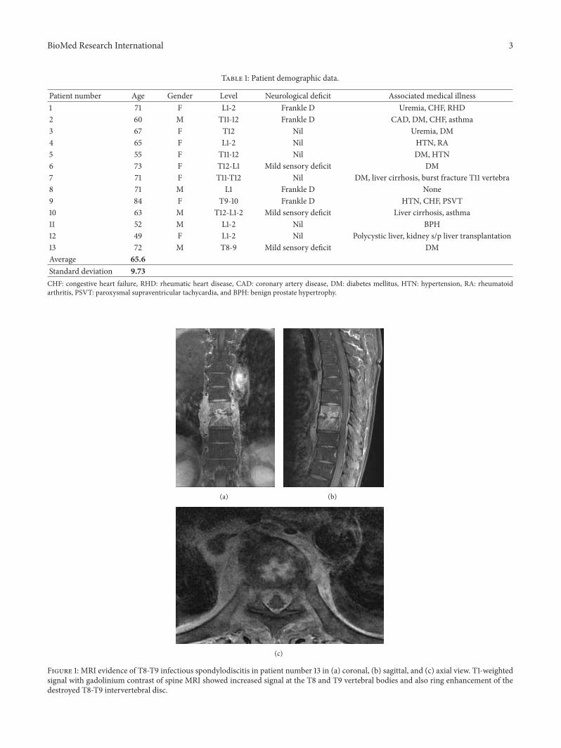
Figure 1: MRI evidence of T8-T9 infectious spondylodiscitis in patient number 13 in (a) coronal, (b) sagittal, and (c) axial view. T1-weightedsignal with gadolinium contrast of spine MRI showed increased signal at the T8 and T9 vertebral bodies and also ring enhancement of thedestroyed T8-T9 intervertebral disc.
4 BioMed Research International
(a) (b)
(c)
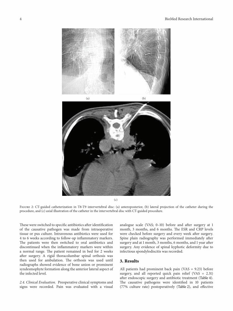
Figure 2: CT-guided catheterization in T8-T9 intervertebral disc: (a) anteroposterior, (b) lateral projection of the catheter during theprocedure, and (c) axial illustration of the catheter in the intervertebral disc with CT-guided procedure.
These were switched to specific antibiotics after identificationof the causative pathogen was made from intraoperativetissue or pus culture. Intravenous antibiotics were used for4 to 6 weeks according to follow-up inflammatory markers.The patients were then switched to oral antibiotics anddiscontinued when the inflammatory markers were withina normal range. The patient remained in bed for 2 weeksafter surgery. A rigid thoracolumbar spinal orthosis wasthen used for ambulation. The orthosis was used untilradiographs showed evidence of bone union or prominentsyndesmophyte formation along the anterior lateral aspect ofthe infected level.
2.4. Clinical Evaluation. Preoperative clinical symptoms andsigns were recorded. Pain was evaluated with a visual
analogue scale (VAS; 0–10) before and after surgery at 1month, 3 months, and 6 months. The ESR and CRP levelswere checked before surgery and every week after surgery.Spine plain radiography was performed immediately aftersurgery and at 1 month, 3 months, 6 months, and 1 year aftersurgery. Any evidence of spinal kyphotic deformity due toinfectious spondylodiscitis was recorded.
3. Results
All patients had prominent back pain (VAS = 9.23) beforesurgery, and all reported quick pain relief (VAS = 2.31)after endoscopic surgery and antibiotic treatment (Table 4).The causative pathogens were identified in 10 patients(77% culture rate) postoperatively (Table 2), and effective

BioMed Research International 5
T8 T9
(a)
(b) (c)
Figure 3: (a) Intraoperative fluoroscopic image demonstrated the working sheath and endoscope within the intervertebral disc space, (b)immediate postoperative AP view demonstrated a 1/4 inch drainage tube in the intervertebral space, and (c) postoperative 3-month AP viewrevealed partially united T8-T9 vertebral body.
Table 2: Surgical procedures.
Patient Level Procedures Bacteria culture1∗ L1-L2 CT-guided catheter + PEDD Delftia acidovorans2∗ T11-T12 CT-guided catheter + PEDD No growth3 T12 CT-guided catheter + PEDD No growth4 L1-L2 CT-guided catheter + PEDD Escherichia coli (ESBL)5 T11-T12 CT-guided catheter + PEDD Staphylococcus aureus6 T12-L1 CT-guided catheter + PEDD Staphylococcus aureus (MRSA)7 T11-T12 CT-guided catheter + PEDD No growth8∗ L1 CT-guided catheter + PEDD Streptococcus anginosus9 T9-10 CT-guided catheter + PEDD Mycobacteria tuberculosis complex10 T12-L1-2 CT-guided catheter + PEDD Klebsiella pneumoniae11 L1-L2 CT-guided catheter + PEDD Staphylococcus aureus12 L1-L2 CT-guided catheter + PEDD Candida albicans, Ecoli13 T8-9 CT-guided catheter + PEDD Klebsiella pneumoniaePEDD: percutaneous endoscopic discectomy drainage.∗Dying of other medical problems during next few years.

6 BioMed Research International
Table 3: ESR and CRP levels before and after surgery.
Patient number ESR (mm/1 hr) CRP (mg/dL)Preop Postop 1m Postop 3m Last F/U Preop Postop 1m Postop 3m
1 51 67 20 37 6.62 0.38 2.842 76 18 21 13 22.16 0.15 0.183 115 86 78 72 32.11 2.70 0.604 79 41 9 8 4.96 0.45 0.035 108 71 25 21 6.33 0.31 0.086 123 115 64 36 23.72 1.24 0.557 112 93 86 84 1.44 0.18 0.298 75 42 14 7 18.19 2.80 0.959 53 28 18 28 6.61 1.94 0.7510 125 85 37 30 0.19 0.07 0.0611 92 16 2 2 7.45 0.23 0.1212 108 74 50 9 4.84 0.70 0.1113 104 12 11 11 16.53 0.25 0.35Average 93.02 57.53 33.46 27.53 11.63 0.88 0.53Standard deviation 25.00 33.44 27.51 25.26 9.85 0.98 0.75
antibiotics were administered according to the sensitivity testof isolated pathogens. The blood culture result of patient7 was methicillin-sensitive Staphylococcus aureus infection,but the results of the endoscopic specimen were negative.Oxacillin was used for infection control according to thesensitivity test report of blood culture. The other 2 patientshad no known culture results, and empirical antibioticsweremaintained until the inflammatory parameters returnedto normal ranges. In patient 9, anti-TB medication wasmaintained for 9months according to TB treatment protocol.The ESR and CRP levels of all patients decreased significantlyafter endoscopic surgery and 3 months of intravenous andoral antibiotic treatment. No patient had relapse of spinalinfection at the treated level during the follow-up period(Table 3).
No surgery-related complications were noted duringthe follow-up period (average, 42.46 months; range, 8–70months). No recurrence of infection developed in thesepatients, and no open spinal surgery was needed. Deformityof the spine was evaluated from plain film radiographsobtained before and after surgery and at the last follow-up.The kyphotic angle was measured as the angle between theupper end plate of the first vertebral body above the involvedlevel and the lower end plate of the first vertebral body belowthe involved level. Only 1 patient had a kyphotic changegreater than 10∘. The other 12 patients had no significantchanges in spinal deformity (Table 5; Figure 4).
4. Discussion
Infectious spondylodiscitis is an increasingly prevalent dis-ease, especially in elderly or immunocompromised patientsor those with medical comorbidities. Conservative treatmentconsisting of antibiotic administration and bed rest is thestandard choice in cases of mild infection. CT-guided biopsyis a less invasive procedure used to obtain specimens for
Table 4: Back pain level before and after surgery.
Patient number Visual analog scale (0–10) ComplicationPreop Postop 1m Postop 3m
1 10 5 2 Nil2 9 4 2 Nil3 10 5 3 Nil4 10 4 1 Nil5 10 5 4 Nil6 10 5 3 Nil7 10 4 3 Nil8 9 5 1 Nil9 9 4 3 Nil10 10 5 4 Nil11 8 1 1 Nil12 8 3 2 Nil13 7 3 1 NilAverage 9.23 4.08 2.31Standard deviation 1.01 1.19 1.11
pathogen identification. Drainage function also contributesto infection control. However, the rate of pathogen identifica-tion varies widely, from 36% to 91% [2–4, 10–12], and the rateof successful infection control is also unsatisfactory. Whenconservative treatment fails, the treatment approach shifts toopen surgery. However, traditional anterior debridement andreconstruction with or without posterior instrumentationare a major operation with a high rate of postoperativecomplications, especially in immunocompromised patientswith many comorbid diseases or in the elderly.
Percutaneous endoscopic discectomy was first appliedfor lumbar disc herniation in the 1980s and now is a well-established surgical procedure. This procedure has also beenused to treat infectious spondylodiscitis in recent years and

BioMed Research International 7
Table 5: Changes in kyphosis angle (∘).
Patient number Level Preoperative Postoperative LastF/U
1 L1-2 −6∘ −2∘ −1∘
2 T11-12 10∘ 8∘ 10∘
3 T12 16∘ 19∘ 19∘
4 L1-2 2∘ −2∘ −1∘
5 T11-12 6∘ 4∘ 1∘
6 T12-L1 13∘ 13∘ 24∘
7 T11-12 11∘ 14∘ 12∘
8 L1 12∘ 5∘ 4∘
9 T9-10 0∘ 3∘ 3∘
10 T12-L1-2 −13∘ −12∘ −12∘
11 L1-2 7∘ 8∘ 12∘
12 L1-2 17∘ 16∘ 16∘
13 T8-9 12∘ 14∘ 15∘
Average 8.69 6.77 7.85Standard deviation 6.68 8.72 9.91Kyphosis angle was obtained by measuring the sagittal angles with the Cobbmethod.
(a) (b)
Figure 4: The plain films 6 months after operation. (a) AP viewand (b) lateral view revealed prominent syndesmophyte formationat anterior and lateral aspects of infective area without significantkyphotic deformity.
has proved to be as effective as open surgery for infection con-trol [4, 6, 8, 13]. However, percutaneous endoscopic surgeryfor infectious spondylodiscitis has been limited to caseswith lower lumbar spine infection for reasons of operativesafety [7]. Percutaneous endoscopic surgery carries risks ofinjury to the surrounding visceral organs, the spinal cord,and major vessels of the thoracic and upper lumbar spinewhen used to treat lesions above the L2 level. In this study,a combination of a CT-guided angiographic catheter andpercutaneous endoscopic surgery was found to be effectiveand safe for the treatment of infectious spondylodiscitis of
the upper lumbar and thoracic spine. CT provides real-timeimages that can be used to insert the guidewire and catheter,which helps prevent injury to the spinal cord,major vessels, orvisceral organs. Once the endoscope is inserted safely alongthe CT-guided catheter, percutaneous endoscopic surgerycan be performed safely to carry out radical debridementor obtain a sufficient specimen culture, and a large-diameterdrainage tube can be left in the infected level. Infection waswell controlled with antibiotics in all patients, regardless ofthe culture results. No complications occurred during theoperation or follow-up period. Moreover, no open surgerywas needed in these patients, whereas anterior debridementand fusion surgery was previously performed in 19% to 25%of patients [4, 6].
In view of the risks of general anesthesia and tra-ditional major surgery for infectious spondylodiscitis ofthe thoracic and upper lumbar spine, minimally invasiveendoscopic surgery represents a good alternative treatmentfor immunocompromised patients. Percutaneous endoscopicsurgery assisted by a CT-guided catheter is a safe and effectiveoperation using local anesthesia and a small incision wound(<1 cm).
5. Conclusion
Percutaneous endoscopic discectomy, debridement, irriga-tion, and drainage with the assistance of a CT-guided catheterare a safe and effective alternative treatment for infectiousspondylodiscitis of the thoracic and upper lumbar spine.It prevents many complications normally associated withpercutaneous endoscopic surgery in the thoracic and upperlumbar area. Endoscopic surgery not only has a high rate ofthe causative-pathogen identification but also provides goodinfection control and pain relief, even if causative pathogencannot be identified. The safe and minimally invasive natureof this procedure broadens the application of operativetreatment for infectious spondylodiscitis, even in the thoracicand upper lumbar level, and prevents progressive deformitywhen prolonged antibiotic treatment is used. We have moreconfidence when treating patients in poor health, immuno-compromised or elderly patients, and can nowprovide amorepromising and safe treatment strategy. Motor weakness ofthe lower limbs may be the only contraindication for thisprocedure.
Conflict of Interests
The authors declare no conflict of interests regarding thepublication of this paper.
References
[1] J. S. Fischgrund, OKU 9: Orthopaedic Knowledge Update,American Academy of Orthopaedic Surgeons, Rosemont, Ill,USA, 2008.
[2] S.-C. Yang, T.-S. Fu, L.-H. Chen, W.-J. Chen, and Y.-K.Tu, “Identifying pathogens of spondylodiscitis: percutaneousendoscopy or CT-guided biopsy,” Clinical Orthopaedics andRelated Research, vol. 466, no. 12, pp. 3086–3092, 2008.

8 BioMed Research International
[3] F. S. Chew and M. J. Kline, “Diagnostic yield of CT-guidedpercutaneous aspiration procedures in suspected spontaneousinfectious diskitis,” Radiology, vol. 218, no. 1, pp. 211–214, 2001.
[4] T.-S. Fu, L.-H. Chen, and W.-J. Chen, “Minimally invasive per-cutaneous endoscopic discectomy and drainage for infectiousspondylodiscitis,” Biomedical Journal, vol. 36, no. 4, pp. 168–174,2013.
[5] M. Ito, K. Abumi, Y. Kotani, K. Kadoya, and A. Minami,“Clinical outcome of posterolateral endoscopic surgery forpyogenic spondylodiscitis: results of 15 patients with seriouscomorbid conditions,” Spine, vol. 32, no. 2, pp. 200–206, 2007.
[6] S.-C. Yang, T.-S. Fu, H.-S. Chen, Y.-H. Kao, S.-W. Yu, andY.-K. Tu, “Minimally invasive endoscopic treatment for lumbarinfectious spondylitis: a retrospective study in a tertiary referralcenter,” BMCMusculoskeletal Disorders, vol. 15, no. 1, article 105,2014.
[7] S.-C. Yang, T.-S. Fu, L.-H. Chen, C.-C. Niu, P.-L. Lai, and W.-J.Chen, “Percutaneous endoscopic discectomy and drainage forinfectious spondylitis,” International Orthopaedics, vol. 31, no.3, pp. 367–373, 2007.
[8] G. Bavinzski, A. Schoeggl, S. Trattnig et al., “Microsurgicalman-agement of postoperative disc space infection,” NeurosurgicalReview, vol. 26, no. 2, pp. 102–107, 2003.
[9] J. C. Chiu, N. Javidan, and J. Thacker, “Posteriorlateral endo-scopic thoracic microdecompressive discectomy,” The InternetJournal of Minimally Invasive Spinal Technology, vol. 2, no. 3,2008.
[10] L. M. Parker, P. C. McAfee, I. L. Fedder, J. C. Weis, and W.P. Geis, “Minimally invasive surgical techniques to treat spineinfections,” Orthopedic Clinics of North America, vol. 27, no. 1,pp. 183–199, 1996.
[11] J. J. Rankine, D. A. Barron, P. Robinson, P. A. Millner, and R. A.Dickson, “Therapeutic impact of percutaneous spinal biopsy inspinal infection,” Postgraduate Medical Journal, vol. 80, no. 948,pp. 607–609, 2004.
[12] G. Staatz, G. B. Adam, P. Keulers, D. Vorwerk, and R. W.Gunther, “Spondylodiskitic abscesses: CT-guided percutaneouscatheter drainage,” Radiology, vol. 208, no. 2, pp. 363–367, 1998.
[13] A. G. Hadjipavlou, J. T. Mader, J. T. Necessary, and A. J.Muffoletto, “Hematogenous pyogenic spinal infections andtheir surgical management,” Spine, vol. 25, no. 13, pp. 1668–1679,2000.

Submit your manuscripts athttp://www.hindawi.com
Stem CellsInternational
Hindawi Publishing Corporationhttp://www.hindawi.com Volume 2014
Hindawi Publishing Corporationhttp://www.hindawi.com Volume 2014
MEDIATORSINFLAMMATION
of
Hindawi Publishing Corporationhttp://www.hindawi.com Volume 2014
Behavioural Neurology
EndocrinologyInternational Journal of
Hindawi Publishing Corporationhttp://www.hindawi.com Volume 2014
Hindawi Publishing Corporationhttp://www.hindawi.com Volume 2014
Disease Markers
Hindawi Publishing Corporationhttp://www.hindawi.com Volume 2014
BioMed Research International
OncologyJournal of
Hindawi Publishing Corporationhttp://www.hindawi.com Volume 2014
Hindawi Publishing Corporationhttp://www.hindawi.com Volume 2014
Oxidative Medicine and Cellular Longevity
Hindawi Publishing Corporationhttp://www.hindawi.com Volume 2014
PPAR Research
The Scientific World JournalHindawi Publishing Corporation http://www.hindawi.com Volume 2014
Immunology ResearchHindawi Publishing Corporationhttp://www.hindawi.com Volume 2014
Journal of
ObesityJournal of
Hindawi Publishing Corporationhttp://www.hindawi.com Volume 2014
Hindawi Publishing Corporationhttp://www.hindawi.com Volume 2014
Computational and Mathematical Methods in Medicine
OphthalmologyJournal of
Hindawi Publishing Corporationhttp://www.hindawi.com Volume 2014
Diabetes ResearchJournal of
Hindawi Publishing Corporationhttp://www.hindawi.com Volume 2014
Hindawi Publishing Corporationhttp://www.hindawi.com Volume 2014
Research and TreatmentAIDS
Hindawi Publishing Corporationhttp://www.hindawi.com Volume 2014
Gastroenterology Research and Practice
Hindawi Publishing Corporationhttp://www.hindawi.com Volume 2014
Parkinson’s Disease
Evidence-Based Complementary and Alternative Medicine
Volume 2014Hindawi Publishing Corporationhttp://www.hindawi.com