clinical findings and management of myopia control and
TRANSCRIPT

1
American Academy of Optometry: Case Report 4
Clinical Findings and Management of Myopia Control and Orthokeratology
Candidate’s Name, O.D.
Candidate’s Address
Candidate’s phone number
Candidate’s email
Abstract: Orthokeratology, also known as corneal refractive therapy, describes a refractive intervention that reduces the amount of myopia development and progression. With the increasing percentage of the myopic population as well as the risk factors associated with myopia, investigative research has been performed to find treatment plans to reduce this known progression and related ocular disease. This case presentation summarizes the etiology of this ocular condition as well as treatment and management approaches. Key Words: Myopia Progression, Myopia Control, Orthokeratology, Corneal Refractive Therapy, Low-Dose Atropine, Soft Multifocal Contact Lenses

2
Introduction Myopia occurs when light focuses in front of the retina, therefore distance objects look blurred. There are two mechanisms that may cause this. Refractive myopia occurs when the ocular refractive power does not match the focal length.1 Axial myopia occurs when the axial length of the eye does not match the focal length formed by the optical components.1 Axial length has proven to be a key indicator of myopia progression as this length commonly increases as myopia increases.2,3 During development, the ultimate objective to correct for refractive error is performed with emmetropization. The process of emmetropization is when the focal length of the optical components of the eye match the length of the eye.1 Due to the increasing prevalence of myopia and the correlated ocular diseases associated with myopia, many optical and pharmacological strategies have been developed to control and impede myopia progression. Case Report Initial Visit (04/10/2018) A 16-year-old Asian male presented as a new patient for a comprehensive examination with a complaint of blurred distance vision. He reported that he had previously worn corneal refractive therapy (CRT) contact lenses at night that he had fit in China two years prior. He had recently lost his left eye contact lens two weeks ago and then stopped wearing his right eye contact lens due to the asymmetric vision he was experiencing. His last ocular examination was 11 months prior. His ocular history included myopia and astigmatism. He denied any family ocular history. The patient’s medical history was negative and he was not taking any medications. He had no known drug allergies. Social history was negative for tobacco, alcohol, or recreational drug use. He was oriented to person, place, and time, and his mood was appropriate. Distance visual acuity following discontinuation of his orthokeratology contact lenses for one week in the right eye and two weeks in the left eye measured as 20/200-2 OD and 20/150-1 OS. He did not have a pair of glasses. Uncorrected near visual acuity was 20/20 OD and 20/20-1 OS. Confrontation visual fields were full to finger counting in both eyes. Extraocular muscle testing showed full range of motion without pain or diplopia. Pupils were equal, round, and reactive to light with no afferent pupillary defect noted. Icare tonometry measured 19 mmHg OD, 19 mmHg OS at 8:57 A.M. Manifest refraction was -4.00 -1.00 x 005 OD and -3.75 -1.50 x 170 OS with best corrected visual acuity at distance as 20/20 OD and 20/20 OS and best corrected visual acuity at near as 20/20 OD and 20/20 OS. Negative relative accommodation performed over the manifest refraction was +2.50 D.S. OU and positive relative accommodation performed over the manifest refraction was -2.50 D.S. OU. Near point of convergence through the manifest refraction in a trial frame was to the nose three times. Slit lamp biomicroscopy revealed normal adnexae, lids, lashes, puncta, and palpebral and bulbar conjunctiva in both eyes. The corneas were intact and clear through all layers in both eyes and a corneal topography was ordered to evaluate and monitor for corneal changes from past

3
CRT contact lens wear(Figures 1 and 2). Keratometry readings were measured as 40.25 @ 177 / 41.50 @ 087 OD and 40.25 @ 168 / 42.00 @ 078 OS. Both irides were flat and brown in both eyes. The anterior chambers appeared deep and quiet with open angles estimated at 4+ using the Von Herrick method. The patient was dilated following consent from his father using one drop of Proparacaine 0.5%, one drop of Mydriacyl 1%, and one drop of Phenylephrine 2.5% in both eyes at 9:13 A.M. Upon full dilation, an evaluation of the posterior segment by slit lamp with 90D lens and by Binocular Indirect Ophthalmoscope with 20D lens was performed. The media of the lenses in both eyes were noted as clear. Assessment of the fundus revealed the optic nerve heads were well perfused with good rim tissue and C/D ratios of 0.25/0.25 in both eyes. A flat and intact macula was noted in both eyes. Retina and retinal vasculature was flat and intact with no pathology noted in both eyes.
Figures 1 and 2. Corneal topography composites measured from an Oculus Keratograph showing anterior topographical maps, including tangential and axial/sagittal maps. Figure 1 shows topographical mapping of the right eye and Figure 2 shows topographical mapping of the left eye. A mild treatment ring from past orthokeratology wear remains in both eyes, however, it was noted to be greater in the right eye. The treatment differentials included: Single Vision Spectacles Single Vision Contact Lenses Multifocal Spectacles, including Flat-Top Bifocals or Progressive Addition Lenses Soft Multifocal Contact Lenses Orthokeratology Low-Dose Atropine All treatment options were reviewed in depth with the patient and his father. Following consultation, orthokeratology was selected as the treatment of choice. The patient reported good success with his previous orthokeratology wear and wanted to continue with that treatment plan at this time. Further review of all of the treatment options is included in the discussion section. The patient and his father were thoroughly educated regarding the examination findings. It was recommended that the patient discontinue CRT contact lens wear until his next

4
scheduled follow up appointment. Also, it was recommended that a corneal baseline should be reestablished prior to the refitting of the corneal refractive therapy contact lenses as the corneal topography still showed partial treatment effect OD>OS. Therefore, he was fit in Clariti 1 Day soft daily disposable silicone hydrogel contact lenses to wear until his next follow up to provide corrected visual acuity. -4.25 spherical Clariti 1 Day lenses were fit in both eyes with good centration and movement and provided distance visual acuity of 20/25 OD, 20/25 OS, and 20/20-2 OU. Ten days of daily disposable trials were dispensed. The patient was educated and trained regarding handling and caring for soft contact lenses, as he had not been fit with those in the past. The patient was scheduled to follow up in a week for reevaluation of the corneal topography, manifest refraction, and a CRT fitting, if deemed necessary at that visit. Follow Up #1 (04/17/2018) The patient presented one week later for a corneal topography, refraction, contact lens fitting, and dispense. Distance visual acuity following discontinuation of CRT contact lenses for two weeks OD and three weeks OS measured as 20/200-2 OD and 20/150-1 OS. The patient did not have a pair of glasses. Uncorrected near visual acuity was 20/20 OD and 20/20-1 OS. Manifest refraction was -4.25 -1.00 x 003 OD and -3.75 -1.50 x 166 OS with best corrected visual acuity at distance as 20/20 OD and 20/20 OS and best corrected visual acuity and near as 20/20 OD and 20/20 OS. Keratometry readings were measured as 40.75 @ 002 / 2.00 @ 92 OD and 40.25 @ 168 / 42.25 @ 078 OS. The corneal topography showed reduction of the treatment zone in comparison to the initial visit. Using the Paragon CRT fitting guide and initial lens selector, the diagnostic CRT lenses were selected. Initial Diagnostic Lenses: Eye Base Curve Diameter Return Zone Diameter Landing Zone Angle OD 9.30 10.5 0.525 -32
OS 9.30 10.5 0.525 -32 The initial diagnostic lenses were inserted and evaluated. The Paragon CRT contact lenses showed good centration and movement. The centration was noted limbus-to-limbus in relation to the pupil. The “bull’s eye” pattern was present with a fluorescein pooling pattern from “black to green to black to green.” Additionally, a three to four-millimeter central treatment zone was present. There was adequate edge lift with tear film touch in the mid-periphery(Figures 3-6). No design or power modifications were made at this visit. Additional lens handling, caring and training was initiated. The patient was educated regarding the solution care systems, including both Clear Care and Boston Simplus. The trial lenses were dispensed. The patient was instructed to restart wear that evening prior to bed. He was scheduled to return to the clinic in one day in the early morning wearing the lenses.

5
Figures 3 and 4. Anterior segment photographs of the right eye showing the centration, “bull’s eye” pattern, four-millimeter central treatment zone, and adequate edge lift at 16X magnification. Figure 3 was captured without a Wratten filter and Figure 4 was taken with a Wratten filter.
Figures 5 and 6. Anterior segment photographs of the left eye showing the centration, “bull’s eye” pattern, four-millimeter central treatment zone, and adequate edge lift at 16X magnification. Figure 5 was captured without a Wratten filter and Figure 6 was taken with a Wratten filter. Follow Up #2 (04/18/2018) The patient returned to the clinic the next morning for a corneal evaluation and contact lens progress examination. The patient reported comfortable wear during the night for approximately eight hours of wear. Distance visual acuity performed with the contact lenses was 20/20-2 in the right eye and 20/20 in the left eye. Slit lamp biomicroscopy revealed normal adnexae, lids, lashes, puncta, and palpebral and bulbar conjunctiva in both eyes. The corneas were intact and clear through all layers in both eyes and a corneal topography was ordered to evaluate and monitor for corneal changes from one night of CRT contact lens wear. The contact lenses still maintained good centration, a
6
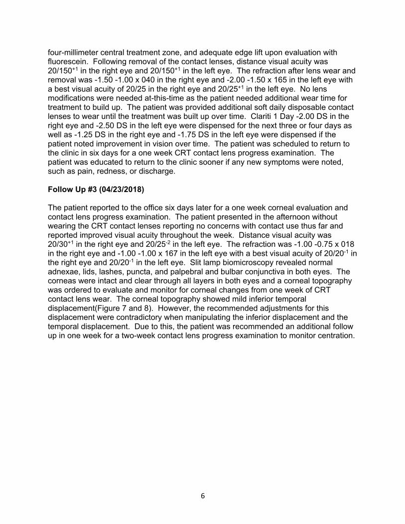
four-millimeter central treatment zone, and adequate edge lift upon evaluation with fluorescein. Following removal of the contact lenses, distance visual acuity was 20/150+1 in the right eye and 20/150+1 in the left eye. The refraction after lens wear and removal was -1.50 -1.00 x 040 in the right eye and -2.00 -1.50 x 165 in the left eye with a best visual acuity of 20/25 in the right eye and 20/25+1 in the left eye. No lens modifications were needed at-this-time as the patient needed additional wear time for treatment to build up. The patient was provided additional soft daily disposable contact lenses to wear until the treatment was built up over time. Clariti 1 Day -2.00 DS in the right eye and -2.50 DS in the left eye were dispensed for the next three or four days as well as -1.25 DS in the right eye and -1.75 DS in the left eye were dispensed if the patient noted improvement in vision over time. The patient was scheduled to return to the clinic in six days for a one week CRT contact lens progress examination. The patient was educated to return to the clinic sooner if any new symptoms were noted, such as pain, redness, or discharge. Follow Up #3 (04/23/2018) The patient reported to the office six days later for a one week corneal evaluation and contact lens progress examination. The patient presented in the afternoon without wearing the CRT contact lenses reporting no concerns with contact use thus far and reported improved visual acuity throughout the week. Distance visual acuity was 20/30+1 in the right eye and 20/25-2 in the left eye. The refraction was -1.00 -0.75 x 018 in the right eye and -1.00 -1.00 x 167 in the left eye with a best visual acuity of 20/20-1 in the right eye and 20/20-1 in the left eye. Slit lamp biomicroscopy revealed normal adnexae, lids, lashes, puncta, and palpebral and bulbar conjunctiva in both eyes. The corneas were intact and clear through all layers in both eyes and a corneal topography was ordered to evaluate and monitor for corneal changes from one week of CRT contact lens wear. The corneal topography showed mild inferior temporal displacement(Figure 7 and 8). However, the recommended adjustments for this displacement were contradictory when manipulating the inferior displacement and the temporal displacement. Due to this, the patient was recommended an additional follow up in one week for a two-week contact lens progress examination to monitor centration.

7
Figures 7 and 8. Corneal topography measured from an Oculus Keratograph displaying anterior tangential maps showing mild inferior temporal displacement of the treatment zone. Figure 7 shows topographical mapping of the right eye and Figure 8 shows topographical mapping of the left eye. Follow Up #4 (04/30/2018) The patient returned to the clinic one week later for a corneal evaluation and contact lens progress examination. The patient reported some dryness and mild irritation when he would remove his contact lenses in the morning. Distance visual acuity was 20/20-1 in the right eye, 20/25 in the left eye, and 20/20-1 in both eyes. The refraction was Plano -0.50 x 033 in the right eye and Plano -1.00 x 173 in the left eye with a best visual acuity of 20/20-1 in the right eye and 20/20-1 in the left eye. Slit lamp biomicroscopy revealed normal adnexae, lids, lashes, puncta, and palpebral and bulbar conjunctiva in both eyes. The corneas were intact and clear through all layers with no signs of epithelial defects in either eye and a corneal topography was ordered to evaluate and monitor for corneal changes from two weeks of CRT contact lens wear. Again, the corneal topography showed mild inferior temporal displacement (Figure 9 and 10). A consultation was completed with the lab sharing the corneal topography images, original keratometry, and refraction measurements, as well as the current visual acuity obtained. The lab recommended no parameter changes and to continue monitoring. They advocated for more lubrication when the patient inserted and removed his lenses. The patient was educated regarding lubrication use. This would also be beneficial for the dryness and irritation he reported when he would remove his lenses. A sample of Boston Rewetting Drops were provided for the patient to use. The patient was instructed to instill a drop in both eyes upon waking and bumping the bottom of the lens through his lid with his index finger for easier, more comfortable removal. The patient was scheduled for a follow up in two weeks for his one month contact lens progress examination.

8
Figures 9 and 10. Corneal topography measured from an Oculus Keratograph displaying anterior tangential maps showing some inferior temporal displacement of the treatment zone. Figure 9 shows topographical mapping of the right eye and Figure 10 shows topographical mapping of the left eye. Follow Up #5 (05/14/2018) The patient returned to the clinic two weeks later for a corneal evaluation and contact lens progress examination. The patient reported improvement in the dryness he reported in the mornings during removal of his contact lenses. Distance visual acuity was 20/20-1 in the right eye, 20/25+2 in the left eye, and 20/20-1 in both eyes. The refraction was -0.25 -0.50 x 015 in the right eye and -0.25 -0.50 x 163 in the left eye with a best visual acuity of 20/20-1 in the right eye and 20/20-1 in the left eye. Slit lamp biomicroscopy revealed normal adnexae, lids, lashes, puncta, and palpebral and bulbar conjunctiva in both eyes. The corneas were intact and clear through all layers with no signs of epithelial defects in both eyes and a corneal topography was ordered to evaluate and monitor for corneal changes from four weeks of CRT contact lens wear. Again, the corneal topography showed mild inferior temporal displacement (Figure 11 and 12). An additional consultation was completed with the lab sharing the corneal topography images, original keratometry, and refraction measurements as well as the current visual acuity obtained. The lab recommended no parameter changes. They reported that the patient was doing well regarding his presenting manifest refraction and keratometry measurements. The contact lenses were finalized at this time. The patient was recommended to return to the clinic in two to three months for a corneal evaluation and contact lens progress examination.

9
Figures 11 and 12. Corneal topography measured from an Oculus Keratograph displaying anterior tangential maps showing some inferior temporal displacement of the treatment zone. Figure 11 shows topographical mapping of the right eye and Figure 12 shows topographical mapping of the left eye. Discussion Orthokeratology was first introduced by Wesley and Jessen in the 1950’s.4 They described corneal reshaping following hard contact lens wear as “spectacle blur.” More developments in regards to oxygen permeability within rigid gas permeable lenses were made in the 1970’s, however, they were still not proficient in effectively correcting for myopia.4 In 1989, Richard Wlodyga developed reverse geometry lenses that improved centration and associated myopia correction.4 The reverse geometry design proved to be efficient in molding the cornea into a plateau shape creating a flat central base curve.4 The epithelial cells in the central cornea were redistributed into the mid-periphery due to the positive pressure centrally. This flattening effect changed refractive outcomes by creating myopic peripheral defocus.4
Recent animal studies performed with infant monkeys show that visual signals within the retinal periphery have an impact on emmetropization.5 These studies also demonstrate that the mechanisms of myopia progression are influenced by the entirety of the retina, not just being limited to the foveal area.6 Constant visual feedback and the ever-changing visual experience have been found to have profound effects on eye growth.1 The induction of hyperopic peripheral defocus can influence the central refractive error and has been studied to further develop myopia. Therefore, certain optical interventions can have a strong impact regarding the development of advancing refractive error.1,5,6
Myopia increases the risk of associated ocular diseases, such as glaucoma, cataracts, retinal detachment, and myopic maculopathy. Low myopes still show risk for these ocular diseases, however, high myopes are at an even higher risk proving the dose-

10
response relationship.3,7 The odds ratio increases as the amount of myopia progresses, therefore, reducing the amount of myopia remains beneficial.3,7 Investigators compare myopia’s risk for associated ocular disease with hypertension and smoking as risk factors for cardiovascular disease.6
Current research describes the etiology of myopia as consisting of both genetic factors as well as environmental factors.8 Parental myopia has shown to be a factor for an increased risk of myopia in children. Furthermore, an increased risk exists for children that have two myopic parents.9 Refractive errors that are less hyperopic than +0.75 at age five show an increased risk of myopia development.9 Also, literature shows an associated risk of myopia with reduced time spent outdoors.10 More time spent outdoors during ages of three to nine years old have a positive correlation with reduced myopia incidence in ages ten to fifteen years old.10 The incidence and prevalence of myopia has vastly increased in recent years. Literature reveals that myopia within the United States has increased from 25% in the 1970’s to 41% over a thirty year period.1,3,8,11 More recent research in 2000 estimated that 22.9% of the world population had myopia and 2.7% of the world population had high myopia consisting of -5.00 D or higher.11,12 Moreover, it is predicted that by 2050 49.8% of the world population will have myopia and 9.8% of the world population will have high myopia.11,12 The highest prevalence of myopia has been reported in Asia, especially East Asia and South-East Asia.1,2
Single vision glasses or contact lenses are noted as the first treatment option offered to patients and parents because this treatment method provides correction for the nearsighted vision and has little to no side effects.1 A treatment plan with single vision correction is not a form of myopia control and has been classically used as the control in myopia control studies.1 Undercorrection of myopia has also been investigated as a form of myopia control. However, research suggests that undercorrection of -0.75 D boosts myopia progression rather than slow it down.1 Next, multifocal spectacles, either flat-top bifocals or progressive lenses, have been studied in regard to their effect on myopia control. The Correction of Myopia Evaluation Trial (COMET) found that the myopia progression difference between children wearing a progressive versus single vision lens was clinically insignificant.1,13 However, the progressive lens design provides clear central vision while peripheral myopic defocus and relative plus power are incorporated. This design shows potential to be a beneficial treatment, especially if it is combined with other myopia control options. Lastly, a promising trial utilized the combination of prism and bifocals to reduce myopia progression.6 It resulted in favorable outcomes, however, continued study is needed. The most common prescribing habits for myopia control currently consist of soft multifocal contact lenses, low dose atropine, and orthokeratology. 37% of eye care providers within the United States have been actively practicing myopia control.11 Of those eye care providers, 51% have utilized soft multifocal contact lenses while 44% have incorporated orthokeratology into their practices.11 Additional studies are being

11
completed for atropine use. The most commonly known form of myopia control worldwide is reported to be orthokeratology.14 Soft multifocal contact lenses typically are designed concentrically as the power alternates from distance correction to plus power zones. This design stimulates two focal planes providing clear distance correction while inducing retinal myopic defocus.1 The Bifocal Lenses in Nearsighted Kids (BLINK) study is a current, on-going, randomized trial that is examining the effectiveness of soft multifocal center-distance contact lenses with a +2.50 add power.11 Although soft multifocal contact lenses are an off-label method, they have become a well-accepted form of myopia control because the patient can maintain good visual acuity while simultaneously inducing myopic peripheral defocus. Atropine has been extensively studied for its place within myopia control. It is a non-selective anti-muscarinic pharmaceutical agent that acts on the M1-M5 receptors located in the ciliary body and pupillary sphincter.1 Atropine’s exact mechanism of action is currently unknown as further research is on-going. This is due to the widespread distribution of muscarinic receptors within the eye that the agent could be impacting.1 Side effects that have been noted with atropine use are photophobia and near blur because atropine produces mydriasis and cycloplegic effects.1 Because of these side effects, it is common that patients will be prescribed progressive lenses in combination. The Atropine for the Treatment of Myopia studies (ATOM1 and ATOM2) tested different concentrations of Atropine for effectiveness as well as side effects. It was noted that after discontinuation of 0.5% and 0.1% atropine, there was a myopic rebound.15 However, lower dose 0.01% atropine did not show this rebound and has less side effects associated.15 Therefore, 0.01% has been utilized as it provides an optimal balance between effectiveness and safety.15 Orthokeratology, as described above, consists of overnight wear of a rigid gas permeable contact lens. Ultimately, this lens reshapes the corneal topography correcting for mild to moderate amounts of myopia while inducing peripheral myopia defocus.1 Many studies have proven the efficacy and safety over long periods of wear for this form of myopia control showing that myopia was controlled by 30% more when compared to a soft contact lens wearer over a ten year period.4,16 Orthokeratology lenses have been shown to inhibit axial elongation more effectively when compared to single vision correction.2,17 Microbial keratitis remains the most concerning orthokeratology complication today.18 This can be avoided with proper education, fitting, compliance with the wear and care, appropriate follow-up care, and adequate treatment when complications arise.18 Orthokeratology provides patients with freedom from spectacle or contact lens wear during the day and can provide treatment of 50% reduction of myopia after even just one night of wear.17
Conclusion

12
In summary, with the growing prevalence of myopia and its recognition as a public health concern, incorporating myopia control treatment options into clinical practice is becoming essential. This case report reviews the different options of myopia control, which is important when educating patients and their parents. In addition, the benefits of employing the assistance of lab consultants when determining parameter changes for specialty contact lens fittings is also demonstrated. It is important for providers to stay abreast of the literature as further studies are performed on this topic in the future. References 1. Kang P. Optical and pharmacological strategies of myopia control. Clinical and Experimental Ophthalmology. 2018;101:321-332.

13
2. Na M, Yoo A. The effect of orthokeratology on axial length elongation in children with myopia: Contralateral comparison study. Japanese Journal of Ophthalmology. 2018. 3. Huang J, et al. Efficiency Comparison of 16 Interventions for Myopia Control in Children: A Network Meta-analysis. Ophthalmology. 2016;123(4):697-708. 4. Lee YC, Wang JH, Chiu CJ. Effect of Orthokeratology on myopia progression: twelve-year results of a retrospective cohort study. BMC Ophthalmology. 2017;17:243. 5. Smith EL, Hung LF, Huang J. Relative Peripheral Hyperopic Defocus Alters Central Refractive Development in Infant Monkeys. Vision Research. 2009;49(19):2386-2392. 6. Flitcroft D. The complex interactions of retinal, optical, and environmental factors in myopia aetiology. Progress in Retinal and Eye Research. 2012;31(6):622-660. 7. Aller TA. Clinical management of progressive myopia. Eye. 2014;28(2):147-153. 8. Vitale S, Sperduto RD, Ferris III FL. Increased Prevalence of Myopia in the United States Between 1971-1977 and 1999-2004. Archives of Ophthalmology. 2009;127(12):1632-1639. 9. Jones-Jordan LA, Sinnott LT, Manny RE, et al. Early Childhood Refractive Error and Parental History of Myopia as Predictors of Myopia. Investigative Ophthalmology and Visual Science. 2010;51(1):115-121. 10. Shah RL, Huang Y, Guggenheim JA, Williams C. Time Outdoors at Specific Ages During Early Childhood and the Risk of Incident Myopia. Investigative Ophthalmology and Visual Science. 2017;58(2):1158-1166. 11. Schulle KL, Berntsen DA, Sinnott LT, Bickle KM, Gostovic AT, Pierce GE, Jones-Jordan LA, Mutti DO, Walline JJ, Bifocal Lenses in Nearsighted Kids (BLINK) Study Group. Visual Acuity and Over-Refraction in Myopic Children Fitting with Soft Multifocal Contact Lenses. Optometry and Vision Science. 2018;95(4):292-298. 12. Holden BA, Fricke TR, Wilson DA, et al. Global prevalence of myopia and high myopia and temporal trends from 2000 through 2050. Ophthalmology. 2016;123:1036-1042. 13. Berntsen DA, Barr CD, Mutti DO, et al. Peripheral Defocus and Myopia Progression in Myopic Children Randomly Assigned to Wear Single Vision and Progressive Addition Lenses. Investigative Ophthalmology and Visual Science. 2013;54:5761-5770.

14
14. Cheung SW, Lam C, Cho P. Parents’ knowledge and perspective of optical methods for myopia control in children. Optometry and Vision Science. 2014;91:634-641. 15. Chia A, Chua WH, Cheung YB, Wong WL, Lingham A, Fong A, Tan D. Atropine for the treatment of childhood myopia: safety and efficacy of 0.5%, 0.1%, and 0.01% doses (Atropine for the Treatment of Myopia 2). Ophthalmology. 2012;119(2):347-354. 16. Hiraoka T, Sekine Y, Okamota F, Mihashi T, Oshika T. Safety and efficacy following 10-years of overnight orthokeratology for myopia control. Ophthalmic and Physiological Optics. 2018;38:281-289. 17. Cho P, Cheung SW. Discontinuation of orthokeratology on eyeball elongation (DOEE). Contact Lens and Anterior Eye. 2017;40(2):82-87. 18. Liu YM, Xie P. The Safety of Orthokeratology—A Systematic Review. Eye and Contact Lens. 2016;93(4):336-343.