clinical features and research opportunities in rheumatoid arthritis clinical immunology march 26,...
TRANSCRIPT

Clinical features and research opportunities in rheumatoid arthritis
Clinical Immunology
March 26, 2013
HARVARDMEDICAL SCHOOL

Overview
• Clinical characteristics and pathophysiology
• Differential diagnosis
• Exam and laboratory studies
• Treatment strategy
• Research opportunities

Overview
• Clinical characteristics and pathophysiology
• Differential diagnosis
• Exam and laboratory studies
• Treatment strategy
• Research opportunities
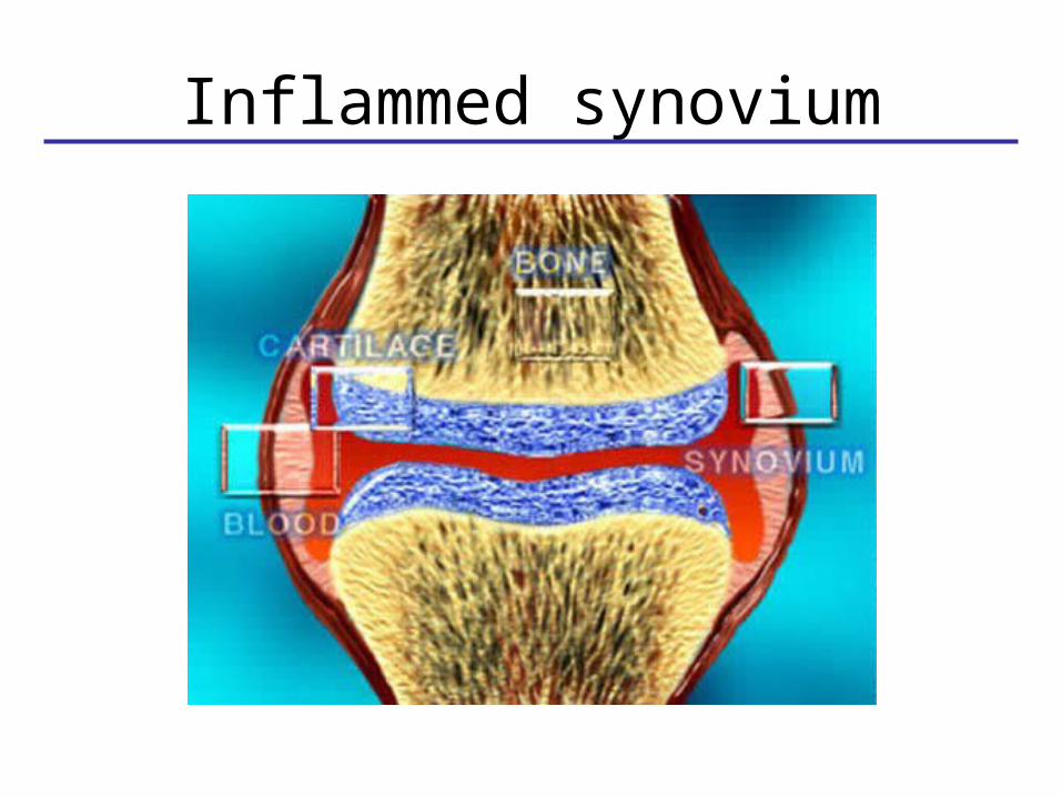
Inflammed synovium

Rheumatoid Synovium
Normal Synovium
Lining
Sublining

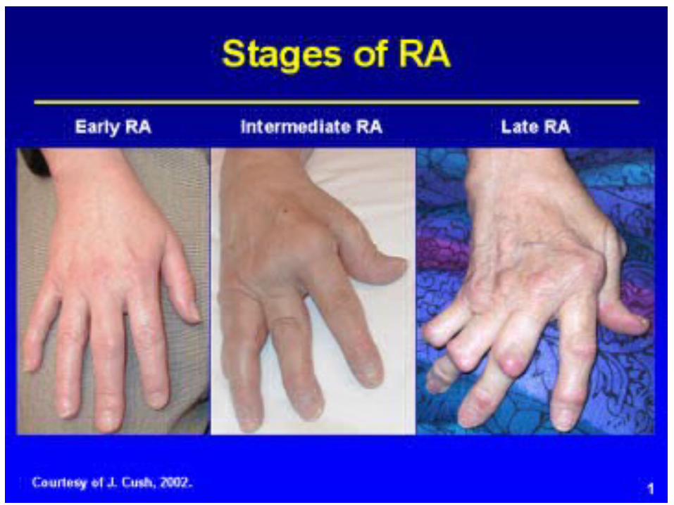
The 1-2-3 of Rheumatoid Arthritis
Lee, Kiener and Brenner, Synoviocytes 2004

Understanding pathogenesis
Klareskog et al Lancet 2009

Clinical characteristics
• Systemic chronic inflammatory disease• Mainly affects synovial joints• Variable expression • Extra-articular manifestations (e.g., nodules, ILD,
ocular)
• Prevalence ~1%• Worldwide distribution• Female: Male ratio 3:1• Peak age of onset 30 – 50 years (median in 40’s)

ACR Criteria for Diagnosis
• Four or more of the following criteria must be present:
• Morning stiffness >1 hour• Arthritis of >3 joint areas• Arthritis of hand joints (MCPs, PIPs, wrists)• Symmetric swelling (arthritis)
• Serum rheumatoid factor• Rheumatoid nodules• Radiographic changes
First four criteria must be present for 6 weeks or more

Overview
• Clinical characteristics and pathophysiology
• Differential diagnosis
• Exam and laboratory studies
• Treatment strategy
• Research opportunities

Differential Diagnosis• Rheumatoid Arthritis
• Psoriatic Arthritis• Inflammatory bowel disease• Ankylosing spondylitis
• Crystal – Gout, Pseudogout
• SLE, Vasculitis
• PMR-GCA
• Any “immune complex” illness
• Paraneoplastic syndrome
• Viral – Parvovirus, HepBSAg, HCV, Rubella
• Bacterial – Lyme, GC, chlamydia
• Osteoarthritis, bursitis, tendonitis

Conceptual organization
• Inflammatory vs. non-inflammatory– synovitis vs structural
• Articular vs. non-articular
• Systemic vs. regional
• Polyarticular vs. monarticular
• Extra-articular manifestationsNote: Older patients need more careful history and physical exam-labs often confusing

Pertinent historical features• Duration
– acute vs chronic– gradual vs abrupt onset
• Pattern– symmetrical vs asymetrical– large vs small joints– morning stiffness– effect of activity
• Joint distribution– DIP vs PIP/MCP
DIP
PIP
MCP

Overview
• Clinical characteristics and pathophysiology
• Differential diagnosis
• Exam and laboratory studies
• Treatment strategy
• Research opportunities

• Tenderness
• synovitis = tender joint
• mechanical or periarticular lesions (bursitis and tendonitis) = tenderness often localized
• Swelling
• bony vs. soft tissue swelling
• Pattern
• proximal vs. distal
• asymetric vs. symmetric
• DIP and nail changes
Physical exam of joint

• Swelling is confined to the area of the joint capsule
• Synovial thickening feels like a firm sponge
Proximal InterPhalangeal joint
PIP

MetaCarpal Phalangeal joint
MCP
• Rheumatoid Arthritis
• Psoriatic Arthritis• Inflammatory bowel disease• Ankylosing spondylitis
• Crystal – Gout, Pseudogout
• SLE, Vasculitis
• PMR-GCA
• Any “immune complex” illness
• Paraneoplastic syndrome
• Viral – Parvovirus, HepBSAg, HCV, Rubella
• Bacterial – Lyme, GC, chlamydia
• Osteoarthritis, bursitis, tendonitis
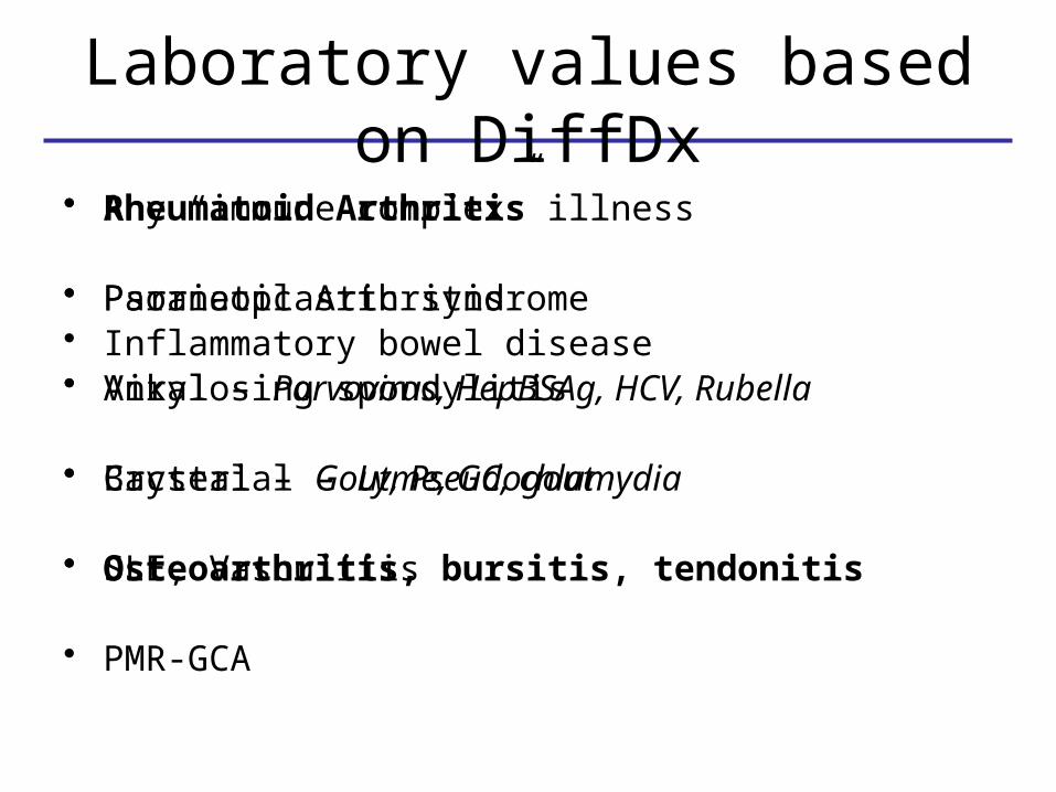
Laboratory values based on DiffDx
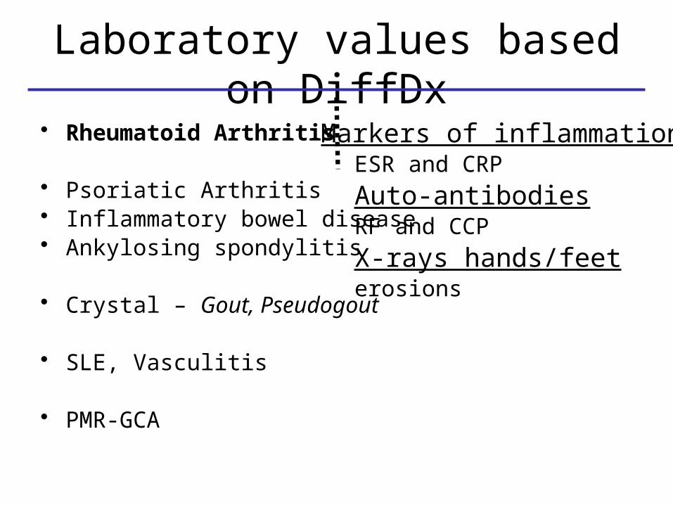
Laboratory values based on DiffDx
• Rheumatoid Arthritis
• Psoriatic Arthritis• Inflammatory bowel disease• Ankylosing spondylitis
• Crystal – Gout, Pseudogout
• SLE, Vasculitis
• PMR-GCA
Markers of inflammationESR and CRP
Auto-antibodiesRF and CCP
X-rays hands/feeterosions

Laboratory values based on DiffDx
• Rheumatoid Arthritis
• Psoriatic Arthritis• Inflammatory bowel disease• Ankylosing spondylitis
• Crystal – Gout, Pseudogout
• SLE, Vasculitis
• PMR-GCA
joint aspirationPresence of crystals
bloodUric acid
X-rayserosions

Laboratory values based on DiffDx
• Rheumatoid Arthritis
• Psoriatic Arthritis• Inflammatory bowel disease• Ankylosing spondylitis
• Crystal – Gout, Pseudogout
• SLE, Vasculitis
• PMR-GCA
autoantibodiesANAANCA
bloodCH50
urineurinalysis
X-rayslack of erosions

• Cell count and differential
• Crystals
• Gram stain and culture
non-inflammatory <1500mildly inflammatory 1500-3500inflammatory >3500possible infection >50,000
Joint fluid analysis

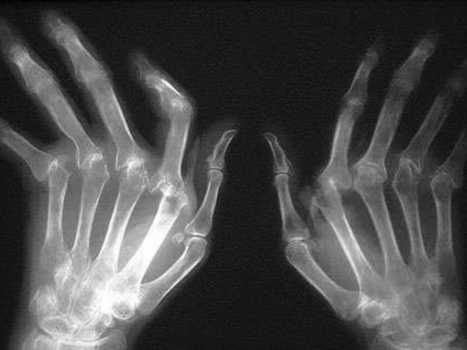
Clinical utility of x-rays• X-rays show only bone, not cartilage or
synovium• Lesions must correlate w/ clinical picture• Erosive pattern (or lack) useful in diff.
diagnosis• Early inflammatory lesions often non-
specific• X-ray changes take months to occur
– avascular necrosis not visible for 6 wks– spondylitis not evident for 2 – 10 yrs
• Valuable for plotting the clinical course in terms of structural changes

Patterns of radiographic changes
RA OAgout
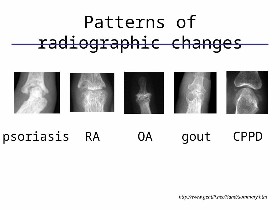
Patterns of radiographic changes
RApsoriasis
http://www.gentili.net/Hand/summary.htm
OA gout CPPD
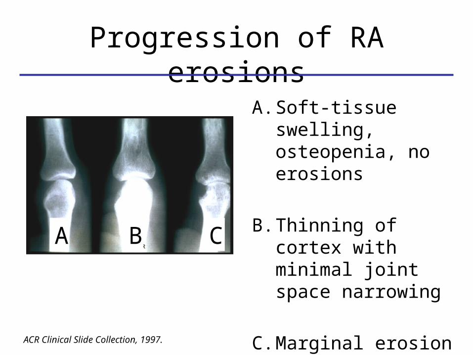
A. Soft-tissue swelling, osteopenia, no erosions
B. Thinning of cortex with minimal joint space narrowing
C. Marginal erosion with joint space narrowing
How fast is joint damage progressing?
ACR Clinical Slide Collection, 1997.
Progression of RA erosions
A B C

Overview
• Clinical characteristics and pathophysiology
• Differential diagnosis
• Exam and laboratory studies
• Treatment strategy
• Research opportunities
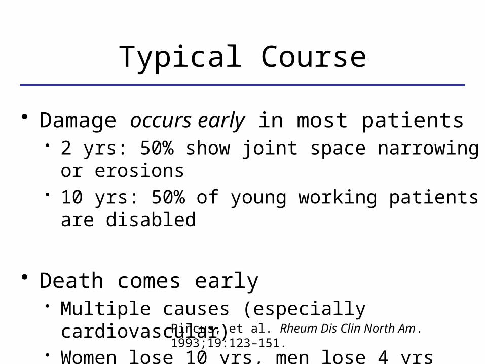
Pincus, et al. Rheum Dis Clin North Am. 1993;19:123–151.
Typical Course
• Damage occurs early in most patients • 2 yrs: 50% show joint space narrowing or
erosions• 10 yrs: 50% of young working patients are
disabled
• Death comes early• Multiple causes (especially cardiovascular)• Women lose 10 yrs, men lose 4 yrs


• Determine spectrum of disease
• Use the safest treatment plan that matches the aggressiveness of the disease
• Monitor treatment for adverse effects
• Monitor disease activity, revise Rx as needed
Treatment principles

General strategy of treatment escalation in RA patients


abatacept(Orencia)

Overview
• Clinical characteristics and pathophysiology
• Differential diagnosis
• Exam and laboratory studies
• Treatment strategy
• Research opportunities

Cost is increasing, productivity is decreasing
Scannell et al Nat Rev Drug Discovery (2012)
We need new drugs to treat RA and other
complex traits!
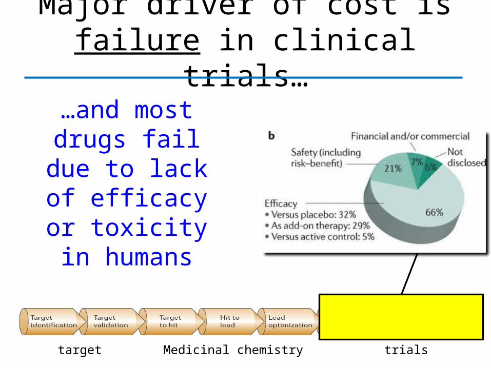
Major driver of cost is failure in clinical trials…
target Medicinal chemistry trials
…and most drugs fail due to lack of efficacy or toxicity
in humans

“Target validation” is key to avoid failure from efficacy/safety
Current models are ineffective at
choosing targets that are safe and
effective in humans
target Medicinal chemistry trials

target Medicinal chemistry trials
We determine dose-response in clinical trials, after many
years and millions of dollars
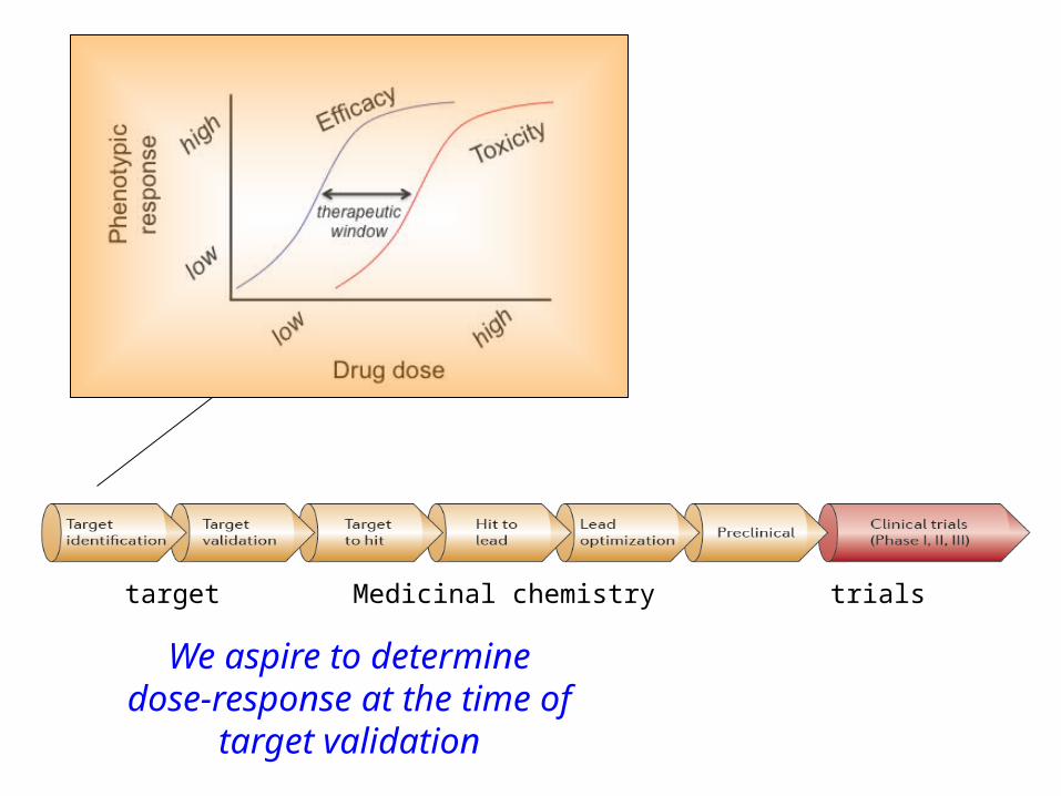
target Medicinal chemistry trials
We aspire to determine dose-response at the time
of target validation

• Nature’s perturbation of many drug targets in the human genome
• Links physiological state in humans (e.g., disease risk) to a target perturbation
• Indicates gain- or loss-of-function• Provides allelic series for range of effect
on perturbing a potential drug target
Human genetics is a unique tool for target validation
Dose-response curves derived from human genetics

44
The history and success of GWAS – illuminating for common phenotypes
2007
Data: www.genome.gov/GWAStudies - slide from Sara Pulit and Paul de Bakker
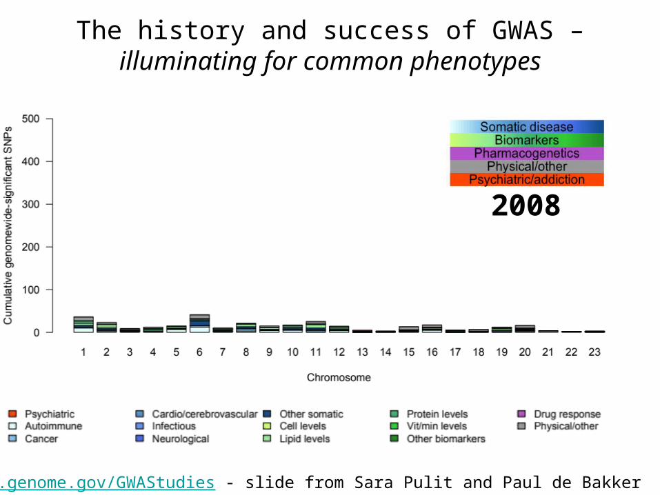
Data: www.genome.gov/GWAStudies - slide from Sara Pulit and Paul de Bakker
2008
The history and success of GWAS – illuminating for common phenotypes
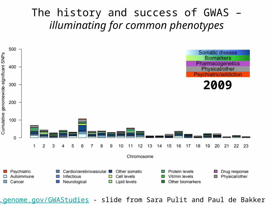
2009
Data: www.genome.gov/GWAStudies - slide from Sara Pulit and Paul de Bakker
The history and success of GWAS – illuminating for common phenotypes

2010
Data: www.genome.gov/GWAStudies - slide from Sara Pulit and Paul de Bakker
The history and success of GWAS – illuminating for common phenotypes
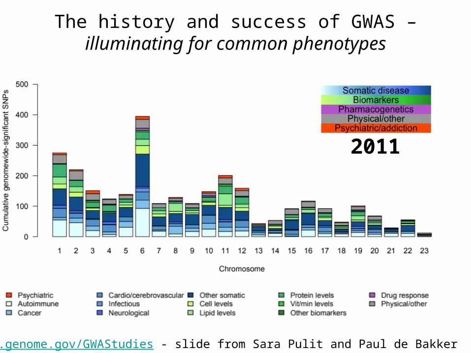
2011
Data: www.genome.gov/GWAStudies - slide from Sara Pulit and Paul de Bakker
The history and success of GWAS – illuminating for common phenotypes

2012
Data: www.genome.gov/GWAStudies - slide from Sara Pulit and Paul de Bakker
The history and success of GWAS – illuminating for common phenotypes

Similarly, great success in unraveling genetics of RA
Plenge et al NEJM 2007
15
10
5
0
Chromosomal position
GWAS 1,522 RA cases, 1,850 controls
No. GWAS hits = 3
Total No. risk loci = 5*
(* includes replication beyond GWAS)

Similarly, great success in unraveling genetics of RA
Raychaudhuri et al Nat Gen 2008
15
10
5
0
Chromosomal position
GWAS 3,393 RA cases, 12,462 controls
No. GWAS hits = 4
Total No. risk loci = 6

Similarly, great success in unraveling genetics of RA
15
10
5
0
Chromosomal position
Stahl et al Nat Gen 2010
GWAS 5,539 RA cases, 20,169 controls
No. GWAS hits = 9
Total No. risk loci = 25

From 1 to 100
Yukinori Okada et al unpublished
15
10
5
0
Chromosomal position
GWAS 19,234 RA cases, 61,565 controls
No. GWAS hits = 56
Total No. risk loci = ~100

Given the wealth of GWAS and other genetic data…how should it be used for drug discovery?

(1) “look-up” method – simple and
suggestive but undisciplined
(2) “Allelic series” method – powerful but
likely infrequent
(3) “pathway” method – powerful and
comprehensive but target ID difficult
Three potential solutions

(1) “look-up” method – simple and
suggestive but undisciplined
(2) “Allelic series” method – powerful but
likely infrequent
(3) “pathway” method – powerful and
comprehensive but target ID difficult
Three potential solutions

(1) “look-up” method – simple and
suggestive but undisciplined
(2) “Allelic series” method – powerful but
likely infrequent
(3) “pathway” method – powerful and
comprehensive but target ID difficult
Three potential solutions

Allelic series in PCSK9: loss-of-fxn, protective for CAD
2
3
1
1. Allelic series of LOF mutations alter PCSK9
2. Lowers LDL cholesterol3. Protects against CAD4. No obvious “ADE”
phenotypes

Allelic series in PCSK9: no obvious “adverse events”
2
3
4
1. Allelic series of LOF mutations alter PCSK9
2. Lowers LDL cholesterol3. Protects against CAD4. No obvious “ADE”
phenotypes

Monoclonal antibodies to PCSK9: dramatically lower LDL levels

(1) “look-up” method – simple and
suggestive but undisciplined
(2) “Allelic series” method – powerful but
likely infrequent
(3) “pathway” method – powerful and
comprehensive but target ID difficult
Three potential solutions

Polygenic architecture but discrete biological pathways
Rossin et al (2011) PLoS Genetics
CD40-CD40L pathway
1. CD40 is expressed on surface of B lymphocytes
2. Pathway is upregulated in inflammed synovial tissue of RA patients
3. CD40 mutations lead to human immunodeficiency

Cell-based phenotype screens to find inhibitors of CD40 signaling
“target” is a pathway, rather than a specific
molecule
Gang Li et al in press PLoS Genetics

Using this HTS assay, test >2000 chemical compounds
luciferase
FDA-approved drugs, other

Identified two “known” and two “novel” compounds
luciferase


• 34-year-old woman • 5-year history of RA• Morning stiffness = 30 minutes• Exam
– Synovitis: 1+ swelling of MCP, PIP, wrist, and MTP joints
– Normal joint alignment• Labs
– ESR and CRP normal– RF positive (CCP negative)
• No erosions seen on x-rays
Case #1

Case #1 (continued)
*
• Assessment• current activity: mild• no sign of damage after 5 years• anticipate minimally progressive course
• Treatment• NSAIDs prn• safer, less potent drugs (SSZ or HCQ) daily• education• ROM, conditioning, and strengthening
exercises

• 34-year-old woman • 1-year history of RA• Morning stiffness = 90 minutes• Exam
– Synovitis: 2+ of MCP, PIP, wrist, knee, and MTP joints
– Normal joint alignment• Labs
– ESR and CRP elevated– RF positive and CCP positive
• Small erosions seen on x-rays
Case #2

Early erosion at the tip of the ulnar styloid
Case #2 (continued)

• Assessment• current activity: moderate with more joint
involvement• early radiographic damage with CCP+• anticipate progressive course
• Treatment• Initial treatment with prednisone, NSAIDs• Start MTX weekly• Education on pregnancy, alcohol, risks,
benefits• ROM, conditioning, and strengthening
exercises
Case #2 (continued)
*
~1/3 of patients will respond adequately

• 34-year-old woman • 2-year history of RA• Morning stiffness = 3 hours• Exam
– Synovitis: 3+ swelling of MCP, PIP, wrist, and MTP joints
– Ulnar deviation, decreased ROM wrists– nodules on elbows
• Labs– ESR and CRP elevated– RF positive and CCP positive
• prednisone (10 mg QD) + MTX (25 mg Qweek)
Case #3

• Assessment• current activity: severe, poorly controlled on
MTX• Clear destruction with CCP+• progressive course
• Treatment• continue prednisone, NSAIDs, MTX• Add anti-TNF therapy• Education on risks of infection• ROM, conditioning, and strengthening
exercises
Case #3 (continued)
*
~1/3 of patients will respond adequately