clinical dataset uveitis dataset the royal college … 5 are particularly relevant to the care of...
TRANSCRIPT

Clinical Dataset
Uveitis Dataset
The Royal College of Ophthalmologists
Authorship Group:
Alastair K. Denniston, Richard W. Lee, Carlos Pavesio, Miles R Stanford, Philip I Murray, Annabelle Okada, H. Nida Sen, Andrew D. Dick
October 2018

2018/PROF/389 2
Date of review: October 2021
Contents
Section page
1 Uveitis and the Principles of a Common Dataset 3
1.1 Introduction 3
1.2 Aims 3
1.3 Principles 4
1.4 Specific Challenges 5
2 The Uveitis Dataset 5
2.1 Type of Uveitis 5
2.2 Current Status 10
2.3 Time-course and complications of uveitis 11
2.4 Major therapeutic interventions 13
2.5 Investigations 16
3 References 16
Appendix A. Summary of Minimum dataset 17
Appendix B. Extended dataset for clinical features: 24
2018/PROF/389 3
1 Uveitis and the Principles of a Common Dataset
1.1 Introduction
Uveitis describes a complex collection of conditions characterized by intraocular inflammation.1 As a group, uveitis is a significant cause of blindness worldwide. In the industrialised world it is thought to account for about 10–15% of the cases of total blindness (World Health Organization definition) and up to 20% of legal blindness.2-3
In addition to the direct visual impairment that may result from intraocular inflammation, the impact of uveitis is compounded by two important factors: first, many forms of uveitis are associated with significant systemic disease;4 and second, many of the sight-threatening forms of uveitis require local and/or systemic therapies that are accompanied by significant drug-related morbidity.5
The study of the clinical practice of uveitis – both for research and auditing purposes – is challenged by the individual scarcity of most of the constituent syndromes,6 and a lack of consensus about what a ‘gold standard’ of optimal care would look like.7 Valuable data does exist but is usually retrospective, being based on the post-hoc interpretation of case-notes, as seen in the Systemic Immunosuppressive Therapy for Eye Diseases (SITE) Cohort Study.8 The relatively few number of studies that are prospective are narrow in scope, usually syndrome-specific prospective studies such as in Birdshot Chorioretinopathy.9 These syndrome-specific studies are valuable in their own right but do not enable inter-syndrome comparisons.
The Royal College of Ophthalmologists has been supporting the development of Datasets for over a decade, with national Datasets for both cataract and diabetic retinopathy, among others. Such Datasets help provide a standardized language for clinical care, and a tool for outcome analysis, clinical audit, revalidation, and research. Although the formation of a Dataset for uveitis is particularly challenging, the future benefits are apparent, with the potential to mark major shifts both in clinical care (including auditing and bench-marking) and the research environment.
1.2 Aims
The overall purpose of this proposal is to provide a Dataset that enables standardization of clinical data collection reflective of routine clinical care and of value for audit and research purposes. Specific aims within this are to provide:
1) a minimum Dataset which is mandatory
2) an extended Dataset which is desirable but not mandatory
It is not within the scope of this work to define a Dataset by which quality of care can be audited for revalidation purposes but it would be anticipated that such a Dataset would be a subset of the minimum dataset, and would take into account the distribution of cases (type
2018/PROF/389 4
and severity), their therapeutic interventions and their visual outcome over time. Patient reported outcomes would also be desirable but are not currently collected in routine practice for most ophthalmic conditions.
1.3 Principles
1.3.1 General
This dataset ascribes to the core principles elucidated in other RCOphth Datasets10 namely that:
• The minimum Dataset should be a subset of information routinely collected so as not to add to the demands on busy clinicians.
• Data Elements should only be included if they are identified as being subjects of interest that will be analysed.
• Elements in common with other College Datasets should be congruent; for the purposes of this report we do not describe these in detail but defer to the relevant Dataset.
• The Dataset should be capable of implementation in an electronic medical record.
1.3.2 The Uveitis Minimum Dataset
The minimum Dataset comprises those elements that should be recorded in every case of uveitis, and would be regarded as standard of care for those conditions. The elements included here are those that were considered essential to defining the type of uveitis, the severity of disease, the major therapeutic interventions and the current status.
• The type of uveitis is defined according to anatomical classification and course (both according to the Standardization of Uveitis Nomenclature recommendations11), and aetiological classification (major headings according to the International Uveitis Study Group1).
• The current status comprises current visual function as measured by visual acuity and the current measures of active inflammation comprising key measures of active inflammation that have been described according to standardized grades (AC cells, AC flare and vitreous haze).
• The severity and time-course of uveitis is assessed by the collection of key measures of active inflammation over time and the presence of potentially sight-threatening complications; the date of the patient’s first presentation and the date of first onset of any sight-threatening complications are also recorded.
• The major therapeutic interventions comprise significant medical and surgical interventions and their approximate dates.
The Uveitis Minimum Dataset is discussed further below and is provided as Appendix A.
1.3.3 The Uveitis Extended Dataset
The extended Dataset comprises the above data elements plus many additional elements that are relevant to the clinical assessment or treatment pathway in some or all types of uveitis. The extended Dataset is included to provide guidance to electronic medical records developers regarding the inclusion and format of those elements of the clinical record that

2018/PROF/389 5
are particularly relevant to the care of patients with uveitis. Although it would be ideal to have the extended Dataset recorded in all patients, it is recognized that in routine clinical practice the capture of different elements will be prioritised over others according to their perceived relevance to the uveitis syndrome in question, and thus completion of these elements will be variable. The provision of the extended Dataset does however provide the opportunity for electronic medical record developers to ensure standardization of fields so that in those cases where clinicians do perceive their assessment and recording to be important, these data can be aggregated and analysed. It is recognized that even the Extended Dataset is not exhaustive, as the nature of uveitis means that a truly comprehensive Dataset would include an unmanageable number of elements of both ophthalmic and systemic disease, their complications and treatments
The Uveitis Extended Dataset is discussed further below and is provided in Appendix B.
1.4 Specific Challenges
It is acknowledged that the development of a core dataset for uveitis is made more challenging due its complexity, heterogeneity, and limitations in the current system of taxonomy and classification which serve to highlight our imperfect understanding of the aetiology of much uveitis. It is however partly because of these challenges – and the variable ways in which much uveitis is recorded and reported – that a standardized Dataset for uveitis would be of immense value, enabling prospective data gathering in a coherent large-scale manner. There will inevitably be debate as to what characteristics are essential vs desirable, but the priority for the mandatory elements that comprise the core dataset has been to collect those variables that are broadly applicable to all forms of uveitis and reflect the overall care of those patients.
2 The Uveitis Dataset
The minimum Dataset comprises those elements that should be recorded in every case of uveitis, and would be regarded as standard of care for those conditions.
2.1 Type of Uveitis
2.1.1 Anatomical classification of uveitis
Mandatory Data element = Anatomical classification of uveitis according to the Standardization of Uveitis Nomenclature11 down to ‘subvalue’ level.
2018/PROF/389 6
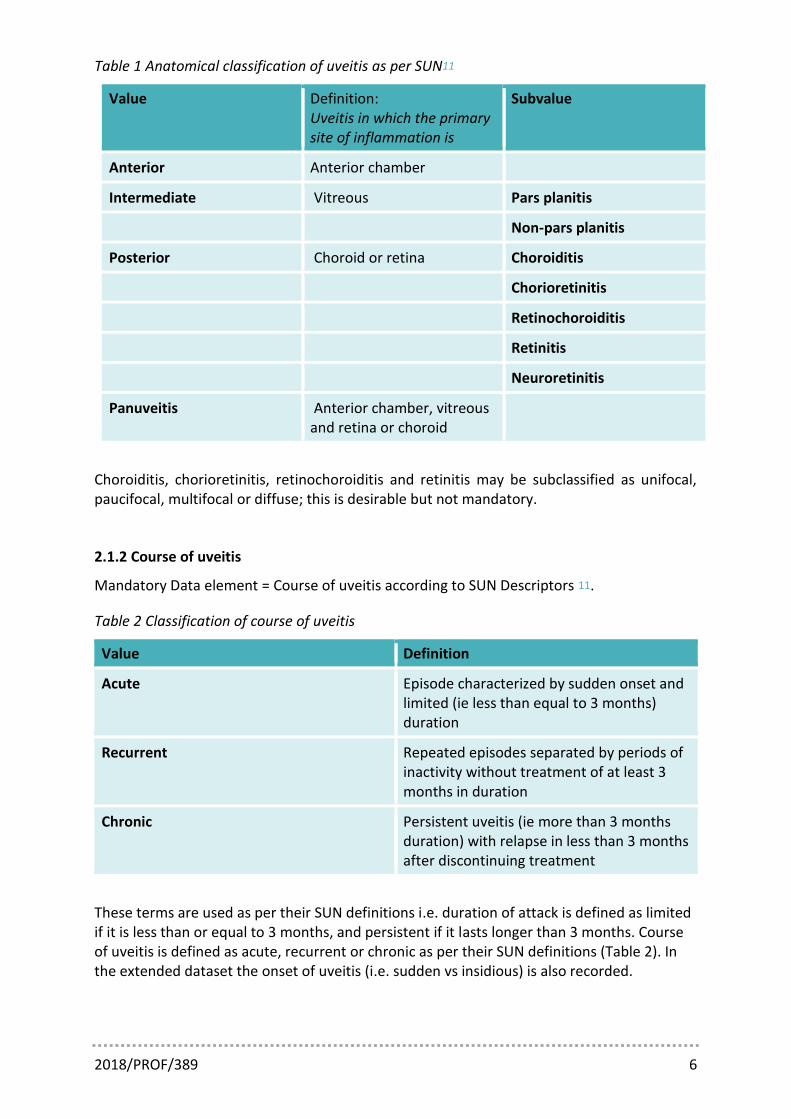
Table 1 Anatomical classification of uveitis as per SUN11
Value Definition: Uveitis in which the primary site of inflammation is
Subvalue
Anterior Anterior chamber
Intermediate Vitreous Pars planitis
Non-pars planitis
Posterior Choroid or retina Choroiditis
Chorioretinitis
Retinochoroiditis
Retinitis
Neuroretinitis
Panuveitis Anterior chamber, vitreous and retina or choroid
Choroiditis, chorioretinitis, retinochoroiditis and retinitis may be subclassified as unifocal, paucifocal, multifocal or diffuse; this is desirable but not mandatory.
2.1.2 Course of uveitis
Mandatory Data element = Course of uveitis according to SUN Descriptors 11.
Table 2 Classification of course of uveitis
Value Definition
Acute Episode characterized by sudden onset and limited (ie less than equal to 3 months) duration
Recurrent Repeated episodes separated by periods of inactivity without treatment of at least 3 months in duration
Chronic Persistent uveitis (ie more than 3 months duration) with relapse in less than 3 months after discontinuing treatment
These terms are used as per their SUN definitions i.e. duration of attack is defined as limited if it is less than or equal to 3 months, and persistent if it lasts longer than 3 months. Course of uveitis is defined as acute, recurrent or chronic as per their SUN definitions (Table 2). In the extended dataset the onset of uveitis (i.e. sudden vs insidious) is also recorded.

2018/PROF/389 7
2.1.3 Clinical and Taxonomic classification
Table 3 Clinical classification reflecting taxonomy
Element Value Subvalue Sub-subvalue Sub-sub-subvalue
Classification by taxonomy
Infectious Bacterial Mycobacterium tuberculosis (TB)
Ocular only With extraocular involvement
Treponema pallidum (Syphilis)
Ocular only With extraocular involvement
Bartonella henselae (Cat-scratch disease)
Ocular only With extraocular involvement
Borrelia burgdorferi (Lyme disease)
Ocular only With extraocular involvement
Other bacterial Specify
Viral HSV1 Anterior uveitis Keratouveitis Acute Retinal Necrosis Progressive Outer Retinal Necrosis
HSV2 Anterior uveitis Keratouveitis Acute Retinal Necrosis Progressive Outer Retinal Necrosis
VZV Anterior uveitis Keratouveitis Acute Retinal Necrosis Progressive Outer Retinal Necrosis
CMV Anterior uveitis Keratouveitis Posterior uveitis

2018/PROF/389 8
Element Value Subvalue Sub-subvalue Sub-sub-subvalue
Viral syndrome (undifferentiated)
Anterior uveitis Keratouveitis Acute Retinal Necrosis Progressive Outer Retinal Necrosis
Other viral Specify
Fungal Candida sp.
Aspergillus sp.
Other fungal Specify
Parasitic Toxoplasma gondii
Toxocara canis
Onchocerca volvulus
Diffuse unilateral subacute necrosis
Other parasitic Specify
Other
Non-Infectious
No systemic disease
Acute/Recurrent Anterior Uveitis
HLA-B27-positive HLA-B27-negative HLA-B27 unknown
Chronic Anterior Uveitis
Fuchs Uveitis Syndrome
Intermediate Uveitis Pars planitis Non-pars planitis
Acute Posterior Multifocal Placoid Pigment Epitheliopathy
Multiple Evanescent White Dot Syndrome
Multifocal Choroiditis with Panuveitis
Punctate Inner Choroidopathy
Ampiginous Choroiditis
Birdshot Chorioretinopathy
Serpiginous Choroiditis

2018/PROF/389 9
Element Value Subvalue Sub-subvalue Sub-sub-subvalue
Other non-infectious (no systemic disease)
Specify
Systemic disease
Ankylosing spondylitis
Sarcoidosis
Multiple sclerosis
Behcet Disease
Sympathetic Ophthalmia
Vogt-Koyanagi-Harada Disease
Tubulointerstitial Nephritis and Uveitis
Juvenile idiopathic arthritis
Other non-infectious (systemic disease)
Specify
Masquerade Neoplastic Specify
Non-neoplastic
Undifferentiated
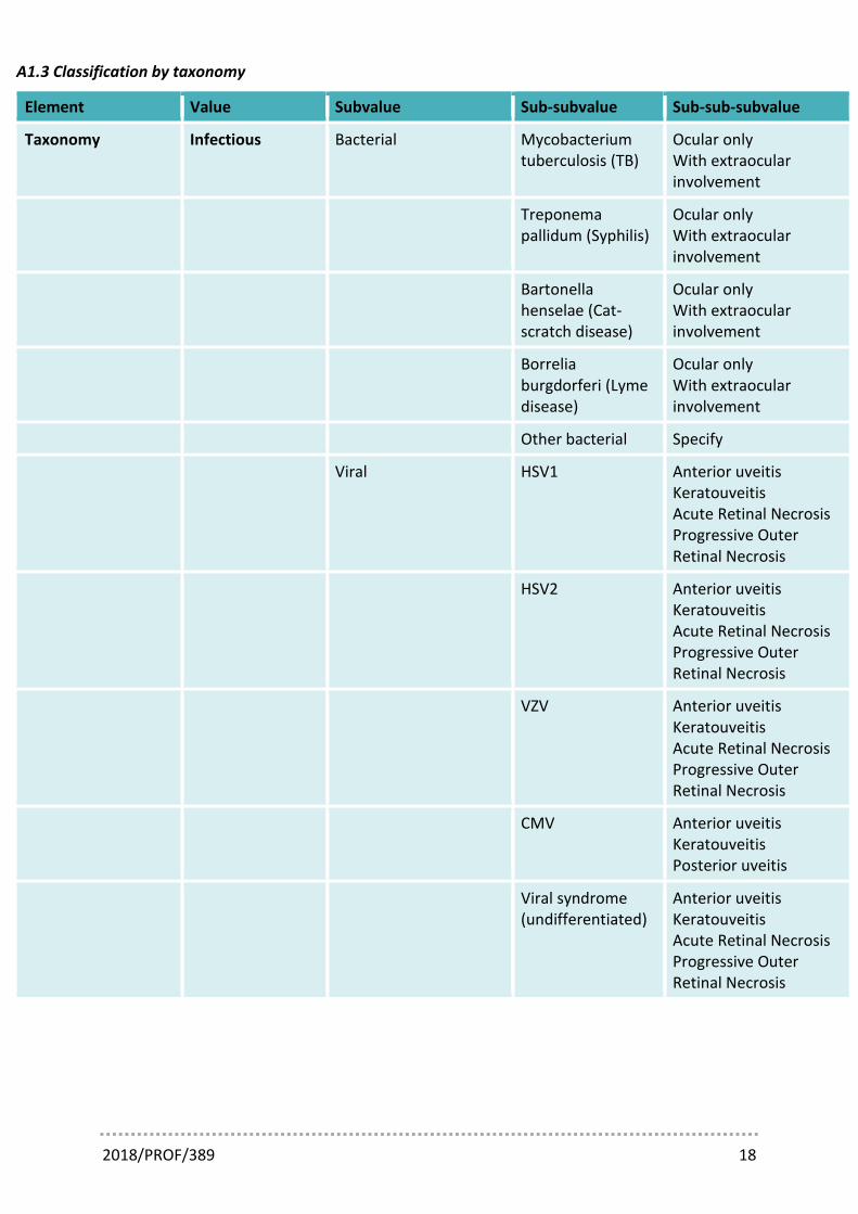
The Clinical Classification of the International Uveitis Study Group1 provide major clinical headings which to some extent reflects aetiology, at least where this is known. It is recognized that, for many of the ‘Non-infectious’ group, aetiology is poorly defined particularly for those syndromes not associated with a systemic disease. For this reason we refer to this as ‘classification by taxonomy’. The formation of a hierarchical sub-classification beyond that provided by the IUSG is difficult, but is clearly desirable in order to support consistency in EMR systems. The system proposed remains consistent with the IUSG outline and the ongoing work of SUN. A couple of points are worth noting in terms of our proposed hierarchical system for EMR use:
• ‘Undifferentiated’ categories: At the point of presentation it may not be possible to accurately classify the type of uveitis using the IUSG system, and therefore ‘Undifferentiated’ (rather than idiopathic) may be the most appropriate category. Additionally there are a number of clinically defined infectious syndromes where an infectious aetiology is recognized but the aetiological agent is not fully established at the point of diagnosis, for example Acute Retinal Necrosis. To enable appropriate capture of this type of data we have provided the sub-category of ‘Viral Syndrome (Undifferentiated)’ with further classification according to the clinical syndromes of Anterior Uveitis, Keratouveitis, Acute Retinal Necrosis and Progressive Outer Retinal Necrosis.
2018/PROF/389 10
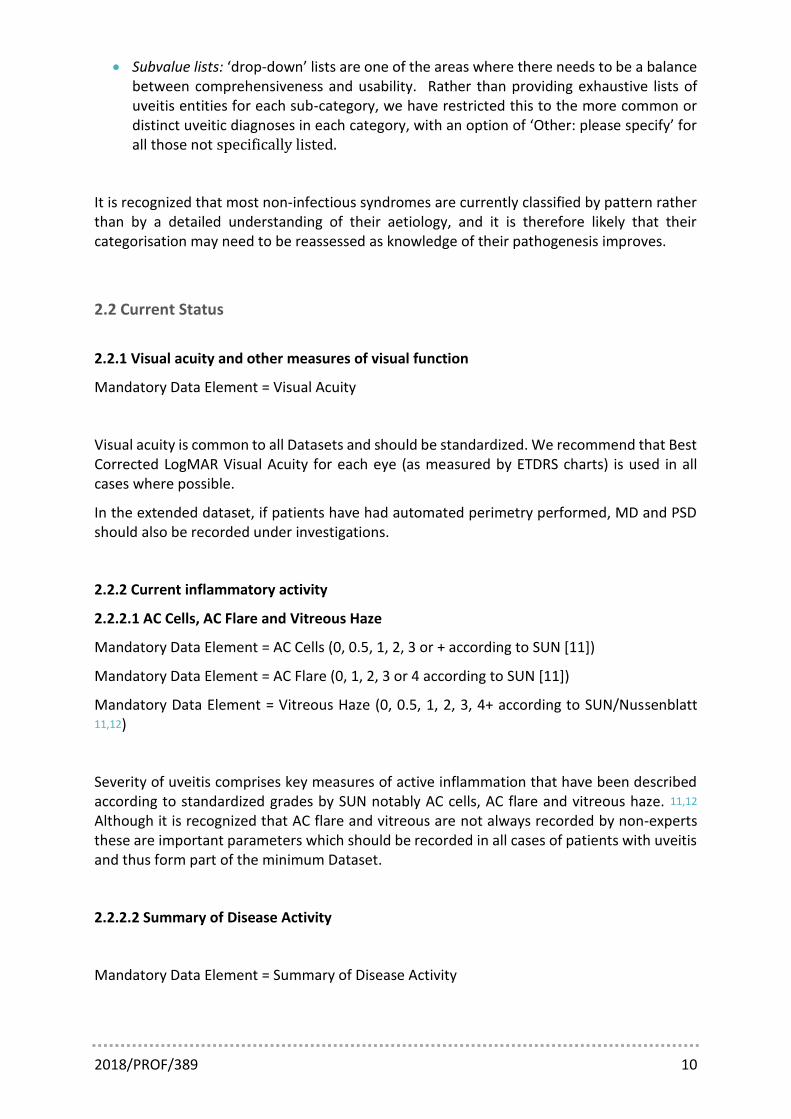
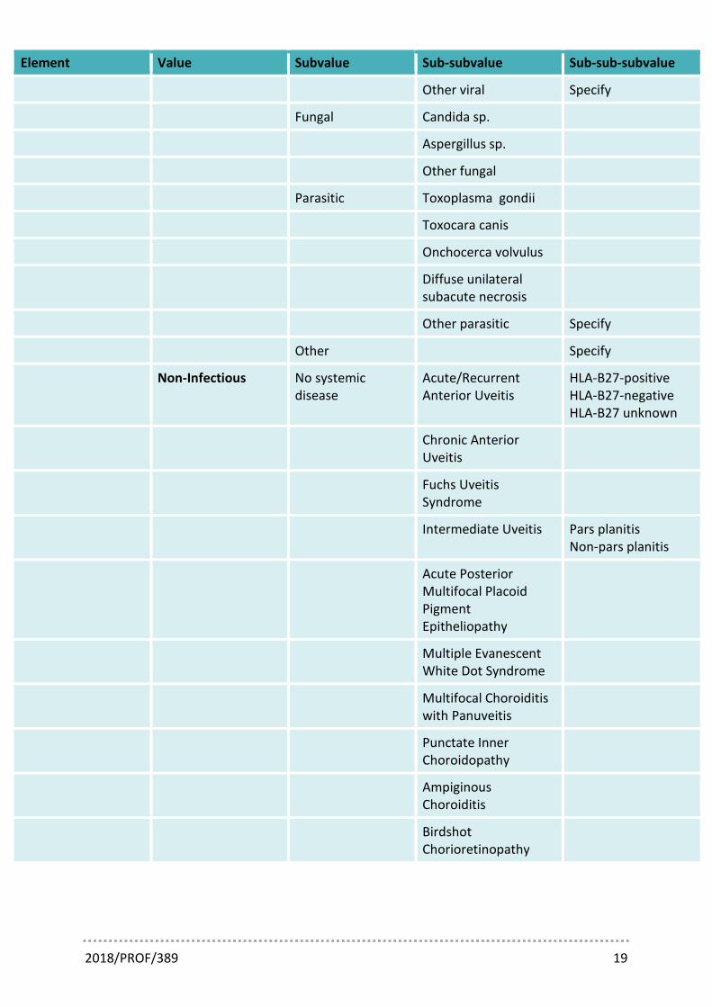
• Subvalue lists: ‘drop-down’ lists are one of the areas where there needs to be a balance between comprehensiveness and usability. Rather than providing exhaustive lists of uveitis entities for each sub-category, we have restricted this to the more common or distinct uveitic diagnoses in each category, with an option of ‘Other: please specify’ for all those not specifically listed.
It is recognized that most non-infectious syndromes are currently classified by pattern rather than by a detailed understanding of their aetiology, and it is therefore likely that their categorisation may need to be reassessed as knowledge of their pathogenesis improves.
2.2 Current Status
2.2.1 Visual acuity and other measures of visual function
Mandatory Data Element = Visual Acuity
Visual acuity is common to all Datasets and should be standardized. We recommend that Best Corrected LogMAR Visual Acuity for each eye (as measured by ETDRS charts) is used in all cases where possible.
In the extended dataset, if patients have had automated perimetry performed, MD and PSD should also be recorded under investigations.
2.2.2 Current inflammatory activity
2.2.2.1 AC Cells, AC Flare and Vitreous Haze
Mandatory Data Element = AC Cells (0, 0.5, 1, 2, 3 or + according to SUN [11])
Mandatory Data Element = AC Flare (0, 1, 2, 3 or 4 according to SUN [11])
Mandatory Data Element = Vitreous Haze (0, 0.5, 1, 2, 3, 4+ according to SUN/Nussenblatt 11,12)
Severity of uveitis comprises key measures of active inflammation that have been described according to standardized grades by SUN notably AC cells, AC flare and vitreous haze. 11,12 Although it is recognized that AC flare and vitreous are not always recorded by non-experts these are important parameters which should be recorded in all cases of patients with uveitis and thus form part of the minimum Dataset.
2.2.2.2 Summary of Disease Activity
Mandatory Data Element = Summary of Disease Activity
2018/PROF/389 11
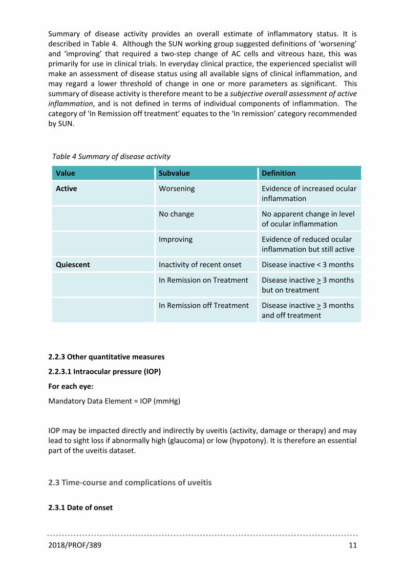
Summary of disease activity provides an overall estimate of inflammatory status. It is described in Table 4. Although the SUN working group suggested definitions of ‘worsening’ and ‘improving’ that required a two-step change of AC cells and vitreous haze, this was primarily for use in clinical trials. In everyday clinical practice, the experienced specialist will make an assessment of disease status using all available signs of clinical inflammation, and may regard a lower threshold of change in one or more parameters as significant. This summary of disease activity is therefore meant to be a subjective overall assessment of active inflammation, and is not defined in terms of individual components of inflammation. The category of ‘In Remission off treatment’ equates to the ‘In remission’ category recommended by SUN.
Table 4 Summary of disease activity
Value Subvalue Definition
Active Worsening Evidence of increased ocular inflammation
No change No apparent change in level of ocular inflammation
Improving Evidence of reduced ocular inflammation but still active
Quiescent Inactivity of recent onset Disease inactive < 3 months
In Remission on Treatment Disease inactive > 3 months but on treatment
In Remission off Treatment Disease inactive > 3 months and off treatment
2.2.3 Other quantitative measures
2.2.3.1 Intraocular pressure (IOP)
For each eye:
Mandatory Data Element = IOP (mmHg)
IOP may be impacted directly and indirectly by uveitis (activity, damage or therapy) and may lead to sight loss if abnormally high (glaucoma) or low (hypotony). It is therefore an essential part of the uveitis dataset.
2.3 Time-course and complications of uveitis
2.3.1 Date of onset

2018/PROF/389 12
For each eye:
Mandatory Data Element = Date of first onset of uveitis
Date of current visit (recorded for all visits) and date of onset of uveitis in each eye are mandatory. It is accepted that date of onset is likely to be an estimate and should be recorded to the nearest month; occasionally it may not be possible to obtain an estimate of the onset of uveitis and for this reason ‘not known’ should be provided as an alternative entry. Duration of disease is calculated from these parameters where known.
2.3.2 Complications
For each eye:
Mandatory Data Element = Presence of cataract
Mandatory Data Element = Presence of glaucoma or ocular hypertension
Mandatory Data Element = Presence of macular oedema
Mandatory Data Element = Presence of any other sight-threatening complication
Table 5 Presence of potentially sight-threatening complications of uveitis
Element Value Subvalue
Cataract Present Visually significant
Visually insignificant
Previous Pseudophakic (autofill)
Aphakic (autofill)
Absent
Glaucoma/OHT Present Glaucoma
OHT without glaucomatous optic neuropathy
Previous Previous OHT with no ongoing treatment requirement
Absent
Macular oedema Present Visually significant
Visually insignificant
Previous
Absent

2018/PROF/389 13
Element Value Subvalue
Epiretinal membrane Present Visually significant
Visually insignificant
Previous
Absent
Any other visually significant complication
Present Please specify
Previous Please specify
Absent
Key events in the time-course of uveitis are also recorded, notably the date of first presentation of uveitis and the presence of any sight-threatening complications; the presence or absence of cataract, glaucoma, macular oedema and epiretinal membrane and whether they are visually significant (Table 5) must be recorded in all cases. Any other potentially sight-threatening complications should be listed under ‘Any other visually significant complication’.
In the extended Dataset, the dates when these complications were first noted are recorded to the nearest month and may be estimated where the precise date is not known.
For each eye (where presence of sight-threatening complication has been noted):
Extended Data Element = Date cataract first noted
Extended Data Element = Date glaucoma first noted
Extended Data Element = Date macular oedema first noted
Extended Data Element = Date epiretinal membrane first noted
Extended Data Element = Date other specified sight-threatening complication first noted
2.4 Major therapeutic interventions
It is essential that both medical and surgical therapies directly related to the uveitis or its complications are recorded. It is recognized that most EMR systems will gather this data routinely (and indeed more comprehensively than outlined below) as part of their standard medical therapy interface pages. It is not expected that the clinician would be required to re-enter any of this data. These variables are however highlighted to support what would be expected to be available in terms of data extraction as they highlight key therapeutic interventions that are of ongoing relevance to the patient.
2018/PROF/389 14
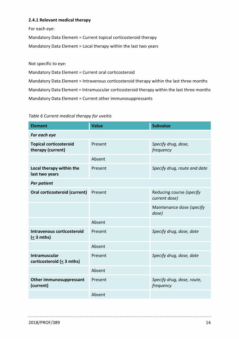
2.4.1 Relevant medical therapy
For each eye:
Mandatory Data Element = Current topical corticosteroid therapy
Mandatory Data Element = Local therapy within the last two years
Not specific to eye:
Mandatory Data Element = Current oral corticosteroid
Mandatory Data Element = Intravenous corticosteroid therapy within the last three months
Mandatory Data Element = Intramuscular corticosteroid therapy within the last three months
Mandatory Data Element = Current other immunosuppressants
Table 6 Current medical therapy for uveitis
Element Value Subvalue
For each eye
Topical corticosteroid therapy (current)
Present Specify drug, dose, frequency
Absent
Local therapy within the last two years
Present Specify drug, route and date
Per patient
Oral corticosteroid (current) Present Reducing course (specify current dose)
Maintenance dose (specify dose)
Absent
Intravenous corticosteroid (< 3 mths)
Present Specify drug, dose, date
Absent
Intramuscular corticosteroid (< 3 mths)
Present Specify drug, dose, date
Absent
Other immunosuppressant (current)
Present Specify drug, dose, route, frequency
Absent
2018/PROF/389 15
In the extended Dataset, any additional active or significant previous ocular treatments should be recorded. In the active group, common additional medications of particular interest are the topical therapies for glaucoma. In the significant previous treatments group, medications of particular interest are previous systemic immunosuppressant drugs, local therapies prior to two years earlier, and intravenous or intramuscular corticosteroid prior to three months earlier. Adverse drug reactions which may influence future treatment decisions in relation to uveitis, such as corticosteroid-induced ocular hypertension or hepatotoxicity due to a specific immunosuppressant, are also recorded in the extended dataset.
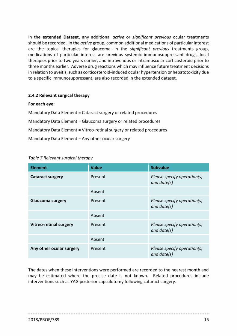
2.4.2 Relevant surgical therapy
For each eye:
Mandatory Data Element = Cataract surgery or related procedures
Mandatory Data Element = Glaucoma surgery or related procedures
Mandatory Data Element = Vitreo-retinal surgery or related procedures
Mandatory Data Element = Any other ocular surgery
Table 7 Relevant surgical therapy
Element Value Subvalue
Cataract surgery Present Please specify operation(s) and date(s)
Absent
Glaucoma surgery Present Please specify operation(s) and date(s)
Absent
Vitreo-retinal surgery Present Please specify operation(s) and date(s)
Absent
Any other ocular surgery Present Please specify operation(s) and date(s)
The dates when these interventions were performed are recorded to the nearest month and may be estimated where the precise date is not known. Related procedures include interventions such as YAG posterior capsulotomy following cataract surgery.

2018/PROF/389 16
2.5 Investigations
Although not included in the minimum dataset, the extended dataset includes selected investigations that are important either for diagnosis or monitoring of disease. The gathering of this data is encouraged both for assessing the current usage of these tests for particular uveitic syndromes and may provide some estimate of the diagnostic utility of these investigations in this context. Quantitative data from common serial assessments that are used for disease monitoring, such as central macular thickness as measured by optical coherence tomography, is also recorded.
3 References
1. Deschenes J, Murray PI, Rao NA, Nussenblatt RB; International Uveitis Study Group. International Uveitis Study Group (IUSG): clinical classification of uveitis. Ocul Immunol Inflamm. 2008 Jan-Feb;16(1):1-2. doi: 10.1080/09273940801899822. PubMed PMID: 18379933.
2. Durrani OM, Tehrani NN, Marr JE, Moradi P, Stavrou P, Murray PI. Degree, duration, and causes of visual loss in uveitis. Br J Ophthalmol. 2004 Sep;88(9):1159-62. PubMed PMID: 15317708; PubMed Central PMCID: PMC1772296
3. ten Doesschate J. Causes of blindness in the Netherlands. Doc Ophthalmol 1982;52:270–85 4. Barisani-Asenbauer T, Maca SM, Mejdoubi L, Emminger W, Machold K, Auer H. Uveitis- a rare disease often
associated with systemic diseases and infections- a systematic review of 2619 patients. Orphanet J Rare Dis. 2012 Aug 29;7:57. doi: 10.1186/1750-1172-7-57. Review. PubMed PMID: 22932001; PubMed Central PMCID: PMC3503654.
5. Barry RJ, Nguyen QD, Lee RW, Murray PI, Denniston AK. Pharmacotherapy for uveitis: current management and emerging therapy. Clin Ophthalmol. 2014 Sep 22;8:1891-911. doi: 10.2147/OPTH.S47778. eCollection 2014. Review. PubMed PMID: 25284976; PubMed Central PMCID: PMC4181632.
6. Jones NP. The Manchester Uveitis Clinic: the first 3000 patients—epidemiology and casemix. Ocul Immunol Inflamm. 2015 Apr;23(2):118-26. doi: 10.3109/09273948.2013.855799. Epub 2013 Dec 2. PubMed PMID: 24295124.
7. Sreekantam S, Denniston AK, Murray PI. Survey of expert practice and perceptions of the supporting clinical evidence for the management of uveitis-related cataract and cystoid macular oedema. Ocul Immunol Inflamm. 2011 Oct;19(5):353-7. doi: 10.3109/09273948.2011.592260. Epub 2011 Aug 8. PubMed PMID: 21823935.
8. Kempen JH, Daniel E, Gangaputra S, Dreger K, Jabs DA, Kaçmaz RO, Pujari SS, Anzaar F, Foster CS, Helzlsouer KJ, Levy-Clarke GA, Nussenblatt RB, Liesegang T, Rosenbaum JT, Suhler EB. Methods for identifying long-term adverse effects of treatment in patients with eye diseases: the Systemic Immunosuppressive Therapy for Eye Diseases (SITE) Cohort Study. Ophthalmic Epidemiol. 2008 Jan-Feb;15(1):47-55. doi: 10.1080/09286580701585892. PubMed PMID: 18300089.
9. Monnet D, Brézin AP, Holland GN, Yu F, Mahr A, Gordon LK, Levinson RD. Longitudinal cohort study of patients with birdshot chorioretinopathy. I. Baseline clinical characteristics. Am J Ophthalmol. 2006 Jan;141(1):135-42. PubMed PMID: 16386987
10. The Informatics and Audit Committee of the Royal College of Ophthalmologists. The Royal College of Ophthalmologists Dataset Guidelines. https://www.rcophth.ac.uk/wp-content/uploads/2014/12/2013_PROF_246_College-Dataset-Guidelines-August-2013.pdf
11. Jabs DA, Nussenblatt RB, Rosenbaum JT; Standardization of Uveitis Nomenclature (SUN) Working Group. Standardization of uveitis nomenclature for reporting clinical data. Results of the First International Workshop. Am J Ophthalmol. 2005 Sep;140(3):509-16. Review. PubMed PMID: 16196117
12. Nussenblatt RB, Palestine AG, Chan CC, Roberge F. Standardization of vitreal inflammatory activity in intermediate and posterior uveitis. Ophthalmology. 1985 Apr;92(4):467-71. PubMed PMID: 4000641.
2018/PROF/389 17
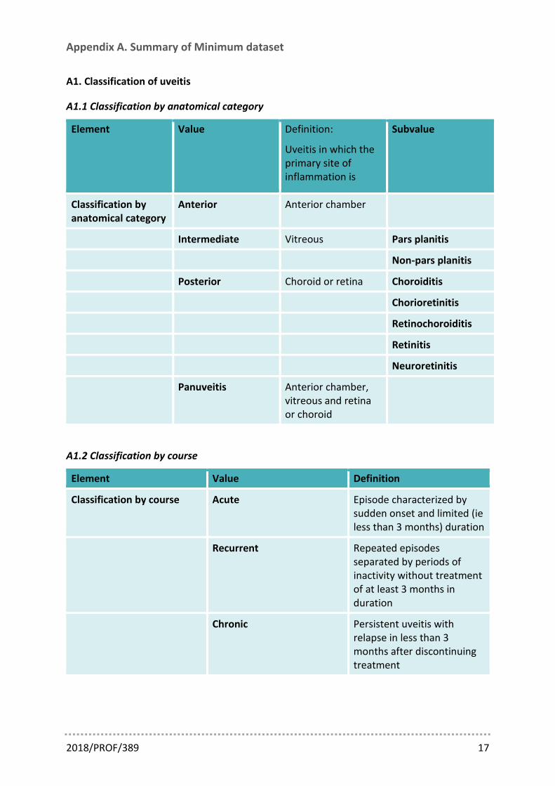
Appendix A. Summary of Minimum dataset
A1. Classification of uveitis
A1.1 Classification by anatomical category
Element Value Definition:
Uveitis in which the primary site of inflammation is
Subvalue
Classification by anatomical category
Anterior Anterior chamber
Intermediate Vitreous Pars planitis
Non-pars planitis
Posterior Choroid or retina Choroiditis
Chorioretinitis
Retinochoroiditis
Retinitis
Neuroretinitis
Panuveitis Anterior chamber, vitreous and retina or choroid
A1.2 Classification by course
Element Value Definition
Classification by course Acute Episode characterized by sudden onset and limited (ie less than 3 months) duration
Recurrent Repeated episodes separated by periods of inactivity without treatment of at least 3 months in duration
Chronic Persistent uveitis with relapse in less than 3 months after discontinuing treatment
2018/PROF/389 18
A1.3 Classification by taxonomy
Element Value Subvalue Sub-subvalue Sub-sub-subvalue
Taxonomy Infectious Bacterial Mycobacterium tuberculosis (TB)
Ocular only With extraocular involvement
Treponema pallidum (Syphilis)
Ocular only With extraocular involvement
Bartonella henselae (Cat-scratch disease)
Ocular only With extraocular involvement
Borrelia burgdorferi (Lyme disease)
Ocular only With extraocular involvement
Other bacterial Specify
Viral HSV1 Anterior uveitis Keratouveitis Acute Retinal Necrosis Progressive Outer Retinal Necrosis
HSV2 Anterior uveitis Keratouveitis Acute Retinal Necrosis Progressive Outer Retinal Necrosis
VZV Anterior uveitis Keratouveitis Acute Retinal Necrosis Progressive Outer Retinal Necrosis
CMV Anterior uveitis Keratouveitis Posterior uveitis
Viral syndrome (undifferentiated)
Anterior uveitis Keratouveitis Acute Retinal Necrosis Progressive Outer Retinal Necrosis
2018/PROF/389 19
Element Value Subvalue Sub-subvalue Sub-sub-subvalue
Other viral Specify
Fungal Candida sp.
Aspergillus sp.
Other fungal
Parasitic Toxoplasma gondii
Toxocara canis
Onchocerca volvulus
Diffuse unilateral subacute necrosis
Other parasitic Specify
Other Specify
Non-Infectious No systemic disease
Acute/Recurrent Anterior Uveitis
HLA-B27-positive HLA-B27-negative HLA-B27 unknown
Chronic Anterior Uveitis
Fuchs Uveitis Syndrome
Intermediate Uveitis Pars planitis Non-pars planitis
Acute Posterior Multifocal Placoid Pigment Epitheliopathy
Multiple Evanescent White Dot Syndrome
Multifocal Choroiditis with Panuveitis
Punctate Inner Choroidopathy
Ampiginous Choroiditis
Birdshot Chorioretinopathy

2018/PROF/389 20
Element Value Subvalue Sub-subvalue Sub-sub-subvalue
Serpiginous Choroiditis
Other non-infectious (no systemic disease)
Specify
Systemic disease Ankylosing spondylitis
Sarcoidosis
Multiple sclerosis
Behcet Disease
Sympathetic Ophthalmia
Vogt-Koyanagi-Harada Disease
Tubulointerstitial Nephritis and Uveitis
Juvenile idiopathic arthritis
Other non-infectious (systemic disease)
Specify
Masquerade Neoplastic Specify
Non-neoplastic Specify
Undifferentiated

2018/PROF/389 21
A2. Current status
A2.1 Visual acuity for each eye
Element Value
Visual acuity
Best corrected LogMAR value
A2.2 Current inflammatory activity for each eye
Element Value
AC cells 0-4 as per SUN
AC flare 0-4 as per SUN
Vitreous haze 0-4 as per SUN/Nussenblatt
A2.3 Intraocular pressure
Element Value
Intraocular pressure Numerical (mmHg)
A2.4 Summary of disease activity for each eye
Element Value Subvalue Definition
Summary of disease activity
Active Worsening Evidence of increased ocular inflammation
No change No apparent change in level of ocular inflammation
Improving Evidence of reduced ocular inflammation but still active
Quiescent Inactivity of recent onset
Disease inactive < 3 months
In Remission on Treatment
Disease inactive > 3 months but on treatment
In Remission off Treatment
Disease inactive > 3 months and off treatment

2018/PROF/389 22
A3. Time-course and complications of uveitis
A3.1 Time-course
Element Value Subvalue
Date of first onset of uveitis (per eye)
Specify
Date of current visit Specify
Duration of disease since first onset
Calculated
A3.2 Complications for each eye
Element Value Subvalue
Cataract Present Visually significant Visually insignificant
Previous Pseudophakic (autofill)
Aphakic (autofill)
Absent
Glaucoma/OHT Present Glaucoma OHT without glaucomatous optic neuropathy
Previous Previous OHT with no ongoing treatment requirement
Absent
Macular oedema Present Visually significant Visually insignificant
Previous
Absent
Epiretinal membrane Present Visually significant Visually insignificant
Previous
Absent

2018/PROF/389 23
Element Value Subvalue
Any other visually significant complication
Present Please specify
Previous Please specify
Absent
A4. Major therapeutic interventions
A4.1 Relevant Medical Therapies
Element Value Subvalue
For each eye
Topical corticosteroid therapy (current)
Present Specify drug, dose, frequency
Absent
Local therapy within the last two years
Present Specify drug, route and date
Per patient
Oral corticosteroid (current) Present Reducing course (specify current dose)
Maintenance dose (specify dose)
Absent
Intravenous corticosteroid (< 3 mths)
Present Specify drug, dose, date
Absent
Intramuscular corticosteroid (< 3 mths)
Present Specify drug, dose, date
Absent
Other immunosuppressant (current)
Present Specify drug, dose, route, frequency
Absent
2018/PROF/389 24
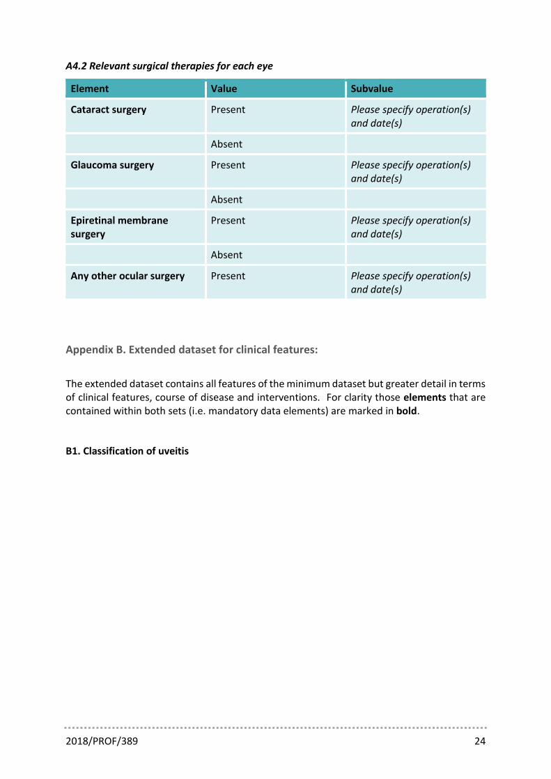
A4.2 Relevant surgical therapies for each eye
Element Value Subvalue
Cataract surgery Present Please specify operation(s) and date(s)
Absent
Glaucoma surgery Present Please specify operation(s) and date(s)
Absent
Epiretinal membrane surgery
Present Please specify operation(s) and date(s)
Absent
Any other ocular surgery Present Please specify operation(s) and date(s)
Appendix B. Extended dataset for clinical features:
The extended dataset contains all features of the minimum dataset but greater detail in terms of clinical features, course of disease and interventions. For clarity those elements that are contained within both sets (i.e. mandatory data elements) are marked in bold.
B1. Classification of uveitis

2018/PROF/389 25
B1.1 Classification by anatomical category
Element Value Definition:
Uveitis in which the primary site of inflammation is
Subvalue Sub-subvalue
Classification by anatomical category
Anterior Anterior chamber
Intermediate Vitreous Pars planitis
Non-pars planitis
Posterior Choroid or retina Choroiditis Unifocal Paucifocal Multifocal Diffuse
Chorioretinitis Unifocal Paucifocal Multifocal Diffuse
Retinochoroiditis Unifocal Paucifocal Multifocal Diffuse
Retinitis Unifocal Paucifocal Multifocal Diffuse
Neuroretinitis
Panuveitis Anterior chamber, vitreous and retina or choroid

2018/PROF/389 26
B1.2 Classification by course
Element Value Definition
Classification by course Acute Episode characterized by sudden onset and limited (ie less than 3 months) duration
Recurrent Repeated episodes separated by periods of inactivity without treatment of at least 3 months in duration
Chronic Persistent uveitis with relapse in less than 3 months after discontinuing treatment
Element Value
Descriptor of uveitis onset Sudden
Insidious

2018/PROF/389 27
B1.3 Classification by aetiology/clinical syndrome
Element Value Subvalue Sub-subvalue Sub-sub-subvalue
Classification by Taxonomy
Infectious Bacterial Mycobacterium tuberculosis (TB)
Ocular only With extraocular involvement
Treponema pallidum (Syphilis)
Ocular only With extraocular involvement
Bartonella henselae (Cat-scratch disease)
Ocular only With extraocular involvement
Borrelia burgdorferi (Lyme disease)
Ocular only With extraocular involvement
Other bacterial Specify
Viral HSV1 Anterior uveitis Keratouveitis Acute Retinal Necrosis Progressive Outer Retinal Necrosis
HSV2 Anterior uveitis Keratouveitis Acute Retinal Necrosis Progressive Outer Retinal Necrosis
VZV Anterior uveitis Keratouveitis Acute Retinal Necrosis Progressive Outer Retinal Necrosis
CMV Anterior uveitis Keratouveitis Posterior uveitis
Viral syndrome (undifferentiated)
Anterior uveitis Keratouveitis Acute Retinal Necrosis Progressive Outer Retinal Necrosis

2018/PROF/389 28
Element Value Subvalue Sub-subvalue Sub-sub-subvalue
Other viral Specify
Fungal Candida sp.
Aspergillus sp.
Other fungal Specify
Parasitic Toxoplasma gondii
Toxocara canis
Onchocerca volvulus
Diffuse unilateral subacute necrosis
Other parasitic Specify
Other Specify
Non-Infectious No systemic disease
Acute/Recurrent Anterior Uveitis
HLA-B27-positive HLA-B27-negative HLA-B27 unknown
Chronic Anterior Uveitis
Fuchs Uveitis Syndrome
Intermediate Uveitis Pars planitis Non-pars planitis
Acute Posterior Multifocal Placoid Pigment Epitheliopathy
Multiple Evanescent White Dot Syndrome
Multifocal Choroiditis with Panuveitis
Punctate Inner Choroidopathy
Ampiginous Choroiditis
Birdshot Chorioretinopathy

2018/PROF/389 29
Element Value Subvalue Sub-subvalue Sub-sub-subvalue
Serpiginous Choroiditis
Other non-infectious (no systemic disease)
Specify
Systemic disease Ankylosing spondylitis
Sarcoidosis
Multiple sclerosis
Behcet Disease
Sympathetic Ophthalmia
Vogt-Koyanagi-Harada Disease
Tubulointerstitial Nephritis and Uveitis
Juvenile idiopathic arthritis
Other non-infectious (systemic disease)
Specify
Masquerade Neoplastic Specify
Non-neoplastic Specify
Undifferentiated
B2. Current status
B2.1 Visual function
Element Value Subvalue
Visual acuity
VA - Unaided LogMAR value
VA - Best corrected LogMAR value
VA - Pin-hole LogMAR value
Colour vision Number of Ishihara plates (x/y)
RAPD Present
Absent

2018/PROF/389 30
B2.2 Current inflammatory activity and other clinical features for each eye
Symptoms
Element Value Subvalue
Photophobia Present
Absent
Other eye pain Present
Absent
Blurred vision Present
Absent
Floaters Present
Absent
Photopsia Present
Absent
Blind-spots Present
Absent
Distortion Present
Absent
Other visual symptoms Present Please specify
Absent
Other non-visual ocular symptoms
Present Please specify
Absent

2018/PROF/389 31
Conjunctiva and sclera
Element Value Subvalue Sub-subvalue
Conjunctival injection
Present Diffuse
Severity (0-4)
Sectoral Severity (0-4)
Circumlimbal Severity (0-4)
Absent
Episcleritis Present Nodular
Non-nodular Diffuse Sectoral
Absent
Anterior Scleritis Present Non-necrotising Diffuse Nodular
Necrotising With evident inflammation Without evident inflammation (scleromacia perforans-type)
Absent
Cornea
Element Value Subvalue Sub-subvalue
Keratic precipitates Present Regular Diffuse Inferior Focal
Granulomatous Diffuse Inferior Focal
Stellate Diffuse Inferior Focal
Band keratopathy Present Involves visual axis
Not involving visual axis
Absent
Not done
2018/PROF/389 32
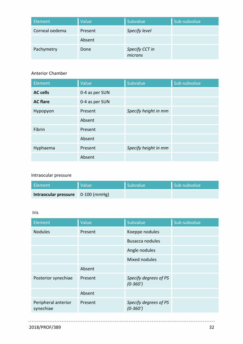
Element Value Subvalue Sub-subvalue
Corneal oedema Present Specify level
Absent
Pachymetry Done Specify CCT in microns
Anterior Chamber
Element Value Subvalue Sub-subvalue
AC cells 0-4 as per SUN
AC flare 0-4 as per SUN
Hypopyon Present Specify height in mm
Absent
Fibrin Present
Absent
Hyphaema Present Specify height in mm
Absent
Intraocular pressure
Element Value Subvalue Sub-subvalue
Intraocular pressure 0-100 (mmHg)
Iris
Element Value Subvalue Sub-subvalue
Nodules Present Koeppe nodules
Busacca nodules
Angle nodules
Mixed nodules
Absent
Posterior synechiae Present Specify degrees of PS (0-360’)
Absent
Peripheral anterior synechiae
Present Specify degrees of PS (0-360’)
2018/PROF/389 33
Element Value Subvalue Sub-subvalue
Absent
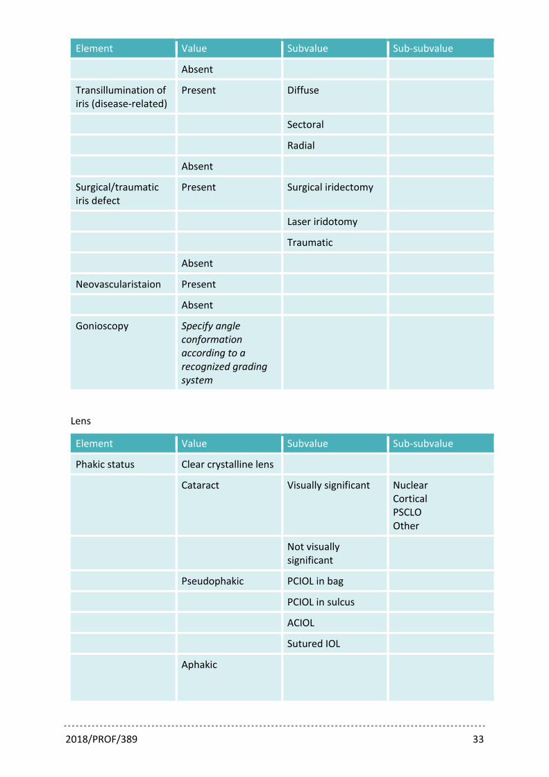
Transillumination of iris (disease-related)
Present Diffuse
Sectoral
Radial
Absent
Surgical/traumatic iris defect
Present Surgical iridectomy
Laser iridotomy
Traumatic
Absent
Neovascularistaion Present
Absent
Gonioscopy Specify angle conformation according to a recognized grading system
Lens
Element Value Subvalue Sub-subvalue
Phakic status Clear crystalline lens
Cataract Visually significant Nuclear Cortical PSCLO Other
Not visually significant
Pseudophakic PCIOL in bag
PCIOL in sulcus
ACIOL
Sutured IOL
Aphakic

2018/PROF/389 34
Element Value Subvalue Sub-subvalue
Posterior capsule status (where relevant)
Clear
PCO Visually significant
Not visually significant
Anterior capsule status (where relevant)
Normal
Anterior capsular phimosis
Lens stability Central
Subluxed
Vitreous
Element Value Subvalue Sub-subvalue
Vitreous haze 0-4 as per SUN
Vitreous cells Present
Absent
Vitreous snowballs Present
Absent
Pars plana exudates Present
Absent
Vitreous haemorrhage
Present
Absent
Posterior vitreous detachment
Present
Absent
Fundus - Macula
Element Value Subvalue Sub-subvalue
Macular oedema Present
Absent
2018/PROF/389 35
Element Value Subvalue Sub-subvalue
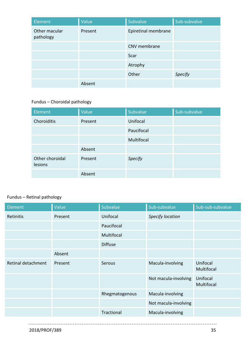
Other macular pathology
Present Epiretinal membrane
CNV membrane
Scar
Atrophy
Other Specify
Absent
Fundus – Choroidal pathology
Element Value Subvalue Sub-subvalue
Choroiditis Present Unifocal
Paucifocal
Multifocal
Absent
Other choroidal lesions
Present Specify
Absent
Fundus – Retinal pathology
Element Value Subvalue Sub-subvalue Sub-sub-subvalue
Retinitis Present Unifocal Specify location
Paucifocal
Multifocal
Diffuse
Absent
Retinal detachment Present Serous Macula-involving Unifocal Multifocal
Not macula-involving Unifocal Multifocal
Rhegmatogenous Macula-involving
Not macula-involving
Tractional Macula-involving
2018/PROF/389 36
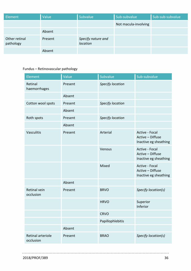
Element Value Subvalue Sub-subvalue Sub-sub-subvalue
Not macula-involving
Absent
Other retinal pathology
Present Specify nature and location
Absent
Fundus – Retinovascular pathology
Element Value Subvalue Sub-subvalue
Retinal haemorrhages
Present Specify location
Absent
Cotton wool spots Present Specify location
Absent
Roth spots Present Specify location
Absent
Vasculitis Present Arterial Active - Focal Active – Diffuse Inactive eg sheathing
Venous Active - Focal Active – Diffuse Inactive eg sheathing
Mixed Active - Focal Active – Diffuse Inactive eg sheathing
Absent
Retinal vein occlusion
Present BRVO Specify location(s)
HRVO Superior Inferior
CRVO
Papillophlebitis
Absent
Retinal arteriole occlusion
Present BRAO Specify location(s)

2018/PROF/389 37
Element Value Subvalue Sub-subvalue
CRAO
Absent
Neovascularisation Present NVD Specify location(s)
NVE Specify location(s)
Absent
Optic disc
Element Value Subvalue Sub-subvalue
Cup-Disc Ratio 0-1.0
Features suspicious of glaucoma
Present Specify features of concern
Absent
Disc colour Normal
Hyperaemic
Pallor
Disc margin Normal
Disc oedema
Other Specify
Disc haemorrhages Present
Absent
Disc vessels Normal
Disc collaterals
NVD
B2.3 Summary of disease activity for each eye
Element Value Subvalue Definition
Summary of disease activity
Active Worsening Evidence of increased ocular inflammation
No change No apparent change in level of ocular inflammation

2018/PROF/389 38
Element Value Subvalue Definition
Improving Evidence of reduced ocular inflammation but still active
Quiescent Inactivity of recent onset
Disease inactive < 3 months
In Remission on Treatment
Disease inactive > 3 months but on treatment
In Remission off Treatment
Disease inactive > 3 months and off treatment
B3. Time-course and complications of uveitis
B3.1 Time-course
Element Value Subvalue
Date of first onset of uveitis (per eye)
Specify
Date of current visit Specify
Duration of disease since first onset
Calculated
B3.2 Complications for each eye
Element Value Subvalue
Cataract Present Visually significant Visually insignificant
Previous Pseudophakic (autofill)
Aphakic (autofill)
Absent
Glaucoma/OHT Present Glaucoma OHT without glaucomatous optic neuropathy
2018/PROF/389 39
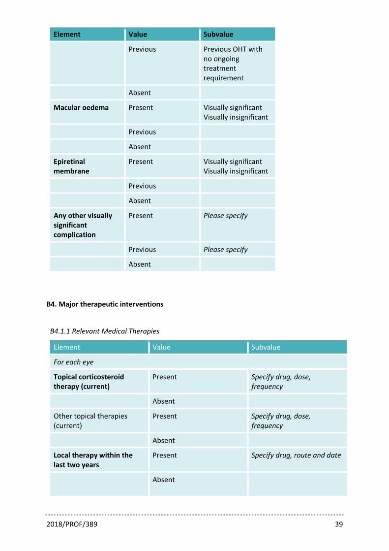
Element Value Subvalue
Previous Previous OHT with no ongoing treatment requirement
Absent
Macular oedema Present Visually significant Visually insignificant
Previous
Absent
Epiretinal membrane
Present Visually significant Visually insignificant
Previous
Absent
Any other visually significant complication
Present Please specify
Previous Please specify
Absent
B4. Major therapeutic interventions
B4.1.1 Relevant Medical Therapies
Element Value Subvalue
For each eye
Topical corticosteroid therapy (current)
Present Specify drug, dose, frequency
Absent
Other topical therapies (current)
Present Specify drug, dose, frequency
Absent
Local therapy within the last two years
Present Specify drug, route and date
Absent

2018/PROF/389 40
Element Value Subvalue
Local therapy prior to two years previously
Present Specify drug, route and date
Absent
Per patient
Oral corticosteroid (current) Present Reducing course (specify current dose)
Maintenance dose (specify dose)
Absent
Oral corticosteroid (previous)
Present Specify dose-ranges and dates of episodes of treatment
Absent
Intravenous corticosteroid (within last 3 months)
Present Specify drug, dose, date
Absent
Intravenous corticosteroid (prior to 3 months previously)
Present Specify drug, dose, date
Absent
Intramuscular Corticosteroid (within last 3 months)
Present Specify drug, dose, date
Absent
Intramuscular Corticosteroid (prior to 3 months previously)
Present Specify drug, dose, date
Absent
Other immunosuppressant (current)
Present Specify drug, dose, route, frequency
Absent
Other immunosuppressant (previous)
Present Specify dose-ranges, dates of episodes of treatment, and reason for cessation
2018/PROF/389 41
Element Value Subvalue
Absent
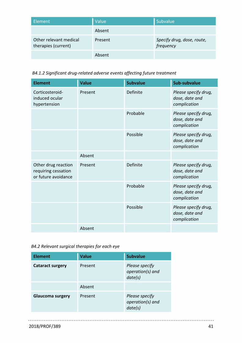
Other relevant medical therapies (current)
Present Specify drug, dose, route, frequency
Absent
B4.1.2 Significant drug-related adverse events affecting future treatment
Element Value Subvalue Sub-subvalue
Corticosteroid-induced ocular hypertension
Present Definite Please specify drug, dose, date and complication
Probable Please specify drug, dose, date and complication
Possible Please specify drug, dose, date and complication
Absent
Other drug reaction requiring cessation or future avoidance
Present Definite Please specify drug, dose, date and complication
Probable Please specify drug, dose, date and complication
Possible Please specify drug, dose, date and complication
Absent
B4.2 Relevant surgical therapies for each eye
Element Value Subvalue
Cataract surgery Present Please specify operation(s) and date(s)
Absent
Glaucoma surgery Present Please specify operation(s) and date(s)

2018/PROF/389 42
Element Value Subvalue
Absent
Vitreo-retinal surgery
Present Please specify operation(s) and date(s)
Absent
Any other ocular surgery
Present Please specify operation(s) and date(s)
B5. Investigations
B5.1 Quantitative data from common serial assessments for each eye
Element Value
Central macular thickness on OCT Numerical
MD on 24-2 automated perimetry Numerical
PSD on 24-2 automated perimetry Numerical
B5.2 Genotypic data
Element Value
HLA-A29 Positive
Negative
Not tested
HLA-B27 Positive
Negative
Not tested
HLA-B51 Positive
Negative
Not tested
HLA-DR4 Positive
Negative
Not tested
Other genotypic data Specify

2018/PROF/389 43
B5.3 Other investigations
Element Value Subvalue Sub-subvalue
ACE Positive
Negative
Not tested
Syphilis serology Positive
Negative
Not tested
Interferon gamma release assay
Positive
Negative
Inconclusive
Not tested
Mantoux Positive
Equivocal
Negative
Not tested
CXR Suggestive of pathology related to uveitis
Specify
Abnormal but unrelated to uveitis
Normal
Not tested
CT Thorax Suggestive of pathology related to uveitis
Specify
Abnormal but unrelated to uveitis
Normal
Not tested
MRI Brain Suggestive of pathology related to uveitis
Specify

2018/PROF/389 44
Element Value Subvalue Sub-subvalue
Abnormal but unrelated to uveitis
Normal
Not tested
Aqueous humour PCR PCR positive Specify organism
PCR negative
PCR not done
Other findings Specify
Vitreous humour PCR PCR positive Specify organism
PCR negative
PCR not done
Other findings Specify
Other relevant investigations
Specify