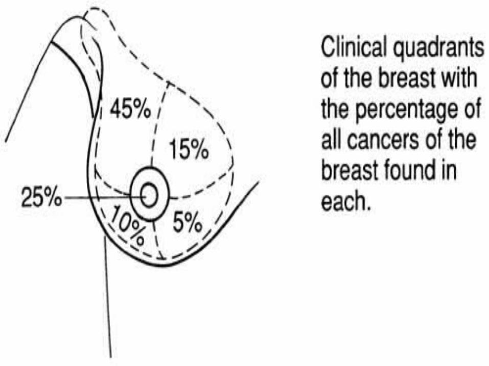
clinical assessment of breast
TRANSCRIPT

Clinical Examination of Breast
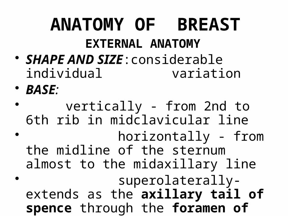
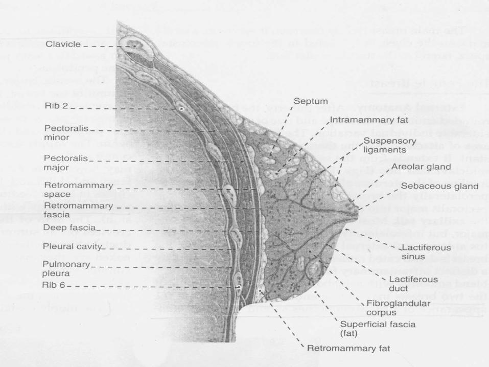
ANATOMY OF BREASTEXTERNAL ANATOMY
• SHAPE AND SIZE:considerable individual variation
• BASE:• vertically - from 2nd to 6th rib in
midclavicular line • horizontally - from the midline of
the sternum almost to the midaxillary line• superolaterally- extends as the
axillary tail of spence through the foramen of langer

• THE AREOLA:pigmented circular area of skin around the base of the nipple
It contains a number of subcutaneous glands which enlarge during pregnancy and are called tubercles of Montgomery
• THE NIPPLE{papilla mammaria}:cylindrical or conical structure projecting from the center of the areola
It contains an elaborate subcutaneous network of of smooth muscle cells and elastic fibers


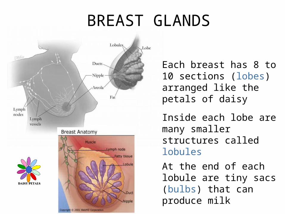
BREAST GLANDS
Each breast has 8 to 10 sections (lobes) arranged like the petals of daisy
Inside each lobe are many smaller structures called lobules
At the end of each lobule are tiny sacs (bulbs) that can produce milk

ARTERIAL SUPPLY
It is supplied by:• Lateral thoracic branch of 2nd part of
axillary artery• Medial mammary branches of internal
thoracic artery• Superior thoracic branch of axillary artery• Lateral branches of 2nd,3rd,4th posterior
intercostal arteries

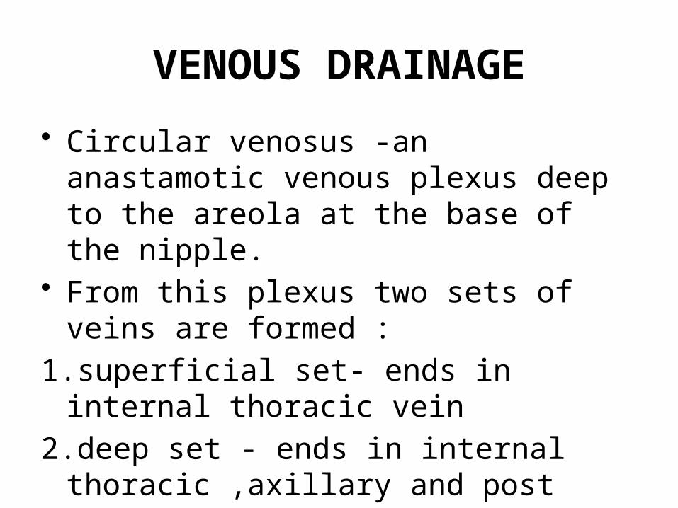
VENOUS DRAINAGE
• Circular venosus -an anastamotic venous plexus deep to the areola at the base of the nipple.
• From this plexus two sets of veins are formed :
1.superficial set- ends in internal thoracic vein
2.deep set - ends in internal thoracic ,axillary and post intercostal veins


LYMPHATIC DRAINAGE• 5 Groups: ANTERIOR (PECTORAL) SET: Situated along the lateral thoracic vein under the ant
axillary fold,they lie mainly on the 3rd rib POSTERIOR (SCAPULAR) SET: Lies on the post axillary fold in relation to the subscapular
vessels LATERAL (AXILLARY VEIN) SET: Along the upper part of humerus in relation to the
axillary vein CENTRAL SET:
Situated in the fat of the upper Axilla. APICAL or INFRACLAVICULAR SET:
Lie deep to the clavipectoral fascia along the axillary vesels


History
• Lump – duration,onset,rate of growth• Pain • Fever• Discharge from nipple• Retraction of nipple• Trauma• Loss of weight/appetite

History• Swelling elsewhere• Related to metastasis- bone pain,
jaundice ,cough with hemoptysis,• Similar episodes• Smoking• Alcoholism• Diet habits(high fat diet)• Breast feeding• Drug intake

History - CA risk factors• Age: older• History: family, prior dz• Abortion• Late menopause• Obese• Nulliparity• Early menarche
A H
istory A
LO
NE

•Pt removes upper body clothing•Expose/•inspect theopposite sideso can comparefor asymmetry.
Expose

•Ask pt. if Tenderness before start touching them.•Warm your hands
Tenderness
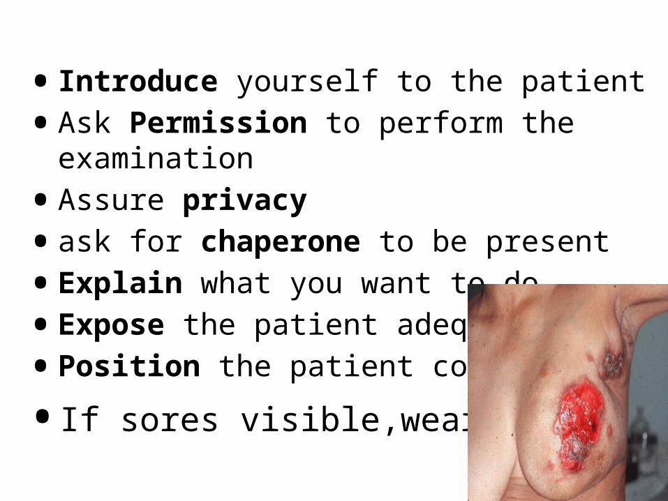
• Introduce yourself to the patient
•Ask Permission to perform the examination
•Assure privacy
•ask for chaperone to be present
•Explain what you want to do
•Expose the patient adequately
•Position the patient correctly
•If sores visible,wear gloves.

Methods of Inspection
• Sitting position,arms at sides
• Arms overhead
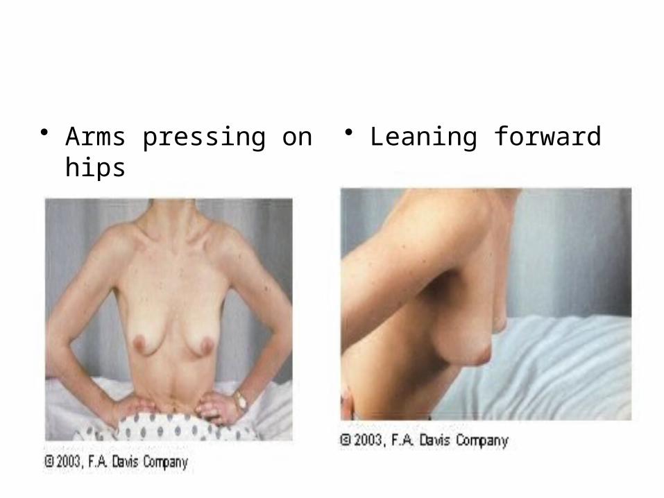
• Arms pressing on hips
• Leaning forward
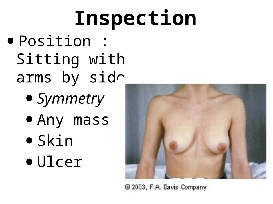
Inspection
•Position : Sitting with arms by side
•Symmetry
•Any mass
•Skin
•Ulcer
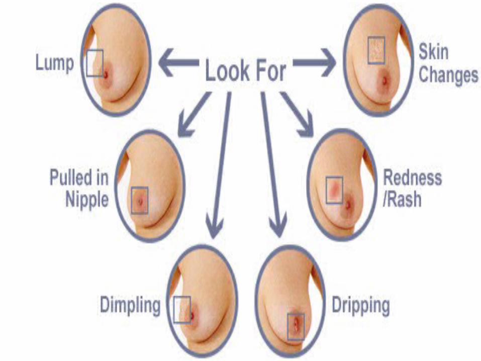

skinSkin retraction
Dimpling
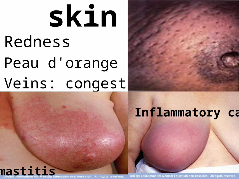
skinRednessPeau d'orange (ca)
Veins: congestion
mastitis
Inflammatory ca

Nipples•Nipple number, position•Inversion retraction; (fibrosis, CA, normal)
Slit like

Nipples•Red, bleeding •(Paget's dz of nipple). •Discharge

•Ask patient to raise arms and place hands behind head
•Change in a mass's relative position.
•Nipple or skin tethering

Inspect whole skin• Raise the breast to inspect
the undermined skin.

Inspect the axilla
Examine axilla while pt's arms are raised;
• axillary tail• axillary LNs • any mass, ulcer• Edema,nodules• Cancer en cuirase
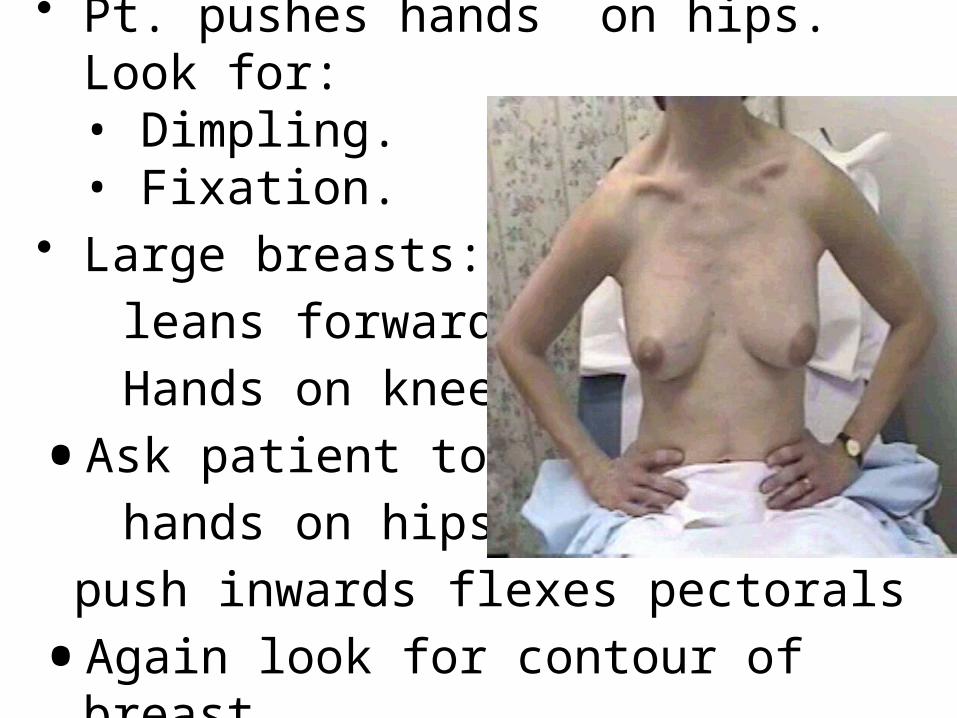
• Pt. pushes hands on hips. Look for:• Dimpling.• Fixation.
• Large breasts: pt.
leans forward
Hands on knees
•Ask patient to put
hands on hips and
push inwards flexes pectorals
•Again look for contour of breast

Palpation-Use fingerpads of middle 3 fingers-Palpation should not elicit pain-Consistency is highly variable

Sitting position•First examine sitting
•Examine ‘normal’ side first
•Place hand behind head
•One quadrant at a time

Supine position•Spreads the breast more evenly across chest •Examine lying down
•Use one or two hands to elicit lumps
•If felt define lump with fingertips
•See Examining A Mass

•Press breast against chest wall•Rolling fingers in small, circular motions.
•Press lightly for superficial layers•Medium pressure for middle layer•Firmer pressure for deepest layers•Start at sternoclavicular junction. •Move in overlapping vertical strips until all 4 breast quadrants are covered.

Evaluation of Breast Mass Characteristics• Location
• Size
• Shape
• Number
• Consistency
• Definition• Mobility• Tenderness• Erythema• Dimpling or
retraction• Lymphadenopathy
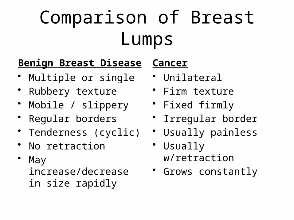
Comparison of Breast Lumps
Benign Breast Disease• Multiple or single• Rubbery texture• Mobile / slippery• Regular borders• Tenderness (cyclic)• No retraction• May increase/decrease in
size rapidly
Cancer• Unilateral• Firm texture• Fixed firmly• Irregular border• Usually painless• Usually w/retraction• Grows constantly
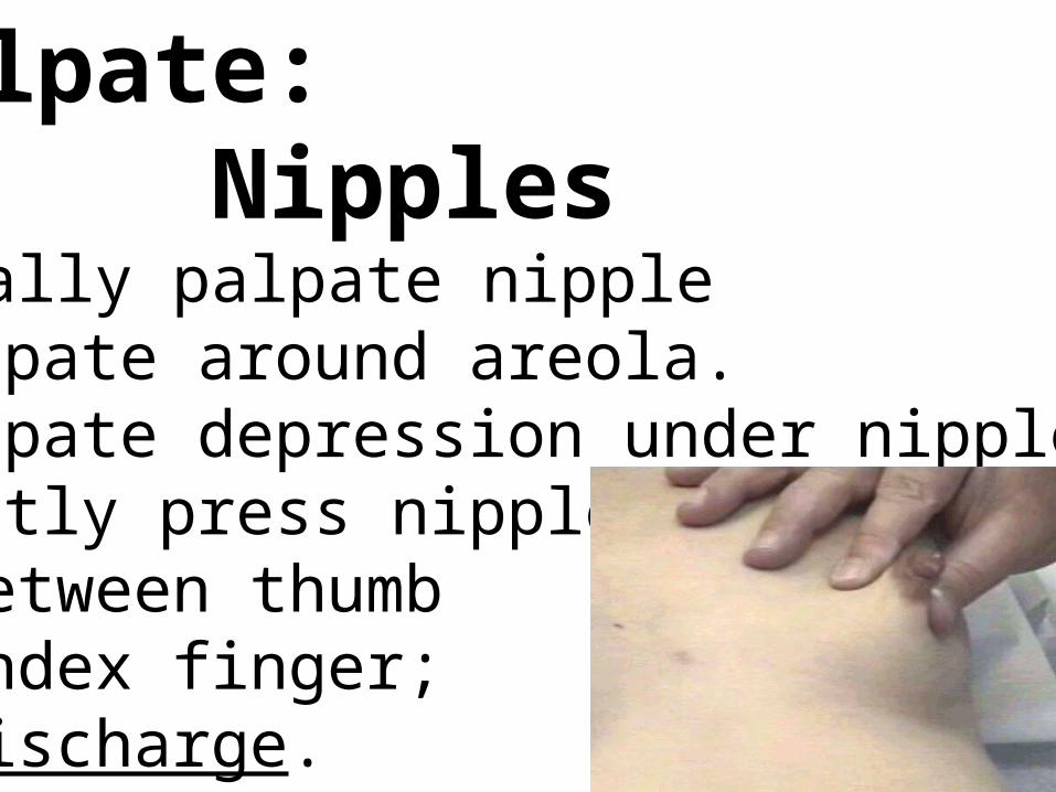
Palpate: NipplesFinally palpate nipple•Palpate around areola.•Palpate depression under nipple.•Gently press nipple between thumb index finger; Discharge.
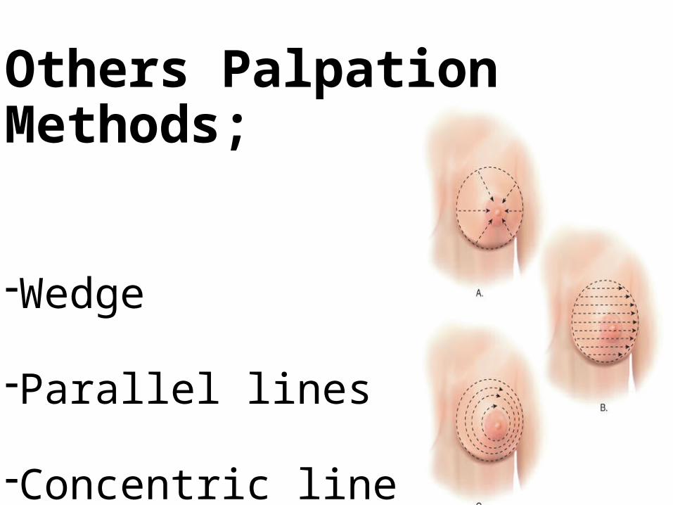
Others Palpation Methods;
-Wedge
-Parallel lines
-Concentric lines

Examination of Axilla

palpate the axilla•Support patient’s arm•Palpate tail between fingers and thumb.•Palpate axillary lymph nodes•Supraclavicular nodes.•Palpable lymph nodes less than 1 cm in
diameter usually are clinically insignificant
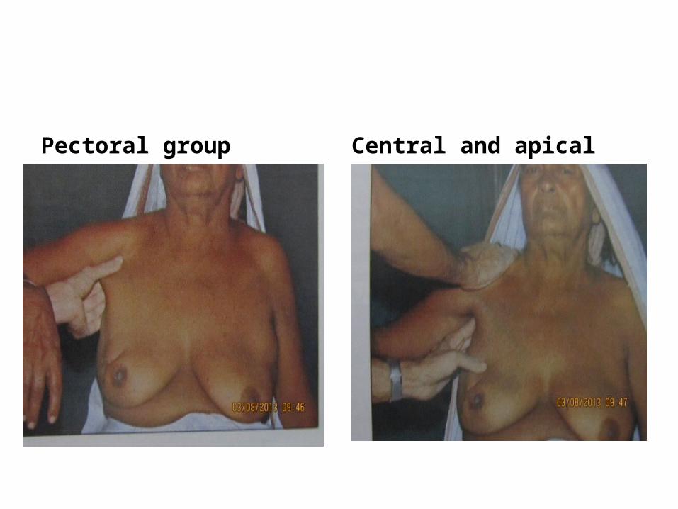
Pectoral group Central and apical

Posterior group
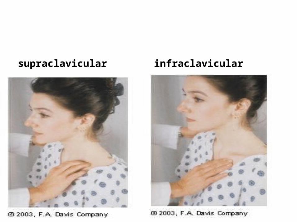
supraclavicular infraclavicular

BREAST SELF EXAM
• GOAL: Early detection• IN PREPARATION FOR TEACHING:• Assess: knowledge base , motivation • fears and concerns• family history• risk factors• TEACHING: Use show and tell; use finger
pads • EXAM: monthly, day 5-7 of menstrual cycle;
after menopause same day each month• Use in conjunction with mammography & CBE
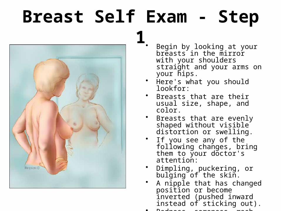
Breast Self Exam - Step 1• Begin by looking at your breasts in
the mirror with your shoulders straight and your arms on your hips.
• Here's what you should lookfor:• Breasts that are their usual size,
shape, and color.• Breasts that are evenly shaped
without visible distortion or swelling.
• If you see any of the following changes, bring them to your doctor's attention:
• Dimpling, puckering, or bulging of the skin.
• A nipple that has changed position or become inverted (pushed inward instead of sticking out).
• Redness, soreness, rash, or swelling

Breast Self Exam - Step 2 and 3• Raise your arms and look
for the same changes.• While you're at the mirror,
gently squeeze each nipple between your finger and thumb and check for nipple discharge (this could be a milky or yellow fluid or blood).

Breast Self Exam - Step 4 • Feel your breasts while
lying down, using your right hand to feel your left breast and then your left hand to feel your right breast. Use a firm, smooth touch with the first few fingers of your hand, keeping the fingers flat and together.
• Cover the entire breast from top to bottom, side to side—from your collarbone to the top of your abdomen, and from your armpit to your cleavage


Breast Self Exam - Step 5 • Finally, feel your breasts
while you are standing or sitting. Many women find that the easiest way to feel their breasts is when their skin is wet and slippery, so they like to do this step in the shower. Cover your entire breast, using the same hand movements described in Step 4.
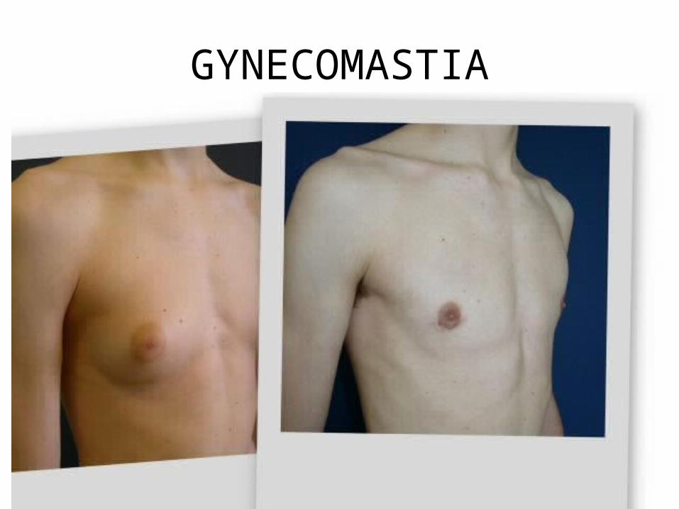
GYNECOMASTIA


Mondor’s disease: thrombophlebitis

Phylloides tumor

