clinical and histopathological correlations of the - rjme.ro
TRANSCRIPT

Romanian Journal of Morphology and Embryology 2009, 50(1):67–72
OORRIIGGIINNAALL PPAAPPEERR
Clinical and histopathological correlations of the modifications of fetal membranes
in amniochorial infection ANCA PĂTRAŞCU1), SABINA BERCEANU1), CARMEN FLORINA POPESCU2),
V. GHEORMAN1), C. BERCEANU1)
1)Department of Obstetrics and Gynecology, University of Medicine and Pharmacy of Craiova
2)Laboratory of Pathological Anatomy and Cytology, Emergency County Hospital, Craiova
Abstract The paper aims at analyzing the histopathological modifications induced by the amniotic infection on present fetal membranes and in the absence of clinical signs of chorioamnionitis. Such an evaluation is important in the context of postpartum fetomaternal complications. The objective was to determine the relation between the microbial invasion in the amniotic cavity and the severity of inflammatory lesions of the placenta, of the membranes and the umbilical chord. Keywords: chorioamnionitis, placenta, amniotic membranes, amniotic fluid.
Introduction
Chorioamnionitis is the inflammation of the chorioamniotic structures of bacterial, viral, parasitic, chemical and hypoxic etiology [1, 2]. Chorioamnionitis with ruptured membranes represents the most common form; the contamination direction is generally the ascending one, and the gravity of the infection depends on the time elapsed from the moment of ruptured membranes, as well as on the virulence of the germs in the vagina [1, 3, 4]. The microbial invasion in the amniotic cavity can also result from the dissemination at local, chorio-decidual level [1, 5]. In addition, the microbial aggression by products of microbial degradation, such as proteases, determines lesion of fetal membranes for both parts. The microbial contamination of the amniotic fluid is correlated to the frequency of vaginal examinations and the presence of ruptured membranes [1, 2, 6].
Recent research estimate the relation between histological chorioamnionitis and four proteomic markers in the amniotic fluid specific to the inflammation. All these markers are strictly related to the presence of third stage chorioamnionitis, independently of race and the gestational age [7].
If clinical signs of bacterial colonization of amniotic fluid are always accompanied by histological modifications of the amniochorial membranes, the converse relation is not always valid. Therefore, there is a so-called latent amnionitis, clinically asymptomatic, but with positive cultures from the amniotic fluid and with present histological modifications, as there can be histopathological modifications of the placenta and of amniotic membranes as the only sign of chorioamnioninits [1, 6].
Material and Methods
The analysis was carried out for placentas and ovular membranes sampled from 26 women with complicated births by chorioamnionitis, which took place in the Second Clinic of the “Filantropia” Hospital, Craiova, in 2007.
These cases were selected of the total number of 1932 births, which took place in 2007, which represents 1.33%.
The placentas were analyzed macroscopically and tissue fragments were sampled. These were later processed by the classic method of paraffin embedding, in the Research Center for Microscopic Morphology and Immunology, University of Medicine and Pharmacy of Craiova.
The fetal membranes were examined clinically and histopathologically. At the same time, there were also taken samples for microbial cultures of the amniotic fluid, of the placenta, the umbilical chord and the membranes, and the achieved results were correlated with clinical and histopathological aspects.
Results
Clinical data
In the studied cases, 20 patients presented histo-pathological modifications of fetal membranes. These patients had ruptured membranes for over 6 hours but with no clinical symptoms.
For 16 of the studied patients, the cultures of the amniotic fluid were positive, while for the others it was impossible to sample amniotic fluid or the amniotic fluid was contaminated with blood or meconium.
Anca Pătraşcu et al.
68
In the study we carried out, the following germs were identified: Escherichia coli in 18.42% of cases, Staphylococcus aureus in 27.88% of cases, β-hemolytic Streptococcus for 3.26% of the patients, and Bacteroides for 2.4% of them. According to these data, the patients with ruptured membranes for a greater interval than 6 hours and with more than four vaginal examinations were selected for antibiotic prophylaxis in order to obtain a reduction in the febrile morbidity and in the infection of perineal incision or post-caesarean section. The treatment was carried out depending on the antibiogram.
Modifications of the umbilical chord and of amniotic membranes
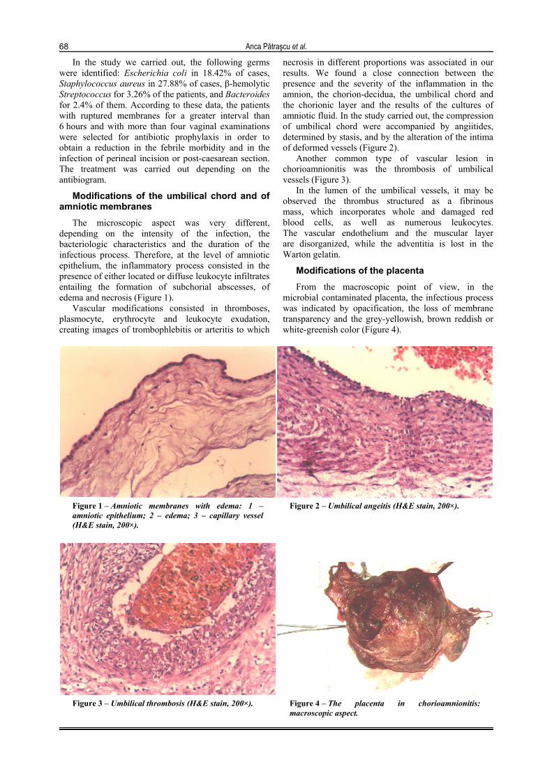
The microscopic aspect was very different, depending on the intensity of the infection, the bacteriologic characteristics and the duration of the infectious process. Therefore, at the level of amniotic epithelium, the inflammatory process consisted in the presence of either located or diffuse leukocyte infiltrates entailing the formation of subchorial abscesses, of edema and necrosis (Figure 1).
Vascular modifications consisted in thromboses, plasmocyte, erythrocyte and leukocyte exudation, creating images of trombophlebitis or arteritis to which
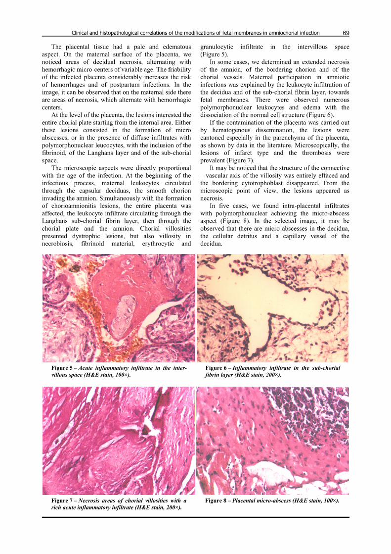
necrosis in different proportions was associated in our results. We found a close connection between the presence and the severity of the inflammation in the amnion, the chorion-decidua, the umbilical chord and the chorionic layer and the results of the cultures of amniotic fluid. In the study carried out, the compression of umbilical chord were accompanied by angiitides, determined by stasis, and by the alteration of the intima of deformed vessels (Figure 2).
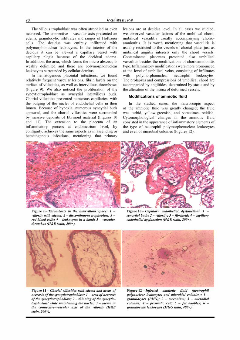
Another common type of vascular lesion in chorioamnionitis was the thrombosis of umbilical vessels (Figure 3).
In the lumen of the umbilical vessels, it may be observed the thrombus structured as a fibrinous mass, which incorporates whole and damaged red blood cells, as well as numerous leukocytes. The vascular endothelium and the muscular layer are disorganized, while the adventitia is lost in the Warton gelatin.
Modifications of the placenta
From the macroscopic point of view, in the microbial contaminated placenta, the infectious process was indicated by opacification, the loss of membrane transparency and the grey-yellowish, brown reddish or white-greenish color (Figure 4).
Figure 1 – Amniotic membranes with edema: 1 – amniotic epithelium; 2 – edema; 3 – capillary vessel (H&E stain, 200×).
Figure 2 – Umbilical angeitis (H&E stain, 200×).
Figure 3 – Umbilical thrombosis (H&E stain, 200×). Figure 4 – The placenta in chorioamnionitis: macroscopic aspect.
Clinical and histopathological correlations of the modifications of fetal membranes in amniochorial infection
69
The placental tissue had a pale and edematous aspect. On the maternal surface of the placenta, we noticed areas of decidual necrosis, alternating with hemorrhagic micro-centers of variable age. The friability of the infected placenta considerably increases the risk of hemorrhages and of postpartum infections. In the image, it can be observed that on the maternal side there are areas of necrosis, which alternate with hemorrhagic centers.
At the level of the placenta, the lesions interested the entire chorial plate starting from the internal area. Either these lesions consisted in the formation of micro abscesses, or in the presence of diffuse infiltrates with polymorphonuclear leucocytes, with the inclusion of the fibrinoid, of the Langhans layer and of the sub-chorial space.
The microscopic aspects were directly proportional with the age of the infection. At the beginning of the infectious process, maternal leukocytes circulated through the capsular deciduas, the smooth chorion invading the amnion. Simultaneously with the formation of chorioamnionitis lesions, the entire placenta was affected, the leukocyte infiltrate circulating through the Langhans sub-chorial fibrin layer, then through the chorial plate and the amnion. Chorial villosities presented dystrophic lesions, but also villosity in necrobiosis, fibrinoid material, erythrocytic and
granulocytic infiltrate in the intervillous space (Figure 5).
In some cases, we determined an extended necrosis of the amnion, of the bordering chorion and of the chorial vessels. Maternal participation in amniotic infections was explained by the leukocyte infiltration of the decidua and of the sub-chorial fibrin layer, towards fetal membranes. There were observed numerous polymorphonuclear leukocytes and edema with the dissociation of the normal cell structure (Figure 6).
If the contamination of the placenta was carried out by hematogenous dissemination, the lesions were cantoned especially in the parenchyma of the placenta, as shown by data in the literature. Microscopically, the lesions of infarct type and the thrombosis were prevalent (Figure 7).
It may be noticed that the structure of the connective – vascular axis of the villosity was entirely effaced and the bordering cytotrophoblast disappeared. From the microscopic point of view, the lesions appeared as necrosis.
In five cases, we found intra-placental infiltrates with polymorphonuclear achieving the micro-abscess aspect (Figure 8). In the selected image, it may be observed that there are micro abscesses in the decidua, the cellular detritus and a capillary vessel of the decidua.
Figure 5 – Acute inflammatory infiltrate in the inter-villous space (H&E stain, 100×).
Figure 6 – Inflammatory infiltrate in the sub-chorial fibrin layer (H&E stain, 200×).
Figure 7 – Necrosis areas of chorial villosities with a rich acute inflammatory infiltrate (H&E stain, 200×).
Figure 8 – Placental micro-abscess (H&E stain, 100×).
Anca Pătraşcu et al.
70
The villous trophoblast was often atrophied or even necrosed. The connective – vascular axis presented an edema, granulocytic infiltrates and ranges of Hofbauer cells. The decidua was entirely infiltrated with polymorphonuclear leukocytes. In the interior of the decidua it can be viewed a capillary vessel with capillary plegia because of the decidual edema. In addition, the area, which forms the micro abscess, is weakly delimited and there are polymorphonuclear leukocytes surrounded by cellular detritus.
In hematogenous placental infections, we found relatively frequent vascular lesions, fibrin layers on the surface of villosities, as well as intervillous thromboses (Figure 9). We also noticed the proliferation of the syncytiotrophoblast as syncytial intervillous buds. Chorial villosities presented numerous capillaries, with the bulging of the nuclei of endothelial cells in their lumen. Because of hypoxia, numerous syncytial buds appeared, and the chorial villosities were surrounded by massive deposits of fibrinoid material (Figures 10 and 11). The extension to the placenta of an inflammatory process at endometrium level, by contiguity, achieves the same aspects as in ascending or hematogenous infections, mentioning that primary
lesions are at decidua level. In all cases we studied, we observed vascular lesions of the umbilical chord, umbilical vasculitis usually accompanying chorio-amnionitis. It is worth mentioning that vasculitis is usually restricted to the vessels of chorial plate, just as umbilical angiitis interests only the chord vessels. Contaminated placentas presented also umbilical vasculitis besides the modifications of chorioamnionitis type. Inflammatory modifications were more pronounced
at the level of umbilical veins, consisting of infiltrates with polymorphonuclear neutrophil leukocytes. The prolapsus and compressions of umbilical chord are accompanied by angiitides, determined by stasis and by the alteration of the intima of deformed vessels.
Modifications of amniotic fluid In the studied cases, the macroscopic aspect
of the amniotic fluid was greatly changed; the fluid was turbid, yellow-greenish, and sometimes reddish. Cytomorphological changes in the amniotic fluid consisted in the appearance of inflammatory elements of the type of neutrophil polymorphonuclear leukocytes and even of microbial colonies (Figures 12).
Figure 9 – Thrombosis in the intervillous space: 1 – villosity with edema; 2 – discontinuous trophoblast; 3 – red blood cells; 4 – leukocytes in a band; 5 – vascular thrombus (H&E stain, 200×).
Figure 10 – Capillary endothelial dysfunction: 1 – syncytial buds; 2 – villosity; 3 – fibrinoid; 4 – capillary endothelial dysfunction (H&E stain, 200×).
Figure 11 – Chorial villosities with edema and areas of necrosis of the syncytiotrophoblast: 1 – area of necrosis of the syncytiotrophoblast; 2 – thinning of the syncytio-trophoblast while maintaining the nuclei; 3 – edema in the connective–vascular axis of the villosity (H&E stain, 200×).
Figure 12 – Infected amniotic fluid (neutrophil polynuclear leukocytes and microbial colonies): 1 – granulocytes (PMN); 2 – meconium; 3 – microbial colonies; 4 – prismatic cell; 5 – fat bubbles; 6 – granulocytic leukocytes (MGG stain, 400×).

Clinical and histopathological correlations of the modifications of fetal membranes in amniochorial infection
71
Cytopathological changes were related to normal cytomorphological components of the amniotic fluid.
In the present study, the cytomorphological modifications of inflammatory type of the amniotic fluid varied depending on the age of the pregnancy: 74% of the cases concerned preterm births, and 35% of term births with preterm membrane rupture.
Simultaneously with the modifications of inflammatory type in the amniotic fluid, we determined, in the same percentage, lesions of inflammatory type of amniotic membranes in the proximity of the continuity solution. Chorioamnionitis lesions extended from the placenta and the umbilical chord, having a maximum acuteness in the area of insertion of the chord. The acuteness of inflammatory lesions is reduced while the pregnancy advances, however the vasculopathies aggravate.
Discussion
As far as the microbiological study of the inferior genital tract and of the amniotic fluid in patients with preterm asymptomatic infection, the literature proves that anaerobe germs are prevalent [8].
The umbilical chord appeared edematous, friable, dull dark colored, possibly reaching a genuine necrotic aspect [9]. In the umbilical chord, there were polymorph nuclear leukocytes as a response to microbial aggression. The membranes presented similar located or diffuse lesions. Diffuse lesions interested all structures (the amnion, the chorion and the decidua), the inflammatory reaction of the decidua causing a maternal leukocyte flux towards amniochorial structures.
These macro- and microscopic features are not specific to the infection, they can be observed in severe hypoxia, in modifications induced by proteolytic enzymes and by chemical action of the meconium. The inflammatory modifications are produced after introducing glucosed or chlorinated solutions and iodate substances in the amniotic cavity.
A study was carried out with reference to the relation between the inflammatory lesions of the preterm placenta and the microbiology of the amniotic fluid. There is a close connection between the presence and the severity of the inflammation in the amnion, the chorion–decidua, the umbilical chord and the chorionic layer and the results of the cultures of amniotic fluid. Several authors quoted in Cunningham FG and contributory, in Williams Obstetrics, showed that acute inflammation of the chorionic layer was the most sensible sign of microbial invasion of the amniotic cavity (96.9%) [2].
The prolapsus and compression of umbilical chord were accompanied by angeitides, determined by stasis, and by the alteration of the intima of deformed vessels [1, 10].
Usually, placental lesions were prevalent in the basal plate and the neighboring villosities. Chorial villosities were intumescent, with hydropic degeneration and with dystrophic lesions of the trophoblast. In the connective–vascular axis of villosities, there were
rendered evident septic granulomas, consisting of leukocyte agglomerations, also partly interesting the structures of the umbilical chord) [9, 11].
Both the infiltration of membranes with neutrophil polynuclear leucocytes, and umbilical angiitis can also be induced by the extended action of meconium, its irritant action being well known [1, 6].
Normal cytomorphological components of the amniotic fluid are prismatic cells (epithelial cells of Malpighian type in the fetus teguments), cubic cells (epithelial cells in amniotic membranes), lanugo, meconium granules, and fat bubbles. The most frequent cytopathological modifications rendered evident at the level of microbial contaminated amniotic fluid consist in the identification of granulocytic leukocytes, of meconium granules, of fat bubbles and of microbial colonies [12, 13].
In the present study, the cytomorphological modifications of inflammatory type of the amniotic fluid varied depending on the age of the pregnancy: 74% of the cases concerned preterm births, and 35% of term births with preterm membrane rupture, values that are comparable with the data of technical literature [1, 2, 5, 10].
Simultaneously with the modifications of inflammatory type in the amniotic fluid, we determined, in the same percentage, lesions of inflammatory type of amniotic membranes in the proximity of the continuity solution. Chorioamnionitis lesions extended from the placenta and the umbilical chord, having a maximum acuteness in the area of insertion of the chord. The acuteness of inflammatory lesions is reduced while the pregnancy advances, however the vasculopathies aggravate. Therefore, histopathological modifications at the umbilical chord level develop without chorionic inflammation [13, 14].
Conclusions
The lesions of amniochorial membranes in the amniotic infection are not specific, they represent an inflammatory process.
Histopathological modifications of amniotic membranes are markers of the microbial invasion of chorioamniotic cavity.
Reduced or missing clinical symptoms causes the chorioamnionitis diagnosis to be often established after the delivery of the placenta and only by the morphological and histopathological aspects it presents.
Morphopathological and bacteriological modifica-tions in the amniochorial infections vary depending on the entirety of ovular membranes and on the duration of infectious aggression.
The identification of these modifications can contribute to setting up maternal and prophylactic fetal treatment.
References [1] PĂTRAŞCU ANCA, Chorioamnionitis, Didactic and Pedagogic
Publishing House, Bucharest, 1998. [2] CUNNINGHAM F. G., MACDONALD P. C., GANT N. F.,
LEVENO K. J., GILSTRAP L. C., Williams Obstetrics, McGraw–Hill/Appleton & Lange, 1993.

Anca Pătraşcu et al.
72
[3] PĂTRAŞCU ANCA, VOICULESCU C., Study on bactericide and bacteriostatic capacity of amniotic fluid depending on the time elapsed from the moment of membrane rupture, Bacteriology, Virusology, Parasitology, Epidemiology, 1998, 43(3):167–170.
[4] PATRASCU A., SURTEA L., BERCEANU S., BANITA M., Studies into amniotic fluid in chorioamnionitis, XVI FIGO World Congress of Gynecology and Obstetrics, September 3–8, Washington DC, Int J Gynecol Obstet, 2000, 70(Suppl 1):A66.
[5] GABBE S. G., SIMPSON J. L., NIEBYL J. R., GALAN H., GOETZL L., JAUNIAUX E. R. M., LANDON M. (eds), Obstetrics: normal and problem pregnancies, 5th edition, Churchill Livingstone, 2007.
[6] BUHIMSCHI I. A., ZAMBRANO E., PETTKER C. M., BAHTIYAR M. O., PAIDAS M., ROSENBERG V. A., THUNG S., SALAFIA C. M., BUHIMSCHI C. S., Using proteomic analysis of the human amniotic fluid to identify histologic chorioamnionitis, Obstet Gynecol, 2008, 111(2 Pt 1):403–412.
[7] MCDONALD H. M., CHAMBERS H. M., Intrauterine infection and spontaneous midgestation abortion: is the spectrum of microorganisms similar to that in preterm labor?, Infect Dis Obstet Gynecol, 2000, 8(5–6):220–227.
[8] ROMERO R., MAZOR M., SEPULVEDA W., AVILA C., COPELAND D., WILLIAMS J., Tumor necrosis factor in preterm and term labor, Am J Obstet Gynecol, 1992, 166(5):1576–1587.
[9] QUEENAN J. T., SPONG C. Y., LOCKWOOD C. J. (eds), Management of high-risk pregnancy: an evidence-based approach, 5th edition, Wiley–Blackwell, 2007, 333–353.
[10] CHELLAM V. G., RUSHTON D. I., Chorioamnionitis and funiculitis in the placentas of 200 births weighing less than 2.5 kg, Br J Obstet Gynaecol, 1985, 92(8):808–814.
[11] PĂTRAŞCU ANCA, Syphilitic chorioamnionitis in a full term pregnant woman age 26 years, Arch Balkan Med Union, 1998, 33(1):37–38.
[12] PĂTRAŞCU ANCA, VOICULESCU C., Immunological studies on amniotic fluid, Clinical Laboratory – Medical Technique Review, IIIrd year, 1998, 3:25–26.
[13] HECHT J. L., ALLRED E. N., KLIMAN H. J., ZAMBRANO E., DOSS B. J., HUSAIN A., PFLUEGER S. M., CHANG C. H., LIVASY C. A., ROBERTS D., BHAN I., ROSS D. W., SENAGORE P. K., LEVITON A.; ELGAN STUDY INVESTIGATORS, Histological characteristics of singleton placentas delivered before the 28th week of gestation, Pathology, 2008, 40(4):372–376.
[14] PATRASCU A., BERCEANU S., SURTEA L., BANITA M., The value of interleukine 6 in the diagnosis of chorio-amnionitis, XVI FIGO World Congress of Gynecology and Obstetrics, September 3–8, Washington DC, Int J Gynecol Obstet, 2000, 70(Suppl 1):A108.
Corresponding author Anca Pătraşcu, Associate Professor, MD, PhD, Department of Obstetrics and Gynecology, University of Medicine and Pharmacy of Craiova, “Filantropia” Municipal Hospital, 3 Constantin Brâncuşi Street, 200136 Craiova, Romania; Phone +40251–417 820, e-mail: [email protected] Received: November 10th, 2008
Accepted: January 25th, 2009