cleft lip and palate anomaly in india: clinical profile, risk .... cleft lip...dr. sushma sagar dr....
TRANSCRIPT

Cleft Lip and Palate Anomaly in India: Clinical Profile, Risk Factors and
Current Status of Treatment: A Hospital Based Study
Indian Council of Medical Research
Task Force Project Report
Indian Council of Medical Research (ICMR), New Delhi
and Centre for Dental Education and Research
All India Institute of Medical Sciences (AIIMS), New Delhi

Cleft Lip and Palate Anomaly in India: Clinical Profile, Risk Factors and
Current Status of Treatment: A Hospital Based Study
Indian Council of Medical Research
Task Force Project Report
Coordinating Centre
Centre for Dental Education and Research All India Institute of Medical Sciences, New Delhi
Contributing Centres
Centre for Dental Education and Research All India Institute of Medical Sciences, New Delhi
Safdarjung Hospital, New Delhi
Medanta- The MEDICITY Hospital, Gurgaon

Pre-pilot study
Duration- 2 years
March 2010 to March 2011
extended upto March 2012
Pilot study
Duration- 3 years
April 2012 to March 2014
extended upto June, 2015
Published By
Indian Council of Medical Research, New Delhi Division of Non Communicable Diseases
Dr. Bela Shah Former Head (Upto 11th Feb 2013 and
12th Feb 2016 – 31st May 2016) Dr. D. K. Shukla Former Head (12th Feb 2013 – 11th Feb 2016) Dr. R. S. Dhaliwal Head (1st June 2016 – till date) Dr. Ashoo Grover Scientist ‘E’, Programme Officer
Centre for Dental Education and Research All India Institute of Medical Sciences, New Delhi
Professor O. P. Kharbanda CHIEF - Centre for Dental Education and Research Head, Division of Orthodontics and Dentofacial Deformities
Published in 2016

Cleft lip and palate anomaly in India: Clinical profile risk factors and
current status of treatment: A hospital based study
PRINCIPAL INVESTIGATOR AND COORDINATOR
Prof. O. P. Kharbanda
Centre for Dental Education and Research All India Institute of Medical Sciences, New Delhi
CONTRIBUTORS
Centre for Dental Education and Research, AIIMS, New Delhi
Chief Investigator
Co-investogators
Prof. O. P. Kharbanda
Dr. S. C. Sharma
Dr. Madhulika Kabra
Dr. Sushma Sagar
Dr. Maneesh Singhal
Dr. Neerja Gupta
Dr. Manju Mehta
Safdarjung Hospital, New Delhi
Chief Investigator
Co-investogators
Dr. Karoon Agrawal
Dr. N. N. Mathur
Medanta-The MEDICITY Hospital, Gurgaon
Chief Investigator
Co-investogators
Dr. Rakesh Khazanchi
Dr. K. K. Handa

CONTRIBUTORS
Panel of experts
Dr. Anil Kohli, Delhi Dr. S.G. Damle, Mullana, Haryana
Dr. Ashok Utreja, Chandigarh Dr. K. Sreedharan, Chennai
Dr. T. Samraj, Salem Dr. I. C. Verma, Delhi
Dr. G. S. Meena, Delhi
Research staff engaged in the project
Institute Names Designation
ICMR HQs None
CDER, AIIMS, New Delhi 1. Dr. Neeraj Wadhwan Senior Research Officer
2. Ms. Parul J. Rathod Senior Research Fellow
3. Ms. B. Aarthi Data Entry Operator
4. Mr. Netra Pal Dental Technician
5. Ms. Nisha Bansal Computer Programmer
6. Ms. Neha Takhi Computer Programmer
7. Ms. Pooja Maurya Computer Programmer
Safdarjung Hospital, Delhi Dr Parul Narang Research Assistant
Medanta- The MEDICITY
Hospital Gurgaon Dr Upaasna Vinayak Research Assistant
ICMR
Dr. Bela Shah
Dr. D. K. Shukla Dr. R. S. Dhaliwal
Dr. Ashoo Grover
Dr. Ravinder Singh
NIC
Dr. Savita Dawar

Executive Summary
Cleft treatment requires a multidisciplinary approach extended from the birth until adulthood.
Many of these children are born in rural areas where resources for treatment and awareness
on cleft care are limited. Consequently, many patients may receive limited or suboptimal care due to various reasons. This study was aimed at evaluating and identifying the patterns of
the congenital defects of face, cleft lip and palate among patients visiting three major hospitals across Delhi and the National Capital Region (NCR). The objectives included
establishing baseline data on a spectrum of clinical profile of cleft patients, treatment protocols, quality of treatment and their residual treatment needs. The experience gained
from a study of these three target centres would then be used to lay a framework to conduct
a nationwide multicentre study in terms of logistics, feasibility and difficulties. The study titled Cleft Lip and Palate anomaly in India: Clinical profile, Risk factors and Current status of treatment: a Hospital-based study was started in 2010 as a task force project of ICMR. The pilot phase, which started in 2012, encompassed three cleft centres across Delhi and NCR,
namely, AIIMS, Safdarjung Hospital and Medanta-The MEDICITY. The relevant data for 126
subjects exhibiting non-syndromic cleft lip and palate was recorded on a specially designed performa. Each case was evaluated by a team of specialists comprising of a Plastic Surgeon,
an Orthodontist, an ENT Surgeon, a Dental Surgeon, a Speech therapist and an Audiologist. Clinical records included the subject’s profile and intraoral photos, dental study models,
audiometric and speech evaluation data. The current report highlights that among the samples of the cleft patients assessed in the project, the treatment needs were significantly
high. There was a wide variation in age at primary lip and palate surgery with a significant
percentage of cases requiring lip and nose revision surgeries. Fifty five percent cases had post-surgical oro-nasal fistula and a large proportion of operated UCLP cases had complex
orthodontic treatment needs.
There seems to be an urgent need to devise strategies to improve the delivery of
quality care to the afflicted subjects, with the joint efforts of all the experts and health care
providers. It must be mentioned here that the data is not representative of the outcome of the three centres. It also tries to highlight that larger multicentre studies are needed in the
Indian setup so that the patients not only receive treatment but also the quality of the treatment is monitored for better outcomes. The results indicate a lack of uniform protocol
followed in providing care to cleft patients. A great variation was found in the quality of
treatment received by many of the patients.

Forward
A comprehensive management of cleft patients requires a multidisciplinary approach
extended from birth until adulthood. Many of these children are born in rural areas where resources for treatment and awareness on cleft care are limited. Consequently, many patients
may receive limited or suboptimal care due to multitudes of reasons. This study was aimed to evaluate and identify pattern of the congenital birth defects of face, cleft lip and palate
among patients visiting three major hospitals across Delhi and National Capital Region (NCR). The study titled “Cleft Lip and Palate anomaly in India: Clinical profile, Risk factors and
Current status of treatment: a Hospital based study” was started in 2010 as a Task Force
project of ICMR. Pre-Pilot phase of the study was conducted between 2010 to 2012 in the Department of Orthodontics and Dentofacial Deformities and ENT, AIIMS, New Delhi. The
Pilot phase, which started in 2012, encompassed three cleft centres across Delhi and NCR, namely, AIIMS, Safdarjung Hospital and Medanta-The MEDICITY. Current report highlights
that larger multicentre studies are needed in the Indian setup so that the patients do not only
receive treatment but also the quality of the treatment is monitored for better treatment outcomes. The results indicate a lack of uniform protocol followed in providing care to cleft
patients. A great variation was found in the quality of treatment received by many of the
patients.
Report of three centres of Delhi and NCR presents the situation of the status of cleft
care in India. The profilometric analysis of cleft care has provided a glimpse on the ground realities related to the treatment of cleft patient in different parts of India. It is hoped that
this Report would be useful for researchers and planners in their endeavor to work towards strengthening the management of cleft lip and palate anomaly in the country and work out
guidelines /protocols for proper management of CLP in the Indian social milieu that is ailing
with limited health care resources.
Dr. Soumya Swaminathan
Director General, ICMR

Preface
Cleft lip and palate is the most common congenital deformity of the craniofacial region with
an average worldwide incidence of 1 in 700. Its incidence in Asian population is reported to be around 2.0 per 1000 live births or higher. In India, though national epidemiological data is
not available, many studies from different parts have reported a variation in the incidence of cleft anomaly. Based on rough estimates, it has been suggested that approximately 35,000
newborn cleft patients are added every year to the Indian population. With many patients having less than optimum care in a not-so-organized setup, the cumulative burden of persons
affected with this birth defect is huge. Although India has a large and extended network of
medical facilities, interdisciplinary cleft care is provided in only a few hospitals. Day-to-day interactions with these patients exhibit significant variation in treatment provided and the
quality of outcome, with some having had excellent treatment outcomes while many patients, unfortunately, received suboptimum, limited or no treatment at all. The reasons for this are
many and varied. The awareness in the society and amongst the health professionals on the
critical aspects of interdisciplinary care of this anomaly may be lacking. Affordability and availability of experts may also contribute to the quality of treatment. There is a lack of
interdisciplinary approach in majority of the centres, and hence, there is a need for better interaction amongst the specialists. This lack of interdisciplinary approach and the need for it
in the Indian setup has been stressed previously also. This ongoing Task Force Project was initiated by the Indian Council of Medical Research to evaluate the current status of treatment
and treatment needs of cleft patients. The ultimate aim is to work out a national registry and
guidelines for cleft care in India.
The long term objectives are to initiate a National Registry for the patients with
congenital birth defects of the face and jaws and also to establish strategies that will address a multitude of challenges associated with the prevention and treatment of this deformity. The
aim is to improve the quality of life (QOL) of children suffering from Cleft lip palate and such
deformities so as to offer them a hope for a normal living.
Professor O.P. Kharbanda
CHIEF - Centre for Dental Education & Research Head - Division of Orthodontics and Dentofacial Deformities
All India Institute of Medical Sciences, New Delhi PRINCIPAL INVESTIGATOR/ COORDINATOR- Task Force Project DIRECTOR- WHO Collaborating Centre for Oral Health Promotion

Acknowledgement
We gratefully acknowledge the valuable contribution of the Chairperson and the Members of the Task Force Group for providing continuous guidance and support in implementing the
pre-pilot and pilot phase of the study. We also acknowledge the investigators engaged in
undertaking the ICMR funded Task Force Study on “CLEFT LIP AND PALATE ANOMALY IN INDIA: CLINICAL PROFILE, RISK FACTORS AND CURRENT STATUS OF TREATMENT: A
HOSPITAL BASED STUDY (2012-2014)” and providing meaningful outcome in the form of ‘IndiCleft Tool’. The tool is ready to be taken up further in the multicentric nationwide study
which will be useful in achieving aims and objectives during the main phase. The members
enthusiastically participated in discussions and provided immensely useful inputs drawn from their vast experience in the subject. We also thank the Reviewers for their suggestions and
timely advice.
We are grateful to Director General, ICMR for envisioning the Task Force Study in the
Indian context and encouraging us to take the initiative. We would like to thank Scientist – ‘F’ from National Informatics Centre for her valuable efforts. We also thank the Head, Division of
Non-Communicable Diseases (NCD) for being a constant guide and support.
Our special thanks to the administrative staff of Division of Non-Communicable Diseases and the financial staff of ICMR headquarters for smooth implementation and timely
release of grants to make the project a success.
Oct., 2015 Dr. Ashoo Grover
Dr. R. S. Dhaliwal

Abbreviations followed uniformly in text
UCLP : Unilateral Cleft Lip and Palate
BCLP : Bilateral Cleft lip and Palate
CP : Cleft Palate
CL : Cleft Lip
CLA : Cleft Lip and Alveolus

List of tables Table 1-Distribution of sample according to age
Table 2- Distribution of sample according to the type of cleft & sex
Table 3-Classification of cleft (Nagpur classification)
Table 4- Distribution of sample according to Nagpur classification
Table 5- Patients with positive familial history of Cleft
Table 6-History of medical problems in mother during 1st trimester of affected pregnancy
Table 7- History of Drug usage in mother during affected pregnancy
Table 8- History of radiation exposure to mother during 1st trimester of a affected
pregnancy
Table 9- Use of intoxicants by mother during 1st trimester of a affected pregnancy
Table 10- History of exposure to smoke during 1st trimester
Table 11- Effect of cleft deformity on the social acceptability of the patient
Table 12- Post natal counseling of parents with regards to feeding of child with cleft and
his treatment possibilities related to cleft
Table 13- Distribution of the patients who received correct advice for at least one of the
evaluated variables
Table 14- Age wise distribution of lip repair
Table 15- Age at palatal repair excluding alveolus
Table 16- Previous history of pre-surgical orthopaedic treatment
Table 17- Previous history of dental treatment
Table 18- Positive history of post surgical orthodontic treatment
Table 19- Supernumerary teeth
Table 20- Presence of anterior crossbite
Table 21- Presence of posterior crossbite in the sample of 55 cases
Table 22 - Overjet in the sample
Table 23- Overbite in the sample
Table 24- Goslon Yardstick scores and their interpretation
Table 25- Distribution of subjects according to the Goslon Yardstick
Table 26- Widest gap in the cleft, palate + alveolus
Table 27- Length of the palate
Table 28- Length of scar in unilateral clefts
Table 29- Angulation of scar in unilateral cleft
Table 30- Length of scar in bilateral cleft
Table 31- Angulation of scar in bilateral cleft
Table 32- Assessment of lip seal
Table 33- Evaluation of lip symmetry
Table 34- Overall appearance of lip
Table 35- Overall appearance of nose

Table 36- Evaluation of nasal septum in unilateral clefts
Table 37- Evaluation of nasal septum - Bilateral clefts
Table 38- Evaluation of nostril floor width
Table 39- Evaluation of the length of the palate in the sample
Table 40- Evaluation of post surgical scarring of the palate in the sample
Table 41- Mobility of the palate in the sample
Table 42- Status of uvula in operated cases of cleft palate
Table 43- Presence of fistula in the sample
Table 44- Assessment whether the fistula is symptomatic or not
Table 45- Evaluation of the size of the oronasal fistula in the sample
Table 46- Speech abnormality due to presence of the fistula
Table 47- Evaluation whether the fistula has been operated previously or not in the
sample
Table 48- Status of Tonsils in the sample according to type of cleft
Table 49- Incidence of Ear Discharge
Table 50- Status of tympanic membrane in the sample in unilateral cleft
Table 51- Status of tympanic membrane-Bilateral cleft
Table 52- Prevalence of hearing abnormalities in the sample as a function of type of cleft
Table 53- Degree of hearing loss in the sample
Table 54- Relation of hearing loss with different types of cleft – Unilateral Cleft
Table 55- Relation of hearing loss with different types of cleft – Bilateral Cleft
Table 56- Impedance Audiometry and their inference
Table 57- Status of middle ear based on Impedance
Table 58- Status of middle ear based on Impedance- Bilateral cleft
Table 59- Distribution of the sample according to nasality of speech
Table 60- Speech Articulation in the sample
Table 61- Status of Affected articulation in the sample
Table 62- Description of speech sample
Table 63- Overall speech intelligibility in various cleft types

List of figures
Figure 1- Cleft patient assessment tool
Figure 2- Intraoral photographs
Figure 3- Supplemental intraoral photographs
Figure 4- Dental study model
Figure 5- Complete organizational setup of the project
Figure 6- “The Indicleft Team”
Figure 7- Schedule and timing protocol of cleft care
Figure 8 (1-12)- Pedigree Charts
Figure 9- Distribution of proposed centres in multicentre study

List of graphs
Graph 1- Distribution of sample according to age
Graph 2- Distribution of sample according to the type of cleft & sex
Graph 3- Distribution of sample according to Nagpur classification
Graph 4- Patients with positive familial history of Cleft
Graph 5- History of medical problems in mother during 1st trimester of affected pregnancy
Graph 6- History of Drug usage in mother during affected pregnancy
Graph 7- History of radiation exposure to mother during 1st trimester of a affected pregnancy
Graph 8- History of exposure to smoke during first trimester
Graph 9- Distribution of the patients who received correct advice for at least one of the evaluated variables
Graph 10- Age wise distribution of lip repair
Graph 11- Age at palatal repair excluding alveolus
Graph 12- Presence of anterior crossbite
Graph 13- Goslon score
Graph 14- Evaluation of lip symmetry
Graph 15- Overall appearance of lip
Graph 16- Overall appearance of nose
Graph 17- Evaluation of nasal septum in unilateral clefts
Graph 18- Evaluation of nasal septum - Bilateral clefts
Graph 19- Evaluation of nostril floor width
Graph 20- Evaluation of the length of the palate in the sample
Graph 21- Evaluation of post surgical scarring of the palate in the sample
Graph 22- Presence of fistula in the sample
Graph 23- Evaluation of the size of the oronasal fistula in the sample
Graph 24- Status of Tonsils in the sample according to type of cleft
Graph 25- Prevalence of hearing abnormalities in the sample as a function of type of cleft
Graph 26- Degree of hearing loss in the sample
Graph 27- Distribution of the sample according to nasality of speech
Graph 28- Speech Articulation in the sample
Graph 29- Overall speech intelligibility in various cleft types

Contents
1 Introduction …………………………………………………………………………... 1
2. Aims and objectives ………………………………………………………………… 2
3. Subjects and methods
➢ Pre Pilot Phase 2012-1012 …………………………………………….
➢ Pilot Phase 2012 -2014
• Sample and methodology ……………………………….………
• Structuring of an expert team: “The Indicleft Team” ...
• Patient evaluation ………………………….……………………….
• Assessment of etiology of cleft …….………………………….
• Dental history and examination ……………………………….
• Orthodontic Treatment History ………………………………..
• Evaluation of primary cleft deformity …………..…………..
• Evaluation of secondary cleft deformity …………………..
• Evaluation of lip ……………………………………………………..
• Evaluation of nose ………………………………….………………
• Evaluation of secondary palate ………………………………..
• Assessment of post-surgical palatal fistula ………………..
• ENT evaluation ………………………………………………………
• Speech assessment ………………………………………………..
3
6
6
9
9
9
9
10
10
10
11
11
11
12
12
4. Analysis and results
• Distribution of sample according to age and sex ……………..
• Distribution and types of cleft ……………………………..………..
• Classification of cleft …………………………….………………………
• Etiology of cleft: genetic and environmental risk factors ….
• Effect of cleft deformity on the social acceptability of the patient ………………………………………………………………………..
• Post natal counseling of parents with regards to feeding and treatment possibilities related to cleft ………………………
• Age wise distribution of primary lip and palate repair ………
• Previous history of dental and orthodontic treatment ………
• Dental examination ………………………………………………………
• GOSLON Yardstick ……………………………………………………….
• Examination of primary cleft ………………………………………….
• Examination of Secondary Cleft Deformity ………………………
• Examination of the Nose ……………………………………………….
• ENT Examination ………………………………………………………….
• Hearing evaluation ……………………………………………………….
• Speech assessment ………………………………………………………
13
13
13
16
28
28
28
29
34
35
35
41
46
57
60
60
5. Conclusions ……….…………………………………………………………………… 70
6. Future directions …………………………………………………………………….. 71
7. S & T benefits occurred
• Lists of research publications with complete details ……..…. 73
8. Procurement and Usage of equipments …….………………………………. 74

1 Indian Council of Medical Research Task Force Project
1 Introduction
Cleft of the lip and /or the palate (CL±P) is a
congenital birth defect which is characterized
by complete or partial cleft of the lip and/ or
the palate. The severity of the cleft may vary
from the trace of notching of the upper lip to a
complete non- fusion of the lip, the primary
palate and the secondary palate. Cleft lip and
palate anomaly constitutes nearly one-third of
all congenital malformations of the
craniofacial region with an average worldwide
incidence of 1 in 700. Its incidence in Asian
population is reported to be around 2.0 per
1000 live births or higher. In India, even
though a national epidemiological data is not
available, many studies from different parts of
the country have reported a variation in the
incidence of cleft anomaly. Sidhu and
Deshmukh reported the incidence of Cleft Lip
and Palate (CL+P) at AIIMS, New Delhi to be
1.4 per 1000 live births. Mossey and Little
estimated from various multicentric studies
across India that the incidence of CLP in India
ranges from around 0.93-1.3 for cleft lip and
palate.
Based on rough estimates, it is
suggested that approximately 35,000 newborn
cleft patients are added every year to the
Indian population. With many patients having
less than optimum care in a not so organized
setup, the cumulative burden of persons
affected with this birth defect is huge.
Although India has a large and extended
network of medical facilities, interdisciplinary
cleft care is provided in only a few hospitals.
Day-to-day interactions with these patients
exhibit significant variations in treatment
provided and the quality of outcome with
some having excellent outcomes while many
patients received sub-optimum, limited or no
treatment. The reasons are many and varied.
The awareness in the society and amongst the
health professionals on critical aspects of
interdisciplinary care of this anomaly may be
lacking. Affordability and availability of
experts may also contribute to the quality of
treatment. Gopalakrishna and Agrawal (2010),
following a national survey on trends in the
management of patients with CLP in India,
concluded in their study, “Management of
Cleft Lip and Palate (CLP) differs in India.
Primary surgical practices are almost similar
to other studies. There is a lack of
interdisciplinary approach in majority of the
centres and hence, there is a need for better
interaction amongst the specialists.” This lack
of interdisciplinary approach and the need for
it in the Indian setup has been stressed
previously also. This taskforce project was
initiated by the Indian Council of Medical
Research, to evaluate the current status of
treatment vis-à-vis CLP and the medical needs
of cleft patients. The aim is to work out a
national registry and guidelines for cleft care
in India.

2 Indian Council of Medical Research Task Force Project
2 Aims and Objectives
To assess the multidisciplinary, multicentre
project titled Cleft Lip and Palate anomaly in
India: Clinical profile Risk factors and current
status of treatment: A hospital based study was
initiated under the aegis of Indian Council of
Medical Research (ICMR), New Delhi in 2010
as a pre-pilot study aimed to assess the
feasibility of a larger pilot study. The pre-pilot
phase was successfully completed in the
Department of Orthodontics & Dentofacial
Deformities and ENT, AIIMS, New Delhi
(2010-12).
The pilot phase of the study (2012-14) was
commissioned by the ICMR encompassing
three cleft-care centres with high case load
across Delhi and National Capital Region
(NCR). The study is essentially aimed to
evaluate the socio-economic, demographic
details of patients suffering from cleft lip and
palate (visiting the 3 enrolled hospitals), their
treatment profile and the residual treatment
needs of the same subjects. The objectives of
the pilot phase were:
i. To utilize the study tools developed in the
pre-pilot phase for the evaluation of the
clinical profile of CLP anomaly. The
experience gained would result in its
suitable modifications for its further use in
a nationwide multicentre study.
ii. To establish a methodology for a
nationwide collection of data for the
clinical profile for Cleft lip and palate
anomalies and its major treatment needs
including the logistics, feasibility and
difficulties expected in the execution of the
multicentre project.
The report of the Pre-pilot and Pilot
phase would be used to formulate a
multicentre study which will be aimed to:
1. Identify patterns of the congenital birth
defects of the face, cleft lip and palate in
India.
2. Establish the baseline data of a spectrum of
problems of cleft patients, discuss the
protocols of treatment given to these
children and their actual treatment needs.
3. Ascertain the risk factors associated with
congenital defects of the face: nutritional,
environmental and genetic.
The long term objectives are to initiate a
‘National Registry’ for the patients with
congenital birth defects of the face and jaws
and also to establish strategies that will
address the multitude of challenges associated
with the prevention and treatment of this
deformity. These will include:
a. Identify risk factors associated with the
development of cleft lip and palate
b. Develop strategies to minimize risk factors,
thereby reducing the incidence of Cleft Lip
and Palate (CLP) anomaly
c. Develop protocols for interdisciplinary care
which is feasible and affordable in India
with minimum burden on patients, parents
and service providers
d. Improve treatment outcomes
The ultimate aim is to improve the quality of
life (QOL) of children suffering from Cleft lip
palate and such deformities so that these
children can have a better living.

3 Indian Council of Medical Research Task Force Project
3 Subjects and Methods
A. Pre pilot phase 2010-2012
This phase of study was conducted at the
Department of Orthodontics and
Dentofacial Deformities, Centre for Dental
Education and Research (CDER) and ENT,
at the All India Institute of Medical
Sciences (AIIMS), New Delhi, from April
2010 to March 2012. The key highlights
were:
1. On 2nd June 2007, Prof. O.P. Kharbanda
wrote to Dr Jagdish Kaur (of DGHS,
MOHFW) to request for the inclusion of
cleft lip and palate, along with speech
and hearing defects, in the national
health surveys.
2. Core group experts of oral health met at
the ICMR headquarters and
recommended to Prof. Kharbanda to
initiate a pre-pilot study for 1 year to
focus on the spectrum of cleft patients to
develop a pilot proposal for 3 to 4
centres.
3. On 23rd July 2009, an application was
sent to ICMR for the initiation of the
task force project.
4. On 28th January 2010, an ethical
clearance was received.
5. On 23rd March 2010, the project was
initiated.
6. On 31st March 2012, the project was
completed.
7. An exhaustive study tool was developed
which had seven sections (Figure-1).
The multidisciplinary clinical
examination was to be performed by the
following specialists. Surgeon/Plastic
Surgeon, Orthodontist, Dental Surgeon,
ENT specialist and Speech therapist.
8. Standard operating procedure (SOP) for
recording the extraoral and intraoral
clinical photographs (Figure- 2 and 3),
dental study models (Figure- 4) and
investigations on hearing defect and
evaluation of speech were developed.
9. Planning was carried out to extend the
project onto the other hospitals across
Delhi.
Figure 1- Cleft patient assessment tool

Cleft Lip and Palate Anomaly in India: Clinical Profile, Risk Factors and Current Status of Treatment: A Hospital Based Study
4 Indian Council of Medical Research Task Force Project
Figure 2- Intraoral photographs
Figure 3- Supplemental intraoral photographs

Subjects and Methods
5 Indian Council of Medical Research Task Force Project
Figure 4- Dental study model
Figure 5-Complete organizational setup of the project

Cleft Lip and Palate Anomaly in India: Clinical Profile, Risk Factors and Current Status of Treatment: A Hospital Based Study
6 Indian Council of Medical Research Task Force Project
B. Pilot phase 2012 -2014
1. Sample and methodology
This study was conducted with the active
collaboration of All India Institute of
Medical Sciences, New Delhi, Safdarjung
Hospital, New Delhi, Medanta- The
MEDICITY Hospital Gurgaon, Haryana,
INDIA. A total number of 164 cases with cleft
lip and palate anomaly were recorded from
these three hospitals involved in the project
(55 from AIIMS, 54 from Safdarjung, 55 from
Medanta- The MEDICITY). The sample
consisted of 99 males and 65 females with a
wide age distribution (18 months to 516
months; a mean of 147.4 months) (Table 2).
The majority of the subjects belonged to the
age group of 5 (>6yrs -<=12yrs) (53 subjects)
(Table 1).
Data was recorded from three high
volume cleft care centres in Delhi and
National Capital Region (NCR) which
involved two public funded and one private
hospital.
The main highlights of the centres were:
• AIIMS: located in South Delhi; is an
autonomous institute which is a tertiary
care centre too. The combined cleft clinic
was established in the orthodontic unit in
the 1970’s.
• Safdarjung hospital; also located in South
Delhi; is a high volume centre supported by
the central government. It has a renowned
plastic and burns unit where cleft patients
are treated for primary and secondary
surgeries.
• Medanta- The MEDICITY; is also a
renowned hospital with an advanced plastic
surgery unit.
At each of the centres, the Departments of
Plastic surgery, Orthodontics and ENT were
the major input holders for support and
coordination of the study. The collection of the
data was carried out by the specifically
designated “Indicleft Team”. The complete
organizational setup of the project is indicated
in Figure 5.
2. Structuring of an expert team: “The Indicleft Team”
The “Indicleft team” included experts from
various medical specialties (Figure-6). It was
divided into a supervisory team (called
investigators) and a mobile team of research
staff. The members of the supervisory team
were based in three locations: AIIMS,
Safdarjung and Medanta- The MEDICITY
hospitals. The mobile team included a doctor,
an audiologist & a speech therapist and a
dental lab technician. Apart from the mobile
team, research staff was also deputed at the
three centres to work under the guidance of
supervisory staff. The research staff at AIIMS,
Safdarjung and Medanta- The MEDICITY
hospital performed the local data collection in
the form of taking down the medical history,
making impressions and other project-related
work at the respective centres. The research
staff at the centres also was responsible to
coordinate with the technical unit of CDER
and for the follow-up of the cleft patients of
their own centres. The mobile research team
would go to the various centres for
coordinating with the staff at the centres.
As previously mentioned, the team
comprised experts from various fields. This
was very important because cleft patients need
diverse types of treatment which can only be
provided by experts of the fields (Figure-7).
The individual role of each expert in the team
can be summarized below:
• The role of plastic surgeon: The plastic
surgeon is one of the most important
members of the team. He is responsible
for the primary and the secondary
surgeries of the defect. The surgeries are
particularly important because a good
primary surgery has a major effect on the
final outcome of the treatment and for
post-surgical maxillary growth. Poor
repair of the soft palate leads to
velopharyngeal incompetence and nasal
intonation along with nasal regurgitation.
The fistula many a times results from
poor surgical technique which can be best
understood by a plastic surgeon. The
fistula may also lead to nasal
regurgitation and nasal intonation. To
evaluate all these defects, the services of a
surgeon, preferably a plastic surgeon
would be required for proper assessment
of the gravity of the situation. Thus the
role of the plastic surgeon cannot be
under estimated in the project.

Subjects and Methods
7 Indian Council of Medical Research Task Force Project
• The role of orthodontist: The role of an
orthodontist cannot be overemphasized
when dental management of the cleft
child is concerned. Since nearly all of the
cleft children have dental deformities
secondary to cleft lip and palate (barring
perhaps just a few cases with isolated CP
and submucous clefts), all cleft children
need extensive orthodontic support for
management of their dentition right from
the start of the eruption of the primary
teeth to the time of growth completion at
18 years or till the final surgical
correction of the cleft defect. Thereafter,
the retentive aspects of orthodontic
correction take over and the patient is
followed in an adult life for over viewing
the alignment of teeth achieved earlier. In
many centres, the orthodontist has been
chosen as a team leader in the
multidisciplinary cleft team.
• The role of ENT specialist: Since many
of the patients with cleft have a defective
Eustachian tube function secondary to the
altered muscle attachments on the tube
and due to persistent nasal regurgitation,
many suffer from hearing defects
including CSOM (Chronic Suppurative
Otitis Media), tympanic membrane
defects and sometimes advanced hearing
loss resulting from adhesions of the ear
bones. It is of utmost importance to have
an ENT expert to examine the cases for
necessary interventions and referrals.
• The role of speech pathologist: As
mentioned earlier, due to velopharyngeal
incompetence, palatal fistulas as well as
poor repair of the lip, many patients have
distorted speech sounds, misarticulation
and most even have hypernasality. It is
indeed a necessity to evaluate the defects
in speech by an expert who can diagnose
the speech alteration, counsel the patient
and provide necessary speech therapy.
• The role of medical/dental graduate:
Overall assessment of the medical history
condition and /or dental condition
requires dedicated manpower support so
that the experts can focus on their specific
fields of interest; this leads to increased
efficiency of the experts and better quality
of recording of the data. Hence, support
by a medical/dental graduate to the
constituted team of experts cannot be
discounted.
Figure 6- The ‘Indicleft’ team

Cleft Lip and Palate Anomaly in India: Clinical Profile, Risk Factors and Current Status of Treatment: A Hospital Based Study
8 Indian Council of Medical Research Task Force Project
Ort
ho
gn
ath
ic S
urg
ery
an
d
Rh
ino
pla
sty
Ort
ho
do
nti
cs
Bo
ne
gra
ftin
g j
aw
Sp
ee
ch
the
rap
y/P
ha
ryn
go
pla
sty
Tym
pa
no
sto
my
tu
be
Pa
late
re
pa
ir
Pri
ma
ry c
left
lip
Su
rge
ry
? P
ala
tal
ob
tura
tor
/Fe
ed
ing
A
pp
lia
nce
18
y
17
y
16
y
15
y
14
y
13
y
12
y
11
y
10
y
9 y
8 y
7 y
6 y
5 y
4 y
3 y
2 y
1 y
9 m
6 m
3 m
0 m
Ag
e
Ort
ho
gn
ath
ic S
urg
ery
an
d
Rh
ino
pla
sty
Ort
ho
do
nti
cs
Bo
ne
gra
ftin
g j
aw
Sp
ee
ch
the
rap
y/P
ha
ryn
go
pla
sty
Tym
pa
no
sto
my
tu
be
Pa
late
re
pa
ir
Pri
ma
ry c
left
lip
Su
rge
ry
? P
ala
tal
ob
tura
tor
/Fe
ed
ing
A
pp
lia
nce
18
y
17
y
16
y
15
y
14
y
13
y
12
y
11
y
10
y
9 y
8 y
7 y
6 y
5 y
4 y
3 y
2 y
1 y
9 m
6 m
3 m
0 m
Ag
e

Subjects and Methods
9 Indian Council of Medical Research Task Force Project
3. Patient evaluation
The record keeping for the cases involved
prior consent and approval. Each patient
inducted in the study was subjected to the
following investigations, all of which were
non-invasive type.
1. Evaluation of the patient using the
specially-designed performa developed
during the pre-pilot phase of the project.
The said performa evaluated the
following aspects of cleft care (Figure-4):
a. General details: the personal details
like the patient’s name and address,
contact details
b. Socio-demographic profile
c. Evaluation of the risk factors
associated with etiology of cleft.
d. Evaluation of dental status of cleft
patients and their orthodontic
treatment profile
e. Evaluation of primary and secondary
cleft deformity
f. Assessment of hearing and speech
evaluation
2. The patients’ extraoral and intraoral
standard clinical photographs were
recorded.
3. Study models of each patient above 5
years of age were prepared for the project
using alginate impression material and
poured in orthodontic grade white dental
stone.
4. Assessment of etiology of cleft
The assessment was done by direct interview
of the parents/guardians of the subject. The
interview consisted of various parameters
known to be associated with genesis of the
cleft. Broadly, they could be divided as genetic
or environmental.
a. Genetic: Evaluation of genetic factors
consisted of pedigree analysis. The
pedigree was formed as per the interview
with the subjects’ parents and it was
evaluated whether the subject had any
predisposition to familial occurrence of
the cleft.
b. Environmental factors: The
environmental factors were also evaluated
by interview with the subjects’ parents.
The time of the 1st trimester of pregnancy
was particularly evaluated for the
presence of any risk factors associated
with the genesis of cleft. The interview
consisted of the following evaluations:
I. Exposure to radiation
II. Exposure to medication
III. History of sickness in the 1st trimester
IV. Use of intoxicants/ smoking
V. Exposure to smoke by either use of
chulha or passive smoking.
VI. History of recurrent abortions
5. Dental history and examination
The dental history and examination consisted
of verbal evaluation as well as clinical
examination. The subjects were evaluated for
any history of previous dental treatment by
way of direct interview as well as clinical
examination of dentition for presence of any
fillings or root canal treatment. Further, the
maxillary arch was evaluated for the presence
of supernumerary teeth and their position was
noted in relation to cleft.
6. Orthodontic treatment history
This aspect of taking history involved
evaluation of whether the subject had any
previous orthodontic treatment in his lifetime.
This included evaluation for pre-surgical
orthopedics, orthodontic treatment with
removable appliances or fixed appliances.
a. History of pre-surgical orthopedic
treatment: This was evaluated by
interview with subject’s parents. Parents
were asked whether any orthopedic
procedures like nasoalveolar moulding
were carried out on the child before the
primary surgery was done. The response
was noted as yes or no.
b. Previous history of orthodontic
treatment: This was evaluated by an
interview with the subject’s parents.
Parents were asked whether any
orthodontic procedures like arch
expansion, treatment with removable
plates, fixed orthodontic treatment were
carried out at any time during the
subject’s lifetime. The response was
noted as yes or no.
c. Examination of Overjet and Overbite
along with Goslon Index: Overjet in
the dentition of the subjects was

Cleft Lip and Palate Anomaly in India: Clinical Profile, Risk Factors and Current Status of Treatment: A Hospital Based Study
10 Indian Council of Medical Research Task Force Project
evaluated on the dental cast articulated
in the centric relation position. The
overjet was measured in mm between
the most proclined tooth in the upper
arch and the most retroclined tooth in
the lower arch. The overjet could be
positive or negative depending upon the
relation of the dentitions. Similarly, the
overbite was measured in millimeters
(mm) as the vertical distance of overlap
between the upper and the lower
anterior teeth.
The orthodontic treatment
need was assessed using GOSLON
Yardstick according to the criteria
defined by Mars et al and grouped into
categories 1-5. The inclusion criteria for
GOSLON assessment included:
i. All cases with UCLP anomaly
ii. Age more than 6 years
iii. All cases operated for primary clefts
iv. No history of previous orthodontic
treatment
v. Non syndromic
d. Presence of cross bites: Cross bites can
be of two types: anterior and posterior.
The anterior dental segment comprises
from canine to canine tooth and
comprises 6 teeth. The posterior
segment consists of teeth posterior to
the canine tooth i.e. the premolars and
the molars. In our analysis, we studied
the presence of cross bite as single
tooth, 1-3 teeth, or more than 3 teeth.
However, since single tooth cross bites
are usually not indicative of underlying
growth disturbance, these types of cross
bites were grouped with the category of
“cross bite absent” when assessing the
posterior segment relations. Cross bites
of more than 1 tooth were taken to be
indicators of growth restriction/collapse
of segment and were grouped together
for ease of analysis and data
presentation.
7. Evaluation of the primary cleft deformity
The Cleft deformity was evaluated by a Plastic
surgeon. Cases with primary and secondary
cleft deformities were evaluated on separate
parameters. Cases with primary cleft were
evaluated for the following parameters:
a. Maximum Width of the cleft in the
palatal region (including alveolus): It was
evaluated objectively by measuring the
maximum width of cleft in the palate. The
width was noted in mm and classified into
three categories: <2mm, ≥2- <5mm and
≥5mm.
b. Length of the palate: The length of the
soft palate was judged subjectively by
asking the patient to open the mouth wide
and say ‘aaaahhhh’ repetitively while
noting the elevation of the soft palate
during this maneuver. The soft palates were
classified as short or inadequate in length.
8. Evaluation of secondary cleft deformity
This was done for all cases which were
operated for cleft lip and/or palate. As
previously, the lip and palate were evaluated
separately.
9. Evaluation of the lip
a. Evaluation of lip scar: It was done for
both unilateral and bilateral cases of cleft
which involved the lip. It consisted of
evaluation of width of scar and was
measured in mm directly during clinical
examination. For unilateral cases, only the
affected side was evaluated while for
bilateral cleft both the sides were
evaluated separately. It was classified as
below:
<=0.5mm >0.5-<=1mm >1mm
b. Angulation of the scar: It was done by
direct clinical examination for both
unilateral and bilateral cases of cleft
which involved the lip. The evaluation
was done by direct clinical examination.
The cases were classified as vertical and
oblique cleft of lip.
c. Quality of lip scar repair: This was also
done by direct visual examination for all
cases involving the lip. The rating was
subjective and based on the width of the
scar, the quality of scar tissue, presence of
cross hatches across the scar line,
amongst other criteria. For the bilateral
cases, however, the right and the left sides
were not evaluated separately. Instead the
upper lip was examined in entirety and

Subjects and Methods
11 Indian Council of Medical Research Task Force Project
the lips were classified as: poor, fair,
good, very good and excellent.
d. Evaluation of lip seal: Lip seal was
evaluated by asking the patient to relax
his lips in normal posture and observing
whether the lips meet at rest or not
completely. Further, the patient was asked
to blow his cheeks with the lips sealed
with each other and it was observed
whether the patient could maintain
effective lip seal or not during the task.
The lip seal was classified as present or
absent.
e. Evaluation of lip symmetry: Lip
symmetry was subjectively evaluated
clinically by comparing the left and the
right side of the upper lip and noting
whether they are symmetrical or not.
f. Overall appearance of lip: It was done
for both unilateral and bilateral cases of
cleft which involved the lip. Overall lip
appearance was judged subjectively on
the basis of the lip symmetry, thickness of
the vermilion border, presence of
crosshatches across the scar line and the
width (length of the scar). The lips were
classified as poor repair, fair, good, very
good and excellent.
10. Evaluation of nose
a. Overall appearance of nose: The
appearance of the nose depended upon the
nasal symmetry, deviation of the tip of
nose and nasal septum, length of the
columella, width and symmetry of the alar
bases, amongst other factors. The nose
repair was classified as poor, fair, good,
very good and excellent.
b. Evaluation of nasal septum: The nasal
septum was evaluated clinically for any
deviation to either side of the midline. It
was classified as deviated or not deviated.
c. Evaluation of nostril floor width: This
evaluation was carried out for all cases of
cleft involving the lip and palate. The
nostril floor width was evaluated
subjectively by comparing the affected
side with the normal side. The nostril
floor width was classified as equal or
unequal.
11. Evaluation of the secondary palate
a. Length of the palate: The length of the
soft palate was also judged subjectively
by asking the patient to open the mouth
wide and say ‘aaaahhhh’ repetitively a
few times and noting the elevation of the
soft palate during this procedure. The soft
palates were classified as short or
adequate in length.
b. Post surgical scarring of the palate:
This evaluation was subjective and was
carried out during clinical examination by
observing the palatal contour, amount of
scarring and fibrosis. It was classified as
little, acceptable or too much.
c. Mobility: Criteria of evaluation were
same as in point 3a. The mobility was
classified as satisfactory and
unsatisfactory.
d. Status of uvula: Uvula was examined by
direct intraoral examination. The shape
and size of uvula was noted subjectively
and classified as well formed, not well
formed and bifid.
e. 12. Assessment of post surgical palatal fistula
Fistula assessment was done by direct clinical
examination and the presence or absence of
fistula was noted. In cases with palatal fistula,
the following additional parameters were also
evaluated:
a. Size of fistula: It was noted by measuring
the longest diameter of the fistula using
blunt-ended calipers. The assessment was
routinely done during the clinical intraoral
examination. However, in cases where the
fistula was located in an inaccessible area
or where caliper cannot reach safely like
the soft palate, the size of fistula was
noted on the dental cast using a suitable
caliper. The size was noted in mm.
b. Whether the fistula is symptomatic or
not: This assessment was subjective and
based on patient interview. The patient
was asked whether there is a nasal
regurgitation of fluids during daily
activity. In the cases with positive history
of regurgitation the fistula was regarded
as symptomatic. In cases with non-
specific response water holding test was

Cleft Lip and Palate Anomaly in India: Clinical Profile, Risk Factors and Current Status of Treatment: A Hospital Based Study
12 Indian Council of Medical Research Task Force Project
performed where by the patient was asked
to hold water in mouth for around 10-15
seconds. During this period the nasal
regurgitation of fluid was noted and the
fistula was categorized accordingly.
c. Speech abnormality due to fistula: The
examination consisted of clinical
evaluation wherein the speech of the
subject was evaluated without obliterating
the fistula. In the 2nd phase the fistula was
obliterated with moist gauze or an
orthodontic relief wax and the speech was
again evaluated. Both the speech samples
were noted for any change in nasalance or
articulation. Consequently, the speech
abnormality due to fistula was categorized
as yes or no.
13. ENT evaluation
ENT evaluation was done by an
otolaryngologist. The evaluation is consisted
of:
a. Assessment of tonsils: The tonsils were
evaluated clinically and were classified
into 4 grades, based on Neiminen study,
2002 titled ‘Snoring and Obstructive
Sleep Apnea in young children’ Grade I
tonsils within tonsillar fossa, Grade II
tonsils not reaching the mid line between
anterior faucial pillar and uvula, Grade III
tonsils medially from the midline and
Grade IV tonsils with in maximally 4
millimeters in between.
b. Status of tympanic membrane: Its
evaluation was done by an ENT surgeon
using an Otoscope. In each patient, both
ears were examined irrespective of the
side or type of cleft. The tympanic
membranes were classified as normal,
retracted or perforated.
c. Assessment of hearing ability: This
evaluation was only done in ears with an
intact tympanic membrane. The
assessment was done in both ears using a
combined pure tone and impedance
audiometry unit (Interaccoustics, USA).
The cases were classified according to the
presence or absence of hearing loss, and
the type of hearing loss i.e. conductive,
sensorineural and mixed type.
14. Speech assessment
Speech assessment was done by a speech
therapist and each subject was assessed for
hypernasality, articulation defects and overall
speech intelligibility.
a. Assessment of hypernasality: This was
subjective and hypernasality was
classified as present or absent.
b. Articulation defects: The speech was
assessed for presence of articulation
defects using a predefined articulation test
which is standardized on the Hindi
language. The type of misarticulation was
judged using the following categories:
substitution, omission, deletion and
addition.
c. Speech intelligibility: Overall speech
intelligibility was judged on the basis of a
predefined criterion which is standardized
on Hindi language.

13 Indian Council of Medical Research Task Force Project
4 Analysis and Results
This three centre pilot phase of ICMR-funded
Task Force project was undertaken to assess
the feasibility and difficulties encountered in
undertaking such a study across India and to
establish a protocol for the same. This report is
focused to highlight the current treatment
profile and the residual treatment needs of the
patients with cleft anomaly visiting the three
prominent cleft care hospitals across Delhi and
NCR. The data analysis of cases pooled from
the three centres exhibited significant variation
in the timings and outcome of surgery,
complexity of orthodontic treatment and
speech and hearing defects. It is pertinent to
mention that the cases recorded at each of the
centres were a mix of those who had their
treatment at their respective centre and those
cases which were treated elsewhere but were
referred/sought further treatment. Hence, the
results of this pooled data do not necessarily
reflect the treatment outcome of the three
centres alone. They only reflect the quality of
care which many of the cleft patients in our
society end up receiving.
A. Distribution of sample according to age and sex
In our study, out of the 164 cases, the 42 cases
(25.6%) belonged to age group less than 6
years, 51 cases (31.1%) were between 6-12
years of age, 32 cases (19.5%) were between
12-18 years of age and 39 cases (23.8%) were
more than 18 years of age (Table 1, Graph 1).
Among the 164 cases, 99 cases (60.4%) were
male and 65 (39.6%) were female.
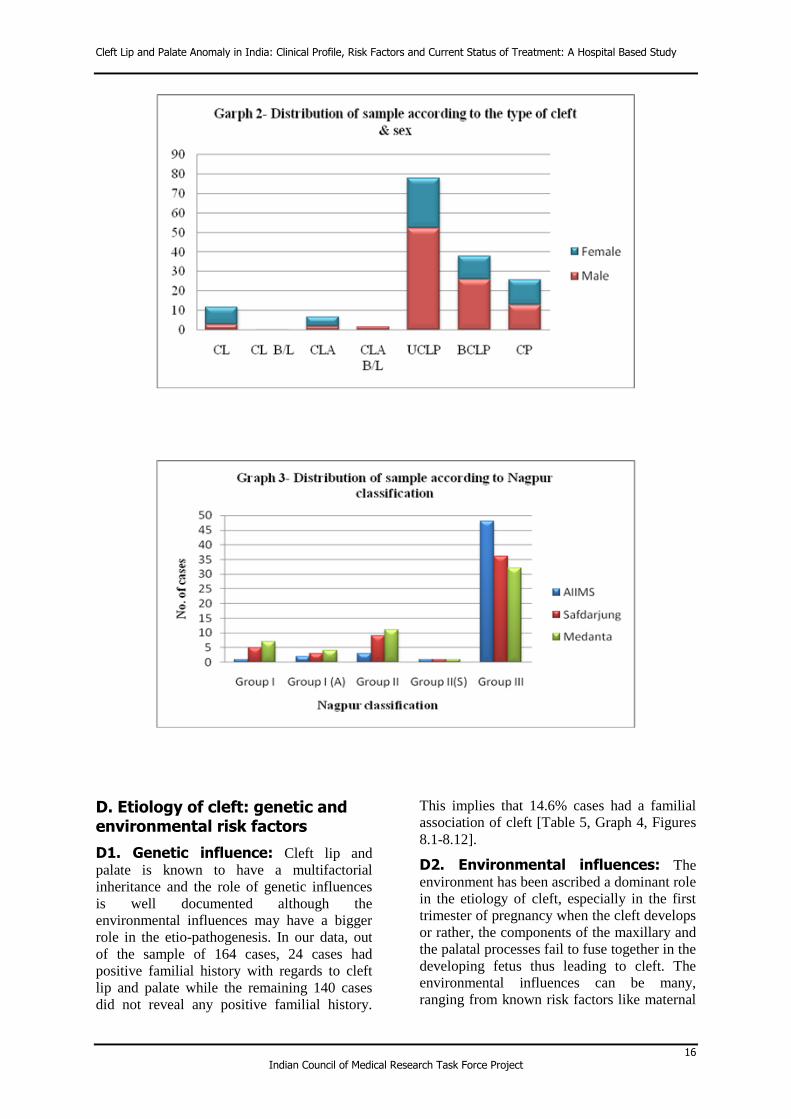
B. Distribution and type of cleft
When the sample was analyzed according to
the type of cleft, UCLP was found to be the
biggest category with 78 cases followed by
BCLP (38 cases) and CP (26 cases). When the
type of cleft was analyzed as a function of
cleft; amongst the males, the majority of the
cases belonged to the UCLP (52 cases)
category followed by BCLP (26 cases) and CP
(13 cases). Amongst the females, 26 cases had
UCLP, 12 had BCLP while 13 cases had cleft
palate (CP) (Table 2, Graph 2)
C. Classification of cleft
In our study, the Nagpur classification system
was used to classify the types of clefts (Table
3), both operated and unoperated. It was found
that majority of the cases (116 cases) were
found to be belonging to Group-III of Nagpur
Classification (unilateral and bilateral cleft lip
and palate) (Table 4, Graph 3).
Table 1-Distribution of sample according to age
Centre
>18mo -<=6yrs
(4)
>6yrs -<=12yrs
(5)
>12yrs - <=18yrs
(6)
>18 yrs
(7) Total
AIIMS 9 24 12 10 55
Safdarjung 14 12 10 18 54
Medanta-
The MEDICITY 19 15 10 11 55
Total 42
(25.6%)
51
(31.1%)
32
(19.5%)
39
(23.8%)
164
(100%)
n=164; Data represents no. of patients in each category

Cleft Lip and Palate Anomaly in India: Clinical Profile, Risk Factors and Current Status of Treatment: A Hospital Based Study
14 Indian Council of Medical Research Task Force Project
Table 2- Distribution of sample according to the type of cleft & sex
CL CL B/L CLA CLA B/L UCLP BCLP CP Total
Male 3 1 2 2 52 26 13 99
(60.4%)
Female 9 0 5 0 26 12 13 65
(39.6%)
Total 12
(7.3%)
1
(0.6%)
7
(4.3%)
2
(1.2%)
78
(47.6%)
38
(23.1%)
26
(15.9%)
164
(100%)
n=164; Data represents no. of patients in each category

Analysis and Results
15 Indian Council of Medical Research Task Force Project
Table 3-Classification of cleft (Nagpur classification)
Group I Cleft of lip
Group I (A) Cleft of lip with cleft of alveolus
Group II Cleft of palate alone
Group II(S) Submucous cleft of palate
Group III Cleft of lip and palate
Table 4- Distribution of sample according to Nagpur classification
No. of cases Group I Group I
(A) Group II
Group II
(S) Group III Total
AIIMS 1 2 3 1 48 55
Safdarjung 5 3 9 1 36 54
Medanta- The MEDICITY
7 4 11 1 32 55
Total 13
(7.9%)
9
(5.5%)
23
(14.1%)
3
(1.8%)
116
(70.7%)
164
(100%)
n=164; Data represents no. of patients in each category
Cleft Lip and Palate Anomaly in India: Clinical Profile, Risk Factors and Current Status of Treatment: A Hospital Based Study
16 Indian Council of Medical Research Task Force Project
D. Etiology of cleft: genetic and environmental risk factors
D1. Genetic influence: Cleft lip and
palate is known to have a multifactorial
inheritance and the role of genetic influences
is well documented although the
environmental influences may have a bigger
role in the etio-pathogenesis. In our data, out
of the sample of 164 cases, 24 cases had
positive familial history with regards to cleft
lip and palate while the remaining 140 cases
did not reveal any positive familial history.
This implies that 14.6% cases had a familial
association of cleft [Table 5, Graph 4, Figures
8.1-8.12].
D2. Environmental influences: The
environment has been ascribed a dominant role
in the etiology of cleft, especially in the first
trimester of pregnancy when the cleft develops
or rather, the components of the maxillary and
the palatal processes fail to fuse together in the
developing fetus thus leading to cleft. The
environmental influences can be many,
ranging from known risk factors like maternal

Analysis and Results
17 Indian Council of Medical Research Task Force Project
smoking and alcohol consumption, exposure to
smoke leading to foetal hypoxia, nutritional
deficiencies, medical illnesses, use of oral
contraceptives, certain medications, amongst
other risk factors.
In our study, we evaluated a few of the
known risk factors including maternal
smoking and alcohol consumption, intake of
drugs during the first trimester of pregnancy
and exposure to smoke during the same time
by the use of chulha at home or due to passive
smoking. The results of our study are
mentioned below.
D2.1. History of medical problems in mother during 1st trimester of affected pregnancy
In the sample of 164 subjects, 64 cases (39%)
were found where the mother had a positive
history of illness during 1st trimester of the
affected pregnancy. When the type of disease
was enquired, a variable pattern was found
with responses ranging from non-specific
fever to viral fever, tuberculosis, thyroid,
vomiting, etc. (Table 6, Graph 5).
D2.2. History of drug intake during the 1st trimester of affected pregnancy
Out of the 164 cases, 44 (26.8%) had a
positive history of maternal use of drugs
during the first trimester of affected
pregnancy, but were unaware about its dosage
and duration of use. (Table 7, Graph 6)
D2.3. History of maternal radiation exposure and use of intoxicants during the 1st trimester of affected pregnancy
Out of 164 subjects, only 4 cases gave a
positive but incomplete history of radiation
exposure of mother during 1st trimester of
affected pregnancy (Table 8, Graph 7) while 1
case revealed use of some intoxicants during
1st trimester of affected pregnancy. (Table 9)
D2.4. History of exposure to smoke and mode of cooking during 1st trimester
In the recent past, the role of maternal
exposure to smoke in the first trimester of
pregnancy has been implicated in the etiology
of cleft palate possibly due to fetal hypoxia
leading to interference in palatal shelf fusion.
In the Indian scenario, this is especially valid
for the rural setups where it has been
postulated that exposure to smoke emanating
from the use of chulha for cooking might be
related to increased incidence of cleft in the
progeny. In our sample, out of 164 cases, 89
cases (54.26%) gave positive history of
exposure to smoke during 1st trimester of
pregnancy (Table 10, Graph 8). This sample
included mothers exposed to smoke from
cigarette smoking, passive smoking and/or use
of chulha during the 1st trimester of the
affected pregnancy.

Cleft Lip and Palate Anomaly in India: Clinical Profile, Risk Factors and Current Status of Treatment: A Hospital Based Study
18 Indian Council of Medical Research Task Force Project
Figure 8.1 Pedigree Chart
Figure 8.2 Pedigree Chart

Analysis and Results
19 Indian Council of Medical Research Task Force Project
Figure 8.3 Pedigree Chart
Figure 8.4 Pedigree Chart

Cleft Lip and Palate Anomaly in India: Clinical Profile, Risk Factors and Current Status of Treatment: A Hospital Based Study
20 Indian Council of Medical Research Task Force Project
Figure 8.5 Pedigree Chart
Figure 8.6 Pedigree Chart

Analysis and Results
21 Indian Council of Medical Research Task Force Project
Figure 8.7 Pedigree Chart
Figure 8.8 Pedigree Chart

Cleft Lip and Palate Anomaly in India: Clinical Profile, Risk Factors and Current Status of Treatment: A Hospital Based Study
22 Indian Council of Medical Research Task Force Project
Figure 8.9 Pedigree Chart
Figure 8.10 Pedigree Chart

Analysis and Results
23 Indian Council of Medical Research Task Force Project
Figure 8.11 Pedigree Chart
Figure 8.12 Pedigree Chart

Cleft Lip and Palate Anomaly in India: Clinical Profile, Risk Factors and Current Status of Treatment: A Hospital Based Study
24 Indian Council of Medical Research Task Force Project
Table 5- Patients with positive familial history of Cleft
Familial history positive
(1)
Familial history negative
(2)
Total
24
(14.6%)
140
(85.4%)
164
(100%)
n=164; Data represents no. of patients in each category
Table 7- History of Drug usage in mother during 1st trimester of
affected pregnancy
Centre Yes No Total
AIIMS 11 44 55
Safdarjung 21 33 54
Medanta- The MEDICITY 12 43 55
Total 44
(26.8%)
120
(73.2%)
164
(100%)
n=164; Data represents no. of patients in each category
Table 6-History of medical problems in mother during 1st trimester of affected pregnancy
Centre Yes No Total
AIIMS 12 43 55
Safdarjung 13 41 54
Medanta- The MEDICITY 39 16 55
Total 64
(39%)
100
(61%)
164
(100%)
n=164; Data represents no. of patients in each category

Analysis and Results
25 Indian Council of Medical Research Task Force Project
Table 8- History of radiation exposure to mother during 1st trimester of affected pregnancy
Centre Yes No
AIIMS 1 54
Safdarjung 3 51
Medanta- The MEDICITY 0 55
Total 4
(2.5%)
160
(97.5%)
n=164; Data represents no. of patients in each category
Table 9- Use of intoxicants by mother during 1st trimester of affected pregnancy
Centre Yes (Code 1) No (Code 2)
AIIMS 0 55
Safdarjung 1 53
Medanta- The MEDICITY 0 55
Total 1
(0.6%)
163
(99.4%)
n=164; Data represents no. of patients in each category
Table 10- History of exposure to smoke during 1st trimester
Centre Yes (Code 1) No (Code 2)
AIIMS 27 28
Safdarjung 40 14
Medanta- The MEDICITY 22 33
Total 89
(54.3 %)
75
(45.7%)
n=164; Data represents no. of patients in each category

Cleft Lip and Palate Anomaly in India: Clinical Profile, Risk Factors and Current Status of Treatment: A Hospital Based Study
26 Indian Council of Medical Research Task Force Project

Analysis and Results
27 Indian Council of Medical Research Task Force Project

Cleft Lip and Palate Anomaly in India: Clinical Profile, Risk Factors and Current Status of Treatment: A Hospital Based Study
28 Indian Council of Medical Research Task Force Project
E. Effect of cleft deformity on the social acceptability of the patient
In the modern society, the value of facial
aesthetics and a beautiful face cannot be
underemphasized. In other words, in the
current scenario, facial beauty has more
importance than ever before. People with
pleasing smiles are more likely to be
professionally successful and have higher self-
esteem compared to people with compromised
facial esthetics. The problem of facial beauty
has even more relevance in patients with cleft
anomaly as they not only have to deal with the
issues of facial scar but also problems affected
due to articulation of speech, higher nasalance
which hampers communication and therefore,
lead to lowered self-esteem. Hence, this aspect
was given its due importance in our study as
the primary objective of the study is to
document the treatment needs of patients with
cleft. The problem becomes even more severe
in the Indian setup where many patients
receive less than optimal primary surgeries and
many receive only partial treatments.
In our sample, only the adult (18 years
and above) patients were selected for
evaluation in this category where 39 cases
were found. Of the 39 cases, only 32 cases
could be evaluated (7 cases from Safdarjung
not applicable) and the results were quite on
the expected lines. When the sample of 32
subjects were enquired whether the cleft had
affected their professional life, 29 of the
responded gave a positive reply (Table 11).
F. Post natal counselling of parents with regards to feeding and treatment possibilities related to cleft
One of the significant aspects of cleft care is
the post natal counselling of the parents
regarding the anomaly, the effects of the
anomaly and the treatment possibilities. Since,
many of the parents have never seen such an
anomaly before in life, they are many times
not aware that the defect is correctable and that
the treatment must follow a certain course to
give expected results. Also, the feeding of the
cleft child is difficult, challenging and has to
be done in a specific way. Many of the parents
are not well versed with feeding such a child
and consequently the child suffers from
various problems like malnutrition, inadequate
weight gain, poor health, respiratory and nasal
diseases and even possibility of death due to
aspiration while feeding. The problem is more
acute in rural areas where access to medical
facilities and online resource is limited.
In our study we evaluated whether the
parents of the child with cleft received post
natal counselling or not. We specifically
attempted to know whether they received
correct advice in the following parameters: a.
Feeding of the cleft new born, b. timeline for
surgical interventions, c. possible speech
defects that may occur and its correction, d.
dental and orthodontic interventions.
In our evaluation, we found that out of
the 164 cases, parents of 91 cases received
correct post natal counselling for one or more
of the parameters we evaluated (mentioned
above) while 73 cases did not receive any post
natal advice regarding the management of the
deformity (Table 12). Amongst these 73 cases,
many received advice later from various
sources like family elders, other patients with
cleft or from hospitals they went for treatment.
A few of the cases even reported that they
were told by the dais, which assisted in
delivery that the child would not survive.
Out of the 91 cases who received post-
delivery advice, only 9 received correct advice
for all the parameters evaluated, namely
feeding, surgical correction of deformity,
dental and hearing and speech abnormalities
(Table 13, Graph 9). The remaining 82 cases
received incorrect advice for at least one of the
parameters. Interestingly, hearing and speech,
and dental and orthodontic treatment were the
criteria which were paid the least attention to
amongst the defined criteria when the post
natal parental counselling was done.
G. Age wise distribution of primary lip and palate repair
The primary lip and palatal surgeries of the
cleft are extremely important in deciding the
outcome of the treatment as it is one of the
most important parameters dictating the post-
surgical growth of the maxilla in all the 3
planes. Growth restriction following primary
surgery of palate is common in many patients
leading to sagittal maxillo-mandibular
discrepancy causing development of Class III
malocclusion. Under normal circumstances,
the primary lip repair should be carried out
between 3-6 months of age.

Analysis and Results
29 Indian Council of Medical Research Task Force Project
1) Primary Lip Repair
When the age at primary lip surgery was
analyzed, of the 164 cases, 26 cases of CP and
2 unoperated cases were excluded. For the
remaining 136 cases, the age of primary lip
surgery varied considerably ranging from 2
months to 180 months. Majority of cases were
operated within 6 months of birth (75 cases;
45.7%) while 61 cases (37.19%) were operated
beyond 6 months of birth (Table 14, Graph
10).
2) Age at primary palatal surgery
When the age of primary palatal surgery was
analyzed, out of the 164 cases, 142 cases had a
cleft of the palate. Out of these, 20 cases
remained un-operated for the palate at the time
of presentation. The remaining 122 cases,
when analyzed, showed significant variation in
timing of the palatal surgery varying from 3
months to 228 months. 72 out of 122 cases
were operated within 18 months of birth (59%)
while the remaining received surgery after 50
months (41%) cases (Table 15, Graph 11).
Pre-surgical orthopaedics is indicated
in few cases for correction of severe cleft
defects so that the surgeon can easily
approximate the cleft segments without risking
the stretching of the tissue. It is indicated only
in the first few months after birth and although
recently its efficacy has been questioned, it
remains a useful technique in selected cases. In
our sample of 164 cases, when the history for
pre-surgical orthopaedic treatment was
recorded, it was found that not even a single
case received the said treatment (Table 16).
H. Previous history of dental and orthodontic treatment
Cleft patients usually have a multitude of
dental problems including missing teeth,
supernumerary teeth, abnormal tooth positions
and inclinations, increased incidence of
impacted teeth, amongst other problems. In
many cases increased incidence of caries,
gingival and periodontal disease is seen.
Hence any cleft child should be put under the
supervision of a dentist after the eruption of
the first primary tooth. i.e. age of 1 year
onwards. Orthodontic intervention is usually
indicated after the age of 6 years, when the
first permanent molars have completely
erupted. The treatment usually involves
expansion of the upper arch so that by the age
of 9-10 years the child can undergo alveolar
bone grafting (ABG). Full fixed orthodontic
treatment is usually indicated after the eruption
of complete permanent dentition i.e. 12-13
years.
Out of 164 cases, 58 cases (35.4%)
reported that they sought some sort of dental
treatment during their lifetime while a majority
of the cases, as many as 106 (64.6%), did not
report any history of dental treatment (Table
17). The dental treatment included any sort of
filling, scaling, root canal treatment, extraction
of teeth, etc.
Out of 164 cases, 42 cases fell in age
group 18 months-6 years. Since this age group
has little relevance in orthodontic treatment,
they were excluded from analysis. Of the
remaining 122 cases above 6 years, 28 cases
(23%) reported that they sought some sort of
orthodontic treatment during their lifetime
while a majority of 94 cases did not reported
any history of orthodontic treatment. Out of
the 12 cases who sought orthodontic treatment,
the age range for seeking treatment varied
widely between 4 years to 22 years (Table 18).

Cleft Lip and Palate Anomaly in India: Clinical Profile, Risk Factors and Current Status of Treatment: A Hospital Based Study
30 Indian Council of Medical Research Task Force Project
Table 11- Effect of cleft deformity on the social acceptability of the patient
Centre Yes
(Code 1)
No
(Code 2)
Sample considered/ total sample
AIIMS 10 0 10/55
Safdarjung 9 2 11/54
Medanta-
The MEDICITY 10 1 11/55
Total 29
(90.6%)
3
(9.4%)
32/164
*Safdarjung 43cases NA
n=32; Data represents no. of patients in each category
Table 12- Post natal counseling of parents with regards to feeding of child with cleft
and his treatment possibilities related to cleft
Centre
Patients who received
correct advice for at least 1 parameter
% Patient who
did not receive correct advice
%
AIIMS 37 67.3 18 33.7
Safdarjung 26 48.1 28 52.9
Medanta- The MEDICITY 28 50.9 27 49.1
Total 91 55.4 73 44.6
n=164; Data represents no. of patients in each category
Table 13- Distribution of the patients who received correct advice for at least one of the evaluated variables
Centre All the
parameters
Feeding Surgical
correction Dental
Hearing and
speech
More than one
parameter
AIIMS 3 13 12 4 3 2
Safdarjung 0 1 7 0 0 18
Medanta-
The MEDICITY 6 3 2 0 0 17
Total 9 17 21 4 3 37
n=164; Data represents no. of patients in each category

Analysis and Results
31 Indian Council of Medical Research Task Force Project
Table 14- Age wise distribution of lip repair
Centre ≤6 months >6 to ≤12
months >12 - ≤18
months >18 months -
≤6 years >6 years
AIIMS 26 16 2 6 0
Safdarjung 12 12 7 9 4
Medanta-
The MEDICITY 37 3 0 2 0
Total 75
(55.2%)
31
(22.8%)
9
(6.6%)
17
(12.5%)
4
(2.9%)
n=136; Data represents no. of patients in each category
Table 15- Age at palatal repair excluding alveolus
Centre ≤6 months >6 to ≤12
months >12 - ≤18
months >18 months - ≤6 years
>6 years
AIIMS 2 14 11 18 3
Safdarjung 2 7 6 18 3
Medanta-
The MEDICITY 2 21 7 7 1
Total 6
(4.9%)
42
(34.4%)
24
(19.7%)
43
(35.2%)
7
(5.7%)
AIIMS- 52 with Cleft palate-4 unoperated; Safdarjung-46 with Cleft palate-10 unoperated; Medanta- The MEDICITY-44 with Cleft palate-6 unoperated
n=122; Data represents no. of patients in each category
Table 16- Previous history of pre-surgical orthopaedic
treatment
Centre Yes No
AIIMS 0 55 (100%)
Safdarjung 0 54 (100%)
Medanta- The MEDICITY 0 55 (100%)
Total 0 164 (100%)
n=164; Data represents no. of patients in each category

Cleft Lip and Palate Anomaly in India: Clinical Profile, Risk Factors and Current Status of Treatment: A Hospital Based Study
32 Indian Council of Medical Research Task Force Project
Table 17- Previous history of dental treatment
Centre Yes
(1)
No
(2)
AIIMS 19 36
Safdarjung 13 41
Medanta-The MEDICITY 26 29
Total 58 (35.4%) 106 (64.6%)
n=164; Data represents no. of patients in each category
Table 18- Positive history of post surgical orthodontic treatment
Centre Yes No
AIIMS 12 43
Safdarjung 5 49
Medanta- The MEDICITY 11 44
Total 28 (17.1%) 136 (82.9%)
n=164; Data represents no. of patients in each category

Analysis and Results
33 Indian Council of Medical Research Task Force Project

Cleft Lip and Palate Anomaly in India: Clinical Profile, Risk Factors and Current Status of Treatment: A Hospital Based Study
34 Indian Council of Medical Research Task Force Project
I. Dental examination
1) Presence of supernumerary teeth
Supernumerary teeth are common in cleft
patients because the surgical intervention
frequently leads to aberrations in tooth
formation leading to various developmental
anomalies like missing teeth, supernumerary
teeth, malformed teeth, dilacerations of roots,
impacted teeth, etc.
In the sample of 164 cases, 12 cases had a
completely unoperated cleft. None of these
had any supernumerary teeth. Of the
remaining 152 cases with operated cleft, 132
cases (86.8%) did not have supernumerary
teeth while 20 cases had supernumerary teeth
(13.2%) (Table 19). Of the 20 subjects with
supernumerary teeth, 19 teeth were in relation
to cleft while 1 teeth was unrelated to cleft.
2) Presence of dental cross bite in the sample
Patients with cleft frequently suffer from
maxillo-mandibular growth anomalies leading
to restriction of maxillary growth in all three
planes while the mandible may grow normally.
This causes development of Angle Class III
malocclusion in the sagittal plane, cross bites
in both the anterior and posterior segments of
the dentition and loss of vertical maxillary
height in many cases.
The anterior dental segment comprises
of 6 teeth from canine to canine. The posterior
segment consists of teeth posterior to the
canine tooth i.e. the premolars and the molars.
In our analysis we studied the presence of
cross bite as single tooth, 1-3 teeth, or more
than 3 teeth. However, since single tooth cross
bites are usually not indicative of an
underlying growth disturbance; these types of
cross bites were grouped with the category of
“cross bite absent” when assessing the
posterior segment relations. Cross bites of
more than 1 tooth were taken to be indicator of
growth restriction/collapse of segment and
were grouped together for ease of analysis and
data presentation.
3) Growth restriction in the anterior segment of maxilla (as a function of anterior cross bite)
In the sample of 164 cases, 21 cases of cleft
were unoperated, 6 did not cooperate while 6
could not be assessed and therefore, excluded
from further analysis. Of the remaining 131
cases, 48 cases (32.9%) did not have an
anterior dental cross bite while the remaining
98 cases (67.1%) had cross bite ranging from
single tooth to complete anterior segment
(Table 20, Graph 12). Most of the cases with
cross bite of more than single tooth belonged
to UCLP (52%) and BCLP (30.6%). If we
consider presence of cross bite in more than 3
teeth as an indicator of sagittal growth
restriction of maxilla, 43 cases (43.9%) had
significant restriction of growth of maxilla in
the sagittal plane, all belonging to UCLP and
BCLP categories.
4) Growth restriction in the posterior segment of maxilla (as a function of posterior cross bite)
In the sample of 164 cases, 21 cases of cleft
were unoperated, 6 did not cooperate and 6
could not be assessed and therefore excluded
from further analysis. Of the remaining 131
cases, 73 cases (55.7%) did not have posterior
cross bite while 58 cases (44.3%) had
significant posterior cross bite (more than 1
tooth). Amongst the 58 cases with cross bite,
39 cases (67.24%) had bilateral cross bite
while 19 cases (32.75%) had unilateral cross
bite.
When the distribution of posterior cross
bite was seen across the various cleft types,
cases with UCLP and BCLP had the maximum
proportion of cases with cross bites more than
1 tooth. In UCLP cases 34 out of 62 cases
(54.8%) had posterior cross bite while 28 did
not have posterior cross bite. In BCLP, out of
34 cases, 22 cases (64.7%) had posterior cross
bite. Interestingly, out of these 22 cases, 20
had a bilateral posterior cross bite (Table 21).
5) Overjet and Overbite in the sample
Overjet is the representation of the relation of
anterior teeth in the sagittal plane. In normal
circumstances, overjet is positive with the
upper anterior teeth in front of lower anterior
teeth. Overbite represents the vertical overlap
of anterior teeth. In ideal circumstances both
overjet and overbite remains between 1-2 mm.
In cleft patients however the maxillary growth
is retarded and this may lead to alteration in
the ideal overjet and overbite relations. With
growth restriction of maxilla, the overjet may
reduce and even become negative which
implies a greater treatment challenge.

Analysis and Results
35 Indian Council of Medical Research Task Force Project
In our sample, out of 164 cases, 19 cases
could not be evaluated. Of the remaining 145
cases, 74 cases (51.4%) showed a positive
favourable overjet of more than 1 mm while
25 cases (17.24%) had an overjet of -3mm or
lesser (Table 22). These cases pose a
significant challenge for orthodontic treatment
and many of these would ultimately require
combine orthodontic and orthognathic surgery.
When the overbite in the sample was assessed
it was seen that 10 cases out of 145 (6.8%) had
an anterior open bite while 93 cases (64.13%)
had 0-2mm of overbite. 42 cases (29%) had
more than 2mm of overbite (Table 23). It must
be stressed here that a positive overbite is
indicative of better maxillary growth and thus
cases with deep bite (more than 2 mm) are
considered more favourable during treatment
while cases with 0-1 mm overbite may
progress to anterior open bite if the future
growth of maxilla is unfavourable.
J. GOSLON YARDSTICK
The outcome of the primary cleft surgeries on
the maxillo-mandibular growth can be
evaluated using the GOSLON yardstick. The
Goslon yardstick essentially evaluates the
sagittal maxillo-mandibular dental arch
relations and reflects, in part, the effect of cleft
surgeries on the sagittal growth restriction of
maxilla. It also reveals the complexity of
orthodontic treatment anticipated. Goslon
yardstick has been devised to be selectively
used in cases with UCLP anomaly only.
The dental arch relations in our study
were assessed using Goslon Yardstick. All
cases with operated UCLP anomaly above 7
years of age were selected for the evaluation.
Of the 164 cases, only 40 cases fit the
inclusion criteria used for applying the Goslon
Yardstick. Dental study models of all these
cases were evaluated for the anteroposterior
dental arch relations and grouped into
categories 1-5 (Table 24).
The findings revealed that 20 (50%) out
of 40 cases fell in Goslon category 3, 10 in
Goslon category 4 (25% cases), and 9 cases in
Goslon category 2 (22.5%) (Table 25, Graph
13). Only 1 case was found in category 5.
Thus, it was seen that 75% of the cases with
operated UCLP had complex orthodontic
treatment needs while 28% cases required
combined orthognathic surgery and
orthodontic treatment for optimal outcome.
Only 25% cases had minor orthodontic
treatment needs.
K. Examination of primary cleft
Although in today’s social setup, many cleft
cases receive surgical intervention for the
primary clefts, there are still patients who end
up receiving partial or no surgeries at all
especially if we move to semi urban and rural
setups. Such cases remain unoperated for
periods significantly beyond the expected date
of lip and/or palate surgery. This segment of
the report covers the clinical profile of such
cases. In our sample, 12 cases were unoperated
and 9 cases were partially operated.
1) Widest gap in the cleft palate (including alveolus)
Out of the sample of 21 cases, only 10 could
be evaluated. Of the 10 cases, the widest gap
in the cleft palate (including alveolus) was also
more than 1 cm in 8 cases (80%) (Table 26).
2) Length of the palate
Out of the 21 unoperated/partially-operated
cases, 15 could be assessed. Amongst the 15
cases, 6 cases (40%) had adequate palatal
length while 9 (60%) had inadequate palatal
length (Table 27).

Cleft Lip and Palate Anomaly in India: Clinical Profile, Risk Factors and Current Status of Treatment: A Hospital Based Study
36 Indian Council of Medical Research Task Force Project
Table 19- Supernumerary teeth
Centre
Present
Absent Related to cleft
Not related to cleft
Both
AIIMS 9 0 0 43
Safdarjung 8 1 1 40
Medanta- The MEDICITY 1 0 0 49
Total 18 (11.8%) 1(0.7%) 1 (0.7%) 132(86.8%)
* AIIMS- 3 cases completely unoperated, Safdarjung- 4 cases completely unoperated & Medanta- The MEDICITY 5 cases completely unoperated not included
n=152; Data represents no. of patients in each category
Table 20- Presence of anterior cross bite
Type of cleft Absent N Single tooth >1-<=3 Teeth >3 teeth
CL 11 0 0 0
CLA 5 2 1 0
UCLP 11 8 16 26
BCLP 6 3 11 16
CP 9 2 3 0
Total 42
(32.3%)
15
(11.6%)
31
(23.8%)
42
(32.3%)
*AIIMS-4 cases unoperated; Safdarjung-10 cases unoperated and 6 could not be assessed; Medanta- The MEDICITY 7 cases unoperated and 6 cases non cooperative.
n=130; Data represents no. of patients in each category

Analysis and Results
37 Indian Council of Medical Research Task Force Project
Table 21- Presence of posterior cross bite in the sample of 55 cases
Type of cleft
Present
Absent Unilateral Bilateral
CL 0 0 10
CL B/L 0 0 1
CL A 0 0 6
CLA B/L 1 0 1
UCLP 16 18 28
BCLP 2 20 12
CP 0 1 15
Total 19
(14.5%)
39
(29.8%)
73
(55.7%)
*AIIMS-4 cases unoperated; Safdarjung-10 cases unoperated and 6 could not be assessed; Medanta- The MEDICITY 7 cases unoperated and 6 cases non cooperative.
n=131; Data represents no. of patients in each category
Table 22 - Overjet in the sample
CL CLA UCLP BCLP CP Total
Less than equal to -3mm
0 0 13 12 0 25
-2mm to 0 0 2 29 11 4 46
1mm and above 12 6 27 12 17 74
Total 12
(8.3%)
8
(5.5%)
69
(47.6%)
35
(24.1%)
21
(14.5%)
145
(100%)
n= 145 *9 cases in Medanta- The MEDICITY and 10 cases in SJ NA

Cleft Lip and Palate Anomaly in India: Clinical Profile, Risk Factors and Current Status of Treatment: A Hospital Based Study
38 Indian Council of Medical Research Task Force Project
Table 23- Overbite in the sample
CL CLA UCLP BCLP CP
Total GIVE IN %
Less than 0 0 0 8 2 0 10
0-2mm 9 3 42 22 17 93
More than 2mm 3 5 19 11 4 42
Total 12
(8.3%)
8
(5.5%)
69
(47.6%)
35
(24.1%)
21
(14.5%)
145
(100%)
n= 145 *9 cases in Medanta- The MEDICITY and 10 cases in SJ NA
Table 24- Goslon Yardstick scores and their interpretation
Goslon category
Usual Dental
relation Orthodontic Treatment need
Treatment outcome
1 Positive overjet
Mild malocclusion; minimal or no orthodontic treatment required
Excellent
2 Positive overjet
Mild malocclusion; minimal orthodontic treatment required
Good
3 Edge to edge bite
Complex orthodontic treatment to correct the Class III
malocclusion but a good result can be anticipated
Fair
4
Negative
overjet
1-3 mm
Complex orthodontic treatment
needs; future orthognathic surgery may be required
Poor
5
Negative
overjet >3 mm
Definite requirement of orthognathic surgery
Very poor

Analysis and Results
39 Indian Council of Medical Research Task Force Project
Table 25- Distribution of subjects according to the Goslon Yardstick
Goslon score Number of cases
1 0
2 9 (22.5%)
3 20 (50%)
4 10 (25%)
5 1 (2.5%)
Total 40
n=40; Data represents no. of patients in each category
Table 26- Widest gap in the cleft , palate + alveolus
Type of cleft ≤0.5cm >0.5 - ≤1cm
> 1cm Total
CLA B/L 0 0 0 0
UCLP 1 1 5 7
BCLP 0 0 0 0
CP 0 0 3 3
Total 1 1 8 10
* 3 cases of AIIMS excluded (Submucus CP), 1 case of Safdarjung excluded (Submucus CP) & Medanta- The MEDICITY- 4 cases of submucus CP, 1 case of CL and 2 cases did not cooperated are excluded. n=10; Data represents no. of patients in each category
Table 27- Length of the palate
Type of cleft Adequate Inadequate
UCLP 4 3
BCLP 0 0
CP 2 6
Total 6 9
n=15 * 5cases could not be assessed and 1case of CL excluded.

Cleft Lip and Palate Anomaly in India: Clinical Profile, Risk Factors and Current Status of Treatment: A Hospital Based Study
40 Indian Council of Medical Research Task Force Project

Analysis and Results
41 Indian Council of Medical Research Task Force Project
L. Examination of secondary cleft deformity
A lot of cases in today’s setup get operated for
their primary cleft, at various ages. That,
however, does not mean the end of treatment.
A host of issues crop up after the primary
surgeries, some related to surgery, some
unrelated to surgery. The outcome is gravely
dependent on the quality of surgical repair as
that dictates the long term growth
characteristics of the maxilla. Apart from that,
the Eustachian tube function, altered dental
development, residual scars, palatal function
and altered speech are few of the aspects
which merit consideration. This section of the
report aims to put some light on these issues.
i) Evaluation of lip (and scar)
Surgical repair of cleft lip result in scar
formation which may vary from a fine
imperceptible line to thick, ugly and fibrous
scars. The amount of scar tissue depends upon
many variables including the severity of the
cleft, the quality of surgery, timing of the
repair, etc. Lip scars can create both esthetic
and functional problems. A heavy lip scar can
make for an unsightly facial appearance
leading to psychosocial issues while functional
problems can result due to altered speech,
improper lip seal, altered orofacial muscular
balance and malocclusion due to pressure from
fibrosed tissue.
The upper lip, in our study, was evaluated
for bilateral symmetry, the amount and quality
of scar tissue, the size (length) of scar and its
angulation. The overall quality of repair was
evaluated on the basis of these parameters and
subjective evaluation to rate the outcome from
poor through very good.
ii) Evaluation of the lip scar
When the width (length) of the lip scar was
evaluated in patients with unilateral cleft
(UCL, UCLA and UCLP), 97 cases were
detected; out of which 2 case were unoperated.
Among the remaining 95 operated cases, 14
cases (15%) had a scar less than (equal to) 0.5
mm while 46 cases (48.4%) had a scar varying
between 0.5-1 mm (Table 28). As many as 35
cases had a scar width more than 1 mm.
When the angulations of the lip scar
were evaluated, vertical scars (57 cases (60%)
were more common than oblique scars (38
cases; 40%) (Table 29). The quality of scar
repair, when evaluated subjectively, depending
upon multiple factors, it was found that the
quality of scar repair varied between poor to
good while the categories of ‘very good’ and
‘excellent’ remained blank.
Amongst the cases with bilateral lip
involvement (BCL, BCLA and BCLP), the
right and the left side were evaluated
separately. Amongst the 41 cases, on the right
side, 16 patients (39%) had a scar more than 1
mm in length which is obvious to create
esthetic problems. Only 10 cases (24.4%) had
a scar less than 0.5mm (Table 30) while 15
cases had a scar length between 0.5-1mm
(36.6%). The predominant angulation of the
scar was vertical (75.6%) (Table 31).On the
left side, 20 out of 41 scars (48.8%) had scar
width more than 1 mm while only 8 cases
(19.5%) had an aesthetic scar less than 0.5 mm
wide. In angulation, 29 (70.7%) out of 41 scars
had vertical angulation.
iii) Evaluation of lip seal: at rest and while blowing
Achieving lip seal in cleft patients can be
quite a challenge in patients with cleft
anomaly especially if the quality of repair is
not satisfactory, or if the scar is overwhelming
and restricts mobility of the lip or if the
premaxilla is protruded significantly. Lip seal
while blowing indicates if the perioral muscles
are able to make tight seal at lips to prevent air
escape when lips are stressed. In cases with
cleft lip proper approximation of muscle fibers
while surgical correction of defect is necessary
for proper lip function; in cases with improper
lip function, lip seal while blowing may be
absent. Additionally, presence of excessive
scar tissue, reduced height of upper lip as well
as protruding premaxilla may also lead to
absence of lip seal while blowing.
In our sample, although the quality of
scar repair was not good in majority of the
sample but lip seal was present in most of the
subjects. Of the sample of 136 cases, 2 cases
were unoperated for lip and were excluded. Of
the remaining 134 cases assessed for lip seal at
rest, 14 cases (10.4%) were found to have
deficient lip seal at rest while only 5 cases out
of 124 cases lacked proper seal while blowing
(Table 32). Note that the sample for lip seal at
rest and while blowing is not same as 9 cases

Cleft Lip and Palate Anomaly in India: Clinical Profile, Risk Factors and Current Status of Treatment: A Hospital Based Study
42 Indian Council of Medical Research Task Force Project
were partially unoperated cleft were excluded
while 1 did not cooperate for blowing.
iv) Evaluation of lip symmetry
Lip symmetry was analyzed to judge the
overall symmetry of the upper lip, irrespective
of the side of involvement. In unilateral cases,
it involved comparing the affected side to the
normal side. In bilateral cases, it involved
comparing not only both the sides but also
both the sides to a normal unaffected lip. In
our sample of 164 cases, 26 had CP while 2
cases were unoperated and thus were
excluded. Of the remaining 134 cases, only 38
cases (28.4%) had good lip symmetry while 49
cases (36.6%) had fair lip symmetry. 30 cases
(22.4%) had poor lip symmetry while only 4
cases had excellent lip symmetry (Table 33,
Graph 14).
v) Overall appearance of lip
Overall appearance of the lip was
considered a combined result of lip
symmetry, width of the scar, cross hatches
across scar line and thickness of the
vermilion border. The lips were
subjectively evaluated by a single expert
for each of the 3 institutions. In our sample
of 164 cases, 26 cases of CP and 2
unoperated cases for lip were excluded. Of
the remaining 134 cases, 33 cases (24.6%)
cases had a poor overall lip appearance, 54
(40.3%) had fair appearance while 43
(32.1%) cases had good or very good lip
appearance (Table 34, Graph 15).
Table 28- Length of scar in unilateral clefts
Type of cleft ≤0.5mm >0.5 - ≤1mm >1mm
CL U/L 1 6 4
CL A U/L 1 4 2
UCLP 12 36 29
Total 14 46 35
*AIIMS- 22 cases did not have cleft lip and 1 case unoperated, Medanta- The MEDICITY- 27 cases did not have cleft lip and 1 case unoperated, Safdarjung- 18 cases did not have cleft lip n=95; Data represents no. of patients in each category
Table 29- Angulation of scar in unilateral cleft
Type of cleft Vertical Oblique
CL U/L 9 2
CL A U/ L 6 1
UCLP 42 35
Total 57
(60%)
38
(40%)
n=95; Data represents no. of patients in each category

Analysis and Results
43 Indian Council of Medical Research Task Force Project
Table 30- Length of scar in bilateral cleft
Type of cleft
Right Left
≤0.5mm >0.5 - ≤1mm
>1mm ≤0.5mm >0.5 - ≤1mm
>1mm
CL B/L 0 1 0 0 1 0
CL A B/L 0 1 1 1 0 1
BCLP 10 13 15 7 12 19
Total 10 15 16 8 13 20
n=41; Data represents no. of patients in each category
Table 31- Angulation of scar in bilateral cleft
Type of cleft
Right Left
Vertical Oblique Vertical Oblique
CL B/L 1 0 1 0
CL A B/ L 1 1 1 1
BCLP 29 9 27 11
Total 31 10 29 12
n=41; Data represents no. of patients in each category

Cleft Lip and Palate Anomaly in India: Clinical Profile, Risk Factors and Current Status of Treatment: A Hospital Based Study
44 Indian Council of Medical Research Task Force Project
Table 32- Assessment of lip seal
Type of cleft
Lip seal at rest Lip seal while blowing
Present Absent Present Absent
CL 9 1 11 0
CL B/L 1 0 1 0
CLA 5 2 7 0
CL A B/L 1 1 1 1
UCLP 71 5 68 1
BCLP 33 5 32 3
Total 120
(89.6%)
14
(10.4%)
119
(96%)
5
(4%)
1 case could not assessed and 2 cases unoperated
12 cases could not be assessed
6 cases of CP excluded

Analysis and Results
45 Indian Council of Medical Research Task Force Project
Table 33- Evaluation of lip symmetry
Poor Fair Good Very good Excellent Total
CL 0 3 5 2 0 10
CL B/L 0 0 0 0 1 1
CL A 1 3 3 0 0 7
CLA B/L 0 2 0 0 0 2
UCLP 18 24 22 9 3 76
BCLP 11 17 8 2 0 38
Total 30
(22.4%)
49
(36.6%)
38
(28.4%)
13
(9.7%)
4
(2.9%) 134
26 cases of CP excluded and 2 cases unoperated for lip
n=134; Data represents no. of patients in each category
Table 34- Overall appearance of lip
Type of cleft Poor Fair Good Very good Excellent Total
CL 0 6 2 2 0 10
CL B/L 0 0 0 0 1 1
CL A 1 5 1 0 0 7
CLA B/L 1 1 0 0 0 2
UCLP 14 31 19 9 3 76
BCLP 17 11 9 1 0 38
Total 33
(24.6%)
54
(40.3%)
31
(23.1%)
12
(9%)
4
(3%) 134
26 cases of CP excluded and 2 cases unoperated for lip
n=134; Data represents no. of patients in each category

Cleft Lip and Palate Anomaly in India: Clinical Profile, Risk Factors and Current Status of Treatment: A Hospital Based Study
46 Indian Council of Medical Research Task Force Project
M. Evaluation of the nose
i) Overall appearance of nose
Like the previous parameter this also evaluates
the overall aesthetic outcome of nose
following surgeries for cleft lip and palate. The
examination was subjectively done by a single
expert. Various parameters were taken into
account while doing the analysis, including
deviation of nasal septum, length of columella,
alar base width, bilateral symmetry etc.
In our sample of 164 cases, 26 cases had
cleft palate only and 2 cases were unoperated
for lip. These were excluded from further
analysis. Analysis of the remaining 134 cases
revealed that 10 cases (7.5%) had a very good
result while 25 cases (18.7%) had a good
result. As many as 47 cases (35%) had a poor
outcome for appearance of nose (Table 35,
Graph 16).
ii) Evaluation of nasal septum
Patients with cleft lip and palate anomaly
frequently show deviation of nasal septum. In
unoperated cases this happens due to altered
anatomical relationships which causes
deviation in growth direction, while in
operated cases it happens predominantly due
to the stretch created by scar tissue.
In our sample of 164 cases, 12 cases
could not be assessed (Table 36, Graph 17)
and were excluded from further analysis.
Analysis of the remaining 152 cases was
separately done for unilateral cleft cases and
other type of clefts. The rest of the cases were
grouped together. For the unilateral cleft cases,
out of 97 cases, 24 cases (24.7%) had a non-
deviated nasal septum, while 49 cases (50.6%)
had deviation of nasal septum to the opposite
side to that of the cleft.
For the rest of the cases which included
cases of bilateral cleft and CP (Table 37,
Graph 18), out of the 55 cases, 17 had a non-
deviated septum (31% cases) while 19 cases
(34.5%) cases showed deviation of nasal
septum to either side.
iii) Evaluation of width of the nostril floor
Distortion of the alar base and dome
frequently occur following surgical correction
of cleft deformity. This may lead to a variety
of alar malformations like widening, stretching
and depression of the alar dome. In our
analysis we categorized the width of the alar
base as equal and unequal. Of the 164 subjects,
26 cases with CP and 2 unoperated cases of lip
were excluded. Of the remaining 136 cases, 95
cases (69.9%) had an equal nostril floor width
while 41 cases (30.1%) had an unequal width
(Table 38, Graph 19).
Palatal evaluation in secondary cleft deformity
a. Evaluation of the length of soft palate
Adequate length of soft palate is essential for
achieving velopharyngeal competence. In
velopharyngeal incompetence, the posterior
aspect of soft palate does not connect to the
Passavant’s ridge of the posterior pharyngeal
wall. This causes nasal intonation of speech
and nasal escape of liquids. Velophryngeal
incompetence is frequently seen in patients
with cleft lip and palate, especially in operated
cases where the scar tissue and the restriction
of growth due to effects of surgery lead to a
short soft palate and/or inadequate movement
of palate during function.
In our sample out of 164 cases, only
120 could be assessed (20 cases were
unoperated and 15 cases had cleft lip only
while 3 cases could not be evaluated due to
young age, 6 cases of CLA excluded). Of the
remaining 120 cases, 49 cases (40.8%) had a
short palatal length while 71cases (59.2%) had
an adequate length (Table 39, Graph 20). If we
consider the two largest categories UCLP and
BCLP, the percentage of cases with short
palatal length within these categories is even
higher (60% in UCLP; 66% in BCLP).
b. Evaluation of scarring and mobility of the soft palate
In our sample, out of 164 cases, only 121
could be assessed (20 cases were unoperated
and 15 cases had cleft lip only while 2 cases
could not be evaluated due to young age, 6
cases of CLA excluded). Of the remaining 121
cases, only 31 cases (25.6%) had little scaring
of palate. Majority of cases (72 cases; 59.5%)
had an acceptable scaring of palate while 18
cases (14.9%) cases had significant scaring
(Table 40, Graph 21).
The mobility of the soft palate governs the
functional efficacy of the same, as effective lift
of the palate is essential for proper

Analysis and Results
47 Indian Council of Medical Research Task Force Project
velopharyngeal closure. In our sample, out of
164 cases, only 118 could be assessed (20
cases were unoperated and 15 cases had cleft
lip only while 4 cases could not be evaluated
due to young age, 6 cases of CLA excluded.
Of the remaining 118 cases, 17 cases (14.4%)
were deemed to have unsatisfactory mobility
while 101 (83.6%) had satisfactory mobility
(Table 41).
When the uvula was assessed in the
sample, in the 121 cases assessed, the majority
of cases showed a uvula which was not well
formed or bifid (74 cases; 61%) (Table 42).
c. Presence of post-surgical palatal fistula
Palatal fistulas may result in cleft surgeries
due to various factors, both related to surgery
as well as patient-related factors. The residual
fistula is associated with recurrent nasal
infections, nasal regurgitation of liquids and
also may affect the quality of voice.
In our sample, out of 164 cases, 20
cases were unoperated, 1 could not be assessed
due to the age factor. These cases were thus
excluded. Of the remaining 143 cases, only 65
cases (45%) did not have a fistula while 78
cases (55%) had a residual palatal fistula
(Table 43, Graph 22). Amongst the 78 cases
with fistula, majority of fistulas were located
in the peri-alveolar region (65 cases; 83%). Of
the 78 cases with fistula 57 cases (73%) gave
positive history of nasal regurgitation (Table
44).
The maximum diameter/length of the
fistula was noted in each case to evaluate the
size of the fistula. For the purpose of
categorization and representation, the size of
the fistula was classified as follows: a. Less
than 2mm in maximum length, b. 2-5 mm, c.
More than 5 mm. In our sample, out of 78
cases with palatal fistula, 28 cases each
(35.9%) had fistula in category a and b. Fistula
of more than 5mm in length was seen in 22
cases (28.2%). (Table 45, Graph 23).
The incidence of speech abnormality
due to fistula was found to be quite low in the
sample. Only 76 of the 78 cases could be
assessed (Table 46). Of the 76 cases, 35 cases
(46%) with fistula had significant speech
changes when the fistula was blocked
temporarily during speech assessment.
Closure of palatal fistula is frequently
undertaken due to a variety of functional and
psychological reasons like speech abnormality,
recurrent nasal regurgitation of fluids,
recurrent infections and psychological
concerns amongst parents and patients. Of the
77 cases assessed in the category, 26 cases
(33%) had been operated previously for fistula
closure, while 51 did not receive any surgery
for the same (67%). (Table 47)
Table 35- Overall appearance of nose
Type of cleft Poor Fair Good Very good
Excellent Total
CL 0 4 5 1 0 10
CL B/L 0 0 0 1 0 1
CL A 2 4 1 0 0 7
CLA B/L 1 0 1 0 0 2
UCLP 29 27 12 8 0 76
BCLP 15 17 6 0 0 38
Total 47
(35%)
52
(38.8%)
25
(18.7%)
10
(7.5%) 0 134
26 cases of CP excluded and 2 cases unoperated for lip
n=134; Data represents no. of patients in each category

Cleft Lip and Palate Anomaly in India: Clinical Profile, Risk Factors and Current Status of Treatment: A Hospital Based Study
48 Indian Council of Medical Research Task Force Project
Table 36- Evaluation of nasal septum – Unilateral clefts
Non-Deviated Cleft side Non cleft side
CL 3 2 7
CL A 1 3 3
UCLP 20 19 39
Total 24
(24.7%)
24
(24.7%)
49
(50.6%)
n=97; Data represents no. of patients in each category
Table 37: Evaluation of nasal septum - Bilateral clefts
Type of cleft
Non-Deviated
(0)
Left
(1)
Right
(2) Total
CL B/L 1 0 0 1
CLA B/L 1 0 1 2
BCLP 6 16 16 38
CP 9 3 2 14
Total 17
(31%)
19
(34.5%)
19
(34.5%) 55
Cases excluded Medanta- The MEDICITY-12 cases of CP NA
n=55; Data represents no. of patients in each category
Table 38- Evaluation of nostril floor width
Type of cleft Unequal Equal Total
CL 4 7 11
CL B/L 1 0 1
CL A 1 6 7
CL A B/L 1 1 2
UCLP 21 56 77
BCLP 13 25 38
Total 41
(30.1%)
95
(69.9%) 136
26 cases of CP excluded and 2 cases unoperated
n=164; Data represents no. of patients in each category

Analysis and Results
49 Indian Council of Medical Research Task Force Project
Table 39- Evaluation of the length of the palate in the sample
Type of cleft Short Adequate Total
UCLP 26 43 69
BCLP 17 18 35
CP 6 10 16
Total 49
(40.8%)
71
(59.2%) 120
AIIMS-3 cases unoperated, 1 case of CL, 2 cases could not be assessed due to younger age and 2 cases of CLA Safdarjung- 10 cases of unoperated for the palate and 8 cases of CL Medanta- The MEDICITY- 7 cases unoperated, 6 cases of CL, 1 case could not be assessed and 4 cases of CLA
n=120; Data represents no. of patients in each category
Table 40- Evaluation of post surgical scarring of the palate in the sample
Little Acceptable Too much Total
UCLP 15 47 7 69
BCLP 10 19 8 37
CP 6 6 3 15
Total 31
(25.6%)
72
(59.5%)
18
(14.9%) 121
AIIMS-3 cases unoperated, 1 case of CL, 2 cases could not be assessed due to younger age and 2 cases of CLA Safdarjung- 10 cases of unoperated for the palate and 8 cases of CL Medanta- The MEDICITY- 7 cases unoperated, 6 cases of CL and 4 cases of CLA
n=121; Data represents no. of patients in each category
Table 41- Mobility of the palate in the sample
Type of cleft Satisfactory Unsatisfactor
y Total
UCLP 61 7 68
BCLP 27 7 34
CP 13 3 16
Total 101
(85.6%)
17
(14.4%) 118
AIIMS-3 cases unoperated, 1 case of CL, 2 cases could not be assessed due to younger age and 2 cases of CLA Safdarjung- 10 cases of unoperated for the palate, 8 cases of CL and 1 case could not be assessed Medanta- The MEDICITY- 7 cases unoperated, 6 cases of CL, 4 cases of CLA and 2 cases could not be assessed
n=118; Data represents no. of patients in each category

Cleft Lip and Palate Anomaly in India: Clinical Profile, Risk Factors and Current Status of Treatment: A Hospital Based Study
50 Indian Council of Medical Research Task Force Project
Table 42- Status of uvula in operated cases of cleft palate
Type of cleft Well
formed Not well formed
Bifid Total
UCLP 26 40 3 69
BCLP 8 27 2 37
CP 5 7 3 15
Total 39
(32.3%)
74
(61%)
8
(6.7%) 121
AIIMS-3 cases unoperated, 1 case of CL, 2 cases could not be assessed due to younger age and 2 cases of CLA; Safdarjung- 10 cases of unoperated for the palate and 8 cases of CL; Medanta- The MEDICITY- 7 cases unoperated, 6 cases of CL and 4 cases of CLA
n=121; Data represents no. of patients in each category
Table 43- Presence of fistula in the sample
Type of cleft Absent Peri-
alveolar Hard
Palate
Junction of Hard and Soft
Palate
Soft Palate
Multiple fistula
Total
CL U/L 11 0 0 0 0 0 11
CL B/L 1 0 0 0 0 0 1
CL A U/L 6 1 0 0 0 0 7
CLA B/L 0 2 0 0 0 0 2
UCLP 31 32 3 0 2 1 69
BCLP 7 27 2 0 0 1 37
CP 9 3 1 0 3 0 16
Total 65
(45.5%)
65
(45.5%)
6
(4.2%) 0
5
(3.5%)
2
(1.3%) 143
* AIIMS- 3 cases unoperated and 1 can’t assess , Safdarjung- 10cases unoperated/partially unoperated & Medanta- The MEDICITY- 7 cases unoperated/partially operated
n=143; Data represents no. of patients in each category

Analysis and Results
51 Indian Council of Medical Research Task Force Project
Table 44- Assessment whether the fistula is symptomatic or not
Type of cleft Symptomati
c Asymptomatic Total
CL A 0 1 1
CLA B/L 2 0 2
UCLP 25 13 38
BCLP 26 4 30
CP 4 3 7
Total 57
(73 %)
21
(27%) 78
n=78; Data represents no. of patients in each category
Table 45- Evaluation of the size of the oronasal fistula in the sample
Type of cleft <=2mm
(1)
2-5mm
(2)
>5mm
(3) Total
CL A 1 0 0 1
CLA B/L 0 2 0 2
UCLP 16 12 10 38
BCLP 8 12 10 30
CP 3 2 2 7
Total 28
(35.9%)
28
(35.9%)
22
(28.2%) 78
n=98; Data represents no. of patients in each category

Cleft Lip and Palate Anomaly in India: Clinical Profile, Risk Factors and Current Status of Treatment: A Hospital Based Study
52 Indian Council of Medical Research Task Force Project
Table 46- Speech abnormality due to presence of the fistula
Type of cleft Yes
(1)
No
(2) Total
CL A 0 1 1
CLA B/L 0 2 2
UCLP 15 23 38
BCLP 15 14 29
CP 5 1 6
Total 35 (46%) 41(54%) 76
2 cases of Safdarjung could not be assessed
n=76; Data represents no. of patients in each category
Table 47- Evaluation whether the fistula has been operated previously or not in the sample
Type of cleft Yes No
CL A 0 1
CLA B/L 0 12
UCLP 10 17
BCLP 13 17
CP 3 4
Total 26
(33%)
51
(67%)
Patients with fistula: AIIMS-34; 1 case from AIIMS did not remember; Safadarjung-20 cases; Medanta- The MEDICITY- 24 cases n=77; Data represents no. of patients in each category

Analysis and Results
53 Indian Council of Medical Research Task Force Project

Cleft Lip and Palate Anomaly in India: Clinical Profile, Risk Factors and Current Status of Treatment: A Hospital Based Study
54 Indian Council of Medical Research Task Force Project

Analysis and Results
55 Indian Council of Medical Research Task Force Project

Cleft Lip and Palate Anomaly in India: Clinical Profile, Risk Factors and Current Status of Treatment: A Hospital Based Study
56 Indian Council of Medical Research Task Force Project
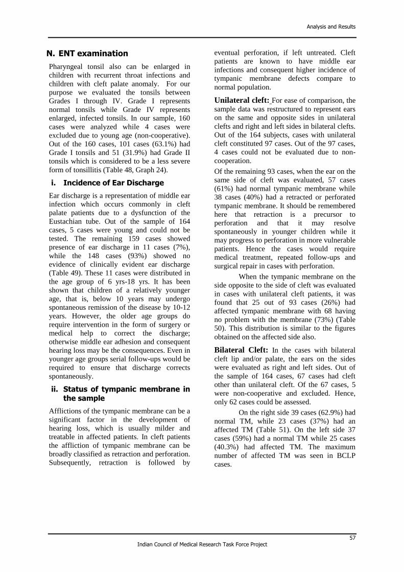
Analysis and Results
57 Indian Council of Medical Research Task Force Project
N. ENT examination
Pharyngeal tonsil also can be enlarged in
children with recurrent throat infections and
children with cleft palate anomaly. For our
purpose we evaluated the tonsils between
Grades I through IV. Grade I represents
normal tonsils while Grade IV represents
enlarged, infected tonsils. In our sample, 160
cases were analyzed while 4 cases were
excluded due to young age (non-cooperative).
Out of the 160 cases, 101 cases (63.1%) had
Grade I tonsils and 51 (31.9%) had Grade II
tonsils which is considered to be a less severe
form of tonsillitis (Table 48, Graph 24).
i. Incidence of Ear Discharge
Ear discharge is a representation of middle ear
infection which occurs commonly in cleft
palate patients due to a dysfunction of the
Eustachian tube. Out of the sample of 164
cases, 5 cases were young and could not be
tested. The remaining 159 cases showed
presence of ear discharge in 11 cases (7%),
while the 148 cases (93%) showed no
evidence of clinically evident ear discharge
(Table 49). These 11 cases were distributed in
the age group of 6 yrs-18 yrs. It has been
shown that children of a relatively younger
age, that is, below 10 years may undergo
spontaneous remission of the disease by 10-12
years. However, the older age groups do
require intervention in the form of surgery or
medical help to correct the discharge;
otherwise middle ear adhesion and consequent
hearing loss may be the consequences. Even in
younger age groups serial follow-ups would be
required to ensure that discharge corrects
spontaneously.
ii. Status of tympanic membrane in the sample
Afflictions of the tympanic membrane can be a
significant factor in the development of
hearing loss, which is usually milder and
treatable in affected patients. In cleft patients
the affliction of tympanic membrane can be
broadly classified as retraction and perforation.
Subsequently, retraction is followed by
eventual perforation, if left untreated. Cleft
patients are known to have middle ear
infections and consequent higher incidence of
tympanic membrane defects compare to
normal population.
Unilateral cleft: For ease of comparison, the
sample data was restructured to represent ears
on the same and opposite sides in unilateral
clefts and right and left sides in bilateral clefts.
Out of the 164 subjects, cases with unilateral
cleft constituted 97 cases. Out of the 97 cases,
4 cases could not be evaluated due to non-
cooperation.
Of the remaining 93 cases, when the ear on the
same side of cleft was evaluated, 57 cases
(61%) had normal tympanic membrane while
38 cases (40%) had a retracted or perforated
tympanic membrane. It should be remembered
here that retraction is a precursor to
perforation and that it may resolve
spontaneously in younger children while it
may progress to perforation in more vulnerable
patients. Hence the cases would require
medical treatment, repeated follow-ups and
surgical repair in cases with perforation.
When the tympanic membrane on the
side opposite to the side of cleft was evaluated
in cases with unilateral cleft patients, it was
found that 25 out of 93 cases (26%) had
affected tympanic membrane with 68 having
no problem with the membrane (73%) (Table
50). This distribution is similar to the figures
obtained on the affected side also.
Bilateral Cleft: In the cases with bilateral
cleft lip and/or palate, the ears on the sides
were evaluated as right and left sides. Out of
the sample of 164 cases, 67 cases had cleft
other than unilateral cleft. Of the 67 cases, 5
were non-cooperative and excluded. Hence,
only 62 cases could be assessed.
On the right side 39 cases (62.9%) had
normal TM, while 23 cases (37%) had an
affected TM (Table 51). On the left side 37
cases (59%) had a normal TM while 25 cases
(40.3%) had affected TM. The maximum
number of affected TM was seen in BCLP
cases.

Cleft Lip and Palate Anomaly in India: Clinical Profile, Risk Factors and Current Status of Treatment: A Hospital Based Study
58 Indian Council of Medical Research Task Force Project
Table 48- Status of Tonsils in the sample according to type of cleft
Type of cleft Grade I
(1)
Grade II
(2)
Grade III
(3)
Grade IV
(4) Total
CL U/L 11 1 0 0 12
CL B/L 1 0 0 0 1
CLA U/L 6 1 0 0 7
CLA B/L 2 0 0 0 2
UCLP 45 28 3 0 76
BCLP 17 15 4 1 37
CP 19 6 0 0 25
TOTAL 101
(63.1%)
51
(31.9%)
7
(4.4%)
1
(0.6%) 160
4 cases from AIIMS could not be assessed due to younger age
n=160; Data represents no. of patients in each category
Table 49- Incidence of Ear Discharge
Type of cleft Yes
(1)
No
(2)
CL U/L 0 12
CL B/L 0 1
CLA U/L 0 7
CLA B/L 0 2
UCLP 6 70
BCLP 4 33
CP 1 23
TOTAL 11
(7%)
148
(93%)
4 cases from AIIMS could not be assessed due to younger age and 1 case of Medanta- The MEDICITY could not be assessed due to younger age. n=159; Data represents no. of patients in each category
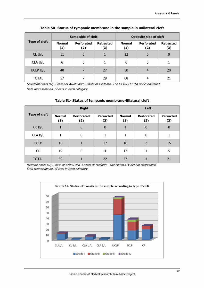
Analysis and Results
59 Indian Council of Medical Research Task Force Project
Table 50- Status of tympanic membrane in the sample in unilateral cleft
Type of cleft
Same side of cleft Opposite side of cleft
Normal
(1)
Perforated
(2)
Retracted
(3)
Normal
(1)
Perforated
(2)
Retracted
(3)
CL U/L 11 0 1 12 0 0
CLA U/L 6 0 1 6 0 1
UCLP U/L 40 7 27 50 4 20
TOTAL 57 7 29 68 4 21
Unilateral cases 97; 2 cases of AIIMS and 2 cases of Medanta- The MEDICITY did not cooperated
Data represents no. of ears in each category
Table 51- Status of tympanic membrane-Bilateral cleft
Type of cleft
Right Left
Normal
(1)
Perforated
(2)
Retracted
(3)
Normal
(1)
Perforated
(2)
Retracted
(3)
CL B/L 1 0 0 1 0 0
CLA B/L 1 0 1 1 0 1
BCLP 18 1 17 18 3 15
CP 19 0 4 17 1 5
TOTAL 39 1 22 37 4 21
Bilateral cases 67; 2 case of AIIMS and 3 cases of Medanta- The MEDICITY did not cooperated
Data represents no. of ears in each category

Cleft Lip and Palate Anomaly in India: Clinical Profile, Risk Factors and Current Status of Treatment: A Hospital Based Study
60 Indian Council of Medical Research Task Force Project
O. Hearing evaluation
1. Prevalence of Hearing defects in the sample
Out of the sample of 164 cases, 30 cases could
not be assessed. Of the remaining 134 cases
that could be assessed for hearing, 74 of the
134 cases (55.2%) had normal hearing while
60 cases (44.8%) had hearing defects in one or
both the ears. Amongst those affected, 45 of
the 60 cases (75%) had bilateral hearing loss
of certain degree while a few of the cases with
unilateral cleft had a hearing loss of the same
side (Table 52, Graph 25). This is remarkable
because hearing loss of the ear of the same
side of cleft is documented but loss on both the
sides in such a percentage is alarming.
Moreover, the sample had many cases which
came to the orthodontic OPD and did not even
know that they had hearing abnormalities.
However, amongst the sample of 60
cases which had a documented hearing loss, 44
cases (73.3%) had a mild level of hearing loss
while 15 (25%) had a moderate loss of hearing
(Table 53, Graph 26). Severe or profound type
of loss was not found in the sample.
In our study we also tried to document
the type of hearing loss in the group. Broadly
we categorized the hearing loss as conductive,
mixed or sensorineural. The cases for ease of
documentation of hearing loss pattern were
divided into unilateral and bilateral cleft cases.
In the 31 unilateral cleft cases, 3 cases
had normal hearing as assessed on PTA.
Remaining 28 cases showed hearing loss on
the same side of the cleft while 24 showed
hearing loss on the opposite of the cleft (Table
54).
Amongst the bilateral cleft and CP
category, 29 had affected hearing, of which 24
ears on right side and 23 on left side had
conductive hearing loss (Table 55).
2. Status of middle ear function
Impedance audiometry is a versatile objective
technique to test the middle ear function as far
as sound conduction is concerned. The
versatility of this technique is highlighted by
the fact that the technique can be carried out in
infants as young as barely a few months. In
our setup, we used the Interaccoustics®
combined audiometry tympanometry unit to
assess the status of middle ear in the sample.
Standardized protocol for the testing was
followed and the middle ear function was
graded on the basis of the type of
tympanogram (Table 56). This test however,
can be done only in cases where the tympanic
membrane is intact with little or no cermuen
accumulation in the external auditory canal.
For ease of data presentation and
segregation, the data is presented separately
for unilateral and bilateral cleft cases with
special reference to UCLP and BCLP as these
were the largest groups. When the sample of
unilateral cleft cases was tested for the middle
ear function on the same side of cleft, it was
found that out of 97 cases; only 66 cases could
be assessed. Of the 66 cases, 38 cases (57.5%)
had normal middle ear function while 28
(42.4%) had affected middle ear function
(Table 57) on the same side as cleft. Of the
affected cases, 20 out of 28 cases (71.4%) had
Type B tympanogram indicating immediate
intervention by an otolaryngologist to prevent
further loss of hearing. For the opposite side
only 64 ears could be examined. Of the 64
ears, 40 cases (62.5%) had normal middle ear
function while 24 (37.5%) had affected middle
ear function (Table 57,58) on the opposite side
as cleft. Of the affected cases, 16 out of 24
cases (66.67%) had Type B tympanogram
indicating immediate intervention by an
otolaryngologist to prevent further loss of
hearing.
In bilateral cleft cases, 48 ears were
evaluated on each side. On the right side, 20
(41.6%) and 18 (37.5%) on right and left side
respectively had normal function (Table 58).
P. Speech assessment
Speech assessment in the sample was
comprehensive and consisted of assessment of
hypernasality, presence of articulation defects
in those with defective speech and assessment
of speech intelligibility.
Hypernasality of speech in cleft cases
occurs due to velopharyngeal incompetence
occurring in cleft patients because of
incompetency, inadequacy or mislearning of
the soft palate. Assessment of hypernasality
was done by a single observer (speech
pathologist) and the assessment was
subjective. The cases were rated only in 3
categories: hypernasal, normal or hyponasal
speech.

Analysis and Results
61 Indian Council of Medical Research Task Force Project
In the sample of 164 cases, 22 cases could
not be assessed due to non-cooperation. Of the
remaining 142 cases, only 27 cases had normal
nasality (19%) while the 114 out of 164
(80.3%) had hypernasality (Table 59, Graph
27). This lends credence to the fact that many
operated cases of cleft lip and palate have
hypernasality due to nasal escape of air during
phonation via either VPI or symptomatic
oronasal fistula. Unfortunately, the assessment
of the nasality was subjective and categorized
as present or absent, which overestimates the
true picture as even those with mild
hypernasality are also categorized as
hypernasal speech. However, the assessment
was done by a single operator and thus
operator bias was excluded from the equation.
1. Defects in speech articulation
Similar results were seen for articulation
defects in the sample. In our study, a detailed
speech analysis was done and articulation
defects in various categories of speech sounds
was evaluated. Of the 164 cases, 24 cases
could not be assessed due to young age.
Amongst the remaining 140 cases, 108
(77.2%) had an articulation defect ranging
from mild to severe problems (Table 60,
Graph 28). The results show that in UCLP,
BCLP and CP the articulation defects span
across all categories of speech sounds. In
UCLP and BCLP the major sounds affected
were palatal, retroflex, dental and alveolar
(Table 61).
2. Speech Intelligibility
Overall speech intelligibility is a subjective
evaluation of speech on the basis of the extent
of comprehensibility of the vocal speech of the
subject by the listener. Speech intelligibility is
a function of both articulation and nasality. In
our settings the assessments was done by a
single operator who was an expert in the field
of speech and language pathology. The
intelligibility ratings were standardised into 7
categories on the basis of pre-defined criteria
(Table 62). In our sample, a total of 139 cases
were evaluated out of 164 cases enrolled for
the report. In these 139 cases, the speech
intelligibility was normal in only 22 cases
(15.8%). However, majority of cases had only
mild speech intelligibility problems. If we
consider patients with I-0 to I-2 to be having
socially acceptable speech with only mild
intelligibility problems, a total of 70 out of 139
patients (50.3%) had clinically acceptable
speech. The remaining 49.7% cases had
clinically significant speech intelligibility
problems which would be detrimental to their
quality of life (Table 63, Graph 29).
Thus, it is evident that patients with
cleft anomaly tend to develop a multitude of
problems including surgical, dental,
orthodontic, hearing and speech, to name a
few. The findings of the pooled data recorded
from the three centres across the National
Capital Region (NCR) highlight the need for
improvement in the quality of cleft care. It is
clear that the outcomes in this sample of
patients are way behind those seen in some of
the good European centres like Copenhagen
and Oslo.
The current pilot project was primarily
aimed at establishing a protocol for a larger
multicentre study and its logistic operative
feasibility. The road ahead includes the
expansion of the project on a pan-India level,
with inclusion of multiple centres representing
the different regions of the nation. It is obvious
that to make this study truly representative of
the population, we would have to make
changes in the study design.

Cleft Lip and Palate Anomaly in India: Clinical Profile, Risk Factors and Current Status of Treatment: A Hospital Based Study
62 Indian Council of Medical Research Task Force Project
Table 52- Prevalence of hearing abnormalities in the sample as a function of type of cleft
Type of cleft Right ear Left ear Both Normal
CL L 0 1 0 8
CL R 0 0 0 1
CL B/L 0 0 1 0
CLA L 0 0 1 4
CLA R 0 0 0 1
CLA B/L 0 0 1 1
UCLP L 3 4 12 20
UCLP R 2 0 8 13
BCLP 1 2 14 17
CP 1 1 8 9
Total 7
(5.2%)
8
(6 %)
45
(33.6%)
74
(55.2%)
AIIMS-6 CNT; Safdarjung-13 CNT and Medanta- The MEDICITY- 11 CNT
n=134; Data represents no. of patients in each category
Table 53- Degree of hearing loss in the sample
Type of cleft Mild
(1)
Moderate
(2)
Moderately
Severe
(3)
Severe
(4)
Profound
(5)
CL U/L 1 0 0 0 0
CL B/L 0 1 0 0 0
CLA U/L 0 1 0 0 0
CLA B/L 1 0 0 0 0
UCLP 20 8 1 0 0
BCLP 12 5 0 0 0
CP 10 0 0 0 0
TOTAL 44
(73.3%)
15
(25%)
1
(1.7%) 0 0
n=60; Data represents no. of patients in each category

Analysis and Results
63 Indian Council of Medical Research Task Force Project
Table 54- Relation of hearing loss with different types of cleft – Unilateral Cleft
Type of cleft
Same side Opposite side
Conductive
(1)
Mixed
(2)
Sensorineural
(3)
Conductive
(1)
Mixed
(2)
Sensorineural
(3)
CL U/L 1 0 0 0 0 0
CLA U/L 0 1 0 0 0 1
UCLP U/L 23 0 3 19 1 3
Total 24 1 3 19 1 4
28 ears affected on same side and 24 ears affected on opposite side
Table 55- Relation of hearing loss with different types of cleft – Bilateral Cleft
Type of cleft
Right Left
Conductive
(1)
Mixed
(2)
Sensorineural
(3)
Conductive
(1)
Mixed
(2)
Sensorineural
(3)
CL B/L 0 0 1 0 0 1
CLA B/L 1 0 0 1 0 0
BCLP 15 2 0 14 2 0
CP 8 0 1 8 0 1
Total 24 2 2 23 2 2
* the value represents the ears and not the cases hence, the sample on right and left may not necessarily be same
Table 56- Impedence Audiometry and their inference
Type of Curve Inference Treatment
need
Type-A Normal middle ear function No
Type-B Negative Middle ear pressure with low compliance
High
Type-C Normal compliance but Negative
Middle ear pressure High
Type-Ad High compliance; normal middle ear pressure
Low
Type-As Normal pressure; low compliance Mild

Cleft Lip and Palate Anomaly in India: Clinical Profile, Risk Factors and Current Status of Treatment: A Hospital Based Study
64 Indian Council of Medical Research Task Force Project
Table 57- Status of middle ear based on Impedance
TYPE OF CLEFT
Same side Opposite side
Type-A Type-B Type-C Type-
Ad Type-
As Type-A Type-B Type-C
Type-Ad
Type-As
CL U/L 7 1 0 0 1 8 0 0 0 0
CLA U/L 4 0 0 0 0 4 0 0 0 0
UCLP 27 19 5 0 2 28 16 6 0 2
TOTAL 38 20 5 0 3 40 16 6 0 2
Table 58- Status of middle ear based on Impedance- Bilateral cleft
TYPE OF CLEFT
Right Left
Type-A Type-B Type-C Type-
Ad Type-
As Type-A Type-B Type-C
Type-Ad
Type-As
CL B/L 0 0 0 0 0 0 0 0 0 0
CLA B/L 2 0 0 0 0 1 0 0 0 1
BCLP 12 16 1 1 0 10 15 2 3 1
CP 6 7 1 0 2 7 6 1 0 1
TOTAL 20 23 2 1 2 18 21 3 3 3
Table 59- Distribution of the sample according to nasality of speech
Type of cleft Normal Hyper
nasality Hypo nasality
CL U/L 10 1 0
CL B/L 1 0 0
CLA U/L 7 0 0
CLA B/L 1 1 0
UCLP 7 59 1
BCLP 1 34 0
CP 0 19 0
TOTAL 27
(19%)
114
(80.3%)
1
(0.7%)
CNT-AIIMS-5 cases; Safdarjung-11 cases and Medanta- The MEDICITY-6 cases

Analysis and Results
65 Indian Council of Medical Research Task Force Project
n=142; Data represents no. of patients in each category
Table 60- Speech Articulation in the sample
Type of cleft Affected Normal
CL U/L 2 9
CL B/L 0 1
CLA U/L 2 5
CLA B/L 1 1
UCLP 52 13
BCLP 33 2
CP 18 1
TOTAL 108 (77.2%) 32 (22.8%)
CNT-AIIMS-6 cases; Safdarjung-11 cases and Medanta- The MEDICITY-7 cases
n=140; Data represents no. of patients in each category
Table 61- Status of affected articulation in the sample
Typ
e o
f
cle
ft
Pa
lata
l
Re
tro
fle
x
De
nta
l
La
bio
-
de
nta
l
Bil
ab
ial
Alv
eo
lar
Glo
tta
l
Ve
lar
Vo
we
ls
CL U/L 1 0 1 0 0 1 0 0 1
CL B/L 0 0 0 0 0 0 0 0 0
CLA U/L 0 0 0 0 1 1 0 0 0
CLA B/L 1 1 1 1 1 1 1 1 1
UCLP 46 34 35 7 25 45 6 18 20
BCLP 33 23 26 10 18 28 8 16 17
CP 14 16 13 3 11 16 4 8 15

Cleft Lip and Palate Anomaly in India: Clinical Profile, Risk Factors and Current Status of Treatment: A Hospital Based Study
66 Indian Council of Medical Research Task Force Project
Table 62- Description of speech sample
Point scale
0 Normal
1 Can understand without difficulty; however, feel speech is not normal
2 Can understand with little effort occasionally need to ask for repetition
3 Can understand with concentration and effort
specially by sympathetic listener
4 Can understand with difficulty and concentration by family but not others
5 Can understand with effort if content is known
6 Cannot understand at all even content is known
Table 63- Overall speech intelligibility in various cleft types
Type of cleft I-0 I-1 I-2 I-3 I-4 I-5 I-6
CL U/L 9 1 1 0 0 0 0
CL B/L 1 0 0 0 0 0 0
CLA U/L 6 1 0 0 0 0 0
CLA B/L 1 0 0 0 0 1 0
UCLP 5 21 11 12 3 10 3
BCLP 0 8 5 5 3 11 2
CP 0 2 3 4 4 4 2
TOTAL 22
(15.8%)
33
(23.8 %)
20
(14.4%)
21
(15.1%)
10
(7.2%)
26
(18.7%)
7
(5%)
CNT-AIIMS-7 cases; Safdarjung-11 cases and Medanta- The MEDICITY-7 cases n=139; Data represents no. of patients in each category

Analysis and Results
67 Indian Council of Medical Research Task Force Project

Cleft Lip and Palate Anomaly in India: Clinical Profile, Risk Factors and Current Status of Treatment: A Hospital Based Study
68 Indian Council of Medical Research Task Force Project

Analysis and Results
69 Indian Council of Medical Research Task Force Project

70 Indian Council of Medical Research Task Force Project
5 Conclusions
A total of 164 cases with cleft lip and palate
anomaly were recorded from three hospitals
involved in the project (55 from AIIMS, 54
from Safdarjung, 55 from Medanta- The
MEDICITY). At each of the centres, the
Departments of Plastic surgery, Orthodontics
and ENT were the major input holders for
support and coordination of the study. The
collection of the data was carried out by the
specifically designated Indicleft Team. The
“Indicleft team” included experts from various
medical specialties. It was divided into a
supervisory team (called investigators) and a
mobile team of research staff. The members of
the supervisory team were based in three
locations: AIIMS, Safdarjung and Medanta-
The MEDICITY hospitals. The Key
observations of the study included:
• Wide variation in age at primary lip (range
2 to 180 months) and palatal surgery (3 to
228 months) were noted.
• A significant percentage of cases required
lip and nose revision surgeries (36% and
35% respectively)
• Fifty five percent cases had a post-surgical
oronasal fistula
• A large proportion (77.5%) of the operated
UCLP cases had complex orthodontic
treatment needs.
• A high proportion of patients had hearing
defects (44.7%) and many of these also had
concomitant tympanic membrane
afflictions also (nearly 40 % cases), in one
or both the ears.
• Around fifty percent (49.7%) cases had
clinically relevant speech intelligibility
problems.
Thus, it can be concluded that in the sample of
cleft patients assessed in the project, the
treatment needs were significantly high. There
seems to be an urgent need to devise strategies
to improve the delivery of quality care with
joint efforts of all experts and health care
providers. It must be mentioned here that the
data is not representative of the outcomes of
the three centres.

71 Indian Council of Medical Research Task Force Project
6 Future Directions
The knowledge and experience gained
from the ongoing task force project of
ICMR to evaluate the treatment needs of
cleft patients will be served as the
foundation to conduct a nationwide
multicentric study consisting of at least
one centre in each representative region
of the country (Figure-9) with the
ultimate aim to enable the cleft-ridden
children to live anormal life by
initiating a National Registry for the
patients with congenital defects of the
face and jaws, and to establish strategies
that will address the multitude of
challenges associated with the
prevention and treatment of this
deformity. The study will lead to the
formulation of national-level guidelines
for the treatment of these birth defects.
The outcome will lead us the way for:
• Quantifying issues and problems in the
delivery of cleft care in India
• Planning strategies for the prevention
and reduction of this anomaly
• Planning and implementing
multispeciality quality care suited to
the Indian scenario
For the better understanding of etiology
of CLP, inclusion of a genome/exome-
wide scans using next-generation
sequencing in future multicentric study
will offer an immense opportunity to
potentially identify novel causative
variants associated with genetic diseases.
Understanding of the genetic basis CLP
will be helpful in taking effective
preventive measures.

72 Indian Council of Medical Research Task Force Project
Figure 9- Distribution of proposed centres in multicentre study

73 Indian Council of Medical Research Task Force Project
7 S&T benefits occurred
List of research publications with complete details
1. Kharbanda OP, Agrawal K, Khazanchi R,
Sharma SC et al. Clinical profile and
Treatment Status of subjects with Cleft
Lip and Palate anomaly in India: Report
of a three centre study. Journal of Cleft
Lip Palate and Craniofacial Anomalies
2014; 1:26-33.
2. Abstract of the Scientific poster titled
“Multicentric Collaborative
Interdisciplinary Research in Cleft Lip
and Palate: Experience from a Pilot
Study” at the 12th International Congress
on Cleft Lip and Palate and related
craniofacial anomalies, 5-10 May,
Florida, USA, 2014.
3. OP Kharbanda, 13th Annual Conference
of Indian Society of Cleft Lip, Palate and
Craniofacial Anomalies
(INDOCLEFTCON 2014)” Lucknow,
India from 14th to 16th February 2014.
4. Parul Rathod, Speech outcome among
Indicleft children 13th Annual Conference
of Indian Society of Cleft Lip, Palate and
Craniofacial Anomalies
(INDOCLEFTCON 2014)” Lucknow,
India from 14th to 16th February 2014.
5. Abstract of scientific paper titled
“Multicentric Collaborative
Interdisciplinary Research in Cleft Lip
and Palate: Experience from a Pilot
Study” at the International
Comprehensive Cleft Care Conference in
Guwahati-Assam, India, 9-10th Nov 2013.
Abstract
6. Abstract of scientific paper titled “Timing
of surgery and dental arch relationships in
patients with UCLP anomaly: preliminary
results from a multicentric study in India”
at the Indian Society for Dental Research,
26th Annual conference, 3rd – 6th
October 2013, New Delhi.
7. Abstract Parul Rathod – Approach to
speech in cleft palate patients GOLDEN
JUBILEE CELEBRATION: SafPlastCon,
Department of Burns, Plastic &
Maxillofacial Surgery, Safdarjung
Hospital and VMMC, New Delhi. March,
2013.
8. Abstract of the Scientific presentation
titled “Timing of surgery and dental arch
relationships in patients with UCLP
anomaly: preliminary results from a
multicentric study in India” at the
Indocleftcon 2013 (12th annual
conference of ISCLPCA) at Nagpur,
Maharashtra, 17-20 Jan 2013.

74 Indian Council of Medical Research Task Force Project
Procurement/usage of equipment S.
No. Name of Equipment Make/Model
Cost
FEE/Rs
Date of Installation
Utilization rate %
1. Impedance audiometry unit
Interaccoustics, USA 4,41,000 07-08-2013 100%
2. Computer desktop HP Compaq 8200 Elite Small Form Factor
43, 877
19-06-2012
100%
3
Canon DSLR camera EOS 700D (18-55mm) 54,900 09-12-2013 100%
4
Canon Macro Ring
Lite
MR-14Ex
33,495
09-12-2013
100%
5.
Canon DSLR camera
With macro ring flash
LP-E8 & Remote
EOS 700D (18-55) IS STM
(MR – 14EX)
95,629 06-11-2013 100%