clathrinpit-mediatedendocytosisofneutrophilelastaseand ... ·...
TRANSCRIPT
Clathrin Pit-mediated Endocytosis of Neutrophil Elastase andCathepsin G by Cancer Cells*
Received for publication, May 25, 2012, and in revised form, August 17, 2012 Published, JBC Papers in Press, August 22, 2012, DOI 10.1074/jbc.M112.385617
Alyssa D. Gregory‡, Pamela Hale§, David H. Perlmutter§, and A. McGarry Houghton‡¶�1
From the Departments of ‡Medicine and §Pediatrics, Division of Pulmonary, Allergy, and Critical Care Medicine, University ofPittsburgh School of Medicine, Pittsburgh, Pennsylvania, 15261, the ¶Clinical Research Division, Fred Hutchinson Cancer ResearchCenter, Seattle, Washington 98109, and the �Division of Pulmonary and Critical Care, University of Washington,Seattle, Washington 98195
Background: Neutrophil elastase (NE)-induced tumor cell proliferation requires endosomal entry of the proteinase.Results:NE and CG bind to the surface of cancer cells and enter into tumor endosomes in a clathrin- and dynamin-dependent,caveolin- and flotillin-independent fashion.Conclusion:Entry ofNE andCG into tumor endosomes occurs via classic clathrin-pitmediated endocytosis, which requires thebinding of the proteinase to a cell surface receptor.Significance: This novel means of cell entry may grant tumor cells access to numerous, as yet unidentified bioactive moleculeslocated in the extracellular environment.
Neutrophil elastase (NE) is a neutrophil-derived serine pro-teinase with broad substrate specificity. We have recently dem-onstrated that NE is capable of entering tumor cell endosomesand processing novel intracellular substrates. In the currentstudy, we sought to determine the mechanism by which NEenters tumor cells. Our results show that NE enters into earlyendosomal antigen-1� endosomes in a dynamin- and clathrin-dependent but flotillin-1- and caveolin-1-independent fashion.Cathepsin G (but not proteinase-3) also enters tumor endo-somes via the same mechanism. We utilized 125I-labeled NE todemonstrate that NE binds to the surface of cancer cells. Incu-bation of radiolabeled NE with lung cancer cells displays a dis-sociation constant (Kd) of 284 nM. Because NE is known to bindto heparan sulfate- and chondroitin sulfate-containing pro-teoglycans,we treated cellswith glycanases to remove these con-founding factors, which did not significantly diminish cell sur-face binding or endosomal entry. Thus, NE and CG bind to thesurface of cancer cells, presumably to a cell surface receptor, andsubsequently undergo clathrin pit-mediated endocytosis.
Neutrophil elastase (NE),2 or human leukocyte elastase(encoded by Elane, formerly Ela2), is a neutrophil-derived ser-ine proteinase with broad substrate specificity (1). The majorphysiologic role for NE resides in its ability to kill engulfedmicroorganisms within neutrophil phagolysosomes (2–5).
However, most studies of NE have focused on its potentiallydestructive nature when released into the extracellular space(6). As is the case withmostmatrix-degrading enzymes, NE hasbeen characterized by its ability to cleave extracellular matrixproteins, which include collagens (types I–III), fibronectin,laminin, entactin, type IV collagen, and the rather inert elasticfiber (7). The identification of nontraditional substrates for NEcontinues to emerge from the literature, with plasminogen,coagulation factors, immunoglobulins, C5a, and TGF�, amongothers (8, 9). Due to its broad substrate specificity and destruc-tive properties when released freely into the extracellularmatrix, NE has been implicated in the pathogenesis of severaldiseases, including acute lung injury, cystic fibrosis, andemphysema (10–12). However, two key aspects of NE release/function are often overlooked. First, NE is rarely dumped intothe extracellularmatrix, but rather is released by the neutrophilin quantummicrobursts for the purpose of focused proteolysis(13). Second,NE frequently binds to heparan sulfate- and chon-droitin sulfate-containing proteoglycans found on the surfaceof PMN,where it is catalytically active and resistant to its inhib-itors (14, 15). Taken together, these findings suggest that theconcentrations of NE commonly encountered in vivo are morelikely to be modest and less likely to be destructive.We have recently shown that thesemodest concentrations of
NE promote lung cancer cell proliferation both in vitro and invivo (16). NE accomplishes this by degrading a novel targetsubstrate, insulin receptor substrate-1 (IRS-1), ultimatelyresulting in phosphoinositol 3-kinase (PI3K) hyperactivity andsubsequent tumor cell proliferation (17). Surprisingly, we local-ized the site of NE:IRS-1 interaction within the tumor cell, andnot in the extracellular space nor on the cell surface. Our reportrepresents the first description of a secreted proteinase gainingaccess to another cell without killing it, as is the case with thegranzymes, for example. Subsequently, NE has been shown toenter into breast cancer cells as well (18).Wewere able to localizeNE beyond the plasmamembrane of
tumor cells and within endosomal structures. Furthermore, we
* This work was supported, in whole or in part, by National Institutes ofHealth Grants R01HL108979 through the NHLBI (to A. M. H.) and5T32HL007563-24 through the NHLBI (to A. D. G.). This work was alsosupported by an American Lung Association senior research award (toA. D. G.).
1 To whom correspondence should be addressed: Clinical Research Division,Fred Hutchinson Cancer Research Center, 1100 Fairview Ave. N., P. O. Box19024, D4-100, Seattle, WA 98109. Tel.: 206-667-3175; Fax: 206-667-5255;E-mail: [email protected].
2 The abbreviations used are: NE, neutrophil elastase; CG, cathepsin G;EEA-1, early endosomal antigen-1; IRS-1, insulin receptor substrate-1;MPO, myeloperoxidase; PMN, polymorphonuclear neutrophil; PR3,proteinase-3.
THE JOURNAL OF BIOLOGICAL CHEMISTRY VOL. 287, NO. 42, pp. 35341–35350, October 12, 2012© 2012 by The American Society for Biochemistry and Molecular Biology, Inc. Published in the U.S.A.
OCTOBER 12, 2012 • VOLUME 287 • NUMBER 42 JOURNAL OF BIOLOGICAL CHEMISTRY 35341
by guest on July 12, 2018http://w
ww
.jbc.org/D
ownloaded from
showed that entry of NE into tumor endosomes was requiredfor its cell proliferative effects. These findings provide a novelmeans by which a secreted proteinase can impact the behaviorof neighboring cells. Additionally, it increases the list of poten-tial substrates for NE (and possibly for other secreted protei-nases) to include novel targets that would previously have beenconsidered restricted access. The purpose of the current studyis to determine the means by which NE gains access to cancercells. Here, we show that NE binds to the cancer cell surfacewith specificity and subsequently undergoes classic clathrinpit-mediated endocytosis.
EXPERIMENTAL PROCEDURES
Materials—Human neutrophil elastase (NE, also known ashuman leukocyte elastase), cathepsin G (CG), proteinase-3(PR3), and myeloperoxidase (MPO), all purified from humansputum, were purchased from Elastin Products Co. (Owens-ville, MO). Transferrin, dynasore, nystatin, guanidine, sodiummetabisulfite, cytochrome c, and phenylmethylsulfonyl fluo-ride (PMSF) were purchased from Sigma. Trypsin was pur-chased from Prospec (East Brunswick, NJ). Heparitinases I, II,and III (from Flavobacterium heparinum) and chondroitinaseABC (from Proteus vulgaris) were purchased from SeikagakuCorporation (Tokyo, Japan). Na-125I was obtained fromPerkinElmer Life Sciences. Synthetic peptides were generatedby the University of Pittsburgh Peptide Synthesis Core facility:P1 � ASEIVGGRRARPHAWPFMVSLQL; P2 � GGHFCGA-TLIAPNFVMSAAHC; P3 � LGAHNLSRREPTRQVFAVQ-RIFE; P4 � LNDIVILQLNGSATIN; P5 � CFGDSGSPL-VCNGLIHGIASF; P6 � GGCASGLYPDAFAPVAQFVNWI;P7 � NVQNAQLPAQGRRLGNGVQC; P8 � MGWGLLG-RNRGIASVLQEL.Cells—Three human lung adenocarcinoma cell lines were
utilized, A549, 343T, andH23. Each cell line was obtained fromATCC (Manassas, VA). Chinese hamster ovary (CHO) cells,xylosyltransferase I-deficient (proteoglycan-deficient) CHOcells (CHO-pgsa) (19), and CMP-sialic acid transporter mutant(sialic acid-deficient) CHO cells (CHO-Lec-2) (20) were alsoobtained from ATCC. All cells were cultured in T-75 flasks inDMEM plus 10% FBS and 1� penicillin/streptomycin. For theassays, cells were plated at 1 � 105 cells/well in 24-well platesand grown to confluence (�4.0� 105 cells/well) prior to chang-ing to serum-free conditions.Thymidine Incorporation Assay—H23 or A549 cells were
plated at a concentration of 1 � 105 cells/well in 24-well platebefore treatment with NE or CG at concentrations from 1 to 40nM for 60 min in serum-free media. The cells were incubatedwith 1 �Ci/ml thymidine (PerkinElmer Life Sciences) for 18 h.Assays were terminated by washing with PBS, fixing with 5%trichloroacetic acid, and washing with tap water. Cells werelysed with 200 mM NaOH neutralized with equimolar HCl andtransferred to scintillation vials. Results are from a representa-tive experiment in triplicate.Radiolabeling of NE—NE was iodinated using the IODO-
BEAD method according to the manufacturer’s instructions(Pierce), as described previously (21). Briefly, two beads werewashed in 500�l of PBS for 5min and dried on filter paper. Thebeads were then preincubated with 2 mCi of Na-125I in PBS for
5 min at which time the protein (NE or CG, in separate exper-iments) was added (200 �g) at a concentration of 10 �g/�l.After an 8-min incubation, the reaction was terminated withsodium metabisulfite. Cytochrome c was added as a marker,and the entire reaction mixture was run on a Sephadex G-25column to separate bound 125I from free 125I. The column flow-through was collected in fractions and subjected to autoradiog-raphy to ensure that the collected radioligand was free of unla-beled 125I. Specific activity for 125I-NE was 29.4 cpm/fmol.Radioligand Binding Assay—Cells were plated at 1 � 105
cells/well in a 24-well plate and grown to confluence prior toculturing under serum-free conditions. All binding assays wereperformed at 4 ºC in a walk-in cold room. The cells werewashed in PBS buffer containing 1 mM CaCl2 and 1 mM MgCl2before incubation with 125I-NE for 2 h. The incubation bufferconsisted of PBS with 0.5% BSA and 0.01% v/v Tween 80. Ini-tially, this experiment was performed using a fixed concentra-tion of radiolabeled NE (25 nM) and increasing concentrationsof unlabeled NE. At the conclusion of the experiment, the cellswere washed in PBS buffer and lysed with 500 mM NaOH. Thelysates were subject to �-counting. Assays performed in tripli-cate and replicated in a separate experiment.Determination of Dissociation Constant (Kd)—To determine
the dissociation constant (Kd) of radiolabeled NE from the can-cer cell surface, we repeated the binding assay in A549 cellsdescribed above using increasing concentrations of radiola-beled NE. A549 cells were incubated with the radiolabeled NEin both the absence and presence of 100-foldmolar excess unla-beled NE. The results are presented as specific binding or totalbinding minus nonspecific (the amount remaining in the pres-ence of 100-fold molar excess unlabeled ligand) binding. Thesedata were analyzed using a nonlinear regression (curve fit) todetermine the Kd and Bmax (maximum number of binding sitesper cell). For context, we also performed a linear regression ontransformed data to generate a Scatchard plot depicting boundNE versus bound/free NE. Assays were performed in triplicateand replicated in a separate experiment.Determination of KI—To determine the half-maximal inhib-
itory concentration (IC50) and inhibition of binding affinity (KI)of unlabeled NE and CG as inhibitors, we treated A549 cellswith a constant concentration of 125I-labeled NE (25 nM) in thepresence of increasing concentrations of unlabeled NE and CG(in separate experiments) ranging from 1 nM to 10�M. The datawere analyzed using a nonlinear progression (Prism software).The “top” plateau was set as the cpmmeasurement using 25 nM125I-NE in the absence of unlabeled ligand. The KI was calcu-lated from this value using the formula KI � IC50/(1 � c/Kd),where cwas the concentration of the radioligand, andKdwas itsdissociation constant. Results are presented in logarithmicscale. Assays were performed in triplicate and replicated in aseparate experiment.Glycanase Treatment—NE has been previously reported to
bind to the sulfate groups of heparan sulfate- and chondroitinsulfate-containing proteoglycans on the surface of PMNs (15).Therefore, we chose to perform the binding assays andNE traf-ficking experiments both before and after removing thesepotentially contaminating sulfate groups. For all glycanaseexperiments, cells were plated on type I collagen-coated plates
Endosomal Entry of Neutrophil Elastase and Cathepsin G
35342 JOURNAL OF BIOLOGICAL CHEMISTRY VOLUME 287 • NUMBER 42 • OCTOBER 12, 2012
by guest on July 12, 2018http://w
ww
.jbc.org/D
ownloaded from
(BD Biosciences) overnight and serum-starved for 1 h prior totreatment. Heparan sulfate and chondroitin sulfate groupswere removed using 3.5 units/ml heparitinase I and 200 milli-units/ml chondroitinase ABC in a buffer composed of 50 mM
NaCl, 4 mM CaCl2, 40 mM sodium acetate, 0.1% BSA in PBS for1 h at 37C. Cleavage of heparan and chondroitin sulfate groupswas verified using flow cytometry. Cell staining was performedusing anti-�-heparan sulfate (1:100, 3G10 clone; Seikagaku) andanti-chondroitin sulfate (1:100, CS-56 clone; Abcam) for 2 h at4 ºC followed by secondary antibody stain (Alexa Fluor 488-anti-mouse IgG/IgM, 1:1000; Invitrogen). Verification of pro-teoglycan removal employed the above antibodies with theknowledge that the chondroitin sulfate antibody recognizedshed proteoglycans, and the heparan sulfate antibody recog-nizes the exposed protein core on the cell surface once theheparan sulfate-containing proteoglycans have been shed (22).The samples were analyzed using a BD FacsCalibur instrumentand CellQuest Pro software. Gates were set based on appropri-ate isotype controls.Competition of Radiolabeled NE Binding—The above bind-
ing assays were repeated using a constant concentration of125I-NE (25 nM) in the absence or presence of potentially com-petitive ligands in 100-fold molar excess. These ligandsincluded proteins of neutrophil origin including CG, PR3, andMPO, as well as NE itself, both alone and in conjunction withthe serine proteinase inhibitor PMSF. Transferrinwas also usedas an irrelevant protein control. Finally, this assay was repeatedusing synthetic peptides from different regions of NE as thecompetitor, all in 100-fold molar excess. Assays were per-formed in triplicate and replicated in a separate experiment.Immunofluorescence Labeling and Confocal Microscopy—A549,
H23, and 343T cells (5 � 104 cells) were plated on tissue cul-ture-treated coverslips (Thermo Fisher) in 24-well plates,allowed to adhere overnight, and transferred to serum-freemedia for 24 h. NE was fluorescently labeled using the AlexaFluor 488 protein labeling kit (Invitrogen) according to themanufacturer’s instructions. Cells were treated with AlexaFluor 488-NE (8 nM) for the times indicated, immediately fixedin 2% paraformaldehyde in PBS, and permeabilized with 0.1%Triton X-100. Cells were co-stained for phalloidin (Invitrogen),LAMP-1 (Developmental Studies Hybridoma Bank, Iowa City,IA), early endosomal antigen-1 (EEA-1; Abcam, Cambridge,MA), clathrin, caveolin-1, or flotillin-1 (all Cell Signaling, Dan-vers, MA). Donkey anti-rabbit 594 was used as the secondaryantibody (Invitrogen). Cells were imaged using an OlympusFluoview 100 upright confocal microscope.Small Interfering RNA—To assess the dependence of NE
endosomal entry on caveolin-1, flotillin-1 and clathrin, thesegenes were silenced using siRNA, in separate experiments.A549 cells were plated at a concentration of 1 � 104 cells/mland transfected with siRNA for caveolin-1 (80 nM, Qiagen,Valencia, CA; Hs_CAV1_10), clathrin heavy chain (80 nM, Qia-gen, Hs_CLTC_10), flotillin-1 (60 nM, Santa Cruz Biotechnol-ogy, Santa Cruz, CA), or scrambled (SCR) control (Qiagen)using Lipofectamine (Invitrogen) according to themanufactur-ers’ instructions. Cells were transfected for 5 h on days 1 and 2.Adequacy of gene silencing was assessed using Western blot-ting. Immunofluorescence experiments (as above) were per-
formed 48 h following initial transfection. NE entry into A549cells in the presence of siRNA was quantified in at least 20 cellsfrom four separate fields.Immunoblotting—Proteins were separated by standard 10%
SDS-PAGE followed by transfer of protein to nitrocellulosemembranes. The following antibodies were used, all from CellSignaling Technology: IRS-1 (1:250, 2382), pAkt (1:250, 9271),Akt (1:250, 9272), and �-actin (1:500, 4967).Statistics—Assays were performed in duplicate or triplicate
and replicated in a separate experiment. Data are expressed asthe mean values � S.E. Simple pairwise comparisons were ana-lyzed using the Student’s t test (two-tailed distribution withtwo-sample equal variance). Formultiple comparisons, we usedone-way ANOVA with Newman-Keuls post test. A p value of�0.05 was considered significant. Linear transformation ofbinding data were accomplished using Prism data analysissoftware.
RESULTS
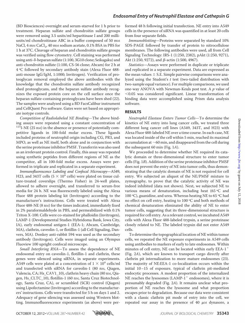
Neutrophil Elastase Enters Tumor Cells—To determine thekinetics of NE entry into lung cancer cells, we treated threedifferent lung cancer cell lines (A549, 343T, and H23) withAlexa Fluor 488-labeledNEover a time course. In each case, NEwas located inside of the cell within 5 min, reached its maximalaccumulation at�60min, and disappeared from the cell duringthe subsequent 60 min (Fig. 1A).We proceeded to determine whether NE required its cata-
lytic domain or three-dimensional structure to enter tumorcells (Fig. 1B). Addition of the serine proteinase inhibitor PMSFdoes not prevent the entry of NE into tumor cells, thus demon-strating that the catalytic domain of NE is not required for cellentry. We subjected an aliquot of the NE/PMSF mixture tozymography, to prove that the catalytic activity of NE wasindeed inhibited (data not shown). Next, we subjected NE tovarious means of denaturation, including heat (65 ºC and100 ºC), guanidine, and urea.Whereas heating NE to 65 ºC hadno effect on cell entry, heating to 100 ºC and both methods ofchemical denaturation eliminated the ability of NE to entertumor cells, suggesting that its three-dimensional structure isrequired for cell entry. As a relevant control, we incubatedA549cells with Alexa Fluor 488-labeled trypsin, a serine proteinaseclosely related to NE. The labeled trypsin did not enter A549cells.To determine the topographical location of NEwithin tumor
cells, we repeated the NE exposure experiments in A549 cellsusing antibodies to markers of early to late endosomes. Withinminutes of cell entry, NE can be located within early EEA-1�
(Fig. 2A), which are known to transport cargo directly afterclathrin pit internalization to more mature endosomes (23).The majority of NE:EEA-1 co-localization occurs within theinitial 10–15 of exposure, typical of clathrin pit-mediatedendocytic processes. A modest proportion of the internalizedNE reaches the lysosomes (LAMP-1� endosomes), where it ispresumably degraded (Fig. 2A). It remains unclear what pro-portion of NE reaches the lysosome and what proportionescapes prior to degradation. Because our data were consistentwith a classic clathrin pit mode of entry into the cell, werepeated our assay in the presence of 40 �M dynasore, a
Endosomal Entry of Neutrophil Elastase and Cathepsin G
OCTOBER 12, 2012 • VOLUME 287 • NUMBER 42 JOURNAL OF BIOLOGICAL CHEMISTRY 35343
by guest on July 12, 2018http://w
ww
.jbc.org/D
ownloaded from
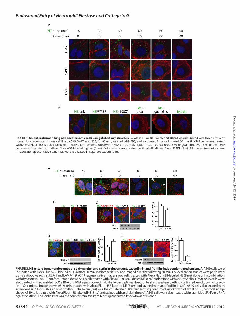
FIGURE 1. NE enters human lung adenocarcinoma cells using its tertiary structure. A, Alexa Fluor 488-labeled NE (8 nM) was incubated with three differenthuman lung adenocarcinoma cell lines, A549, 343T, and H23, for 60 min, washed with PBS, and incubated for an additional 60 min. B, A549 cells were treatedwith Alexa Fluor-488-labeled NE (8 nM) in native form or denatured with PMSF (1:100 molar ratio), heat (100 ºC), urea (8 M), or guanidine-HCl (6 M); or the A549cells were incubated with Alexa Fluor 488-labeled trypsin (8 nM). Cells were counterstained with phalloidin (red) and DAPI (blue). All images (magnification,�1200) are representative data that were replicated in separate experiments.
FIGURE 2. NE enters tumor endosomes via a dynamin- and clathrin-dependent, caveolin-1- and flotillin-independent mechanism. A, A549 cells wereincubated with Alexa Fluor 488-labeled NE (8 nM) for 60 min, washed with PBS, and imaged over the following 60 min. Co-localization studies were performedusing antibodies against EEA-1 and LAMP-1. B, A549 representative images show cells treated with Alexa Fluor 488-labeled NE (8 nM) alone or in combinationwith dynasore (40 nM). C, confocal image shows A549 cells treated with Alexa Fluor 488-labeled NE (8 nM) and stained with anti-caveolin-1 (red). A549 cells werealso treated with scrambled (SCR) siRNA or siRNA against caveolin-1. Phalloidin (red) was the counterstain. Western blotting confirmed knockdown of caveo-lin-1. D, confocal image shows A549 cells treated with Alexa Fluor 488-labeled NE (8 nM) and stained with anti-flotillin-1 (red). A549 cells also treated withscrambled siRNA or siRNA against flotillin-1. Phalloidin (red) was the counterstain. Western blotting confirmed knockdown of flotillin-1. E, confocal imageshows A549 cells treated with Alexa Fluor 488-labeled NE (8 nM) and stained with anti-clathrin (red). A549 cells were also treated with scrambled siRNA or siRNAagainst clathrin. Phalloidin (red) was the counterstain. Western blotting confirmed knockdown of clathrin.
Endosomal Entry of Neutrophil Elastase and Cathepsin G
35344 JOURNAL OF BIOLOGICAL CHEMISTRY VOLUME 287 • NUMBER 42 • OCTOBER 12, 2012
by guest on July 12, 2018http://w
ww
.jbc.org/D
ownloaded from
dynamin inhibitor that prevents the formation of early endo-somes (24). Accordingly, dynamin inhibition prevents the entryof NE into tumor cells (Fig. 2B), consistent with a clathrin pit toearly endosome mechanism of cellular entry for NE.We entertained two additional possible mechanisms for the
entrance of NE into tumor vesicles. First, we considered thecaveolar system, known to grant tumor cell access to extracel-lular proteins (25). We were unable to observe co-localizationof Alexa Fluor 488-labeled NE with caveolin-1 over a timecourse (Fig. 2C). Additionally, the silencing caveolin-1 usingsiRNA did not impact the entrance of NE into A549 cells (Fig.2C). Second, we evaluated the possibility that NE entered earlyendosomes in a dynamin-dependent, yet clathrin-independentfashion. Although this type of endocytosis is still poorlydescribed, it is often flotillin-1 dependent, and does not neces-sarily utilize a cell surface receptor, as is typically the case forclathrin pit-mediated endocytosis (26, 27). Silencing of flotil-lin-1 did not inhibit the ability of NE to enter tumor endosomes(Fig. 2D). Furthermore, we were unable to co-localize AlexaFluor 488-labeledNEwith flotillin-1 using confocalmicroscopy(Fig. 2D). However, we were able to co-localize Alexa Fluor488-labeled NE with clathrin using confocal microscopy and toinhibit the entry of NE into tumor endosomes by silencing theclathrin heavy chain using siRNA approaches (Fig. 2E). Thus,NE enters tumor endosomes in a dynamin- and clathrin-depen-dent, yet caveolin-1- and flotillin-1-independent fashion.Endosomal Entry of NE Is Required for Tumor Cell
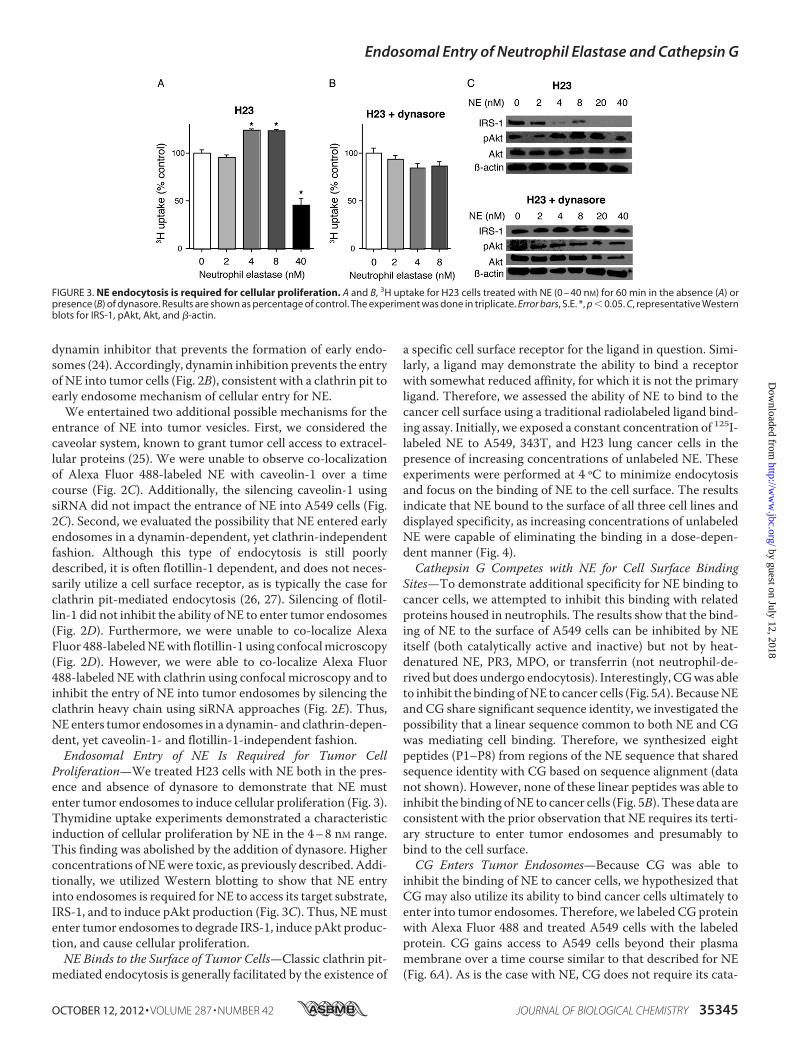
Proliferation—We treated H23 cells with NE both in the pres-ence and absence of dynasore to demonstrate that NE mustenter tumor endosomes to induce cellular proliferation (Fig. 3).Thymidine uptake experiments demonstrated a characteristicinduction of cellular proliferation by NE in the 4–8 nM range.This finding was abolished by the addition of dynasore. Higherconcentrations ofNEwere toxic, as previously described. Addi-tionally, we utilized Western blotting to show that NE entryinto endosomes is required for NE to access its target substrate,IRS-1, and to induce pAkt production (Fig. 3C). Thus, NEmustenter tumor endosomes to degrade IRS-1, induce pAkt produc-tion, and cause cellular proliferation.NE Binds to the Surface of Tumor Cells—Classic clathrin pit-
mediated endocytosis is generally facilitated by the existence of
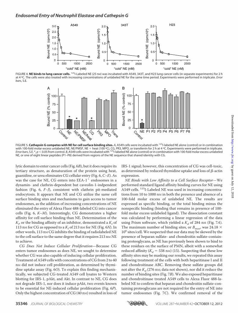
a specific cell surface receptor for the ligand in question. Simi-larly, a ligand may demonstrate the ability to bind a receptorwith somewhat reduced affinity, for which it is not the primaryligand. Therefore, we assessed the ability of NE to bind to thecancer cell surface using a traditional radiolabeled ligand bind-ing assay. Initially, we exposed a constant concentration of 125I-labeled NE to A549, 343T, and H23 lung cancer cells in thepresence of increasing concentrations of unlabeled NE. Theseexperiments were performed at 4 ºC to minimize endocytosisand focus on the binding of NE to the cell surface. The resultsindicate that NE bound to the surface of all three cell lines anddisplayed specificity, as increasing concentrations of unlabeledNE were capable of eliminating the binding in a dose-depen-dent manner (Fig. 4).Cathepsin G Competes with NE for Cell Surface Binding
Sites—To demonstrate additional specificity for NE binding tocancer cells, we attempted to inhibit this binding with relatedproteins housed in neutrophils. The results show that the bind-ing of NE to the surface of A549 cells can be inhibited by NEitself (both catalytically active and inactive) but not by heat-denatured NE, PR3, MPO, or transferrin (not neutrophil-de-rived but does undergo endocytosis). Interestingly, CGwas ableto inhibit the binding ofNE to cancer cells (Fig. 5A). BecauseNEand CG share significant sequence identity, we investigated thepossibility that a linear sequence common to both NE and CGwas mediating cell binding. Therefore, we synthesized eightpeptides (P1–P8) from regions of the NE sequence that sharedsequence identity with CG based on sequence alignment (datanot shown). However, none of these linear peptides was able toinhibit the binding ofNE to cancer cells (Fig. 5B). These data areconsistent with the prior observation that NE requires its terti-ary structure to enter tumor endosomes and presumably tobind to the cell surface.CG Enters Tumor Endosomes—Because CG was able to
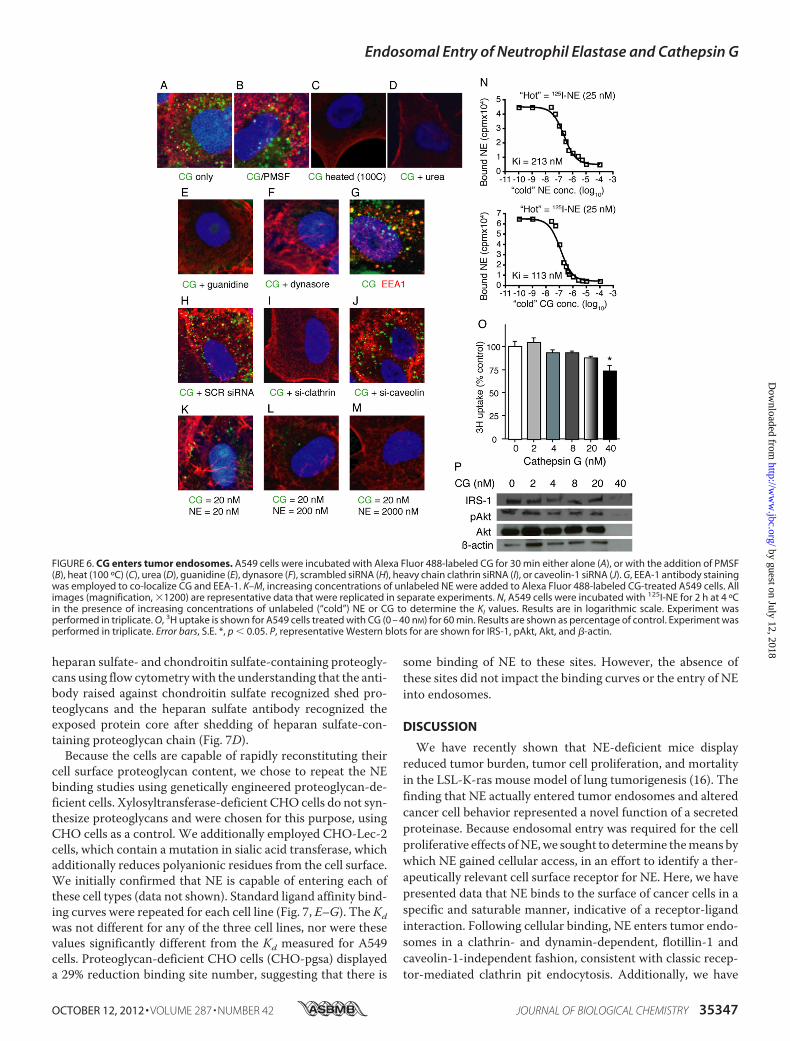
inhibit the binding of NE to cancer cells, we hypothesized thatCG may also utilize its ability to bind cancer cells ultimately toenter into tumor endosomes. Therefore, we labeled CG proteinwith Alexa Fluor 488 and treated A549 cells with the labeledprotein. CG gains access to A549 cells beyond their plasmamembrane over a time course similar to that described for NE(Fig. 6A). As is the case with NE, CG does not require its cata-
FIGURE 3. NE endocytosis is required for cellular proliferation. A and B, 3H uptake for H23 cells treated with NE (0 – 40 nM) for 60 min in the absence (A) orpresence (B) of dynasore. Results are shown as percentage of control. The experiment was done in triplicate. Error bars, S.E. *, p � 0.05. C, representative Westernblots for IRS-1, pAkt, Akt, and �-actin.
Endosomal Entry of Neutrophil Elastase and Cathepsin G
OCTOBER 12, 2012 • VOLUME 287 • NUMBER 42 JOURNAL OF BIOLOGICAL CHEMISTRY 35345
by guest on July 12, 2018http://w
ww
.jbc.org/D
ownloaded from
lytic domain to enter cancer cells (Fig. 6B), but it does require itstertiary structure, as denaturation of the protein using heat,guanidine, or urea eliminates CG cellular entry (Fig. 6,C–E). Aswas the case for NE, CG enters into EEA-1� endosomes in adynamin- and clathrin-dependent but caveolin-1-indpendentfashion (Fig. 6, F–J), consistent with clathrin pit-mediatedendocytosis. It appears that NE and CG utilize the same cellsurface binding sites and mechanisms to gain access to tumorendosomes, as the addition of increasing concentrations of NEeliminated the entry of Alexa Fluor 488-labeled CG into cancercells (Fig. 6, K–M). Interestingly, CG demonstrates a higheraffinity for cell surface binding than NE. Determination of theKI, or the binding affinity of an inhibitor, demonstrated a KI of113 nM for CG as opposed to a KI of 213 nM for NE (Fig. 6N). Inother words, 113 nMCG inhibits the binding of radiolabeledNEto the cell surface to the same degree that it requires 213 nMNEto achieve.CG Does Not Induce Cellular Proliferation—Because CG
enters tumor endosomes as does NE, we sought to determinewhether CG was also capable of inducing cellular proliferation.Treatment ofA549 cells with concentrations ofCG from2 to 40nM did not induce cell proliferation, as determined by thymi-dine uptake assay (Fig. 6O). To explain this finding mechanis-tically, we subjected CG-treated A549 cell lysates to Westernblotting for IRS-1, pAkt, and Akt. In contrast to NE, CG doesnot degrade IRS-1, nor does it induce pAkt, two events knownto be essential for NE-induced cellular proliferation (Fig. 6P).Only the highest concentration of CG (40 nM) resulted in loss of
IRS-1 signal; however, this concentration of CG was cell-toxic,as determined by reduced thymidine uptake and loss of �-actinexpression.NE Binds with Low Affinity to a Cell Surface Receptor—We
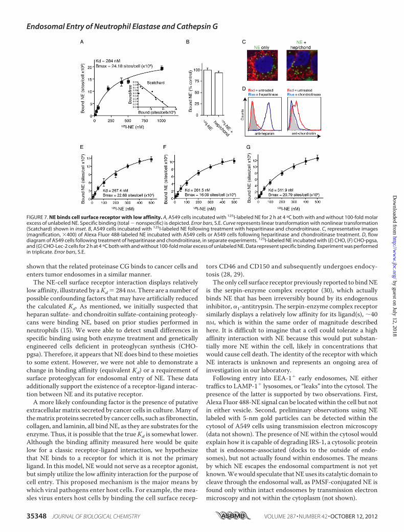
performed standard ligand affinity binding curves for NE usingA549 cells. 125I-Labeled NE was used in increasing concentra-tions from 10 to 1000 nM in both the presence and absence of a100-fold molar excess of unlabeled NE. The results areexpressed as specific binding, or the total binding minus thenonspecific binding (binding that remains in presence of 100-fold molar excess unlabeled ligand). The dissociation constantwas calculated by performing a linear regression of the datausing Prism software, which yielded a Kd of 284 nM (Fig. 7A).The maximum number of binding sites, or Bmax, was 24.18 �106 sites/cell.We suspected that our datamay be skewed by thepresence of heparan sulfate- and chondroitin sulfate-contain-ing proteoglycans, as NE has previously been shown to bind tothese residues on the surface of PMN, albeit with a somewhatreduced affinity (Kd � 538 nM) (15). Suspecting that these lowaffinity sites may bemasking our results, we repeated this assayfollowing treatment of the cells with both heparitinase I and IIand chondroitinase ABC. Removing these sulfate groups didnot alter the Kd (276 nM; data not shown), nor did it reduce thenumber of binding sites (Fig. 7B).We also exposed heparitinaseand chondroitinase treated A549 cells to Alexa Fluor 488-la-beled NE to confirm that heparan and chondroitin sulfate-con-taining proteoglycans are not required for the entry of NE intotumor endosomes (Fig. 7C). We confirmed removal of the
FIGURE 4. NE binds to lung cancer cells. 125I-Labeled NE (25 nM) was incubated with A549, 343T, and H23 lung cancer cells (in separate experiments) for 2 hat 4 ºC. The cells were also treated with increasing concentrations of unlabeled NE for the same time period. Experiments were performed in triplicate. Errorbars, S.E.
FIGURE 5. Cathepsin G competes with NE for cell surface binding sites. A, A549 cells were incubated with 125I-labeled NE alone (control) or in combinationwith 100-fold molar excess unlabeled NE, NE/PMSF, NE � heat (100 ºC), CG, PR3, MPO, or transferrin for 2 h at 4 ºC. Experiments were performed in triplicate.Error bars, S.E. *, p � 0.05 from control. B, A549 cells were incubated with 125I-labeled NE alone (control) or in combination with 100-fold molar excess unlabeledNE, or one of eight linear peptides (P1–P8) derived from regions of the NE sequence that shared identity with CG.
Endosomal Entry of Neutrophil Elastase and Cathepsin G
35346 JOURNAL OF BIOLOGICAL CHEMISTRY VOLUME 287 • NUMBER 42 • OCTOBER 12, 2012
by guest on July 12, 2018http://w
ww
.jbc.org/D
ownloaded from
heparan sulfate- and chondroitin sulfate-containing proteogly-cans using flow cytometrywith the understanding that the anti-body raised against chondroitin sulfate recognized shed pro-teoglycans and the heparan sulfate antibody recognized theexposed protein core after shedding of heparan sulfate-con-taining proteoglycan chain (Fig. 7D).Because the cells are capable of rapidly reconstituting their
cell surface proteoglycan content, we chose to repeat the NEbinding studies using genetically engineered proteoglycan-de-ficient cells. Xylosyltransferase-deficient CHO cells do not syn-thesize proteoglycans and were chosen for this purpose, usingCHO cells as a control. We additionally employed CHO-Lec-2cells, which contain a mutation in sialic acid transferase, whichadditionally reduces polyanionic residues from the cell surface.We initially confirmed that NE is capable of entering each ofthese cell types (data not shown). Standard ligand affinity bind-ing curves were repeated for each cell line (Fig. 7, E–G). The Kdwas not different for any of the three cell lines, nor were thesevalues significantly different from the Kd measured for A549cells. Proteoglycan-deficient CHO cells (CHO-pgsa) displayeda 29% reduction binding site number, suggesting that there is
some binding of NE to these sites. However, the absence ofthese sites did not impact the binding curves or the entry of NEinto endosomes.
DISCUSSION
We have recently shown that NE-deficient mice displayreduced tumor burden, tumor cell proliferation, and mortalityin the LSL-K-ras mouse model of lung tumorigenesis (16). Thefinding that NE actually entered tumor endosomes and alteredcancer cell behavior represented a novel function of a secretedproteinase. Because endosomal entry was required for the cellproliferative effects ofNE,we sought to determine themeans bywhich NE gained cellular access, in an effort to identify a ther-apeutically relevant cell surface receptor for NE. Here, we havepresented data that NE binds to the surface of cancer cells in aspecific and saturable manner, indicative of a receptor-ligandinteraction. Following cellular binding, NE enters tumor endo-somes in a clathrin- and dynamin-dependent, flotillin-1 andcaveolin-1-independent fashion, consistent with classic recep-tor-mediated clathrin pit endocytosis. Additionally, we have
FIGURE 6. CG enters tumor endosomes. A549 cells were incubated with Alexa Fluor 488-labeled CG for 30 min either alone (A), or with the addition of PMSF(B), heat (100 ºC) (C), urea (D), guanidine (E), dynasore (F), scrambled siRNA (H), heavy chain clathrin siRNA (I), or caveolin-1 siRNA (J). G, EEA-1 antibody stainingwas employed to co-localize CG and EEA-1. K–M, increasing concentrations of unlabeled NE were added to Alexa Fluor 488-labeled CG-treated A549 cells. Allimages (magnification, �1200) are representative data that were replicated in separate experiments. N, A549 cells were incubated with 125I-NE for 2 h at 4 ºCin the presence of increasing concentrations of unlabeled (“cold”) NE or CG to determine the KI values. Results are in logarithmic scale. Experiment wasperformed in triplicate. O, 3H uptake is shown for A549 cells treated with CG (0 – 40 nM) for 60 min. Results are shown as percentage of control. Experiment wasperformed in triplicate. Error bars, S.E. *, p � 0.05. P, representative Western blots for are shown for IRS-1, pAkt, Akt, and �-actin.
Endosomal Entry of Neutrophil Elastase and Cathepsin G
OCTOBER 12, 2012 • VOLUME 287 • NUMBER 42 JOURNAL OF BIOLOGICAL CHEMISTRY 35347
by guest on July 12, 2018http://w
ww
.jbc.org/D
ownloaded from
shown that the related proteinase CG binds to cancer cells andenters tumor endosomes in a similar manner.The NE-cell surface receptor interaction displays relatively
low affinity, illustrated by aKd � 284 nM. There are a number ofpossible confounding factors that may have artificially reducedthe calculated Kd. As mentioned, we initially suspected thatheparan sulfate- and chondroitin sulfate-containing proteogly-cans were binding NE, based on prior studies performed inneutrophils (15). We were able to detect small differences inspecific binding using both enzyme treatment and geneticallyengineered cells deficient in proteoglycan synthesis (CHO-pgsa). Therefore, it appears that NE does bind to thesemoietiesto some extent. However, we were not able to demonstrate achange in binding affinity (equivalent Kd) or a requirement ofsurface proteoglycan for endosomal entry of NE. These dataadditionally support the existence of a receptor-ligand interac-tion between NE and its putative receptor.A more likely confounding factor is the presence of putative
extracellularmatrix secreted by cancer cells in culture.Many ofthematrix proteins secreted by cancer cells, such as fibronectin,collagen, and laminin, all bind NE, as they are substrates for theenzyme. Thus, it is possible that the trueKd is somewhat lower.Although the binding affinity measured here would be quitelow for a classic receptor-ligand interaction, we hypothesizethat NE binds to a receptor for which it is not the primaryligand. In this model, NE would not serve as a receptor agonist,but simply utilize the low affinity interaction for the purpose ofcell entry. This proposed mechanism is the major means bywhich viral pathogens enter host cells. For example, themea-sles virus enters host cells by binding the cell surface recep-
tors CD46 and CD150 and subsequently undergoes endocy-tosis (28, 29).The only cell surface receptor previously reported to bindNE
is the serpin-enzyme complex receptor (30), which actuallybinds NE that has been irreversibly bound by its endogenousinhibitor,�1-antitrypsin. The serpin-enzyme complex receptorsimilarly displays a relatively low affinity for its ligand(s), �40nM, which is within the same order of magnitude describedhere. It is difficult to imagine that a cell could tolerate a highaffinity interaction with NE because this would put substan-tially more NE within the cell, likely in concentrations thatwould cause cell death. The identity of the receptor with whichNE interacts is unknown and represents an ongoing area ofinvestigation in our laboratory.Following entry into EEA-1� early endosomes, NE either
traffics to LAMP-1� lysosomes, or “leaks” into the cytosol. Thepresence of the latter is supported by two observations. First,Alexa Fluor 488-NE signal can be locatedwithin the cell but notin either vesicle. Second, preliminary observations using NElabeled with 5-nm gold particles can be detected within thecytosol of A549 cells using transmission electron microscopy(data not shown). The presence of NE within the cytosol wouldexplain how it is capable of degrading IRS-1, a cytosolic proteinthat is endosome-associated (docks to the outside of endo-somes), but not actually found within endosomes. The meansby which NE escapes the endosomal compartment is not yetknown.Wewould speculate thatNEuses its catalytic domain tocleave through the endosomal wall, as PMSF-conjugated NE isfound only within intact endosomes by transmission electronmicroscopy and not within the cytoplasm (not shown).
FIGURE 7. NE binds cell surface receptor with low affinity. A, A549 cells incubated with 125I-labeled NE for 2 h at 4 ºC both with and without 100-fold molarexcess of unlabeled NE. Specific binding (total � nonspecific) is depicted. Error bars, S.E. Curve represents linear transformation with nonlinear transformation(Scatchard) shown in inset. B, A549 cells incubated with 125I-labeled NE following treatment with heparitinase and chondroitinase. C, representative images(magnification, �400) of Alexa Fluor 488-labeled NE incubated with A549 cells or A549 cells following heparitinase and chondroitinase treatment. D, flowdiagram of A549 cells following treatment of heparitinase and chondroitinase, in separate experiments. 125I-labeled NE incubated with (E) CHO, (F) CHO-pgsa,and (G) CHO-Lec-2 cells for 2 h at 4 ºC both with and without 100-fold molar excess of unlabeled NE. Data represent specific binding. Experiment was performedin triplicate. Error bars, S.E.
Endosomal Entry of Neutrophil Elastase and Cathepsin G
35348 JOURNAL OF BIOLOGICAL CHEMISTRY VOLUME 287 • NUMBER 42 • OCTOBER 12, 2012
by guest on July 12, 2018http://w
ww
.jbc.org/D
ownloaded from
The finding that CG binds to the cancer cell surface andenters tumor endosomes is entirely novel. CG, like NE, wasreported to bind to heparan sulfate- and chondroitin sulfate-containing proteoglycans on the surface of neutrophils. Ofnote, MPO and PR3, both neutrophil-derived, were capable ofpartially competing for these binding sites on PMN, which wasnot the case here. The fact that CG, but not PR3, was capable ofcompeting for NE binding sites raises questions regarding thethree-dimensional configuration of the enzymes in vivo. Thedegree of sequence homology among the three enzymes is sim-ilar. Linear peptide sequences displaying sequence homologybetween NE and CG failed to inhibit NE binding. Finally, dena-turing NE with heat or chemicals eliminates binding. There-fore, there is likely a unique three-dimensional characteristic inNE and CG that has not been preserved in PR3. The only othernotable difference is that NE andCG are both polycationic pro-teins, whereas PR3 is more neutral.In contrast to NE, CG is unable to induce cellular proliferation.
As illustrated here, NE-induced cell proliferation is dependentupon four events: (i) cell surface binding, (ii) entrance into tumorendosomes, (iii) degradationof IRS-1, and (iv) enhancedpAktpro-duction. CG is capable of binding cancer cells and entering endo-somes, but is incapable of degrading IRS-1 in this context, andtherefore, subsequent pAkt production is not observed. It is likelythat CG processes tumor-derived substrates that could impacttumor cell behavior. The nature of these events is currently underinvestigation in our laboratory.In summary, we have provided evidence that NE and CG
bind to the surface of cancer cells and subsequently undergoclathrin- and dynamin-dependent, and caveolin-1- and flotil-lin-1-independent endocytosis. These results strongly suggestthe presence of a cell surface receptor capable of binding NEand CG with relatively low affinity, that allows for endosomalentry via clathrin pit-mediated endocytosis. The presence ofsuch a receptor may provide endosomal access to numerous, asyet unidentified ligands, capable of altering cell behavior, as isclearly the case for NE, and may be for CG. Expression cloningapproaches are underway in our laboratory to definitively iden-tify the cell surface receptor in question. The identification ofthis receptor bears clinical significance. Because NE is a potentantimicrobial agent, inhibiting NE directly would theoreticallycarry some risk of immunocompromise. The ability to targetthe receptor that grants NE access to cancer cells would elimi-nate the tumor promoting effects of NEwithout compromisingthe enzymes ability to kill engulfedmicroorganismswithin neu-trophil phagolysosomes.
REFERENCES1. Baugh, R. J., and Travis, J. (1976) Human leukocyte granule elastase: rapid
isolation and characterization. Biochemistry 15, 836–8412. Belaaouaj, A., McCarthy, R., Baumann, M., Gao, Z., Ley, T. J., Abraham,
S. N., and Shapiro, S. D. (1998) Mice lacking neutrophil elastase revealimpaired host defense against Gram-negative bacterial sepsis. Nat. Med.4, 615–618
3. Belaaouaj, A., Kim, K. S., and Shapiro, S. D. (2000) Degradation of outermembrane protein A in Escherichia coli killing by neutrophil elastase.Science 289, 1185–1188
4. Weinrauch, Y., Drujan, D., Shapiro, S. D., Weiss, J., and Zychlinsky, A.(2002) Neutrophil elastase targets virulence factors of enterobacteria.Na-ture 417, 91–94
5. Gabay, J. E., Scott, R. W., Campanelli, D., Griffith, J., Wilde, C., Marra,M. N., Seeger, M., and Nathan, C. F. (1989) Antibiotic proteins of humanpolymorphonuclear leukocytes. Proc. Natl. Acad. Sci. U.S.A. 86,5610–5614
6. Lee, W. L., and Downey, G. P. (2001) Leukocyte elastase: physiologicalfunctions and role in acute lung injury.Am. J. Respir. Crit. CareMed. 164,896–904
7. Hedstrom, L. (2002) Serine protease mechanism and specificity. Chem.Rev. 102, 4501–4524
8. Lane, A. A., and Ley, T. J. (2003) Neutrophil elastase cleaves PML-RAR�
and is important for the development of acute promyelocytic leukemia inmice. Cell 115, 305–318
9. Kohri, K., Ueki, I. F., and Nadel, J. A. (2002) Neutrophil elastase inducesmucin production by ligand-dependent epidermal growth factor receptoractivation. Am. J. Physiol. Lung Cell Mol. Physiol. 283, L531–540
10. Kaynar, A. M., Houghton, A. M., Lum, E. H., Pitt, B. R., and Shapiro, S. D.(2008) Neutrophil elastase is needed for neutrophil emigration into lungsin ventilator-induced lung injury. Am. J. Respir. Cell Mol. Biol. 39, 53–60
11. Shapiro, S.D., Goldstein,N.M.,Houghton,A.M., Kobayashi, D. K., Kelley,D., and Belaaouaj, A. (2003) Neutrophil elastase contributes to cigarettesmoke-induced emphysema in mice. Am. J. Pathol. 163, 2329–2335
12. Senior, R. M., Tegner, H., Kuhn, C., Ohlsson, K., Starcher, B. C., andPierce, J. A. (1977) The induction of pulmonary emphysema with humanleukocyte elastase. Am. Rev. Respir. Dis. 116, 469–475
13. Liou, T. G., andCampbell, E. J. (1996)Quantumproteolysis resulting fromrelease of single granules by human neutrophils: a novel, nonoxidativemechanism of extracellular proteolytic activity. J. Immunol. 157,2624–2631
14. Owen, C.A., Campbell,M.A., Sannes, P. L., Boukedes, S. S., andCampbell,E. J. (1995) Cell surface-bound elastase and cathepsin G on human neu-trophils: a novel, nonoxidativemechanismbywhich neutrophils focus andpreserve catalytic activity of serine proteinases. J. Cell Biol. 131, 775–789
15. Campbell, E. J., and Owen, C. A. (2007) The sulfate groups of chondroitinsulfate- and heparan sulfate-containing proteoglycans in neutrophilplasma membranes are novel binding sites for human leukocyte elastaseand cathepsin G. J. Biol. Chem. 282, 14645–14654
16. Houghton, A. M., Rzymkiewicz, D. M., Ji, H., Gregory, A. D., Egea, E. E.,Metz, H. E., Stolz, D. B., Land, S. R., Marconcini, L. A., Kliment, C. R.,Jenkins, K. M., Beaulieu, K. A., Mouded, M., Frank, S. J., Wong, K. K., andShapiro, S. D. (2010) Neutrophil elastase-mediated degradation of IRS-1accelerates lung tumor growth. Nat. Med. 16, 219–223
17. Metz, H. E., and Houghton, A. M. (2011) Insulin receptor substrate regu-lation of phosphoinositide 3-kinase. Clin. Cancer Res. 17, 206–211
18. Mittendorf, E. A., Alatrash, G., Qiao, N., Wu, Y., Sukhumalchandra, P., StJohn, L. S., Philips, A. V., Xiao, H., Zhang, M., Ruisaard, K., Clise-Dwyer,K., Lu, S., and Molldrem, J. J. (2012) Breast cancer cell uptake of theinflammatorymediator neutrophil elastase triggers an anticancer adaptiveimmune response. Cancer Res. 72, 3153–3162
19. Esko, J. D., Stewart, T. E., and Taylor, W. H. (1985) Animal cell mutantsdefective in glycosaminoglycan biosynthesis. Proc. Natl. Acad. Sci. U.S.A.82, 3197–3201
20. Eckhardt, M., Gotza, B., and Gerardy-Schahn, R. (1998) Mutants of theCMP-sialic acid transporter causing the Lec2 phenotype. J. Biol. Chem.273, 20189–20195
21. Joslin, G., Griffin, G. L., August, A. M., Adams, S., Fallon, R. J., Senior,R. M., and Perlmutter, D. H. (1992) The serpin-enzyme complex (SEC)receptor mediates the neutrophil chemotactic effect of �1-antitrypsin-elastase complexes and amyloid-� peptide. J. Clin. Invest. 90, 1150–1154
22. David, G., Bai, X. M., Van der Schueren, B., Cassiman, J. J., and Van denBerghe, H. (1992) Developmental changes in heparan sulfate expression:in situ detection with mAbs. J. Cell Biol. 119, 961–975
23. Rubino, M., Miaczynska, M., Lippé, R., and Zerial, M. (2000) Selectivemembrane recruitment of EEA-1 suggests a role in directional trans-port of clathrin-coated vesicles to early endosomes. J. Biol. Chem. 275,3745–3748
24. Macia, E., Ehrlich, M., Massol, R., Boucrot, E., Brunner, C., and Kirch-hausen, T. (2006) Dynasore, a cell-permeable inhibitor of dynamin. Dev.Cell 10, 839–850
Endosomal Entry of Neutrophil Elastase and Cathepsin G
OCTOBER 12, 2012 • VOLUME 287 • NUMBER 42 JOURNAL OF BIOLOGICAL CHEMISTRY 35349
by guest on July 12, 2018http://w
ww
.jbc.org/D
ownloaded from
25. Nabi, I. R., and Le, P. U. (2003) Caveolae/raft-dependent endocytosis.J. Cell Biol. 161, 673–677
26. McMahon, H. T., and Boucrot, E. (2011)Molecular mechanism and phys-iological functions of clathrin-mediated endocytosis. Nat. Rev. Mol. CellBiol. 12, 517–533
27. Glebov, O. O., Bright, N. A., and Nichols, B. J. (2006) Flotillin-1 defines aclathrin-independent endocytic pathway in mammalian cells. Nat. CellBiol. 8, 46–54
28. Buchholz, C. J., Koller, D., Devaux, P.,Mumenthaler, C., Schneider-Schau-lies, J., Braun, W., Gerlier, D., and Cattaneo, R. (1997) Mapping of the
primary binding site of measles virus to its receptor CD46. J. Biol. Chem.272, 22072–22079
29. Erlenhoefer, C., Wurzer, W. J., Löffler, S., Schneider-Schaulies, S., terMeulen, V., and Schneider-Schaulies, J. (2001) CD150 (SLAM) is a recep-tor for measles virus but is not involved in viral contact-mediated prolif-eration inhibition. J. Virol. 75, 4499–4505
30. Perlmutter, D. H., Joslin, G., Nelson, P., Schasteen, C., Adams, S. P., andFallon, R. J. (1990) Endocytosis and degradation of �1-antitrypsin-prote-ase complexes is mediated by the serpin-enzyme complex (SEC) receptor.J. Biol. Chem. 265, 16713–16716
Endosomal Entry of Neutrophil Elastase and Cathepsin G
35350 JOURNAL OF BIOLOGICAL CHEMISTRY VOLUME 287 • NUMBER 42 • OCTOBER 12, 2012
by guest on July 12, 2018http://w
ww
.jbc.org/D
ownloaded from
Alyssa D. Gregory, Pamela Hale, David H. Perlmutter and A. McGarry HoughtonCancer Cells
Clathrin Pit-mediated Endocytosis of Neutrophil Elastase and Cathepsin G by
doi: 10.1074/jbc.M112.385617 originally published online August 22, 20122012, 287:35341-35350.J. Biol. Chem.
10.1074/jbc.M112.385617Access the most updated version of this article at doi:
Alerts:
When a correction for this article is posted•
When this article is cited•
to choose from all of JBC's e-mail alertsClick here
http://www.jbc.org/content/287/42/35341.full.html#ref-list-1
This article cites 30 references, 15 of which can be accessed free at
by guest on July 12, 2018http://w
ww
.jbc.org/D
ownloaded from