classification schemes for arteriovenous malformations
TRANSCRIPT

Classif icationSchemes forArteriovenousMalformations
Jason M. Davies, MD, PhDa, Helen Kim, PhDb,c,William L. Young, MDa,b,c,d, Michael T. Lawton, MDa,c,*KEYWORDS
� Arteriovenous malformation � Classification� Grading system � Spetzler-Martin
Judicious patient selection is essential to avoidsurgical complications and poor neurologicoutcomes with microsurgical resection of brainarteriovenous malformations (AVMs). The widevariety of AVM anatomy, size, location, and clinicalpresentation makes patient selection for surgerya difficult process. Neurosurgeons have analyzedtheir surgical experiences to identify factors thatdetermine the risks of surgery to assist them inthis selection process. Numerous classificationschemes have been developed, each withits own emphasis, accuracy, advantages, anddisadvantages. Some are complex and otherssimple, each striving to predict surgical risk andto achieve bedside applicability. These classifica-tion schemes have value because they transformcomplex decisions into algorithms. In this review,the important grading schemes that have contrib-uted to management of patients with brain AVMsare described, and our current approach to patientselection is outlined.
a Department of Neurological Surgery, University of CalifFrancisco, CA 94143-0112, USAb Department of Anesthesia and Perioperative Care, UAvenue, C450, San Francisco, CA 94143-0648, USAc Center for Cerebrovascular Research, University of CalifSan Francisco, CA 94110, USAd Department of Neurology, University of California San FCA 94143-0110, USA* Corresponding author.E-mail address: [email protected]
Neurosurg Clin N Am 23 (2012) 43–53doi:10.1016/j.nec.2011.09.0021042-3680/12/$ – see front matter � 2012 Elsevier Inc. All
PRE–SPETZLER-MARTIN CLASSIFICATIONSCHEMES
The first major AVM grading scheme developed byLuessenhop and Gennarelli1 in 1977 formulateda grade from I to IV based on the number offeeding arteries for which there is standardizednomenclature. The score was determined bycounting the number of tertiary arteries feedingthe AVM from a single vascular territory, like themiddle cerebral artery (MCA), anterior cerebralartery (ACA), or posterior cerebral artery (PCA)territory. When the AVM was supplied by multipleterritories, the grade was determined by thevascular territory with the largest number offeeders. No additional grade was assigned forlarge AVMs with more than 4 arteries, becausethese lesions were deemed inoperable. Therewere several exceptions in this scheme: lenticu-lostriate vessels were counted as named arteries;choroid plexus-based AVMs were deemed gradeIII because they are supplied by 1 anterior and 2
ornia San Francisco, 505 Parnassus Avenue, M780, San
niversity of California San Francisco, 521 Parnassus
ornia San Francisco, 1001 Potrero Avenue, Box-1371,
rancisco, 505 Parnassus Avenue, M798, San Francisco,
rights reserved. neurosurgery.th
eclinics.com
Davies et al44
posterior choroidal arteries; and corpus callosumAVMs were deemed grade II when supplied bypericallosal arteries and grade III when suppliedby the PCA. The investigators made allowancesfor clinical status to supplement anatomic gradingscale, but clear guidelines for integrating clinicaland anatomic factors were lacking. Surgicalresults in 49 patients showed that grade I AVMswere associated with low risk, higher-gradeAVMs were associated with increasing risks, andgrade IV AVMs were managed nonoperatively.Luessenhop and Rosa2 simplified this grading
scheme in 1984 by considering only the angio-graphic size of the AVM, which was believed tobe easier than counting arterial feeders. The newgrades were assigned based on nidus diameter:grade I, less than 2 cm; grade II, 2 to 4 cm; gradeIII, 4 to 6 cm; and grade IV, greater than 6 cm. Theoriginal classification scheme excluded AVMs inthe cerebellum, brain stem, and region of thevein of Galen malformations, whereas the newscheme included cerebellar AVMs. In a surgicalseries consisting of 90 patients, the investigatorsshowed low morbidity and mortality with grades Iand II AVMs, and therefore recommended surgicalresection for these lesions, with minimal consider-ation of nidus location, age, or comorbidities. Theinvestigators recommended more conservativemanagement of patients with high-grade lesions(grade III and IV) and careful consideration of theseother anatomic and clinical factors.Shi and Chen3 presented an alternative classifi-
cation scheme in 1986 that considered AVM size,location and depth, arterial supply, and venousdrainage. Each of these 4 aspects was dividedinto 4 grades. Specifically, size was graded asless than 2.5 cm (grade I), 2.5 to 5 cm (grade II),5 to 7.5 cm (grade III), or greater than 7.5 cm(grade IV). Location and depth were graded assuperficial/nonfunctional (grade I), superficial/functional (grade II), deep (grade III), and deep/vital(grade IV). Arterial supply was graded as singlesuperficial branch of MCA or ACA (grade I),multiple superficial branches of MCA or ACA(grade II), PCA branches or deep MCA or ACAbranches (grade III), and branches of all 3 cerebralarteries or vertebrobasilar artery (grade IV).Venous drainage was graded as single superficial(grade I), multiple superficial (grade II), deep (gradeIII), and deep with variceal dilatation (grade IV). Thefinal AVM grade was “matched to the appropriatehighest grade when at least 2 criteria are in thatgrade,” or was a mixed or intermediary grade ifonly one was in the highest grade. Although thisclassification scheme has 4 grades, this methodof grading led to 6 different groupings in the inves-tigators’ surgical series of 100 patients. Excellent
results were achieved in patients with grade I, Ito II, and II AVMs, with increasing morbidity/mortality in patients with AVMs grade II to III, III,and III to IV. This classification scheme incorpo-rated similar anatomic features as the Spetzler-Martin4 scheme, but it failed to gain acceptancebecause of its complexity, with grading withingrades and mixed final grades.
SPETZLER-MARTIN CLASSIFICATION SCHEME
In 1986, Spetzler and Martin4 published what hasbecome the predominant classification schemefor brain AVMs. After considering a range offactors including size, number of feeding arteries,location, operative accessibility, shunt flow,vascular steal, location, and venous drainage,these investigators settled on a simplified schemeusing only size, eloquence of surrounding brainparenchyma, and venous drainage pattern.Simplicity, applicability at the bedside, and accu-rate outcome prediction were the investigators’principal objectives.Each factor in the grading scale was scored
independently. Size was divided into 3 categories,with small AVMs less than 3 cm assigned 1 point,medium AVMs 3 to 6 cm assigned 2 points, andlarge AVMs greater than 6 cm assigned 3 points.Venous drainage was considered superficial if itdrained into cortical veins and convexity sinusesand assigned 0 points, or deep if it drained intoveins that coursed to the vein of Galen (ie, internalcerebral veins, basal veins of Rosenthal, and pre-central cerebellar vein) and assigned 1 point.AVM eloquence was assessed anatomically basedon the presumed function of surrounding braintissue, with 1 point assigned to lesions locatedin sensorimotor cortex, language areas, visualcortex, hypothalamus, internal capsule, brainstem, cerebellar peduncle, or deep cerebellarnuclei. AVMs not in these regions were assigned0 points for eloquence. The final AVM grade wasthe sum of points across the 3 domains, witha range from I to V. AVMs that are too complexfor resection, like intrinsic brain stem and holohe-mispheric AVMs, were deemed grade VI.The Spetzler-Martin grading system was initially
evaluated in a retrospective analysis of the investi-gators’ surgical experience in 100 consecutivesurgically resected AVMs. Outcomes were cate-gorized as “no deficit,” “minor deficit” (includingtemporary worsening of neurologic function, mildataxia, or mild increase in brain stem deficit), or“major deficit” (including aphasia, hemianopsia,or hemiparesis). There were no major deficits andonly 1 minor deficit in patients with low-gradeAVMs (grades I and II, Table 1). Patients with

Table 1Grading scheme of Spetzler andMartin correlatedwith operative outcome by grade in 100 consecutivepatients
Grade Patients (n) No Deficit (n) (%) Minor Deficit (n) (%) Major Deficit (n) (%) Death (%)
I 23 23 100 0 9 0 0 0
II 21 20 95 1 5 0 0 0
III 25 21 84 3 12 1 4 0
IV 15 11 73 3 20 1 7 0
V 16 11 69 3 19 2 12 0
Total 100 86 86 10 10 4 4 0
Adapted from Spetzler RF, Martin NA: A proposed grading system for arteriovenous malformations. J Neurosurg1986;65:476–83.
Classification Schemes for AVMs 45
high-grade AVMs experienced higher rates ofboth major (grade IV, 7%; and grade V, 12%) andminor deficits (grade IV, 20%; and grade V, 19%).A subsequent prospective analysis by the seniorauthor5 as well as independent analyses by othergroups6–8 confirmed the accuracy of the grad-ing system to predict operative morbidity andmortality.
This grading system has been widely acceptedby neurosurgeons and other clinicians for itssimplicity and practicality, quickly providing esti-mates of surgical risk. Furthermore, its easy appli-cability has made the grading system an integralpart of the description and language of brainAVMs. Experience with this grading has identified3 distinct groups of patients. Low-grade AVMs(grades I and II) have low morbidity associatedwith their resection and are frequently treatedsurgically. High-grade AVMs (grades IV and V)have highmorbidity associated with their resectionand are frequently managed conservatively. GradeIII AVMs have intermediate morbidity associatedwith their surgical resection, and treatment recom-mendations require an individualized approachwith multimodality treatment.
MODIFICATIONS OF THE SPETZLER-MARTINCLASSIFICATION SCHEME
One deficiency of the Spetzler-Martin gradingsystem is its handling of grade III AVMs. A 2-cmthalamic AVM with deep drainage has the samescore as a 7-cm right frontal pole AVM with super-ficial drainage, yet these 2 different lesions havethe same grade. Grade III AVMs are the mostheterogeneous of the 5 grades, with 4 differentcombinations of size, venous drainage, andeloquence and one-third of all combinations ofthese elements of the grading scale. In addition,these AVMs are technically challenging, at the limit
of what many neurosurgeons are willing to acceptin terms of technical difficulty and potentialmorbidity. The Spetzler-Martin grading systemgroups these diverse lesions together and fails toprovide the clarity in management that it does forlow-grade and high-grade AVMs.
In an analysis of a consecutive series of 76grade III AVMs, Lawton9 confirmed that neurologicoutcomes varied according to the subtype ofgrade III AVM (Table 2). Small grade III AVMs,with 1 point assigned for size, deep venousdrainage, and eloquence (S1V1E1), were themost common and had the lowest surgicalrisk (3% morbidity). In contrast, medium-sized/eloquent grade III AVMs (S2V0E1) had the highestsurgical risk (15% morbidity). Medium-sized/deepgrade III AVMs (S2V1E0) had an intermediatesurgical risk (7%). Large grade III AVMs (S3V0E0)are rare, with none in this surgical experience.
Based on these data, grade III AVMs should notbe considered a homogeneous group with equiva-lent risks for all subtypes, as the Spetzler-Martinscheme suggests. Instead, grade III AVMs shouldbe analyzed according to the 4 subtypes definedby the Spetzler-Martin scheme. Lawton proposeda modification to the Spetzler-Martin scheme toemphasize these differences. Small grade IIIAVMs were designated grade III� because theirsurgical risk was less than the average grade IIIlesion, more like a grade II lesion. Medium/eloquent grade III AVMs were designated gradeIII1 because their surgical risk was more thanthe average grade III lesion, more like a grade IVlesion. Medium/deep grade III AVMs were desig-nated plain grade III because their surgical riskwas the same as the average grade III lesion.Large grade III AVMs were designated grade III*because they are lesions fabricated by the gradingsystem with an incidence so low that their surgicalrisk is unclear and their clinical relevance minimal.

Table 2Surgical risk, according to the type of Spetzler-Martin grade III arteriovenous malformationa
Grade III Type
No. of Patients
TotalImproved or Unchanged(Number [%])
New Deficitor Death Modified Spetzler-Martin Grade
S1V1E1 34 33 (97.1) 1 (2.9) III�S2V1E0 14 13 (92.9) 1 (7.1) III
S2V0E1 27 23 (85.2) 4 (4.8) III1
S3V0E0 0 0 0 III*
Overall 75 69 (92.0) 6 (8.0)
a Patients lost to follow-upmonitoring, for whom outcome data were not available, were excluded from the calculations.Adapted from LawtonMT: Spetzler-Martin Grade III arteriovenousmalformations: surgical results and amodification of
the grading scale. Neurosurgery 2003;52:740–8 [discussion: 748–9].
Davies et al46
Large, superficial AVMs in noneloquent brain canoccur only in the frontal or temporal poles; largeAVMs in other locations are likely to have a deepdraining vein or abut eloquent brain tissue.This modification to the Spetzler-Martin system
facilitates treatment recommendations. GradeIII� and III AVMs are typically managed surgically,whereas grade III1 lesions are managed conser-vatively. This modification improves the capacityof the Spetzler-Martin grading system for bedsidedecision making.
NEW AND SUPPLEMENTARY CLASSIFICATIONSCHEMES
Although the original Spetzler-Martin classificationscheme has become an established part of theAVM literature5,10–14 Spetzler himself recognizedthat there was redundancy in the grading system,with low-grade AVMs being managed similarlywith surgery and high-grade AVMs managedconservatively. Therefore, Spetzler and Ponce15
condensed the original 5-tier grading system into3 tiers: grades I and II were combined into classA; grade III became class B; and grades IV and Vbecame class C. In a compilation of 1476 casesfrom the literature, the investigators showed bypair-wise comparison that the differences insurgical outcomes were smallest between gradesI and II AVMs and again between grades IV andV AVMs. The predictive power, as measured bycalculated area under the receiver operating char-acteristic (ROC) curve, for the 3-tiered system wasidentical to that for the 5-tiered system (0.713 and0.711, respectively), supporting this condensedclassification system.Ponce and Spetzler made broad treatment
recommendations based on AVM class. Class A
lesions are managed surgically with a goal ofcomplete resection; class B are managed withmultimodality treatment that includes endovascu-lar and radiosurgical intervention in addition tosurgical resection; and class C lesions are largelydeemed nonsurgical, with exceptions made forrecurrent hemorrhage, progressive neurologicdeficits, steal-related symptoms, and AVM-related aneurysm. Proponents of this simplificationof the grading system emphasize that fewerclasses group together patients with a raredisorder and that the larger groups correspondmore directly with treatment recommendations.Opponents of this simplification argue that itdoes not simplify the analysis of the AVM becausethe same scoring steps of the original Spetzler-Martin scale are required with an additional stepto reclassify the AVM. Opponents also emphasizethat the class-specific recommendations arevague and encumbered with exceptions. Forexample, the class system does nothing to shedlight on the heterogeneous group of grade IIIlesions. Patient selection is a sophisticatedprocess that requires more complexity, not less.To counter this trend toward simplification, new
and supplementary classification schemes havebeen proposed. Additional factors are importantto patient selection but are not part of theSpetzler-Martin grading scale, including hemor-rhagic presentation, age, deep perforating arterysupply, and diffuseness. Presentation with hemor-rhage not only indicates AVMs with high risk ofrehemorrhage but also facilitates surgery. Hema-tomas help separate AVMs from adjacent brain;evacuation of hematoma creates working spacearound the AVM that can minimize transgressionof normal brain or access a deep nidus thatmight otherwise have been unreachable; and
Classification Schemes for AVMs 47
hemorrhage can obliterate some of the arterialsupply of the AVM to reduce its flow duringresection. AVM hemorrhage and microsurgerycan injure brain tissue, but young age and plas-ticity can enhance a patient’s ability to recoverneurologic function. Compact AVMs with tightlywoven arteries and veins often have distinctborders that separate cleanly from the adjacentbrain, whereas diffuse AVMs with ragged bordersand intermixed brain force the neurosurgeon toestablish dissection planes that can extend intonormal brain. Deep perforating arteries are thin,fragile, and difficult to occlude with cautery.Bleeding during surgery can escape into deepwhite matter tracts and cause significant deficits.All of these factors (hemorrhagic presentation,young age, compactness, and absence of deepperforator supply) have been identified aspredictors of good outcomes after microsurgicalresection.1,2,16–19
Alternative AVM classification schemes havebeen proposed since the introduction of theSpetzler-Martin grading system to incorporatethese other factors and improve surgical riskprediction and patient selection. Tamaki andcolleagues17 identified age as a significantpredictor of outcome, but did not include it in thegrading system. They assigned points for size(small or large), number of feeding artery systems(1–2 or �3), and location (superficial or deep),stratifying AVMs into 5 grades ranging from 0 to4 that correlated with surgical difficulty. Theirgrading system was too similar to the Spetzler-Martin system and they failed to use age in theirclassification scheme.
After finding that neither AVM size nor venousdrainage pattern influenced outcome in theirexperience, Hollerhage19 proposed a gradingsystem based on 5 territories of feeding arterysupply (ACA, MCA, PCA, Rolandic branches, andanterior communicating artery shunt flow). Thisgrading system was among the first to incorporatea clinical variable in addition to these anatomicvariables, assigning 1 point for Hunt and Hess20
grades I to II and 2 points for Hunt and Hessgrades III to V. The Hunt and Hess grade containedwithin this AVM grading system is a surrogate forhemorrhagic presentation, but Hunt and Hess de-signed their scale for patients with aneurysm withsubarachnoid hemorrhage, and its application topatients with AVM was awkward. Despite thepossibility of an AVM grade as high as 7, gradesranged from 1 to 4 in this study and correlatedwith Glasgow Outcome Scale (GOS) scores.However, by measuring surgical results by finalGOS rather than changes in GOS scores, thisgrading scale failed to recognize the surgical
advantages associated with hemorrhagicpresentation.
Perhaps the most comprehensive gradingsystem for AVMs was proposed by Pertuiset andcolleagues.16 In addition to angiographic factorslike AVM location and feeding artery supply, thissystem analyzed AVM sectors, caliber of feedingarteries, nidus volume, and hemodynamic factorsincluding cerebral steal and circulatory velocity ofradiolabeled red blood cells. This system includedage and previous hemorrhage in its clinical vari-ables. Using elaborate tables, each variable wascoded, and these codes were added to generateoperability scores ranging between 3 and 69,with AVM scores less than 30 considered oper-able. The investigators concluded that the scoresystem was too complicated and too impracticalfor clinical use.
The University of Toronto Brain AVM StudyGroup18 developed a discriminative predictionmodel of neurologic outcomes associated withAVM resection that recognized nidus diffusenessas a critical predictor variable. The Toronto modelincorporated just 3 variables, weighted them withrounded odds ratios (eloquence 5 4, diffuse-ness 5 3, and deep venous drainage 5 2), andadded points to form a 9-point stratified riskscore. Discrimination of this model for predictingpermanent disabling neurologic outcomes washigh (area under the ROC curve, 0.79), and betterthan the Spetzler-Martin scale (area under theROC curve, 0.69). The grading system of theToronto group is simple, but it has not beenwidely applied in the years since its publicationbecause it competes with the Spetzler-Martingrading system, reaffiliating eloquence andvenous drainage with the newer scale.
Lawton and colleagues21 envisioned a gradingsystem that would supplement rather than replacethe already entrenched Spetzler-Martin gradingsystem (Table 3). This supplementary gradingsystem has its own unique variables separatefrom the Spetzler-Martin scale. Points were as-signed for patient age, hemorrhagic presentation,and AVM diffuseness, analogous to the Spetzler-Martin scoring system. Pediatric patients (age<20 years) were assigned 1 point; adults (age20–40 years) were assigned 2 points; and olderpatients (>40 years) were assigned 3 points.Patients presenting with unruptured AVMs wereassigned 1 point and ruptured AVMs 0 points.Diffuse AVMs were assigned 1 point and compactAVMs 0 point. These points were added togetherfor a supplementary AVM grade that ranged from1 to 5. Simplicity is a critical aspect of a populargrading scale, and a supplementary grading scalewas designed with this in mind. In addition, the 2
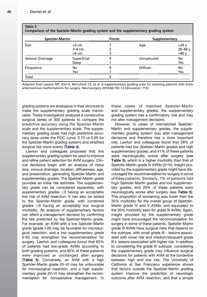
Table 3Comparison of the Spetzler-Martin grading system and the supplementary grading system
Spetzler-Martin Points Supplementary
Size <3 cm 1 Age <20 y3–6 cm 2 20–40 y>6 cm 3 >40 y
Venous Drainage Superficial 0 Unruptured NoDeep 1 Yes
Eloquence No 0 Diffuse NoYes 1 Yes
Total 5
Adapted from Lawton MT, Kim H, McCulloch CE, et al. A supplementary grading scale for selecting patients with brainarteriovenous malformations for surgery. Neurosurgery 2010;66:702–13 [discussion: 713].
Davies et al48
grading systems are analogous in their structure tomake the supplementary grading scale memo-rable. These investigators analyzed a consecutivesurgical series of 300 patients to compare thepredictive accuracy using the Spetzler-Martinscale and the supplementary scale. The supple-mentary grading scale had high predictive accu-racy (area under the ROC curve, 0.73 vs 0.65 forthe Spetzler-Martin grading system) and stratifiedsurgical risk more evenly (Table 4).Lawton and colleagues proposed that this
supplementary grading system be used to improveand refine patient selection for AVM surgery. Clin-ical decisions begin with an analysis of nidussize, venous drainage, location, diffuseness, age,and presentation, generating Spetzler-Martin andsupplementary grades. The Spetzler-Martin gradeprovides an initial risk estimate. The supplemen-tary grade can be considered separately, withsupplementary grades �3 having an acceptablylow risk of AVM resection. It can also be addedto the Spetzler-Martin grade, with combinedgrades �6 having an acceptably low surgicalmorbidity. An analysis of supplementary factorscan affect a management decision by confirmingthe risk predicted by the Spetzler-Martin grade.For example, an AVM with a low Spetzler-Martingrade (grade I–III) may be favorable for microsur-gical resection, and a low supplementary grade(I–III) may strengthen the recommendation forsurgery. Lawton and colleagues found that 62%of patients had low-grade AVMs according toboth grading systems, and 85% of these patientswere improved or unchanged after surgery(Table 5). Conversely, an AVM with a highSpetzler-Martin grade (IV–V) may be unfavorablefor microsurgical resection, and a high supple-mentary grade (IV–V) may strengthen the recom-mendation for nonoperative management. In
these cases of matched Spetzler-Martinand supplementary grades, the supplementarygrading system has a confirmatory role and maynot alter management decisions.However, in cases of mismatched Spetzler-
Martin and supplementary grades, the supple-mentary grading system may alter managementdecisions and therefore has a more importantrole. Lawton and colleagues found that 28% ofpatients had low Spetzler-Martin grades and highsupplementary grades, and 41% of these patientswere neurologically worse after surgery (seeTable 5), which is a higher morbidity than that ofSpetzler-Martin grade IV AVMs (31%). Insight pro-vided by the supplementary grade might have dis-couraged the recommendation for surgery in someof these patients. Similarly, 7% of patients hadhigh Spetzler-Martin grades and low supplemen-tary grades, and 29% of these patients wereneurologically worse after surgery (see Table 5).This proportion of worsening was lower than the35% morbidity for the overall group of Spetzler-Martin grade IV and V AVMs, and equivalent tothe 30% morbidity seen for grade III AVMs. Again,insight provided by the supplementary grademight have encouraged the recommendation forsurgery in some of these patients. Spetzler-Martingrade III AVMs have surgical risks that depend onthe subtype, with small grade III� lesions associ-ated with lower risk and medium/eloquent gradeIII1 lesions associated with higher risk. In additionto considering the grade III subtype, consideringthe supplementary grade may influence surgicaldecisions for patients with AVM at the borderlinebetween high and low risk. The University ofCalifornia at San Francisco experience showsthat factors outside the Spetzler-Martin gradingsystem improve the prediction of neurologicoutcome after AVM resection, and that a simple
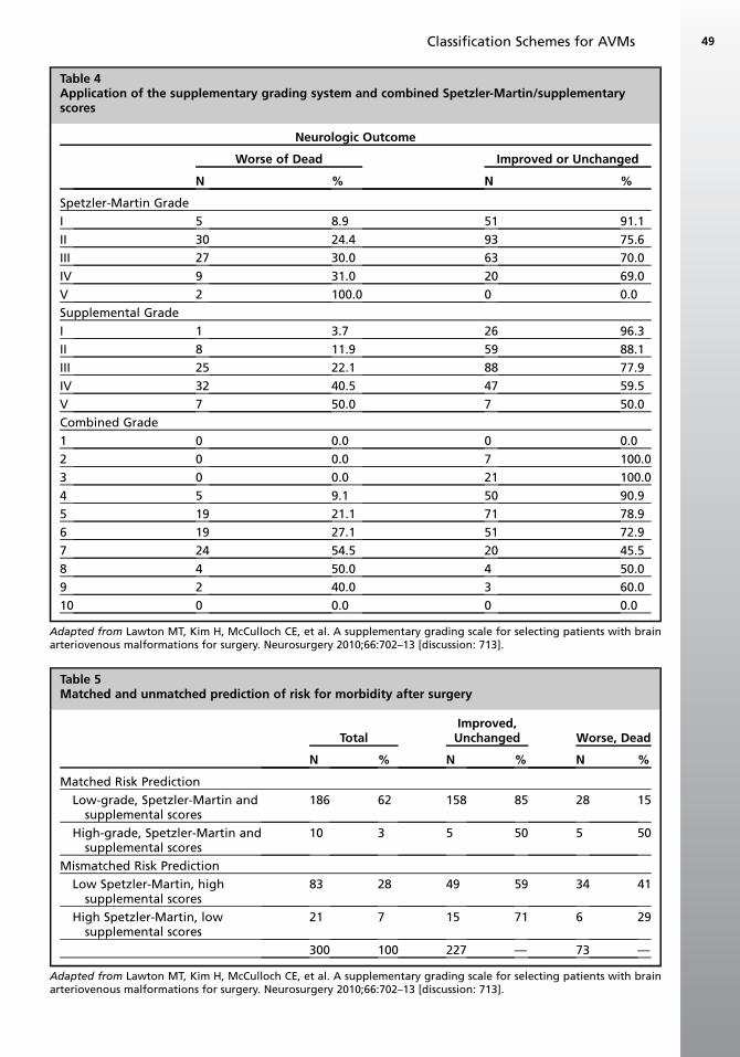
Table 4Application of the supplementary grading system and combined Spetzler-Martin/supplementaryscores
Neurologic Outcome
Worse of Dead Improved or Unchanged
N % N %
Spetzler-Martin Grade
I 5 8.9 51 91.1
II 30 24.4 93 75.6
III 27 30.0 63 70.0
IV 9 31.0 20 69.0
V 2 100.0 0 0.0
Supplemental Grade
I 1 3.7 26 96.3
II 8 11.9 59 88.1
III 25 22.1 88 77.9
IV 32 40.5 47 59.5
V 7 50.0 7 50.0
Combined Grade
1 0 0.0 0 0.0
2 0 0.0 7 100.0
3 0 0.0 21 100.0
4 5 9.1 50 90.9
5 19 21.1 71 78.9
6 19 27.1 51 72.9
7 24 54.5 20 45.5
8 4 50.0 4 50.0
9 2 40.0 3 60.0
10 0 0.0 0 0.0
Adapted from Lawton MT, Kim H, McCulloch CE, et al. A supplementary grading scale for selecting patients with brainarteriovenous malformations for surgery. Neurosurgery 2010;66:702–13 [discussion: 713].
Table 5Matched and unmatched prediction of risk for morbidity after surgery
TotalImproved,Unchanged Worse, Dead
N % N % N %
Matched Risk Prediction
Low-grade, Spetzler-Martin andsupplemental scores
186 62 158 85 28 15
High-grade, Spetzler-Martin andsupplemental scores
10 3 5 50 5 50
Mismatched Risk Prediction
Low Spetzler-Martin, highsupplemental scores
83 28 49 59 34 41
High Spetzler-Martin, lowsupplemental scores
21 7 15 71 6 29
300 100 227 — 73 —
Adapted from Lawton MT, Kim H, McCulloch CE, et al. A supplementary grading scale for selecting patients with brainarteriovenous malformations for surgery. Neurosurgery 2010;66:702–13 [discussion: 713].
Classification Schemes for AVMs 49

Davies et al50
supplementary grading system can be easilyapplied at the bedside to refine patient selectionfor AVM surgery.
NATURAL HISTORY RISK PREDICTION
Neurosurgeons have focused on the prediction ofsurgical risk with classification schemes to helpthem recommend AVM resection. However, thenatural alternative to AVM resection is observa-tion, which is particularly important with high-grade AVMs associated with high surgical risks.Therefore, predicting the natural history risk ofan AVM is as important in the clinical decision aspredicting the surgical risk. An accurate under-standing of the hemorrhagic behavior of untreatedAVMs is essential when comparing conservativeand surgical options.Ondra and colleagues22 examined prospec-
tively a cohort of 160 patients with AVM managednonoperatively over a 33-year period in Finland,drawn from a population that included more than90% of patients with AVM in that country. Thesepatients presented with hemorrhage (71%),seizures without evidence of hemorrhage (24%),or other symptoms including headaches, asymp-tomatic bruits, or other neurologic complaints(5%). The annual rate of AVM hemorrhage was4%, regardless of presentation and constantover a mean follow-up of 23.7 years. The mortalitywas 1% annually and the combined majormorbidity/mortality was 2.7% annually. The meaninterval between presentation and subsequenthemorrhagic events was 7.7 years.Other reports in the literature estimate the
annual risk of AVM hemorrhage to be 2% to 4%(Table 6).22–26 The New York Islands AVMHemorrhage Study,27,28 a prospective survey ofa zip-code–defined population of nearly 10 millioninhabitants, found an AVM detection rate of 1.21/100,000 person-years, with incidence of hemor-rhage being 0.42/100,000 person-years. The
Table 6Summary of natural history studies for arteriovenou
Author Study Design Cases
Graf et al,23 1983 Retrospective 71
Crawford et al,24 1986 Retrospective 217
Brown et al,25 1988 Retrospective 168
Ondra et al,22 1990 Retrospective 160
Mast et al,26 1997 Prospective 139
Adapted from http://www.neurosurgery.ufl.edu/residency/ima
Northern Manhattan Stroke Study29,30 found theincidence of first-ever AVM hemorrhage to be0.55/100,000 person-years. These studies offerinsight into the clinical and anatomic factors thatinfluence AVM natural history. Data from ColumbiaAVM Databank31 showed that deep brain location,exclusively deep venous drainage, and presenta-tion with AVM hemorrhage were all associatedwith increased risk of new hemorrhage. Thehemorrhage risks associated with these factorswere additive, with an average annual rupturerate that ranged from 0.9% with no risk factorsto 34.4% with all 3. In addition, associated arterialaneurysms increased the risk of hemorrhage, witha relative risk of 2.28 for patients with intranidalaneurysms and 1.88 for patients with feedingartery aneurysms.32 Stapf and colleagues also re-ported that borderzone AVMs fed by at least 2arteries from the circle of Willis had a decreasedrisk of hemorrhage (27% vs 60%, respectively).Based on a cohort of 705 patients with AVM, Natafand colleagues33 identified similar angiographicfactors associated with hemorrhage risk: venousrecruitment, venous stenosis, venous reflux,deep venous drainage, and intranidal or juxtanidalaneurysms. A classification scheme incorporatingthese factors showed increasing hemorrhage riskwith grade.Despite these insights, most estimates of
untreated AVM rupture risk are crude at best.There are statistical estimates based on the multi-plicative law of probability for the lifetime riskof hemorrhage34: Rupture risk 5 1 – (risk of nohemorrhage)(life expectancy). This formula assumesa constant yearly risk of hemorrhage with indepen-dent behavior over all years. This formulaicapproach yields an estimate lifetime hemorrhagerisk, but is not a practical bedside tool. Simplifiedformulas are more practical and popular: rupturerisk5 105 � patient age.34,35 The use of simplifiedformulas and the lack of an accepted naturalhistory grading system show that efforts to define
s malformation rupture
Follow-up Term (y) Hemorrhage Rate (%/y)
4.8 4.1
10.4 3.4
8.2 2.3
23.7 1.7
1.0 2.2
ges/Vascular_Malformations.pdf.
Classification Schemes for AVMs 51
the risks of observation have lagged behind effortsto define the surgical risks. The importance of sucha natural history grading system is clear. As theunderlying biology of AVMs and AVM rupture iselucidated, this system will likely include the clin-ical and anatomic factors discussed earlier butalso genetic and hemodynamic factors that influ-ence the ongoing inflammation and angiogenesisinside an AVM nidus.
CLASSIFICATION SCHEMES FORNONSURGICAL THERAPIES
AVMs are treated by other modalities besidessurgery, and with multimodality strategies. Classi-fication schemes predicting surgical risk saynothing about the risks of these alternative thera-pies. However, the usefulness of surgical gradingsystems provided impetus to develop similar ra-diosurgical and endovascular grading systems.
The factors that are important to AVM oblitera-tion by stereotactic radiosurgery are differentfrom the factors in the Spetzler-Martin gradingsystem. Inoue and colleagues36 identified size,morphology, and hemodynamics as factors mostpredictive of radiosurgical outcome. The investi-gators classified AVM hemodynamics as Moya-type (small-caliber feeding arteries, compactnidus, and veins that drain in the venous phaseof the angiogram), shunt type (large-caliberfeeding arteries, indistinct nidus, and veins thatdrain early in the arterial phase), or mixed. AVMmorphology was classified as homogeneousor heterogeneous. Radiosurgical results in 30patients showed that small, homogeneous,Moya-type AVMs had the best obliteration rates,and that other AVMs with shunt and mixedhemodynamics benefit from preradiosurgicalembolization.
Pollock and Flickinger37 developed a morequantitative radiosurgical classification schemecalled the radiosurgery-based AVM gradingsystem. Multivariate analysis of data from 220patients identified 5 variables associated with ra-diosurgical success, defined as complete nidusobliteration without new or worsening neurologicdeficit: AVM volume, age, location, previousembolization, and number of draining veins. Thelatter 2 factors added little to predictive accuracyand were omitted from the final equation foroutcome: AVM score 5 0.1 � (volume in cm3) 10.02 � (patient age in years) 1 0.3 � (location oflesion (frontal or temporal 5 0; parietal, occipital,intraventricular, corpus callosum, or cerebellar5 1;and basal ganglia, thalamic, or brainstem 5 2)).Patients with a composite score of less than 1had excellent outcomes, whereas only 39% of
patients with scores greater than 2 had excellentoutcomes. The predictive equation was validatedat a different institution using a separate patientcohort of 136 patients. To reduce the complexityof the scheme, the location variable was modifiedto be 2-tiered, with lesions in the hemispheres,corpus callosum, and cerebellum assigned0 points, and those in the basal ganglia, thalamus,and brain stem assigned 1 point.38 This modifiedequation was validated on an additional 247patients. Although this grading system was devel-oped at a center using exclusively Gamma Knife(Elekta AB, Sweden) radiosurgery, it has been vali-dated at linear accelerator-based centers aswell.39 The scale was based on outcome data aftera single radiosurgery procedure. Radiosurgery hasa long latency period, and many radiosurgicalpatients require repeat radiosurgery or surgery toachieve complete obliteration of the lesion. There-fore, outcome data used for radiosurgical gradingscales require diligent follow-up.
Endovascular embolization of AVMs has longbeen a surgical adjunct rather than a curativetherapy. Consequently, there have been fewattempts to create an endovascular classificationscheme. TheViennagroup40producedaclassifica-tion based on AVM size, number of feedingarteries, and pial versus perforating feedingarteries. Low-grade AVMs that were small, hadfewer than2 feedingarteries, andwerenot suppliedby perforators were suitable for endovasculartherapy, whereas high-grade AVMs that were large(>4 cm), hadmore than 4 feeding arteries, andweresuppliedbyperforatorswere not suitable for endo-vascular therapy. Improved embolic agents likeOnyx (ev3 Endovascular, Inc, Irvine, CA, USA) andmore navigable microcatheters have increasedthe rates of curative AVM embolization. As tech-nology and techniques evolve and obliterationrates increase, there will undoubtedly be additionalclassification schemes that will help select patientsfor curative endovascular intervention.
SUMMARY
The decision to treat a patient’s AVM is a complexprocess that must consider the individual’s clinicalpresentation, anatomy, and biology. A treatmentstrategy is conceived that may involve 1 or multiplemodalities, and the cumulative risks of the overalltreatment strategy are estimated. Alternative treat-ments and strategies are also conceived andanalyzed for risk. Grading systems are useful inmaking these estimations. The grading systemsthat stand the test of time are simple, applicable,and accurate. Other grading systems that arecomplex, cumbersome, or inaccurate are quickly

Davies et al52
discarded. Final recommendations are derived bycomparing the therapeutic risks with the naturalhistory risks. It is important to not only individualizerecommendations for a particular patient but alsoto individualize grading systems for a particularneurosurgeon and a particular institution. Thereis no value in quoting published results if they differfrom one’s own personal results at one’s own insti-tution. Ultimately, grading systems enable clini-cians to make management recommendations,but patients have the final say. The decision toembark on AVM treatment is difficult and fright-ening to most patients, and their individual prefer-ences and feelings should dictate the finaldecision. Guiding patients through this decisionprocess is an art from that cannot be replacedby grades or outcome data. One of the hardestthings for neurosurgeons to accept is their limita-tions, and grading systems remind us when tosay no.The current crop of grading systems is generally
useful, but imperfect and evolving. As the patho-physiology of AVMs is elucidated throughresearch, grading systems will incorporate theseadvances with genetic information and biologicmarkers. Hemodynamic data may also becomea defining element of rupture risk and futuregrading scales. Therefore, the work of developingAVM grading systems should be viewed as anongoing process, and clinicians should be opento reshaping established, proven grading systems.
REFERENCES
1. Luessenhop AJ, Gennarelli TA. Anatomical grading
of supratentorial arteriovenous malformations for
determining operability. Neurosurgery 1977;1(1):
30–5.
2. Luessenhop AJ, Rosa L. Cerebral arteriovenous
malformations. Indications for and results of surgery,
and the role of intravascular techniques. J Neuro-
surg 1984;60(1):14–22.
3. Shi YQ, Chen XC. A proposed scheme for grading
intracranial arteriovenous malformations. J Neuro-
surg 1986;65(4):484–9.
4. Spetzler RF, Martin NA. A proposed grading system
for arteriovenous malformations. J Neurosurg 1986;
65(4):476–83.
5. Hamilton MG, Spetzler RF. The prospective appli-
cation of a grading system for arteriovenous malfor-
mations. Neurosurgery 1994;34(1):2–6 [discussion:
6–7].
6. Deruty R, Pelissou-Guyotat I, Mottolese C, et al. The
combined management of cerebral arteriovenous
malformations. Experience with 100 cases and
review of the literature. Acta Neurochir (Wien)
1993;123(3–4):101–12.
7. Heros RC, Tu YK. Is surgical therapy needed for un-
ruptured arteriovenous malformations? Neurology
1987;37(2):279–86.
8. Steinmeier R, Schramm J, Muller HG, et al. Evalua-
tion of prognostic factors in cerebral arteriovenous
malformations. Neurosurgery 1989;24(2):193–200.
9. Lawton MT. Spetzler-Martin Grade III arteriovenous
malformations: surgical results and a modification
of the grading scale. Neurosurgery 2003;52(4):
740–8 [discussion: 748–9].
10. Ponce FA, Lozano AM. Highly cited works in neuro-
surgery. Part I: the 100 top-cited papers in neurosur-
gical journals. J Neurosurg 2010;112(2):223–32.
11. Ponce FA, Lozano AM. Highly cited works in neuro-
surgery. Part II: the citation classics. J Neurosurg
2010;112(2):233–46.
12. Hartmann A, Stapf C, Hofmeister C, et al. Determi-
nants of neurological outcome after surgery for brain
arteriovenous malformation. Stroke 2000;31(10):
2361–4.
13. Heros RC, Korosue K, Diebold PM. Surgical excision
of cerebral arteriovenous malformations: late
results. Neurosurgery 1990;26(4):570–7 [discussion:
577–8].
14. Davidson AS, Morgan MK. How safe is arteriove-
nous malformation surgery? A prospective, observa-
tional study of surgery as first-line treatment for brain
arteriovenous malformations. Neurosurgery 2010;
66(3):498–504 [discussion: 504–5].
15. Spetzler RF, Ponce FA. A 3-tier classification of cere-
bral arteriovenous malformations. Clinical article.
J Neurosurg 2011;114(3):842–9.
16. Pertuiset B, Ancri D, Kinuta Y, et al. Classification of
supratentorial arteriovenous malformations. A score
system for evaluation of operability and surgical
strategy based on an analysis of 66 cases. Acta
Neurochir (Wien) 1991;110(1–2):6–16.
17. Tamaki N, Ehara K, Lin TK, et al. Cerebral arterio-
venous malformations: factors influencing the surgi-
cal difficulty and outcome. Neurosurgery 1991;
29(6):856.
18. Anon Arteriovenous malformations of the brain in
adults. N Engl J Med 1999;340(23):1812–8.
19. Hollerhage HG. Cerebral arteriovenous malforma-
tions: factors influencing surgical difficulty and
outcome. Neurosurgery 1992;31(3):604–5.
20. Hunt WE, Hess RM. Surgical risk as related to time of
intervention in the repair of intracranial aneurysms.
J Neurosurg 1968;28(1):14–20.
21. Lawton MT, Kim H, McCulloch CE, et al. A supple-
mentary grading scale for selecting patients with
brain arteriovenous malformations for surgery.
Neurosurgery 2010;66(4):702–13 [discussion: 713].
22. Ondra SL, Troupp H, George ED, et al. The natural
history of symptomatic arteriovenous malformations
of the brain: a 24-year follow-up assessment. J Neu-
rosurg 1990;73(3):387–91.

Classification Schemes for AVMs 53
23. Graf CJ, Perret GE, Torner JC. Bleeding from cere-
bral arteriovenous malformations as part of their
natural history. J Neurosurg 1983;58(3):331–7.
24. Crawford PM, West CR, Chadwick DW, et al. Arterio-
venous malformations of the brain: natural history in
unoperated patients. J Neurol Neurosurg Psychiatry
1986;49(1):1–10.
25. Brown RD, Wiebers DO, Forbes G, et al. The natural
history of unruptured intracranial arteriovenous mal-
formations. J Neurosurg 1988;68(3):352–7.
26. Mast H, Young WL, Koennecke HC, et al. Risk
of spontaneous haemorrhage after diagnosis of
cerebral arteriovenous malformation. Lancet 1997;
350(9084):1065–8.
27. Stapf C, Khaw AV, Sciacca RR, et al. Effect of age on
clinical and morphological characteristics in
patients with brain arteriovenous malformation.
Stroke 2003;34(11):2664–9.
28. Stapf C, Mast H, Sciacca RR, et al. The New York
Islands AVM Study: design, study progress, and
initial results. Stroke 2003;34(5):e29–33.
29. Stapf C, Mohr JP, Sciacca RR, et al. Incident hemor-
rhage risk of brain arteriovenous malformations
located in the arterial borderzones. Stroke 2000;
31(10):2365–8.
30. Stapf C, Labovitz DL, Sciacca RR, et al. Incidence of
adult brain arteriovenous malformation hemorrhage
in a prospective population-based stroke survey.
Cerebrovasc Dis 2002;13(1):43–6.
31. Choi JH, Mast H, Hartmann A, et al. Clinical and
morphological determinants of focal neurological
deficits in patients with unruptured brain arteriove-
nous malformation. J Neurol Sci 2009;287(1–2):
126–30.
32. Stapf C, Mohr JP, Pile-Spellman J, et al. Concurrent
arterial aneurysms in brain arteriovenous malforma-
tions with haemorrhagic presentation. J Neurol Neu-
rosurg Psychiatry 2002;73(3):294–8.
33. Nataf F, Meder JF, Ghossoub M, et al. Hemorrhage in
arteriovenous malformations: clinical and anatomic
data. Neurochirurgie 2001;47(2–3 Pt 2):158–67 [in
French].
34. Kondziolka D, McLaughlin MR, Kestle JR. Simple
risk predictions for arteriovenous malformation
hemorrhage. Neurosurgery 1995;37(5):851–5.
35. Brown RD. Simple risk predictions for arteriovenous
malformation hemorrhage. Neurosurgery 2000;46(4):
1024.
36. Inoue H, Kohga H, Kurihara H, et al. Classification of
arteriovenous malformations for radiosurgery. Ster-
eotact Funct Neurosurg 1995;64(1):110–7.
37. Pollock BE, Flickinger JC. A proposed radiosurgery-
based grading system for arteriovenous malforma-
tions. J Neurosurg 2002;96(1):79–85.
38. Pollock BE, Flickinger JC. Modification of the
radiosurgery-based arteriovenous malformation
grading system. Neurosurgery 2008;63(2):239–43
[discussion: 243].
39. Richling B, Killer M, Al-Schameri AR, et al. Therapy
of brain arteriovenous malformations: multimodality
treatment from a balanced standpoint. Neurosurgery
2006;59(5 Suppl 3):S148–57 [discussion: S3–13].
40. Vinuela F, Duckwiler T, Guglielmi G. Intravascular
embolization of brain arteriovenous malforma-
tions. In: Maciuna R, editor. Endovascular neuro-
logical intervention. Rolling Meadows (IL): The
American Association of Neurological Surgeons;
1995. p. 189–99.