classification of basal cucujoidea (coleoptera:polyphaga
TRANSCRIPT

IntroductionOf the six superfamilies in the series Cucujiformia,Cucujoidea is the most problematic because it seems to havebeen established only for convenience to contain a heteroge-nous group of families with a similar habitus (Figs 1–10).The superfamily presently consists of 32 families (Lawrenceand Newton 1995), when Languriidae and Erotylidae arecombined (Wegrzynowicz 2002; Leschen 2003) andCyclaxyridae recognised at the family level (Lawrence et al.1999b). Cucujoid workers recognise two informal groupingsin the superfamily: the cerylonid series may be a mono-phyletic lineage within the superfamily consisting of eightfamilies (Sen Gupta and Crowson 1973; Ślipiński 1990;Ślipiński and Pakaluk 1992): Bothrideridae, Cerylonidae,Discolomatidae, Coccinellidae, Endomychidae, Alexiidae,Corylophidae, and Latridiidae. The remaining members ofCucujoidea are considered part of the basal Cucujoidea (orlower Cucujoidea, Leschen 1996), a colloquial designationfor what may be the more primitive members of the group(along with enigmatic families such as Helotidae andPassandridae). Among the basal cucujoids, the familyPhloeostichidae, consisting of only a handful of genera, hasbeen controversial with regard to its monophyletic status. Inthis paper we provide a cladistic analysis of the basal groups,mainly to place the Phloeostichidae subfamilies in broaderphylogenetic context.
The family Phloeostichidae was proposed by Sen Guptaand Crowson (1969) for the European PhloeostichusRedtenbacher (Fig. 6, Cucujidae), the New ZealandAgapytho Broun (Fig. 4, Salpingidae), the AustralianHymaea Pascoe (Fig. 5), originally included inTenebrionidae but made the type of the boganiid subfamilyHymaeinae by Sen Gupta and Crowson (1966), and theChilean Rhopalobrachium Boheman (Fig. 8), which hadbeen placed in Oedemeridae or Pythidae. Crowson (1973)added the subfamily Priasilphinae comprised of the NewZealand Priasilpha Broun (Fig. 1) placed by Broun (1893)and Grouvelle (1913) in Nitidulidae, and a newly describedTasmanian genus Priastichus Crowson (Fig. 2). In this samepaper, Crowson (1973: 55) admitted that the phloeostichidsubfamilies Phloeostichinae, Hymaeinae, Agapythinae andPriasilphinae ‘would be better treated as independent fami-lies, with individual affinities to other clavicorn families’.Lawrence (1988) added two other genera to the family basedin part on discussions with Crowson: Tasmosalpingus Lea(1919, Fig. 10) from Salpingidae and Myrabolia Reitter(1876, Fig. 9) from Silvanidae (Blackburn 1903). Lawrenceand Britton (1991) proposed two additional subfamilies,Myraboliinae and Tasmosalpinginae, for their inclusion, andLawrence (1995) added an Australian and Chilean species tothe formerly monotypic Rhopalobrachium. In the past fewyears an additional two genera have been discovered which
Invertebrate Systematics, 2005, 19, 17–73
10.1071/IS04007 1445-5226/05/010017© CSIRO 2005
R. A. B. LeschenA,C, J. F. LawrenceB and S. A.ŚlipińskiB
ANew Zealand Arthropod Collection, Private Bag 92170, Auckland, New Zealand.BCSIRO Entomology, GPO Box 1700, Canberra, ACT, 2601, Australia.
CCorresponding author. Email: [email protected]
Abstract. Phylogenetic relationships among the basal Cucujoidea were reconstructed by a cladistic analysis of adata matrix consisting of 37 exemplar taxa and 99 adult and larval characters. Eight most parsimonious cladogramsprovided evidence for the polyphyly of Phloeostichidae, the paraphyly of Cucujoidea (with respect to the placementof Trogossitidae), and the monophyly of Protocucujidae + Sphindidae, Biphyllidae + Erotylidae, Cryptophagidae,Cucujidae + Silvanidae, Propalticidae + Laemophloeidae, and the Nitidulidae groups (Nitidulidae, Smicripidae, andBrachypteridae). The following families are elevated from subfamily to family status: Agapythidae (one genus),Phloeostichidae (four genera; the subfamilies Phloeostichinae and Hymaeinae are supressed), Priasilphidae (threegenera), Tasmosalpingidae (one genus), and Myraboliidae (one genus). These families are described in detail andadult and larval keys to all families of Cucujoidea are provided. The genus Bunyastichus, gen. nov. (type species:B. monteithi, sp. nov.) is described in the family Phloeostichidae and the family Priasilphidae is revised with thefollowing new taxa: Chileosilpha, gen. nov. (type species: C. elguetai, sp. nov.), Priasilpha (P. angulata, sp. nov.,P. aucklandica, sp. nov., P. bufonia, sp. nov., P. carinata, sp. nov., P. earlyi, sp. nov., and P. embersoni, sp. nov.),Priastichus (P. crowsoni, sp. nov. and P. megathorax, sp. nov.).
Classification of basal Cucujoidea (Coleoptera:Polyphaga):cladistic analysis, keys and review of new families
www.publish.csiro.au/journals/is
CSIRO PUBLISHING

R. A. B. Leschen et al.18 Invertebrate Systematics
Figs 1–4. Dorsal views of adult Priasilphidae and Agapythidae: 1, Priasilpha obscura; 2, Priastichusmegathorax, sp. nov. (length = 3.6 mm); 3, Chileosilpha elguetai, sp. nov.; 4, Agapytho foveicollis. Scalebars = 1 mm.

Invertebrate Systematics 19
belong in this complex: one from Chile related to thePriasilphinae and one from Australia related to thePhloeostichinae and Hymaeinae; in addition several newspecies have been discovered in the genera Priasilpha andPriastichus. These are described in the present paper.
Although the family Phloeostichidae appears to be a taxonof convenience, consisting mainly of monogeneric Notogeansubfamilies with a suite of basal cucujoid features, it is not atall clear which of these taxa should be elevated to family rank
and which transferred to other known families. In order toshed some light on this problem we have examined 37exemplar genera, including all eight currently included inPhloeostichidae, the two new genera described below, andmembers of all remaining families of basal Cucujoidea. Basedon this analysis we revise the classification for Cucujoidea,review the families Priasilphidae, Phloeostichidae, Aga-pythidae, Tasmosalpingidae and Myraboliidae, and providekeys to adults and larvae of all cucujoid families.
Classification of Cucujoidea
Figs 5–10. Dorsal views of adult basal Cucujoidea. 5, Hymaea magna (length = 4.5 mm); 6, Phloeostichus denticollis (length = 5.0 mm);7, Bunyastichus monteithi, sp. nov. (length = 3.6 mm); 8, Rhopalobrachium penai (length = 11.0 mm); 9, Myrabolia sp. (length = 4.3 mm);10, Tasmosalpingus sp. (length = 1.8 mm).

R. A. B. Leschen et al.20 Invertebrate Systematics
Materials and methods
Terms and measurements
The terms mesoventrite and metaventrite used in keys, descriptions andcharacter discussions were coined by Lawrence (1999) and Lawrenceet al. (1999b) to replace the misapplied terms mesosternum and meta-sternum (see also Campau 1940; Ferris 1940; Beutel and Haas 2000).Wing vein terminology is from Kukalová-Peck and Lawrence (2004);specifically the medial field refers to the area of wing membranebetween the medial bar (posterior wing strut or MP1+2) and the analfold, thus excluding AP3+4; the number of free veins in this field isequivalent to Crowson’s term ‘anal veins in the main group’. The termdecumbent is used for setae that are erect basally but strongly recurvedat apex, and recumbent refers to setae that are strongly declined at baseand more or less adpressed to the surface. The terms proximal gono-coxite and distal gonocoxite are those used by Burmeister (1976, 1980),whereas the terms paraproct (= hemitergite IX or laterotergite IX) andproctiger (tergite X) are taken from Tanner (1927). The terms baculumand bacula are used for the longitudinal supporting struts on each sideof the proctiger and on the ventral or mesal edge of each paraproct andto the transverse or oblique strut at the base of the proximal gonocoxite.Measurements: PL, median length of pronotum; PW, greatest width ofpronotum; EL, length of the elytra along suture; EW, combined greatestwidths of elytra; BL, body length (excluding head) or PL + EL; GD,greatest depth (metaventrite to basal third of elytra).
Sources of material
Abbreviations for collections used in the text are as follows:
AA Albert Allen Collection, Boise, Idaho (private)ANIC Australian National Insect Collection, CSIRO
Entomology, CanberraAMNZ Auckland War Memorial Museum, Auckland (John
Early, Stephen Thorpe)CAS California Academy of Sciences, San Francisco
(Roberta Brett)FMNH Field Museum of Natural History, Chicago (Al Newton,
Phil Parillo)JNIC John Nunn Insect Collection, Dunedin, New Zealand
(private)LUNZ Lincoln University Insect Museum, Lincoln, New
Zealand (John Marris)MCZH Museum of Comparative Zoology, Harvard University,
Cambridge (Phil Perkins)MHNS Museo Nacional de Historia Natural, Santiago (Mario
Elgueta)MV Museum of Victoria, Melbourne: (Ken Walker, Catriona
McPhee)NHML The Natural History Museum, London (Max Barclay)NMNH National Museum of Natural History, Smithsonian
Institution, Washington, D.C. (Steve Lingafelter)NZAC New Zealand Arthropod Collection, Auckland
QM Queensland Museum, Brisbane (Geoff Monteith)SAM South Australian Museum, Adelaide (Eric Matthews)
TUPC Teruhisa Ueno Personal Collection, Kyushu University,Japan (private)
New Zealand localities are arranged according to the two-letter areacodes proposed by Crosby et al. (1998). For specimens of Priasilpha,longitude and latitude positions have been added in parentheses to theoriginal label data.
Terminal taxa used in cladistic analysis
The phylogenetic analysis was prompted by the uncertain relationshipsof Phloeostichidae to other members of the basal Cucujoidea, though
the phylogenetic problem relates to the larger question of monophyly ofCucujoidea and relationships of some cucujoid families to other super-families, namely Cleroidea. A more extensive study than the presentone is necessary for ascertaining whether the basal Cucujoidea orcerylonid series are monophyletic; here we are concerned mainly withthe controversial taxa of Phloeostichidae and the monophyletic status ofits subfamilies, and members of the cerylonid series were not included.Instead of members of the cerylonid series, we decided to include moreprimitive cucujiform taxa representing Bostrichiformia (Derodontidae)and Cleroidae (Trogossitidae) to focus attention on the problems in thebasal Cucujoidea (see below).
Cucujoidea is characterised by several features (e.g. presence of acucujoid aedeagus and pygopod-like 10th abdominal segment of thelarva). These and other characters aid in distinguishing the group fromTenebrionoidea (Lawrence and Britton 1991) but cannot count assynapomorphies because they occur in other superfamilies.Futhermore, the ring-like tegmen (Fig. 31) may be a plesiomorphic con-dition transformed to character states seen in cleroids, cucujoids, andtenebrionoids (Crowson 1955; Lawrence and Leschen 2003). Crowson(1955) listed several other characters, but admitted that none of thecharacters he provided were reliable estimates for the monophyly of thesuperfamily.
To resolve the relationships of basal cucujoids, we use an exemplarapproach to taxon sampling (Yeates 1995) because such morphologicalstudies of a subset of taxa can lead to more robust hypotheses aboutphylogenetic relationship (Bremer et al. 1999). This is also an impor-tant consideration in morphological studies where the ratio of thenumber of informative characters to terminal taxa may be quite lowcompared to molecular studies. In most cases we use a single species torepresent genera as terminals (Wiens 1998), and selected 37 key taxathat are rather generalised and represent typical members of theirfamilies (Appendix 1). All genera of Phloeostichidae are representedand the family Passandridae is represented by two taxa: the larva ofAulonosoma is unknown to us and we used Ancistria to score larvalcharacters. Though most of the species of Priasilpha are wingless, wecoded the only winged species, P. obscura Broun, for adult and larvalcharacters (this species is also a plesiomorphic member of the genus,R. A. B. Leschen and B. Michaux, personal communication). Thescoring of larval characters was based mainly on specimens associatedwith adults in the same habitats, often confirmed by subsequent associ-ations or by a process of elimination in limited faunas. Two genera rep-resenting Cryptophagidae were included in the analysis to test themonophyly of this family because of the differences betweenAtomariinae and Cryptophaginae, and especially of the similaritiesbetween the former subfamily and Hobartiidae (Leschen 1996). Thoughwe coded characters based on the key taxa listed in Appendix 1, we con-firmed the characters and their states in dissected and whole specimensof other taxa available in our collections to clarify the coding ofambiguous characters that were difficult to interpret. To root the trees,we chose two members of the cleroid family Trogossitidae because ofthe similarities of this family to other members of Cucujoidea(Lawrence and Newton 1995; Lawrence and Leschen 2003). As a moredistant outgroup to root trees, we coded Derodontidae (Derodontus), abasal Polyphagan. Derodontidae was chosen as a distant outgroupbecause Caterino et al. (2002) in their DNA analysis showed thatLaricobius (Derodontidae) and Eucinetus (Eucinetidae) are the twomost basal polyphagan clades, whereas Lawrence (1999, 2001) notedthat a membranous joint between the meso- and metathorax, a basalcoleopteran feature by Beutel and Haas (2000), also occurs inDerodontidae, Eucinetoidea and some Staphylinoidea.
Characters
In total, 99 characters were coded and consisted of 66 adult and 33larval characters (Table 1). Larvae of Bunyastichus, gen. nov. and

Invertebrate Systematics 21Classification of Cucujoidea
Table 1. Character state definitions
Adult characters(1) Postocular constriction: (1) absent (Fig. 24); (2) present (Fig. 65).
This constriction of the head may be located immediately behind the eyes or well behind them and it may be abrupt or more gradual, so thatthe temples so formed vary in length and may be more or less rounded to sharply angulate. In some cases the constriction is clearlydemarcated in ventral view but only slightly indicated from above.
(2) Vertexal line: (1) absent (Figs 24, 66); (2) present (Fig. 111).This refers to a relatively sharp transverse line or carina extending across the dorsal part of the head behind the eyes and often abutting the
anterior edge of the pronotum (Leschen 1996, 2003). Lawrence et al. (1999b) referred to this as a transverse occipital ridge.(3) Median occipital stridulatory file: (1) absent (Figs 24, 66); (2) present.
This refers to a single, broad median file lying in front of the occipital foramen. This type of stridulatory file, which occurs in Myraboliaand Tasmosalpingus, and is probably not homologous to the paired files known in several Erotylidae (including Pharaxonotha, seeFig. 111) or in Atomaria among the Cryptophagidae.
(4) Paired occipital incisions: (1) absent (Fig. 66); (2) present (Fig. 24).These paired incisions occur along the dorsal edge of the occipital foramen, thus defining a rounded lobe between them.
(5) Frontoclypeal suture: (1) present; (2) absent.The frontoclypeal or epistomal suture is coded as present only when it is delimited by a clearly impressed line and indicated internally by an
epistomal ridge. When the frontoclypeal region is crossed by a vague impression, it is coded as lacking the suture.(6) Antennal insertions: (1) exposed from above (Fig. 24); (2) concealed by frontal ridge (Fig. 66).
The character states are not very clearly defined in many cucujoid taxa in which the insertions may be barely exposed or barely concealed.A taxon is coded as having concealed insertions when in a dissected specimen with the antenna removed, no portion of the antennalsocket may be seen from directly above.
(7) Subantennal groove: (1) absent (Fig. 65); (2) not extending below or behind eye; (3) extending below or behind eye.This groove lies between the eye and the mandibular and maxillary articulations, and houses the basal antennomere(s); it usually does not
extend beyond the lower edge of the eye, but in some groups it may continue posteriorly behind the eye.(8) Genal projection: (1) absent; (2) anterior, truncate or rounded; (3) anterior, acute (Fig. 25); (4) anterolateral.
That portion of the head capsule just laterad of the maxillary articulation may project anteriorly or anterolaterally and the shape is that asseen from above and not from a lateral perspective.
(9) Anterior cervical sclerites: (1) not contiguous with head capsule or placed in paired emarginations; (2) contiguous with head capsule andusually placed within paired emarginations on ventral edge of occipital foramen (Fig. 25); (3) apparently absent.
The anterior cervical sclerites occur in many basal cucujoids, although they may be quite small and lightly sclerotised. They are usuallyloosely attached to the head capsule and lie in the neck membrane; they may or may not be accompanied by a pair of posterior cervicalsclerites (often remaining attached to the thorax in dissections). In several basal cucujoids, these anterior sclerites are more closelyassociated with the ventral portion of the head capsule, more or less abutting the cuticle and usually lying in emarginations on either sideof the gula.
(10) Apex of mandible: (1) unidentate; (2) bidentate (Fig. 97); (3) tridentate.This character may be difficult to code when the apical teeth are on the same plane and when smaller, subapical teeth are also present on the
cutting edge of the mandible. If in a mandible with three more or less apical teeth, the middle tooth is longest and those on either sideshorter or somewhat subapical, then the mandible is coded as tridentate (3). If, on the other hand, two adjacent teeth are more or lesscoplanar and of a similar size, whereas a third tooth is smaller and more subapical, then this is bidentate (2).
(11) Dorsal surface of mandible: (1) without tubercle fitting into lateral clypeal emargination (Fig. 98); (2) with tubercle fitting into lateralclypeal emargination (Fig. 112).
This tubercle or rounded ridge-like structure projects mesally and fits into an emargination at the base of the clypeus; it usually lies aboveand may partly overlap the mandibular cavity (see following character).
(12) Dorsal surface of mandible: (1) without cavity (Fig. 98); (2) with glabrous cavity; (3) with setose cavity. The area mesad of the dorsal mandibular tubercle may be broadly concave, but this does not constitute a mandibular cavity, which is either
deep and well defined (state 1) or densely lined with setae (state 2). The setose cavity is restricted to members of the families Sphindidaeand Boganiidae, whereas a deep, glabrous one occurs in Hymaeinae and Silvanidae (reduced and lost in most Silvaninae).
(13) Mandibular mola: (1) present (Fig. 98); (2) absent.(14) Galea: (1) at least 2.5× as wide as lacinia; (2) between 1× and 2.5× as wide as lacinia (Figs 68, 100); (3) distinctly narrower than lacinia;
(4) absent.(15) Lacinial uncus: (1) absent; (2) present (Figs 68, 111).
This structure, which is usually bidentate or occasionally tridentate, is located at the apicolateral angle of the lacinia and curves mesally;it may be difficult to distinguish from stout spines lying adjacent to it and below it.
(16) Lateral pronotal carinae: (1) complete (Figs 1, 2); (2) incomplete or absent (Figs 4, 8).(17) Sides of pronotum: (1) without 4–6 sharp teeth (Figs 1, 2); (2) with 4–6 sharp teeth (Figs 6, 72).
The sharp teeth include those at the anterior and posterior angles. These teeth face laterally in most taxa but are curved posteriorly inDerodontus. Among those taxa coded as having state 0, Cryptophagus may have a single tooth in addition to the anterior callosity, but thisvaries in the genus. Other genera may have blunt lobe (Rhopalobrachium), or more numerous fine tubercles or serrations (several genera).
(18) Anterior pronotal angles: (1) absent or not produced forwards (Figs 3, 4); (2) produced forwards (Figs 1, 2).(19) Anterior portion of prosternum at midline: (1) longer than prosternal process (Fig. 26); (2) as long as as prosternal process (Fig. 72);
(3) shorter than prosternal process.
(continued next page)

R. A. B. Leschen et al.22 Invertebrate Systematics
Table 1. (continued)
(20) Anteromesal corner of hypomeron: (1) without tooth; (2) with tooth (Fig. 102).The presence of a tooth occurs in the genera Chileosilpha and Priasilpha.
(21) Apex of prosternal process: (1) without lateral projections; (2) with lateral projections (Fig. 26, 72).The presence of lateral processes are very widespread in basal Cucujoidea, and these may also form a closer of the procoxal cavity when
joined with the postcoxal projections of the hypomeron, a character present in many higher Erotylidae.(22) Shortest distance between procoxal cavities: (1) less than half as great as mid length of cavity (Fig. 72); (2) more than half as great but less
than mid length of cavity; (3) more than mid length of cavity (Fig. 26).(23) Notosternal suture: (1) complete (Fig. 72); (2) incomplete (Fig. 26).(24) Procoxa: (1) without or with short, concealed lateral extension (Figs 26, 72); (2) with long, concealed lateral extension.
An internal coxal extension is said to be long if it is at least 0.75× the length of the exposed portion of the coxa.(25) Protrochantin: (1) exposed (Figs 26, 72); (2) concealed.
The protrochantin can be concealed from ventral view in taxa with concealed lateral extensions of the procoxae (C24–2) or thos taxa havingmore or less rounded procoxal cavities (C26–3), but these three characters do not co-occur and they are treated separately in this study.
(26) Procoxal cavity: (1) strongly transverse (Fig. 26); (2) slightly transverse; (3) about as long as wide.(27) Procoxal cavities externally: (1) open; (2) closed (Fig. 26).(28) Procoxal cavity: (1) without lateral notch, only slightly or gradually narrowed laterally (Fig. 26); (2) with narrow lateral notch (less than
0.25× as wide as mid lenth of cavity).(29) Procoxal cavity: (1) internally open (Fig. 26, 72); (2) internally closed.(30) Elytral punctation: (1) not seriate or striate (Fig. 1); (2) seriate or striate (Fig. 6).(31) Scutellary striole: (1) absent; (2) present.(32) Elytral sutural flange: (1) not widened apically (Fig. 2); (1) widened apically (Fig. 3).
This character is sometimes referred to as a subapical gape (see Leschen 1996).(33) Epipleuron: (1) complete to apex; (2) incomplete or absent.
The epipleural is variable in Cucujoidea, and in many taxa the structure may have the same width throughout its length, but in many taxascored in this study the anterior portion is relatively wide usually up to a level of the posterior edge of the metaventrite.
(34) Anteromesal corner of mesepisternum: (1) without cuticular pocket; (2) with cuticular pocket (Fig. 113). The cuticular pocket is quite variable in the groups that have them (Erotylidae and Cryptophagidae) and may be present as fairly deep fovea
containing setae or a rather shallow impression (Leschen 2003).(35) Shortest distance between mesocoxal cavities: (1) less than half as great as shortest diameter of cavity (Fig. 75); (2) more than half as great
but less than shortest diameter of cavity; (3) more than shortest diameter of cavity.(36) Meso-metaventral junction: (1) dicondylic; 1) monocondylic (Fig. 75); (3) simple.
The meso-metaventral articulation is variable in Cucujoidea. A monocondylic form has a single ball-like process arising from themetaventrite that fits into a corresponding fossa located on the posterior margin of the mesoventrite, whereas a dicondylic form is onethat has two processes The simple or straight-line type lacks well developed anterior processes.
(37) Mesocoxal cavities laterally: (1) open (partly closed by mesepimeron) (Fig. 75); (2) closed (by meeting of mesoventrite and metaventrite).Occasionally the mesoventrite and metaventrite do not quite meet, but the mesepimeron does not extend between them and is well removed
from the coxa and trochantin; in this case the mesocoxal cavities are considered to be laterally closed.(38) Metaventral discrimen: (1) present (Fig. 75); (2) absent.
This structure, often referred to a median or longitudinal suture, is the line of invagination of the metendosternite.(39) Metaventral transverse suture (katepisternal suture): (1) present; (2) absent (Fig. 75).
This suture is said to be present whether or not it crosses the discrimen.(40) Metacoxae: (1) contiguous or narrowly separated (Fig. 75); (2) widely separated.
Metacoxae coded as state (2) are separated by a distance at least a third as great as the metacoxal width (longest coxal diameter).(41) Metacoxae: (1) extending laterally to meet elytral epipleura, ventrite one not in contact with metepimeron; (2) not extending laterally to
meet elytral epipleura, ventrite one in contact with metepimeron (Fig. 75).(42) Metacoxal carina: (1) present; (2) absent (Fig. 75).
The metacoxal carina extends from the coxotrochanteral joint laterally and forms an anterior abutment for the femur. In many basalpolyphagans it extends to the lateral edge of the coxa and may be well developed, forming a distinct coxal plate concealing the femur inrepose. In Cucujoidea, the carina is rarely weakly developed and usually absent.
(43) Metendosternal laminae: (1) well developed; (2) reduced; (3) absent. Reduced laminae (2) tend to be slender and not broad and plate-like.(44) Anterior tendons of metendosternite: (1) narrowly separated; (2) widely separated.(45) Radial cell of hind wing: (1) complete (closed basally, Fig. 63); (2) incomplete (open basally) or absent (Fig. 62).(46) Free veins in medial field of hind wing: (1) five (Fig. 62); (2) four; (3) three or fewer.
The medial field lies between the posterior wing strut (medial bar, MP1+2) and the anal fold. There is a maximum of five free veins in thisfield (MP3, MP4+CuA1, CuA2, AA3 and AA4), but in most cucujiforms this number is reduced to four or fewer.
(47) Tarsi in male: (1) 5–5–5; (2) 5–5–4.(48) Mesotarsomere four: (1) not or slightly reduced and not enclosed within lobe on tarsomere three; (2) highly reduced and partly or entirely
enclosed within ventral lobe on tarsomere three (Fig. 73).State 1 represents the condition known as pseudotetramerous in Chrysomeloidea and Curculionoidea; however the reduction of tarsomere 4
is usually not as great in cucujoids.(49) Apex of terminal tarsomere: (1) without fringe of short setae; (2) with fringe of short setae (Fig. 105).
A fringe of modified setae is present on the terminal tarsomeres of Priasilpha and Priastichus.
(continued next page)

Invertebrate Systematics 23Classification of Cucujoidea
Table 1. (continued)
50) Empodium: (1) projecting between pretarsal claws; (2) absent or not visible between pretarsal claws (Fig. 105).In most cucujoids, the empodium is reduced and rarely extends beyond the bases of the pretarsal claws, although the empodial setae at its
apex may be visible (Fig. 105). In taxa coded as having state 0, the body of the empodium is visible between the claws.(51) Number of basal ventrites connate: (1) none; (2) two.(52) Ventrite 1: (1) not much longer than 2 (Fig. 71); (2) much longer than 2.(53) Abdominal intercoxal process: (1) acute or narrowly rounded (Fig. 71); (2) broadly rounded; angulate or truncate (Fig. 104).(54) Abdominal tergite VII: (1) concealed from above; (2) exposed from above.(55) Functional spiracles on abdominal segment VII: (1) present; (2) absent.(56) Tergite VIII in male: (1) completely dorsal; (2) with sides curved ventrally; (3) with sides and apex curved ventrally to form genital capsule.(57) Anterior edge of sternite VIII in male: (1) without median strut; (2) with median strut.(58) Apex of sternite IX in male: (1) with mesal lobe (Fig. 35); (2) without mesal lobe.(59) Anterior edge of sternite IX in male: (1) without spiculum gastrale; (2) with spiculum gastrale (Fig. 35).(60) Base of tegmen: (1) broadly rounded; (2) narrowly rounded to acute (Fig. 55); (3) produced anteriorly forming strut.(61) Parameres: (1) free from one another (Figs 28, 29); (2) fused into single piece.(62) Parameres: (1) articulated to phallobase (Figs 28, 31); (2) fused to phallobase.(63) Penis: (1) not divided into distinct basal and apical sections (Fig. 78); (2) divided into distinct basal and apical sections (Figs 29, 30).
The division into sections is a character present in only a few basal Cucujoidea, including all Priasilphinae and Agapytho.(64) Basal portion of penis: (1) distinctly wider than apical portion (Fig. 36); (2) as wide as or narrower than apical portion (Fig. 81).(65) Base of penis: (1) without median carina (Fig. 78); (2) with median carina (Fig. 41).(66) Anterior edge of penis: (1) without struts (Figs 29, 78); (2) with paired struts; (3) with single strut.
Larval characters(67) Posterior edge of head capsule: (1) not, or only slightly, emarginate (Fig. 87); (2) distinctly emarginate.(68) Bases of frontal arms: (1) contiguous; (2) well separated (Figs 79, 87).(69) Median endocarina: (1) absent (Fig. 87); (2) present.(70) Paired endocarinae: (1) absent (Fig. 87); (2) present.(71) Stemmata: (1) six; (2) five; (3) four or fewer.(72) Antennal length: (1) less than 0.15× head width; (2) 0.15–0.5× head width; (3) more than 0.5× head width (Figs 80, 87).(73) Sensorium: (1) shorter than apical antennomere (Figs 87, 80); (2) longer than apical antennomere.(74) Labrum: (1) separated from head capsule by complete suture (Fig. 87); (2) partly or completely fused to head capsule (suture incomplete or
absent).(75) Apex of mandible: (1) unidentate; (2) bidentate; (3) tridentate (Fig. 106).
This character varies among specimens of Priasilphidae that may have lost teeth due to wear (Fig. 91, Priastichus) or where a subapical lobemay be present (compare Figs 84 and 106 for Priasilpha).
(76) Accessory ventral process of mandible: (1) absent; (2) present (Fig. 90).This is the accessory ventral condyle of Böving and Craighead (1931) and it appears to be a plesiomorphic feature in basal Polyphaga and
Myxophaga (Lawrence et al. 1999a).(77) Mesal surface of mandibular base: (1) with asperate or tuberculate mola (Figs 106, 107); (2) with 1–3 hyaline processes; (3) simple.(78) Ventral mouthparts: (1) strongly retracted (distance between mandibular and maxillary articulations greater than width of stipes, Figs 80,
88); (2) protracted or slightly retracted (distance between mandibular and maxillary articulations less than width of stipes).(79) Maxillary articulating area: (1) well developed (Figs 80, 88); (2) highly reduced or absent.(80) Inner apical angle of mala or lacinia: (1) rounded or truncate; (2) more or less acute (falciform) (Figs 82, 92).
The mala in the larva of Myrabolia is weakly falcate and this has been coded as acute.(81) Number of labial palpomeres: (1) two (Figs 80, 88, 108); (2) one.(82) Labial palps: (1) contiguous or separated by less than width of first palpomere; (2) separated by more than width of first palpomere
(Figs 80, 88).(83) Ligula: (1) absent; (2) present (Fig. 93).(84) Hypopharyngeal sclerome: (1) absent; (2) present.(85) Hypostomal rods: (1) subparallel; (2) diverging (Figs 80(86) Ventral epicranial ridges: (1) absent (Figs 80, 88); (2) present.(87) Gula: (1) wider than long (Figs 80, 88); (2) longer than wide.(88) Thoracic and most abdominal terga: (1) without long lateral processes; (2) with long lateral processes (Figs 79, 109).(89) Mesocoxae separated by: (1) less than two coxal diameters; (2) more than two coxal diameters.(90) Number of pretarsal setae: (1) two (Fig. 94); (2) one.(91) Abdominal tergites: (1) without rows of asperities (Fig. 79); (2) with curved rows of asperities.(92) Abdominal tergum IX: (1) not forming articulated plate (Figs 85, 109); (2) forming articulated plate (Fig. 86).(93) Abdominal tergum IX: (1) simple; (2) with paired urogomphi (Figs 86, 109).(94) Urogomphi: (1) straight (Figs 79, 109; (2) curved upwards.(95) Urogomphi: (1) subparallel (Fig. 79); (2) strongly diverging (Fig. 109).(96) Sternum IX: (1) partly or entirely exposed (Fig. 85); (2) completely concealed or apparently absent.(97) Segment X and anal opening: (1) posterior or terminal; (2) posteroventral; (3) ventral.(98) Spiracles: (1) annular (Fig. 81); (2) annular–biforous.(99) Abdominal spiracles: (1) not at ends of spiracular tubes; (2) at ends of short spiracular tubes on segments I–VIII; (3) at ends of long
spiracular tubes on segments I–VIII (Fig. 79); (4) at end of spiracular tubes on segment VIII only.

R. A. B. Leschen et al.24 Invertebrate Systematics
Tab
le 2
.D
ata
mat
rix
used
for
gen
erat
ing
clad
ogra
ms
for
basa
l Cuc
ujoi
dea
Cha
ract
er a
nd s
tate
num
bers
cor
resp
ond
to th
ose
in T
able
1 a
nd in
the
text
Taxa
Cha
ract
ers
1020
3040
5060
7080
90
Der
odon
tus
1111
2113
1211
1211
2131
2111
1121
1221
2113
1111
1131
2111
1111
1111
2211
1111
1221
1112
1122
1112
1222
3211
1111
2211
122
Thy
mal
us11
1121
3212
11
1211
1131
1211
1111
11–1
1122
1111
1122
1211
1111
1111
1223
2112
1221
2121
1121
2121
1111
1111
1211
2211
221
Ero
nyxa
1112
2221
12
1112
1111
3111
1111
1111
–121
1311
1112
3112
1111
1111
1111
2311
1212
2121
3211
2121
2111
1111
2122
1122
1132
1H
elot
a21
1122
3312
11
1221
1211
2311
2321
2221
2131
1112
2122
1211
1111
2112
1223
2212
1212
1112
1222
1111
1222
2211
1111
2121
222
Am
artu
s11
1212
2211
11
1311
1111
1111
1111
21–1
2123
1112
2121
2312
1211
2223
1221
1112
1311
1132
1111
1221
2222
2111
1211
1??1
121
Aus
tral
aeth
ina
1212
2232
12
1114
2111
3122
1111
2222
1121
2311
1222
3123
1212
1122
2312
2322
1213
1211
3211
2111
2122
2231
1121
1122
1122
1Sm
icri
ps11
1111
2313
11
1421
1111
1211
1111
21–1
2123
1222
2231
2311
1212
2223
1221
1112
1322
1132
1121
1221
2112
1121
2211
2211
221
Rhi
zoph
agus
1112
2233
22
1113
2111
1122
1111
2122
2121
1211
2122
2123
2112
1212
1312
2122
1212
1211
3211
3211
1212
2222
1111
1122
1122
1E
ricm
odes
2112
1222
13
1112
2111
2122
1111
2112
2121
1211
1122
1111
2111
1111
1212
2322
1212
1111
2221
3211
1212
2221
1111
1122
2122
2P
roto
sphi
ndus
2111
1133
13
2312
2121
2122
1111
2112
2111
2111
1122
1111
1112
1111
1212
1222
1212
1111
1211
3211
1112
1221
1111
1122
1121
1Pa
racu
cuju
s11
1111
1113
23
1311
1131
1111
1111
1221
2113
1111
2221
1212
1211
1112
2122
1112
1211
1113
1112
1112
1222
1111
1111
2111
121
Chi
leos
ilph
a11
1221
1322
11
1211
1112
2321
1121
11–2
2121
1121
2221
2211
1221
1111
1122
1121
21??
????
????
????
????
????
????
????
???
Pri
asil
pha
1112
2112
22
1112
1112
1213
1111
1111
–221
3111
2222
1111
2122
2121
1111
2211
2121
1211
2311
3211
1212
1221
1221
1121
2122
3P
rias
tich
us11
1221
3222
11
1111
1211
1311
1111
11–1
2133
1222
2222
??21
2221
2111
1122
1121
2112
1133
1122
1112
1212
2112
2111
2111
223
Hym
aea
2112
2114
22
2212
1121
1111
1113
1212
2221
1211
2122
1121
2112
1111
1112
2311
1221
2211
1212
3211
1212
2221
1121
2122
1132
1R
hopa
lobr
achi
um21
1221
1422
22
1212
1111
1121
1212
11–2
2112
1121
2211
2121
1221
1111
1221
1112
1122
1112
1232
1112
1222
2111
2121
2211
321
Bun
yast
ichu
s21
1212
1423
21
1211
2121
2111
1121
1222
1112
1121
2211
2122
1211
1111
1223
1112
11??
????
????
????
????
????
????
????
???
Phl
oeos
tich
us21
1222
1422
11
1211
2111
1111
1111
1222
1113
1121
2211
2121
1211
1111
1111
1111
1122
1112
1232
1112
1222
2111
2111
2211
321
Aga
pyth
o21
1222
1122
11
1212
1111
2111
1212
11–2
2112
1121
2221
2121
1211
1111
1221
2221
1121
1122
1122
1112
1222
2111
1111
2221
221
Cry
ptam
orph
a22
1112
1122
22
1211
1111
1222
2321
1221
2123
1121
2222
2112
1221
1111
1211
1112
1111
1113
1132
1112
1222
2111
1112
2112
114
Aha
sver
us21
1122
1322
11
1111
1111
1322
2321
2211
1123
1122
2232
2311
1221
2111
1222
1212
1111
1113
1132
1112
1222
2111
2211
1??2
111
Cuc
ujus
2212
2213
22
1112
2111
1112
1212
1211
?111
3311
2112
2111
2112
1111
1111
2311
2221
1111
1311
2212
1212
2221
1121
1222
2231
1Pa
ssan
drid
ae21
1222
3131
11
2221
1211
2312
2321
1211
1113
1121
2211
2311
1211
1111
1211
1112
1212
1121
1211
3221
1211
2111
2211
1??1
222
Myr
abol
ia11
2221
2322
11
1211
1111
2222
2312
1212
1122
2121
2221
2221
1212
1111
1121
1111
1221
1132
1131
2212
1221
2111
2111
2211
311
Tasm
osal
ping
us11
2121
2323
11
2221
1211
1211
1111
21?2
2123
1222
2222
2321
1211
2121
1222
1212
1222
1222
2131
2221
1221
2121
1211
1??1
3?1
Cyc
laxy
ra11
1221
3123
11
1121
1231
1211
1111
21?1
1133
1221
2222
2321
1211
1121
1222
1212
1321
1222
1131
2221
1221
2121
2111
1??1
221
Lam
ingt
oniu
m11
1221
1223
11
2121
1211
1211
1212
1211
2123
1221
2221
1211
1211
1111
1223
1112
1121
1212
2131
2221
1111
2121
2111
2211
224
Pro
palt
icus
2211
2111
33
1111
1112
1113
2222
1121
?121
3321
2222
3223
1112
1121
1112
2111
1213
2212
3111
2122
211?
?121
?121
121?
?131
1L
aem
ophl
oeus
1211
1113
13
1111
1111
1123
1223
1121
?111
3311
2222
3223
2112
1121
1112
1121
1213
2212
2211
3122
2112
2111
2122
2221
2131
1A
cylo
mus
1112
2213
32
1111
1112
3111
1123
1122
1121
2321
2122
2223
1112
1111
1112
2122
1211
1211
2221
3112
2112
2121
2122
1122
2211
1H
obar
tius
1112
1122
12
2112
1112
2112
1111
1111
?121
1211
1122
2123
2112
1211
1112
2111
1211
1111
2211
2211
1222
2221
1111
1122
1122
2C
avog
nath
a11
1221
1113
11
1212
1121
1211
2212
11?2
1233
1121
2221
2111
1211
1111
1213
1112
1112
1112
1131
2111
1211
2111
1111
2111
221
Ato
mar
ia11
1111
1312
11
1211
1111
1321
2212
11?2
2223
2122
2221
2311
1212
2121
1211
2212
2111
1132
1122
1112
1222
2111
1111
1??1
111
Cry
ptop
hagu
s12
1221
1312
11
1211
1211
1211
2311
11?2
2221
2122
2221
2321
1212
2111
1222
2212
1112
1132
1122
1112
2222
2111
1111
2211
221
Pha
raxo
noth
a11
1122
1213
11
1211
1111
1211
2311
21?1
1222
2121
2222
1211
1211
1111
1111
1112
1211
2122
1122
1112
1222
2211
1111
2211
221
Xer
asia
1112
2111
12
1112
1111
3111
1111
2221
?121
1311
1122
2212
1211
1111
1122
2312
1212
1111
1212
2111
1212
2221
1112
111?
?112
1A
ncho
rius
1212
2232
12
1112
1112
1111
1122
2122
2111
2221
2122
1212
1112
1111
1121
2322
1212
1111
1212
2211
1212
2222
1112
111?
?112
4
Invertebrate Systematics 25
Chileosilpha, gen. nov. are not known and larval characters for thesewere coded as unknown (?). Also coded as unknown for certain charac-ters are the following: Propalticus (82, 83, 87; these characters were notrecorded from a single specimen residing in the BMNH), andTasmosalpingus (98; body of presumed larva lost before coding thischaracter). Other details about character coding are in Table 1.
Analytical methods
The data were initially coded and entered into DELTA (Dallwitz et al.2000) and converted to NEXUS files for use in MacClade version 3(Maddison and Maddison 1992) for character analysis and PAUP*version 4.0 (Swofford 2003) for tree construction. DELTA files werealso converted to Hennig86 files for use with Hennig86 (Farris 1988),Winclada (Nixon 1999) and Nona (Goloboff 1999) for confirmingcharacters and trees (the results are the same among the different pro-grammes). The complete data matrix is provided in Table 2. The settingsused in PAUP* for heuristic tree searches included random additionsequences (100 replicates) with steepest descent; character states weretreated as unordered. The trees were rooted by default using the firsttaxon in the matrix (Derodontus) while executing the searching inPAUP* (see methods for rooting in Nixon and Carpenter 1993).
To date there is no completely satisfactory statistical method todetermine confidence intervals for branches on a cladogram (seeSanderson 1995), though for parsimony analyses the co-occurrence ofminimised character state changes on branches are acceptable as ade-quate and testable hypotheses for determining the relationships amongtaxa (Farris 1983). Bremer support (Bremer 1988), which measures thenumber of additional steps required to collapse a branch of a tree, wasdetermined by Autodecay 4.0.2’ppc (Eriksson 2000; see also Erikssonand Wikström 1995). Bootstrap (Felsenstein 1985; Sanderson 1995)and jackknife analyses (Farris et al. 1996), which determine branchsupport by resampling a certain percentage of characters or taxa perreplication, were done with 1000 replications to determine support forsome of the major clades we refer to in this paper. Search settings inPAUP* for these analyses were the same as above, except simplesearches were performed holding one tree at each step and swapping onbest tree.
Accelerated transformation (ACCTRAN) of character states withambiguous reconstructions was selected in PAUP* while executing treesearches. However, because different optimisations for ambiguouscharacters will indicate alternative ancestral reconstructions and char-acter support for the clades we recognise below, characters were alsooptimised onto trees using standard DELTRAN (delayed transforma-tion) optimisations (Maddison et al. 1984) and the alternative recon-structions were examined in MaClade. In the discussion below,character states that reverse or transform to other states are indicated bysuperscripts Cr and Ct, respectively.
The data matrix (in nexus format), tree files and bootstrap and jack-knife trees are available as Accessory material on the InvertebrateSystematics website.
Results of the cladistic analysis
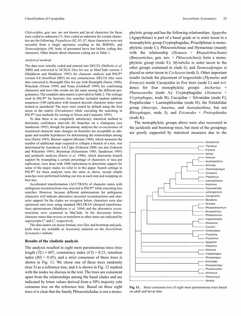
The analysis resulted in eight most parsimonious trees (treelength (TL) = 607, consistency index (CI) = 0.21; retentionindex (RI) = 0.45); and a strict consensus of these trees isshown in Fig. 11. We chose one of these trees randomly(tree 3) as a reference tree, and it is shown in Fig. 12 markedwith the nodes we discuss in the text. The trees are consistentapart from the relationships among the basal clades and areindicated by lower values derived from a 50% majority ruleconsenus tree on the reference tree. Based on these eighttrees it is clear that the family Phloeostichidae is not a mono-
phyletic group and has the following relationships: Agapytho(Agapythinae) is part of a basal grade or is sister taxon to amonophyletic group Cryptophagidae; Priasilphinae is mono-phyletic (node C); Phloeostichinae and Hymaeinae (mainlywith the relationship (Hymaea + Rhopalobrachium)(Bunyastichus, gen. nov. + Phloeostichus)) form a mono-phyletic group (node E); Myrabolia is sister taxon to theother groups contained in clade G; and Tasmosalpingus isplaced as sister taxon to Cyclaxyra (node I). Other importantresults include the placement of trogossitids (Thymalus andEronyxa) inside Cucujoidea in five trees (node L) and evi-dence for four monophyletic groups: Anchorius +Pharaxonotha (node A); Cryptophagidae (Atomaria +Cryptophagus, node B); Cucujidae + Silvanidae (node F);Propalticidae + Laemophloeidae (node H); the Nitidulidaegroup (Smicrips, Amartus, and Australaethina, but notRhizophagus, node J); and Ericmodes + Protosphindus(node K).
The monophyletic groups above were also recovered inthe jackknife and bootstrap trees, but most of the groupingsare poorly supported by statistical measures due to the
Classification of Cucujoidea
Fig. 11. Strict consensus tree of eight most parsimonious trees basedon adult and larval data.
Derodontus
Thymalus
Eronyxa
Helota
Amartus
Australaethina
Smicrips
Tasmosalpingus
Cyclaxyra
Propalticus
Laemophloeus
Acylomus
Passandridae
Lamingtonium
Cavognatha
Myrabolia
Hymaea
Rhopalobrachium
Bunyastichus
Phloeostichus
Cryptamorpha
Ahasverus
Cucujus
Chileosilpha
Priasilpha
Priastichus
Agapytho
Hobartius
Atomaria
Cryptophagus
Rhizophagus
Ericmodes
Protosphindus
Pharaxonotha
Anchorius
Paracucujus
Xerasia

R. A. B. Leschen et al.26 Invertebrate Systematics
paucity of synapomorphies for each group. The lack of sup-portive characters, however, tends to be in the internal nodesand if a single taxon is deleted from the analysis(e.g. Myrabolia that has uncertain relationships), the phylo-genetic arrangement changes significantly though the majorlineages we discuss below hold. We believe that instabilitymay be related in part to the poor signal in larval data (thoughthere are fewer larval characters anyway). A 50% majorityrule consensus tree based on adult characters is shown inFig. 13 (204 trees, TL = 417, CI = 0.20, RI = 0.09). In thisadult-based tree some of the major results are consistent withthe combined analysis (nodes B, C, E, H, I, and K are presentalso in this tree), though the relationships differ. Comparethese relationships to the 23 larval trees, shown as a 50%majority rule consensus tree in Fig. 14 (TL = 146, CI = 0.28,RI = 0.17). The larval data supports the inclusion of a mono-phyletic Trogossitidae in Cucujoidea and also nodes C, E,and F. They also support the sister relationship of Biphyllidae
and Byturidae, which is not supported by adult characters orin the combined analysis (see below). Otherwise the larvalcharacters, though being less in number, appear to supportsome spurious combinations of taxa (e.g. Passandridae +Amartus). More importantly, though, larval characters incombination with adult characters support many of thegroups we recognise below, and should not be disregarded(e.g. the monophyly of Hymaeinae + Phloeostichinae andPriasilphinae are supported by three unambiguous larvalcharacters).
Major lineages and the subfamilies of Phloestichidae
There have been numerous hypotheses proposed concerningrelationships among basal Cucujoidea, and these are to befound in the papers by Roy Crowson and Tapan Sen Gupta(see References) that are narrative and not based onHennigian argumentation (see discussion in Leschen 2003).Their narrative descriptions may be confusing to readersbecause they are often inconsistent and many taxonomicconclusions for controversial taxa are based on similarities torather generalised groups such as Cryptophagidae and
Fig. 12. Reference tree with marked nodes referred to in the text. Allnodes are consistent among the original eight parsimonious trees,except those that are indicated by the values derived from a 50%majority consensus tree, that also includes a trichotomy (*) ofAgapytho, Priasiliphidae and taxa included at nodes D and G. Bremersupport is indicated at consistent nodes.
Derodontus
Thymalus
Eronyxa
Xerasia
Paracucujus
Helota
Ericmodes
Protosphindus
Amartus
Australaethina
Smicrips
Tasmosalpingus
Cyclaxyra
Propalticus
Laemophloeus
Acylomus
Passandridae
Lamingtonium
Cavognatha
Myrabolia
Hymaea
Rhopalobrachium
Bunyastichus
Phloeostichus
Cryptamorpha
Ahasverus
Cucujus
Chileosilpha
Priasilpha
Priastichus
Agapytho
Atomaria
Cryptophagus
Hobartius
Rhizophagus
Pharaxonotha
AnchoriusA
B
75
75
75
75*
75
75
75
62
62
C
D
E
F
G
H
I
J
K
L
1
1 4
3
14
13
1
1
11
1
1
1
1
41
3
4
1
4
3
Fig. 13. 50% majority rule consensus tree of most 204 parsimonioustrees derived from adult characters.
Derodontus
Thymalus
Helota
Rhizophagus
Ericmodes
Protosphindus
Australaethina
Amartus
Smicrips
Tasmosalpingus
Cyclaxyra
Cryptamorpha
Ahasverus
Propalticus
Laemophloeus
Passandridae
Myrabolia
Acylomus
Hobartius
Atomaria
Cryptophagus
Pharaxonotha
Anchorius
Chileosilpha
Priasilpha
Priastichus
Cucujus
Hymaea
Bunyastichus
Phloeostichus
Rhopalobrachium
Agapytho
Cavognatha
Lamingtonium
Eronyxa
Paracucujus
Xerasia
55
85
100
87
67
55
100
65100
50
89
5597
100
100
87
94
100
100100
93
100
100
100100
73

Invertebrate Systematics 27
Cucujidae. In the following sections we limit reference toprevious statements about relationships and refer to the treeshown in Fig. 12.
Subfamilies of Phloestichidae
Agapythinae
The genus Agapytho is placed in a basal grade of taxa (seeFig. 11) or as sister taxon to Cryptophagidae in two of theparsimonious trees based on three characters: 61–2(parameres fused into a single piece), 62–2 (parameres fusedto phallobase), 89–1 (larval mesocoxae separated by lessthan two coxal diameters, ACCTRAN). Sen Gupta andCrowson (1966) listed Agapytho as an enigmatic genus ofHobartiidae, though later included it in a monotypic subfam-ily of Phloeostichidae (Sen Gupta and Crowson 1969).Grouping Agapytho at the bases of Priasilphinae orPhloeostichinae + Hymaeinae in Fig. 12 results in a one-stepincrease in tree length and there are relatively low bootstrap(20) and jackknife (24) values for the inclusion of the genusat the base of Phloeostichinae + Hymaeinae. The genusAgapytho differs from these taxa in many characteristics (see
description below) and is defined by 13 characters withunambiguous characters indicated by an asterisk (*): *1–2,*6–2, *8–1, *16–2, *21–2, *22–1, 22–1, 28–2, 35–1, 63–2,64–1, *67–2, 71–2, *95–2.
Priasilphinae
This subfamily is shown as a monophyletic group in all ofthe trees produced. It is rather isolated from other membersof Phloeostichidae, and in jacknife and bootstrap trees itforms a sister taxon to Cucujidae. The monophyly ofPriasilphinae is supported by the following synapomorphies(node C, Fig. 12): 20–2r (anteromesal corner of hypomeronwith tooth; reverses in Priastichus), 22–3 (shortest distancebetween procoxal cavities more than midlength of cavity),26–1 (procoxal cavity strongly transverse), 28–1 (procoxalcavity without narrow lateral notch, ACCTRAN), 36–1t
(meso-metaventral junction simple; transforms to state 3 inPriastichus), 51–2 (two basal ventrites connate), 58–1 (apexof sternite IX in male with lobe), 60–2t (base of tegmen nar-rowly rounded; transforms to state 3 in Priasilpha), 63–2(penis divided into basal and apical portions, DELTRAN),64–1 (basal portion of penis distinctly wider than apicalportion, DELTRAN), 65–2 (base of penis with mediancarina), 72–3 (antennal length more than 0.5× head width inlarva, ACCTRAN), 83–1 (larval ligula absent, ACCTRAN;coded as ? in Chileosilpha, gen. nov.), 88–2 (thoracic andmost abdominal terga with long lateral processes,ACCTRAN; coded as ? in Chileosilpha, gen. nov.), 94–1(urogomphi straight, ACCTRAN; coded as ? in Chile-osilpha, gen. nov.), 99–3 (abdominal spiracles at ends of longspiracular tubes, ACCTRAN; coded as ? in Chileosilpha,gen. nov.).
There are two unique larval characters that support themonophyly of Priasilphinae (88–2, 99–3, see larval tree inFig. 14) and the support for this group is rather robust(Bremer support (BS) = 3, Bootstrap support (BO) = 60,Jackknife (JS) = 72). Sen Gupta and Crowson (1969) thoughtat one time that Priasilpha was related to Helota; but basedon our study Priasilphinae falls nowhere near to helotids.
Phloeostichinae + Hymaeinae
These two subfamilies consistently occur together in allof the trees produced, and were often grouped as sister taxonto the Cucujidae group at node D (Fig. 12). The relationshipsare fairly consistent with the combination of (Hymaea +Rhopalobrachium) and (Bunyastichus, gen. nov. +Phloeostichus) shown in most trees. This group is supportedby the following synapomorphies (node E, Fig. 12): 8–4(genal projection lateral), 11–2r (dorsal surface of mandiblewith tubercle fitting into lateral clypeal emargination,ACCTRAN; reverses in Phloeostichus), 17–2r (sides ofpronotum with lateral teeth, ACCTRAN; reverses inRhopalobrachium), 22–1 (shortest distance between pro-coxal cavities less than half as great as mid length of cavity),
Classification of Cucujoidea
Fig. 14. 50% majority rule consensus tree of 23 most parsimonioustrees derived from larval characters.
Derodontus
Thymalus
Eronyxa
Myrabolia
Tasmosalpingus
Acylomus
Laemophloeus
Propalticus
Cyclaxyra
Lamingtonium
Smicrips
Amartus
Passandridae
Australaethina
Cryptophagus
Rhizophagus
Protosphindus
Cavognatha
Paracucujus
Cryptamorpha
Cucujus
Ahasverus
Priasilpha
Priastichus
Hymaea
Rhopalobrachium
Phloeostichus
Atomaria
Xerasia
Anchorius
Ericmodes
Agapytho
Hobartius
Pharaxonotha
Helota
96
96
96
100
100
100
100
100
100
100
100
100
100100
100
100
100
100
100
100
100
100
100100
100
100100
100100

R. A. B. Leschen et al.28 Invertebrate Systematics
35–1 (shortest distance between mesocoxal cavities less thanhalf as great as shortest diameter of cavity), 43–1(metendosternal laminae well developed), 67–2 (posterioredge of larval head capsule distinctly emarginate; coded as ?in Bunyastichus, gen. nov.), 68–2 (bases of frontal arms wellseparated in larval head capsule, DELTRAN; coded as ? inBunyastichus, gen. nov.), 74–2 (labrum fused to headcapsule, partly fused in Rhopalobrachium; coded as ? inBunyastichus, gen. nov.).
There are no unique characters that support the mono-phyly of Phloeostichinae and Hymaeinae, though two char-acters supporting node D are not common in basal cucujoids(43–1, 74–2). The group is relatively well supported by sta-tistical values (BS = 3, BO = 57, JS = 67).
Myraboliinae
The genus Myrabolia is placed at a consistent locationamong the trees at the base of node G (Fig. 12). The genus isnot associated with other members of Phloeostichidae andhas numerous characters that separate it from other familiesof Cucujoidea (see Description below) and is defined by thefollowing 18 characters (unambiguous characters are indi-cated by an asterisk (*)): *3–2, *7–2, *21–2, *23–2, *24–2,*26–3, *37–2, 46–2, *52–2, *58–1, 64–1, *66–2, *67–2,68–1, 71–3, 78–2, 97–3, *98–1.
Tasmosalpinginae
The genus Tasmosalpingus is placed consistently in all ofthe trees as sister taxon to Cyclaxyra (apart from the larvaltree where it is placed as sister taxon to Acylomus) based onthe following characters (node I, Fig. 12): 9–2 (anteriorcervical sclerites contiguous with head capsule, DELTRAN(transformed from state 3) and ACCTRAN (transformedfrom state 1), 38 (metaventral discrimen absent, DELT-RAN), 43–2 (metendosternal laminae reduced, ACCTRAN),47–2 (tarsi in male 5–5-4), 60–2 (base of tegmen narrowlyrounded), 62–2 (parameres fused to phallobase), 70–2(paired endocarinae present in the ventral head capsule oflarvae, DELTRAN), 93–1 (abdominal tergum IX simple inlarva).
Tasmosalpingus is not closely related to other members ofPhloeostichidae and has numerous characters that separate itfrom other groups of Cucujoidea (see Description below)and is defined by 9 characters (unambiguous characters indi-cated by an asterisk (*)): *3–2, 4–1, *13–2, *14–2, *32–2,*66–2, *73–2, *89–1, *97–3. The relationship ofTasmosalpingus to Cyclaxyra may be incorrect because thereare no unique characters that support their sister relationshipeven though the statistical support is relatively high (BS = 3,BO = 58, JS = 62). It is also possible that Cyclaxyra may bemore closely related to Lamingtoniidae (Lawrence andLeschen 2003) as shown in the larval tree (Fig. 14) whereTasmosalpingus is also grouped with Acylomus. MovingTasmosalpingus to a position as sister taxon to Acylomus in
the combined tree (Fig. 12) results in a tree that is an addi-tional 13 steps.
Other basal Cucujoidea
Erotylidae + Biphyllidae
The genera Pharaxonotha and Anchorius are consistentlygrouped as sister taxa in the eight parsimonious trees basedonly on adult characters (Fig. 12, node A): 8–2 (genal pro-jection anterior, truncate or rounded), 21–1 (apex of pro-sternal process without lateral projections, DELTRAN),25–2 (protrochantin concealed), 29–2 (procoxal cavity inter-nally closed, DELTRAN), 33–1 (epipleuron complete toapex), 35–2 (shortest distance between mesocoxal cavitiesmore than half as great but less than shortest diameter ofcavity), 37–2 (mesocoxal cavities laterally closed), 44–2(anterior tendons of metendosternite widely separated), 58–1(apex of sternite IX in male with mesal lobe).
In the past, Biphyllidae and Erotylidae were classifiedtogether in a very broad definition of the family (e.g.Ganglbauer 1899; Arrow 1929; Rymer-Roberts 1958) andthe relationships between these two groups is relativelyweakly supported in the combined analysis (BS = 1, BO =23, JS = 24). These results may be suprising sinceBiphyllidae and Byturidae were thought to be sister taxa(Falcoz 1926; Lawrence and Newton 1995), a relationshipsupported in the larval trees (Fig. 14) by nine unambiguouscharacters (27–2, 44–2, 45–1, 46–2, 57–2, 60–3, 66–2, 74–2,and 90–2).
Cryptophagidae
The monophyly of Cryptophagidae (Atomaria andCryptophagus) is clearly supported in this study based on thefollowing characters (node B, Fig. 12): 25–2 (protrochantinconcealed), 34–2 (anteromesal corner of mesepisternumwith cavities), 35–2 (shortest distance between mesocoxalcavities more than half as great but less than shortestdiameter of cavity, DELTRAN), 37–2 (mesocoxal cavitieslaterally closed by metaventrite), 40–2 (metacoxae widelyseparate), 53–2 (abdominal intercoxal process broadlyrounded), 71–3 (four or fewer larval stemmata, DELTRAN).
There are no unique characters that support the mono-phyly of Cryptophagidae, though statistical values are rela-tively high (BS = 4, BO = 72, JS = 65).
Cucujidae and Silvanidae
The group Cucujus (Ahasverus + Cryptomorpha) is sup-ported in the combined analysis based on the following char-acters (node F, Fig. 12): 2–2r (vertexal line present,ACCTRAN, reverses in Ahasverus), 6–2 (antennal insertionsconcealed by frontal ridge, DELTRAN), 24–2 (procoxa withlong, concealed lateral extension), 32–1 (elytral suturalflange not widened apically), 36–3 (meso-metaventral junc-tion simple), 68–1 (bases of frontal arms contiguous in larva,
Invertebrate Systematics 29
ACCTRAN), 72–3 (larval antennal length more than 0.5×head width), 92–2r (larval abdominal tergum IX forming anarticulated plate, ACCTRAN; reverses in Ahasverus), 96–2(larval sternum IX completely concealed or apparentlyabsent), 98–1 (larval spiracles annular).
The Cucujidae and Silvanidae are sometimes includedtogether in a broadly defined Cucujidae (along withPassandridae and Laemophloeidae, e.g. Forbes 1926),though they have been treated separately (Böving andCraighead 1931; Lawrence and Newton 1995). The mono-phyly of the Cucujidae + Silvanidae is relatively poorly sup-ported in the combined analysis (BS = 1) and it is alsorecovered with different relationships in the larval tree(Fig. 14).
Laemophloeidae and Propalticidae
Laemophloeus and Propalticus are consistently placed assister taxa based on the following characters (node H, Fig.12): 2–2 (vertexal line present), 4–1 (paired occipital inci-sions absent, DELTRAN), 22–3 (shortest distance betweenprocoxal cavities, DELTRAN), 24–2 (procoxa with longconcealed extensions), 35–3 (shortest distance betweenmesocoxal cavities more than shortest diameter of cavity),43–3 (metendosternal laminae absent, DELTRAN), 70–2(larval head capsule with paired endocarinae, DELTRAN),92–2 (abdominal tergum IX forming an articulated plate),94–1 (urogomphi straight, ACCTRAN; coded as unknown inPropalticus), 97 (larval segment X and anal openingventral), 98–1 (larval spiracles annular, DELTRAN).
Though the larva of Propalticidae has not been formallydescribed (the one described in Crowson and Sen Gupta(1969) was based on a misidentification), Lawrence andNewton (1995), R. A. Crowson (unpublished data), andLawrence and Newton (1995) proposed that the familiesPropalticidae and Laemoploeidae are sister taxa. Support forthe monophyly of this group is relatively robust (BS = 4,BO = 86, JS = 85), though it is not supported in the larvalanalysis (Fig. 14).
Nitidulidae group
The Nitidulidae families (Brachypteridae, Nitidulidae, andSmicripidae) are supported by the following synapo-morphies (node J, Fig. 12): 5–1r (frontoclypeal suturepresent, ACCTRAN; reverses in Australaethina), 9–1 (ante-rior cervical sclerite not contiguous with head capsule orplaced in paired emarginations, DELTRAN), 14–4t (galeaabsent; transforms to state 3 in Amartus), 18–1 (anteriorpronotal angles absent or not produced forwards), 44–1(anterior tendons of metendosternite narrowly separated),54–2 (abdominal tergite VII exposed in dorsal view), 56–3(tergite VIII in male with sides curved ventrally forming agenital capsule), 70–1 (paired endocarinae absent in theventral head capsule of larvae, ACCTRAN), 71–3 (numberof stemmata four or fewer), 75–2t (apex of larval mandible
bidentate; transforms to state 1 in Amartus), 77–1 (mesalsurface of larval mandible with asperate or tuberculatemola), 81–2 (labial palpi 1-segmented in larva), 84–3t (larvalhypopharyngeal sclerome an irregular molar-like tooth;transforms to state 2 in Australaethina).
The Nitidulidae group is supported by one unique charac-ter (14–4) and two others found only in this group andMonotomidae (54–2, 56–3) and with relatively strong statis-tical support (BS = 4, BO = 58, JS = 67). Crowson (1955)(see also Sen Gupta 1988) included Monotomidae in theNitidulidae group, though in later years (R. A. Crowson,unpublished data) he thought that the relationship toMonotomidae was erroneous, and this hypothesis is sup-ported here. Moving Monotomidae to the base of theNitidulidae group at node J (Fig. 12) increases the tree by 13steps. Crowson also excluded the Nitidulidae group fromCucujoidea and was planning to erect it as a separate super-family Nitiduloidea in an unpublished manuscript (Audisio1993), but there is no evidence for this change in the classi-fication.
Protocucujidae + Sphindidae
The close relationship between these two families(Ericmodes and Protosphindus) is well accepted (Sen Guptaand Crowson 1979; McHugh 1993; Ślipiński 1998) and issupported by the following characters (node K, Fig. 12): 5–1(frontoclypeal suture present), 10–3 (apex of mandible tri-dentate), 19–2 (anterior portion of prosternum at midline aslong as prosternal process), 29–1 (procoxal cavity internallyopen, ACCTRAN), 43–1 (metendosternal laminae welldeveloped), 46–1 (5 free veins present in medial area), 75–3(apex of larval mandible tridentate) and 86–1 (ventral epi-cranial ridges of larval head capsule absent). This group isnot well supported by unique characters and statistical valuesare relatively low (BS = 1, BO = 48, JS = 46).
Changes in classification and future work
Several important changes in Cucujoidea classificationare warranted based on the phylogenetic conclusions above.First and foremost are the changes in ranks for most of thesubfamilies of Phloeostichidae to family: Agapythidae,Myraboliidae, Phloeostichidae, Priasilphidae, and Tasmo-salpingidae. The subfamily names Hymaeinae (that previ-ously included Hymaea and Rhopalobrachium) andPhloeostichinae (for Phloeostichus) are unnecessary, thoughthese could be reinstated if the larva of Bunyastichus,gen. nov. is discovered and the evidence would add furthersupport for a closer kinship of it to Phloeostichus.
The inclusion of Cleroidea within Cucujoidea is sup-ported here and elsewhere (Beutel and Pollock 2000; Beuteland Ślipiński 2001) suggesting that the two superfamiliesCucujoidea and Cleroidea should be synonymised. Thesesuperfamilies are classified separately, despite the sharing ofcharacters by basal cleroids and cucujoids (e.g. Crowson
Classification of Cucujoidea

R. A. B. Leschen et al.30 Invertebrate Systematics
1964; Lawrence and Newton 1995; Lawrence and Leschen2003). Historically the members of these superfamilies weremixed. One group was referred to by one name (e.g.Clavicornia) with the others consisting of derived cleroidshaving soft bodies like Melyridae being placed inMalacodermata with unrelated cantharoids and other taxa(e.g. Lameere 1900; see review by Lawrence et al. 1995).Any action to synonymise the two superfamilies would bepremature for several reasons and a more appropriate studyshould be designed to include additional taxa representingthe major lineages of trogossitids (a heterogenous group) andthe families of Cleroidea.
For this study we have concentrated efforts to place theformer subfamilies of Phloeostichidae by scoring charactersthat are found in these subfamilies as well as many key char-acters used repeatedly by cucujoid workers. Others have notbeen used (e.g. presence of a sublateral lines present on thepronotum that would support the sister relationship betweenLaemophloeus and Propalticus) and the study of additionalcharacter systems is needed. Moreover, many questionsabout phylogenetic relationships remain, including the basalrelationships of Cucujoidea, the monophyly of the basalfamilies, and the relationships of the monogeneric familiesAgapythidae, Cyclaxyridae, Lamingtoniidae, Myraboliidae,and Tasmosalpingidae. Likewise, the phylogenetic positionof Helotidae has always posed problems for cucujoidworkers and it has been closely allied with Erotylidae,Cucujidae, Nitidulidae, Cleroidea, and other taxa (Crowson1995; Kirejtshuk 2000): It has even been considered amember of its own superfamily (R. A. Crowson, unpublisheddata). Though we have been able to address the monophyleticstatus of Phloeostichidae (sensu lato), cucujoid workers willneed to polarise characters based on multiple outgroups(e.g. Ślipiński 1998; Leschen 1996, 2003). If anything, theanalytical results may help to guide future studies of cucu-joids and other members of Cucujiformia.
Taxonomy
Family PRIASILPHIDAE Crowson
Priasilphinae Crowson, 1973: 56 (Phloeostichidae).
Adult diagnosis (Figs 1–3, 15–26, 29–63, 97–105, 110)
Head without postocular constriction, stridulatory file orfrontoclypeal suture. Antennal insertions exposed and sub-antennal grooves absent (except in Priastichus); genae pro-jecting forwards, rounded or acute. Anterior cervicalsclerites contiguous with head capsule. Mandible withoutdorsal tubercle or cavity, with mola. Lacinia with uncus.Pronotum with complete, simple lateral carinae; prosternumlong in front of coxae; prosternal process broad, not apicallyexpanded. Procoxae transverse, with exposed trochantins,cavities internally open, externally narrowly open or nar-
rowly closed. Elytral punctation not seriate; sutural flangeapically widened in winged species. Mesocoxal cavitieswidely separated, laterally open; meso-metaventral junctiondicondylic or simple (Priastichus). Metacoxae narrowly towidely separated. Hind wing with or without closed radialcell and with four or five free veins in medial field; wingsabsent in all Priastichus and most Priasilpha. Tarsi usually5–5–4 in male (5–5–5 in Chileosilpha, gen. nov.); tarsomereswithout ventral lobes. Abdomen with basal two ventritesconnate. Sternite VIII in male without median strut; spicu-lum gastrale present. Tegmen anteriorly narrowly rounded,acute or produced forming strut; parameres separate andarticulated. Penis distinctly divided into basal and apical sec-tions, base with median carina, without anterior struts.
Adult description
Length ~3.3–5.5 mm. Body narrowly oblong to broadlyovate, slightly to moderately convex; dorsal surfaces smoothand even or with tubercles or carinae on elytra; derm more orless shiny, but often covered with exudate and debris; vesti-ture of fine, recumbent hairs or recurved or erect, stoutbristles, evenly distributed or forming clusters dorsally. Headas long as wide or somewhat elongate, slightly flattened, not,or only slightly, declined, without postocular constriction,vertexal line or stridulatory file; posterior edge dorsally withpair of weak or strong incisions just above occipital foramen.Tentorial arms relatively close together, joined by moder-ately broad corporotentorial bridge. Frontoclypeal sutureabsent; clypeus extending well in front of antennal inser-tions, sides subparallel or slightly convergent, without lateralemarginations; apex truncate. Eyes moderately large, verti-cally oval and slightly emarginate to small and circular,prominent or not, moderately to coarsely facetted, withoutinterfacetal setae. Antennae 11-segmented with distinct3-segmented, pubescent club; scape slightly to distinctlyelongate and asymmetrically inflated; insertions widely sep-arated, exposed or barely concealed from above; subantennalgrooves present or absent; genae produced forwards and nar-rowly rounded or acute. Gular sutures well separated; pregu-lar region flat or slightly concave. Cervical sclerites present,sometimes enlarged. Labrum strongly transverse and slightlyinclined. Mandible usually bidentate, occasionally uniden-tate or obliquely tridentate, with well developed, transverselyridged mola and extensive, setose prostheca; without dorsaltubercle or setose cavity. Maxilla with broad, setose galeaand lacinia with setose inner edge and apical bifid uncus; ter-minal maxillary and labial palpomeres fusiform, narrowedapically with rounded apex. Mentum strongly transverse;ligula laterally expanded, apically emarginate, membranousand setose. Pronotum subquadrate to strongly transverse,broadest at middle or posterior third; sides not to stronglyexplanate and usually irregularly undulate, with distinctlateral carinae; anterior angles not to slightly produced for-wards and angulate or broadly rounded; posterior angles

Invertebrate Systematics 31
more or less right or slightly produced to fit over elytralhumeri; posterior edge straight to strongly bisinuate, notmargined. Disc slightly, to strongly convex, usually some-what uneven with large, shallow impressions. Hypomeronwith anteroventral angle slightly, to strongly produced andnot at same level as anterior edge of prosternum. Scutellumslightly, to strongly transverse, usually abruptly raised atbase, rounded to broadly angulate posteriorly. Elytra oblongto somewhat ovate, complete, widest at middle or anteriorthird; sides subparallel or somewhat rounded, sometimesstrongly narrowed posteriorly; apices broadly or narrowlyrounded; humeral region rounded or angulate, sometimeswith slight notch for posterior pronotal angles; base withcarina extending from scutellum laterally, below humerusand posteriorly forming upper edge of epipleuron. Discevenly convex, sometimes explanate laterally, with orwithout tubercles or longitudinal costae; punctation irregu-lar; sutural flange widened apically, except in winglessforms. Epipleura narrow to moderately broad at base, nar-rowing posteriorly and not extending to apex. Prosternumwell developed in front of coxae, at least as long as andusually longer than mid coxal length; anterior edge usuallyconcave, sometimes with narrow notch at each end, rarelybisinuate, forming short, broad chin piece flanked by broad
emarginations; prosternal process very broad, slightlycurved behind coxae, with sides subparallel, slightlyexpanded at middle or rarely abruptly expanded at apex,which may be truncate or slightly emarginate. Notosternalsuture almost always complete. Procoxae not projecting ven-trally, without long internal extension. Procoxal cavities
Classification of Cucujoidea
Figs 15–21. Dorsal views of adult Priasilpha: 15, P. obscura; 16, P. aucklandica, sp. nov.; 17, P. angulata, sp. nov.; 18, P. carinata, sp. nov.; 19, P.earlyi; 20, P. embersoni; 21, P. bufonia, sp. nov. Scale bars = 1 mm.
Figs 22-23. Dorsal views of adult Priastichus. 22, P. crowsoni, sp.nov.; 23, P. tasmanicus. Scale bars = 1 mm.

R. A. B. Leschen et al.32 Invertebrate Systematics
strongly transverse, without narrow lateral notch, internallyopen, externally narrowly open or closed; postcoxal projec-tions of hypomera relatively long and acute or rarely inter-locking with lateral projections of prosternal process;trochantins exposed. Mesoventrite simple or with pairedlongitudinal carinae, rarely defining a median shallow con-cavity; anterior edge with pair of strongly transverse,slightly, to strongly declined procoxal rests. Mesocoxalcavities slightly transverse, separated by a distance morethan half as great as and sometimes greater than shortestdiameter of cavity, open laterally (partly closed bymesepimeron), with exposed trochantins; meso-metaventraljunction a simple straight line or with paired metaventralknobs. Metaventrite moderately convex to flattened, withoutpost-coxal lines; discrimen usually absent, rarely long.Exposed portion of metepisternum long and narrow, slightlywidened anteriorly. Metendosternite with moderately to veryshort, broad stalk, long arms, reduced laminae and anteriortendons arising close together or moderately to widely sepa-rated. Metacoxae transverse, narrowly to widely separated.Hind wing, if present, relatively long and narrow, more than3× as long as wide; apical field more than half total winglength, containing long extensions of RA and RP; radial cell
complete or incomplete basally, with short, broad scleritenear its apical end; r3, if present, almost longitudinal; cross-vein r4 bisected by longitudinal sclerite; radio-medial loopforming acute angle; MP1+2 with long, slender apical exten-sion; medial field with four or five free veins; medial fleckand wedge cell absent; anal notch distinct. Legs relativelylong and slender; trochanterofemoral joint strongly oblique;femur slightly expanded near middle; tibia not or slightlyexpanded at apex, without carinae, teeth or spines, occasion-ally denticulate or spiculate, with or without subapical cteni-dium; tibial spurs relatively small, paired. Tarsi 5–5–5 infemale and 5–5–4 in male (rarely 5–5–5 in both sexes); tar-someres simple, 1 shorter than 2, 1–4 together shorter than toalmost twice as long as 5; terminal tarsomere usually withfringe of short setae at apex; claws simple; empodium rela-tively small and not projecting between claws. Abdomenwith five ventrites, the first two of which are more or lessconnate; ventrite 1 not much longer than 2, without post-coxal lines; intercoxal process usually broadly rounded ortruncate, rarely narrowly rounded; tergites I–VI lightly tomoderately pigmented, VII and VIII more heavily pig-mented; lateral wing-folding patches present on III or V toVII in winged forms, reduced in wingless forms. Abdominalspiracles: seven, first six located in pleural membrane, 7th ontergite. Sternite VIII in male without anterior median strut.Sternite IX in male deeply emarginate apically, with articu-lated median process, basally with anterior strut (spiculumgastrale). Aedeagus of inverted cucujiform type, withparameres ventral to penis; basal portion of tegmen attenuateanteriorly forming narrowly rounded to subacute plate, butnot drawn out to form anterior strut, apical portion welldeveloped, with short, broad to moderately elongate, articu-lated parameres. Penis divided into distinct basal (anterior)and apical (posterior) sections, the former broad, somewhatflattened, rounded or truncate basally, with or withoutmedian, basal carina; the latter narrower, slightly flattened tocylindrical or laterally compressed, parallel-sided or con-verging apically, with sclerotised endoskeleton and withapex emarginate or produced and acute; endophallus usuallywith one or more sclerites. Female sternite VIII with welldeveloped spiculum ventrale. Ovipositor short to moderatelylong and broad, with paired longitudinal bacula on para-procts and proctiger); proximal gonocoxite short and broad,with oblique, curved or sinuate transverse baculum; distalgonocoxite moderately to very elongate, narrowed apically,lightly to darkly pigmented; stylus moderately long andcylindrical, attached apically or subapically.
Larval diagnosis (Figs 79–83, 87–94, 106–109)
Body oblong to moderately elongate, somewhat ellipticaland narrowed posteriorly, flattened, with long lateral pro-cesses on all thoracic terga and abdominal terga I–VIII, inaddition to well developed urogomphi on tergum IX; uppersurfaces usually moderately well sclerotised, with or without
Figs 24–28. Characters of adults. 24, Chileosilpha elgueti, sp. nov.,dorsal view of head; 25, same, ventral view of head; 26, same, ventralview of prothorax; 27, Bunyastichus monteithi, sp. nov., lateral view oftegmen; 28, same, ventral view of tegmen.

Invertebrate Systematics 33
numerous setiferous tubercles forming straight or curvedrows, which may meet up to form cells (setiferous tuberclesmore developed in late instars of Priasilpha). Head broad,not emarginate posteriorly; epicranial stem absent; frontalarms lyriform, well separated at base; endocarinae absent.Stemmata: six, on raised projections, upper three on singleprojection, lower three on individual projections. Antennaealmost half as long as head length; antennomere 1 slightlylonger than wide, 2 almost twice as long as 1; 3 very short,less than 0.2× as long as 2; sensorium almost as long asapical antennomere. Frontoclypeal suture absent; labrumfree. Mandible tridentate (Priasilpha) or bidentate(Priastichus) (reduced and worn in some specimens), withaccessory ventral process, well developed, tuberculate mola,and narrow-based, acute prostheca. Ventral mouthpartsstrongly retracted, with well developed maxillary articulat-ing area; mala falciform; maxillary palps 3-segmented;labial palps 2-segmented, separated by more than width ofbasal palpomere; ligula short; hypopharyngeal sclerome atransverse bar. Hypostomal rods diverging; ventral epicranialridges absent or weakly developed; gula wider than long.Prothorax on each side with two long, posteriorly curved,acute tergal processes bearing elongate setiferous tubercles(more developed in late instars of Priasilpha); meso- andmetathorax each with one similar process on each side;abdominal tergum I on each side with a short, straight, lateralprocesses bearing a spiracle, terga II–VIII each with a pair ofelongate, slender, posteriorly curved lateral spiracle-bearingprocesses; tergum IX with pair of moderately to very longurogomphi, approximate or separated at base and subparallelor strongly diverging, each with lateral elongate setiferoustubercles, urogomphi of Priasilpha with one (P. earlyi andP. obscura) or two (P. angulata, sp. nov., P. bufonia, sp. nov.,and P. carinata, sp. nov.) long subapical setae that are muchlonger in first instars. Coxae moderately widely separated;pretarsal setae two. Sternum IX exposed, well developed;segment X distinct and sclerotised, at least ventrally, but notvisible from above. Spiracles annular–biforous.
Constitution, geographical distribution andbiological notes
As here redefined and expanded, Priasilphidae includes thesingle Chilean species Chileosilpha elguetai, sp. nov., threespecies of Priastichus from Tasmania and seven species ofPriasilpha from New Zealand. Although no biological infor-mation is available for Chileosilpha, gen. nov., adults andlarvae of Priasilpha and Priastichus species have been col-lected mainly in leaf litter, but also in decayed wood oramong moss on rock or log surfaces. The species may bemycophagous and some members of Priasilpha may befeeding on sooty mould. Specimens of Priasilpha andPriastichus larvae and adults are often found covered bydebris and exudate, which may be held in place by scale-likesetae (Fig. 110) and accumulate in foveae (Fig. 103).
Key to genera of Priasilphidae
1. Body more elongate and narrow, BL/EW greater than 2.25(Fig. 3); pronotum about as long as wide; prosternum pro-duced anteriorly forming chin piece flanked by pair of broademarginations (Fig. 26); procoxal cavities closed behind;tarsi 5–5–5 in both sexes; intercoxal process on ventrite 1narrowly rounded; Chile. . . . . . . . . . Chileosilpha, gen. nov.
Body shorter and broader, BL/EW less than 2.25 (Figs 1, 2);pronotum less than 0.75× as long as wide; chin piece, ifpresent, flanked by pair of narrow, deep notches (Fig. 102);procoxal cavities narrowly open behind; tarsi 5–5–4 in male;intercoxal process on ventrite 1 broadly rounded to truncate(Fig. 104). . . . . . . . . . . . . . . . . . . . . . . . . . . . . . . . . . . . . . . 2
2. Posterior edge of pronotum relatively straight and posteriorangles not produced over elytra (Fig. 2); scutellum less thanhalf as long as wide; elytral surfaces with tubercles and/orridges (Figs 2, 22, 23); eyes more or less circular andstrongly protruding; Tasmania . . . . . . . Priastichus Crowson
Posterior edge of pronotum sinuate and posterior angles pro-duced over elytra (weakly sinuate in P. aucklandica, sp. nov.)(Fig. 1); elytral surfaces usually smooth (tuberculate inP. aucklandica, sp. nov.) (Figs 15–21); eyes vertically ovaland not strongly protruding; New Zealand . . . . . . . . . . . . . . . . . . . . . . . . . . . . . . . . . . . . . . . . . . . . . . . . Priasilpha Broun
Genus Chileosilpha, gen. nov.
(Figs 3, 24–26, 29–35, 62)
Type species: Chileosilpha elguetai, sp. nov.
Diagnosis
Chileosilpha differs from both Priasilpha and Priastichus inthe more elongate body, distinct prosternal chin pieceflanked by broad emarginations, externally closed procoxalcavities, 5–5–5 tarsi in both sexes, and narrowly roundedintercoxal process on ventite 1.
Description
Length ~3.3–4.3 mm. Body oblong, somewhat narrowedanteriorly and moderately convex; ratio of body length togreatest body width 2.1–2.5. Upper surfaces clothed withmoderately long, fine, decumbent hairs. Head longer thanwide, posterior edge dorsally with a pair of deep incisions,representing lateral cuticular infoldings supporting occipitalforamen on each side. Eyes strongly protuberant, entire.Antennae with scape only slightly longer than wide andslightly longer than pedicel that is shorter than antennomere3. Antennal insertions exposed from above; subantennalgroove absent; genae produced well beyond mandibular atic-ulations and subacute as seen from above. Clypeal regionwith sides slightly convergent. Gula extending forwards toabout middle of eye; pregular area broadly concave, with pairof obliquely longitudinal grooves extending to maxillaryarticulations. Posterior edge of head broadly excavate oneach side of gular base, which extends posteriorly and is sub-acute at apex; each excavation containing an irregularlyovoid, thin and translucent anterior cervical sclerite, which is
Classification of Cucujoidea

R. A. B. Leschen et al.34 Invertebrate Systematics
connected to the gular base, the base of the tentorium and thelateral occipital infoldings. Tentorial bridge bearing a fineanterior median process. Mandible ~1.5× as long as wide,more or less abruptly curved mesally at apical 4th andobliquely tridentate. Apical maxillary and labial palpomereselongate, about twice as long as wide, distinctly narrowedapically. Pronotum about as long as wide, widest posteriorly;sides distinctly sinuate; lateral carinae complete, barelyvisible for their entire lengths from above, simple, withoutmarginal bead; anterior angles not produced forwards;posterior angles more or less right; posterior edge bisinuate,simple, without marginal bead; disc with weak, broadlytransverse impression. Scutellum about half as long as wide,
abruptly elevated basally, rounded laterally and obtuselyangulate posteriorly. Elytra ~1.6× as long as wide and 2.2×as long as pronotum; surface even, except for distincthumerus and slight boss at apical fifth. Prosternum in frontof coxae about twice as long as mid length of a coxal cavity,moderately convex; anterior edge produced forwards atmiddle to form chin piece, which is flanked by pair of broademarginations. Notosternal sutures deeply impressed butincomplete anteriorly; anteromesal corner of hypomeronwith short tooth. Prosternal process parallel-sided, butabruptly expanded laterally at apex, which is subtruncate.Procoxal cavities broadly closed externally by notal projec-tions extending over and fitting into concavities in lateral
Figs 29–35. Characters of adult of Chileosilpha elgueti, sp. nov. 29, dorsal view of penis;30, lateral view of penis; 31, dorsal view of tegmen; 32, dorsal view of ovipositor; 33, ventralview of ovipositor; 34, spermatheca; 35, ventral view of male sternite IX.

Invertebrate Systematics 35
expansions of prosternal process. Mesoventrite anteriorlywith pair of narrow, strongly declined procoxal rests and pos-teriorly with a broad, shallow depression, flanked by longi-tudinal carinae. Mesocoxal cavities separated by distanceequal to shortest diameter of a mesocoxal cavity. Meso-metaventral junction complex, with pair of metaventralknobs fitting into paired mesoventral cavities. Metaventritemoderately long and convex; discrimen long but weaklyindicated. Metacoxae narrowly separated. Metendosternitewith anterior tendons arising close together. Hind wing withincomplete (basally open) radial cell and four free veins(MP4+CuA1, CuA2, AA3 and AA4) in medial field. Legssimple, with weak subapical ctenidium. Tarsi 5–5–5 in bothsexes; tarsomeres 1–4 together 1.5× as long as tarsomere 5.
Abdomen with intercoxal process narrowly rounded.Tegmen with parameres broader apically than at base; ante-rior plate narrowed and subacute. Penis with basal sectionslightly depressed, broadly rounded anteriorly, with mediancarina; apical section laterally compressed and narrowed api-cally, notched at apex; endoskeleton relatively complex,including paired hooks and a semicircular sclerite; endo-phallus with a pair of longitudinal, complexly sinuate, darklypigmented sclerites. Ovipositor about twice as long as wide;combined gonocoxites slightly longer than paraprocts, prox-imal gonocoxite subquadrate and apically oblique withsinuate transverse baculum, distal one slightly longer, dis-tinctly narrowed apically, pigmented on apical half, with welldeveloped stylus attached laterally just before apex.
Classification of Cucujoidea
Figs 36–45. Characters of adult new species of Priasilpha. 36, P. angulata, sp. nov., ventralview of penis; 37, same, lateral view; 38, P. bufonia, sp. nov., ventral view of penis; 39, same,lateral view; 40, P. carinata, sp. nov., ventral view of penis; 41, same, lateral view;42, P. earlyi, ventral view of penis; 43, same, lateral view; 44, same, detail of left paramere;45, same, distal portion of gonocoxite and stylus.

R. A. B. Leschen et al.36 Invertebrate Systematics
Chileosilpha elguetai, sp. nov.
(Figs 3, 24–26, 29–35, 62)
Material examined
Holotype. � ‘Chile, T. Ueno PN Puyehue Anticura, 17 Feb 2003.’(MNHS)
Paratypes. �, Chile. Chiloe: Chiloe I., ii.1990 (AA). � (dis-sected, parts on card and in glycerine), Chile. Llanquihue: P[arque].N[ational], Vicente Perez R[osales], C° [Cerro] Derrumbe [41°11′S,72°15′ W], 8-v-1971, J. Solervicens / formacion Caigue-Tepa (ANIC).
Description
With the characters of the genus. TL = 4.8, 3.85 mm;BL/EW = 2.35, 2.33; PL /PW = 1.0, 0.96; EL/EW = 1.59,1.6; EL/PL = 2.11, 2.18; GD/EW = 0.73, 0.76. Colourreddish-brown; anterior portion of head, palps and legs yel-lowish-brown. Dorsal surfaces smooth and shiny, clothedwith moderately long, recumbent yellow hairs, those onelytra posteriorly directed, those on head and prothoraxmore or less laterally directed from midline; ventral sur-faces clothed with short, recumbent hairs. Punctation ofhead and pronotum moderately fine and dense, which onelytra is fine but somewhat sparser; punctation moderatelyfine and dense on most surfaces, extremely fine and sparseon hypomera, coarse and dense on posterior portion ofmesoventrite. Aedeagus as in Figs 29–31. Ovipositor as inFigs 32, 33.
Distribution
Valdivian region, Chile.
Remarks
The holotype specimen was collected in a forest where thedominant tree species were Nothofagus dombeyi (Fagaceae)and Laurelia philippiana (Atherospermataceae).
Genus Priasilpha Broun
(Figs 1, 15–21, 36–52, 63, 97–105, 110, 80, 83)
Priasilpha Broun, 1893: 1077. Type species, by monotypy,P. obscura Broun.
Diagnosis
Priasilpha species are always shorter and broader thanChileosilpha elguetai, and differ also in having either no chinpiece or one that is shorter, broader and flanked by deepnotches, and in having narrowly open procoxal cavities, abroader abdominal process and 5–5–4 tarsi in the male. Thegenus differs from Priastichus in having the prothorax dis-tinctly wider posteriorly than anteriorly, with a bisinuateposterior edge and somewhat acute and produced posteriorangles; in addition the eyes are vertically oval and notstrongly projecting. In most Priasilpha the elytral surfacesare completely devoid of tubercles or ridges, which charac-
terise species of Priastichus; however these do occur inP. aucklandica, sp. nov. (see below).
Description
Length ~3.4–5.8 mm. Body oblong to broadly ovate,slightly narrowed anteriorly, strongly so posteriorly, moder-ately to strongly convex; ratio of body length to greatestbody width 1.3–2.04. Upper surfaces clothed with short tomoderately long, stout, yellow setae, mainly recumbent butwith a variable number of suberect setal clusters. Head aslong as wide or slightly elongate; posterior edge dorsallywith a pair of shallow emarginations. Eyes vertically ovaland slightly emarginate. Antennae with scape distinctlylonger than wide and distinctly longer than pedicel that isshorter than antennomere 3. Antennal insertions exposedfrom above; subantennal grooves absent; genae moderatelyproduced forwards and rounded. Pregular area flat. Tentorialbridge without median process. Clypeal region with sidesslightly convergent. Mandible slightly longer than wide,more or less abruptly curved mesally at apical 3rd andbidentate. Apical maxillary and labial palpomeres elongate,~1.5× as long as wide, not, or only slightly, narrowed api-cally. Pronotum ~0.50–0.76× as long as wide, widest at orjust behind middle, and wider at base than at apex; sidessomewhat irregularly undulate, not explanate but oftenslightly raised; lateral carinae visible for their entire lengthsfrom above, anterior angles not or slightly produced for-wards and rounded; posterior angles slightly produced andsubacute; disc sometimes with pair of weak elevations.Scutellum slightly more than half as long as wide, usuallyabruptly elevated basally, rounded or acutely angulate later-ally and rounded to sharply angulate posteriorly. Elytra~0.77–1.65× as long as wide and 1.37–3× as long as prono-tum, widest at anterior third; sides slightly converging ante-riorly, strongly so posteriorly; apices broadly to narrowlyrounded; anterior edge with slight notch or depression oneach side for receiving posterior pronotal angles; humerussharply angulate, sometimes laterally produced; disc evenor with irregular elevations; epipleura broad at base,strongly narrowed posteriorly. Prosternum in front of coxae~1.3 to 1.8× as long as mid length of a coxal cavity, moder-ately convex; anterior edge concave, not produced forwardsor slightly produced to form short, broad chin piece, withdeep, narrow notch at each end between sternum andhypomeron; notosternal sutures complete; prosternal punc-tation finely spiculate, in contrast to coarsely punctatehypomeral punctation. Prosternal process shorter than wide,sides diverging apically or rounded (diverging and then con-verging); apex subtruncate or concave, without lateralexpansions. Procoxal cavities narrowly open behind; notalprojections acute, extending beyond middle of coxal cavi-ties but not meeting prosternal process. Mesoventrite withpair of slightly oblique procoxal rests, with or without a pairof weak longitudinal carinae. Mesocoxal cavities separated

Invertebrate Systematics 37
by distance at least 1.2× as great as shortest diameter of amesocoxal cavity. Meso-metaventral junction forming astraight line. Metaventrite less than one-third to slightlymore than half as long as wide; discrimen absent.Metacoxae widely separated. Metendosternite with stalkmuch shorter and anterior tendons more widely separated inwingless forms. Hind wing, if present, with complete(basally closed) radial cell and five free veins (MP3,MP4+CuA1, CuA2, AA3 and AA4) in medial field. Legssimple; tibiae slightly expanded apically, with distinct sub-apical ctenidium. Tarsi 5–5–5 in female, 5–5–4 in male;tarsomeres 1–4 together longer than tarsomere 5. Abdomenwith intercoxal process broadly rounded or truncate.Tegmen with parameres about twice as long as wide andnarrower apically; anterior plate narrowly rounded. Peniswith basal section depressed, broadly rounded anteriorly,with or without median carina; apical section narrowedapically, acute at apex; endoskeleton simple, trapezoidal;endophallus with pair of lightly pigmented, longitudinalsclerites. Ovipositor with proximal gonocoxites sub-quadrate or slightly transverse, distal ones 3× as long andmuch narrower, pigmented except at base, with apicallyattached styli.
Key to species of Priasilpha based on adults
1. Body elongate, length of elytra exceeding 2.5× length of prono-tum (Fig. 15); apical sutural flanges of elytra present (Fig. 1);hind wings present; widely distributed . . . P. obscura Broun
Body shortened or moderately elongate, length of elytra 2.0× orless length of pronotum (Figs 16–21); apical flanges of elytraabsent; hind wing absent; distributions restricted. . . . . . . . 2
2. Posterior edge of pronotum evenly convex; sides of pronotumwithout deep sulci; dorsal surface with tubercles (Fig. 16);Auckland Islands . . . . . . . . . . . . . . . P. aucklandica, sp. nov.
Posterior edge of pronotum sinuate; sides of pronotum with deepfurrows; dorsal surface without tubercles (Figs 17–21) . . . 3
3. Scutellum well developed and rounded; anterior edge of pronotumemarginate; anterior angles produced forwards (Fig. 17);Franz Joseph Glacier, Westland . . . . . . . . P. angulata, sp. nov
Scutellum poorly developed and triangular; anterior edge ofpronotum not emarginate (Figs 18–21); anterior angles notproduced forwards. . . . . . . . . . . . . . . . . . . . . . . . . . . . . . . . 4
4. Elytra twice as long as pronotum; body elongate-oval(Figs 18, 19) . . . . . . . . . . . . . . . . . . . . . . . . . . . . . . . . . . . . 5
Elytra 1.3–1.5× as long as pronotum; body rounded (Figs 20, 21) . . . . . . . . . . . . . . . . . . . . . . . . . . . . . . . . . . . . 6
5. Antennomere 3 as long as 4; asetose area present on centralanterior portion of elytra (Fig. 19); Arthur’s Pass, Westland. . . . . . . . . . . . . . . . . . . . . . . . . . . . . . . . . . P. earlyi, sp. nov.
Antennomere 3 longer than 4; asetose area on elytra absent(Fig. 18); north-west Nelson . . . . . . . . . P. carinata, sp. nov.
6. Elytra distinctly shorter than their combined widths; sides con-verging from middle to apex; humeral rim extending tomiddle (Fig. 21); penis with posterior carina but without ante-rior keel (Figs 38, 39); Marlborough . . . . P. bufonia, sp. nov.
Elytra only slightly shorter than combined widths; sides con-verging before middle; humeral rim short, not extending tomiddle (Fig. 20); penis with anterior keel but without poste-rior carina (Figs 46, 47); Buller . . . . . P. embersoni, sp. nov.
Priasilpha angulata, sp. nov.
(Figs 17, 36, 37, 95)
Material examined
Holotype. �, ‘Fox Glacier, Westland Natl Pk [43°27′S, 170°01′E]16–17.xii 99, T. Ueno leg;’ (NZAC).
Paratypes. New Zealand: South Island. WD, 10, Fox Glacier,Westland Natl Pk, [43°27′S, 170°01′E] 16–17.xii 99, T. Ueno (3 NZAC,7 TUPC); 2, same locality, 12–15.xii 99, T. Ueno leg (TUPC);3, Canavan Knob Walk, 43°23′S, 170°10′E, 13.ii.1999, R. Leschen,R. Hoare, slime flux, RL309 (NZAC); 1, Lake Ianthe [43°03′S,170°37′E] 7.ii.1984, J. C. Watt, sifted litter and wood mould 84/22(NZAC).
Associated larva. 1, Canavan Knob Walk, 43°23′S, 170°10′E,13.ii.1999, R. Leschen, R. Hoare, slime flux, RL309 (NZAC).
Diagnosis
Body moderately elongate, elytra ~1.1× as long as wide and1.8× as long as the pronotum. Dorsal surfaces lacking tuber-cles. Pronotum with lateral rims well developed; side withone notch; deep sublateral sulci present; anterior anglesstrongly produced forwards; two setal patches present atcentre. Scutellum fully exposed. Humeral ridge of elytronshort. Femur not bicolored. This is the only species in whichthe anterior edge of the pronotum is deeply emarginate andthe anterior angles strongly produced forwards.
Description
Length 4.5–5.2 mm (mean = 4.76, n = 10). Body ~1.80× aslong as wide (BL/EW = 1.73–2.06), and greatest depth~0.56× as great as elytral width (GD/EW = 0.52–0.61).Colour dark reddish brown; mouthparts, legs, and clypeusdark red; dorsal surfaces lacking tubercles; vestiture of thickyellow setae; anterior portion of prosternum, all of thehypomeron, and epipleuron covered with microtubercles.Head coarsely and densely punctate posteriorly, more finelyso anteriorly. Antennomeres 3 and 4 subequal in length. Eyecoarsely facetted, greatest length five facets, greatest width11 facets. Pronotum ~0.7× as long as wide (PL/PW =0.64–74), widest at basal 1/3; sides broadly, irregularlysinuate, lateral notch present at middle; lateral rim welldeveloped with deep sublateral sulcus; anterior margindeeply incised with anterior angles strongly produced for-wards, with internal angles right; posterior angles slightlyacute; posterior margin sinuate; disc uneven, glabrous areaspresent in anterior half (in some specimens) and associatedwith sublateral sulci; setal patches present at anterior margin(two, sometimes fused to form a single patch), centre (two),and either side of notch on lateral rim. Hypomeron lackinglarge ovate punctures; well developed ventrally directedtooth present above anterior hypomeral notch; shallowposterior fovea absent. Prosternum without chin piece; widthof prosternal process equal to width of procoxal cavity; beadnot present running parallel to notosternal suture; shorttransverse groove at either side of anterior margin absent.
Classification of Cucujoidea

R. A. B. Leschen et al.38 Invertebrate Systematics
Scutellum fully exposed; rounded and slightly transverse.Elytra ~1.1× as long as wide (EL/EW = 1.07–1.37) and~1.8× as long as pronotum (EL/PL = 1.61–1.97); sidesevenly rounded, widest at middle; humeral ridge present,extending a short distance, not to midline of elytron (1/6length of elytron); 10 setal patches present, posterior patcheslinear; glabrous areas absent; width of epipleuron at level ofmetacoxa 1/2 that of metacoxa; apical sutural flanges absent.Hind wing absent. Width of mesoventral process greater thanthat of mesocoxa. Abdominal process of first ventrite broadand square, width greater than that of metacoxa. Abdominalventrites on different planes. Femur not bicolored, uniformlyred or reddish brown. Penis with anterior hyaline keel andlateral rim extending to anterior 1/3 in anterior portion; pos-teriorly carinate (carina short and rather broad), with dorsaledge evenly rounded and sinuate just before acute apex.Paramere 1.5× longer than wide with many apical setae.
Remarks
The species is restricted to an area known as the Westlandbeech gap, where Nothophagus species are completelyabsent, one of the major biogeographic mysteries in theSouth Island. Three adult specimens were collected with alarva feeding on slime flux oozing from a cut edge of a tree.Gut contents of a dissected specimen contained mainlyunidentified matter and a few fungal hyphae. The namerefers to the strongly angulate and produced anterior anglesof the pronotum.
Distribution
South Island, Westland.
Priasilpha aucklandica, sp. nov.
(Fig. 16)
Material examined
Holotype. �, ‘Fairchilds Garden, Adams Id, Auckland Is[50°35′S, 166°10′E] 20.i.66, litter 66/77, Ent. Div. D.S.I.R. N. Zealand’(NZAC).
Associated larvae. 2, same label data as Holotype, but 2.ii.66(slide mounted, NZAC); 4, same except 2 Feb 1966, litter 66/95 (2 slidemounted, NZAC); 1, Fairchilds Garden, 20.i.66, fellfield, P. M. J.(NZAC).
Diagnosis
Body moderately elongate, elytra ~1.3× as long as wide and2.0× as long as the pronotum. Dorsal surfaces tuberculate.Pronotum without lateral rims or deep sublateral sulci; sidewithout notch; anterior angles produced forwards; four setalpatches present at centre. Scutellum fully exposed. Humeralridge of elytron absent. Femur unicolored. This speciesdiffers considerably from its congeners, most notably by thepresence of dorsal tubercles and the shape of the pronotum.
Description
Length 5.8 mm. Body twice as long as wide (BL/EW = 2),and greatest depth ~0.6× as great as elytral width (GD/EW= 0.62). Colour of body light tan; ventrites and head dark tan;dorsal surfaces tuberculate; vestiture of thick setae; anteriorportion of prosternum and lateral and anterior portion ofhypomeron covered with microtubercles. Head coarsely anddensely punctate. Antennomere 3 longer than 4. Eye coarselyfacetted, greatest length eight facets, greatest width 12facets. Pronotum ~0.8× as long as wide (PL/PW = 0.76),widest at middle; sides evenly rounded and slightly sinuatewith carina explanate with lateral bead; lateral notch, rim anddeep sublateral sulci absent; anterior margin shallowlyincised with anterior angles produced forwards and rounded,interior angle oblique; posterior angles acute; posteriormargin weakly sinuate and more or less convex; disc uneven,glabrous areas absent; setal patches on tubercles and presentat centre (4), longer setae present on carina at basal andapical 1/3 and along anterior margin. Hypomeron lackinglarge ovate punctures absent; well developed ventrallydirected tooth present above anterior hypomeral notchpresent; shallow posterior fovea absent. Prosternum withoutchin piece; width of prosternal process slightly less thanwidth of procoxal cavity; bead present running parallel tonotosternal suture; short transverse groove at either side ofanterior margin absent. Scutellum fully exposed; angulateand transverse. Elytra ~1.3× as long as wide (EL/EW = 1.34)and ~2× as long as pronotum (EL/PL = 2.05); sides evenlyrounded, widest at middle; humeral ridge absent; nine setalpatches present on rounded tubercles, posterior and lateraltubercles shallower than those at centre; glabrous areasabsent; width of epipleuron at level of metacoxa 1/2 that ofmetacoxa; apical sutural flanges absent. Hind wing absent.Width of mesoventral process narrower than that of meso-coxa. Abdominal process of first ventrite broad and square,width less than that of metacoxa. Abdominal ventrites on dif-ferent planes. Femur unicolorous light tan.
Remarks
This species is known from one female adult specimen andfive larvae, which were in alcohol up until the start of thisstudy. All specimens were bleached, apparently due toimproper storage in sunlight, and the larvae were very fragileand four were slide mounted for better preservation andfuture study. Priasilpha aucklandica is known only fromAdams Island, the large and pristine southernmost island ofthe subantarctic Auckland Islands. The Auckland Islandarchipelago is the largest of all New Zealand’s subantarcticislands, and is the only island group with a forest canopy(composed of Metrosideros umbellata or Southern Rata). Ofthe four slide mounted larvae, the gut of one was empty,otherwise they were filled with undetermined debris and thedebris affixed to the body was undetermined, apart form

Invertebrate Systematics 39
spores found on the body of one individual. The name isbased on the type locality.
Distribution
Auckland Island, Adams Island.
Priasilpha bufonia, sp. nov.
(Figs 21, 38, 39, 95)
Material examined
Holotype. �, ‘Mt Duppa, Nelson, 3500’ [41°13S, 173°30E]16–3–38, E. S. Gourlay, E. S. Gourlay Acc. 1970 Ent. Div’ (NZAC).
Paratypes. New Zealand: South Island. MB, 3, Fell Pk,Richmond Ra, 1296 m, [41°27′S, 173°24′E] 13.iii.1969, A. C. Eyles,litter (NZAC). NN, 3, Third House, Dun Mt track, Nelson [41°19′S,173°19′E], 14.ix.1971, G. W. Ramsay, litter (NZAC); 2, Dun Mt,Nelson [41°19′S, 173°22′E] 9.xi.1957, R. M. Bull Collection (NZAC).
Associated larvae. 1, MB, Fell Pk, Richmond Ra, 1296 m[41°27′S, 173°24′E], 13.iii.1969, A. C. Eyles, litter (NZAC); 9, NN,Third House, Dun Mt track, Nelson [41°19′S, 173°19′E], 14.ix.1971,G. W. Ramsay, litter (NZAC).
Diagnosis
Body round, elytra ~0.8× as long as wide and 1.5× as long asthe pronotum. Dorsal surfaces lacking tubercles. Pronotumwith lateral rims well developed; side with one notch; deepsublateral sulci present; anterior angles reduced; four setalpatches present at centre. Scutellum reduced. Humeral ridgeof elytron elongate. Femur not bicolored. This is the onlyspecies with an elongate humeral ridge that extends to themidlength of the elytron.
Description
Length 3.4–3.9 mm (mean = 3.63, n = 7). Body ~1.38× aslong as wide (BL/EW = 1.30–1.43), and greatest depth~0.54× as great as elytral width (GD/EW = 0.50–0.58).Colour of body dark reddish brown or brown; mouthparts,legs, and clypeus dark red to light brown; dorsal surfaceslacking tubercles; vestiture of thick yellow setae; anteriorportion of prosternum, lateral and anterior portion ofhypomeron, and epipleuron covered with microtubercles.Head coarsely and densely punctate. Antennomeres 3 and 4subequal in length. Eye coarsely facetted, greatest length fivefacets, greatest width 12 facets. Pronotum ~0.6× as long aswide (PL/PW = 0.54–64), widest at basal 1/3; sides broadly,irregularly sinuate, lateral notch present at middle; lateralrim well developed with deep sublateral sulcus; anteriormargin weakly incised with anterior angles weakly producedforwards; posterior angles rounded; posterior marginsinuate; disc uneven, glabrous areas not present; setalpatches present at anterior margin (two fused to form a singlepatch), centre (four), and either side of notch on lateral rim.Hypomeron with posteromesal surface glabrous, large ovatepunctures absent; poorly developed ventrally directed tooth
present above anterior hypomeral notch; shallow posteriorfovea absent. Prosternum without chin piece; width ofprosternal process subequal to width of procoxal cavity;bead present running parallel to notosternal suture; shorttransverse groove at either side of anterior margin present.Scutellum partly exposed; triangular. Elytra ~0.8× as long aswide (EL/EW = 0.77–0.89) and ~1.5× as long as pronotum(EL/PL = 1.37–1.67); sides subparallel to middle andabruptly converging to apex; humeral ridge present, extend-ing a long distance to midline of elytron (~1/2 length ofelytron); five rounded setal patches present; glabrous areaspresent in basal 1/3 of some specimens; width of epipleuronat level of metacoxa slightly wider than that of metacoxa;apical sutural flanges absent. Hind wings absent. Width ofmesoventral process greater than that of mesocoxa.Abdominal process of first ventrite broad and square, widthgreater than that of metacoxa. Abdominal ventrites onslightly different planes. Femur not bicolored, uniformlyreddish or light brown. Penis without anterior hyaline keeland lateral rim extending to anterior third; posteriorly cari-nate (carina narrow), with dorsal edge evenly rounded andsinuate just before apex, apex acute. Paramere twice as longas wide with few apical setae.
Remarks
The species is restricted to Richmond Range in theMarlborough area at the northern end of the South Island.The gut of a single dissected specimen was filled with darksooty mould fungal spores that are essentially long chainshyphae that fragment into spores (P. Johnston, personal com-munication). Scrapings of the surface debris of the samespecimen also contained the same hyphae, which can be seenin some of the pinned specimens. A single early instar fromthe Richmond Range had a gut filled with undeterminedmaterial. The species name is taken from the Latin word bufofor toad and refers to the toad-like body form.
Distribution
South Island, Nelson and Marlborough regions.
Priasilpha carinata, sp. nov.
(Figs 18, 40, 41, 95)
Material examined
Holotype. �, ‘Paradise Peak, 4500′ [40°55′S, 172°38′E] 6.11.38,E. S. Gourlay’ (NZAC).
Paratypes. New Zealand: South Island. NN, 5, Mt Arthur(Salisbury), 3000′ [41°11′S, 172°44′E] 23.x.1943, E. S. Gourlay(4, NZAC; 1 slide mounted, ANIC); 1, Mt Arthur (G.), 4500′ [41°11′S,172°44′E] 17.i.1943, E. S. Gourlay (NZAC); 1, Mt Arthur, KahurangiNat. Pk [41°11′S, 172°44′E] 25–28.xi.1999, T. Ueno (TUPC); 1, Bushbehind Mytton Hut, Cobb Valley 4000′ [41°06′S, 172°35′E]13.xii.1967, litter, S. Edridge (NZAC);
Classification of Cucujoidea
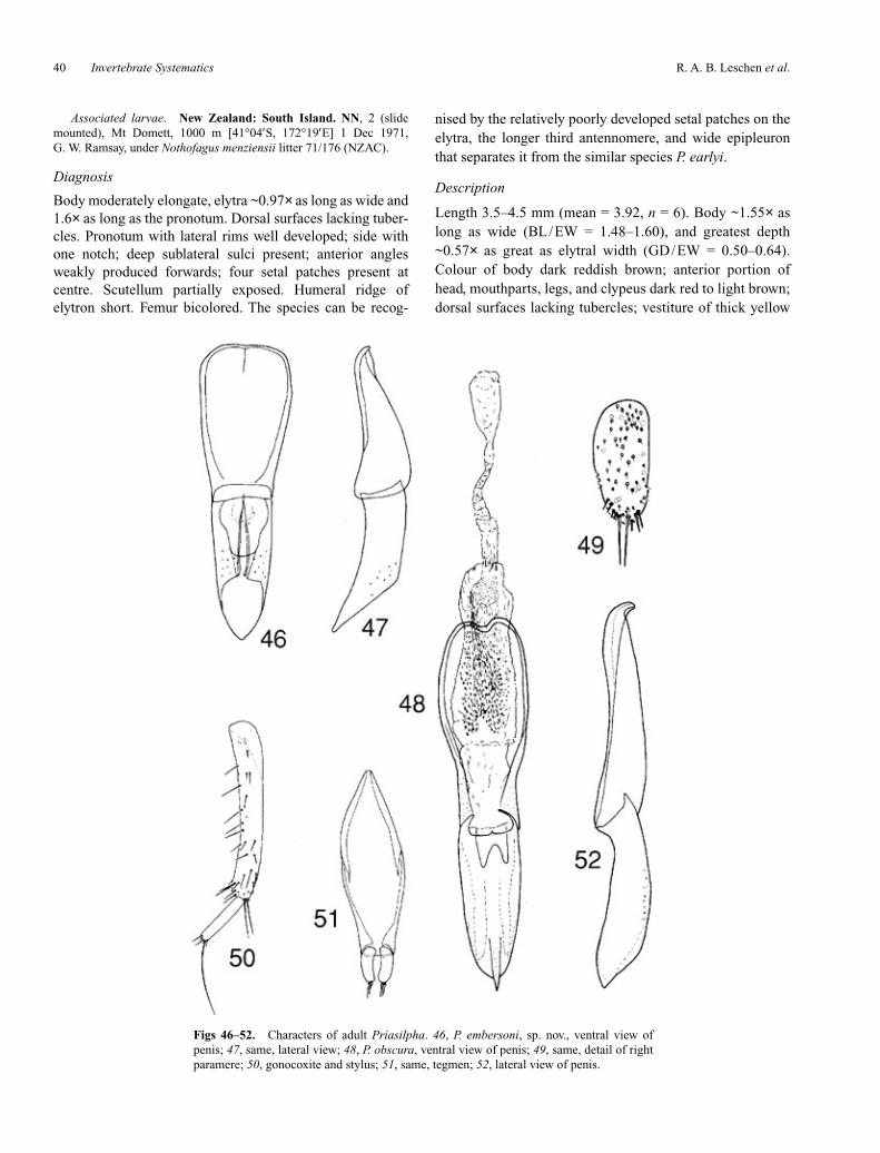
R. A. B. Leschen et al.40 Invertebrate Systematics
Associated larvae. New Zealand: South Island. NN, 2 (slidemounted), Mt Domett, 1000 m [41°04′S, 172°19′E] 1 Dec 1971,G. W. Ramsay, under Nothofagus menziensii litter 71/176 (NZAC).
Diagnosis
Body moderately elongate, elytra ~0.97× as long as wide and1.6× as long as the pronotum. Dorsal surfaces lacking tuber-cles. Pronotum with lateral rims well developed; side withone notch; deep sublateral sulci present; anterior anglesweakly produced forwards; four setal patches present atcentre. Scutellum partially exposed. Humeral ridge ofelytron short. Femur bicolored. The species can be recog-
nised by the relatively poorly developed setal patches on theelytra, the longer third antennomere, and wide epipleuronthat separates it from the similar species P. earlyi.
Description
Length 3.5–4.5 mm (mean = 3.92, n = 6). Body ~1.55× aslong as wide (BL/EW = 1.48–1.60), and greatest depth~0.57× as great as elytral width (GD/EW = 0.50–0.64).Colour of body dark reddish brown; anterior portion ofhead, mouthparts, legs, and clypeus dark red to light brown;dorsal surfaces lacking tubercles; vestiture of thick yellow
Figs 46–52. Characters of adult Priasilpha. 46, P. embersoni, sp. nov., ventral view ofpenis; 47, same, lateral view; 48, P. obscura, ventral view of penis; 49, same, detail of rightparamere; 50, gonocoxite and stylus; 51, same, tegmen; 52, lateral view of penis.

Invertebrate Systematics 41Classification of Cucujoidea
Figs 53–61. Terminalia of adult Priastichus. 53, P. crowsoni, sp. nov., dorsal view ofpenis; 54, same, lateral view of penis; 55, same, dorsal view of tegmen; 56, P. mega-thorax, sp. nov., dorsal view of penis; 57, same, lateral view of penis; 58, same, dorsalview of tegmen; 59, P. tasmanicus, dorsal view of penis; 60, same, lateral view of penis;61, same, dorsal view of tegmen.

R. A. B. Leschen et al.42 Invertebrate Systematics
setae; anterior portion of prosternum, lateral and anteriorportion of hypomeron, and epipleuron covered with micro-tubercles. Head coarsely and densely punctate. Antenno-mere 3 longer than 4. Eye coarsely facetted, greatest lengthfive facets, greatest width 12 facets. Pronotum ~0.6× aslong as wide (PL/PW = 0.59–67); sides broadly, irregularlysinuate, lateral notch present at middle; lateral rim welldeveloped with deep sublateral sulcus; anterior marginweakly incised with anterior angles slightly produced for-wards; posterior angles slightly rounded; posterior marginsinuate; disc uneven, glabrous areas present in anterior half(in some specimens); setal patches present at anteriormargin (two, weakly produced), centre (four, two centralones reduced in two specimens), and either side of notch onlateral rim. Hypomeron with posteromesal surface glabrousand large ovate punctures absent; ventrally directed toothpresent above anterior hypomeral notch, well or poorlydeveloped; shallow posterior fovea absent. Prosternumwithout chin piece; width of prosternal process slightlywider than width of procoxal cavity; bead present runningparallel to notosternal suture; short transverse groove ateither side of anterior margin absent. Scutellum partiallyexposed; triangular. Elytra ~0.97× as long as wide (EL/EW= 0.92–0.97) and ~1.6× as long as pronotum (EL/PL =1.53–1.84); widest at basal 1/3, sides converging apically;humeral ridge present, extending a short distance, not tomidline of elytron (1/6 length of elytron); 7–9 round setal
patches present, posterior and lateral patches reduced andmay be diffuse in some specimens; glabrous areas absent;width of epipleuron at level of metacoxa subequal to that ofmetacoxa; apical sutural flanges absent. Hind wing absent.Width of mesoventral process greater than that of mesocoxa.Abdominal process of first ventrite broad and square, widthgreater than that of metacoxa. Abdominal ventrites onslightly different planes. Femur bicolored, basal third dark.Penis with anterior hyaline keel and with lateral rim extend-ing to base; posteriorly carinate (carina rather broad), withdorsal edge evenly narrowed and slightly sinuate just beforeapex, apex acute. Paramere 1.5× longer than wide withmany apical setae.
Remarks
The species is restricted to the Nelson region of the SouthIsland. The gut of one adult specimen contained fungalhyphae and unidentified matter. Two early instars from MtDomett had little unrecognisable debris in their guts and onthe outside of their bodies. The species name is based on theLatin word carina and refers to the hyaline keel on the ante-rior portion of the penis.
Distribution
South Island, Nelson region.
Priasilpha earlyi, sp. nov.
(Figs 19, 42, 43, 95)
Material examined
Holotype. �, ‘Arthurs Pass Natl Pk, Kellys Ck [42°59′S,171°35′E] 14.i.1983, J. W. Early, moss from stones on bush floor,LCNZ 83/3’ (LUNZ).
Paratypes. New Zealand: South Island. WD, 2, same data asHolotype (LUNZ, NZAC).
Associated larvae. New Zealand: South Island. WD, 1 (slidemounted), Arthurs Pass Natl Pk, Kellys Ck [42°59′S, 171°35′E]18.v.1970, C. S. Horning (NZAC).
Diagnosis
Body moderately elongate, elytra ~0.95× as long as wide and1.6× as long as the pronotum. Dorsal surfaces lacking tuber-cles. Pronotum with lateral rims well developed; side lackingdeep notch; deep sublateral sulci present; anterior anglesweakly produced forwards; four setal patches present atcentre (two additional ones present posteriorly). Scutellumpartially exposed. Humeral ridge of elytron short. Femurbicolored. The species is most similar to P. carinata (seeComments of that species).
Description
Length 3.5–3.8 mm (mean = 3.65, n = 2). Body ~1.59×as longas wide (BL/EW = 1.58–1.61), and greatest depth ~0.63× as
Figs 62–64. Wings of basal Cucujoidea. 62, Chileosilpha elguetai,sp. nov.; 63, Priasilpha obscura; 64, Bunyastichus monteithi, sp. nov.

Invertebrate Systematics 43
great as elytral width (GD/EW = 0.62–0.65). Colour of bodydark reddish brown; lateral portions of pronotum, posteriorportion of head, mouthparts, legs, and clypeus dark to yellowbrown; dorsal surfaces lacking tubercles; vestiture of thickyellow setae; anterior portion of prosternum, lateral and ante-rior portion of hypomeron, and epipleuron covered withmicrotubercles. Head finely punctate. Antennomeres 3 and 4subequal. Eye coarsely facetted, greatest length five facets,greatest width 10 facets. Pronotum ~0.7× as long as wide(PL/PW = 0.66), widest at basal 1/3; sides irregularly sinuatewith two weak lateral notches with the deeper present in basal1/2; lateral rim well developed with deep sublateral sulcus;anterior margin weakly incised with anterior angles slightlyproduced forwards; posterior angles slightly produced andangulate; posterior margin weakly sinuate; disc uneven,glabrous areas absent; setal patches present at anterior margin(two), centre (four plus two weaker patches posteriorly), andeither side of deeper notch on lateral rim. Hypomeron withposteromesal surface glabrous and large ovate puncturesabsent; ventrally directed tooth present above anteriorhypomeral notch; shallow posterior fovea absent. Prosternumwithout chin piece; width of prosternal process slightly widerthan width of procoxal cavity; bead present running parallel tonotosternal suture; short transverse groove at either side ofanterior margin absent. Scutellum partially exposed; triangu-lar. Elytra ~0.95× as long as wide (EL/EW = 0.95–1.00) and~1.6× as long as pronotum (EL/PL = 1.50–1.71), widest inbasal 1/3, sides converging apically; humeral ridge present,extending a short distance (1/6 length of elytron); nine roundsetal patches present; glabrous area present at centre of basal1/3; width of epipleuron at level of metacoxa narrower thanthat of metacoxa; apical sutural flanges absent. Hind wingabsent. Width of mesoventral process greater than that ofmesocoxa. Abdominal process of first ventrite broad andsquare, width subequal to that of metacoxa. Abdominalventrites not on different planes. Femur bicolored, basal tipdark, as is the trochanter. Penis without anterior hyaline keel;posteriorly carinate (carina rather narrow), with dorsal edgeevenly narrowed towards rounded apex. Paramere 1.5× longerthan wide with many apical setae.
Remarks
The species is known only from Arthur’s Pass in the SouthIsland. The aedeagus of the dissected male appears to bedeformed and when additional specimens are collected thestructure of the anterior portion of the structure needs to bedescribed. The gut of one adult specimen contained uniden-tified matter and one short strand of sooty mould (?) fungalhyphae. The species name is a patronym for John Early, col-lector of the species and in recognition for his contributionsto New Zealand entomology.
Distribution
South Island, Westland near Arthur’s Pass.
Priasilpha embersoni, sp. nov.
(Figs 20, 46, 47, 95)
Material examined
Holotype. �, ‘Paparoa Range, Moonlight V track, 600 m[42°04′S, 171°34′E] 14.xi 1997, R. M. Emberson, ex litter fromNothofagus fusca/Dacrydium forest’ (LUNZ).
Paratype. New Zealand: South Island. BR, 1, Fletchers Ck,6 km SW of Rotokoku [41°59′S, 171°50′E] 25.i.1972, J. McBurney,litter 72/84, Beech Forest Utilisation Project (NZAC).
Diagnosis
Body oval, elytra ~0.6× as long as wide and 1.7× as long asthe pronotum. Dorsal surfaces lacking tubercles. Pronotumwith lateral rims well developed; side with one notch; deepsublateral sulci present; anterior angles weakly produced for-wards; four setal patches present at centre. Scutellum par-tially exposed. Humeral ridge of elytron short. Femurbicolored. This species is most similar to P. bufonia (bothbeing rather convex with an oval to round body outline), butcan be distinguished from it by many characters, includingthe short humeral ridge and bicolored femur.
Description
Length 3.5 mm (n = 2). Body ~1.5× as long as wide(BL/EW = 1.52), and greatest depth 0.5× as great as elytralwidth (GD/EW = 0.52). Colour of body red to dark brown;anterior portion of head, mouthparts, legs, and clypeus darkred to light brown; ventral portions darker; dorsal surfaceslacking tubercles; vestiture of thick yellow setae; anteriorportion of prosternum, lateral and anterior portion ofhypomeron, and epipleuron covered with microtubercles.Head finely and densely punctate anteriorly and coarselypunctate at vertex. Antennomere 3 longer than 4. Eyecoarsely facetted, greatest length five facets, greatest width10 facets. Pronotum ~0.6× as long as wide (PL/PW = 0.59),widest at basal 1/3; sides broadly, irregularly sinuate, lateralnotch present at middle; lateral rim well developed withdeep sublateral sulcus; anterior margin weakly incised withanterior angles slightly produced forwards; posterior anglesslightly rounded; posterior margin sinuate; disc uneven, twoglabrous areas present in anterior half; setal patches presentat anterior margin (two, weakly produced), centre (four),and either side of notch on lateral rim. Hypomeron withposteromesal surface glabrous and large ovate puncturesabsent; ventrally directed tooth present above anteriorhypomeral notch, well or poorly developed; shallow poste-rior fovea absent. Prosternum without chin piece; width ofprosternal process subequal to width of procoxal cavity;bead present running parallel to notosternal suture; shorttransverse groove at either side of anterior margin present.Scutellum partially exposed; triangular. Elytra ~0.96× aslong as wide (EL/EW = 0.96) and ~1.7× as long as pro-notum (EL/PL = 1.69), widest in basal 1/2; sides converg-
Classification of Cucujoidea

R. A. B. Leschen et al.44 Invertebrate Systematics
ing apically; humeral ridge present, extending a short dis-tance (1/6 length of elytron); 4–6 round setal patchespresent, with central-most linear, posterior and lateral onesreduced and sometimes diffuse; glabrous areas present;width of epipleuron at level of metacoxa narrower than thatof metacoxa; apical sutural flanges absent. Hind wingabsent. Width of mesoventral process greater than that ofmesocoxa. Abdominal process of first ventrite broad andsquare, width greater than that of metacoxa. Abdominalventrites not on different plane. Femur bicolored, basal thirddark. Penis with anterior hyaline keel and lateral rim extend-ing to basal 1/3; posteriorly acarinate, with dorsal edge nar-rowed obliquely, apex acute. Paramere 3× longer than widewith few apical setae.
Remarks
The species is known only from the Buller area in the SouthIsland. The gut of one adult specimen contained unidentifiedmatter and unidentified fungal hyphae (not sooty mould).The species name is a patronym for Rowan Emberson, col-lector of the species and for his contributions to NewZealand zoology.
Distribution
South Island, Buller region.
Priasilpha obscura Broun
(Figs 1, 15, 48–52, 63, 80–83, 95, 97–110)
Priasilpha obscura Broun, 1893: 1078. Type locality: Mount Cook,New Zealand. Type with following labels: 1, Type [round labelwith red border]/1920./ Hermitage Mt Cook./New ZealandBroun Coll. Brit. Mus. 1922–482./Priasilpha obscura (NHML).
Diagnosis
Body elongate, elytra ~1.78× as long as wide and 2.6× aslong as the pronotum. Dorsal surfaces lacking tubercles.Pronotum without lateral rims, sulci, and notches; anteriorangles weakly produced; two setal patches present at centre(an additional two may be present). Scutellum fully exposed.Humeral ridge of elytron absent. Femur unicolored. This isthe only winged species of the genus known, and associatedwith flight-capability are several easily recognisable charac-ters that are useful for identification, such as the apicallywidened elytral sutural flange.
Redescription
Length 4.1–4.8 mm (mean = 4.43, n = 10). Body ~1.94× aslong as wide (BL/EW = 1.75–2.04), and greatest depth~0.64× as great as elytral width (GD/EW = 0.59–0.69).Colour of body dark or red brown to black; anterior portionof head, mouthparts, tibiae, and clypeus dark to light brown;dorsal surfaces lacking tubercles; vestiture of thick yellowsetae; anterior portion of prosternum, lateral and anterior
Figs 65–71. Adult characters of Bunyastichus monteithi, sp. nov. 65, Ventral view of head; 66, same,dorsal view; 67, dorsal view of left mandible; 68, ventral view of right maxilla; 69, antenna; 70, ventralview of labium; 71, ventral view of abdomen.

Invertebrate Systematics 45
portion of hypomeron, and epipleuron covered with micro-tubercles. Head finely punctate anteriorly, coarsely posteri-orly. Antennomere 3 slightly longer than 4. Eye finelyfacetted, greatest length 10 facets, greatest width 13 facets.Pronotum ~0.6× as long as wide (PL/PW = 0.5–67), widestat basal 1/3; sides broadly, irregularly sinuate, lateral notchabsent; lateral rim and deep sublateral sulcus absent; anteriormargin weakly incised with weak anterior angles; posteriorangles slightly angulate; posterior margin sinuate; discuneven, glabrous areas absent; setal patches present at ante-rior margin at centre (four, two posterior ones absent in manyspecimens, and all four rarely absent). Hypomeron withposteromesal surface tuberculate and with large ovate punc-tures; anteriorly directed tooth present above anteriorhypomeral notch; shallow posterior fovea present.Prosternum with weak chinpiece; width of prosternalprocess subequal to width of procoxal cavity; bead presentrunning parallel to notosternal suture; short transversegroove at either side of anterior margin absent. Scutellumfully exposed; rounded and transverse. Elytra ~1.41× as longas wide (EL/EW = 1.25–1.65) and ~2.6× as long as prono-tum (EL/PL = 2.38–3.00), widest at basal 1/3, sides con-verging apically; humeral ridge absent; up to 11 round setalpatches present, may be reduced in number in many speci-mens and rarely completely absent; glabrous areas absent;width of epipleuron at level of metacoxa much narrower thanthat of metacoxa; apical sutural flanges present. Hind wingpresent. Width of mesoventral process narrower than that ofmesocoxa. Abdominal process of first ventrite apically
convex, width less than that of metacoxa. Abdominal ven-trites on same plane. Femur unicolored. Penis without ante-rior hyaline keel and with lateral rim extending to base;posteriorly carinate (carina narrow), with dorsal edge evenlynarrowed and tapering to pointed apex. Paramere 3× longerthan wide with few apical setae.
Records
New Zealand. North Island: BP, 1, Whinray SR, 7 km N of Motu,38°14′S, 177°33′E, 28.xi.1997, M-C. Lariviere & A. Larochelle, litter97/14 (NZAC); 1, Mamaku Ra., Rotorua, 38°06′S, 176°05′E,18.i.1972, G. W. Ramsay, litter (NZAC); 1, Opotiki, 38°00′S, 177°17′E,7.xii.1912, T. Broun Collection (NZAC); 4, Upper Kaimai, 37°52′S,175°56′E, 1.i.1931, A. E. Brookes (NZAC); 1, Upper Kaimai, 37°52′S,175°56′E, 20.ii.1938, A. E. Brookes Collection (NZAC). GB, 58 (+ 14larvae), Hikurangi Hut, 260 m, 38°17′S, 178°13′E, 4.v.1996,P. Poortman, L4459 (AMNZ); 2, Mt Maungapohatu, 762 m, 38°34′S,177°58′E, 3.iii.1971, I. Townsend, litter (NZAC); 1 (+ 1 larva), HuiarauRa., 914 m, 38°37′S, 177°60′E, 17.i.1972, G. W. Ramsay, litter 72/22.RI, 1, Mangaweka Apiti Scenic Res, 39°59′S, 175°52′E, xi.1999,G. M. Coombe, Pit trap 7.32, ha hinau, five-finger, mahoe, Dicksoniafibrosa forest remnant, NZMS 260 T22 593365 (AMNZ). TK, 2,Potaema Walk, Mt Egmont, 39°17′S, 174°04′E, 9 12 95, in leaf litter,J. T. Nunn (JNIC); 2 (+ 2 larvae), Mt Egmont, 914 m, 39°17′S,174°04′E, 23.i.1972, litter, G. W. Ramsay, (NZAC); 8, Dawson Falls,914 m, 39°20′S, 174°06′E, 23.i.1972, Litter 72/68, G. W. Ramsay(NZAC); 1, Wilkies Pools Track, Dawson Falls, 3200’, 39°19′S,174°06′E, 16.vi.1965, litter, J. I. Townsend (NZAC). TO, 1, 19 km E ofTaumarunui, 701 m, 38°52′S, 175°32′E, 20.xi.1965, J. I. Townsend(NZAC); 1, Waituhi Sdl, 38°52′S, 175°32′E, 1.ix.1993, in leaf litter,J. T. Nunn (JNIC); 2, Raurimu, 39°07′S, 175°23′E, 25.xii.1940,C. E. Clark Collection (AMNZ). WA, 2, Waiohine Gorge, Rd end,41°02′S, 175°24′E, 10.vi.1995, in leaf litter, J. T. Nunn (JNIC). WN, 2,
Classification of Cucujoidea
Figs 72–78. Adult characters of Bunyastichus monteithi, sp. nov. 72, ventral view of prothorax;73, middle leg; 74, spermatheca; 75, ventral view of meso- and metaventrites; 76; ventral view ofovipositor; 77, ventral view of penis; 78, lateral view of penis.

R. A. B. Leschen et al.46 Invertebrate Systematics
Akatarawa Rd Smt, 40°58′S, 175°06′E, 21.viii.1993, in decayed wood,J. T. Nunn (JNIC); same locality, 19.viii.1991, J. T. Nunn (NZAC);6, Akatarawa Sdle, 600 m, 40°58′S, 175°06′E, 7.iii.1978, S. B. Peck,litter (NZAC); 1, same locality, 21.x.1990, ex leaf litter mostlyWeinmannia, G. W. Gibb NZAC), 1, same locality, 17.i.1984, litter 5/84,H. P. McColl (NZAC); 1, Silverstream, 41°09′S, 175°01′E, 19.xi.1951,E. S. Gourlay (NZAC); 2 larvae, Haurangi, Aorangi Mts, Wellington,670 m, 41°30′S, 175°20′E, 2.ix.1965, Litter 65/460, J. T. Townsend,(NZAC). WO, 1, Taumatatotara Te Anga, 38°15′S, 174°55′E,31.x.1967, B. tawa & Hedycarya, B. M. May (NZAC). South IslandBR, 2, Rotoiti, ~500–700 m, 41°48′S, 172° 49′E, 23–27.xii.1983, sedgetussock litter, P. M. Hammond (RALC); 1 larva, Mawhera SF, 42°27′S,171°29′E, 10.xi.1971, Litter 71/143, J. S. Dugdale (NZAC); 3, LakeRotoiti, nr. Paddys Hut, 900 m, 41°48′S, 172°49′E, 9.i.1993, beatenfrom dead Nothofagus fusca and N. solandri cliffortioides branches,J. W. M. Marris (LUNZ); 1, Upper Grey V, 370 m, 42°20′S, 171°43′E,7.vi.1982, Nothofagus fusca litter, J. W. Early, LCNZ 82/14 (LUNZ);4 larvae, Lake Rotoiti, Paddys Lookout, 2600 m, 41°48′S, 172°49′E,12.x.1969, Nothofagus fusca litter, G. W. Ramsay (NZAC); 2, LakeRotoiti, 2800′, 41°48′S, 172°49′E, 21.iii.1965, leaf litter, N. A. Walker(NZAC); 4, Maruia Springs, 2500′, 42°23′S, 172°20′E, 18.xi.1961,61/3, G. Kuschel (NZAC); 2, Waipuna, Grey Valley, Nelson, 42°20′S,171°43′E, 21.iv.1971, leaf litter, 71/97, J. S. Dugdale (NZAC), 2,Capleston, Inangahua V., 42°04′S, 171°55′E, 8–9.xi.1972, at light,J. S. Dugdale, Beech Forest Utilisation Project (NZAC); 2 + 58 larvae,Lake Rotoiti, 41°32′S, 172°51′E, 13.xi.1999, ex compost pile, RL467,R. Leschen & R. Hoare (NZAC); 5 larvae (3 slide mounted), Rahu Sdle,Reefton, 670 m, 42°19′S, 172°07′E, 1.vi.1965, moss, 65/334,J. I. Townsend (NZAC); 1, Rapahoe, 42°22′S, 171°15′E, 3.xi.1940, E.S. Gourlay (NZAC). FD, 1 larva, N. Te Anau, Southland, 45°15′S,167°45′E, 19.ii.1965, litter, 65/47, N. A. Walker (NZAC); 2 larvae,Spey River, W. Manapouri, 180 m, 45°32′S, 167°13′E, 19.i.1970, mats,70/61, A. C. Eyles (NZAC); 1, W. side of Mt Burns, Hunter Mts, 755 m,45°45′S, 167°25′E, i.1979, J. I. Townsend, Manapouri Exp. (NZAC);1, Wolfe Flat, Turret Ra, 600 m–680 m, 45°33′S, 167°18′E, i.1979,moss, J. McBurney, Manapouri Exp. (NZAC); 1, Mica Burn,180 m–230 m, 45°31′S, 167°13′E, i.1979, beating, G. Kuschel,Manapouri Exp. (NZAC); 1, Sweep Flat, 1/2 way up to Wolfe Flat,45°33′S, 167°18′E, i.1979, A. C. Eyles, Manapouri Exp. (NZAC); 25 +5 larvae, Hollyford Tk, 44°42′S, 168°08′E, 22.i.1998, flood debrisNothofagus/coastal forest, RL109, R. Leschen & C. Carlton (NZAC);1 larva, Waikaia River, Piano Flat, Southland, 45°33′S, 169°11′E,16.x.1966, Black beech litter 66/370 (NZAC); 1, L. Hauroko, 46°00′S,167°02′E, 19.ii.2003, rotting Fomitopsis, RL756, R. Leschen (NZAC).MC, 1, Sharplin Falls, Staveley, 43°39′S, 171°26′E, 25.x.1997,J. T. Nunn (JNIC); 1 larva, McLennans Bush, Canterbury, 43°34′S,171°32′E, 28.vii.1966, litter, 66/223, A. D. Lowe (NZAC); 1,McLennans, 23.iv.1912, Broun Coll., 1922–482 (NHML); 1, Mt Hutt,43°28′S, 171°32′E, 28.xii.1912, T. Broun Collection, A. E. BrookesCollection (NZAC); 1, Pudding Hill, 43°34′S, 171°32′E, 4.v.1912,Broun Coll., 1922–482 (NHML); 1, Puddg. (Pudding Hill), 43°34′S,171°32′E, 4–5.xii.1920, A. E. Brookes Collection (NZAC); 1, Oakden,43°18′S, 171°26′E, 23.ix.1913, T. Broun Collection. A. E. BrookesCollection (NZAC); 5, Wilberforce Riv., 1950’, 43°14′S, 171°23′E,15.i.1948, E. S. Gourlay (NZAC); 2, Moa, 43°05′S, 171°15′E,20.x.1913, T. Broun Collection. A. E. Brookes Collection (NZAC).MK, 2, Sealy Range, Hooker Valley, Southern Alps, 43°43′S, 170°04′E,10–15.iii.1953, A. E. Brookes Collection (NZAC). NC, 1 + 1 larva,4.5 km S.E. Arthur’s Pass, 725 m, 42°55′S, 171°33′E, 11.i.1998,Nothofagus solandri leaf litter/flood debris, RL046, R. Leschen &C. Carlton (NZAC). NN, 1, Abel Tasman Natl Pk, Bark Bay, 40°55′S,173°03′E, 5.ii.1981, under bark Pinus, S. P. Worner, (LUNZ); 1, MtArthur, Salisbury Crossing, 3000′, 41°13′S, 174°21′E, 18.i.1943,E. S. Gourlay (NZAC); 9 + 1 larva, Oparara R., nr. Karamea,
13.iii.1939, A. E. Brookes Collection (NZAC); 1 larva, Oparara R.,Karamea, 11.iii.1971, litter, 71/47, J. I. Townsend (NZAC). OL, 1, MtAlfred, 44°45′S, 168°22′E, 1914, A. E. Brookes Collection (NZAC); 2,Routeburn, Otago, 44°44′S, 168°15′E, 22.iii.1943, E. Fairburn(NZAC); 4, Clippings, South Island (Bush, Kingston), 45°16′S,168°40′E, 28.i.1914, T. Broun Collection. A. E. Brookes Collection(NZAC). SC, 1, Kelsey’s Bush, Waimate, 700′, 44°41′S, 170°57′E,20.i.1966, litter in forest, J. I. Townsend (NZAC). SL, 1, Purakaunui, nrOwaka, 46°31′S, 169°34′E, 31.xii.1995, J. T. Nunn (JNIC); 1, CathedralCaves Rd, Catlins, 46°36′S, 169°22′E, 6.vi.1999, in forest floor litter,J. T. Nunn (JNIC); 3, Thomsons Bush, Invercargill, 46°22′S, 168°21′E,30.vi.1997, in decayed wood, J. T. Nunn (JNIC, NZAC); 10, WapatiBeach, 38 km SW of Owaka, 46°39′S, 169°23′E, 15,19.i.1978, siftedlitter and rotten wood, 78/33, 78/43, G. Kuschel (NZAC); 1, Owaka,Table Hill, 46°30′S, 169°40′E, 17.i.1978, sifted litter, rotten wood andground plants 78/38, G. Kuschel (NZAC); 3, Mistake (probablyChaslands Mistake), 9.x.1913, T. Broun Collection, A. E. BrookesCollection (NZAC). WD, 2, L. Ianthe, lowland forest, 43°03′S,170°37′E, 7.ii.1984, litter, P. M. Hammond (NZAC); 2, L. Ianthe ScenicRes, 43°03′S, 170°37′E, 29.x.1978, leaf litter, M. R. Butcher (LUNZ);2, Westland Natl Pk, adj Canavans Knob, 140 m, 43°23′S, 170°10′E,19.viii–12.ix.1982, malaise trap, A. B. Miller, (LUNZ); 6 + 3 larvae,Devils Punch Bowl Tr. Arthurs Pass, 2600′, 42°56′S, 171°35′E,16.xi.1966, Nothofagus litter, 66/412, A.K. Walker (NZAC); 4, L.Ianthe, 43°03′S, 170°37′E, 18.i.1957, E. S. Gourlay (NZAC); 1, samelocality, 7.ii.1984, fungus, 84/16, J. C. Watt & W. Pond (NZAC);2, same locality, sifted litter and wood mould, 84/22, J. C. Watt (NZAC);1, Franz Josef, 43°24′S, 170°11′E, 2.xi.1965, A. C. Eyles (NZAC);1, Callery gorge, Franz Josef, 43°24′S, 170°11′E, 2.xi.1965, at night,A. C. Eyles (NZAC); 1, L. Mahinapua, Jum Michel Tk 42°47′S,170°55′E, 12.ii.1999, berlesate leaf litter/fungusy logs, RL305,R. Leschen & R. Hoare (NZAC); 1, Cole’s Flat (could be Cole Creek),43°44′S, 169°11′E, 9.x.1938, E. S. Gourlay (NZAC). OutlyingIslands. CH, 2, Chatham Is., 44°00′S, 176°30′, W, Broun,A. E. BrookesCollection (probably mislabelled).
Remarks
Priasilpha obscura is also very widespread in the SouthIsland and known as far north as Lake Waikaramoana in thecentral North Island. A slide mounted adult specimen hadsooty mould spores contained within the debris in its body,and some contained within the hypomeral fovea. A singlelarva collected from Arthur’s Pass had a gut filled with unde-termined material and a few fungal spores. A selection offour larvae from a series collected at Lake Rotoiti had gutsthat were filled with undetermined material and spores(mainly of sooty moulds) and debris affixed to their bodieswas mostly unrecognisable material and sooty mould spores.This species was described by Broun (1893) who was‘indebted to Mr. H. Suter for a good series of this curiousinsect’. We could only locate one definitive specimen fromthe type locality, Mount Cook, although five additionalspecimens were also in the Broun collection (NHML).
Distribution
Widespread from the central portion of the North Island andmost of the South Island .
Invertebrate Systematics 47
Genus Priastichus Crowson
(Figs 2, 22, 23, 53–61, 79, 87–94)
Priastichus Crowson, 1973: 56. Type species, by original designa-tion, P. tasmanicus Crowson.
Diagnosis
Priastichus species differ from Chileosilpha elguetai inlacking a chin piece and having narrowly open procoxalcavities, a broader abdominal intercoxal process and 5–5–4tarsi in the male. The genus differs from Priasilpha in havingthe prothorax less narrowed anteriorly, with a straight poste-rior edge posterior angles more or less at right angles, abut-ting the elytral humeri, and the eyes more circular and morestrongly projecting. The elytra are always armed with ridgesor tubercles, lacking in almost all Priasilpha species.Priastichus superficially resemble some Ulodidae andZopheridae that are similarly tuberculate and often covered indebris; however the latter groups are characterised by having5–5–4 tarsi in both sexes and a tenebrionoid type of aedeagus.
Description
Length ~3.3–5.3 mm. Body oblong and moderately convex;ratio of body length to greatest body width 1.75 to 2.15, andratio of greatest depth to body width 0.64–0.78. Upper sur-faces shiny, punctate and clothed with short, recumbentand/or decumbent, yellow setae, sometimes forming clus-ters, but often obscured by exudate to which dirt and debrisadhere; lower surfaces shiny and punctate or finely granulateand spiculate, clothed with short, recumbent setae. Headabout as long as wide; posterior edge with pair of shallowemarginations. Eyes small, round, strongly protuberant.Frontal ridges on each side slightly elevated, barely conceal-ing antennal insertions from above; subantennal groovesmoderately well developed, extending below and behindeyes; genae slightly produced forwards, subacute to rounded.Pregular area flat or concave. Tentorial bridge withoutmedian process. Clypeal region with sides more or lessparallel. Antennae with scape and pedicel subequal in length,the latter distinctly longer than antennomere 3. Pronotumstrongly transverse, broadest at posterior third; sides dis-tinctly explanate and slightly raised; anterior angles slightlyproduced forwards and rounded to sharply angulate; lateralcarina variably undulate with finely crenulate edge; posteriorangles abutting elytral humeri; posterior edge very weaklycurved; disc usually evenly convex except for an anterior andposterior impression at base of explanate side, sometimeswith additional weak elevations and impressions. Scutellumstrongly transverse, with sides apically converging and apexbroadly angulate. Elytra ~1.2–1.45× as long as wide and1.8–2.4× as long as pronotum; sides weakly curved or sub-parallel, and apices broadly rounded; epipleuron extendingto base of last ventrite, epipleural carina sometimes slightlyexplanate and elevated; disc moderately convex and bearing
series of rounded or elongate tubercles, sometimes forminglongitudinal ridges, broken in several places; punctationirregular; decumbent setae usually concentrated on tuberclesor ridges; sutural flanges not apically widened. Prosternumin front of coxae about as long as mid length of a coxalcavity, moderately convex; not produced forwards to formchin piece; notosternal sutures complete; prosternal puncta-tion moderately coarse, in contrast to very fine, spiculatehypomeral punctation. Prosternal process shorter than wide,sides subparallel or slightly rounded (diverging and thenconverging); apex truncate. Mesoventrite simple, withslightly declined procoxal rests; mesocoxal cavities sepa-rated by a distance equal to or slightly less than shortestdiameter of a cavity; mesometaventral junction forming astraight line. Metaventrite short, about a third as long aswide, somewhat flattened. Metendosternite with anteriortendons widely separated on lateral arms. Hind wings absent.Legs with distinctly denticulate or spiculate surfaces; tibiaebarely expanded apically, without subapical ctenidium.Abdomen with ventrites on different planes, intercoxalprocess on ventrite 1 broadly truncate. Tegmen withparameres short and broad, more or less parallel-sided andbroadly rounded apically; anterior plate narrowly rounded tosubacute. Penis with apical section distinctly shorter thanbasal one, slightly depressed and parallel-sided, broadlyemarginate at apex, with paired, subapical, longitudinalsclerites; basal section depressed, with distinct, basal,median carina; endoskeleton consisting of pair of dorso-ventrally oriented, forked processes, which may or may notmeet at midline; endophallus with single, transverse, curvedsclerite. Ovipositor with proximal gonocoxites subquadrateor slightly transverse, distal ones 2× as long and much nar-rower, pigmented except at base, with slightly subapicallyattached styli.
Key to species of Priastichus
1. Greatest width of prothorax distinctly greater than combinedelytral widths (Fig. 2); sides of elytra subparallel or onlyslightly curved; most elytral prominences circular to oval andtuberculate; tegmen less than twice as long as wide (Fig. 58) . . . . . . . . . . . . . . . . . . . . . . P. megathorax, sp. nov.
Greatest width of prothorax less than combined elytral widths(Figs 22, 23); sides of elytra more strongly curved; mostelytral prominences elongate and costate; tegmen more thantwice as long as wide (Figs 55, 61). . . . . . . . . . . . . . . . . . . 2
2. Elytra shorter and broader, less than 1.33× as long as wide(Fig. 22); lateral edges of prothorax finely undulate formingvariable number of weak lobes; base of tegmen subacute(Fig. 55); basal carina of penis shorter, only slightly elevated(Fig. 54); southern Tasmania. . . . . . . . . P. crowsoni, sp. nov.
Elytra longer and narrower, more than 1.33× as long as wide(Fig. 23); lateral edges of prothorax even or more broadlyundulate, with fewer weak lobes; base of tegmen narrowlyrounded (Fig. 61); basal carina of penis longer, stronglyelevated (Fig. 60); northern Tasmania . . . . . . . . . . . . . . . . . . . . . . . . . . . . . . . . . . . . . . . . . . . . . P. tasmanicus Crowson
Classification of Cucujoidea

R. A. B. Leschen et al.48 Invertebrate Systematics
Priastichus crowsoni, sp. nov.
(Figs 22, 23, 53–55, 87–94, 96)
Material examined
Holotype. �, ‘42.41 S, 146.37 E Mt. Field N. P. nr. Lake FentonTAS, 1000m, 31 Jan. 1980, Lawrence & Weir, / Berlesate ANIC 667litter, Nothofagus gunni Arthrotaxis selaginoides’ (ANIC).
Paratypes. Australia. Tasmania: 2, no specific locality (MV);1, Lake Dobson Road, 42°41′S, 146°37′E, 960 m, 25.ii.1980, L. Hill(ANIC); 1, Mt Wellington, 42°54′S, 147°14′E, Griffith Collection(SAM); 1, Mt Wellington, Summit, 42°54′S, 147°14′, E, H. H. Griffith,Lea 2743 (SAM).
Larval material. Australia.Tasmania: 3, Mt Field N. P., nr. LakeFenton, 42.41′S, 146.37′E, 1000 m, 31.i.1980, Lawrence & Weir, ANICBerl. 667, litter, Nothofagus gunni, Arthrotaxis selaginoides (ANIC);4, Mt Field N. P., Wombat Moor, E. edge, 1060 m, 30.i–5.ii.1980,berlese leaf litter, A. Newton, M. Thayer (ANIC).
Diagnosis
Priastichus crowsoni is similar to P. tasmanicus and differentfrom P. megathorax in having mainly elongate, ridge-likeprominences on the elytra and in lacking the laterallyexpanded prothorax; it differs from P. tasmanicus in havingshorter and broader elytra, more finely undulate lateralpronotal edges, a subacute tegminal base and shorter, lessprominent basal penile carina.
Description
Length 4.2–5.3 mm. Body ~1.85× as long as wide (BL/EW= 1.91–2.09), and greatest depth ~0.66× as great as elytralwidth (GD/EW = 0.64–0.68). Colour dark reddish-brownabove, usually lighter at sides of pronotum; under surfaces,antennae, palps and legs yellowish-brown to reddish brown.Dorsal vestiture of short recumbent or decumbent, stout,yellowish setae, those on elytral ridges usually decumbent;setae on ventral surfaces much shorter. Antennomere 3 sub-equal in length to 4; first club segment ~1.15× as long aswide. Pronotum ~0.63× as long as wide (PL/PW = 0.60–64);anterior angles produced, right but rounded at apex; sidesdiverging to posterior third, then abruptly converging,unevenly undulate forming variable number of weak lobes,with broad emargination at about middle and another beforeposterior angle, which is oblique. Elytra ~1.25× as long aswide (EL/EW = 1.19–1.32) and ~2.05× as long as pronotum(EL/PL = 1.94–2.15); sides weakly curved; humeri stronglydeveloped and distinctly elevated; disc with combination oflong and short ridges grading into tubercles; mesal suturalridge broken into six short pieces and extending almost toapex; lateral ridge almost complete but very weak in places;second ridge with five short pieces, third one with four.Aedeagus as in Figs 53–55.
Distribution
South-eastern Tasmania.
Priastichus megathorax, sp. nov.
(Figs 2, 56–60, 79, 96)
Priastichus tasmanicus Lawrence & Britton, 1991: 650, figs 35.46J,35.50B. – Lawrence & Britton, 1994: 124, figs 46J, 50B;Lawrence et al., 1999a, 1999b. Misidentification.
Material examined
Holotype. �, ‘SW Tasmania, Lower Gordon R., 42.56 S 145.50 E42.54 S, 145.54 E Howard …Hill / H.E.C. Survey, 12L.820, Feb. 1977,moss’ (ANIC).
Paratypes. Australia. Tasmania: 1, Cradle Mountain CampGround, 41°35′S, 145°56′E, 880 m, Site 2, Tree 2, 14.xi.1989, pyrethrinknockdown, H. Mitchell (ANIC); 3, Lake St Clair, 42°06′S, 146.10′E,750 m, 25–27.i.1980, ANIC Berl. 664, litter under tree ferns andNothofagus, J. Lawrence, T. Weir (ANIC); 2, Lower Gordon River,42°31′–32′ S, 145°45′–47′ E, H.E.C. Survey, 3L.60, i.1978, moss,Howard, Hill (ANIC); 6, Lower Gordon River, 42°31′–32′S,145°45′–47′E, H.E.C. Survey, 3R.750, 19.i.1978, moss, litter, Howard,Hill (ANIC); 1, Lower Gordon River, 42°37′–38′S, 145°53′–56′E,H.E.C. Survey, 14L.2650, i.1978, moss, Howard, Hill (ANIC); 1, LowerGordon River, 42°43′ S, 145°45′–50′E, H.E.C. Survey, 12L.1140,ii.1977, moss, Howard, Hill (ANIC); 1, Lower Gordon River, 42°43′S,145°45′–145°50′E, H.E.C. Survey, 12L.4500, i.1977, moss, Howard,Hill (ANIC); 1, Lower Gordon River, 42°43′S 145°45′–50′, H.E.C.Survey, 2L.4350, i.1977, moss, Howard, Hill (ANIC); 1, Lower GordonRiver, 42°54′–56′S, 145°50′–54′E, H.E.C. Survey, 12L.820, ii.1977,moss, Howard, Hill (ANIC); 1, Lower Gordon River, 42°48.5′S,145°51′–54′E, H.E.C. Survey, 5R.2200, iii.1977, moss, Howard, Hill(ANIC); 1, Lower Gordon River, 42°31′–32′S, 145°45′–47′E, H.E.C.Survey, 3R.10, i.1978, litter, Howard, Hill (ANIC); 1, Lower GordonRiver, 42°48.5′S, 145°51′–54′E, H.E.C. Survey, 5R.2480, iii.1977,moss, Howard, Hill (ANIC); 1, Mt Murchison, 41°50′ S, 145°37′E,18.iv.1989, pyrethrin knockdown, P. Greenslade (ANIC); 1, Pelion Hut,3 km S Mt Oakleigh, 41°50′ S, 146°03′E, 18–23.xi.1991, at night,I. D. Naumann (ANIC); 1, Strahan, 42°09′S, 145°19′ E, 26.xii.1953,C. Oke (MV).
Associated larvae. Australia. Tasmania: 2, Lake St Clair,42°06′S, 146.10′E, 750 m, 25–27.i.1980, ANIC Berl. 664, litter undertree ferns and Nothofagus, J. Lawrence, T. Weir (ANIC); 1, Pelion Hut,3 km S Mt Oakleigh, 41°50′S, 146°03′E, iv–v.1992, pyrethrin knock-down logs, P. Greenslade (ANIC).
Diagnosis
This species is easily distinguished from either P. tasman-icus or P. crowsoni by the expanded prothorax, which iswider than the combined elytra, and by the tuberculatenature of most elevations on the elytra (longitudinal ridgesin the other two species). The tegmen is also shorter andbroader in this species.
Description
Length 3.3–4.25 mm (mean = 3.63, n = 22). Body about twiceas long as wide (BL/EW = 1.91–2.09), and greatest depth~0.7× as great as elytral width (GD/EW = 0.66–0.76). Colourdark reddish-brown above, usually lighter at sides of prono-tum; under surfaces, antennae, palps and legs yellowish-brown to reddish brown. Dorsal vestiture of short recumbentor decumbent, stout, yellowish setae, those on elytral tuber-cles usually decumbent; setae on ventral surfaces much

Invertebrate Systematics 49
shorter. Antennomere 3 subequal in length to 4; first clubsegment ~1.25× as long as wide. Pronotum ~0.6× as long aswide (PL/PW = 0.55–0.65); anterior angles produced, rightbut rounded at apex; sides diverging to posterior third, thenmore abruptly converging, with few weak undulations andmoderately deep emargination at about middle and anotherbefore posterior angle, which is more or less right. Elytra~1.35× as long as wide (EL/EW = 1.25–1.44) and ~2.1× aslong as pronotum (EL/PL = 1.82–2.45); sides subparallelanteriorly, converging apically; humeri weakly developed andslightly elevated; disc with slightly elongate tubercles and afew short ridges; in general tubercles more elevated than inother species, and lateral tubercles tend to be more elongate;sutural row contains five, second row four, third one three andlateral one four. Aedeagus as in Figs 56–58.
Distribution
Highlands of central and south-western Tasmania.
Priastichus tasmanicus Crowson
(Figs 23, 59–61, 96)
Priastichus tasmanicus Crowson, 1973: 57. Type locality: Highlandof Burnie, Tasmania. Type in MCZH (?).
Records
Australia. Tasmania: 1, Ben Lomond, 41°34′S, 147°39′E, ~5000 ft,iii.1957, P. J. Darlington (paratype, female, MCZH?); 6, CradleMountain, 41°35′S, 145°56′E, J. W. Armstrong (ANIC); 1, Drys Bluff,41°42′S, 146°49′E, top, 4.ix.1981, under bark, S. Fearn (ANIC); 1�,Highland of Burnie, 41°04′S, 145°54′E, iii.1957, P. J. Darlington (holo-type, female, MCZH?).
Diagnosis
Priastichus tasmanicus differs from P. megathorax in havingthe prothorax slightly narrower than the combined elytralwidths and the elytral elevations weak and more or less elon-gate and ridge-like. It is similar to P. crowsoni, but differsfrom that species in having more elongate elytra and morebroadly undulate lateral pronotal edges; in addition, the base
Classification of Cucujoidea
Figs 79–86. Larval features. 79, Priastichus megathorax, sp. nov., dorsal habitus (length = 3.5 mm); 80, Priasilphaobscura, ventral head capsule; 81, same, abdominal spiracle; 82, same, ventral view of maxilla; 83, same, ventral viewof mandible; 84, same, inner view of incisor lobe of mandible; 85, dorsal view of abdominal apex of Rhinosimus sp.(Salpingidae); 86, ventral view of abdominal apex of larval Litochrus sp. (Phalacridae).

R. A. B. Leschen et al.50 Invertebrate Systematics
of the tegmen is narrowly rounded (rather than acute) and thepenile carina is longer and more prominent.
Redescription
Length 4.1–4.9 mm (mean = 4.58; n = 6). Body about twice aslong as wide (BL/EW = 1.95–2.11) and greatest depth ~0.72×elytral width (GD/EW = 0.68–0.75). Colour dark reddish-brown to black above, sometimes slightly lighter anteriorly;under surfaces, antennae, palps and legs yellowish-brown toreddish brown. Vestiture of short recumbent or decumbent,stout, yellowish setae, those on ventral surfaces much shorterthan those above.Antennomere 3 distinctly longer than 4; firstclub segment ~0.8× as long as wide. Pronotum ~0.65× as longas wide (PL/PW = 0.62–0.71); anterior angles produced andsharply right-angled; sides broadly rounded and very weakly,unevenly undulate, each with broad, shallow emargination atabout middle and another in front of posterior angle that isslightly obtuse; disc moderately convex and even, finely, mod-erately densely punctate, the punctures longitudinally orobliquely reniform. Elytra ~1.4× as long as wide (EL/EW =1.36–1.43) and ~2.2× as long as pronotum (EL/PL =2.07–2.31); sides weakly rounded; humeri strongly developedand distinctly elevated; disc with four weak longitudinalridges, the first (sutural) and fourth extending almost to apex,the second extending to posterior fifth, the third to posteriorfourth. Aedeagus as in Figs 59–61.
Remarks
Although Crowson (1973: 57) indicated that both femalespecimens (one dissected and slide mounted) were in the
MCZH, these specimens cannot now be located. It is possi-ble that they were never returned to that institution and areamong Crowson’s specimens maintained at the NHML.
Distribution
Northern Tasmania.
Family PHLOEOSTICHIDAE Reitter
(Figs 5–8, 27, 28, 66–78)
Phloeostichinae Reitter, 1911: 48 (Cucujidae). – Crowson 1955:103 (Cucujidae).
Phloeostichidae Sen Gupta & Crowson, 1969: 578.Hymaeinae Sen Gupta & Crowson, 1966: 77 (Boganiidae). –
Sen Gupta & Crowson, 1969: 580 (Phloeostichidae); Crowson,1973 (Phloeostichidae); Lawrence, 1995 (Phloeostichidae).
Adult diagnosis
Head with postocular constriction; without stridulatory file;frontoclypeal suture absent (except in Bunyastichus, gen.nov.). Antennal insertions exposed or concealed; subantennalgrooves absent. Genae produced laterally. Anterior cervicalsclerites contiguous with head capsule. Mandible bidentate(tridentate in Bunyastichus, gen. nov.), with dorsal tubercle(except in Phloeostichus), with dorsal glabrous cavity inHymaea and Rhopalobrachium; mola present. Lacinia withuncus. Pronotum with lateral carinae bearing four to sixsharp teeth (except in Rhopalobrachium). Prosternum longin front of coxae; procoxal cavities strongly transverse toalmost as long as wide, with narrow lateral notch in Hymaea
Figs 87–94. Larval characters of Priastichus crowsoni, sp. nov. 87, Dorsal view of head capsule;88, ventral view of head capsule; 89, detail of left antenna; 90, left mandible; 91, inner view of incisorlobe of mandible; 92, ventral view of maxilla; 93, ventral view of submentum and labium; 94, ventro-posterior view of leg.

Invertebrate Systematics 51
and Rhopalobrachium, internally open, externally open(except in Bunyastichus, gen. nov.). Elytral punctation seriatewith scutellary striole (except in Rhopalobrachium).Mesocoxal cavities narrowly separated, laterally open; meso-metaventral junction usually monocondylic (simple in
Bunyastichus, gen. nov.). Metacoxae subcontiguous. Hindwing without closed radial cell; five free veins in medialfield. Tarsi in male 5–5–4; penultimate tarsomere reducedand antepenultimate ventrally lobed in Bunyastichus, gen.nov. Ventrites usually free (first two connate inRhopalobrachium). Sternite VIII in male without anteriorstrut; sternite IX in male with mesal lobe and spiculum gas-trale (except in Phloeostichus). Tegmen broadly roundedanteriorly or with strut; parameres separate and articulated;penis not divided, without anterior struts, rarely with mediancarina.
Adult description
Length ~2.4–15 mm. Body 2.35–4.0× as long as wide,narrowly oblong to elongate, usually parallel-sided, slightlyto moderately convex; dorsal surfaces often with tubercleson pronotum and tubercles and/or carinae on elytra; dermmore or less shiny; vestiture of fine, erect or recumbent hairs,evenly or unevenly distributed and sometimes very sparse.Head at least as long as wide (usually longer), not or onlybarely declined at base, but anterior portion usually bentventrally, so that pregular region is concave and mouthpartsanteroventrally oriented, postocular constriction usuallypresent but often weak, temples well developed and abrupt ormore gradual; posterior edge dorsally with pair of weak orstrong incisions just above occipital foramen. Tentorial armsrelatively close together, corporotentorial bridge short,narrow to moderately broad, without median process.Frontoclypeal suture indistinct or absent; clypeus extendingwell in front of antennal insertions, sides subparallel or withlateral emarginations; apex truncate. Eyes moderately large,prominent, moderately finely to coarsely facetted, withoutinterfacetal setae. Antennae 11-segmented with weak3-segmented club; exposed from above or concealed byfrontal ridges; subantennal grooves absent; genae producedlaterally. Gular sutures well separated; pregular regionslightly concave to transversely grooved. Anterior cervicalsclerites closely adpressed to head capsule, prominent or not.Labrum strongly transverse, often slightly declined.Mandible at least 1.5× as long as wide; outer edge strongly,more or less abruptly curved; dorsal surface usually withmesally directed tubercle, fitting into lateral clypeal notch,and glabrous cavity (with tubercle only in Bunyastichus, gen.nov. or with neither in Phloeostichus); apex usually bidentate(tridentate in Bunyastichus, gen. nov.), cutting edge some-times with rounded or angulate projection; prostheca usuallymembranous and setose (reduced in Bunyastichus, gen.nov.); mola well developed and transversely ridged. Maxillawith galea slightly wider than to twice as wide as lacinia,which has bifid uncus; terminal maxillary palpomeresusually fusiform, occasionally expanded and truncate.Mentum slightly to strongly transverse, subtrapezoidal, withbroad, shallow, apical emargination, with curved, transversecarina in Phloeostichus; terminal labial palpomeres cylindri-
Classification of Cucujoidea
Figs 95–96. Distribution maps. 95, New Zealand Priasilpha spp.(white triangle, P. angulata, sp. nov.; white circle, P. bufonia, sp. nov.;black square, P. carinata, sp. nov.; white diamond, P. earlyi, sp. nov.;white square, P. embersoni, sp. nov.; black circle, P. obscura);96, Tasmanian Priastichus spp. (black circle, P. crowsoni, sp. nov.;black triangle, P. megathorax, sp. nov.; black square, P. tasmanicus).

R. A. B. Leschen et al.52 Invertebrate Systematics
cal to subulate, rarely expanded apically. Pronotum0.75–1.55× as long as wide, usually longer than wide, basedistinctly narrower than elytral bases (except in Hymaea suc-cinifera), broadest at middle or posterior third; sides usuallyirregularly dentate or tuberculate; lateral carinae oftenabsent, incomplete or vaguely defined (well developed inPhloeostichus); anterior angles not produced forwards;posterior angles rounded or obtuse; posterior edge straight,margined or not. Disc slightly to strongly convex, usuallywith tubercles and/or impressions. Scutellum usuallybroadly rounded or truncate posteriorly. Elytra 1.55–2.85× aslong as wide and 1.7–4.35× as long as pronotum, parallel-sided, with well developed humeri and apically widenedsutural flange (laterally curved with weakened humeri andno widened flange in Hymaea succinifera). Disc slightlyconvex or somewhat flattened, usually tuberculate and occa-sionally with short carinae; punctation usually seriate, withscutellary striole (puncture rows obscured or absent inRhopalobrachium). Epipleura narrow and usually extendingalmost to apex. Prosternum well developed in front of coxae,distinctly longer than coxal cavity; prosternal processmoderately broad, at least 0.25× as wide as mid length ofcoxal cavity, slightly curved behind coxae, with sides slightlyto strongly expanded laterally at apex, which may be truncateor rounded. Notosternal suture complete to absent. Procoxaenot projecting ventrally. Procoxal cavities strongly transverseto subcircular, with or without narrow lateral extension,internally open, externally widely open to narrowly closed;postcoxal projections of hypomera short and angulate, longand acute or rarely interlocking with lateral projections ofprosternal process; trochantins exposed. Mesoventritesimple, slightly convex; with pair of weakly developed,slightly declined anterior procoxal rests. Mesocoxal cavitiessubovate, separated by 0.3 to more than 0.5× shortestdiameter of cavity, open laterally (partly closed bymesepimera), with exposed trochantins; meso-metaventraljunction usually with a single metaventral knob fitting intocavity at apex of mesoventral process (a straight line inPhloeostichus). Metaventrite usually slightly transverse toelongate and slightly to moderately convex, with well devel-oped discrimen (strongly transverse without discrimen inH. succinifera). Exposed portion of metepisternum long andnarrow, slightly widened anteriorly. Metendosternite withmoderately long, broad stalk, long arms, broad or slenderlaminae and anterior tendons arising close together.Metacoxae strongly transverse and narrowly separated(widely separated in H. succinifera). Hind wing relativelylong and narrow (absent in H. succinifera), apical field morethan half total wing length, usually with long, vaguely indi-cated, extensions of RA and RP; with transverse sclerite justbeyond radial cell in Phloeostichus; radial cell incompletebasally; cross-vein r3 longitudinal or absent, r4 complete,usually bisected by longitudinal sclerite; radio-medial loopforming acute angle; MP1+2 with short or long, slender apical
extension; medial field with five free veins; medial fleck andwedge cell absent; anal notch distinct. Legs relatively longand slender; trochanterofemoral joint strongly oblique;femur weakly to strongly inflated near middle; tibia not orslightly expanded at apex, with or without apical ctenidium;mesotibia or pro- and mesotibiae sometimes with innerapical or subapical tooth in male; tibial spurs paired. Tarsi5–5–5 in female and 5–5–4 in male; tarsomeres usuallysimple, with 1 longer than 2 and penultimate only slightlyshorter than antepenultimate; tarsomeres 2 and 3 with ventrallobes and 4 highly reduced in Bunyastichus, gen. nov.; maleprotarsomeres 1–3 with densely pubescent ventral pads inRhopalobrachium, pretarsal claws simple; empodium rela-tively small and usually not projecting between claws.Abdomen with five free ventrites (first three connate inH. succinifera), ventrite 1 not much longer than 2, withoutpost-coxal lines; intercoxal process usually acute (truncate inH. succinifera); tergites I–VI lightly to moderately pig-mented, VII and VIII more heavily pigmented. Abdominalspiracles: seven, first six located in pleural membrane, 7th ontergite. Sternite IX in male deeply emarginate apically, witharticulated median process, basally with narrow plate oranterior strut (spiculum gastrale). Aedeagus of cucujiformtype, inverted or not; basal portion of tegmen attenuate ante-riorly forming narrowly rounded to subacute plate, occa-sionally drawn out to form anterior strut, apical portion welldeveloped, with short, broad to moderately elongate, articu-lated parameres. Penis not subdivided; usually narrower andparallel-sided basally, without basal struts, broader apicallyand angulate to acute at apex; endophallus usually with arod-like sclerite. Female sternite VIII with well developedspiculum ventrale. Ovipositor with gonocoxites subequal toor longer than paraprocts; proximal gonocoxite subquadrateor slightly longer than wide, distal one 2–3× as long and dis-tinctly narrower or gradually narrowed apically, usually pig-mented in apical half; styli well developed and apicallyattached; internal tract with spermatheca sclerotised, curvedand narrowed at both ends, with attached accessory gland.
Larval diagnosis
Body elongate, parallel-sided and flattened, lightly pig-mented, except for mandibles, urogomphi and sometimespart or all of tergum IX; vestiture of scattered setae. Headbroad, as wide as or slightly wider than prothorax; epicranialstem absent; frontal arms lyriform, well separated at base;endocarinae absent. Stemmata: six on each side. Antennaemoderately long, with antennomere 1 subquadrate or slightlylonger than wide, 2 about twice as long as 1 and 3 ~0.75× aslong as 2; sensorium very short, only ~0.2× as long asantennomere 3. Frontoclypeal suture absent; labrum fused tohead capsule. Mandible tridentate with large tuberculatemola, accessory ventral process, and acute prostheca. Ventralmouthparts strongly retracted; maxillary articulating areawell developed; mala with acute inner apical angle; palp

Invertebrate Systematics 53
3-segmented. Labial palps 2-segmented, separated by morethan width of first palpomere; ligula well developed, roundedor truncate. Hypopharyngeal sclerome a transverse bar.Hypostomal rods diverging; ventral epicranial ridges absent;gula wider than long. Legs widely separated; pretarsus withtwo setae. Abdominal terga and sterna I–VII each bearing apair of curved rows of asperities in Hymaea andRhopalobrachium. Tergum IX slightly shorter than VIII,moderately heavily pigmented with four small plates at ante-rior edge in Hymaea; with two small pigmented platesbetween urogomphi in Rhopalobrachium, and with small pitbetween urogomphi in Phloeostichus; urogomphi long,widely separated, subparallel or slightly inwardly curved,upturned at apex, occasionally with small tubercles nearbase. Sternum IX well developed, ventral. Spiraclesannular–biforous, not at ends of spiracular tubes.
Constitution, geographical distribution andbiological notes
As here constituted, the family Phloeostichidae contains fourgenera and eight species that may be distinguished using thekey below. Phloeostichus denticollis is a rare species incentral Europe (Vogt 1967) and has also been recorded fromthe Carpathian region and the Caucasus (Nikitsky 1991) andfrom the Sikhotealin Range in eastern Siberia(Krivolushkaya 1992). Larvae were described briefly byWeise (1897) from specimens collected in mossy bark of oldmaple trees (Acer), and larvae were collected byN. B. Nikitsky under the bark of Acer. The larval specimenused in the interactive key by Lawrence et al. (1999a) and thetwo larvae described as P. denticollis by Kolibáč (2003) areconsidered to be misidentified. Rhopalobrachium containsone Australian species and two more from Chile (Lawrence1995), whereas Hymaea includes the wingless TasmanianH. succinifera Pascoe, and two winged species from the eastcoast of mainland Australia. The larva of Rhopalobrachiumclavipes Boheman was described by Sen Gupta and Crowson(1969) and Cekalovic (1976), and that of H. magna wasdescribed by Sen Gupta and Crowson (1966). Larvae ofHymaea and Rhopalobrachium are usually found under barkof decayed logs, but those of Hymaea succinifera have alsobeen collected in the galleries of the ambrosia beetlePlatypus subgranosus Schedl (Candy 1990). The hindgut ofa dissected male specimen of H. succinifera was packed withvery dark, elliptical fungal spores lacking an apiculus.
Key to genera and species of Phloeostichidae
1. Procoxal cavities externally closed (Fig. 72); frontoclypealsuture present; mandible with dorsal tubercle but no cavity(Fig. 67); tarsomeres 2 and 3 expanded ventrally formingsetose lobes (Fig. 73); tarsomere 4, if present, highly reducedand not extending beyond apex of lobe on 3; NorthQueensland . . . . Bunyastichus monteithi, gen. nov., sp. nov.
Procoxal cavities externally open; frontoclypeal suture absent; ifmandible with dorsal tubercle, then cavity also present;tarsomeres 2 and 3 simple. . . . . . . . . . . . . . . . . . . . . . . . . . 2
2. Antennal insertions slightly concealed from above (Fig. 6);mandible with neither dorsal tubercle nor cavity; sides ofpronotum with at least four sharp teeth; procoxal cavitywithout narrow lateral notch; surfaces of pronotum andelytra without tubercles or carinae; elytral epipleuron com-plete to apex; tarsomere 1 much shorter than 2; Europe. . . . . . . . . . . . . . . . . . . . Phloeostichus denticollis Redtenbacher
Antennal insertions exposed from above (Figs 5, 8); mandiblewith dorsal tubercle and cavity; sides of pronotum without orwith fewer than four sharp teeth; procoxal cavity with narrowlateral notch; surfaces of pronotum and elytra more or lesstuberculate or carinate; elytral epipleuron incomplete; tarso-mere 1 not, or only slightly shorter than, 2; southernhemisphere . . . . . . . . . . . . . . . . . . . . . . . . . . . . . . . . . . . . . 3
3. Total length greater than 5 mm; last three antennomeres dis-tinctly elongate, at least 1.8× as long as wide; eyes relativelylarge, more than 0.35× as long as head width behind eyes,moderately prominent and hemispherical; sides of pronotumexpanded to form elongate, rounded or truncate processes;elytral punctation not distinctly seriate, puncture rows indis-tinct or interrupted . . . . . . . . . . . . . . . . . . . . . . . . . . . . . . . 4
Total length less than 4.5 mm; last three antennomeres not, oronly slightly, elongate; eyes smaller, less than 0.3× as long ashead width behind eyes, very prominent and elliptical; tem-poral regions more abruptly narrowed posteriorly or reduced;sides of pronotum simple or with three sharp teeth on eachside; elytral punctation distinctly seriate. . . . . . . . . . . . . . . 6
4. Total length less than 7 mm; pronotum without lateral processes;procoxal cavities very narrowly open behind; metaventriteand abdomen strongly convex; elytra sparsely clothed witherect hairs only, with well developed, sharp carinae andtubercles; Eastern Australia . . . . . . . . . . . . . . . . . . . . . . . . . . . . . . . . . . . . . . . . . . Rhopalobrachium crowsoni Lawrence
Total length greater than 9 mm (Fig. 8); pronotum with distinctlateral processes behind middle; procoxal cavities morebroadly open behind; metaventrite and abdomen slightlyconvex; elytra with short, decumbent hairs (sometimes inscattered patches) in addition to scattered erect hairs andwith low, rounded carinae or tubercles; southern SouthAmerica. . . . . . . . . . . . . . . . . . . . . . . . . . . . . . . . . . . . . . . . 5
5. Antennomere 2 more than 2.5× as long as wide and longer than1; antennomere 3 ~5× as long as wide; elytra densely clothedwith decumbent, yellow hairs and armed with numeroussmall tubercles; inner edge of fore and mid tibiae in malewith subapical tooth; apical maxillary palpomere in malestrongly inflated and obliquely truncate . . . . . . . . . . . . . . . . . . . . . . . . . . . . . . . . . . . . Rhopalobrachium penai Lawrence
Antennomere 2 less than 1.5× as long as wide and shorter than1; antennomere 3 ~3.5× as long as wide; elytra subglabrous(with few patches of white decumbent hairs and scatterederect hairs only) and armed with a few vaguely definedtubercles and irregular costae; inner edge of fore and midtibiae in male with apical tooth; apical maxillary palpomerein both sexes slightly expanded apically and with straightsides . . . . . . . . . . . . . . Rhopalobrachium clavipes Boheman
6. Pronotum more than half as long as elytra, without lateral teeth;sides of elytracurved, humeri indistinct and sutural flangesnot apically widened; metaventrite strongly transverse,without discrimen; metacoxae well separated; abdominalventrites 1–3 connate; Tasmania . . . . . . . . . . . . . . . . . . . . . . . . . . . . . . . . . . . . . . . . . . . . . . . . Hymaea succinifera Pascoe
Classification of Cucujoidea

R. A. B. Leschen et al.54 Invertebrate Systematics
Pronotum less than half as long as elytra, with distinct lateral teeth(Fig. 5); sides of elytra subparallel, humeri distinct and suturalflanges apically widened; metaventrite not strongly transverse,with distinct discrimen; metacoxae more or less contiguous;abdominal ventrites free; mainland Australia . . . . . . . . . . . . 7
7. Pronotum with 10 tubercles (Fig. 5); elytra dark brown to blackwith pair of transversely oblique yellowish maculae at ante-rior third; antennomeres 9 and 10 transverse; length morethan 3.6 mm . . . . . . Hymaea magna Sen Gupta & Crowson
Pronotum with two tubercles; elytra yellowish-brown with fourpairs of dark maculae; antennomeres 9 and 10 longer thanwide; length less than 3.6 mm. . . . Hymaea parallela Carter
Genus Bunyastichus, gen. nov.
(Figs 7, 27, 28, 64–78)
Type species: B. monteithi, sp. nov.
Diagnosis
This genus may be distinguished from all otherPhloeostichidae by the externally closed procoxal cavities,mandible with a dorsal tubercle but no associated cavity, anddistinctive tarsi with tarsomeres 2 and 3 expanded and setoseand tarsomere 4 reduced. Bunyastichus superficially resem-bles some brontine Silvanidae, but differs from them inhaving 5–5–4 tarsi in the male, externally closed procoxalcavities, apically widened elytral sutural flange, and a dorsalmandibular tubercle.
Description
Length ~2.4–2.7 mm. Body elongate, more or less parallel-sided, and somewhat flattened. Upper surfaces clothed withsuberect or slightly recurved, fine, hairs. Head slightlylonger than wide, posterior edge dorsally with pair of deepincisions. Vertex with very weak transverse impression justbehind eyes, extending ventrally on each side to meet maxil-lary articulations. Eyes strongly protuberant, subcircular.Antennae with scape only slightly longer than wide andslightly longer than pedicel that is shorter than antennomere3. Frontoclypeal suture slightly impressed and slightlycurved; clypeus slightly transverse, sides notched for recep-tion of mandibular tubercles, apex subtruncate. Gula short,ending well behind eyes; pregular area flat, except for trans-verse groove at base of strongly transverse submentum.Corporotentorial bridge slender and arched, without medianprocess. Mandible ~1.5× as long as wide, outer edge stronglyand more or less evenly curved and apex obliquely tridentate;dorsal surface with mesally directed tubercle, broadlyconcave between tubercle and mola but without distinctcavity; mesal edge produced to form rounded lobe and pros-theca reduced to short membrane and few setae between lobeand mola; molar surface transversely ridged. Apical maxil-lary and labial palpomeres elongate, subulate; mesal edge oflacinia with three adjacent, stout setae at apical third, havingthe appearance of a second uncus. Pronotum ~0.9× as long
as wide, widest at middle; sides distinctly emarginateforming three sharp teeth, a smaller one at anterior angle andtwo larger ones at apical and basal third; lateral carinae com-plete but barely visible for their entire lengths from above,without marginal bead; anterior angles not produced for-wards; posterior angles slightly obtuse; posterior edgestraight, simple, without marginal bead; disc more or lessevenly convex. Scutellum slightly transverse, weakly ele-vated basally, rounded laterally and obtusely angulate poste-riorly. Elytra ~1.9× as long as wide and 2.7× as long aspronotum; humeri well developed; surface even, except forslight transverse impression at basal third; punctation seriate,with scutellary striole and 11 more or less complete puncturerows, epipleura narrow, ending before apex. Prosternum infront of coxae slightly longer than mid length of a coxalcavity, moderately convex; anterior edge simple; notosternalsutures complete. Prosternal process slightly narrowed tomiddle, then expanded to apex, which is subtruncate, ~0.25×as wide at middle as mid length of coxal cavity, distinctlyarched. Procoxal cavities broadly closed externally by notalprojections extending over and fitting into concavities inlateral expansions of prosternal process. Mesoventrite trans-verse and slightly convex, simple, with anterior pair ofnarrow, slightly declined procoxal rests. Mesocoxal cavitiesseparated by ~0.3× shortest diameter of a mesocoxal cavity.Meso-metaventral junction complex, with single metaventralknob fitting into cavity at apex of mesoventral process.Metaventrite about two-thirds as long as wide, moderatelyconvex; discrimen about half as long as mid length of ven-trite. Exposed portion of mesepisternum ~4.5× as long aswide, tapering posteriorly to a point. Metacoxae narrowlyseparated, laterally extending almost to elytral epimera. Hindwing ~3.5× as long as wide; apical field more than half aslong as total wing length, without sclerotisations but withtwo long, vaguely defined radial extensions; radial cellincomplete (basally open); cross-vein r3 absent, r4 completeand basal portion of RP short; medial field with five freeveins; anal notch present. Legs simple; femora and tibiaesubequal in length; tibial apices with well developed ctenid-ium and paired tibial spurs; mesotibial apex in with mesaltooth between tibial spurs; tarsi 5–5–5 in female, 5–5–4 inmale; tarsomere 1 distinctly longer than 2; 2 and 3 with welldeveloped ventral lobes; 4 highly reduced (absent in male);terminal tarsomere as long as 2 and 3 combined; pretarsalclaws slightly enlarged basally; empodium not obvious.Abdomen with intercoxal process narrowly acute; ventrite 1only slightly longer than 2; laterosternites sharply delimited;first six spiracles in pleural membrane, 7th at edge of tergite;tergite VIII in male enlarged and well sclerotised; sterniteVIII much shorter and strongly tranverse; tergites IX and Xmembranous; spiculum gastrale well developed, slightlyasymmetrical. Tegmen inverted, with parameres ventral topenis and anterior strut dorsal; parameres ~3.5× as long aswide, slightly narrowed and rounded apically with long ter-

Invertebrate Systematics 55
minal seta and several shorter setae; anterior plate stronglynarrowed forming short strut. Penis ~4.8× as long as wide,strongly curved, not divided into basal and apical sclerites;slightly expanded subapically and abruptly narrowed andangulate at apex; base broadly rounded, without struts;paired oblique internal sclerites between middle and apex.Ovipositor ~4.5× as long as wide and about as long as ven-trites 3–5 combined; combined gonocoxites ~1.75× as longas paraprocts, proximal gonocoxite slightly longer thanwide, lightly sclerotised, distal one ~2.7× as long, narrowedapically and slightly darker on apical half; styli well devel-oped, apically attached; internal tract expanded apically toform bursa; spermatheca sclerotised, curved, narrowed ateach end.
Bunyastichus monteithi, sp. nov.
(Figs 7, 27, 28, 64–78)
Material examined
Holotype. �, ‘QLD: 16°35′S, 145°16′E Upper Leichhardt Creekupper 28 May 2003, G. B. Monteith, Pyrethrum on Bunya Pinetrunks 11311’ (QM).
Paratypes. Australia. Queensland: 2 �, same data as holotype(QM, ANIC); 3 �, same locality, 18.xii.1997, G. B. Montieth,11311 (QM, ANIC).
Description
With the characters of the genus. TL = 2.45–2.7 mm;BL/EW = 2.61–2.74; PL/PW = 0.85–0.92; EL/EW =1.87–1.96; EL/PL = 2.42–2.53; GD/EW = 0.57–0.58.Colour yellowish-brown; elytra darker along suture. Dorsalsurfaces smooth and shiny, clothed with moderately long,decumbent or suberect yellow hairs, ventral surfaces clothedwith short, recumbent hairs. Punctation moderately coarseand dense. Aedeagus as in Figs 27, 28, 77, 78. Ovipositor asin Fig. 76.
Distribution
Known only from the type locality in northern Queensland.
Family AGAPYTHIDAE Sen Gupta & Crowson
(Fig. 4)
Agapythinae Sen Gupta & Crowson, 1969: 579 (Phloeostichidae).
Adult diagnosis
Head with postocular constriction, without stridulatory file.Antennal insertions concealed; subantennal grooves absent.Genae not projecting. Anterior cervical sclerites contiguouswith head capsule. Mandible bidentate, without dorsal tuber-cle or cavity; mola present. Lacinia with uncus. Sides ofpronotum simple, without lateral carinae. Prosternum longbefore coxae; prosternal process expanded laterally at apex;
procoxal cavities slightly transverse, internally and exter-nally open, without narrow lateral extensions; trochantinexposed. Elytral punctation neither seriate nor striate; suturalflange apically widened. Mesocoxal cavities narrowly sepa-rated, laterally open; meso-metaventral junction mono-condylic. Metacoxae subcontiguous. Hind wing withoutclosed radial cell; five free veins in median field. Male tarsi5–5–4; tarsi simple. Ventrites free; intercoxal process acute.Sternite VIII in male without anterior strut; sternite IX inmale with spiculum gastrale but no mesal lobe. Base oftegmen rounded; parameres fused into single piece and fusedto phallobase. Penis divided into two sections, without ante-rior struts or carina.
Adult description
Length 2.3–3.0 mm. Body 2.5–2.7× as long as wide, more orless parallel-sided, moderately convex; dorsal surfaces rela-tively smooth and even, except for a pair of oblique impres-sions on pronotum and elytra; vestiture of fine, recumbenthairs. Head slightly longer than wide, not or only barelydeclined at base, but anterior portion slightly bent ventrally,so that pregular region is concave and mouthparts antero-ventrally oriented, postocular constriction moderately welldeveloped, temples about two-thirds as long as eyes; poste-rior edge dorsally with pair of incisions just above occipitalforamen. Tentorial arms well separated, corporotentorialbridge long, slender and straight, without median process.Frontoclypeal suture absent; clypeus extending well in frontof antennal insertions, sides without lateral emarginations;apex truncate. Eyes not very large, subcircular, prominent,moderately coarsely facetted, without interfacetal setae.Antennae 11-segmented with weak 3-segmented club; inser-tions concealed from above; subantennal grooves absent;genae not produced. Gular sutures well separated; pregularregion slightly concave, with pair of curved, longitudinalgrooves extending from maxillary articulations to near endsof gular sutures. Anterior cervical sclerites closely adpressedto head capsule, not prominent. Labrum strongly transverseand slightly declined. Mandible 1.5× as long as wide at base,abruptly bent mesally, without dorsal tubercle or cavity; apexbidentate, cutting edge simple or slightly angulate; mola welldeveloped and transversely ridged; prostheca consisting ofsmall apical tuft of setae and narrow membrane. Maxillawith galea about twice as wide as lacinia, which has bifiduncus; terminal maxillary palpomere ~2.5× as long as wide,widest near base. Mentum strongly transverse; apical labialpalpomere ~1.5× as long as wide, slightly expanded andtruncate apically. Pronotum 0.93–1× as long as wide, basedistinctly narrower than elytral bases; sides more or lessswollen at middle; lateral carinae incomplete, extending toanterior edge of lateral swelling; anterior and posteriorlyangles absent; posterior edge straight, with distinct margin.Disc with moderately deep, oblique impression on either sideof midline at posterior third. Scutellum strongly transverse,
Classification of Cucujoidea

R. A. B. Leschen et al.56 Invertebrate Systematics
Figs 97–105. Adult Priasilpha obscura features. 97, mandible, ventral view; 98, same, dorsal view; 99, dorsal view of hypo-pharyngeal region; 100, dorsal view of maxilla; 101, same, ventral view; 102, left anterolateral corner of prosternum; 103, fovealocated in left posterior area of hypomeron; 104, intercoxal process on ventrite 1; 105, apical region of tarsus, ventral view.

Invertebrate Systematics 57Classification of Cucujoidea
Figs 106–113. Adult and larval characters of basal Cucujoidea. 106, larva of Priasilpha obscura, dorsal view of rightmandible; 107, same, ventral view of left mandible; 108, same, dorsal view of hypopharyngeal area; 109, same, urogomphi;110, adult of Priasilpha obscura, view of elytral setal patch; 111, Loberonotha olivascens (Broun) (Erotylidae), dorsal view ofadult head; 112, Athertonium parvum Crowson (Boganiidae), anterior view of head; 113, Leucohimatium arundinaceus(Forskål) (Erotylidae), mesoventrite.

R. A. B. Leschen et al.58 Invertebrate Systematics
apically widened and broadly angulate. Elytra 1.8–1.95× aslong as wide and 2.5–2.7× as long as pronotum, with welldeveloped humeri and apically widened sutural flange. Discslightly convex, with weak, oblique impression at basal third;punctation moderately fine, dense and confused. Epipleuranarrow and incomplete. Prosternum well developed in frontof coxae, about twice as long as a coxal cavity; prosternalprocess moderately broad, slightly curved behind coxae,with sides strongly expanded laterally at apex, which is veryslightly produced and rounded at midline. Notosternal sutureincomplete. Procoxae not projecting ventrally. Procoxal cav-ities slightly transverse, with narrow lateral extension, inter-nally open, externally narrowly open; postcoxal projectionsof hypomera relatively long and acute; trochantins exposed.Mesoventrite simple, slightly convex; with pair of weaklydeveloped, slightly declined anterior procoxal rests.Mesocoxal cavities subovate, separated by ~0.5× shortestdiameter of cavity, open laterally (partly closed bymesepimera), with exposed trochantins; meso-metaventraljunction complex, with a single metaventral knob fitting intocavity at apex of mesoventral process. Metaventrite usuallyslightly transverse slightly convex, with well developed dis-crimen. Exposed portion of metepisternum long and narrow,slightly widened anteriorly. Metendosternite with moder-ately long, broad stalk, long arms, slender laminae and ante-rior tendons arising close together. Metacoxae stronglytransverse and separated by ~0.37× longest coxal diameter.Hind wing relatively long and narrow; apical field more thanhalf total wing length, without sclerotisations; radial cellincomplete basally; cross-vein r3 absent, r4 complete; radio-medial loop forming acute angle; MP1+2 with very shortapical extension; medial field with five free veins; medialfleck and wedge cell absent; anal notch distinct. Legs rela-tively long and slender; trochanterofemoral joint stronglyoblique; femur only weakly inflated near middle; tibia not orslightly expanded at apex; tibial spurs paired. Tarsi 5–5–5 infemale and 5–5–4 in male; tarsomeres simple, with 1 longerthan 2 and penultimate only slightly shorter than antepenul-timate; pretarsal claws simple; empodium small and not pro-jecting between claws. Abdomen with five free ventrites,ventrite 1 not much longer than 2, without post-coxal lines;intercoxal process acute; tergites I–VI lightly pigmented, VIIand VIII more heavily pigmented. Abdominal spiracles:seven, first six located in pleural membrane, 7th on tergite.Sternite IX in male apically membranous, basally with ante-rior strut (spiculum gastrale). Aedeagus uninverted; tegmennarrowly rounded anteriorly; parameres more or less fusedtogether and to tegmen. Penis subdivided into short basalsection, broadly rounded anteriorly, without struts, and anapical section that is twice as long, and somewhat angulate atapex; endophallus usually with at least one sclerite. Femalesternite VIII with well developed spiculum ventrale.Ovipositor ~1.6× as long as wide and almost as long as ven-trites 3–5 combined; combined gonocoxites ~1.4× as long as
paraprocts, proximal gonocoxite shorter and more trans-verse, distal one one long and narrow; styli well developed,apically attached; internal tract expanded apically to formbursa; spermatheca sclerotised, c-shaped, narrowed at thedistal end.
Larval diagnosis
Body elongate, parallel-sided, slightly flattened, lightly pig-mented, except for head, mandibles and tips of urogomphi;vestiture of scattered, long, fine setae. Head slightly flat-tened, slightly emarginate posteriorly; epicranial stemabsent, frontal arms more or less contiguous at base, lyri-form, extending to antennal insertions; median or pairedendocarinae absent. Stemmata: five on each side. Antennaemoderately long; 1st antennomere short and broad, 2nd 3× aslong as 1st, 3rd about two-thirds as long as 2nd, and senso-rium less than half as long as 3rd. Frontoclypeal sutureabsent; labrum free. Mandible bidentate, with accessoryventral process, mandibular base with distinct basal andacute prostheca. Ventral mouthparts strongly retracted;maxillary articulating area well developed; apex of malafalciform; maxillary palps 3-segmented; labial palps2-segmented, separated by more than width of firstpalpomere; ligula present. Hypopharyngeal slcerome molar-like. Hypostomal rods moderately long and diverging;ventral epicranial ridges absent; gula wider than long.Thoracic and abdominal terga without lateral processes;mesocoxae separated by slightly more than two diameters;number of pretarsal sertae, two. Abdominal terga withoutrows of asperities; tergum IX not forming articulated plate,with moderately long urogomphi, approximate basally,diverging and upcurved, with dorsal and lateral tubercle atbase of each. Sternum IX exposed, posteroventrally oriented.Spiracles annular–biforous, not at ends of tubes.
Constitution, geographic distribution and biological notes
This family contains the single species Agapytho foveicollisBroun, which is known from the South Island of NewZealand and has been associated with ‘fumagine’ or sootymould (Ascomycetes: Dothideales) on the bark ofNothofagus and a dissected female specimen containedspores of sooty mould in its hindgut. The larva has beendescribed by Sen Gupta and Crowson (1969).
Family MYRABOLIIDAE Lawrence & Britton
(Fig. 9)
Myraboliinae Lawrence & Britton, 1991: 650 (Phloeostichidae).
Adult diagnosis
Head without postocular constriction, with stridulatory file.Antennal insertions exposed; subantennal grooves present.Genae projecting anteriorly, acute. Anterior cervical scleritescontiguous with head capsule. Mandible bidentate, without
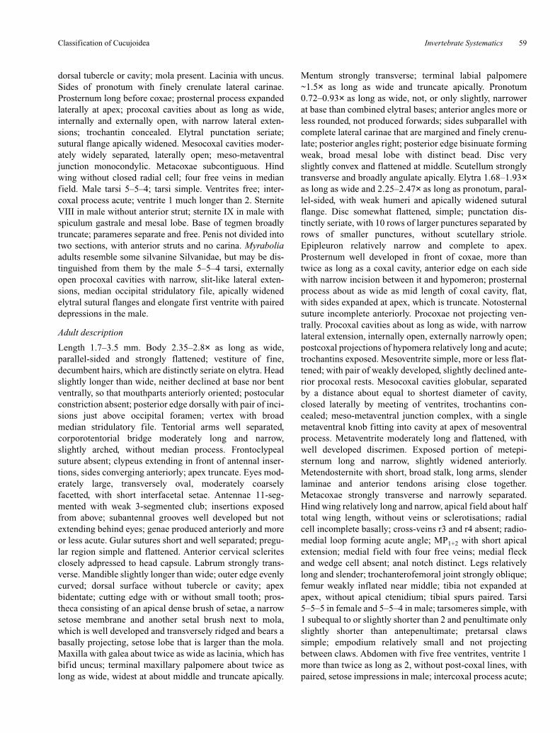
Invertebrate Systematics 59
dorsal tubercle or cavity; mola present. Lacinia with uncus.Sides of pronotum with finely crenulate lateral carinae.Prosternum long before coxae; prosternal process expandedlaterally at apex; procoxal cavities about as long as wide,internally and externally open, with narrow lateral exten-sions; trochantin concealed. Elytral punctation seriate;sutural flange apically widened. Mesocoxal cavities moder-ately widely separated, laterally open; meso-metaventraljunction monocondylic. Metacoxae subcontiguous. Hindwing without closed radial cell; four free veins in medianfield. Male tarsi 5–5–4; tarsi simple. Ventrites free; inter-coxal process acute; ventrite 1 much longer than 2. SterniteVIII in male without anterior strut; sternite IX in male withspiculum gastrale and mesal lobe. Base of tegmen broadlytruncate; parameres separate and free. Penis not divided intotwo sections, with anterior struts and no carina. Myraboliaadults resemble some silvanine Silvanidae, but may be dis-tinguished from them by the male 5–5–4 tarsi, externallyopen procoxal cavities with narrow, slit-like lateral exten-sions, median occipital stridulatory file, apically widenedelytral sutural flanges and elongate first ventrite with paireddepressions in the male.
Adult description
Length 1.7–3.5 mm. Body 2.35–2.8× as long as wide,parallel-sided and strongly flattened; vestiture of fine,decumbent hairs, which are distinctly seriate on elytra. Headslightly longer than wide, neither declined at base nor bentventrally, so that mouthparts anteriorly oriented; postocularconstriction absent; posterior edge dorsally with pair of inci-sions just above occipital foramen; vertex with broadmedian stridulatory file. Tentorial arms well separated,corporotentorial bridge moderately long and narrow,slightly arched, without median process. Frontoclypealsuture absent; clypeus extending in front of antennal inser-tions, sides converging anteriorly; apex truncate. Eyes mod-erately large, transversely oval, moderately coarselyfacetted, with short interfacetal setae. Antennae 11-seg-mented with weak 3-segmented club; insertions exposedfrom above; subantennal grooves well developed but notextending behind eyes; genae produced anteriorly and moreor less acute. Gular sutures short and well separated; pregu-lar region simple and flattened. Anterior cervical scleritesclosely adpressed to head capsule. Labrum strongly trans-verse. Mandible slightly longer than wide; outer edge evenlycurved; dorsal surface without tubercle or cavity; apexbidentate; cutting edge with or without small tooth; pros-theca consisting of an apical dense brush of setae, a narrowsetose membrane and another setal brush next to mola,which is well developed and transversely ridged and bears abasally projecting, setose lobe that is larger than the mola.Maxilla with galea about twice as wide as lacinia, which hasbifid uncus; terminal maxillary palpomere about twice aslong as wide, widest at about middle and truncate apically.
Mentum strongly transverse; terminal labial palpomere~1.5× as long as wide and truncate apically. Pronotum0.72–0.93× as long as wide, not, or only slightly, narrowerat base than combined elytral bases; anterior angles more orless rounded, not produced forwards; sides subparallel withcomplete lateral carinae that are margined and finely crenu-late; posterior angles right; posterior edge bisinuate formingweak, broad mesal lobe with distinct bead. Disc veryslightly convex and flattened at middle. Scutellum stronglytransverse and broadly angulate apically. Elytra 1.68–1.93×as long as wide and 2.25–2.47× as long as pronotum, paral-lel-sided, with weak humeri and apically widened suturalflange. Disc somewhat flattened, simple; punctation dis-tinctly seriate, with 10 rows of larger punctures separated byrows of smaller punctures, without scutellary striole.Epipleuron relatively narrow and complete to apex.Prosternum well developed in front of coxae, more thantwice as long as a coxal cavity, anterior edge on each sidewith narrow incision between it and hypomeron; prosternalprocess about as wide as mid length of coxal cavity, flat,with sides expanded at apex, which is truncate. Notosternalsuture incomplete anteriorly. Procoxae not projecting ven-trally. Procoxal cavities about as long as wide, with narrowlateral extension, internally open, externally narrowly open;postcoxal projections of hypomera relatively long and acute;trochantins exposed. Mesoventrite simple, more or less flat-tened; with pair of weakly developed, slightly declined ante-rior procoxal rests. Mesocoxal cavities globular, separatedby a distance about equal to shortest diameter of cavity,closed laterally by meeting of ventrites, trochantins con-cealed; meso-metaventral junction complex, with a singlemetaventral knob fitting into cavity at apex of mesoventralprocess. Metaventrite moderately long and flattened, withwell developed discrimen. Exposed portion of metepi-sternum long and narrow, slightly widened anteriorly.Metendosternite with short, broad stalk, long arms, slenderlaminae and anterior tendons arising close together.Metacoxae strongly transverse and narrowly separated.Hind wing relatively long and narrow, apical field about halftotal wing length, without veins or sclerotisations; radialcell incomplete basally; cross-veins r3 and r4 absent; radio-medial loop forming acute angle; MP1+2 with short apicalextension; medial field with four free veins; medial fleckand wedge cell absent; anal notch distinct. Legs relativelylong and slender; trochanterofemoral joint strongly oblique;femur weakly inflated near middle; tibia not expanded atapex, without apical ctenidium; tibial spurs paired. Tarsi5–5–5 in female and 5–5–4 in male; tarsomeres simple, with1 subequal to or slightly shorter than 2 and penultimate onlyslightly shorter than antepenultimate; pretarsal clawssimple; empodium relatively small and not projectingbetween claws. Abdomen with five free ventrites, ventrite 1more than twice as long as 2, without post-coxal lines, withpaired, setose impressions in male; intercoxal process acute;
Classification of Cucujoidea
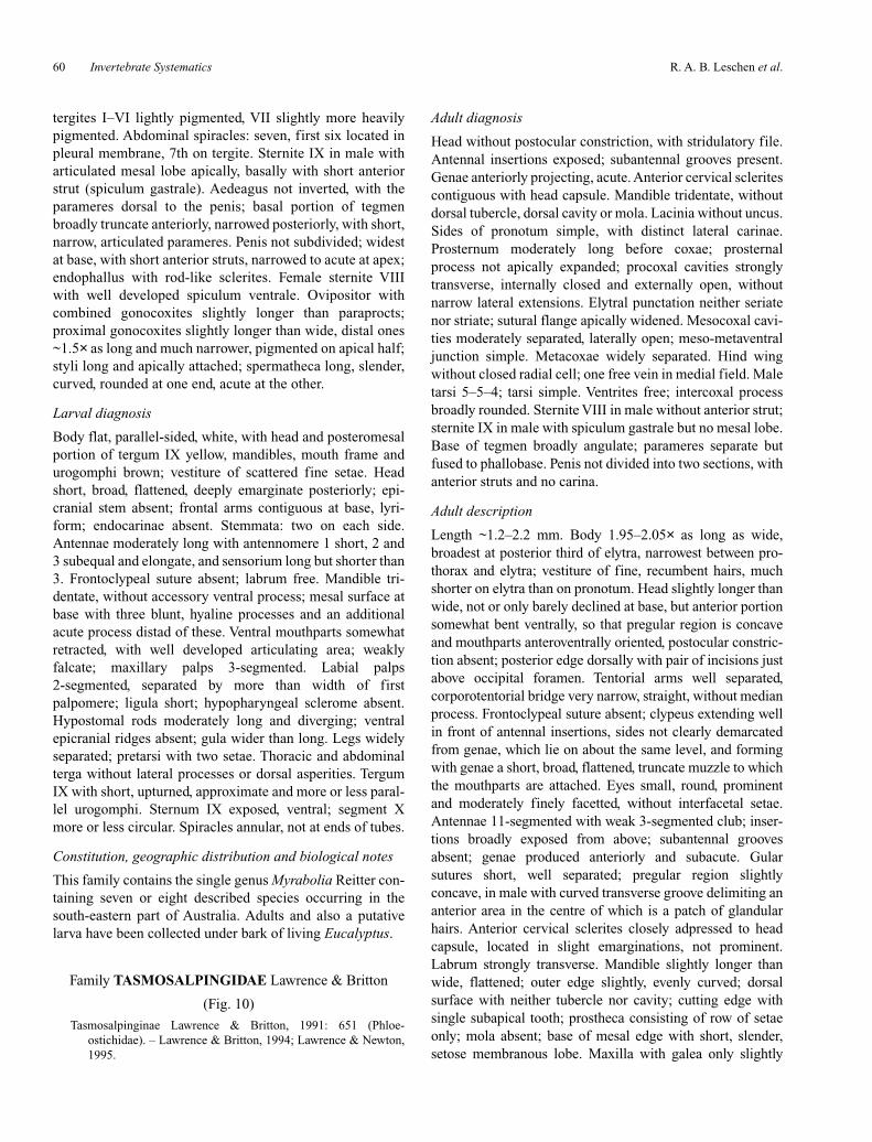
R. A. B. Leschen et al.60 Invertebrate Systematics
tergites I–VI lightly pigmented, VII slightly more heavilypigmented. Abdominal spiracles: seven, first six located inpleural membrane, 7th on tergite. Sternite IX in male witharticulated mesal lobe apically, basally with short anteriorstrut (spiculum gastrale). Aedeagus not inverted, with theparameres dorsal to the penis; basal portion of tegmenbroadly truncate anteriorly, narrowed posteriorly, with short,narrow, articulated parameres. Penis not subdivided; widestat base, with short anterior struts, narrowed to acute at apex;endophallus with rod-like sclerites. Female sternite VIIIwith well developed spiculum ventrale. Ovipositor withcombined gonocoxites slightly longer than paraprocts;proximal gonocoxites slightly longer than wide, distal ones~1.5× as long and much narrower, pigmented on apical half;styli long and apically attached; spermatheca long, slender,curved, rounded at one end, acute at the other.
Larval diagnosis
Body flat, parallel-sided, white, with head and posteromesalportion of tergum IX yellow, mandibles, mouth frame andurogomphi brown; vestiture of scattered fine setae. Headshort, broad, flattened, deeply emarginate posteriorly; epi-cranial stem absent; frontal arms contiguous at base, lyri-form; endocarinae absent. Stemmata: two on each side.Antennae moderately long with antennomere 1 short, 2 and3 subequal and elongate, and sensorium long but shorter than3. Frontoclypeal suture absent; labrum free. Mandible tri-dentate, without accessory ventral process; mesal surface atbase with three blunt, hyaline processes and an additionalacute process distad of these. Ventral mouthparts somewhatretracted, with well developed articulating area; weaklyfalcate; maxillary palps 3-segmented. Labial palps2-segmented, separated by more than width of firstpalpomere; ligula short; hypopharyngeal sclerome absent.Hypostomal rods moderately long and diverging; ventralepicranial ridges absent; gula wider than long. Legs widelyseparated; pretarsi with two setae. Thoracic and abdominalterga without lateral processes or dorsal asperities. TergumIX with short, upturned, approximate and more or less paral-lel urogomphi. Sternum IX exposed, ventral; segment Xmore or less circular. Spiracles annular, not at ends of tubes.
Constitution, geographic distribution and biological notes
This family contains the single genus Myrabolia Reitter con-taining seven or eight described species occurring in thesouth-eastern part of Australia. Adults and also a putativelarva have been collected under bark of living Eucalyptus.
Family TASMOSALPINGIDAE Lawrence & Britton
(Fig. 10)
Tasmosalpinginae Lawrence & Britton, 1991: 651 (Phloe-ostichidae). – Lawrence & Britton, 1994; Lawrence & Newton,1995.
Adult diagnosis
Head without postocular constriction, with stridulatory file.Antennal insertions exposed; subantennal grooves present.Genae anteriorly projecting, acute. Anterior cervical scleritescontiguous with head capsule. Mandible tridentate, withoutdorsal tubercle, dorsal cavity or mola. Lacinia without uncus.Sides of pronotum simple, with distinct lateral carinae.Prosternum moderately long before coxae; prosternalprocess not apically expanded; procoxal cavities stronglytransverse, internally closed and externally open, withoutnarrow lateral extensions. Elytral punctation neither seriatenor striate; sutural flange apically widened. Mesocoxal cavi-ties moderately separated, laterally open; meso-metaventraljunction simple. Metacoxae widely separated. Hind wingwithout closed radial cell; one free vein in medial field. Maletarsi 5–5–4; tarsi simple. Ventrites free; intercoxal processbroadly rounded. Sternite VIII in male without anterior strut;sternite IX in male with spiculum gastrale but no mesal lobe.Base of tegmen broadly angulate; parameres separate butfused to phallobase. Penis not divided into two sections, withanterior struts and no carina.
Adult description
Length ~1.2–2.2 mm. Body 1.95–2.05× as long as wide,broadest at posterior third of elytra, narrowest between pro-thorax and elytra; vestiture of fine, recumbent hairs, muchshorter on elytra than on pronotum. Head slightly longer thanwide, not or only barely declined at base, but anterior portionsomewhat bent ventrally, so that pregular region is concaveand mouthparts anteroventrally oriented, postocular constric-tion absent; posterior edge dorsally with pair of incisions justabove occipital foramen. Tentorial arms well separated,corporotentorial bridge very narrow, straight, without medianprocess. Frontoclypeal suture absent; clypeus extending wellin front of antennal insertions, sides not clearly demarcatedfrom genae, which lie on about the same level, and formingwith genae a short, broad, flattened, truncate muzzle to whichthe mouthparts are attached. Eyes small, round, prominentand moderately finely facetted, without interfacetal setae.Antennae 11-segmented with weak 3-segmented club; inser-tions broadly exposed from above; subantennal groovesabsent; genae produced anteriorly and subacute. Gularsutures short, well separated; pregular region slightlyconcave, in male with curved transverse groove delimiting ananterior area in the centre of which is a patch of glandularhairs. Anterior cervical sclerites closely adpressed to headcapsule, located in slight emarginations, not prominent.Labrum strongly transverse. Mandible slightly longer thanwide, flattened; outer edge slightly, evenly curved; dorsalsurface with neither tubercle nor cavity; cutting edge withsingle subapical tooth; prostheca consisting of row of setaeonly; mola absent; base of mesal edge with short, slender,setose membranous lobe. Maxilla with galea only slightly

Invertebrate Systematics 61
wider than lacinia, which lacks an uncus; terminal maxillarypalpomere about twice as long as wide, widest at base,strongly narrowed and narrowly rounded at apex. Mentumslightly transverse with rounded anterior angles; terminalpalpomere narrowly rounded at apex. Pronotum 0.78–0.85×as long as wide, base distinctly narrower than combinedelytral bases, broadest at about middle; sides straight or veryslightly curved; lateral carinae complete anteriorly and visiblefrom above, their edges weakly crenulate, but interruptedposteriorly by pair of large, deep, laterally opening setosecavities; anterior angles produced forwards and rounded butpartly obscured from above by anterior edge of pronotal disc;posterior angles slightly obtuse; posterior edge slightlycurved, unmargined. Disc moderately convex and simple.Scutellum strongly transverse, broadly rounded posteriorly.Elytra 1.34–1.40× as long as wide and 2.05–2.2× as long aspronotum; sides moderately rounded, strongly convergentposteriorly, with weak humeri and apically widened suturalflange. Disc slightly convex; punctation not distinctly seriate.Epipleura narrow and extending at about middle. Prosternumwell developed in front of coxae, slightly longer than coxalcavity; prosternal process almost as wide as mid length ofcoxal cavity, flat, with sides slightly expanded towards apex,which is emarginate. Notosternal suture complete. Procoxaenot projecting ventrally. Procoxal cavities strongly transverse,without narrow lateral extensions, internally closed, exter-nally broadly open, postcoxal projections of hypomera shortand acute; trochantins exposed. Mesoventrite strongly trans-verse, slightly convex; with pair of weakly developed, slightlydeclined anterior procoxal rests. Mesocoxal cavities sepa-rated by a distance almost as great as shortest diameter ofcavity, open laterally (partly closed by mesepimera); meso-metaventral junction a straight line. Metaventrite stronglytransverse and slightly convex, without discrimen. Exposedportion of metepisternum long and narrow, barely widenedanteriorly. Metendosternite with short, broad stalk, long arms,slender laminae and anterior tendons well separated.Metacoxae strongly transverse and widely separated. Hindwing short and broad, about twice as long as wide, widest atapical third; apical field very short and without sclerotisation;radial cell; cross-veins r3 and r4, and radio-medial loopabsent; medial field with a single free vein; medial fleck,wedge cell and anal notch absent. Legs relatively slender;trochanterofemoral joint oblique; femur weakly inflated; tibianot or slightly expanded at apex, without apical ctenidium;tibial spurs paired. Tarsi 5–5–5 in female and 5–5–4 in male;tarsomeres simple, with 1 longer than 2 and penultimate onlyslightly shorter than antepenultimate; empodium relativelysmall and often not projecting between claws. Abdomen withfive free ventrites, ventrite 1 not much longer than 2, withoutpost-coxal lines; intercoxal process broadly rounded; tergitesI–VI lightly to moderately pigmented, VII more heavily pig-mented. Abdominal spiracles well developed on segmentsI–V, reduced and non-functional on VI, located in pleural
membrane; absent on VII. Sternite IX in male membranousapically, without articulated median process, basally withanterior strut (spiculum gastrale). Aedeagus with basalportion of tegmen broadly angulate anteriorly, apical portionwell developed, with short, broad parameres, which are sepa-rate from one another but not basally articulated. Penis notsubdivided; narrow and parallel-sided basally, with shortbasal struts, broader apically and angulate or acute at apex;endophallus usually with long, rod-like sclerite. Female ster-nite VIII with well developed spiculum ventrale. Ovipositorwith combined gonocoxites subequal to paraprocts; proximalgonocoxite slightly longer than wide, distal one ~1.33× aslong and much narrower, pigmented on apical half; styli welldeveloped and apically attached; internal tract with stronglycurved, slender, sclerotised spermatheca.
Larval diagnosis
Body flat, parallel-sided, white with yellow head and vesti-ture if long, fine setae. Head flattened, deeply emarginateposteriorly, with pair of short incisions above occipitalforamen; frontal arms separated at base, lyriform and joinedanteriorly by transverse line; paired endocarinae relativelynarrowly separated. Stemmata: five with cluster of three andtwo behind them. Antennae relatively long, with antenno-mere 2 about twice as long as 1 and 3 slightly shorter than 1,with sensorium longer than 3. Frontoclypeal suture absent;labrum free. Mandible tridentate, without accessory ventralprocess; base with three hyaline processes. Ventral mouth-parts protracted or only slightly retracted, mala blunt; maxil-lary articulating area absent; maxillary palps 3-segmented;labial palps 2-segmented, separated by more than width offirst palpomere; ligula short; hypopharyngeal scleromeabsent. Gula longer than wide. Hypostomal rods diverging;ventral epicranial ridges absent. Prothorax with ventral,longitudinal median endocarina; legs moderately widelyseparated; pretarsus with single seta. Tergum IX simple;segment X more or less ventrally oriented, not visible fromabove. Spiracles annular, not at ends of tubes.
Constitution, geographical distribution and biological notes
The single genus Tasmosalpingus Lea includes two des-cribed species (T. promiscuus Lea and T. quadrispilotus Lea)from Tasmania but is also known from Victoria. Adults havebeen collected in Malaise traps, and a putative larva was col-lected by pyrethrum fogging the ark of Phyllocladus aspleni-ifolius (Labilladière) Hooker (Podocarpaceae). The hindgutof a dissected male was packed with fungal hyphae and pos-sibly spores of a hyphomycete.
Key to families of Cucujoidea
The following keys include all families of Cucujoidea (sensustricto, excluding Tenebrionoidea) plus some other groupsthat have been or may be confused with cucujoids (i.e.
Classification of Cucujoidea

R. A. B. Leschen et al.62 Invertebrate Systematics
Jacobsoniidae, basal or atypical Cleroidea, Tenebrionoideawith four or fewer mesotarsomeres). All non-cucujoidfamilies have the superfamily in brackets. All other taxa thatare likely to be confused with cucujoids have either a trilo-bate, staphylinoid or bostrichoid type of aedeagus and oftenhave functional 8th spiracles and distinctly excavate meta-coxae. Strepsiptera have been added to the larval key becauseof their likely confusion with some modified Cucujoidea.
Adults (larvae on p. 66)
1. Metacoxae extending laterally to meet edges of elytral epi-pleura; metepimeron not meeting anterior angles of ven-trite 1; tarsomeres never lobed; wings never with medialfleck; procoxae nearly always transverse with broadlyexposed trochantins, or if not, then tarsi 4-segmented andantennae 8-segmented with 1-segmented club;empodium usually well developed . CLEROIDEA (part)
Metacoxae not meeting edges of elytral epipleura;metepimeron meeting anterior angle of ventrite 1; if pro-coxae transverse with broadly exposed trochantins, thenother characters different; empodium usually absent orconcealed. . . . . . . . . . . . . . . . . . . . . . . . . . . . . . . . . . . . 2
2(1). Metaventrite at least as long as prothorax and mesoventritecombined; tarsi not lobed, apical tarsomere longer thanprevious ones combined; lateral pronotal carinae obso-lete; trochanters elongate; wings without medial fleck;abdominal segment VIII with functional spiracles . . . . . . . . . . . . . . . . . . . . Jacobsoniidae (DERODONTOIDEA)
Metaventrite very rarely as long as prothorax and meso-ventrite combined; segment VIII without functional spir-acles. . . . . . . . . . . . . . . . . . . . . . . . . . . . . . . . . . . . . . . . 3
3(2). Metaventrite with complete transverse suture; tarsi 5–5–5,without lobed segments, tarsomere 5 longer than previ-ous ones combined; procoxae globular, their cavitiesexternally closed behind; mesocoxal cavities laterallyopen; metacoxae slightly transverse, widely separatedand weakly excavate. . . . . . . . . . . . . . . . . . . . . Helotidae
Transverse suture of metaventrite incomplete or absent; withother characters different. . . . . . . . . . . . . . . . . . . . . . . . 4
4(3). Aedeagus with tegmen incomplete, not forming ring aroundpenis; tarsi 4–4–4 (or 3–4–4 in male), very rarely withlobed segments; antennal insertions often concealed;basal 2–4 ventrites often connate . . . . . . . . . . . . . . . . . . . . . . . . . . . . . . . . . . . . . . . . TENEBRIONOIDEA (part)
Aedeagus with tegmen forming ring around penis; if tarsi4–4–4, then antennal insertions rarely concealed; allventrites usually free . . . . . . . . . . . . . . . . . . . . . . . . . . . 5
5(4). Tarsi 5–5–5, without lobed segments; form convex androunded dorsally, flattened ventrally; procoxae stronglytransverse, with exposed trochantins, their cavities inter-nally closed; protibiae outwardly keeled and crenulate. . . . . . . . . . . . Trogossitidae: Rentoniinae (CLEROIDEA)
If tarsi so formed, then other characters different . . . . . . . . 66(5). Tarsi 5–5–5 at least in female; wings often with five free
veins in medial field (between MP1+2 and anal fold);elytra often with scutellary striole; protrochantins oftenexposed . . . . . . . . . . . . . . . . . . . . . . . . . . . . . . . . . . . . . 7
Tarsi 4–4–4 or 3–3–3; wings rarely with five free veins inmedial field; elytra very rarely with scutellary striole;protrochantins rarely exposed . . . . . . . . . . . . . . . . . . . 39
7(6). Mesocoxal cavities laterally open (not closed by meeting ofmesoventrite and metaventrite) . . . . . . . . . . . . . . . . . . . 8
Mesocoxal cavities laterally closed by meeting of meso-ventrite and metaventrite; protrochantins very rarelyexposed . . . . . . . . . . . . . . . . . . . . . . . . . . . . . . . . . . . . 32
8(7). Procoxae transverse with broadly exposed trochantins; galeavery slender or absent; outer edge of protibia usually withspines or denticles; head usually with subocular antennalgrooves; elytra often truncate, exposing abdominal apex(pygidium), never with scutellary striole. . . . . . . . . . . . 9
If procoxae transverse with exposed trochantins, then galeabroad and setose and outer edge of protibial apex neitherspinose nor dentate . . . . . . . . . . . . . . . . . . . . . . . . . . . 13
9(8). Antennae 9- or 10-segmented with 1- or apparently2-segmented club; elytra truncate, exposing at least onetergite. . . . . . . . . . . . . . . . . . . . . . . . . . . . . . . . . . . . . . 10
Antennae almost always 11-segmented; if rarely10-segmented, then antennal club 3-segmented; elytravariable . . . . . . . . . . . . . . . . . . . . . . . . . . . . . . . . . . . . 12
10(9). Antennal club consisting of single segment that is basallyglabrous and apically clothed with rings of pubescence;galea present, with few apical setae; tibial apex expandedand dentate; male tarsi 5–5–4; 7th abdominal spiraclesdistinct; temperate parts of northern hemisphere . . . . . . . . . . . . . . . . . . . . . . . . . . Monotomidae (Rhizophaginae)
Antennal club consisting of single, entirely pubescentsegment preceded by strongly transverse, glabrous,cupule-like segment; galea absent; tibial apex narrow andsimple; tarsi 5–5–5 in both sexes; 7th abdominal spira-cles not distinct . . . . . . . . . . . . . . . . . . . . . . . . . . . . . . 11
11(10). Antennae 10-segmented; cupule relatively broad, so that clubappears 2-segmented; elytra exposing pygidium only;south-east Asia . . . . . . . . . . . . Nitidulidae (Calonecrus)
Antennae 9-segmented; cupule very slender; elytra exposingthree abdominal tergites; tropical Africa . . . . . . . . . . . . . . . . . . . . . . . . . . . . . . . . . . . . . Nitidulidae (Maynipeplus)
12(11). Galea present; antennal club weak; subocular antennalgrooves weak or absent; frontoclypeal suture distinct;elytra truncate, exposing last two abdominal tergites;outer edge of protibia not dentate, with few or no spines. . . . . . . . . . . . . . . . . . . . . . . . . . . . . . . . . . . Kateretidae
Galea absent; antennal club usually strong; if elytra exposinglast two abdominal tergites, then subocular groovesstrong. . . . . . . . . . . . . . . . . . . . . . . . . . . . . . . Nitidulidae
13(8). Ventrite 1 always with femoral lines; pronotum almostalways with sublateral carinae; procoxal cavities trans-verse, closed behind, trochantins partly concealed; preg-ular region with pair of deep, laterally-opening pockets;outer edge of protibia dentate or spinose; wings withclosed radial cell and four free veins in medial field, theanterior one running into medial fleck . . . . . . . . . . . . . . . . . . . . . . . . . . . . . . . . . . . . . . . Biphyllidae (major part)
Ventrite 1 rarely with femoral lines, and if so then pronotumwithout sublateral carinae; pregular region never withlaterally-opening pockets; outer edge of protibia rarelyspinose or dentate . . . . . . . . . . . . . . . . . . . . . . . . . . . . 14
14(13). Pronotum with sublateral carinae; elytra usually with threeraised, longitudinal ridges; procoxal cavities small,rounded; tarsi never with lobed segments; antennal clubweak or absent . . . . . . . . . Laemophloeidae (major part)
Pronotum rarely with sublateral carinae; if present, then, tarsiand antennae of a different type . . . . . . . . . . . . . . . . . 15
15(14). Antennae 10-segmented with 1- or 2-segmented club; elytratruncate, exposing pygidium; galea slender, with fewapical setae; procoxal cavities closed behind, trochantinsconcealed; tarsomere 1 often very small, so that tarsi

Invertebrate Systematics 63
appear to be 4–4–4 or 4–4–3 (male) . . . . . . . . . . . . . . . . . . . . . . . . . . . . . . . . . Monotomidae (Monotominae part)
If antennae 10-segmented, then elytra completely concealingabdominal apex; galea broader with multisetose apex . . . . . . . . . . . . . . . . . . . . . . . . . . . . . . . . . . . . . . . . . . . . . 16
16(15). Antennae 10-segmented with 1-segmented club; outer edgeof protibia with stout spines; tarsi 5–5–5 in both sexes,without lobed segments; segment 1 shortest; mandiblewithout mola; procoxae transverse, their cavities closedbehind, with narrow lateral extensions; wingless specieswith short metaventrite and metepisterna widened poste-riorly; Australia, New Zealand, New Hebrides, NewCaledonia. . . . . . . . . . . . . Phycosecidae (CLEROIDEA)
Antennae not so formed; outer edge of protibia rarelyspinose; mandible rarely without mola; without othercharacters in combination . . . . . . . . . . . . . . . . . . . . . . 17
17(16). Tarsomeres 2 and 3 each with membranous ventral lobe,which extends well beyond apex of tarsomere; procoxaetransverse, their cavities internally and externally closed,with narrow lateral extensions partly exposingtrochantins; wings with closed radial cell and four freeveins in medial field (between MP1+2 and anal fold), theanterior one running into medial fleck . . . . . . Byturidae
Tarsomeres 2 and 3 without membranous ventral lobes;wings never with vein running into medial fleck . . . . 18
18(17). Procoxal cavities rounded, trochantins completely con-cealed; sutural flanges of elytra not at all widened api-cally; tarsi 5–5–5 in both sexes; antennal club indistinctor procoxal cavities closed behind; wings without closedradial cell or medial fleck. . . . . . Silvanidae (major part)
Procoxal cavities with lateral extensions at least partlyexposing trochantins; without other characters incombination. . . . . . . . . . . . . . . . . . . . . . . . . . . . . . . . . 19
19(18). Mandible with dorsal tubercle and associated setose cavity;frontoclypeal suture deeply impressed; clypeus sub-quadrate or slightly elongate with sides emarginatebasally; sutural flanges of elytra not widened apically;aedeagus without articulated parameres . . . . . . . . . . . 20
If mandible with dorsal tubercle, then associated cavityglabrous or absent; if clypeus subquadrate with basolat-eral emarginations, then elytra with sutural flangeswidened apically and/or aedeagus with articulatedparameres . . . . . . . . . . . . . . . . . . . . . . . . . . . . . . . . . . 21
20(19). Frontoclypeal suture distinctly arcuate; antennal club strong,as long as previous five or six antennomeres combined,and more or less uniformly, densely clothed with setifer-ous punctures and sensilla, creating a matt appearance;procoxal cavities externally closed or antennae10-segmented; tarsomeres all simple; male tarsi 5–5–4 . . . . . . . . . . . . . . . . . . . . . . . . . . . . . . . . . . . . . Sphindidae
Frontoclypeal suture not, or only slightly, arcuate; antennae11-segmented; antennal club weak or apparently absent,its segments sparsely setose and shiny basally with sen-silla concentrated at or near apex; procoxal cavities exter-nally open; tarsomere 3 distinctly lobed below, 4 reducedand simple; tarsi 5–5–5 in both sexes; Australia andsouthern Africa . . . . . . . . . . . . . . . . . . . . . . . Boganiidae
21(19). Tarsomeres 1–3 expanded and lobed below, 4 reduced andsimple; mandible without dorsal tubercle or cavity;antennal insertions concealed; procoxal cavities exter-nally closed; elytral sutural flanges not widened apically;body elongate and parallel-sided, usually somewhat flat-tened; hind wing with closed radial cell and five freeveins in medial field; aedeagus without articulatedparameres; Chile and Australia . . . . . . . . Protocucujidae
If tarsi with expanded basal tarsomeres and reduced tarso-mere 4, then mandible with dorsal tubercle and antennalinsertions exposed; elytral sutural flanges usuallywidened apically; if parameres not articulated, thenwithout closed radial cell and with a single vein in medialfield . . . . . . . . . . . . . . . . . . . . . . . . . . . . . . . . . . . . . . . 22
22(21). Mandible with distinct dorsal tubercle that fits into basolat-eral emargination of clypeus; Chile and Australia . . . 23
Mandible without dorsal tubercle or clypeal emargination. . . . . . . . . . . . . . . . . . . . . . . . . . . . . . . . . . . . . . . . . . . . . 24
23(22). Mandible with glabrous dorsal cavity or with mesal edgeproduced forming rounded lobe; antennal club veryweak; prothorax subquadrate to elongate, without or withweak lateral carinae but often with lateral tubercles orteeth; procoxal cavities narrowly open or narrowly closedexternally; elytral sutural flanges apically widened(except in wingless species); wings, if present, with atleast three free veins in medial field . . . . . . . . . . . . . . . . . . . . . . . . Phloeostichidae (Hymaea, Rhopalobrachium,. . . . . . . . . . . . . . . . . . . . . . . . . . . . . . . . . Bunyastichus)
Mandible with neither dorsal cavity nor rounded lobe onmesal edge; antennal club well developed; prothorax dis-tinctly transverse, with complete crenulate or denticulatelateral carinae; procoxal cavities broadly open externally;elytral sutural flanges not apically widened; wings withonly one free vein in medial field. . . . . . . . . Hobartiidae
24(22). Body broadly ovate, convex and glabrous; elytral suturalflanges not widened apically; epipleuron broad at basewith elongate, deep cavity (often filled with solidexudate); subantennal grooves long, deep and parallel,extending well behind eyes; New Zealand . . . . . . . . . . . . . . . . . . . . . . . . . . . . . . . . . . . . . . . . . . . . . Cyclaxyridae
Body oblong to elongate; epipleuron without deep cavity;subantennal grooves usually absent or, if present, notextending behind eyes . . . . . . . . . . . . . . . . . . . . . . . . . 25
25(24). Procoxal cavities externally broadly open . . . . . . . . . . . . 26Procoxal cavities externally narrowly open or closed; south-
ern hemisphere . . . . . . . . . . . . . . . . . . . . . . . . . . . . . . 2926(25). Procoxal cavities internally closed; occiput with broad,
median stridulatory file; pronotal disc with pair ofposterolateral setose cavities; aedeagus with parameresless than twice as long as wide at base and not distinctlyarticulated; Tasmania and Victoria . . . Tasmosalpingidae
Procoxal cavities internally open; occiput without stridula-tory file; pronotal disc without setose cavities; aedeaguswith parameres at least twice as long as wide and dis-tinctly articulated. . . . . . . . . . . . . . . . . . . . . . . . . . . . . 27
27(26). Tarsomere 1 much shorter than 2; labrum externally appar-ent; elytral sutural flange widened apically; elytralpunctures seriate, with scutellary striole; central andsouthern Europe and Russia and Russian Far East . . . . . . . . . . . . . . . . . . . . . . . . Phloeostichidae (Phloeostichus)
Tarsomere 1 at least as long as 2; labrum not externallyapparent, either completely concealed beneath clypeus orwith a very narrow anterior strip exposed; elytral suturalflange not widened apically; if elytral punctation seriate,scutellary striole absent . . . . . . . . . . . . . . . . . . . . . . . . 28
28(27). Body not strongly flattened; head with oblique, raised frontalcarinae; antennal club well marked; elytral puncturesseriate; tarsi 5–5–5 in both sexes; mesometaventral junc-tion between mesocoxae with single, broad, internalmetaventral knob; Australia . . . . . . . . . . Lamingtoniidae
Body strongly flattened; head without frontal carinae; anten-nal club weak or absent; elytral punctures confused; male
Classification of Cucujoidea

R. A. B. Leschen et al.64 Invertebrate Systematics
tarsi 5–5–4; mesometaventral junction between meso-coxae without internal knob . . . . . . . . . . . . . . Cucujidae
29(26). Procoxal cavities with very narrow lateral slits, trochantinslargely concealed; mandible with one or two laterally-opening internal vesicles; frons usually with a pair ofdeep pits, often joined by arcuate groove; procoxal cavi-ties narrowly open; male tarsi 5–5–5; Australia, NewZealand, Brazil and Chile . . . . . . . . . . . . Cavognathidae
If procoxal cavities with narrow lateral slits, then ventrite 1as long as next two combined; mandible withoutlaterally-opening vesicles; frons without deep pits orgroove; male tarsi almost always 5–5–4, if 5–5–5, thenprocoxal cavities externally closed . . . . . . . . . . . . . . . 29
30(29). Occiput with stridulatory file; procoxal cavities with narrowlateral slits, largely concealing trochantins; ventrite 1 atleast as long as next two ventrites combined; body elon-gate, parallel-sided and flattened; elytral punctation dis-tinctly seriate; parameres long and narrow; penis notdivided into basal and apical sections; Australia. . . . . . . . . . . . . . . . . . . . . . . . . . . . . . . . . . . . . . . . . Myraboliidae
Occiput without stridulatory files; if protrochantins largelyconcealed, then then body not flattened and elytral punc-tation not seriate; ventrite 1 never as long as next twocombined; parameres shorter and broader; penis dividedinto distinct basal and apical sections . . . . . . . . . . . . . 31
31(30). Protrochantins fully exposed; prosternal process wider thanshortest diameter of a procoxal cavity; head without post-ocular constriction; Australia, New Zealand and Chile. . . . . . . . . . . . . . . . . . . . . . . . . . . . . . . . . . . . Priasilphidae
Protrochantins partly concealed; prosternal process muchnarrower than shortest diameter of a procoxal cavity;head with weak postocular constriction; New Zealand. . . . . . . . . . . . . . . . . . . . . . . . . . . . . . . . . . . . . Agapythidae
32(7). Elytral punctation confused, sutural flanges apically widened(except in apterous species); epipleura oblique, obsoleteposteriorly; trochanters more or less elongate; ventrite 1much longer than 2; wings never with closed radial cellor medial fleck. . . . . . . . . . Cryptophagidae (major part)
Elytral sutural flanges rarely apically widened, if so thenwith distinct striae; if ventrite 1 much longer than 2, thenother characters different. . . . . . . . . . . . . . . . . . . . . . . 33
33(32). Elytra truncate, exposing pygidium, which is slotted alongmidline; head sharply constricted behind eyes; antennae10-segmented with 1-segmented club; galea very slenderwith few apical setae; procoxal cavities broadly closedbehind; New Zealand. . . . . . . . . . Monotomidae (Lenax)
Elytra not truncate, fully concealing abdomen from above;head not or weakly constricted behind eyes; galea neververy slender, multisetose at apex; antennae usually11-segmented with 3-segmented club . . . . . . . . . . . . . 34
34(33). Mandible with dorsal tubercle and setose cavity; head withstrongly arcuate frontoclypeal suture; male tarsi 5–5–4;antennae 10-segmented; procoxal cavities closed behind;eastern hemisphere . . . . . . . . Sphindidae (Aspidiphorus)
Mandible without dorsal tubercle or cavity; frontoclypealsuture straight or absent; if male tarsi 5–5–4, thenprocoxal cavities open behind; antennae usually11-segmented . . . . . . . . . . . . . . . . . . . . . . . . . . . . . . . 35
35(34). Maxillae largely concealed by expansions of head capsule;gular sutures confluent; labrum not evident; antennaethick, with club weak or absent; sutural flanges of elytraapically widened; wings with four or five free veins inmedial field (between MP1+2 and anal fold), medial fleckabsent; radial cell often closed; male tarsi 5–5–5; form
elongate, parallel-sided, usually glabrous above . . . . . . . . . . . . . . . . . . . . . . . . . . . . . . . . . . . . . . . . . Passandridae
Maxillae exposed; gular sutures not confluent; labrumusually evident; antennae usually thinner with distinctclub; sutural flanges not widened apically; if wings withmore than three free veins in medial field, then medialfleck present or male tarsi 5–5–4 . . . . . . . . . . . . . . . . 36
36(35). Pronotum with sublateral carinae; each elytron with threeraised, longitudinal ridges; male tarsi 5–5–4, withoutlobed tarsomeres, claws simple; antennae 11-segmentedwith loose, 3-segmented club; wings without closedradial cell, with 1 free vein in medial field (betweenMP1+2 and anal fold); New World and Africa . . . . . . . . . . . . . . . Laemophloeidae (Lathropus and Carinophloeus)
Pronotum without sublateral carinae; if male tarsi 5–5–4,then some tarsomeres lobed below or tarsal clawstoothed; without other characters in combination . . . . 37
37(36). Procoxae widely separated, their cavities small and rounded,externally and internally open behind; form broadlyovate, strongly flattened; prothorax very large; elytrawith raised longitudinal ridges; antennae slender, clubsegments elongate; male tarsi 5–5–5, without lobedtarsomeres . . . . . . . . . . . . . . . . . . . . . . . . . Propalticidae
If procoxae widely separated, then their cavities externallyclosed behind; if form broadly ovate, then not at all flat-tened; prothorax not so large; antennae thicker, club seg-ments not elongate. . . . . . . . . . . . . . . . . . . . . . . . . . . . 38
38(37). Form highly convex and usually glabrous above, flattenedbelow; procoxal cavities externally open, internallyclosed; claws basally toothed; wings never with closedradial cell or medial fleck; mesometaventral junctionbetween mesocoxae without internal knob; lacinia verynarrow, with few apical setae; without occipital stridula-tory file . . . . . . . . . . . . . . . . . . . . . . . . . . . . . Phalacridae
Form less convex dorsally, less flattened ventrally; clawsrarely toothed; wings often with closed radial cell ormedial fleck; lacinia broader, apically plurisetose;occiput usually with stridulatory file . . . . . . . Erotylidae
39(6). Tegmen not completely surrounding penis; medial fleck, ifpresent, not bisected by vein; elytra often with scutellarystriole; two or more basal ventrites often connate. . . . . . . . . . . . . . . . . . . . . . . . . . . . . . . . . TENEBRIONOIDEA
Tegmen either completely surrounding penis or rarelyabsent; medial fleck, if present, bisected by vein; elytrawithout scutellary striole; basal ventrites rarely connate. . . . . . . . . . . . . . . . . . . . . . . . . . . . . . . . . . . . . . . . . . . 40
40(39). Procoxae with fully exposed trochantins; mesocoxal cavitiesnot closed laterally by meeting of ventrites; galea absent;tarsi 4–4–4; elytra exposing pygidium from above ortarsomere 3 lobed beneath. . . . . . . . . . . . . . . . . . . . . . 41
Protrochantins concealed; mesocoxal cavities often closedlaterally by meeting of ventrites; galea present; if tarsi4–4–4 and elytra exposing pygidium, then mesocoxalcavities closed outwardly by meeting of ventrites. . . . 42
41(40). Procoxal cavities externally closed behind; elytra truncate,not concealing pygidium from above; head without sub-ocular grooves; tarsomeres not lobed below; form elon-gate, parallel-sided, flattened . . . . . . . . . . . . Smicripidae
Procoxal cavities externally open behind; elytra not truncate,at least partly concealing pygidium from above; headwith subocular antennal grooves; tarsomeres 2 and 3lobed below; form rounded, very convex . . . . . . . . . . . . . . . . . . . . . . . . . . . . . . . . . Nitidulidae (Cybocephalinae)
42(40). Elytral sutural flanges apically widened; frontoclypeal suturepresent; ventrite 1 as long as 2 and 3 combined;

Invertebrate Systematics 65
trochanters elongate; Afrotropical and eastern Palae-arctic regions . . . . . . . . . . . . . . . . . . . . . . . . . . . . . . . . . . . . . . . . Cryptophagidae (Cryptafricus and Atomaroides)
Elytral sutural flanges not apically widened; without othercharacters in combination . . . . . . . . . . . . . . . . . . . . . . 43
43(42). Elytra truncate exposing pygidium from above; antennae 10-segmented with 1- or 2-segmented club; male tarsi appar-ently 4–4–3; galea very slender with few apical setae . . . . . . . . . . . . . . . . . . . Monotomidae (Monotominae part)
Elytra rarely exposing pygidium, or if so then antennae dif-ferent; tarsi 4–4–4 or 3–3–3 in both sexes; galea welldeveloped. . . . . . . . . . . . . . . . . . . . . . . . . . . . . . . . . . . 44
44(43). Procoxal cavities broadly closed behind externally; meso-coxal cavities laterally open (not closed laterally bymeeting of ventrites); pregular region with paired, later-ally-opening pockets; tarsomere 2 with setose ventrallobe; body elongate and somewhat flattened; Australia . . . . . . . . . . . . . . . . . . . . . . . . . . . . . . . Biphyllidae (part)
If procoxal cavities closed behind, then mesocoxal cavitiesnearly always closed laterally by ventrites; pregularregion without paired pockets . . . . . . . . . . . . . . . . . . . 45
45(44). Form usually elongate and cylindrical, if somewhat flattened,then trochanterofemoral joint strongly oblique andtrochanter sometimes partly concealed by base of femur;protibiae markedly widened and angulate at outer apex;tarsi 4–4–4, without lobed tarsomeres; antennal clubusually 1-segmented; elytra with striae or rows ofpunctures . . . . . . . . . . . . . . . . . . . . . Bothrideridae (part)
Form never elongate and cylindrical; trochanters neverstrongly oblique or concealed; protibiae rarely apicallywidened and angled. . . . . . . . . . . . . . . . . . . . . . . . . . . 46
46(45). Antennae 8-segmented with 1-segmented club; antennalinsertions concealed beneath sides of frons; empodiumwell developed, bisetose; meso- and metacoxae very nar-rowly separated; apterous species with very shortmetaventrite lacking femoral lines . . . . . . . . . . . . . . . . . . . . . . . . . . Trogossitidae (Colydiopeltis and Parapeltis) . . . . . . . . . . . . . . . . . . . . . . . . . . . . . . . . (CLEROIDEA)
Antennae, if 8-segmented and with 1-segmented club, notinserted under sides of frons; without other characters incombination. . . . . . . . . . . . . . . . . . . . . . . . . . . . . . . . . 47
47(46). Tarsi 4–4–4 . . . . . . . . . . . . . . . . . . . . . . . . . . . . . . . . . . . . 48Tarsi 3–3–3 . . . . . . . . . . . . . . . . . . . . . . . . . . . . . . . . . . . . 62
48(47). Body elongate and strongly flattened; head abruptlynarrowed behind eyes forming distinct temples; antennaelong and filiform with scape more than 3× as long aspedicel; sides of pronotum dentate . . . . . . . . . . . . . . . . . . . . . . . . . . . . . . . . . . . . . . . . . . Uleiota (part) Silvanidae
If body elongate and flattened, then antennae different;without other characters in combination . . . . . . . . . . . 49
49(48). Mesocoxal cavities closed laterally by meeting of ventrites . . . . . . . . . . . . . . . . . . . . . . . . . . . . . . . . . . . . . . . . . . . . 50
Mesocoxal cavities not closed laterally by ventrites . . . . . 5750(49). Procoxal cavities externally and internally closed . . . . . . 51
Procoxal cavities, if closed externally, open internally . . . 5251(50). Head moderately large and exposed from above, more than
half as wide as pronotal base; body moderately convex;antennae shorter than head width; maxilla with distinctgalea and lacinia; abdomen with five pairs of spiracles;Central and South America . . . . . . . . . . . . . . . . . . . . . . . . . . . . . . . . . . . . . . . . . . . . . Endomychidae (Acritosoma)
Head very small and usually concealed from above, less thanhalf as wide as pronotal base; if head exposed fromabove, then body strongly flattened; antennae longer than
head width; maxilla with single lobe; abdomen withseven pairs of spiracles . . . . . . . . . . Corylophidae (part)
52(50). Apical edge of last abdominal ventrite inflexed and linedwith dorsoventrally oriented grooves, so that it appearscrenulate in ventral view; spiracles on first five abdomi-nal segments. . . . . . . . . . . . . . . . . . . . Cerylonidae (part)
Apical edge of last abdominal ventrite smooth; spiracles onfirst five or seven abdominal segments . . . . . . . . . . . . 53
53(52). Frontoclypeal suture absent; maxillary and labial palps acic-ulate; mesal portions of metacoxae concealed byexpanded intercoxal process of ventrite 1; Nepal, Indiaand Thailand . . . . . . . . . . . Cerylonidae (Loebliorylinae)
Frontoclypeal suture present; maxillary and labial palpssubulate . . . . . . . . . . . . . . . . . . . . . . . . . . . . . . . . . . . . 54
54(53). Body distinctly elongate and usually subcylindrical; protibiaexpanded apically and spinose along outer edge; antennalclub 2–3 –segmented; tarsomeres always simple . . . . . . . . . . . . . . . . . . . . . . . . . . . . . . Bothrideridae (Teredinae)
If body elongate and protibia somewhat expanded, thenantennal club 1-segmented . . . . . . . . . . . . . . . . . . . . . 55
55(54). Procoxal cavities externally closed and internally open;tarsomere 2 never lobed below; spiracles on first sevenabdominal segments . . . . . . . . Cerylonidae (Euxestinae)
Procoxal cavities externally open and internally closed;tarsomere 2 usually more or less lobed below. . . . . . . 56
56(55). Frontoclypeal suture present; maxilla with distinct galea andlacinia; metaventrite often with postcoxal pits; spiracleson first five abdominal segments; head larger and notconcealed by pronotum . . . . Endomychidae (major part)
Frontoclypeal suture absent; maxilla with single lobe;metaventrite never with postcoxal pits; spiracles on firstseven abdominal segments; head very small and more orless concealed by pronotum . . . . . . . . . . . . . . . . . . . . . . . . . . . . . . . . . . . . . . . . . . . . Corylophidae (Peltinodinae)
57(49). Mesoventrite, metaventrite and abdomen completely fused,without sutures or lines separating them; body flattenedand disc-like; antennae 8-segmented with 4-segmentedclub; scutellum not visible; abdominal spiracles: five;eastern Africa. . . . . . . . . . Corylophidae (Cleidostethini)
Mesoventrite, metaventrite and abdomen distinctly sepa-rated; without other characters in combination . . . . . . 58
58(57). Head without frontoclypeal suture; metaventrite and abdom-inal ventrite 1 almost always with curved femoral lines . . . . . . . . . . . . . . . . . . . . . . . . . . . . . . . . . . . . . . . . . . . . 59
Head with frontoclypeal suture; femoral lines rarely present. . . . . . . . . . . . . . . . . . . . . . . . . . . . . . . . . . . . . . . . . . . 60
59(58). Antennae relatively short, usually not, or only slightly, longerthan head width; antennal club weak, compact, shorterthan preceding antennomeres combined, without vesi-cles; ventrites 1 and 2 more or less connate; tegmen withmedian posterior projection (basal lobe) lying betweenparameres and with articulated anterior strut (trabes);penis (sipho) slender and curved, with enlarged base andno endophallic armature . . . . . . . . . . . . . . Coccinellidae
Antennae much longer than head width; antennal clubstrong, loosely-organised and longer than precedingantennomeres combined, with vesicles; all ventrites free;tegmen reduced, without median projection or articulatedstrut; penis broader with complex endophallic armatureCorylophidae (minor part)
60(58). Tarsomeres simple, not lobed below; body elongate to sub-cylindrical; antenna 11-segmented with compact2-segmented club; procoxal cavities externally and inter-nally open . . . . . . . . . . . Bothrideridae (Xylariophilinae)
Classification of Cucujoidea

R. A. B. Leschen et al.66 Invertebrate Systematics
Tarsomere 2 at least slightly lobed below; if simple thenantenna 10-segmented with 3-segmented club; bodybroadly oval, never subcylindrical. . . . . . . . . . . . . . . . 61
61 (60). Procoxal cavities externally and internally open; spiracles onfirst seven abdominal segments; antennae 10-segmentedwith 3-segmented club; tarsomere 2 not, or only slightly,lobed below; apical maxillary palpomere inflated andsecuriform; Southern Europe, North Africa and AsiaMinor . . . . . . . . . . . . . . . . . . . . . . . . . . . . . . . . Alexiidae
Procoxal cavities externally open and internally closedbehind; spiracles on first five abdominal segments only;apical maxillary palpomere subulate. . . . . . . . . . . . . . . . . . . . . . . . . . . . . . . . . . . . . . . . . . . . Endomychidae (part)
62(47). Metacoxae externally very small; metaventrite broadly meetingventrite 1 laterally; gland openings along lateral edges ofprothorax and elytra; antennae 9- or 10-segmented with1-segmented club . . . . . . . . . . . . . . . . . . Discolomatidae
Metacoxae larger; metaventrite not meeting ventrite 1 later-ally; lateral edges of prothorax and elytra without glandopenings . . . . . . . . . . . . . . . . . . . . . . . . . . . . . . . . . . . 63
63(62). Apical edge of terminal abdominal ventrite inflexed and linedwith dorsoventrally oriented grooves, so that it appearscrenulate in ventral view; tarsomeres simple, not lobedbelow . . . . . . . . . . . . . . . . . . . . Cerylonidae (minor part)
Apical edge of terminal abdominal ventrite smooth, if itappears as crenulate then tarsomere 2 distinctly lobedbelow . . . . . . . . . . . . . . . . . . . . . . . . . . . . . . . . . . . . . . 64
64(63). Procoxal cavities externally and internally closed; labrumlaterally expanded beyond anterior angles of clypeus;lacinia vestigial; elytral punctures in rows; spiracles onfirst seven abdominal segments. . . . . . . . . . . . . . . . . . . . . . . . . . . . . . . . . . . . . . . . . . . . . . Latridiidae (major part)
Procoxal cavities externally open . . . . . . . . . . . . . . . . . . . 6565(64). Procoxal cavities internally open; spiracles on first seven
abdominal segments; wings absent; eyes reduced orabsent. . . . . . . . . . . . . . . . . . . . . . . . . . . . . . . . . . . . . . 66
Procoxal cavities internally closed; spiracles on first fiveabdominal segments; wings and eyes almost always welldeveloped. . . . . . . . . . . . . . . . . . . . . . . . . . . . . . . . . . . 67
66(65). Protibia widened and spinose at outer apical angle; labrumnot laterally expanded; Europe, North Africa and AsiaMinor, widely introduced elsewhere . . . . . . . . . . . . . . . . . . . . . . . . . . . . . . . . . . . . . Bothrideridae (Anommatinae)
Protibia neither widened nor spinose at outer apex; labrumlaterally expanded; Western North America . . . . . . . . . . . . . . . . . . . . . . . . . . . . . . . Latridiidae (Akalyptoischion)
67(65). Frontoclypeal suture absent; ventrites 1 and 2 connate;femoral lines present on metaventrite and abdominalventrite 1; tegmen with median posterior projection(basal lobe) lying between parameres and with articu-lated anterior strut (trabes); penis (sipho) slender andcurved, with enlarged base . . . . . . . Coccinellidae (part)
Frontoclypeal suture present; ventrites freely articulated;femoral lines rarely present; aedeagus of a different type. . . . . . . . . . . . . . . . . . . . . . . . . . . . Endomychidae (part)
Larvae
1. Larva of triungulin type, with dorsum evenly sclerotised andbearing posteriorly-projecting spines; urogomphi absent;maxillary articulating area absent; mandible withoutmola; labrum usually fused to clypeus . . . . . . . . . . . . . 2
Larva not of triungulin type; dorsum without strong,posteriorly-projecting spines; without other characters incombination. . . . . . . . . . . . . . . . . . . . . . . . . . . . . . . . . . 6
2(1). Maxillary palps absent; abdominal segment VIII without dis-tinct spiracles; antenna with short single segment andlong terminal bristle; stemmata: 3–5. . . . . . . . . . . . . . . . . . . . . . . . . . . . . . . . . . . . . . . . . Order STREPSIPTERA
Maxillary palps distinct; abdominal segment VIII with dis-tinct spiracles; antenna 2- or 3-segmented; stemmatavariable . . . . . . . . . . . . . . . . . . . . . . . . . . . . . . . . . . . . . 3
3(2). Stemmata: five or six on each side . . . . . . . . . . . . . . . . . . 4Stemmata: one or two on each side. . . . . . . . . . . . . . . . . . . 5
4(3). Abdominal spiracles annular–biforous; ventral mouthpartsslightly retracted, distance between mandibular and max-illary articulations less than width of stipes; pretarsuswithout adhesive organ . . . . . . . . . . . Passandridae (part)
Abdominal spiracles annular; ventral mouthparts stronglyretracted distance between mandibular and maxillaryarticulations greater than width of stipes; pretarsus withadhesive organ . . Rhipiphoridae (TENEBRIONOIDEA)
5(3). Pretarsus bisetose; head with distinct coronal suture (epicra-nial stem); maxillary palp 3-segmented; antenna withsensorium short, attached to apex of antennomere 2 . . . . . . . . . . . . . . . . . . . . . Meloidae (TENEBRIONOIDEA)
Pretarsus unisetose; head without coronal suture; maxillarypalps 2-segmented; antenna with sensorium long,attached near base of antennomere 2. . . . . . . . . . . . . . . . . . . . . . . . . . . . . . . . . . . . . . . . . . . . . Bothrideridae (part)
6(1). Larva ectoparasitic, unpigmented, fleshy; broader at middleand narrowed at either end; legs short or absent; stem-mata absent; mandible without mola; maxillary articu-lating area obsolete; spiracles annular. . . . . . . . . . . . . . 7
Larva not ectoparasitic, usually with at least head capsulepigmented; stemmata usually present; legs always welldeveloped; without other characters in combination. . . 8
7(6). Ventral mouthparts slightly retracted, distance betweenmandibular and maxillary articulations less than width ofstipes; hypostomal rods distinct, strongly divergent; legsdistinct . . . . . . . . . . . . . . . . . . . . . . . Passandridae (part)
Ventral mouthparts strongly retracted, distance betweenmandibular and maxillary articulations less than width ofstipes; hypostomal rods indistinct; legs sometimes absent. . . . . . . . . . . . . . . . . . . . . . . . . . . . . Bothrideridae (part)
8(6). Maxillary mala deeply cleft so that partially fused galea andlacinia are still evident; dorsal surfaces granulate andusually with setiferous tubercles; mandible with acces-sory ventral process, well developed, asperate molaextending onto ventral surface and acute hyalineprostheca; stemmata: six; ventral epicranial ridgesusually present. . . . . . . . . . . . . . . . . . . . . . . . . . . . . . . . 9
Maxillary mala undivided or cleft only at apex, separategalea and lacinia not evident . . . . . . . . . . . . . . . . . . . . 10
9(8). Labrum partly fused to head capsule; hypostomal rodspresent; segment X ventral, membranous; spiracles notborne on tubes . . . . . . . . . . . . . . . . . . . . . . . . . Helotidae
Labrum free; hypostomal rods absent; abdominal segment Xterminal, usually with sclerotised tergum; spiracles borneon short tubes. . . . Derodontidae (DERODONTOIDEA)
10(8). Frontal arms more or less V-shaped, sometimes slightlycurved or sinuate but rarely lyriform; usually with eitherlong epicranial stem or long median endocarina; pairedendocarinae rarely present and then combined withretracted ventral mouthparts; mandibular mola absent,mandibular base simple or with one or more hyaline pro-cesses; ventral mouthparts often protracted; maxillarymala often with pedunculate seta; pretarsus always uni-setose; tergum IX not forming articulated plate; body

Invertebrate Systematics 67
always elongate and more or less parallel-sided . . . . . . . . . . . . . . . . . . . . . . . . . . . . . . . . . . . . . . . (CLEROIDEA)
Frontal arms usually lyriform with epicranial stem short orabsent; base of mandible rarely with one or more hyalineprocesses instead of a mola; paired endocarinae usuallyassociated with protracted ventral mouthparts in combi-nation with two pretarsal setae, lyriform frontal arms,rings of abdominal asperities or an articulated 9thtergum; pedunculate malar setae sometimes present intaxa with retracted ventral mouthparts, an oblong or pos-teriorly tapered body and an expanded or flattened pre-tarsal seta . . . . . . . . . . . . . . . . . . . . . . . . . . . . . . . . . . . 11
11(10). Ventral mouthparts protracted or slightly retracted, distancebetween mandibular and maxillary articulations less thanwidth of stipes; maxillary articulating area highlyreduced or absent; labial palps 2-segmented; mandibleoften without mola, or with mola but no prostheca; max-illary mala obtuse; base of epicranium often deeplyemarginate and with median or paired endocarinae;coxae widely separated; abdominal segment X small,transverse, not forming typical pygopod. . . . . . . . . . . 12
If ventral mouthparts protracted or slightly retracted, thenlabial palps 1-segmented and/or maxillary articulatingarea well developed . . . . . . . . . . . . . . . . . . . . . . . . . . . 17
12(11). Urogomphi absent; abdomen without pigmented terga;mesal surface of mandibular base with cluster of hyalineprocesses . . . . . . . . . . . . . . . . . . . . . . . . . . . . . . . . . . . 13
Urogomphi present; abdomen usually with tergum IX orterga VIII and IX pigmented; mesal surface of mandib-ular base varying . . . . . . . . . . . . . . . . . . . . . . . . . . . . . 15
13(12). Antenna very short, antennomeres 1 and 2 transverse, 3much longer and narrow; paired endocarinae parallel;tergum IX unusually short, with pair of articulations atbase; spiracles annular . . . . . . . . . . . . . . . . Propalticidae
Antenna longer, with antennomere 2 distinctly longer thaneither 1 or 3; tergum IX not unusually short or artic-ulated; spiracles annular–biforous. . . . . . . . . . . . . . . . 14
14(13). Posterior edge of head capsule gradually emarginate; pairedendocarinae contiguous at base and anteriorly divergent;sensorium subequal in length to antennomere 3; pretarsusbisetose. . . . . . . . . . . . . . . . . . . . . . . . . . . . Cyclaxyridae
Posterior edge of head capsule abruptly emarginate; pairedendocarinae distant at base, parallel, and interruptedanteriorly; sensorium longer than antennomere 3; pre-tarsus unisetose. . . . . . . . . . . . . . . . . . Tasmosalpingidae
15(12). Hypostomal rods parallel, very long, almost reaching poste-rior edge of head capsule; cardo indistinct; tergum IXforming articulated plate; sternum IX and segment Xreduced, membranous and partly enclosed withinemargination of sternum VIII; pretarsus unisetose;abdominal spiracles annular . . . . . . . . . Laemophloeidae
Hypostomal rods divergent, usually shorter; cardo usuallydistinct; spiracles annular–biforous. . . . . . . . . . . . . . . 16
16(15). Abdominal spiracle 8 located on long, posteriorly-project-ing, acute process; sternum IX and segment X exposed;head without median endocarina, with paired endocari-nae; pretarsus bisetose . . . . . . . . . . . . . . Lamingtoniidae
Abdominal spiracle 8 not located on posterorly-projectingprocess; sternum IX and segment X more or less con-cealed beneath posteriorly projecting lobe on sternumVIII; head often with median endocarina; pretarsusunisetose . . . . . . . . . . . . . . . . . . . . . . . . . . . . Phalacridae
17(11). Ventral mouthparts strongly retracted, maxillary articulatingarea strongly reduced or absent; hypostomal ridges grad-
ually curved mesally and hypostomal cavity narrowlyrounded posteriorly; cardines longitudinally oriented;prostheca almost always complex, consisting of severalto many processes (rarely simple and broadly angulate orabsent); maxillary mala almost always broadly roundedapically, with subapical bidentate or unidentate processon mesal edge (rarely truncate, without mesal process);labial palps 1-segmented; stemmata: four or fewer; uro-gomphi almost always present, sometimes accompaniedby pregomphi (if urogonmphi absent, then segments VIIIand IX with lateral spiniform processes); if hypostomalrods present, then pretarsus with spatulate seta. . . . . . . . . . . . . . . . . . . . . . . . . . . . . . . . . . . . . . . . . . . . Nitidulidae
If ventral mouthparts strongly retracted, then maxillary artic-ulating area usually distinct, hypostomal ridges abruptlycurved mesally, so that hypostomal cavity more truncateposteriorly and cardines more or less transverselyoriented; without other characters in combination . . . 18
18(17). Ventral mouthparts strongly protracted; cardo not distinct;hypostomal rods long and subparallel; prostheca absent;maxillary mala wider than long, obtuse, simple; labialpalps 1-segmented; with single stemma on each side;body elongate, parallel-sided and flattened; pretarsusunisetose; tergum IX well sclerotised with urogomphi;spiracles annular–biforous . . . . . . . . . . . . . . Smicripidae
Without above characters in combination . . . . . . . . . . . . . 1919(18). Mandible with reduced mola, not extending onto dorsal or
ventral surfaces, and without prostheca or ventral acces-sory process; ventral mouthparts slightly retracted;maxilla with transverse stipes and simple, plapiformmala articulated at base; labial palps 1-segmented;hypostomal rods divergent; pretarsus unisetose; uro-gomphi absent; segment X terminal with short pygopods. . . . . . . . . . . . . . . . . . . . . . . . . . . . . . . . . . . Kateretidae
If mandibular mola reduced and restricted to mesal surface,then prostheca present; if mala articulated at base, thenlabial palps 2-segmented; without other characters incombination. . . . . . . . . . . . . . . . . . . . . . . . . . . . . . . . . 20
20(19). Abdominal segment X more or less transverse, forming bil-abiate structure; stemma not more than 5; mandible neverwith prostheca; molae usually asymmetrical; maxillarymala never falcate (but sometimes acute at inner apex),occasionally cleft; head often with coronal suture (epi-cranial stem); pretarsus rarely unisetose . . . . . . . . . . . . . . . . . . . . . . . . . . . . . . . . . . . . . . . (TENEBRIONOIDEA)
Abdominal segment X usually cylindrical; stemmata: oftensix; prostheca usually present; maxillary mala often acuteat apex; mandibles usually symmetrical; head rarely withcoronal suture; pretarsus often unisetose . . . . . . . . . . 21
21(20). Tergum IX reduced and sternum IX highly reduced and con-cealed or apparently absent; segment X forming elon-gate, cylindrical, pygopod-like structure; spiraclesannular; maxillary mala acute at apex; antennomere 2 atleast twice as long as 1; urogomphi long, slender andstraight or absent; pretarsus bisetose . . . . . . . Silvanidae
Tergum IX well developed; sternum IX exposed; segment Xshort and cylindrical or reduced; if spiracles annular, thenmaxillary mala blunt, urogomphi variable . . . . . . . . . 22
22(21). Body of tergum IX much shorter than tergum VIII, formingheavily sclerotised, articulated plate with forked posteriorprojection or pair of urogomphi; sternum IX very short,slightly to strongly retracted into apex of sternum VIII;segment X scarcely projecting; head wider than pro-thorax, with sides more or less straight and subparallel or
Classification of Cucujoidea

R. A. B. Leschen et al.68 Invertebrate Systematics
converging anteriorly; antennomeres 2 and 3 elongateand usually subequal; sensorium less than 0.2× as long asantennomere 3; body flattened; abdominal segmentswithout rows of asperities and urogomphi, if present,without cavity between them. . . . . . . . . . . . . . Cucujidae
Body of tergum IX not, or only slightly, shorter than tergumVIII, not forming articulated plate; of urogomphi;sternum IX not retracted into apex of sternum VIII;segment X sometimes projecting; if head wider than pro-thorax, sides more rounded and cavity present betweenurogomphi or abdominal segments with curved rows oftergal and sternal asperities; sensorium more than 0.2× aslong as antennomere 3. . . . . . . . . . . . . . . . . . . . . . . . . 23
23(22). Ventral mouthparts slightly retracted; mesal surface ofmandibular base with brush of hairs and fixed, hyalineprocess, mola absent; maxillary mala falcate; endo-carinae absent; hypostomal rods long and divergent; pre-tarsus bisetose; urogomphi short and upturned spiraclesannular; body parallel-sided, flat and lightly sclerotisedexcept for head and urogomphi . . . . . . . . . Myraboliidae
Ventral mouthparts strongly retracted; without other charac-ters in combination . . . . . . . . . . . . . . . . . . . . . . . . . . . 24
24(23). Maxillary mala long, falcate, articulated at base, withoutsetae; mandible with broad-based hyaline prostheca;mandibular mola not extending onto dorsal or ventralsurfaces; urogomphi very short and blunt; segment Xwith pair of long, cylindrical pygopods. . . . . Boganiidae
Maxillary mala shorter, setiferous, not basally articulated;abdominal segment X without paired pygopods; withoutother characters in combination . . . . . . . . . . . . . . . . . 25
25(24). Prostheca with narrow base; maxillary mala blunt; mandib-ular mola well developed with accessory ventral process;spiracles annular or annular-multiforous; terga withoutsetiferous tubercles; dorsal setae simple; urogomphismall and simple or absent . . . . . . . . . . . . . . Sphindidae
If prostheca with narrow base, then maxillary mala more orless acute at apex or spiracles annular–biforous; withoutother characters in combination . . . . . . . . . . . . . . . . . 26
26(25). Pretarsus unisetose; prostheca, if present, consisting ofnumerous comb-hairs or two or more acute, hyaline pro-cesses; if prostheca absent, then base of mola withstrongly projecting, membranous, setose lobe; stemmata:six; mala falciform or somewhat truncate with acuteinner angle . . . . . . . . . . . . . . . . . . . . . . . . . . . . . . . . . . 27
If pretarsus unisetose, then mola absent or prostheca consist-ing of single hyaline process; if mola is present withoutprostheca, then there is no membranous, setose projec-tion at its base . . . . . . . . . . . . . . . . . . . . . . . . . . . . . . . 28
27(26). Mandible without accessory ventral process; prosthecaeither absent or consisting of a few simple hyaline pro-cesses; pigment and sculpture on dorsal surfaces more orless uneven; 8th spiracles lateral . . . . . . . . . . . Byturidae
Mandible with accessory ventral process; prostheca consist-ing of numerous comb-hairs; dorsal surfaces more or lessevenly pigmented and sculptured; 8th spiracles dorsalnear edge of tergum . . . . . . . . . . . . . . . . . . . Biphyllidae
28(26). Mola, if present, accompanied by prostheca with narrowbase and acute apex; if mola absent, mesal surface ofmandibular base with two hyaline lobes, one acute andone fimbriate; maxillary mala more or less acute at innerapex; pretarsus bisetose; spiracles usuallyannular–biforous . . . . . . . . . . . . . . . . . . . . . . . . . . . . . 29
Mola, if present, almost always accompanied by prosthecawith broad base and broadly angulate apex (prostheca
rarely absent); if mola absent, mesal surface of mandibu-lar base either simple or with membranous lobe; maxil-lary mala often blunt at inner apex; urogomphi oftenabsent; spiracles often annular . . . . . . . . . . . . . . . . . . 36
29(28). Head with distinct ventral epicranial ridges; stemmata: noneor two; mandible with well developed mola and narrowlyacute or occasionally bidentate prostheca; cutting edgeoften serrate cutting edge; labial palps usually 1-seg-mented; abdominal terga usually granulate or with seti-ferous tubercles, sometimes forming transverse rows;urogomphi usually broad and blunt or divided, rarelyabsent . . . . . . . . . . . . . . . . . . . . . . . . . . . . Monotomidae
Ventral epicranial ridges absent; stemmata: usually morethan two, or if not, then mola replaced by an acute andfimbriate hyaline process and urogomphi simple andupturned. . . . . . . . . . . . . . . . . . . . . . . . . . . . . . . . . . . . 30
30(29). Labial palps 1-segmented; dorsal surfaces with setosetubercles; stemmata: five; prostheca small and acute;maxillary mala falcate or obtuse with acute inner angle;urogomphi simple and upturned; spiraclesannular–biforous, borne on distinct spiracular tubes . . . . . . . . . . . . . . . . . . . . . . . . . . . . . . . . . . . . . . Hobartiidae
If labial palps 1-segmented, dorsal surfaces without setosetubercles and prostheca different; without other charac-ters in combination . . . . . . . . . . . . . . . . . . . . . . . . . . . 31
31(30). Mandibular mola absent; mesal surface of mandibular basewith an acute hyaline process adjacent to a membranous,apically fimbriate lobe; accessory ventral process absent;maxillary mala obliquely truncate but with one or morespines at inner apical angle; urogomphi short, posteriorlydirected, not upturned; head and major portions of allterga (including X) with relatively smooth, more or lessevenly pigmented surface; pretarsus bisetose . . . . . . . . . . . . . . . . . . . . . . . . . . . . . . . . . . . . . . . . . . Cavognathidae
Mandible with well developed, asperate mola; maxillarymala acute at apex; urogomphi, if present, longer andupturned or complex . . . . . . . . . . . . . . . . . . . . . . . . . . 32
32(31). Labial palps 1-segmented or spiracles annular; mandiblewith well developed mola and usually with serratecutting edge; prostheca often bifid, its proximal edgesometimes fimbriate; dorsal surfaces relatively lightlypigmented and smooth or finely granulate, without seti-ferous tubercles; urogomphi simple and upturned orabsent . . . . . . . . . . . . . . . . . . . . . . . . . . . Cryptophagidae
Labial palps 2-segmented; spiracles annular–biforous; molawell developed and asperate; prostheca simple and acute;dorsal surfaces often with setiferous tubercles or rows ofasperities; urogomphi often complex . . . . . . . . . . . . . 33
33(32). Pretarsus strongly toothed at base; abdominal spiraclesannular–biforous, borne on spiracular tubes; trunk tergauniformly sclerotised and pigmented, with anterior trans-verse carinae and setiferous tubercles, without lateraloutgrowths; urogomphi long, with lateral spines and pre-gomphi; stemmata: six . . . . . . . . . . . . . . Protocucujidae
Pretarsus not markedly toothed at base; if trunk terga sclero-tised and pigmented, then they have lateral outgrowths;stemmata variable . . . . . . . . . . . . . . . . . . . . . . . . . . . . 34
34(33). Thoracic and abdominal terga with long lateral outgrowths,those on abdominal terga I–VIII with spiracles at theirends; urogomphi moderately to very long, posteriorly-projecting and slightly to strongly divergent; antenno-mere 2 ~2× as long as 1 and more than 5× as long as 3;stemmata on each side located on one or more projectingtubercles . . . . . . . . . . . . . . . . . . . . . . . . . . . Priasilphidae

Invertebrate Systematics 69
Trunk terga without lateral outgrowths; antennomere 2 notmuch longer than 3; stemmata not located on projectingtubercles . . . . . . . . . . . . . . . . . . . . . . . . . . . . . . . . . . . 35
35(34). Spiracles large, with accessory chambers much longer thanperitreme diameter; labrum freely articulated; urogomphiapproximate at base, upturned and divergent, with lateralspines; body only slightly flattened; abdominal terga andsterna without paired patches of asperities . . . . . . . . . . . . . . . . . . . . . . . . . . . . . . . . . . . . . . . . . . . . . . Agapythidae
Spiracles smaller, with accessory chambers subequal inlength to peritreme diameter; labrum fused to clypeus;urogomphi distant at base, posteriorly directed andupturned at apex only, without lateral spines; bodymarkedly flattened; urogomphi with small cavitybetween them (Phloeostichus) or abdominal terga andsterna I–VII each with paired curved rows of asperities(Hymaea and Rhopalobrachium) . . . . . . Phloeostichidae
36(28). Pretarsus bisetose; maxillary mala acute at inner apex;mandible with well developed mola; prostheca broad-based, sometimes membranous; antenna with sensoriummuch shorter than antennomere 3; spiraclesannular–biforous . . . . . . . . . . . . . . . . . . . . . . . . . . . . . 37
Pretarsus unisetose; without other characters in combination. . . . . . . . . . . . . . . . . . . . . . . . . . . . . . . . . . . . . . . . . . . 40
37(36). Head with long epicranial stem (coronal suture); stemmata:two or fewer; maxillary mala falcate; body more or lesscylindrical; dorsal surfaces lightly pigmented and even,without granulations or setiferous tubercles; urogomphisimple and upturned. . . . . . . . . . Erotylidae (Languriini)
If epicranial stem long; stemmata: five or six and maxillarymala truncate; upper surfaces heavily pigmented and/orwith granulations or setiferous tubercles; urogomphiusually complex, with accessory processes or pre-gomphal tubercles . . . . . . . . . . . . . . . . . . . . . . . . . . . . 38
38(37). Epicranial stem absent; median endocarina present betweenfrontal arms; maxillary mala falcate; dorsal surfaces atleast slightly granulate, without lateral projections; setif-erous tubercles present only on tergum IX; urogomphirelatively simple and upturned, with one or more pairs ofpregomphal tubercles . . . . Erotylidae (Pharaxonothinae)
Median endocarina, if present, combined with long epi-cranial stem and truncate maxillary mala . . . . . . . . . . 39
39(38). Thoracic and abdominal terga with processes extending overspiracles or body distinctly tapered posteriorly; vestitureoften including expanded or frayed setae; tergum IXwithout pregomphal processes; urogomphi short andposteriorly-directed, sometimes with lateral setiferousprocesses; mala falcate; mandibular mola always presentand prostheca apically acute . . . . . . . . . . . . . . . . . . . . . . . . . . . . . . . . . . . . . . . . . . . . Erotylidae (Cryptophilinae)
Lateral tergal processes absent; urogomphi usually stronglyupturned or very long, rarely absent and replaced bymedian spine; upper surfaces granulate, tuberculate, withpatches of asperities or bearing complex processes; malausually more or less truncate, with stout apical teeth orspines; mandibular mola sometimes absent and prosthecabroad and membranous . . . . . . . Erotylidae (major part)
40(36). Mandible and maxilla stylet-like, either entognathous orenclosed within tubular piercing beak; head stronglyhypognathous, often concealed from above; dorsal sur-faces somewhat flattened, usually granulate or withsetose tubercles and often with modified setae; uro-gomphi absent . . . . . . . . . . . Cerylonidae (Ceryloninae)
Mandible of normal chewing type, usually with basal asper-ate mola; urogomphi variable . . . . . . . . . . . . . . . . . . . 41
41(40). Mandibular mola absent or reduced to a tooth-like process;prostheca absent; tibiotarsus almost always bearing spat-ulate, adhesive setae at apex; pretarsus usually wide atbase and abruptly narrowing apically; maxillary articu-lating area obsolete; frontal sutures visible, usually lyri-form; stemmata: three; terga usually pigmented andtuberculate; vestiture often complex; urogomphi rarelypresent . . . . . . . . . . . . . . . . . . . . . . . . . . . . Coccinellidae
Mandible with well developed, asperate mola; tibiotarsuswithout adhesive setae apically; pretarsus graduallynarrowing apically; without other characters incombination. . . . . . . . . . . . . . . . . . . . . . . . . . . . . . . . . 42
42(41). Urogomphi short and complex or acute and upturned . . . 43Urogomphi absent or long and divergent . . . . . . . . . . . . . 46
43(42). Stemmata absent; lateral edges of terga VI–IX more stronglyproduced posteriorly than those of anterior terga; abdom-inal spiracles annular, not placed on tubes . . . . . . . . . . . . . . . . . . . . . . . . . . . . . . . . Bothrideridae (Anommatinae)
Stemmata: five or six; terga equally projecting; spiraclesannular–biforous and placed at ends of spiracular tubes. . . . . . . . . . . . . . . . . . . . . . . . . . . . . . . . . . . . . . . . . . . . 44
44(43). Stemmata: five; urogomphi short, simple and upturned. . . . . . . . . . . . . . . . . . . . . . . . . . . . . . . . . . . . . . . . . Alexiidae
Stemmata: six; urogomphi simple or complex . . . . . . . . . 4545(44). Urogomphi complex; tergum IX with median boss between
and in front of urogomphi; sensorium longer thanantennomere 3; spiracular chambers much longer thanperitreme and extending out onto surface of long spirac-ular tube . . . . . . . . . . . . Bothrideridae (Xylariophilinae)
Urogomphi simple; tergum IX with paired setiferous pro-cesses in front of urogomphi; sensorium shorter thanantennomere 3; spiracular chambers not much longerthan peritreme, spiracular tubes shorter . . . . . . . . . . . . . . . . . . . . . . . . . . . . . . . . . . . . . Bothrideridae (Teredinae)
46(42). Urogomphi long and divergent; terga often with lateral pro-cesses; stemmata: none or two; spiracles placed on shortprocesses. . . . . . . . . . . . . . . . . Cerylonidae (Euxestinae)
Urogomphi absent or inconspicuous; terga usually withoutprocesses; spiracles not on tubes. . . . . . . . . . . . . . . . . 47
47(46). Antenna 2-segmented with sensorium at apex . . . . . . . . . . . . . . . . . . . . . . . . . . . . . . . . . . . . . . . . . . . Discolomatidae
Antenna 3-segmented with sensorium situated laterally onapex of antennomere 2 . . . . . . . . . . . . . . . . . . . . . . . . 48
48(47). Mandible with apical part and prostheca absent; maxillaryarticulating area distinct; stemmata absent; dorsal setaesimple and acute. . . . . . Endomychidae (Anamorphinae)
Mandible complete, with prostheca, or stemmata present; atleast some dorsal setae modified or maxillary articulat-ing area obsolete . . . . . . . . . . . . . . . . . . . . . . . . . . . . . 49
49(48). Maxillary articulating area obsolete . . . . . . . . . . . . . . . . . 50Maxillary articulating area present . . . . . . . . . . . . . . . . . . 51
50(49). Stemmata: usually two; abdomen almost always with glan-dular openings on segments I–VII or on I and VIII; formoften more or less onisciform; pretarsal seta long andspatulate; labial palps usually 2-segmented; mandibleentirely sclerotised. . . . . . . . . . . . . . . . . . . Corylophidae
Stemmata: four or five; abdomen without glandular open-ings; form not onisciform; pretarsal seta short andsimple; labial palps usually 1-segmented; mandible oftenpartly membranous . . . . . . . . . . . . . . . . . . . . Latridiidae
51(49). Body onisciform; head with long median endocarina . . . . . . . . . . . . . . . . . . . . . . . . . . . . Cerylonidae (Mumidiinae)
Body shape not or less strongly onisciform; endocarinaabsent. . . . . . . . . . . . . . . . . . . . . . . Endomychidae (part)
Classification of Cucujoidea

R. A. B. Leschen et al.70 Invertebrate Systematics
Acknowledgments
We thank R. A. Crowson for his initial studies of basalCucujoidea, without which we could not have completed thepresent study. For specimens we thank N. B. Nikitsky for thegift of a larva of Phloeostichus denticollis and D. Telnov forfacilitating the mailing of this specimen from St Petersburgto Gympie, J. Kolibáč for allowing us to examine his putativePhloeostichus larva, A. A. Allen for the gift of the holotypeof Chileosilpha elguetai (that was donated to the MuseoNacional de Historia Natural, Santiago) and for allowing usto remove the abdomen of his female paratype, andM. Elgueta for allowing the completely dissected maleparatype to be deposited in the Australian National InsectCollection. We also thank G. B. Monteith for his specialefforts in obtaining the type series of Bunyastichus monteithiand those curators listed in the Material examined. For helpwith data entry, SEM work, and scanning, we thank G. Hall,B. Rhode, E. Hines, and R. Henderson. Habitus illustrationswere done by D. Helmore (Chileosilpha, Priasilpha spp., andpartial habiti of Priastichus spp.), S. P. Kim (Priastichusmegathorax, Hymaea magna, Rhopalobrachium crowsoni,R. penai, Myrabolia grouvelliana), S. Monteith (Tasmo-salpingus sp.), S. Smith (larva of Priastichus crowsoni), andP. Wegrzynowicz (Phloeostichus denticollis). We thank RolfBeutel and Joe McHugh for reviews of the manuscript.Funding for RABL was supported in part by FRST (contractC09X0002).
References
Arrow, G. J. (1929). On the families of Coleoptera related to theErotylidae, with descriptions of a new family, two new genera, anda few new species. Annals and Magazine of Natural History 4,305–322.
Audisio, P. (1993). ‘Fauna d’Italia. Vol. XXXII. Coleoptera Nitidulidae– Kateretidae.’ (Edizioni Calderini: Bologna, Italy.)
Beutel, R., and Haas, F. (2000). Phylogenetic relationships of the sub-orders of Coleoptera (Insecta). Cladistics 16, 103–141.doi:10.1006/clad.1999.0124
Beutel, R., and Pollock, D. A. (2000). Larval head morphology ofPhycosecis litoralis (Pascoe) (Coleoptera: Phycosecidae) withphylogenetic implications. Invertebrate Taxonomy 14, 825–835.doi:10.1071/IT00030
Beutel, R., and Ślipiński, S. A. (2001). Comparative study of headstructures of larvae of Sphindidae and Protocucujidae (Coleoptera:Cucujoidea). European Journal of Entomology 98, 219–232.
Blackburn, T. (1903). Further notes on Australian Coleoptera withdescriptions of new genera and species. Part xxxii. Transactions ofthe Royal Society of South Australia 28, 91–182.
Böving, A. G., and Craighead, F. C. (1931). An illustrated synopsis ofthe principal larval forms of the order Coleoptera. EntomologicaAmericana 11, 1–351.
Bremer, K. (1988). The limits of amino-acid sequence data inangiosperm phylogenetic reconstruction. Evolution; InternationalJournal of Organic Evolution 42, 795–803.
Bremer, B., Jansen, R. K., Oxelman, B., Backlund, M., Lantz, H., andKim, K. (1999). More characters or more taxa for a robust phylo-geny—case study from the coffee family (Rubiaceae). SystematicBiology 48, 413–435. doi:10.1080/106351599260085
Broun, T. (1893). ‘Manual of the New Zealand Coleoptera. Parts V, VI,VII.’ (New Zealand Institute: Wellington, New Zealand.)
Burakowski, B., and Ślipiński, S. A. (1987). A new species ofProtosphindus (Coleoptera: Sphindidae) from Chile with notes anddescriptions of immature stages of related forms. Annali del MuseoCivico di Storia Naturale Giacomo Doria 86, 605–625.
Burmeister, E. G. (1976). Der Ovipositor der Hydradephaga(Coleoptera) und seine phylogenetische Bedeutung unter beson-derer Berücksichtigung der Dytiscidae. Zoomorphologie 85,165–257. doi:10.1007/BF00993515
Burmeister, E. G. (1980). Funktionsmorphologie und Evolution desOvipositor der Adephaga (Coleoptera). Verhandlungen des natur-wissenschaftlichen Vereines in Hamburg (N.F.) 24(1), 89–184.
Campau, E. J. (1940). The morphology of Chauliognathus pennsyl-vanicus (De Geer) (Coleoptera: Cantharidae). Microentomology 5,57–86.
Caterino, M. S., Shull, V. S., Hammond, P. M., and Vogler, A. P. (2002).Basal relationships of Coleoptera inferred from 18S rDNAsequences. Zoologica Scripta 31, 41–49. doi:10.1046/j.0300-3256.2001.00092.x
Candy, S. G. (1990). Biology of the mountain pinhole borer, Platypussubgranosus Schedl, in Tasmania. M.Sc. Thesis, University ofTasmania, Hobart, Australia.
Cekalovic, K. T. (1976). Nuevo hallazgo de un insecto poco conocidopara Chile (Coleoptera, Phloeostichidae). Bolletín de la Sociedad deBiología de Concepcion 50, 209–211.
Crosby, T. K., Dugdale, J. S., and Watt, J. C. (1998). Area codes forrecording specimen localities in the New Zealand subregion. NewZealand Journal of Zoology 25, 175–183.
Crowson, R. A. (1955). ‘The Natural Classification of the Families ofColeoptera’. (Nathaniel Lloyd: London, UK.)
Crowson, R. A. (1964). A review of the classification of Cleroidea(Coleoptera), with descriptions of two new genera of Peltidae and ofseveral new larval types. Transactions of the Royal EntomologicalSociety of London 116, 275–327.
Crowson, R. A. (1973). Further observations on Phloeostichidae andCavognathidae, with definitions of new genera from Australia andNew Zealand. The Coleopterists Bulletin 27, 54–62.
Crowson, R. A. (1995). Some interesting evolutionary parallels inColeoptera. In ‘Biology, Phylogeny, and Classification ofColeoptera. Papers Celebrating the 80th Birthday of Roy A.Crowson’. (Eds J. Pakaluk and S. A. Ślipiński.) pp. 63–85.(Muzeum i Instytut Zoologii, Polska Academia Nauk: Warsaw,Poland.)
Crowson, R. A., and Sen Gupta, T. (1969). The systematic position ofPropalticidae and of Carinophloeus Lefkovitch (Coleoptera,Clavicornia) with description of a new species of Propalticus and ofits supposed larva. Proceedings of the Royal Entomological Society38, 132–140.
Dallwitz, M., Paine, T. A., and Zurcher, E. J. (2000). Principles of inter-active keys. Available at: http://biodiversity.uno.edu/delta/ (verifiedFebruary 2005).
Eriksson, T. (2000). AutoDecay 4.0.2’ ppc. Available at:http://www.bergianska.se/index_forskning_soft.html (verifiedFebruary 2005).
Eriksson, T., and Wikström, N. (1995). Autodecay (version 3.0).(Program distributed by the first author.) (Department of Botany,Stockholm University: Stockholm, Sweden.)
Falcoz, L. (1926). Position systématique des genres Diphyllus Stephenset Diplocoelus Guerin. In ‘Ecyclopedie Entomologique, Serie B,Mémoires et Notes. I. Coleoptera, Vol. 1, 1925–26’. (Ed P. Lesne.)pp. 69–74. (P. Lechevalier, Paris, France.)
Farris, J. S. (1983). The logical basis of phylogenetic analysis. In‘Advances in Cladistics’. (Eds N.I. Platnick and V. A. Funk.)pp. 7–36. (Columbia University Press: New York, USA.)

Invertebrate Systematics 71
Farris, J. S. (1988). ‘Hennig86 Reference, version 1.5. ComputerProgram and Documentation.’ (J. S. Farris: Stony Brook, New York,USA.)
Farris, J. S., Albert, V. A., Kallersjö, M., Lipscomb, D., and Kluge, A. G.(1996). Parsimony jackknifing outperforms neighbor-joining.Cladistics 12, 99–124. doi:10.1006/clad.1996.0008
Felsenstein, J. (1985). Confidence limits on phylogenies: an approachusing the bootstrap. Evolution; International Journal of OrganicEvolution 39, 783–791.
Ferris, G. F. (1940). The myth of the thoracic sternites of insects.Microentomology 5, 87–90.
Forbes, W. T. M. (1926). The wing folding patterns of the Coleoptera.Journal of the New York Entomological Society 34, 42–139.
Ganglbauer, L. (1899). ‘Die Käfer von Mitteleuropa. Vol. 3.’ (CarlGerold’s Sohn: Vienna, Austria.)
Goloboff, P. (1999). ‘NONA, Version 2.0.’ (Instituto Miguel Lillo:Sierra Madre de Tucuman, Argentina.) Available fromhttp://www.cladistics.com/ (verified February 2005).
Goodrich, M. A., and Springer, C. A. (1992). A revision of the familyBiphyllidae (Coleoptera) for America, north of Mexico. TheColeopterists Bulletin 46(4), 361–377.
Grouvelle, A. (1913). ‘Coleopterorum Catalogus. Pars 56: Byturidae,Nitidulidae: 1. Cateretinae, 2. Meligethinae, 3. Carpophilinae, 4.Nitidulinae, 5. Cryptarchinae, 6. Cybocephalinae’. (W. Junk: Berlin,Germany.)
Hayashi, N. (1986). [Key to Coleoptera larvae]. In ‘The Coleoptera ofJapan in Color. Volume I’. (Eds. K. Morimoto and N. Hayashi.)pp. 202–217. (Hoikusha Publ. Co.: Osaka, Japan.)
Kirejtshuk, A. G. (2000). On origin and early evolution of the super-family Cucujoidea (Coleoptera, Polyphaga). Comments on thefamily Helotidae. The Kharkov Entomological Society Gazette 8,8–38.
Kolibáč, J. (2003). Description of a larva of Phloeostichus denticollisRedtenbacher, 1842 (Coleoptera, Phloeostichidae). EntomologicaBasiliensia 25, 135–140.
Krivolushkaya, G. O. (1992). Cem. Cucujidae – Ploskotelki. In‘Opredelitel′ Nasekomikh dal′nego Vostoka SSSR v Shesti Tomakh.Tom III. Zhestkokrilie, ili Zhuyki. Chast′ 2’. (Ed. P. A. Ler.)pp. 233–245. (“Nauka”: St. Petersburg, Russia.) [In Russian.]
Kukalová-Peck, J., and Lawrence, J. F. (2004). Relationships amongcoleopteran suborders and major neopteran lineages: Evidence fromhind wing characters. European Journal of Entomology 101,95–104.
Lameere, A. (1900). Notes pour la classification des Coléoptères.Annales de la Société Entomologique de Belgique 44, 355–377.
Lawrence, J. F. (1988). Notes on the classification of some Australiancucujoidea (Coleoptera). Journal of the Australian EntomologicalSociety 27, 53–54.
Lawrence, J. F. (1995). Two new species of Rhopalobrachium Boheman(Coleoptera: Phloeostichidae: Hymaeinae) from Australia andChile. In ‘Biology, Phylogeny, and Classification of Coleoptera.Papers Celebrating the 80th Birthday of Roy A. Crowson’.(Eds J. Pakaluk and S. A. Ślipiński.) pp. 433–447. (Muzeum iInstytut Zoologii, Polska Academia Nauk: Warsaw, Poland.)
Lawrence, J. F. (1999). The Australian Ommatidae (Coleoptera:Archostemata): new species, larva and discussion of relationships.Invertebrate Taxonomy 13, 369–390.
Lawrence, J. F. (2001). A new genus of Valdividian Scirtidae(Coleoptera) with comments on Scirtoidea and beetle suborders.Special Publications of the Japanese Coleoptera Society Osaka 1,351–361.
Lawrence, J. F., and Britton, E. B. (1991). Coleoptera (beetles). In‘Insects of Australia. A Textbook for Students and ResearchWorkers. Second Edition. Vol. 2’. (Ed. CSIRO Division of
Entomology.) pp. 543–683. (Melbourne University Press:Melbourne, Australia.)
Lawrence, J. F., and Britton, E. B. (1994). ‘Australian Beetles’.(Melbourne University Press: Melbourne, Australia.)
Lawrence, J. F., and Hlavac, T. F. (1979). Review of the Derodontidae(Coleoptera: Polyphaga) with new species from North America andChile. The Coleopterists Bulletin 33, 369–414.
Lawrence, J. F., and Leschen, R. A. B. (2003). Review ofLamingtoniidae (Coleoptera: Cucujoidea) with descriptions of twonew species. In ‘Systematics of Coleoptera: Papers Celebrating theRetirement of Ivan Löbl’. (Eds G. Cuccodoro and R. A. B. Leschen.)pp 905–919. (International Associated Publishers, Memoirs onEntomology, International, Volume 17.) (Associated Publishers:Gainesville, FL, USA.)
Lawrence, J. F., and Newton, A. F. (1995). Families and subfamilies ofColeoptera (with selected genera, notes, references and data onfamily-group names). In: ‘Biology, Phylogeny and Classification ofColeoptera: Papers Celebrating the 80th Birthday of Roy A.Crowson’. (Eds J. Pakaluk and S. A. Ślipiński.) pp. 779–1006.(Muzeum i Instytut Zoologii, Polska Academia Nauk: Warsaw,Poland.)
Lawrence, J. F., Ślipiński, S. A., and Pakaluk, J. (1995). From Latreilleto Crowson: a history of the higher level classification of beetles. In:‘Biology, Phylogeny and Classification of Coleoptera: PapersCelebrating the 80th Birthday of Roy A. Crowson’. (Eds J. Pakalukand S. A. Ślipiński.) pp. 87–154. (Muzeum i Instytut Zoologii,Polska Academia Nauk: Warsaw, Poland.)
Lawrence, J. F., Hastings, A. M., Dallwitz, M. A., Paine, T. A., andZurcher, E. J. (1999a). ‘Beetle Larvae of the World. Descriptions,Illustrations, and Information Retrieval for Families andSubfamilies’. CD-ROM, Version 1.1 for MS-WINDOWS. (CSIROPublishing: Melbourne, Australia.)
Lawrence, J. F., Hastings, A. M., Dallwitz, M. A., Paine, T. A., andZurcher, E. J. (1999b). ‘Beetles of the World. A Key andInformation System for Families and Subfamilies.’ CD-ROM,Version 1.0 for MS-WINDOWS. (CSIRO Publishing: Melbourne,Australia.)
Lea, A. M. (1919). Descriptions of new species of AustralianColeoptera. Records of the South Australian Museum 2, 271–308.
Leschen, R. A. B. (1996). Phylogeny and revision of the genera ofCryptophagidae (Coleoptera: Cucujoidea). University of KansasScience Bulletin 55(15), 549–634.
Leschen, R. A. B. (2003). ‘Fauna of New Zealand, Vol. 47. Erotylidae(Insecta: Coleoptera: Cucujoidea): Phylogeny and Review.’(Ed. T. Crosby.) (Manaaki Whenua Press: Canterbury, NewZealand.)
Maddison, W. P., and Maddison, D. R. (1992). ‘MacClade: Analysis ofPhylogeny and Character Evolution, Version 3.0.’ (SinauerAssociates: Sunderland, MA, USA.)
Maddison, W. P., Donoghue, M. J., and Maddison, D. R. (1984).Outgroup analysis and parsimony. Systematic Zoology 33, 83–103.
McHugh, J. V. (1993). A revision of Eurysphindus LeConte(Coleoptera: Cucujoidea: Sphindidae) and a review of sphindidclassification and phylogeny. Systematic Entomology 18, 57–92.
Nikitsky, N. B. (1991). News on the genus Triphyllia Reitter, 1898(Coleoptera, Tetratomidae). Elytron 5, 159–168.
Nixon, K. C. (1999). Winclada (BETA) ver. 1.00.08. K. C. Nixon,Ithaca, New York.
Nixon, K. C., and Carpenter, J. (1993). On outgroups. Cladistics 9,413–426. doi:10.1006/clad.1993.1028
Reitter, E. (1876). Neue Gatungen und Arten aus der Familie derCucujidae. Coleopterologische Hefte 15, 37–64.
Reitter, E. (1911). ‘Fauna Germanica. Die Käfer des DeutschenReiches. Vol. 3.’ (K. G. Lutz: Stuttgart, Germany.)
Classification of Cucujoidea

R. A. B. Leschen et al.72 Invertebrate Systematics
Rymer Roberts, A. W. (1958). On the taxonomy of Erotylidae(Coleoptera), with special reference to the morphological charactersof the larvae, II. Transactions of the Royal Entomological Society ofLondon 110, 245–284.
Sanderson, M. J. (1995). Objections to bootstrapping phylogenies: a cri-tique. Systematic Biology 44, 299–320.
Sen Gupta, T. (1988). Review of the genera of the familyRhizophagidae (Clavicornia: Coleoptera) of the world. Memoirs ofthe Zoological Survey of India 17, 1–58.
Sen Gupta, T., and Crowson, R. A. (1966). A new family of cucujoidbeetles based on six Australian and one New Zealand genera. Annalsand Magazine of Natural History 9, 61–85.
Sen Gupta, T., and Crowson, R. A. (1969). Further observations on thefamily Boganiidae, with definition of two new familiesCavognathidae and Phloeostichidae. Journal of Natural History 3,571–590.
Sen Gupta, T., and Crowson, R. A. (1973). A review of the classificationof Cerylonidae (Coleoptera, Clavicornia). Transactions of the RoyalEntomological Society of London 124, 365–446.
Sen Gupta, T., and Crowson, R. A. (1979). The coleopteran familySphindidae. The Entomologist’s Monthly Magazine 113, 177–191.
Ślipiński, S. A. (1998). Revision and phylogeny of Protocucujidae(Coleoptera: Cucujoidea). Annales Zoologici (Warszawa) 48,275–298.
Ślipiński, S. A. (1990). A monograph of the world Cerylonidae(Coleoptera; Cucujoidea). Part I – Introduction and higher classi-fication. Annali del Museo Civico di Storia Naturale GiacomoDoria 88, 1–273.
Ślipiński, S. A., and Pakaluk, J. (1992). Problems in the classificationof the cerylonid series of Cucujoidea (Coleoptera). In: ‘Advances inColeopterology’. (Eds M. Zunino, X. Bellés and M. Blas.)pp. 79–88. (Asociacion Europea de Coleopterolgia: Barcelona,Spain.)
Steiner, W. E., and Singh, B. P. (1987). Redescription of an ergot beetle,Acylomus pugetanus Casey, with immature stages and biology(Coleoptera: Phalacridae). Proceedings of the EntomologicalSociety of Washington 89(4), 744–758.
Swofford, D. L. (2003). ‘PAUP* Phylogenetic Analysis UsingParsimony (*and Other Methods), Version 4.’ (Sinauer Associates:Sunderland, MA, USA.)
Tait, S. M., Dahlsten, D. L., Gill, R. J., and Doyen, J. T. (1990). Lifehistory of the incense cedar scale, Xylococculus macrocarpae(Homoptera: Margarodidae), on incense cedar in California with adescription of the larvae of one of its common predators, Eronyxaexpansus Van Dyke (Coleoptera: Trogossitidae). Hilgardia 58(2),1–19.
Tanner, V. M. (1927). A preliminary study of the genitalia of femaleColeoptera. Transactions of the American Entomological Society53, 5–50.
Tomaszewska, W., and Ślipiński, S. A. (1995). A review of the familyHobartiidae (Coleoptera: Cucujoidea). Genus 6(3–4), 303–325.
Vogt, H. (1967). Cucujidae. In ‘Die Käfer Mitteleuropas. Band 7.Clavicornia’. (Eds H. Freude, K. W. Harde and G. A. Lohse.)pp. 83–104. (Goecke and Evers: Krefeld, Germany.)
Wegrzynowicz, P. (2002). Morphology, phylogeny and classification ofthe family Erotylidae based on adult characters (Coleoptera:Cucujoidea). Genus 13(4), 435–504.
Weise, J. (1897). Biologische Mitteilungen. Deutsche EntomologischeZeitschrift 1897, 389–395.
Wiens, J. J. (1998). The accuracy and methods for coding and samplinghigher-level taxa for phylogenetic analysis: a simulation study.Systematic Biology 47, 397–413. doi:10.1080/106351598260789
Yeates, D. K. (1995). Groundplans and exemplars: paths to the tree oflife. Cladistics 11, 343–357. doi:10.1016/0748-3007(95)90020-9
Manuscript received 10 March 2004; revised and accepted31 December 2004.

Invertebrate Systematics 73Classification of Cucujoidea
http://www.publish.csiro.au/journals/is
Appendix 1. Terminal taxaA list of taxa examined for the cladistic analysis follows. Supplemental references that include descriptions of larvae or adults are included for
some entries
Acylomus Sharp, 1888 (Phalacridae: Phalacrinae). Based on adultsand larvae of A. ergoti Casey, and larval description ofA. pugetanus Casey by Steiner and Singh (1987).
Agapytho Broun, 1921 (Phloeostichidae: Agapythinae). Based onadults and larvae of A. foveicollis Broun.
Ahasverus Gozis, 1881 (Silvanidae: Silvaninae). Based on adults andlarvae of A. advena (Waltl).
Amartus LeConte, 1861 (Kateretidae). Based on adults of A. rufipesLeConte and larvae of Amartus sp.
Anchorius Casey, 1900 (Biphyllidae). Based on adults and larvae ofA. lineatus Casey and published material in Goodrich and Springer(1992).
Atomaria Stephens, 1829 (Cryptophagidae: Atomariinae). Based onadults and larvae of Atomaria lewisi Reitter. (Leschen 1996)
Aulonosoma Motschulsky, 1858 and Ancistria Erichson, 1845(Passandridae). Based on adults of Aulonosoma tenebriodesMotschulsky and a first instar larva of Ancistria sp. from NewGuinea.
Australaethina Kirejtshuk & Lawrence, 1990 (Nitidulidae:Nitidulinae). Based on adults and larvae of Australaethina froggatti(Kirejtshuk & Lawrence).
Bunyastichus, gen. nov. Based on adults of B. monteithi, sp. nov.Cavognatha Crowson, 1964 (Cavognathidae). Based on adults and
larvae of C. pullivora Crowson.Chileosilpha, gen. nov. Based on adults of C. alleni, sp. nov.Cryptamorpha Wollaston, 1854 (Silvanidae: Brontindae). Based on
adults and larvae of C. desjardinsi (Guérin-Méneville).Cryptophagus Herbst, 1972 (Cryptophagidae: Cryptophaginae).
Based on adults of C. cellaris (Scopoli) and larvae of C. lycoperdiHerbst. (Leschen 1996).
Cucujus Fabricius, 1775 (Cucujidae). Based on adults and larvae ofCucujus clavipes Fabricius.
Cyclaxyra Broun, 1893 (Cyclaxyridae). Based on adults and larvae ofa Cyclaxyra assumed to be C. politula Broun.
Derodontus LeConte, 1861 (Derodontidae). Based on adults andlarvae of D. maculatus (Melsheimer), and published larvaldescriptions and illustrations in Böving and Craighead (1931) andLawrence and Hlavac (1979).
Ericmodes Reitter, 1878 (Protocucujidae). Based on adults ofE. sylvaticus (R. Philippi, larvae of Ericmodes species from Chileand Australia, and published material in Ślipiński (1998).
Eronyxa Reitter, 1876 (Trogossitidae: Lophocaterinae). Based onadults and larvae of E. pallida (Motschulsky) and larval descriptionand illustrations of E. expansa Van Dyke in Tait et al. (1990).
Helota Macleay, 1825 (Helotidae). Based on adults of Helota sp. fromWest Sumatra and larval descriptions and illustrations in RymerRoberts (1958) and Hayashi (1986).
Hobartius Sen Gupta & Crowson, 1966 (Hobartiidae). Based onadults and larvae of H. eucalypti (Blackburn) (Tomaszewska andŚlipiński 1995).
Hymaea Pascoe, 1869 (Phloeostichidae: Hymaeinae). Based on adultsof H. magna Sen Gupta & Crowson and larvae of H. succiniferaPascoe.
Laemophloeus Dejean, 1835 (Laemophloeidae). Based on adults andlarvae of L. biguttatus (Say).
Lamingtonium Sen Gupta & Crowson, 1969 (Lamingtoniidae). Basedon adults and larvae of L. thayerae Lawrence & Leschen (Lawrenceand Leschen 2003).
Myrabolia Reitter, 1876 (Phloeostichidae: Myraboliinae). Based onadults of various species and a tentatively identified Myrabolialarva from the Brindabella Range, ACT.
Paracucujus Sen Gupta & Crowson, 1966 (Boganiidae:Paracucujinae). Based on adults and larvae of P. rostratusSen Gupta & Crowson.
Pharaxonotha Reitter, 1875 (Erotylidae: Pharaxonothinae). Based onadults and larvae of P. floridana (Casey) (Leschen 2003).
Phloeostichus Redtenbacher, 1842 (Phloeostichidae: Phloeostichinae).Based on adults of P. denticollis Redtenbacher, and a presumedlarva from Guzeripl, Adygeya Autonomous Region (north-westernCaucasus), Russia.
Priasilpha Broun, 1893 (Phloeostichidae: Priasilphinae). Based onadults and larvae of P. obscura Broun.
Priastichus Crowson, 1973 (Phloeostichidae: Priasilphinae). Based onadults and larvae of P. megathorax, sp. nov.
Propalticus Sharp, 1879 (Propalticidae). Based on adults of P. doddiJohn and notes on a presumed larva of a Propalticus sp. collectedin Lamington National Park, Queensland, under bark of deadCasuarina, by R. A. Crowson and preserved on a slide inCrowson’s collection.
Protosphindus Sen Gupta & Crowson, 1979 (Sphindidae:Protosphindinae). Based on adults and larvae of P. chilensisSen Gupta & Crowson and published material in Burakowski andŚlipiński (1987).
Rhizophagus Herbst, 1793 (Monotomidae: Rhizophaginae). Based onadults of Rhizophagus sp. from New York and larvae ofRhizophagus sp. from Mt Rainier National Park, Washington.
Rhopalobrachium Boheman, 1858 (Phloeostichidae: Hymaeinae).Based on adults and larvae of R. clavipes Boheman.
Smicrips LeConte, 1878 (Smicripidae). Based on adults and larvae ofSmicrips spp. (from Panama and Arizona).
Tasmosalpingus Lea, 1918 (Phloeostichidae: Tasmosalpinginae).Based on adults of T. quadrispilotus Lea and tentatively identifiedlarvae of this species from Bruny Island, Tasmania.
Thymalus Latreille, 1802 (Trogossitidae: Peltinae). Based on adultsand larvae of T. marginicollis Chevrolat.
Xerasia Lewis, 1895 (Byturidae: Byturinae). Based on adults andlarvae of X. grisescens (Jayne).