class ii correction with the cantilever bite jumper
TRANSCRIPT
The Angle Orthodontist: Vol. 79, No. 2, pp. 221–229.
Class II Correction with the Cantilever Bite Jumper
A Variant of the Herbst
Alexandre Moro;a Guilherme Janson;b Marcos Roberto de Freitas;b José Fernando Castanha Henriques;b Nicolau Eros
Petrelli;c José Pereira Laurisd
ABSTRACT
Objective: To identify the skeletal, dentoalveolar, and soft tissue changes that occur during Class II correction with the Cantilever Bite Jumper (CBJ).
Materials and Methods: This prospective cephalometric study was conducted on 26 subjects with Class II division 1 malocclusion treated with the CBJ appliance. A comparison was made with 26 untreated subjects with Class II malocclusion. Lateral head films from before and after CBJ therapy were analyzed through conventional cephalometric and Johnston analyses.
Results: Class II correction was accomplished by means of 2.9 mm apical base change, 1.5 mm distal movement of the maxillary molars, and 1.1 mm mesial movement of the mandibular molars. The CBJ exhibited good control of the vertical dimension. The main side effect of the CBJ is that the vertical force vectors of the telescope act as lever arms and can produce mesial tipping of the mandibular molars.
Conclusions: The Cantilever Bite Jumper corrects Class II malocclusions with similar percentages of skeletal and dentoalveolar effects. (Angle Orthod. 2009:79; )
KEY WORDS: Class II, Herbst appliance, Cantilever Bite Jumper.
Accepted: March 2008. Submitted: December 2007
INTRODUCTION Return to TOC
Since the time that the Herbst appliance was reintroduced by Pancherz,1 many variations in the design2–4 of this appliance have occurred over the years. According to a survey, the Herbst appliance with crowns is the functional appliance that is used most often in the United States.5
One variation of the Herbst appliance is the Cantilever Bite Jumper (CBJ; Ormco Corporation, Orange, Calif), which was introduced by Mayes in 1994.4 The CBJ offers many advantages4,6 over other Herbst designs. Mixed dentition transition into the permanent dentition is easier. Cantilevering the lower axle forward and gingivally allows for a longer rod and tube assembly. This helps to keep the rod from coming out of the tube. It is much easier to keep clean than a bonded acrylic appliance. However, only a few studies7–9 have shown the treatment effects of the cantilever design, and these studies have some shortcomings. Croft et al7 included 17 months of the retention period in study measurements. Burkhardt et al8 treated only seven patients for whom the cantilever design was used, and VanLaecken et al9 used an Edgewise Herbst with alignment of the teeth during treatment. These studies were not selected in a recent systematic review10 about the effects of the crown or the banded Herbst appliance.
Therefore, the purpose of this study was to evaluate the skeletal, dentoalveolar, and soft tissue changes that occur during Class II malocclusion correction with the CBJ.
MATERIALS AND METHODS Return to TOC
Sample
Thirty children participated in this prospective clinical study. Recruitment was conducted at the orthodontic clinic at Bauru Dental School, University of São Paulo, after 200 patients with Class II malocclusion were clinically evaluated (age range, 9 to 14 years). Criteria for inclusion included an improvement in facial profile after the lower jaw was slid forward, according to Mayes.4 During CBJ treatment, one patient did not adapt to the appliance and discontinued treatment. Another moved from the city, and two others needed fixed appliances for upper incisor alignment and were excluded from the sample.
The final sample consisted of 26 (15 male and 11 female; mean initial age, 12.5 years; range, 9.5 to 14 years) Class II division 1 patients who were treated for 12 months with the CBJ. All appliances had four first molar crowns with a cantilever on the mandibular molars (Figure 1 ). A transpalatal arch was used on the maxillary molars, and a lingual arch wire that was resting on the cingulum of the mandibular incisors was attached to the mandibular molars. No occlusal rests were placed for the lingual arch wire. The construction bite was registered to an edge-to-edge incisor position with a mean mandibular advancement of 7.2 mm (maximum, 10 mm; minimum, 4 mm) in one step. Seven patients were treated before the maximum pubertal growth spurt (stages MP3-E and MP3-F),11,12 eight patients were treated during the maximum pubertal growth spurt (stages MP3-FG and MP3-G), and eleven patients were treated after the maximum pubertal growth spurt (stages MP3-H and MP3-I). Eighteen patients required maxillary expansion with a Hyrax appliance (Dentaurum, Pforzheim, Germany) for a mean period of 4 months. This appliance was removed before CBJ placement.
The untreated Class II malocclusion control group consisted of 26 subjects (15 male and 11 female; mean initial age, 9.8 years; range, 9 to 11 years) from the longitudinal records of the same

department. Unfortunately, an older control group was not found. Hand radiographs were not available for this group. Lateral cephalograms for each subject were measured at two different stages: T1 = pretreatment; T2 = at least 1 week after removal of CBJ, or end of control period. The mean observation period was 21 months for the treated group and 20 months for the control group. This extended period was due to a delay of 5 months in the start of treatment.
Cephalometric Analysis
Pretreatment and posttreatment lateral cephalograms were subjected to conventional cephalometric analysis with 35 variables (Figures 2 through 5 ). The main goal of this analysis was to characterize the skeletal, dental, and soft tissue changes that had occurred during treatment. In addition, detailed regional superimposition (Johnston analysis)13 was applied to quantify the source of anteroposterior correction of the molar relationship and overjet.
Lateral cephalometric tracings were performed on acetate paper by a single investigator and then were digitized (DT-11 Digitizer; Houston Instruments, Austin, Tex). These data were stored on a Pentium IBM computer and were analyzed with Dentofacial Planner, version 7.02 (Dentofacial Planner Software Inc, Toronto, Ontario, Canada), which corrected the image magnification factor. This software was also used to generate averaged tracings (Figure 6 ).
Johnston analysis cephalometric tracings were made by hand. No digitization was used. All linear measurements were executed to the nearest 0.1 mm.
Statistical Analysis
Means and standard deviations were calculated for all cephalometric variables. To compare groups at the
pretreatment stage, t-tests were used. Results were considered significant for P < .05.
Because the present study examined individuals of both sexes with a wide range of starting ages, coefficients of the expected growth unit (EGU)14,15 derived from the integration of sex-specific incremental growth curves16 served as covariates for adjustment of the comparison of samples. These particular curves were developed by Rocky Mountain Data Systems to generate age-, sex-, and interval-specific growth increments. For each year of development, the areas under the appropriate curve were divided by the area of the minimum prepubertal year (male-female average), taken to represent the intensity of a standard year of growth. This standard year is denominated as an EGU.14,15 This coefficient is proportional to the expected intensity of growth—and the attendant change in form—that an untreated subject of the same age and sex would be expected to experience during the specified interval. These coefficients, when summed over the treatment interval, represent the starting age, sex, and treatment time and thus would be more highly correlated with several measures of physical growth than with treatment time.
To compare treatment changes with normal growth and Johnston analysis variables, analysis of covariance (ANCOVA), in which EGU served as the covariate, was used to assess differences between treated and control groups. ANCOVA tests for differences between means have been adjusted to equalize the effects of expected growth.
Error Study
With the aid of a table of random numbers, ten two-film series were selected and reanalyzed. Dahlberg's formula was used to calculate the error standard deviations,17 and paired t-tests were performed to compare the first and second measurements. Various linear descriptive measurements generally had error standard deviations that were less than 1 mm; for the angular

measurements, these deviations were less than 2 degrees. Paired t-tests revealed no significant differences.
RESULTS Return to TOC
The CBJ group presented greater maxillary and mandibular lengths and a significantly greater Class II basal anteroposterior discrepancy than did controls at treatment onset (Wits), as well as greater anterior (N-Me) and posterior (S-Go) facial heights (Table 1 ). The occlusal plane-to-SN angle was greater in the control group. The maxillary and mandibular dentition was more anteriorly positioned for the CBJ group. The Herbst group showed greater upper and lower lip protrusion and facial convexity.
A significantly greater mandibular length increase was noted, along with mandibular anterior projection, improvement in the apical base relationship, and clockwise rotation of the occlusal plane, in the treatment group in relation to the control group (Co-Gn, Ar-Gn, SNB, ANB, Wits, and SN-Occlusal plane; Table 2 ).
Treatment effects on the maxillary incisors produced significant palatal tipping, retrusion, forward displacement restriction, and greater vertical development as compared with the control group (Table 2 ). The maxillary molars had a significant anterior displacement restriction and a smaller vertical displacement when compared with the control group.
The mandibular incisors in the experimental group showed significant labial tipping, protrusion, and vertical development restriction, and the mandibular molars had greater mesial displacement, as compared with the control group.
The CBJ group exhibited statistically greater upper lip retrusion and reduction of facial convexity in relation to controls.
Class II Correction

Descriptive statistics (means and standard deviations) for the Johnston analysis variables are shown in Table 3 , together with F-ratios from ANCOVA. Linear changes measured along the mean functional occlusal plane during treatment are depicted diagrammatically in the pitchforks (Figure 7 ).
An improvement in the sagittal occlusal relationship was observed after CBJ therapy. The 5.7 mm molar correction was accomplished with a 2.9 mm apical base change, 1.5 mm distal movement of the maxillary molars, and 1.1 mm mesial movements of the mandibular molars.
Maxillary molar inclination was greater in the CBJ group, and this difference (2.1 mm) was statistically significant (P < .001). Mandibular molar inclination was greater in the CBJ group, and this difference (1.1 mm) was statistically significant (P < .001).
Overjet correction also was largely a result of apical base changes combined with 0.9 mm distal movement of the maxillary incisors and 1.3 mm mesial movement of the mandibular incisors.
DISCUSSION Return to TOC
The average initial age was 12.5 years for the CBJ group and 9.8 years for the control group, and, most likely, both groups exhibited different skeletal maturity at the start of treatment. However, EGU was used to “fine-tune” the groups. Alternatively, we could have used annualized change (change per year) or even change per month to compare treatments. However, this approach would not have been as good as ANCOVA with EGU as the covariate, because it would not have adjusted for age differences in growth intensity.15 A delay in the beginning of treatment caused an extension of the total treatment interval, and one must consider this when reviewing the results.
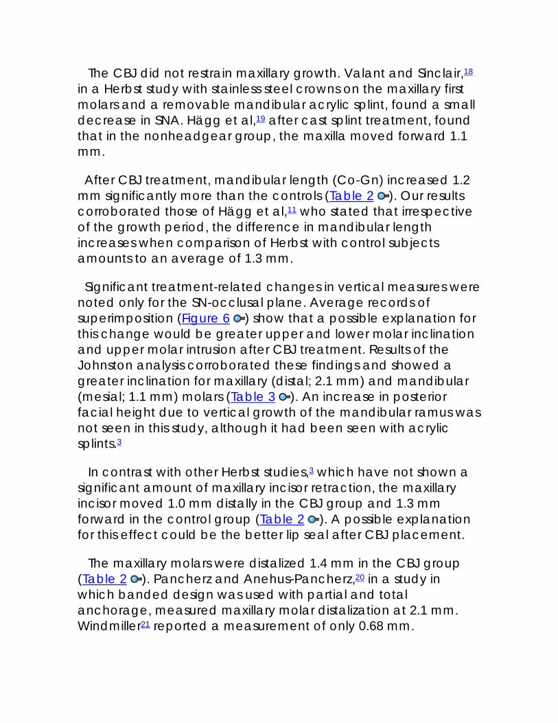
The CBJ did not restrain maxillary growth. Valant and Sinclair,18 in a Herbst study with stainless steel crowns on the maxillary first molars and a removable mandibular acrylic splint, found a small decrease in SNA. Hägg et al,19 after cast splint treatment, found that in the nonheadgear group, the maxilla moved forward 1.1 mm.
After CBJ treatment, mandibular length (Co-Gn) increased 1.2 mm significantly more than the controls (Table 2 ). Our results corroborated those of Hägg et al,11 who stated that irrespective of the growth period, the difference in mandibular length increases when comparison of Herbst with control subjects amounts to an average of 1.3 mm.
Significant treatment-related changes in vertical measures were noted only for the SN-occlusal plane. Average records of superimposition (Figure 6 ) show that a possible explanation for this change would be greater upper and lower molar inclination and upper molar intrusion after CBJ treatment. Results of the Johnston analysis corroborated these findings and showed a greater inclination for maxillary (distal; 2.1 mm) and mandibular (mesial; 1.1 mm) molars (Table 3 ). An increase in posterior facial height due to vertical growth of the mandibular ramus was not seen in this study, although it had been seen with acrylic splints.3
In contrast with other Herbst studies,3 which have not shown a significant amount of maxillary incisor retraction, the maxillary incisor moved 1.0 mm distally in the CBJ group and 1.3 mm forward in the control group (Table 2 ). A possible explanation for this effect could be the better lip seal after CBJ placement.
The maxillary molars were distalized 1.4 mm in the CBJ group (Table 2 ). Pancherz and Anehus-Pancherz,20 in a study in which banded design was used with partial and total anchorage, measured maxillary molar distalization at 2.1 mm. Windmiller21 reported a measurement of only 0.68 mm.

Differences in the design of the appliances could explain these results.
The CBJ appliance restricted the vertical development of the maxillary molars. Pancherz and Anehus-Pancherz20 reported 0.7 mm of maxillary molar intrusion. Lai and McNamara3 found 1.2 mm of intrusion of the maxillary first molars. When the CBJ was compared with other designs, it was seen that it provided good control of vertical maxillary molar position.
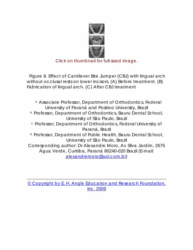
Significant anterior movement of the mandibular incisors was noted in the CBJ group (L1-PogPerp = 1.46 mm; IMPA = 6.6 degrees). It was also observed that the lingual arch without occlusal rests was not able to withstand the inclination forces placed on the mandibular molars by the cantilevers. In many cases, the lingual arch slipped down on the cingulum of the mandibular incisors and landed on the gingiva behind them. In these cases, the lingual arch created sores in the gingiva and contributed to procline the mandibular incisors (Figure 8 ). However, the CBJ did not cause the mandibular incisors to protrude as much as they did with other designs9,19,22 of the Herbst appliance.
CBJ treatment moved the mandibular first molars farther forward (1.3 mm) than did use of only premolar anchorage (1.0 mm)22; less forward movement was seen with cast splint anchorage (2.5 mm),22 the acrylic splint (1.7 mm),21 and the Cantilever Herbst without lingual arch (3.8 mm).9
In the CBJ group, the mandibular molars showed almost the same amount of vertical development in the treated and control groups. Compared with other designs,3 the CBJ exhibited good control of the vertical mandibular molar position.
Profile measurements showed that CBJ treatment had a good impact on facial profile. This study corroborates the findings of Pancherz and Anehus-Pancherz,23 who reported a reduction in facial soft tissue profile convexity. Furthermore, the upper lip

became retrusive, and the lower lip remained, on average, unchanged in relation to the E line.23
Average Class II molar correction of 5.7 mm and overjet correction of 5.3 mm were achieved during CBJ therapy (Table 3 and Figure 7 ). Similar molar and overjet corrections have
been reported for other Herbst samples. Pancherz and Hansen24 reported, on average, a molar correction of 6.3 mm and an overjet correction of 6.9 mm. Valant and Sinclair18 described a 7.1 mm molar correction; Lai and McNamara3 achieved an average molar correction of 5.7 mm and an overjet correction of 4.5 mm.
When the contributions of dental and skeletal changes were compared with the 5.7 mm of molar correction seen in the CBJ group, it was noted that dental changes contributed to 48.2%, and skeletal changes contributed to 50.8%, of the molar correction. Valant and Sinclair18 found that apical base change accounted for 56.5% of the molar correction in their study, whereas Lai and McNamara3 found that growth accounted for 55% of the molar correction. These findings were larger than the Pancherz and Hansen24 determination of 35% for banded Herbst treatment. The average treatment duration was 6 months as reported by Pancherz and Hansen,24 10 months in the study of Valant and Sinclair,18 and 12 months in the trial of Lai and McNamara3 and in the present study. These differences in treatment time and anchorage design may account for the observation that skeletal changes contributed less to the Class II correction in the banded Herbst study.
In the CBJ group, dental movement accounted for 45.3% of the overjet correction. We can conclude that apical base change was responsible for 54.7% of the overjet correction in the CBJ group.
Clinical Implications

When compared with the acrylic splint Herbst design, the CJB exhibited good control of the vertical dimension without the disadvantage of the bulky removable acrylic splint.
The CBJ should not be recommended for very young children. The rods and tubes are very bulky. This size gives strength and makes the appliance almost indestructible, but it is difficult to get used to it. With this appliance, it is important to use a mandibular lingual arch with occlusal rests on premolars or on deciduous molars to prevent cantilever inclination and mandibular incisor proclination. The use of occlusal rests without a lingual arch does not prevent mandibular anchorage loss.
Standard bands to be used with the Herbst appliance should be reinforced with solder, extra band material, or an occlusal support wire soldered around it for stability. Even with these modifications, banded Herbst appliances have a greater breakage rate. Some manufacturers have developed heavier band materials, but these are not as resistant as stainless steel crowns. However, crowns are much more difficult to remove from the teeth. The main clinical advantage of the CBJ is the fact that it comes with preattached axles, and its use can reduce laboratory time.
CONCLUSIONS Return to TOC
• Class II correction achieved by the Cantilever Bite Jumper (CBJ) appliance was accomplished by 2.9 mm apical base change, 1.5 mm distal movement of the maxillary molars, and 1.1 mm mesial movement of the mandibular molars.
• Overjet correction was a result of apical base changes combined with distal movement of the maxillary incisors and mesial movement of the mandibular incisors.
• CBJ provided good control of vertical dimension.

• The main side effect of the CBJ is that the vertical force vector of the telescopes acting as lever arms can produce mesial tipping of the mandibular molars.
REFERENCES Return to TOC
1. Pancherz H. Treatment of Class II malocclusion by jumping the bite with the Herbst appliance: a cephalometric investigation. Am J Orthod. 1979; 76:423–442. [PubMed Citation] 2. Wieslander L. Intensive treatment of severe Class II malocclusions with a headgear-Herbst appliance in the early mixed dentition. Am J Orthod. 1984; 86:1–13. [PubMed Citation] 3. Lai M, McNamara JA Jr. An evaluation of two-phase treatment with the Herbst appliance and preadjusted Edgewise therapy. Semin Orthod. 1998; 4:46–58. [PubMed Citation] 4. Mayes JH. Improving appliance efficiency with the Cantilever Herbst: a new answer to old problems. Clin Impressions. 1994; 3:2–5. 17–19. 5. Keim RG, Gottlieb EL, Nelson AH, Vogels DS. 2002 JCO study of orthodontic diagnosis and treatment procedures: part 1, results and trends. J Clin Orthod. 2002; 36:553–568. [PubMed Citation] 6. Mayes JH. The single-appointment preattached Cantilever. Clin Impressions. 1996; 5:14–23. 7. Croft RS, Buschang PH, English J, Meyer R. A cephalometric and tomographic evaluation of Herbst treatment in the mixed dentition. Am J Orthod Dentofacial Orthop. 1999; 116:435–443. [PubMed Citation] 8. Burkhardt DR, McNamara JA Jr, Baccetti T. Maxillary molar distalization or mandibular enhancement: a cephalometric comparison of comprehensive orthodontic treatment including the pendulum and the Herbst appliances. Am J Orthod

Dentofacial Orthop. 2003; 123:108–116. [PubMed Citation] 9. VanLaecken R, Martin CA, Dischinger T, Razmus T, Ngan P. Treatment effects of the edgewise Herbst appliance: a cephalometric and tomographic investigation. Am J Orthod
Dentofacial Orthop. 2006; 130:582–593. [PubMed Citation] 10. Barnett GA, Higgins DW, Major PW, Flores-Mir C. Immediate skeletal and dentoalveolar effects of the crown- or banded-type Herbst appliance on Class II division 1 malocclusion: a systematic review. Angle Orthod. 2008; 78:361–369. [PubMed Citation] 11. Hägg U, Pancherz H, Taranger J. Pubertal growth and orthodontic treatment. In: Carlson DS, Ribbens KA, eds. Craniofacial Growth During Adolescence. Monograph 20,
Craniofacial Growth Series. Ann Arbor, Mich: Center for Human Growth and Development, University of Michigan; 1987:87– 115. 12. Ruf S, Pancherz H. When is the ideal period for Herbst therapy—early or late?. Semin Orthod. 2003; 9:47–56. 13. Johnston LE Jr. Balancing the books on orthodontic treatment: an integrated analysis of change. Br J Orthod. 1996; 23:93–102. [PubMed Citation] 14. Johnston LE Jr. A comparative analysis of Class II treatments. In: McNamara JA Jr, Carlson DS, Vig PS, Ribbens KA, eds. Science and Clinical Judgement in Orthodontics. Monograph 18,
Craniofacial Growth Series. Ann Arbor, Mich: Center for Human Growth and Development, University of Michigan; 1986:103–148. 15. Johnston LE Jr. Growth and the Class II patient: rendering unto Caesar. Semin Orthod. 1998; 4:59–62. [PubMed Citation] 16. Schulhof R, Bagha L. A statistical evaluation of the Ricketts and Johnston growth-forecasting methods. Am J Orthod. 1975; 67:258–276. [PubMed Citation]

17. Houston WJB. The analysis of errors in orthodontic measurements. Am J Orthod. 1983; 83:382–390. [PubMed Citation] 18. Valant JR, Sinclair PM. Treatment effects of the Hersbt appliance. Am J Orthod Dentofacial Orthop. 1989; 95:138–147. [PubMed Citation] 19. Hägg U, Du X, Rabie BM, Bendeus M. What does headgear add to Herbst treatment and to retention?. Semin Orthod. 2003; 9:57–66. 20. Pancherz H, Anehus-Pancherz M. The head-gear effect of the Herbst appliance: a cephalometric long-term study. Am J
Orthod Dentofac Orthop. 1993; 103:510–520. [PubMed Citation] 21. Windmiller EC. The acrylic-splint Herbst appliance: a cephalometric evaluation. Am J Orthod Dentofacial Orthop. 1993; 104:73–84. [PubMed Citation] 22. Weschler D, Pancherz H. Efficiency of three mandibular anchorage forms in Herbst treatment: a cephalometric investigation. Angle Orthod. 2004; 75:23–27. 23. Pancherz H, Anehus-Pancherz M. Facial profile changes during and after Herbst appliance treatment. Eur J Orthod. 1994; 16:275–286. [PubMed Citation] 24. Pancherz H, Hansen K. Occlusal changes during and after Herbst treatment: a cephalometric investigation. Eur J Orthod. 1986; 8:215–228. [PubMed Citation]
TABLES Return to TOC

Table 1. Comparison of Initial Forms (t-tests)a


Table 2. Comparison of Changes in the CBJ and Control Groups (T1–T2, ANCOVA)a
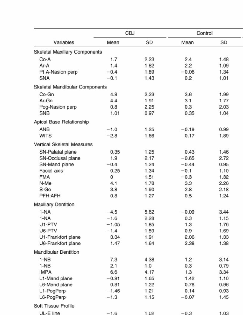
Table 3. Pitchfork Analysisa,b
FIGURES Return to TOC
Click on thumbnail for full-sized image.
Figure 1. The Cantilever Bite Jumper
Click on thumbnail for full-sized image.
Figure 2. Angular measurements
Click on thumbnail for full-sized image.
Figure 3. Linear measurements
Click on thumbnail for full-sized image.
Figure 4. Determination of changes in horizontal and vertical positions of upper first molar and upper central incisor

Click on thumbnail for full-sized image.
Figure 5. Determination of changes in horizontal and vertical positions of lower first molar and central incisor
Click on thumbnail for full-sized image.
Figure 6. Averaged initial and final composite tracing superimpositions. (A) Patients with CBJ. (B) Control patients
Click on thumbnail for full-sized image.
Figure 7. Pitchfork analysis. Comparison between Cantilever Bite Jumper (CBJ) and control subjects
Click on thumbnail for full-sized image.
Figure 8. Effect of Cantilever Bite Jumper (CBJ) with lingual arch without occlusal rests on lower incisors. (A) Before treatment. (B) Fabrication of lingual arch. (C) After CBJ treatment
a Associate Professor, Department of Orthodontics, Federal
University of Paraná and Positivo University, Brazil b Professor, Department of Orthodontics, Bauru Dental School,
University of São Paulo, Brazil c Professor, Department of Orthodontics, Federal University of
Paraná, Brazil d Professor, Department of Public Health, Bauru Dental School,
University of São Paulo, Brazil Corresponding author: Dr Alexandre Moro, Av Silva Jardim, 2675
Água Verde, Curitiba, Paraná 80240-020 Brazil (E-mail: [email protected])
© Copyright by E. H. Angle Education and Research Foundation,
Inc. 2009