circulationaha/2009/898114r2 - ahajournals.org · tre-opn mice were created and expanded by pierre...
TRANSCRIPT

CIRCULATIONAHA/2009/898114R2
Osteopontin expression in cardiomyocytes induces dilated cardiomyopathy
Marie-Ange Renault1 PhD, Fanny Robbesyn1 PhD, Patricia Réant1,2 MD, Victorine Douin3,4
PhD, Danièle Daret1,2 PhD, Cécile Allières1,2 BSc, Isabelle Belloc1,2 BSc, Thierry
Couffinhal1,2 MD PhD, Jean-François Arnal3,4 MD PhD, Karin Klingel5 MD, Claude
Desgranges1,2 PhD, Pierre Dos Santos1,2 MD PhD, Flavien Charpentier6,7 PhD, Alain-Pierre
Gadeau1,2 PhD
1. INSERM, U828, Pessac, France
2. University of Bordeaux Victor Ségalen, IFR4, Bordeaux, France
3. INSERM, U858, Toulouse, France
4. University of Toulouse Paul Sabatier, Toulouse, France
5. Department of molecular pathology, Institute for pathology, University Hospital,
Tübingen, Germany
6. INSERM, U915, Institut du Thorax, Nantes, France
7. University of Nantes and CNRS, ERL3147, Nantes, France
Corresponding author: AP Gadeau
Adress: INSERM U828, Avenue du Haut Lévêque, 33600 Pessac, France
Fax: (33) 556 368 979, Tel: (33) 557 891 977, eMail: [email protected]
Short Title: OPN induces dilated cardiomyopathy
Number of words: 5987
Journal Subject Codes: (11) Other heart failure, (130) Animal models of human disease
ce
E
v
a
ERM, U858, Toulouse, France
versity of Toulouse Paul Sabatier, Toulouse, France
artment of molecular pathology, Institute for pathology, University Hospital,
Germany

CIRCULATIONAHA/2009/898114R2
Abstract
Background - Inflammatory processes play a critical role in myocarditis, dilated
cardiomyopathy and heart failure. The expression of the inflammatory chemokine osteopontin
(OPN) is dramatically increased in cardiomyocytes and inflammatory cells during myocarditis
and heart failure in human and animals. However its role in the development of heart diseases
is not known.
Methods and Results - To understand whether OPN is involved in cardiomyopathies, we
generated a transgenic mouse (MHC-OPN) that specifically overexpresses OPN in
cardiomyocytes using cardiac specific promoter-directed OPN expression.
Young MHC-OPN mice were phenotypically indistinguishable from their control littermates,
but most of them died prematurely with a half life of 12 weeks of age. Electrocardiography
revealed conduction defects. Echocardiography showed LV dilation and systolic dysfunction.
Histological analysis revealed cardiomyocyte loss, severe fibrosis and inflammatory cell
infiltration. Most of these inflammatory cells were activated T cells with Th1 polarization and
cytotoxic activity. Autoantibodies against OPN, cardiac myosin or troponin I, were not found
in the serum of MHC-OPN mice.
Conclusions - These data show that OPN expression in the heart induces in vivo, T cell
recruitment and activation leading to chronic myocarditis, the consequence of which is
myocyte destruction and hence, dilated cardiomyopathy. Thus, OPN might therefore
constitute a potential therapeutic target to limit heart failure.
Key words:
Heart disease, heat failure, myocarditis, inflammation, immune system
ession.
m theeeeiririiriri cococococontntntntntntrorororororoll llll lilllllitttttttterererererermmmmmm
o p
onduction defects. Echocardi ra showed LV dilation and s tolic sfunc
c l
n o
of them died prematurely with a half life of 12 weeks of age. Electrocardiograp
onduction defects. Echocardiography showed LV dilation and systolic dysfunc
cal analysis revealed cardiomyocyte loss, severe fibrosis and inflammatory cell
n. Most of these inflammatory cells were activated T cells with Th1 polarizatio

CIRCULATIONAHA/2009/898114R2
Introduction
Dilated cardiomyopathy (DCM) is a major cause of non ischemic heart failure (HF). Common
causes of DCM include acute ischemia, heart rhythm-related, pericardial, valvular, peripheral
vascular, congenital, and familial disorders1. Inflammatory nature of DCM has also long been
recognized triggered by acute viral or autoimmune myocarditis2,3. The current understanding
of molecular pathogenesis of post viral myocarditis involved a three stages process explaining
the progression from acute injury to chronic DCM. These steps include activation of the
innate immune system, installation of a chronic autoimmune process inducing myocyte
destruction, and finally DCM4.
Osteopontin (OPN), also called cytokine Eta-1, is a chemokine involved in tissue remodeling
and immunity5,6. OPN is highly expressed during chronic inflammatory and autoimmune
diseases and it is thought to play a role in inflammation via the recruitment and retention of
macrophages and T-cells to inflamed sites7. Moreover, OPN regulates the production of
inflammatory cytokines in these cells8. OPN is also involved in the early Th1 response as
demonstrated by the severely impaired type-1 immunity in OPN deficient mice and by
interferon-gamma (IFN- ) and CD40 ligand expression in OPN-stimulated human T cells8,9.
OPN also enhances the survival of activated T cells suggesting that it might promote the
progression of autoimmune diseases10.
Several studies have suggested a role for OPN in cardiovascular diseases, including
atherosclerosis and valvular stenosis7. In the heart, OPN expression is increased during both
acute and chronic diseases11. Indeed after myocardial infarction, OPN expression was
increased in macrophages and during cardiac hypertrophy in both humans and rodents,
cardiomyocytes appear to be the major source of OPN12. This expression probably plays a
1
s inducing myocytytytytytyte e eeee
t d
n e
n n
g
8
tin (OPN), also called cytokine Eta-1, is a chemokine involved in tissue remod
nity5,6. OPN is highly expressed during chronic inflammatory and autoimmune
nd it is thought to play a role in inflammation via the recruitment and retention
ges and T-cells to inflamed sites7. Moreover, OPN regulates the production of
8

CIRCULATIONAHA/2009/898114R2
role in fibrillogenesis13,14. OPN expression coincides also with the transition from myocardial
hypertrophy to HF and its level of expression is correlated with the severity of the HF11.
Moreover OPN expression was increased during viral myocarditis in mouse models15.
Findings from several experiments have therefore converged to establish a link between
cardiomyopathies, including DCM, and OPN expression . 14 However, the direct role of OPN
in the development of DCM has not yet been determined. Here, we hypothesized that OPN
might be involved in the underlying mechanisms of these diseases.
We designed a conditional transgenic mouse model which specifically expresses OPN in the
heart in order to investigate the role of OPN in cardiomyopathies. We evidenced that
expression of OPN in cardiomyocytes induces myocarditis promoting T cell-mediated
inflammation and fibrosis in the heart leading finally to DCM and heart failure.
Materials and Methods
Generation of TRE-OPN transgenic mice
This mouse model was constructed as previously described6. The mouse OPN cDNA (bases 9
to 955, accession number BC057858) was introduced into the multiple cloning site of the
tetracycline transactivator (tTA)-responsive promoter of pBI-G plasmid (Clontech, St
Germain en Laye, France). Transgenic mouse lines (TRE-OPN) were produced by
microinjection of this construct. We obtained 3 independent transgenic lines. These lines were
used after 7 breeding with C57Bl/6 background.
Generation of MHC-OPN mice
TRE-OPN mice were cross-bred with cardiac alpha myosin heavy chain (MHC)-tTA mice
(FVB.Cg-Tg(Myh6-tTA)6Smbf/J, Jackson Laboratory) that express the transcriptional factor
tTA under an MHC promoter. Cross-breeding produced the four following genotypes: WT
2
We evidenced thatt
ng T ccccccelelllelellll-lll-memememememedidididididiatataaaa edededededed
t
s
o
6
tion and fibrosis in the heart leading finally to DCM and heart failure.
s and Methods
on of TRE-OPN transgenic mice
6

CIRCULATIONAHA/2009/898114R2
mice, single transgenic TRE-OPN and MHC-tTA mice and double transgenic MHC-OPN
mice. In all experiments using MHC-OPN mice, we have verified that the behavior of WT,
MHC-tTA and TRE-OPN littermates are not different for their level of OPN expression, their
survival or show only minor differences compared to those associated with OPN expression
(Supplemental Figure S1, S3A, S4). The term "control" refers to a pool of WT, MHC-tTA and
TRE-OPN littermates.
Mice were maintained in a conventional animal facility, on a 12-hour light/12-hour dark
cycle. Food and water were available ad libitum. All procedures were carried out in compliance
with the principles and guidelines established by the French National Institute of Medical
Research (INSERM) and approved by the Institutional Animal Care and Use Committee. When
required, doxycycline hyclate (Dox, Sigma-Aldrich) was added in the drinking water (4 mg/ml).
Mouse genotypes were determined by PCR with experimental conditions and primers
describe in online data.
Enterovirus myocarditis model
A.BY/SynJ mice were bred and kept in pathogen-free conditions at the Department of
Molecular Pathology, University Hospital, Tübingen, Germany. Group B coxsackievirus
infection of mice and further processing of heart and sera were performed as described
previously15.
Assessment of the cardiac function in vivo
Electrocardiography
Six-lead surface ECGs were recorded with 25-gauge subcutaneous electrodes on a computer
through an analog-digital converter (IOX 1.585, EMKA Technologies, Paris, France) for
monitoring and later analysis (ECG Auto 1.5.7, EMKA Technologies). ECG channels were
3
Institute of Medicacaaaaalllll l
nd UsUsUsUsUsUse eeee CoCoCoCoCoCommmmmmmmmmmmitiiiiitteteeeeeeeee.e.e WWW WWW
d g
n
n
doxycycline hyclate (Dox, Sigma-Aldrich) was added in the drinking water (4 mg
notypes were determined by PCR with experimental conditions and primers
n online data.

CIRCULATIONAHA/2009/898114R2
filtered between 0.5 and 250 Hz. Mice were anesthetized with an intraperitoneal injection of
etomidate (30 mg/kg; Janssen-Cilag, Berchem, Belgium). Body temperature was maintained
at 37°C using a servo-controlled heating pad (Harvard Apparatus). The criteria used for
measuring RR, PQ, QRS and QT intervals, as well as P wave duration can be found
elsewhere16. The QT interval was corrected for heart rate using the formula, QTc =
QT/(RR/100)1/2 established for mice; values of QT and RR are expressed in ms17.
Transthoracic echocardiography
Control (n=15) and MHC-OPN (n=5) mice were lightly anesthetized with 1.5% isoflurane.
Echography was performed with a 15-MHz transducer (MI2L General Electric). The percent
of left ventricular (LV) fractional shortening (%FS) was defined as %FS
=[(LVDd LVDs)/LVDd]×100, where LVDd is the LV end-diastolic diameter, and LVDs is
the LV end-systolic diameter.
Histology and immunohistology
For histological analysis, tissue specimens were fixed with 3% paraformaldehyde in PBS.
Sections were stained with Masson's trichrome or Sirius red and observed with a white light
microscope. Myocyte and fibrosis densities were quantified (Sigma Scan software) on
digitized images of Sirius red stained sections.
For immunodetection, paraffin sections (8 μm thick) were incubated for 2 hours with
antibodies directed against: OPN (Sigma Aldrich, Saint Quentin Fallavier, France), collagen I
(Novotec, Saint Martin La Garenne, France), CD31 and CD45 (Pharmingen, San Diego, CA),
CD3 (Santa Cruz, Santa Cruz, CA), CD4, CD8, TNF and IL-17 (Biolegend, SanDiego CA).
Bound biotin-conjugated secondary antibodies (Sigma Aldrich, Saint Quentin Fallavier,
France) were visualized using streptavidin peroxidase-diaminobenzidine (Vector,
4
d with 1.5% isofllururururururanaaaa
ral ElElElElElEleceecececectrtrtrtrtrtriciciciciic)).)))) TTT TTTheheheeee ppp p ppeeeeerer
n
D
d
ntricular (LV) fractional shortening (%FS) was defined as %FS
LVDs)/LVDd]×100, where LVDd is the LV end-diastolic diameter, and LVD
d-systolic diameter.

CIRCULATIONAHA/2009/898114R2
Peterborough, UK) or streptavidin Alexa Fluor 488/568 (Invitrogen, Paisley, UK). OPN area
was measured on digitized images with Sigma Scan software.
Western Blot and ELISA
Autoantibodies against OPN, cardiac myosin and troponin I in mouse serum were detected by
western blot using protein blotted membrane, diluted mouse serum (1%) and biotinylated
anti-mouse IgG. Cardiac myosin was prepared by high ionic solution extraction and water
precipitation as described previously18. Anti-troponin I antibody and troponin I protein were
kindly provided by Dr O. Cazorla (Inserm U637, Montpelliers, France) and Dr G. Pfitzer
(Institute of Vegetative Physiology, University of Cologne, Köln, Germany)19 respectively.
Mouse OPN and OPN ELISA kit was purchased from R&D System (Abingdon, UK).
Apoptosis assay
Tunel assays were performed according to the manufacturer’s recommendations (Roche
Diagnostic, Meylan, France) using the in situ Cell Death Detection Kit.
RT-quantitative PCR gene expression analysis (qPCR)
qPCR was performed by the Molecular Biology Platform of Angers University
(http://www.pftbiotech-angers.com/). OPN qPCR was performed using primers described in
supplemental data.
Statistical analysis
For survival study, a survival plot was used and Log-Rang test performed for statistical
analysis. Censored variable was survival status at the time of analysis. Statistical analysis of
ECG parameters was performed using either one-way ANOVA or two-way ANOVA tests for
5
nce) and Dr G. Pfifiitztztztztztzee
ermaaaaanynynnnyny))))))191919191919 rrrrr resesesesese pppepepp ctctctctctctivivivivivivee
P
s
a
PN and OPN ELISA kit was purchased from R&D System (Abingdon, UK).
s assay
ays were performed according to the manufacturer’s recommendations (Roche

CIRCULATIONAHA/2009/898114R2
repeated measures, both followed up by a Tukey test when appropriate. Other statistical
analyses were performed using either ANOVA followed by a Student's T test when only two
groups have to be compared or using Kruskal-Wallis one-way ANOVA followed by Mann
Whitney U test. A p value <0.05 indicated significant difference between 2 groups. Results
are expressed as mean values ± SEM. Data were analyzed with NCSS statistical software.
Results
Experimental model
OPN is expressed at a very low level in the normal heart of WT mice. To specifically express
the OPN gene in cardiomyocytes, we used double transgenic MHC-OPN mice obtained by
crossing MHC-tTA mice with TRE-OPN mice that carry the mouse OPN gene under the
control of the tTA-responsive promoter (Figure 1A). Unlike in the heart of control mice, OPN
mRNA and protein were strongly expressed in the heart of MHC-OPN mice (Figure 1B, C
and Supplemental Figure S1 and S2). However, the level of OPN mRNA and protein in hearts
of MHC-OPN mice remained in the same range than that observed in hearts of mice during
the acute phase of myocarditis induced by coxsackievirus infection15 (Supplemental Figure
S2). Driven by MHC promoter, OPN was indeed mainly detected in cardiomyocytes where its
distribution appeared patchy (Figure 1C, D). This expression did not change according to the
age of the mouse from the 1st to the 16th week (Supplemental Figure S1). In blood of MHC-
OPN mice OPN concentration remained low while it increased coxsackievirus infected mice
undergoing myocarditis (Figure 1E).
In MHC-OPN mice, OPN gene expression was partially inhibited by oral administration of 2
mg/ml Dox in water and completely blocked by 4 mg/ml (Figure 1B, C).
Genotypic analysis, at weaning age, of offspring resulting from the mating of MHC-tTA mice
with TRE-OPN mice showed a Mendelian inheritance pattern with a gender ratio of 1/1
6
e. To specifically y y y y y exexeeee
OPNNNNNN mmmmmiiciciicice eeeee obobobobobbtatttatatainininninnedededededed
M e
,
n ,
MHC-tTA mice with TRE-OPN mice that carry the mouse OPN gene under the
the tTA-responsive promoter (Figure 1A). Unlike in the heart of control mice,
nd protein were strongly expressed in the heart of MHC-OPN mice (Figure 1B,
emental Figure S1 and S2). However, the level of OPN mRNA and protein in
CIRCULATIONAHA/2009/898114R2
indicating that OPN expression in the heart had no effect on prenatal survival (Supplemental
Table S1).
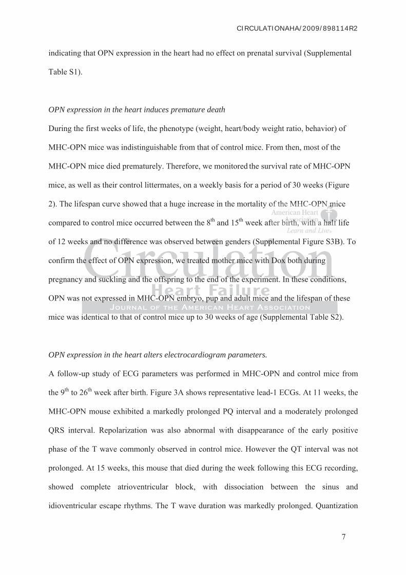
OPN expression in the heart induces premature death
During the first weeks of life, the phenotype (weight, heart/body weight ratio, behavior) of
MHC-OPN mice was indistinguishable from that of control mice. From then, most of the
MHC-OPN mice died prematurely. Therefore, we monitored the survival rate of MHC-OPN
mice, as well as their control littermates, on a weekly basis for a period of 30 weeks (Figure
2). The lifespan curve showed that a huge increase in the mortality of the MHC-OPN mice
compared to control mice occurred between the 8th and 15th week after birth, with a half life
of 12 weeks and no difference was observed between genders (Supplemental Figure S3B). To
confirm the effect of OPN expression, we treated mother mice with Dox both during
pregnancy and suckling and the offspring to the end of the experiment. In these conditions,
OPN was not expressed in MHC-OPN embryo, pup and adult mice and the lifespan of these
mice was identical to that of control mice up to 30 weeks of age (Supplemental Table S2).
OPN expression in the heart alters electrocardiogram parameters.
A follow-up study of ECG parameters was performed in MHC-OPN and control mice from
the 9th to 26th week after birth. Figure 3A shows representative lead-1 ECGs. At 11 weeks, the
MHC-OPN mouse exhibited a markedly prolonged PQ interval and a moderately prolonged
QRS interval. Repolarization was also abnormal with disappearance of the early positive
phase of the T wave commonly observed in control mice. However the QT interval was not
prolonged. At 15 weeks, this mouse that died during the week following this ECG recording,
showed complete atrioventricular block, with dissociation between the sinus and
idioventricular escape rhythms. The T wave duration was markedly prolonged. Quantization
7
of the MHC-OPNN m mmmmmi
fter bibibibibibirtrtrtrtrtrthhhhhh, www witititititithhhhh h aa a hahahahahahalflflflflflf
k B
h
y n
h
ks and no difference was observed between genders (Supplemental Figure S3B
he effect of OPN expression, we treated mother mice with Dox both during
y and suckling and the offspring to the end of the experiment. In these conditio
not expressed in MHC-OPN embryo, pup and adult mice and the lifespan of th

CIRCULATIONAHA/2009/898114R2
of ECG intervals during the last ECG recording before death of MHC-OPN mice and of
control mice demonstrated significant differences of PQ, QRS, QT and QTc but not of RR
between these mice (Figure 3B, C). In contrast, MHC-OPN mice that survive exhibited ECG
parameters within normal values, with a moderate prolongation of PQ and QRS intervals.
None of the groups exhibited either atrial or ventricular arrhythmias (Figure 3B, C). Thus
ECG analysis demonstrated that MHC-OPN mice exhibit a cardiomyopathy that could
explain their premature death.
OPN expression in the heart induces dilated cardiomyopathy
We evaluated the morphological and functional cardiac parameters of MHC-OPN mice by
transthoracic echocardiography. Echocardiographic parameters of 6-week-old mice were
identical to that of control mice (Supplemental Figure S5). However, at 11 weeks, the heart
LV of MHC-OPN mice was severely dilated compared to that of control mice (Figure 4A, B,
C). Moreover, during systole, the LV free wall of MHC-OPN mice was significantly thinner
than in control hearts (Figure 4C) which suggests the development of a hypokinetic DCM.
These structural defects led to systolic dysfunction, observed as reduced LV fractional
shortening (MHC-OPN mice: 13.7 ± 4.0%, n=5; control mice: 29.8 ± 2.0%, n=13; p<0.05). In
vivo imaging was confirmed by a direct observation of cardiac dilation in MHC-OPN mouse
that died during anesthesia (Figure 5A, D).
OPN expression in the heart induces fibrosis and myocyte destruction
We then performed histological analysis. At 6 weeks, the morphology of the hearts of MHC-
OPN mice did not appear to be different from that of control mice (Supplemental Figure S4).
After 11 weeks, the cellular organization in MHC-OPN mouse hearts was altered when
compared to control hearts with evidence of myocyte loss (Figure 5B, E, H), fibrosis (Figure
8
of MHMHMHMHMHMHC-C-CCCC OPOPOPOPOPOPNNN NNN mimimimimimicececececece b b
c e
o e
H A
o n
cic echocardiography. Echocardiographic parameters of 6-week-old mice were
o that of control mice (Supplemental Figure S5). However, at 11 weeks, the he
HC-OPN mice was severely dilated compared to that of control mice (Figure 4Af
over, during systole, the LV free wall of MHC-OPN mice was significantly thin

CIRCULATIONAHA/2009/898114R2
5C, F, I) and interstitial cell infiltration (Figure 5B inset) demonstrating that the disease
developed very rapidly between the 6th and 11th week after birth.
Moreover, while no difference in the number of apoptotic cells in the heart of control and 6-
week-old MHC-OPN mice was observed, the number of apoptotic cells was increased at 11
weeks in the MHC-OPN mouse heart (Figure 5G).
OPN expression induced heart inflammation
The hearts of MHC-OPN mice were infiltrated by a large number of interstitial cells. About
fifty percent of infiltrating cells expressed the panleukocyte CD45 marker (Figure 6A). In
MHC-OPN mice under 6-week-old, the number of CD45+ infiltrated cells in the heart was
very low and was not different from that observed in control mice (Figure 6B). In early heart
lesions focal infiltration spots were observed first in the endomyocardium and the epicardium
(not shown). Ten weeks after birth, CD45+ cells were widespread in all parts of the
myocardium with particular spots at the base of the mitral valve pillars in areas that were also
associated with myocyte destruction (Figure 6C). As a consequence of the myocyte
destruction a strong fibroblast infiltration occurred characterized by vimentin expression
(Figure 6A), explaining the extensive fibrosis.
Among infiltrated leukocytes (CD45+), only a very few, not different than in the control
hearts, were neutrophil granulocytes, as characterized by myeloperoxidase activity (Figure
6D, J), or B lymphocytes (CD19+) (Figure 6J). More unexpectedly, macrophages (Mac-3+)
were rarely observed only in rare small foci (Figure 6E, J). Most of the leukocytes were CD3+
cells, thus demonstrating T cell infiltration (Figure 6F, J). In this population, we identified
helper T cells (CD4+) (Figure 6G) and cytotoxic T cells (CD8+) (Figure 6H) but no IL-17+ T
cells (data not shown). In hearts from control mice, only a very few T cells (CD3+, CD4+ or
CD8+) were detected regardless of age (data not shown). We also observed particularly strong
9
marker (Figure 6A)A)A)A)A)A).... . . I
d cellllllllllllssss s inininiiin t tttttheheheheheh hhh hhheaeaaaaartrtrtrtrtrt wwwww w
a h
c r
n
u e
and was not different from that observed in control mice (Figure 6B). In early h
cal infiltration spots were observed first in the endomyocardium and the epicar
n). Ten weeks after birth, CD45+ cells were widespread in all parts of the
um with particular spots at the base of the mitral valve pillars in areas that were

CIRCULATIONAHA/2009/898114R2
CD44 expression in these inflammatory cells suggesting that they were in an activated stage
(Figure 6I).
Expression of a set of Th1, Th2 and Th17 cytokines in the heart was assessed by qPCR
(Figure 7). The strong induction of the IL-12 p40 subunit and Inf- , and the absence of
induction of IL-4, IL-10, IL-13 and IL-17 suggested that T cells were Th1-polarized.
Moreover, the expression of perforin and granzyme confirmed the existence of a cytotoxic
response as suggested by the presence of large numbers of cytotoxic CD8+ T cells (Figure
6H). Furthermore expression of fractalkine, a molecule able to capture cytotoxic lymphocytes
from blood flow and to promote their emigration, is also strongly increased (Figure 7)20.
These data demonstrate for the first time in vivo, that OPN expressed by cardiomyocytes
triggers directly or indirectly T cells recruitment and activation and retains them in the heart.
To verify whether OPN is just a trigger, we first blocked OPN expression in cardiomyocytes
by administering Dox to 5-week-old MHC-OPN mice before the onset of T cell infiltration.
The survival curve of these mice during a 30-week period after birth was comparable to that
of control mice, and their hearts were morphologically and histologically identical to control
hearts (Supplemental Table S2).
In a second set of experiments, we blocked OPN expression in 11-week-old MHC-OPN mice
when DCM was evidenced (LV diameter >0.41 mm) by echocardiography (Figure 8A). The
survival rate of this population during the next 30 weeks was identical to that of control mice
(Supplemental Table S2). Moreover in Dox treated mice OPN expression (Figure 8B, E, H)
and inflammation (Figure 8C, F, I) were strongly reduced when compared to 11-week-old
MHC-OPN mice. However, fibrosis (Figure 8D, G, J) was still significantly increased
demonstrating that these mice had experienced previous heart injury. The results of these
experiments suggest that transient stimulation of OPN expression is not sufficient to induce or
10
creased ((Figuggg re 77))))))20200.
d by y cacardddddrdioiiiioi mymyyyyyococytytytytytytees
rectly or indirectly T cells recruitment and activation and retains them in the h
w c
i
v
rectly or indirectly T cells recruitment and activation and retains them in the h
whether OPN is just a trigger, we first blocked OPN expression in cardiomyoc
stering Dox to 5-week-old MHC-OPN mice before the onset of T cell infiltrati
val curve of these mice during a 30-week period after birth was comparable to k

CIRCULATIONAHA/2009/898114R2
to sustain myocarditis, and that chronic OPN expression in adult animals is required to sustain
the inflammatory process of myocarditis that leads to DCM.
Chronic OPN expression in cardiomyocytes did not induce a humoral autoimmune response
Experimental autoimmune myocarditis (EAM) is characterized by the presence of
autoantibodies directed against heart proteins including cardiac myosin and troponin I21,22.
Different studies have indicated that OPN promotes disease progression in autoimmune
models including experimental autoimmune encephalomyelitis (EAE)23. Thus we investigated
whether OPN expression in cardiomyocytes induced autoimmune myocarditis. In the injured
hearts from MHC-OPN mice, we did not reveal the presence of immune complexes using
anti-mouse immunoglobulin (not shown). Moreover, we demonstrated that the serum of
MHC-OPN mice did not contain antibodies directed against cardiac myosin, troponin I or
OPN proteins (Supplemental Figure S6).
Altogether these experiments suggest that OPN-mediated chronic inflammation does not
induce a humoral autoimmune response.
Discussion
OPN expression has been found to be increased in the heart during cardiac diseases that lead
to HF and its circulating concentration is now under consideration for use as a biomarker for
the prognosis of HF24. Studies with OPN-/- mice have underlined the potential role of OPN in
heart disease13,14. However, its role not only as a marker but as an actor triggering disease had
not been fully established. To elucidate the role of heart-derived OPN, we generated MHC-
OPN mice that express OPN conditionally in cardiomyocytes. In these mice, a strong increase
in myocardial OPN mRNA and protein expression was observed. Nevertheless, this level
remained relevant to physiopathology since it was closed to that observed in the heart during
11
myocarditis. In thee i iiiiinjnjnnnn
mune ee cococococcompmpmpmpmpmplelelelellexexexeexees s ususususususinininininin
e f
N o
e
r t
e immunoglobulin (not shown). Moreover, we demonstrated that the serum of
N mice did not contain antibodies directed against cardiac myosin, troponin I o
eins (Supplemental Figure S6).
r these experiments suggest that OPN-mediated chronic inflammation does not

CIRCULATIONAHA/2009/898114R2
coxsackievirus-induced myocarditis, one of the best-characterized myocarditis model15.
Interestingly, MHC-OPN mice died prematurely 10 to 15 weeks after birth. Clinical and
histological diagnosis revealed that from the 6th week after birth, they developed spontaneous
myocarditis followed by DCM with electrocardiographic abnormalities and heart block. Heart
block that is found in some idiopathic myocarditis in human2, is not a usual feature in mouse
severe myocarditis models with DCM, stressing the interest of this new mouse model.
Inflammation has been recognized as a critical pathological component of a number of cardiac
diseases. DCM, one of the most common heart diseases, is mainly the consequence of chronic
myocarditis4. In MHC-OPN mice, a chronic inflammation process characterized by CD4+ and
CD8+ T cell infiltration was observed. CD4+ and CD8+ T cells both contribute to
myocarditis: CD4+ helper T cells are required to initiate the pathological process whereas
cytotoxic CD8+ T cells are directly involved in cardiomyocyte damage3. The role of OPN in
T cell recruitment was clearly demonstrated in vitro. Indeed OPN induces T cell chemotaxis,
supports T cell adhesion and enhances their survival10,25. In vivo only a correlation between
OPN expression with T cell infiltration was shown25. Our data clearly demonstrates that, in
vivo, local OPN expression induces T cell recruitment at the site of OPN expression. This
OPN effect could be direct through its chemotactic activity toward T-cells or indirect through
its ability to induce T cell chemotactic factors such as fractalkine since OPN concentration
was not increased in sera. This OPN-mediated T cell recruitment in the heart and Th-1
polarization characterized by high expression of IL-12 and INF- and low expression of IL-4,
IL-10, IL-13, and IL17, lead to myocarditis. Moreover OPN induces the expression of the
cytotoxic proteins perforin and granzyme that are known to be involved in T cell-mediated
cardiomyocyte destruction and consequently DCM.
Myocarditis is described as a progressive disease with three distinct chronologically
successive stages including an initial phase of myocardial injury and innate immune response,
12
haracterized by CDCDCDCDCDCD44
h contntntntntntririiiriribububbbubutetetetetete t tttttoooo oo
t a
C
r t
T e
25
is: CD4+ helper T cells are required to initiate the pathological process wherea
CD8+ T cells are directly involved in cardiomyocyte damage3. The role of OP
ruitment was clearly demonstrated in vitro. Indeed OPN induces T cell chemot
T cell adhesion and enhances their survival10,25. In vivo only a correlation betwe
25

CIRCULATIONAHA/2009/898114R2
a 2nd phase of autoimmune response, and finally a 3rd phase associated with DCM4. OPN
expression is increased in several autoimmune diseases including systemic lupus
erythematosus, rheumatoid arthritis, and inflammatory bowel disease and appears as a critical
factor in the development of EAE10,23. However conflicting results have been published
regarding the role of OPN in EAM. Indeed, OPN expression is dramatically increased26 but
impairment of this expression does not protect these mice from EAM27. Our results show that
chronic overexpression of OPN in the heart is not sufficient to trigger a humoral autoimmune
response against this protein or against cardiac myosin or -troponin I. These data strongly
suggest that OPN does not trigger a humoral autoimmune disease but stimulates an immuno-
inflammatory cellular process. This conclusion is also supported by the different patterns of
inflammatory cells found in EAM, in which macrophages represent the largest fraction of
leucocytes3, and in OPN-mediated myocarditis, in which only T cells are observed.
The level of circulating OPN is recognized as a marker for the shift from myocarditis to HF in
experimental models and in humans24. Since OPN is not increased in the sera of MHC-OPN
mice our work stressed the importance of local OPN in this disease that triggers inflammation
by inducing T cell recruitment, stimulating Th-1 differentiation and inducing production of
cytotoxic compounds, the consequence of which is cardiomyocyte destruction. Moreover the
mouse rescue when recombinant OPN expression was inhibited by Dox at 6 or 11 weeks of
age suggested that OPN is not only a trigger of the myocarditis but is also responsible for the
upholding of the chronicity of the inflammatory process. Thus, we suggest that OPN may be a
key element in the worsening of myocarditis and progression towards HF and that it might
therefore constitute a potential therapeutic target to limit HF.
13
ut stimulates an immmmmmmmmmmmt
the ddddddififififififfffefefefererererererentntntntntnt ppp p ppatatatttteteteteteternrnrnrnrnrnss
t o
s
o
n O
tory cells found in EAM, in which macrophages represent the largest fraction o
s3, and in OPN-mediated myocarditis, in which only T cells are observed.
of circulating OPN is recognized as a marker for the shift from myocarditis to
ntal models and in humans24. Since OPN is not increased in the sera of MHC-O

CIRCULATIONAHA/2009/898114R2
Acknowledgements
We thank Jérôme Guignard (INSERM U828, Pessac) for his excellent technical assistance in
the animal facility. TRE-OPN mice were created and expanded by Pierre Costet, Franck
Simonnet and Emilie Doat at the University of Bordeaux Victor Ségalen transgenic animal
facility. Echocardiography analysis was performed with the equipment of the PTIB facility in
Pessac. The authors wish also to thank Agnès Carcouet (INSERM UMR915, Cardiex-IBISA
facility, Nantes) for her technical assistance in ECG recording and analysis.
Source of Funding
This study was supported by grants from the Conseil Régional d'Aquitaine (action inter-
régionale Aquitaine-Midi Pyrénées); Communauté de Travail des Pyrénées; ANR, program
(ANR-07-PHYSIO-010-02); Fondation Recherche Médicale, program on cardiovascular
aging (DCV20070409258); and the European Vascular Genomics Network (LSHM-CT-2003-
503254).
Disclosures
None
14
uitaininiinine eeee (a(a(((a(actctctctctctioioioioioi nnnn nn ininnnnnteteteteteterrrrr-r-
A r
- r
C -
Aquitaine-Midi Pyrénées); Communauté de Travail des Pyrénées; ANR, progr
-PHYSIO-010-02); Fondation Recherche Médicale, program on cardiovascular
CV20070409258); and the European Vascular Genomics Network (LSHM-CT-

CIRCULATIONAHA/2009/898114R2
References
1. Heart Failure Society Of A. HFSA 2006 Comprehensive Heart Failure Practice
Guideline. Journal of cardiac failure. 2006;12:e1-2.
2. Cooper LT, Jr. Myocarditis. The New England journal of medicine. 2009;360:1526-
1538.
3. Afanasyeva M, Georgakopoulos D, Rose NR. Autoimmune myocarditis: cellular
mediators of cardiac dysfunction. Autoimmun Rev. 2004;3:476-486.
4. Mason JW. Myocarditis and dilated cardiomyopathy: an inflammatory link.
Cardiovasc Res. 2003;60:5-10.
5. Denhardt DT, Noda M, O'Regan AW, Pavlin D, Berman JS. Osteopontin as a means
to cope with environmental insults: regulation of inflammation, tissue remodeling, and
cell survival. J Clin Invest. 2001;107:1055-1061.
6. Lam Shang Leen L, Filipe C, Billon A, Garmy-Susini B, Jalvy S, Robbesyn F, Daret
D, Allieres C, Rittling SR, Werner N, Nickenig G, Deutsch U, Duplaa C, Dufourcq P,
Lenfant F, Desgranges C, Arnal JF, Gadeau AP. Estrogen-stimulated endothelial
repair requires osteopontin. Arterioscler Thromb Vasc Biol. 2008;28:2131-2136.
7. Scatena M, Liaw L, Giachelli CM. Osteopontin: a multifunctional molecule regulating
chronic inflammation and vascular disease. Arterioscler Thromb Vasc Biol.
2007;27:2302-2309.
8. Ashkar S, Weber GF, Panoutsakopoulou V, Sanchirico ME, Jansson M, Zawaideh S,
Rittling SR, Denhardt DT, Glimcher MJ, Cantor H. Eta-1 (osteopontin): an early
component of type-1 (cell-mediated) immunity. Science. 2000;287:860-864.
9. O'Regan AW, Hayden JM, Berman JS. Osteopontin augments CD3-mediated
interferon-gamma and CD40 ligand expression by T cells, which results in IL-12
production from peripheral blood mononuclear cells. J Leukoc Biol. 2000;68:495-502.
15
Osteoeoeoeoeoopopopopopoontntntntntntininininini aaaaaas s aa aaaa memmmmme
g
l
D
Allieres C, Rittling SR, Werner N, Nicken G, Deutsch U, Duplaa C, Dufour
cope with environmental insults: regulation of inflammation, tissue remodeling
ll survival. J Clin Invest. 2001;107:1055-1061.
m Shang Leen L, Filipe C, Billon A, Garmy-Susini B, Jalvy S, Robbesyn F, D
Allieres C, Rittling SR, Werner N, Nickenig G, Deutsch U, Duplaa C, Dufour

CIRCULATIONAHA/2009/898114R2
10. Hur EM, Youssef S, Haws ME, Zhang SY, Sobel RA, Steinman L. Osteopontin-
induced relapse and progression of autoimmune brain disease through enhanced
survival of activated T cells. Nat Immunol. 2007;8:74-83.
11. Singh K, Sirokman G, Communal C, Robinson KG, Conrad CH, Brooks WW, Bing
OH, Colucci WS. Myocardial osteopontin expression coincides with the development
of heart failure. Hypertension. 1999;33:663-670.
12. Graf K, Do YS, Ashizawa N, Meehan WP, Giachelli CM, Marboe CC, Fleck E, Hsueh
WA. Myocardial osteopontin expression is associated with left ventricular
hypertrophy. Circulation. 1997;96:3063-3071.
13. Liaw L, Birk DE, Ballas CB, Whitsitt JS, Davidson JM, Hogan BL. Altered wound
healing in mice lacking a functional osteopontin gene (spp1). J Clin Invest.
1998;101:1468-1478.
14. Satoh M, Nakamura M, Akatsu T, Shimoda Y, Segawa I, Hiramori K. Myocardial
osteopontin expression is associated with collagen fibrillogenesis in human dilated
cardiomyopathy. Eur J Heart Fail. 2005;7:755-762.
15. Szalay G, Sauter M, Haberland M, Zuegel U, Steinmeyer A, Kandolf R, Klingel K.
Osteopontin. A Fibrosis-Related Marker Molecule in Cardiac Remodeling of
Enterovirus Myocarditis in the Susceptible Host. Circulation research. 2009.
16. Royer A, van Veen TA, Le Bouter S, Marionneau C, Griol-Charhbili V, Leoni AL,
Steenman M, van Rijen HV, Demolombe S, Goddard CA, Richer C, Escoubet B,
Jarry-Guichard T, Colledge WH, Gros D, de Bakker JM, Grace AA, Escande D,
Charpentier F. Mouse model of SCN5A-linked hereditary Lenegre's disease: age-
related conduction slowing and myocardial fibrosis. Circulation. 2005;111:1738-1746.
17. Mitchell GF, Jeron A, Koren G. Measurement of heart rate and Q-T interval in the
conscious mouse. Am J Physiol. 1998;274:H747-751.
16
gan BBBBBBLLLLL.L AA AAAAltltltltltltererererererededddeded wwwwwwououoooou
a
9
t a
t e
aling in mice lacking a functional osteopontin gene (spp1). J Clin Invest.
98;101:1468-1478.
toh M, Nakamura M, Akatsu T, Shimoda Y, Segawa I, Hiramori K. Myocardiaa
teopontin expression is associated with collagen fibrillogenesis in human dilate

CIRCULATIONAHA/2009/898114R2
18. Keller DI, Coirault C, Rau T, Cheav T, Weyand M, Amann K, Lecarpentier Y,
Richard P, Eschenhagen T, Carrier L. Human homozygous R403W mutant cardiac
myosin presents disproportionate enhancement of mechanical and enzymatic
properties. J Mol Cell Cardiol. 2004;36:355-362.
19. Kruger M, Pfitzer G, Stehle R. Expression and purification of human cardiac troponin
subunits and their functional incorporation into isolated cardiac mouse myofibrils.
Journal of chromatography. 2003;786:287-296.
20. Nishimura M, Umehara H, Nakayama T, Yoneda O, Hieshima K, Kakizaki M,
Dohmae N, Yoshie O, Imai T. Dual functions of fractalkine/CX3C ligand 1 in
trafficking of perforin+/granzyme B+ cytotoxic effector lymphocytes that are defined
by CX3CR1 expression. J Immunol. 2002;168:6173-6180.
21. Neu N, Rose NR, Beisel KW, Herskowitz A, Gurri-Glass G, Craig SW. Cardiac
myosin induces myocarditis in genetically predisposed mice. J Immunol.
1987;139:3630-3636.
22. Kaya Z, Goser S, Buss SJ, Leuschner F, Ottl R, Li J, Volkers M, Zittrich S, Pfitzer G,
Rose NR, Katus HA. Identification of cardiac troponin I sequence motifs leading to
heart failure by induction of myocardial inflammation and fibrosis. Circulation.
2008;118:2063-2072.
23. Chabas D, Baranzini SE, Mitchell D, Bernard CC, Rittling SR, Denhardt DT, Sobel
RA, Lock C, Karpuj M, Pedotti R, Heller R, Oksenberg JR, Steinman L. The influence
of the proinflammatory cytokine, osteopontin, on autoimmune demyelinating disease.
Science. 2001;294:1731-1735.
24. Rosenberg M, Zugck C, Nelles M, Juenger C, Frank D, Remppis A, Giannitsis E,
Katus H, Frey N. Osteopontin, a New Prognostic Biomarker in Patients With Chronic
Heart Failure. Circ Heart Fail. 2008;1:43-49. 2008;1:43-49.
17
CX3C ligand 1 innnnnn
phocccccytytttytytesesesesess tt tttthahahahahah tttttt araraaaa ee eeee dedededededeff
e
y
CX3CR1 expression. J Immunol. 2002;168:6173-6180.
eu N, Rose NR, Beisel KW, Herskowitz A, Gurri-Glass G, Craig SW. Cardiac
yosin induces myocarditis in genetically predisposed mice. J Immunol.
87;139:3630-3636.

CIRCULATIONAHA/2009/898114R2
25. O'Regan AW, Chupp GL, Lowry JA, Goetschkes M, Mulligan N, Berman JS.
Osteopontin is associated with T cells in sarcoid granulomas and has T cell adhesive
and cytokine-like properties in vitro. J Immunol. 1999;162:1024-1031.
26. Hanawa H, Abe S, Hayashi M, Yoshida T, Yoshida K, Shiono T, Fuse K, Ito M,
Tachikawa H, Kashimura T, Okura Y, Kato K, Kodama M, Maruyama S, Yamamoto
T, Aizawa Y. Time course of gene expression in rat experimental autoimmune
myocarditis. Clin Sci (Lond). 2002;103:623-632.
27. Abel B, Kurrer M, Shamshiev A, Marty RR, Eriksson U, Gunthert U, Kopf M. The
osteopontin - CD44 pathway is superfluous for the development of autoimmune
myocarditis. Eur J Immunol. 2006;36:494-499.
18
ment of autoimmununnnnne e eeee

CIRCULATIONAHA/2009/898114R2
Legends of Figures
Figure 1
MHC-OPN mouse construction and characterization of OPN expression in the heart
and other tissues.
(A) Schematic representation of the integrated sequences in the genome of the two parent
mice. MHC: cardiac specific promoter; tTA: tet-controlled transcriptional activator. (B) Total
OPN mRNA level evaluated by semiquantitative PCR is increased in the MHC-OPN mouse
heart. Doxycycline (Dox), added to the drinking water of the parent mice, suppresses OPN
expression in the heart of MHC-OPN pup mice. (-) : without cDNA; C: cDNA from control
littermate. (C) Immunohistological detection of OPN showed OPN expression in
cardiomyocytes of MHC-OPN mice but not in control mice. Dox (4 mg/ml) inhibited OPN
expression. Bars represent 100 μm. (D) qPCR experiments (n=5) show that in MHC-OPN
mice, OPN is only overexpressed in the heart. CT of the different tissues were adjusted to -
actin data. (E) Serum concentrations of OPN measured by ELISA. (n=3) in MHC-OPN and
control (Cont Bl), A.BY/SnJ infected (infect A.B) control non infected A.BYSnJ mice (Cont
A.B) and OPN deficient mice (OPN-/-). Statistical analyses performed were non parametric
Kruskal Wallis followed up by Mann Whitney U test.
Figure 2
Survey of the survival of MHC-OPN mice.
Offspring from TRE-OPN x MHC-tTA mice were followed during 30 weeks. In Control
group 33 mice were still alive at 30 weeks versus 5 in MHC-OPN group. MHC-OPN mice
died prematurely compared to their control littermates, p<10-5 (Log-Rank test).
19
mice, suppresses s OPOPOOOO
; C: cDcDcDcDcDcDNANANANANANA fff fffrorororororommmm mm cocococococonnn
o P
n P
N t
(C) Immunohistological detection of OPN showed OPN expression in
ocytes of MHC-OPN mice but not in control mice. Dox (4 mg/ml) inhibited OP
n. Bars represent 100 μm. (D) qPCR experiments (n=5) show that in MHC-OP
N is only overexpressed in the heart. CT of the different tissues were adjusted t

CIRCULATIONAHA/2009/898114R2
Figure3
Electrocardiographic characterization of MHC-OPN mice.
(A) Lead I ECG recordings from an 11-week-old control mouse and an MHC-OPN mouse at
11 weeks (left) and 15 weeks (right). At 15 weeks, this transgenic mouse showed complete
atrioventricular block, with complete dissociation between the sinus rhythm and the
idioventricular escape rhythm (not visible in such a short recording, in which P waves follow
QRS complexes). Also note the markedly prolonged T wave duration at this age. (B) Mean
duration (Y-axis) of ECG intervals measured during the last ECG recording (respectively at 9,
11, 11, 14 and 15 weeks) before death in MHC-OPN mice (black bars; n = 5), in 15 week-old
control mice (white bars; n = 23) and in 15 week-old MHC-OPN mice which survived until
the end of the study (grey bars; n = 3). *, **, ***: p< 0.05, p< 0.01 and p< 0.001 respectively.
(C). Evolution of PQ, QRS, QT and QTc intervals as a function of age in control mice (white
circles; n = 23), in the MHC-OPN mice which survived until the end of the study (grey
circles; n = 3) and in the MHC-OPN mice which died during the study (black circles; n = 5 at
the beginning of the study; number of mice later on between brackets). Statistical analysis of
ECG parameters was performed using either one-way ANOVA or two-way ANOVA tests for
repeated measures, both followed up by a Tukey test when appropriate.
Figure 4
Echocardiographic phenotyping of MHC-OPN mice.
Representative echocardiographic photography of the hearts of (A) wild type (control) and
(B) 11-week-old MCH-OPN mice. (C and D) Echocardiographic quantization of LV diameter
(diam) evidences LV dilation in 11-week-old MHC-OPN mice. Absence of LV wall
thickening (thick) during systole in MHC-OPN mice indicated an absence of hypertrophy and
20
ars; n = 5), in 15 wewewwww e
mice eeee whwhwhhhwhiicicicicichhhhhh ssusususus rvrvvvvviviviviviviveededee
f the study (grey bars; n = 3). *, **, ***: p< 0.05, p< 0.01 and p< 0.001 respec
u
=
f the study (grey bars; n = 3). *, **, ***: p< 0.05, p< 0.01 and p< 0.001 respec
ution of PQ, QRS, QT and QTc intervals as a function of age in control mice
= 23), in the MHC-OPN mice which survived until the end of the study
= 3) and in the MHC-OPN mice which died during the study (black circles; n

CIRCULATIONAHA/2009/898114R2
reduced contraction. Statistical analyses performed were non parametric Kruskal Wallis
followed up by Mann Whitney U test. Mean ± SEM, n = 5 per group; * p< 0.05.
Figure 5
Morphological and histological phenotyping of the hearts of MHC-OPN mice.
Hearts from 11-week-old MHC-OPN (A, B, C) and control mice (D, E, F) were compared.
(A, D) Representative specimen of an MHC-OPN mouse heart appears dilated when
compared to a control heart. Bars represent 5 mm. (B, E) Masson's trichrome staining shows
myocyte loss and cell infiltration in the MHC-OPN mouse heart. Bars represent 50 μm. (C, F)
Sirius red staining shows increased fibrosis in the MHC-OPN heart. Bars represent 50 μm.
(G) TUNEL assay showed that the number of apoptotic cells per heart section was increased
in 11-week-old MHC-OPN mice as compared to control mice. This difference was not
observed in 6-week-old mice. Mean ± SEM, n=5 per group, 3 sections. (H, I) Myocyte
density (myocyte/μm²) and fibrosis (% of Sirius red area/total section area) were quantified
using digitized images of Sirius red stained sections. Statistical analyses performed were
ANOVA followed up Student t test. Mean ± SEM, n=14 per group; *: p<0.05.
Figure 6
Characterization of infiltrated cells.
Infiltrated cells were identified using antibodies against (A) vimentin or (A, B, C)
panleucocytes (CD45), (E) macrophages (Mac-3), (F) pan-T cells (CD3), (G) helper T cells
(CD4), (H) cytotoxic T cells (CD8a), (I) activated T cells (CD44) or (D) by assaying the
myeloperoxidase activity of neutrophil granulocytes. Bound antibodies were detected via
peroxidase activity and counterstained by hemalun (B, C, D, E, F) or via fluorescence
techniques (A, G, H, I). Representative images of heart sections from MHC-OPN or control
21
ars represent 50 μμm.m.mmmm (
Barsrsrs rrr repepepeprerereresesesesentntttntt 5 55555000 0 00 μμμμmμm
E
k
i
m f
EL assay showed that the number of apoptotic cells per heart section was incre
k-old MHC-OPN mice as compared to control mice. This difference was not
in 6-week-old mice. Mean ± SEM, n=5 per group, 3 sections. (H, I) Myocyte
myocyte/μm²) and fibrosis (% of Sirius red area/total section area) were quantif

CIRCULATIONAHA/2009/898114R2
mice (B) and cell count (J) demonstrated that infiltrated cells were composed of fibroblasts
and inflammatory cells including mainly helper and cytotoxic T cells. Vimentin+ (A), CD4+
(G), CD8a+ (H) and CD44+ (I) cells are labeled in red, and CD45+ (A) and CD3+ (G, H, I)
cells are green. Nuclei were stained in blue with DAPI. Black bars (50 μm), white bars (20
μm). Bars represent Mean ± SEM.
Figure 7
Determination of Th-1/Th-2 phenotype.
Cytokine expression in the hearts of 11-week-old control (n = 6) and MHC-OPN (n = 9) mice
was evaluated by qPCR. Values are given in an arbitrary unit relative to the expression of a
pool of housekeeping genes including glyceraldehyde-3-phosphate dehydrogenase, heat shock
protein HSP 90-beta, -2-microglobulin precursor and -actin. Cytokine expression suggested
Th-1 polarization with CD8 cytotoxic activity and the absence of Th17 cells. Statistical
analyses performed were non parametric Kruskal Wallis followed up by Mann Whitney U
test. *: p <0.05.
Figure 8
Chronic overexpression of OPN is required to induce DCM
(A) MHC-OPN mice were bred in experimental conditions in which cardiac recombinant
OPN was expressed (-Dox) up to 11 weeks after birth. The existence of dilated
cardiomyopathy was investigated by echocardiography. Six mice with a diastolic LV diameter
superior to 0.41 mm were selected. A subgroup of 3 mice was immediately euthanized (E, F,
G). A second subgroup (n=3) was given Dox in drinking water to inhibit recombinant OPN
expression until the 41st week and then euthanized (H, I, J). Immunohistological analysis of
heart sections with an anti-OPN antibody (B, E, H) and an anti-CD45 antibody (C, F, I)
22
d MHC-OPN (n == 9 999 9 9)
ve to ththththththeee e exexexexprprprpresesesessisiiiiionononononon o o
o
S g
r
performed were non parametric Kruskal Wallis followed up by Mann Whitney
ousekeeping genes including glyceraldehyde-3-phosphate dehydrogenase, heat
SP 90-beta, -2-microglobulin precursor and -actin. Cytokine expression sugg
rization with CD8 cytotoxic activity and the absence of Th17 cells. Statistical
performed were non parametric Kruskal Wallis followed up by Mann Whitney

CIRCULATIONAHA/2009/898114R2
23
demonstrated that blocking OPN reverses the inflammatory process. However, fibrosis
remained present (D, G, J). Quantification was carried out after image digitization on 10
sections from each group (n=3). C: control mice, 11: 11-week-old MHC-OPN mice; 41: 41-
week-old MHC-OPN mice. Statistical analyses performed were non parametric Kruskal
Wallis followed up by Mann Whitney U test. NS: non significant. *: p <0.05.

Figure 1
tTA-RE CMV1CMV2 Lac ZOPN
tTAMHCX
(-) C
0
Dox mg/ml
CControl MHC-OPN
A B
2 4
D
Heart
Liver
Brain
Lung
muscle
Kidney
OP
N/
-act
in(lo
g 10
)
0
2
4
6
MHC-OPN
- dox + dox
EO
PN
μg/
ml
OPN-/-
Cont
BlM
HC-O
PN0
2.0
0.51.0 NS
1.5
Cont
A.B
Infe
ct A.
B
*

0
20
40
60
80
100
0 10 20 30Age (weeks)
% s
urvi
val
Controls n=36
MHC-OPN n=41
Figure 2

A
***
**
***
******
*** **
B C
Control MHC-OPN
(4)(4)
(4)(4)
(2)
(2)(2)
(2)
100 ms 100 ms
11 w 11 w 15 w
Figure 3
ms
020406080
100120140160
RR P wave PQ QRS QT QTc
PQ (m
s)40
50
60
70
80
QR
S (ms) 12
16
20
Age (weeks)10 15 20 25
QT (m
s)
40
50
60
70
80
90
Age (weeks)10 15 20 25
QTc
(ms)
40
50
60
70

Control MHC-OPNA B
C
0
0,1
0,2
0,3
0,4
0,5
Septumthick
Septumthick
LVdiam
LVdiam
LV wallthick
LV wallthick
Size
(mm
) ControlMHC-OPN
Diastole Systole
*
**
Figure 4

Figure 5
G H I
Fibr
otic
are
a (%
)
0OPNWT
48
1216
20 *
Apo
ptot
ic c
ells
12 W0
40
80
120
160
5 W
*OPNWT
Myo
cyte
/μm
²
OPNWT0
48
12
16*20
A
D
B C
E FM
HC
-OPN
Con
trol

Figure 6
D F
I
E
HG
ControlA CB
J
0
100
200
MHC-OPN Control
Cel
l/mm
²
CD45+MacrophagesNeutrophilsT cellsB cells

Figure 7
IL-12
IFN-
Perfori
ne
Granzy
me BIL-
4IL-
10IL-
13IL-
17
Cyt
okin
e ex
pres
sion
0
20
40
60
80
100
120
ControlMHC-OPN
**
*
*600
500
400
300
200
100
0
*
Fractal
kine

Figure 8
Birth11 w
Echography
OPN expression
- Dox + Dox41 wA
Anti-C
D45
Anti-O
PNSirius red
B
C
D
HE
F
G
I
J
Immunohistology
11 weeks 41 weeks
048
1216
Fibr
otic
are
a (%
)
C 11 41
* NS
0102030
OPN
are
a (%
)
C 11 41
**
CD
45+
cells
/fiel
d
0
100
150
C 11 41
**
50
Immunohistology