circnfib1 inhibits lymphangiogenesis and lymphatic
TRANSCRIPT

RESEARCH Open Access
circNFIB1 inhibits lymphangiogenesis andlymphatic metastasis via the miR-486-5p/PIK3R1/VEGF-C axis in pancreatic cancerYao Kong1,2†, Yuting Li3†, Yuming Luo2,4†, Jiang Zhu3, Hanhao Zheng5, Bowen Gao4, Xiaofeng Guo4, Zhihua Li3*,Rufu Chen5* and Changhao Chen2,6*
Abstract
Background: Patients with lymph node (LN)-positive pancreatic ductal adenocarcinoma (PDAC) have extremelypoor survival rates. Circular RNAs (circRNAs), a newly discovered type of endogenous noncoding RNAs, have beenproposed to mediate the progression of diverse types of tumors. However, the role and underlying regulatorymechanisms of circRNAs in the LN metastasis of PDAC remain unknown.
Methods: Next-generation sequencing was used to identify differentially expressed circRNAs between PDAC andnormal adjacent tissues. In vitro and in vivo experiments were conducted to evaluate the functional role ofcircNFIB1. RNA pulldown and luciferase assays were performed to examine the binding of circNFIB1 and miR-486-5p.
Results: In the present study, we identified that a novel circRNA (circNFIB1, hsa_circ_0086375) was downregulatedin PDAC and negatively associated with LN metastasis in PDAC patients. Functionally, circNFIB1 knockdownpromoted lymphangiogenesis and LN metastasis of PDAC both in vitro and in vivo. Mechanistically, circNFIB1functioned as a sponge of miR-486-5p, and partially reversed the effect of miR-486-5p. Moreover, circNFIB1attenuated the oncogenic effect of miR-486-5p and consequently upregulated PIK3R1 expression, which furtherdownregulated VEGF-C expression through inhibition of the PI3K/Akt pathway, and ultimately suppressedlymphangiogenesis and LN metastasis in PDAC.
(Continued on next page)
© The Author(s). 2020 Open Access This article is licensed under a Creative Commons Attribution 4.0 International License,which permits use, sharing, adaptation, distribution and reproduction in any medium or format, as long as you giveappropriate credit to the original author(s) and the source, provide a link to the Creative Commons licence, and indicate ifchanges were made. The images or other third party material in this article are included in the article's Creative Commonslicence, unless indicated otherwise in a credit line to the material. If material is not included in the article's Creative Commonslicence and your intended use is not permitted by statutory regulation or exceeds the permitted use, you will need to obtainpermission directly from the copyright holder. To view a copy of this licence, visit http://creativecommons.org/licenses/by/4.0/.The Creative Commons Public Domain Dedication waiver (http://creativecommons.org/publicdomain/zero/1.0/) applies to thedata made available in this article, unless otherwise stated in a credit line to the data.
* Correspondence: [email protected]; [email protected];[email protected]†Yao Kong, Yuting Li and Yuming Luo contributed equally to this work.3Department of Medical Oncology, Sun Yat-sen Memorial Hospital, 107thYanjiangxi Road, Yuexiu District, Guangzhou, Guangdong 510120, People’sRepublic of China5Department of General Surgery, Guangdong Provincial People’s Hospital,Guangdong Academy of Medical Sciences, Guangzhou, Guangdong 510080,People’s Republic of China2Guangdong Provincial Key Laboratory of Malignant Tumor Epigenetics andGene Regulation, Sun Yat-sen Memorial Hospital, State Key Laboratory ofOncology in South China, Guangzhou, Guangdong 510120, People’sRepublic of ChinaFull list of author information is available at the end of the article
Kong et al. Molecular Cancer (2020) 19:82 https://doi.org/10.1186/s12943-020-01205-6

(Continued from previous page)
Conclusions: Our findings provide novel insight into the underlying mechanism of circRNA-mediated LNmetastasis of PDAC and suggest that circNFIB1 may serve as a potential therapeutic target for LN metastasis inPDAC.
Keywords: circNFIB1, PIK3R1, PI3K/Akt signaling pathway, Lymphatic metastasis, Pancreatic cancer
IntroductionPancreatic ductal adenocarcinoma (PDAC) is one of themost lethal malignant cancers and currently ranks fourthin cancer-related death worldwide [1–3]. Despite the im-proved clinical technology, the prognosis of PDAC re-mains poor and its average five-year survival rate is lessthan 5% [3, 4]. Previous studies have demonstrated thatthe poor prognosis of PDAC is predominantly caused byits early invasion of the lymph nodes, which contributesto devasting dissemination into the peripheral tissuesand metastasis to distant places [5, 6]. Despite the obvi-ous clinical importance of lymphatic metastasis inPDAC, the mechanisms that lead to PDAC spread viathe lymphatic vessels remain largely unknown.Lymphatic vessels serve as the dissemination routes
for cancer cells from the local tumor to the lymphnodes, and subsequently to distant locations via theblood [7]. The expansion of lymphatic vessels, termedlymphangiogenesis, is considered to be the most criticaland the rate-limiting step among the processes by whichcancer cells metastasize via the LNs [8–10]. Previousstudies have demonstrated that lymphangiogenesis ispositively regulated by vascular endothelial growthfactor-C (VEGF-C) and blocking the secretion of VEGF-C via its inhibitor suppresses the formation of newlymphatic vessels, which subsequently limits the LN me-tastasis of tumors [11]. Nevertheless, although VEGF-Cplays a crucial role in lymphangiogenesis and lymphaticmetastasis, little is known of the regulatory mechanismof VEGF-C expression and its induction of lymphangio-genesis in PDAC.Circular RNAs (circRNAs) represent a type of novel
noncoding RNAs with a covalently circular structurearising from the non-canonical splicing of pre-mRNAs.Moreover, circRNAs lack 5′ caps and 3 poly-A tails andare much more stable compared with their linear coun-terparts since they are resistant to exonuclease [12]. Cir-cRNAs are widely expressed in several cancers and playessential roles in regulating cancer progression [13].Yang et al. found that circ-ITCH functioned as a tumorsuppressor in bladder cancer through the circ-ITCH/miR-17, miR-224/p21, PTEN axis [14]. In addition, astudy conducted by Wei et al. demonstrated that cir-cCDYL promoted the malignant transformation of hepa-toma by controlling the tumor-initiating properties ofhepatocellular carcinoma cells via activating the PI3K-
AKT-mTORC1/β-catenin and NOTCH2 pathways [15].Nevertheless, the specific regulatory mechanisms of cir-cRNAs in the LN metastasis of PDAC have not beenfully explored.MicroRNAs (miRNAs) are a kind of small endogenous
non-coding RNAs comprised of 18 ~ 22 nucleotides,which mainly affect the expression of their target genespost-transcriptionally to regulate the progression of hu-man cancers [16]. As a member of miRNA family, miR-486-5p has been reported to function as an oncogene inmultiple cancers. Lopez-Bertoni et al. found that the in-hibition of miR-486-5p reduced tumor size via a PTEN-dependent mechanism in vivo [17]. Hanna et al.reported that miR-486-5p promoted the proliferationand invasion of alveolar rhabdomyosarcoma [18]. Al-though miR-486-5p plays a crucial role in human can-cers, the precise regulatory mechanism of miR-486-5p inLN metastasis of PDAC is still not fully investigated.In this study, we identified a novel circRNA (cir-
cNFIB1, has_circ_0086375), which was downregulated inPDAC and negatively associated with LN metastasis inPDAC patients. Functionally, circNFIB1 knockdown wasfound to promote the lymphangiogenesis and LN metas-tasis of PDAC. Mechanistically, circNFIB1 functioned asan miR-486-5p sponge, and partially reversed the effectof miR-486-5p. Moreover, circNFIB1 attenuated theoncogenic effect of miR-486-5p, consequently upregulat-ing PIK3R1 expression, which further downregulatedVEGF-C expression through inhibition of the PI3K/Aktpathway and ultimately resulted in the suppression oflymphangiogenesis and LN metastasis in PDAC.
MethodsClinical specimensWith the approval of the Protection of Human SubjectsCommittees at Sun Yat-sen Memorial Hospital, SunYat-sen University (Guangzhou, China), paired PDACtissues and non-cancerous tissues (NATs) were collectedfrom 160 PDAC patients who underwent surgery at SunYat-Sen Memorial Hospital, Sun Yat-Sen Universityfrom February 2014 to February 2019. Each sample wasconfirmed by two independent professional pathologists.For RNA extraction, the tissues were immediately snap-frozen in liquid nitrogen and stored at − 80 °C until fur-ther investigation. For immunohistochemistry (IHC), thetissues were fixed in 10% (v/v) neutral-buffered formalin
Kong et al. Molecular Cancer (2020) 19:82 Page 2 of 17

followed by 70% ethanol dehydration and then embed-ded in paraffin. Tumor staging was determined followingthe instruction of the latest edition of the tumor nodemetastasis (TNM) system of the American Joint Com-mittee. Written informed consent was obtained from allthe patients before performing the study. The detailedclinicopathological characteristics of the patients aresummarized in Additional file 1.
Cell lines and cell cultureThe human PDAC cell lines (PANC1, Capan-2,SW1990) and human pancreatic ductal endothelial cells(HPDE) were purchased from the American Type Cul-ture Collection (ATCC, Manassas, VA). The PANC-1,Capan-2 and SW1990 cell lines were maintained in highglucose Dulbecco’s Modified Eagle’s Medium (DMEM,Gibco, USA), supplemented with 10% FBS (BI, Israel).The HPDE cell line was cultured in RPMI 1640 (Gibco,USA), supplemented with 10% FBS. Human lymphaticendothelial cells (HLECs) were obtained from the ATCCand maintained in the Endothelial Cell Medium (ECM,Gibco, USA). All cell lines were cultured under a hu-midified atmosphere containing 5% CO2.
Next-generation sequencingTotal RNA was isolated using TRIzol reagent. DNase Iwas applied to eliminate the genomic DNA and Ribo-Zero rRNA Removal Kits (MRZMB126, Epicentre) wasapplied to deplete ribosomal RNA. We then purifiedPoly(A) + RNA with oligo [4] magnetic beads and frag-mented it into short sequences. The quality of the puri-fied RNA was measured with a BioAnalyzer 2100(AgilenN Technology, Santa Clara, CA). The library wasprepared in accordance with the instructions of the Tru-Seq Stranded mRNA LT 11 Sample Prep Kit (Cat.No.15032612, Illumina). Each library was sequenced onan Illumina HiSeq2500 in 125PE mode 13 (Illumina, SanDiego, CA, USA).
RNA pull-down assayFor the preparation of streptavidin magnetic beads, thebeads were washed three times with an RNase-free lysisbuffer, and blocked with 10 μg/μL BSA and yeast tRNAon a rotator at 4 °C for 3 h. The blocked beads were in-cubated with a biotinylated circNFIB1 probe or oligoprobe at 25 °C for 2 h. Later, approximately 2 × 10⁷
PANC1 and Capan-2 cells were harvested and fixed with1% formaldehyde before being lysed in lysis buffer andincubated with prepared pull-down probes, 50 μL of thecell lysate was collected as the input. The pull-downcomplexes were harvested and tested by quantitativereal-time PCR (qRT-PCR). The probes used in RNApull-down assay were list in Additional file 2.
Popliteal LN metastasis studyAll animal experiments were conducted following theguidelines approved by the Institutional Animal Careand Use Committee of Sun Yat-sen Memorial Hospital,Sun Yat-sen University. four to five weeks of age, 18 g -20 g female BALB/c nude mice were purchased from theExperimental Animal Center. Six mice were included ineach group, and 100 μL PBS suspension of 5 × 10⁶
PANC-1 cells transduced with sh-circNFIB1-luc or sh-NC-luc were incubated into the footpads of the mice.The mice were imaged with an IVIS Spectrum ImagingSystem (PerkinElmer) to monitor lymphatic metastasis.When the tumors reached 200mm3, the primary tumorand popliteal LNs were enucleated, embedded in paraf-fin, and analyzed with an anti-LYVE-1 antibody by IHC.Images were captured with a Nikon Eclipse 80i systemwith NIS-Elements software (Nikon, Japan).
Statistical analysisAll quantitative data are presented as the mean ± stand-ard deviations from at least three independent experi-ments. The cumulative survival time was calculatedusing the Kaplan-Meier method and analyzed with alog-rank test. A multivariate Cox proportional hazardsmodel was used to estimate the adjusted hazard ratiosand 95% confidence intervals, as well as identify inde-pendent prognostic factors. Chi-square tests (χ2 tests)were used to assess the relationships between non-parametric variables, and two-tailed Student’s t-test orone-way ANOVA was used to evaluate the relationshipbetween parametric variables. The threshold for statis-tical significance was set at p < 0.05. For multiple com-parisons, the p-value was corrected by using Bonferronicorrection. All statistical tests were performed with IBM.(Version 19.0.; IBM Corp., Armonk, NY).
Further applied methodsAdditional lentivirus infection and cell transfection,qRT-PCR, HLECs tube formation assay and Transwellassay, subcellular fractionation assay, fluorescence in situhybridization (FISH), Colocalization of circNFIB1 andmiR-486a-5p, luciferase activity assay, Western Blotting,IHC, ELISA-based quantification of secreted VEGF-C,gel electrophoresis analysis are further described in theAdditional file 3.
ResultsIdentification and the characteristics of circNFIB1 in PDACTo identify the essential circRNAs that contribute to theprogression of PDAC, next-generation sequencing was per-formed to analyze five pairs of PDAC tissues and the corre-sponding normal adjacent tissues (NATs)(GSE136569). 13circRNAs were differentially downregulated (fold-change <0.4 and P < 0.05) in PDAC tissues and further validated in a
Kong et al. Molecular Cancer (2020) 19:82 Page 3 of 17

larger cohort of 160-case of PDAC tissues and paired NATsby qRT-PCR analysis (Fig. 1a, b). We then focused on cir-cNFIB1 (hsa_circ_0086375), which was expressed at lowlevel in PDAC tissues, for further study (Fig. 1c).Since circRNAs differ from their counterpart genes re-
garding their secondary structure, we next evaluated thecircular structure of circNFIB1, which was aroused fromexons 16 to 18 of the NFIB gene (chr9: 14146687–14,155,892) (Fig. 1d). Sanger sequencing validated the back-spliced junction of circNFIB1 (Fig. 1e), which was con-sistent with the sequences obtained from circBase, andthe distinct product of circNFIB1 could only be ampli-fied from the cDNA but not from the gDNA (Fig. 1f, g).Furthermore, we found that circNFIB1 expression wassignificantly decreased when oligo-dT primers were usedfor reverse transcription compared with the use of ran-dom primers, indicating the absence of poly-A tail in cir-cRNA, which in construct, exists in linear RNAs (Fig.1h). Using treatment with RNase R, an exoribonucleasewhich could only degrade linear mRNA, NFIB mRNAexpression dramatically decreased, whereas circNFIB1expression remained unaffected, further confirming thatcircNFIB1 consisted of a closed circular structure (Fig.1i). Since RNAs that exist in a circular form are morestable than the linear form, we further analyzed the sta-bility of circNFIB1. We observed a longer half-life of cir-cNFIB1 compared with that of NFIB mRNA in PDACcells treated with Actinomycin D, a transcription inhibi-tor (Fig. 1j, k). Taken together, these results indicate thatcircNFIB1 is a highly stable circRNA derives from exons16 to 18 of the NFIB gene locus.
circNFIB1 is negatively associated with LN metastasis inPDAC patientsTo study the role of circNFIB1 in PDAC, we analyzedcircNFIB1 expression in a cohort of 160 PDAC patients.We found that patients with lymphatic metastasis and ahighly pathological TMN stage had lower circNFIB1 ex-pression (Fig. 2a, b and Additional file 1: Table S1). Inaddition, qRT-PCR analysis showed that there werelower levels of circNFIB1 in metastatic tumor cells inthe LNs compared with the paired primary tumors (Fig.2c). Consistent with the results obtained from clinicalsamples, a lower expression of circNFIB1 was detectedin PDAC cells compared with HPDE (Fig. 2d). More-over, IHC analysis showed a negative correlation be-tween circNFIB1 and lymphatic vessel density (Fig. 2e,f). Together, our findings suggest that circNFIB1 is es-sential for the inhibition of the LN metastasis of PDAC.
circNFIB1 inhibits lymphangiogenesis in vitroGiven that lymphangiogenesis is a key factor for LN me-tastasis, we further explored whether circNFIB1 inhibitslymphangiogenesis in vitro. The expression of circNFIB1
was downregulated by transfection with siRNAs thatspecifically targeted the back-spliced region of circNFIB1without affecting NFIB expression (Fig. 3a, b). Similarly,circNFIB1 was successfully overexpressed by transfectionwith circNFIB1 plasmids, and no obvious changes wereobserved with NFIB (Fig. 3c, d). In vitro assays showedthat the conditioned media from circNFIB1-silencingPDAC dramatically promoted the tube formation andmigration ability of HLECs (Fig. 3e-j). In contrast, condi-tioned media from circNFIB1-transduced PDAC signifi-cantly inhibited the HLEC tube formation and migrationcompared with the control group (Fig. 3k-p). Moreover,given that enhanced migration ability of cancer cellscontributes to LN metastasis, we further examined theeffect of circNFIB1 on the migration of PDAC cells.Transwell assays showed that circNFIB1 knockdownpromoted the migration ability of PANC-1, Capan-2 andSW1990 cells, suggesting that circNFIB1 inhibited themigration of PDAC cells (Additional file 4: Fig. S1a, b).Together, these results indicate that circNFIB1 sup-presses the lymphangiogenesis of PDAC in vitro.
circNFIB1 suppresses LN metastasis of PDAC in vivoNext, we employed a popliteal LN metastasis model toexamine the role of circNFIB1 in the LN metastasis ofPDAC in vivo. Briefly, sh-circNFIB1#1/luc- and sh-NC/luc-transfected PDAC cells were inoculated into thefootpads of nude mice. The effects of circNFIB1 on LNmetastasis were analyzed when the footpad tumor sizereached 200 mm3. Strikingly, the in vivo imaging showedthat circNFIB1 knockdown significantly promoted thepopliteal LN metastasis of PDAC cells (Fig. 4a, b). Thevolume of the popliteal LN was larger in the sh-circNFIB1#1 group compared with the control (Fig. 4c-e). Moreover, sh-circNFIB1#1 group exhibited higherLN metastatic rate than the control (Fig. 4f). Addition-ally, we found that the primary tumor size in sh-NCgroup was lower than the sh-circNFIB1#1 group at thesame time point, suggesting that circNFIB1 suppressedthe tumorigenesis of PDAC (Additional file 4: Fig. S1c).Collectively, our results indicate that circNFIB1 inhibitsthe LN metastasis of PDAC in vivo.
circNFIB1 functions as a miR-486-5p sponge in PDAC cellsSince the localization of RNAs within cells are crucialfor their functional display, we further determined thesubcellular localization of circNFIB1 in PDAC cells.FISH and subcellular fractionation assays revealed thatcircNFIB1 was predominantly located in the cytoplasm(Fig. 5a, b). Cytoplasm-localized circRNAs primarilyfunction as competitive endogenous RNAs (ceRNAs)and contribute to post-transcriptional regulations [12].To determine whether circNFIB1 functions as anmiRNA sponge in PDAC, nine miRNAs that potentially
Kong et al. Molecular Cancer (2020) 19:82 Page 4 of 17

Fig. 1 The identification and characterization of circNFIB1 in PDAC. a Schematic illustration of the identification of circRNAs downregulated inPDAC tissues compared with NATs. b The downregulated circRNAs in PDAC identified from NGS were shown. c qRT-PCR analysis of circNFIB1expression in PDAC tissues (n = 160) paired with NATs (n = 160). The nonparametric Mann-Whitney U test was used to compare different groups.d Schematic illustrations showed the genomic loci of circNFIB1. circNFIB1 was produced by exons 16 to 18 of NFIB. e The back-splice junction ofcircNFIB1 was identified by Sanger sequencing. f and g PCR analysis for circNFIB1 and NFIB in the cDNA and gDNA of PANC-1 (f) or Capan-2 (g)cells. GAPDH was used as NC. h qRT-PCR analysis of circNFIB1 expression using random primers or oligo-dT primers. i circNFIB1 and NFIBexpression in PDAC cells treated with or without RNase R was assessed by qRT-PCR. j and k qRT-PCR analysis of circNFIB1 and NFIB mRNA inPANC-1 (j) or Capan-2 (k) cells treated with actinomycin D at the indicated time points. Significance level was assessed using two-tailed Studentt-tests. Figures with error bars showed the standard deviations of three independent experiments. *p < 0.05 and **p < 0.01
Kong et al. Molecular Cancer (2020) 19:82 Page 5 of 17

bound to circNFIB1 were predicted by CircInteractome[19] (Fig. 5c). Pull-down assays revealed that only miR-486-5p was enriched by circNFIB1 both in Capan-2 andPANC-1 cells (Fig. 5d, e). Moreover, luciferase reporterassays showed that miR-486-5p overexpression signifi-cantly decreased the luciferase activity of circNFIB1compared with the NC group (Fig. 5f-h). After site-directed mutagenesis of the predicted complementarybinding sites on circNFIB1, miR-486-5p failed to affectthe luciferase activity of circNFIB1, which supported thesponge effect of circNFIB1 by binding to miR-486-5p onspecific sequences (Fig. 5h). Furthermore, pull-down as-says with biotin-labeled miR-486-5p and control showedthat circNFIB1 was captured by miR-486-5p in PDACcells, validating the interaction between circNFIB1 andmiR-486-5p (Fig. 5i). Consistently, FISH assays con-firmed the co-localization of circNFIB1 and miR-486-5pin the cytoplasm of PDAC cells (Fig. 5j).Since an interaction between circNFIB1 and miR-486-
5p was determined, we further investigated whether
miR-486-5p mediated lymphangiogenesis in PDAC.miR-486-5p knockdown suppressed PDAC cell-inducedtube formation and the migration ability of HELCs(Fig. 6a, b), whereas miR-486-5p overexpressionenhanced the ability of PDAC to induce HLEC tube for-mation and migration (Fig. 6c, d), suggesting that miR-486-5p promoted lymphangiogenesis in PDAC cells.Taken together, these results indicate that circNFIB1directly binds to miR-486-5p and acts as an miR-486-5psponge in PDAC cells.
circNFIB1 antagonizes the miR-486-5p-mediatedsuppression of PIK3R1 expressionTo identify the downstream targets regulated by miR-486-5p, the potential target genes of miR-486-5p werepredicted by the intersection of TargetScan [20] and mi-Randa [21] (Additional file 4: Fig. S1d). Subsequently,the 95 predicted genes were subjected to pathway ana-lysis using the PANTHER database [22] and 10 of themost enriched signaling pathways were recognized
Fig. 2 CircNFIB1 is downregulated in PDAC and negatively correlates with LN metastasis. a and b circNFIB1 expression in human PDAC tissues(n = 160) according to LN status (a) and tumor stages (b) was assessed by qRT-PCR. The nonparametric Mann-Whitney U test was used tocompare different groups. c qRT-PCR analyzed the expression of circNFIB1 in primary PDAC tissues and paired metastatic LNs (n = 160). Thenonparametric Mann-Whitney U test was used to compare different groups. d The expression of circNFIB1 in PDAC and normal pancreatic ductalepithelial cell lines was detected by qRT-PCR. e and f Representative images (e) and percentages (f) for IHC staining showed that LYVE-1indicated the lymphatic vessel density in PDAC tissues with differential circNFIB1 expression. Scale bars: 50 μm. Significance level was assessedusing one-way ANOVA followed by Dunnett’s tests for multiple comparisons. Figures with error bars showed the standard deviations of threeindependent experiments. *p < 0.05 and **p < 0.01
Kong et al. Molecular Cancer (2020) 19:82 Page 6 of 17

Fig. 3 (See legend on next page.)
Kong et al. Molecular Cancer (2020) 19:82 Page 7 of 17

(See figure on previous page.)Fig. 3 CircNFIB1 suppresses lymphangiogenesis in vitro. a-d qRT-PCR analysis of circNFIB1 and NFIB expression following circNFIB1-silencing (aand b), circNFIB1 overexpression (c and d) or corresponding control PDAC cells. e-j Representative images (e and h) and histogram analysis oftube formation (f and i) and Transwell (g and j) assays by HLECs treated with conditioned medium from circNFIB1-silencing or control PDACcells. Scale bar: 100 μm. k-p Representative images (k and n) and histogram analysis of tube formation (l and o) and Transwell (m and p) assaysby HLECs treated with conditioned medium from circNFIB1-overexpressing or control PDAC cells. Scale bar: 100 μm. Significance level wasassessed using two-tailed Student t-tests and one-way ANOVA followed by Dunnett’s tests for multiple comparisons. Figures with error barsshowed the standard deviations of three independent experiments. *p < 0.05 and **p < 0.01
Fig. 4 CircNFIB1 inhibits LN metastasis of PDAC in vivo. a and b Representative images (a) of bioluminescence and histogram analysis (b) ofpopliteal LN metastasis after silencing circNFIB1 (n = 12 per group). c Representative images for the nude mouse popliteal LN metastasis model.PDAC cells were injected into the footpads of the nude mice and the popliteal LNs were shown. d Representative images of enucleated poplitealLNs (n = 12 per group). e Histogram analysis of the LN volume in the indicated groups. f The ratio of popliteal LN metastasis was calculated for allgroups (n = 12 per group). Significance level was assessed using two-tailed Student t-tests and one-way ANOVA followed by Dunnett’s tests formultiple comparisons. Figures with error bars showed the standard deviations of three independent experiments. *p < 0.05 and **p < 0.01
Kong et al. Molecular Cancer (2020) 19:82 Page 8 of 17

Fig. 5 CircNFIB1 acts as a sponge for miR-486-5p in PDAC. a and b Representative FISH images (a) and subcellular fractionation assays (b)showed the subcellular distribution of circNFIB1 in Capan-2 cells. U6 was used as a nuclear control and 18S rRNA was used as a cytoplasmiccontrol. Scale bar: 100 μm. c the potential target miRNAs of circNFIB1 were predicted using CircInteractome. d and e RNA pulldown assaysrevealed that miRNAs directly interact with circNFIB1 in PANC-1 (d) and Capan-2 (e) cells. f The secondary structure of circNFIB1 was predicted byRNAalifold. g Schematic illustrations showed the alignment of circNFIB1 with miR-486-5p and the red part indicates the mutagenesis nucleotides.h Dual luciferase reporter assays show the luciferase activity of wild-type or mutant circNFIB1 following co-transfection with either the miR-486-5por control mimics. Relative firefly luciferase expression was normalized to that of Renilla luciferase. i RNA pulldown assays showed the RNAscaptured by biotinylated miR-486-5p. j Representative FISH images showed the colocalization of circNFIB1 and miR-486-5p. Scale bar: 100 μm.Significance level was assessed using two-tailed Student t-tests and one-way ANOVA followed by Dunnett’s tests for multiple comparisons.Figures with error bars showed standard deviations of three independent experiments. *p < 0.05 and **p < 0.01
Kong et al. Molecular Cancer (2020) 19:82 Page 9 of 17

Fig. 6 (See legend on next page.)
Kong et al. Molecular Cancer (2020) 19:82 Page 10 of 17

(Additional file 4: Fig. S1e). We further analyzed whetherthe predicted genes involved in these pathways were af-fected by miR-486-5p and found that PIK3R1 was nega-tively co-expressed with miR-486-5p in PDAC cells(Additional file 4: Fig. S1f-i). Moreover, miRNAs sup-press target gene expression through binding to the3’UTR of their mRNAs [23]. We found that the 3’UTRof PIK3R1 harbored sequences complementary to miR-486-5p sequences (Fig. 6e). The luciferase assays demon-strated that miR-486-5p reduced the luciferase activityof the PIK3R1 3’UTR luciferase-construct rather thanthe 3’UTR luciferase-construct with mutant sequencesin the miR-486-5p binding site, indicating that miR-486-5p degraded PIK3R1 by directly targeting the 3’UTR re-gion of PIK3R1 (Fig. 6f).Next, we investigated whether circNFIB1 could in-
duce PIK3R1 expression as a sponge of miR-486-5p.We found that circNFIB1 silencing significantly de-creased the level of PI3KR1 expression in PDAC (Fig.6g), whereas circNFIB1 overexpression increased thelevel of PIK3R1 (Fig. 6h), supporting the regulatoryrole of circNFIB1 on PIK3R1 expression. Importantly,miR-486-5p silencing upregulated PIK3R1 in PDAC,and downregulating circNFIB1 dramatically reversedthis effect (Fig. 6i-l). Collectively, these findings sug-gest that circNFIB1 antagonizes the inhibitory effectof miR-486-5p on PIK3R1 expression.
circNFIB1 inhibits LN metastasis via the miR-486-5p/PI3KR1/VEGF-C axis in PDACPIK3R1 (also known as PI3K p85α), a regulatory sub-unit of PI3K, has been reported to play an inhibitoryrole in the activation of the PI3K/Akt signaling path-way and suppress tumor progression [24, 25]. Thus,we explored whether circNFIB1 extensively regulatesthe activation of the PI3K/Akt signaling pathway inPDAC. The overexpression of circNFIB1 inhibited thephosphorylation of Akt and increased the level ofPIK3R1 and phosphorylated GSK3β (Fig. 7a, b). Con-versely, the downregulation of circNFIB1 expressionfacilitated the phosphorylation of Akt and reducedthe level of PIK3R1 and phosphorylated GSK3β, sug-gesting that circNFIB1 upregulated PIK3R1 and
inhibited activation of the PI3K/Akt signaling pathwayin PDAC cells (Fig. 7c, d).Since the role of circNFIB1 on the activation of the
PI3K/Akt signaling pathway was clarified, the down-stream targets of the PI3K/Akt signaling pathway associ-ated with lymphangiogenesis in PDAC should be furtherdetermined. VEGF-C is required for lymphangiogenesisand considered to be a downstream regulator of thePI3K/Akt signaling pathway [26]. qRT-PCR analysis re-vealed that VEGF-C expression was increased by silen-cing circNFIB1 and decreased by ectopic circNFIB1 inPDAC cells (Fig. 7e, f). Consistently, Western blot andELISA assay verified that the secretion of VEGF-C byPDAC cells was inhibited by circNFIB1 (Fig. 7g, h andAdditional file 4: Fig. S1j-m). Moreover, we found thatcircNFIB1 depletion promoted VEGF-C expression andsecretion; however, following treatment with LY294002,a PI3K/Akt signaling pathway inhibitor, the expressionand secretion of VEGF-C were dramatically depleted(Fig. 7i-l), further confirming that circNFIB1 inducedVEGF-C suppression via inactivation of the PI3K/Aktsignaling pathway.We next evaluated whether circNFIB1-mediated
VEGF-C suppression contributed to the inhibition oflymphangiogenesis and LN metastasis in PDAC. Block-ing VEGF-C with neutralizing antibody, pV1006R-r, dra-matically reversed circNFIB1 depletion-induced HLECtube formation and migration in PDAC (Fig. 8a-f). Con-sistently, in vivo assays showed that circNFIB1 knock-down increased tumor burden in the LNs and treatmentwith pV1006R-r inhibited circNFIB1-depletion-mediatedLN metastasis (Fig. 8g). Moreover, administration ofpV1006R-r decreased the LN metastatic rate of sh-circNFIB1#1 tumor bearing mice, which significantlyprolonged the survival times (Fig. 8h, i). Together, theseresults reveal that circNFIB1 suppresses the LN metasta-sis of PDAC via the miR-486-5p/PI3K/VEGF-C axis.
Clinical relevance of circNFIB1 as miR-486-5p sponge inPDAC patientsGiven that the critical role of circNFIB1 in the inhibitionof LN metastasis via sponging miR-486-5p was deter-mined, we next assessed the clinical significance of
(See figure on previous page.)Fig. 6 CircNFIB1 antagonizes miR-486-5p-mediated repression of PIK3R1 expression. a-d Representative images and a histogram analysis of tubeformation and Transwell assay by HLECs treated with conditioned medium from miR-486-5p-silenced (a and b), miR-486-5p overexpressing (cand d), and corresponding control PDAC cells. Scale bar: 100 μm. e Schematic illustration showed the alignment of miR-486-5p with PIK3R1 andthe red portion indicated the mutagenesis nucleotides. f Dual luciferase reporter assays showed the luciferase activity of wild type or mutantPIK3R1 following co-transfection with miR-486-5p mimic or control mimic. Relative firefly luciferase expression was normalized to that of Renillaluciferase. g and h The effect of circNFIB1-silencing or circNFIB1 overexpression on PIK3R1 expression in PDAC cells was assessed by qRT-PCR. iand j qRT-PCR analyzed the effect of circNFIB1-silencing on miR-486-5p depletion-induced PIK3R1 expression in PDAC cells. k and l Western blotanalysis of circNFIB1-silencing on miR-486-5p depletion-induced PIK3R1 expression in PDAC cells. Significance level was assessed using two-tailedStudent t-tests and one-way ANOVA followed by Dunnett’s tests for multiple comparisons. Figures with error bars showed the standarddeviations of three independent experiments. *p < 0.05 and **p < 0.01
Kong et al. Molecular Cancer (2020) 19:82 Page 11 of 17

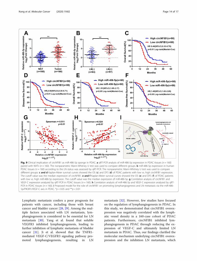
circNFIB1 and miR-486-5p in a cohort of 160 PDAC pa-tients. We found that miR-486-5p was overexpressed inPDAC tissues compared with NATs (Fig. 9a). Moreover,miR-486-5p expression in PDAC patients with LN me-tastasis was higher than those without LN metastasis(Fig. 9b). Importantly, Kaplan-Meier analysis revealedthat high miR-486-5p expression or low circNFIB1 ex-pression was associated with poor overall survival (OS)and disease-free survival (DFS) in 160 PDAC patients
(Fig. 9c-f). Univariate and multivariate analyses demon-strated that circNFIB1 functioned as an independent fac-tor for a good prognosis of PDAC patients(Additional file 5: Table S3 and Additional file 6: TableS4). Furthermore, correlation analysis demonstrated thatVEGF-C levels were negatively correlated with cir-cNFIB1 expression and positively associated with miR-486-5p expression in PDAC (Fig. 9g-h). In conclusion,our results indicate that circNFIB1 acts as a miR-486-5p
Fig. 7 CircNFIB1 downregulates VEGF-C expression through inhibiting the PI3K/Akt signaling pathway. a-d Western blot analysis of PIK3R1, Akt, p-Akt, GSK3β, and p-GSK3β levels after the overexpression (a and b) or silencing (c and d) of circNFIB1 in PDAC cells. e and f The effect ofcircNFIB1-silencing (e) or circNFIB1-overexpression (f) on VEGF-C expression in PDAC was assessed by qRT-PCR. g and h ELISA analysis of VEGF-Csecretion by PDAC after silencing (g) or overexpressing (h) circNFIB1. i and j The effect of LY294002 treatment on circNFIB1 depletion-inducedVEGF-C expression in PDAC cells was assessed by qRT-PCR. k and l ELISA for LY294002 treatment on circNFIB1 depletion-induced VEGF-Csecretion by PDAC cells. Significance level was assessed using two-tailed Student t-tests and one-way ANOVA followed by Dunnett’s tests formultiple comparisons. Figures with error bars showed the standard deviations of three independent experiments. *p < 0.05 and **p < 0.01
Kong et al. Molecular Cancer (2020) 19:82 Page 12 of 17

sponge and mediates the inhibition of LN metastasis viaVEGF-C suppression in PDAC patients.
DiscussionCircRNAs represent a class of noncoding RNAs witha high degree of stability and participate in multiplebiological processes and the regulation of gene ex-pression in human cells [27]. However, little is knownabout the role and underlying mechanisms of cir-cRNAs in the LN metastasis of PDAC. Herein, cir-cNFIB1 was identified to be downregulated in PDAC
and negatively correlated with LN metastasis inPDAC. Functionally, circNFIB1 suppressed lymphan-giogenesis and LN metastasis in PDAC. Mechanistic-ally, circNFIB1 functioned as a ceRNA and modulatedthe miR-486-5p/PI3KR1/VEGF-C axis, which inhibitedthe lymphangiogenesis and LN metastasis of PDAC.To our knowledge, this is the first report to provideinsight into the regulatory mechanism of circRNA-mediated LN metastasis inhibition of PDAC andhighlight that circNFIB1 may serve as a potentialtherapeutic target for LN metastasis in PDAC.
Fig. 8 CircNFIB1 inhibits lymphangiogenesis and LN metastasis of PDAC via VEGF-C suppression. a-f Representative images (a and d) and ahistogram analysis of tube formation (b and e) and Transwell (c and f) assay by HLECs treated with conditioned medium from si-NC, si-circNFIB1,si-circNFIB1 + PBS, and si-circNFIB1 + αVEGF-C PDAC cells. Scale bar: 100 μm. g Histogram analysis of the LN volume in the indicated groups. hThe ratio of popliteal LN metastasis was calculated for all groups (n = 12 per group). i Kaplan-Meier survival curves for indicated groups (n = 12per group). Significance level was assessed using two-tailed Student t-tests and one-way ANOVA followed by Dunnett’s tests for multiplecomparisons. Figures with error bars showed the standard deviations of three independent experiments. *p < 0.05 and **p < 0.01
Kong et al. Molecular Cancer (2020) 19:82 Page 13 of 17
Lymphatic metastasis confers a poor prognosis forpatients with cancer, including those with breastcancer and bladder cancer [28, 29]. Among the mul-tiple factors associated with LN metastasis, lym-phangiogenesis is considered to be essential for LNmetastasis [30]. Yang et al. found that solubleVEGFR3 inhibited lymphangiogenesis, leading tofurther inhibition of lymphatic metastasis of bladdercancer [31]. Ji et al. showed that the TNFR1-mediated VEGF-C/VEGFR3 signaling pathway pro-moted lymphangiogenesis, resulting in LN
metastasis [32]. However, few studies have focusedon the regulation of lymphangiogenesis in PDAC. Inthis study, we demonstrated that circNFIB1 overex-pression was negatively correlated with the lymph-atic vessel density in a 160-case cohort of PDACpatients. Furthermore, circNFIB1 inhibited lym-phangiogenesis in PDAC through reducing the ex-pression of VEGF-C and ultimately limited LNmetastasis in PDAC. Thus, our findings clarified themolecular mechanism underlying circNFIB1 overex-pression and the inhibition LN metastasis, which
Fig. 9 Clinical implication of circNFIB1 as miR-486-5p sponge in PDAC. a qRT-PCR analysis of miR-486-5p expression in PDAC tissues (n = 160)paired with NATs (n = 160). The nonparametric Mann-Whitney U test was used to compare different groups. b miR-486-5p expression in humanPDAC tissues (n = 160) according to the LN status was assessed by qRT-PCR. The nonparametric Mann-Whitney U test was used to comparedifferent groups. c and d Kaplan-Meier survival curves showed the OS (c) and DFS (d) of PDAC patients with low vs. high circNFIB1 expression.The cutoff value was the median expression of circNFIB1. e and f Kaplan-Meier survival curves showed the OS (e) and DFS (f) of PDAC patientswith low vs. high miR-486-5p expression. The cutoff value was the median expression of miR-486-5p. g Correlation analysis of circNFIB1 andVEGF-C expression analyzed by qRT-PCR in PDAC tissues (n = 160). h Correlation analysis of miR-486-5p and VEGF-C expression analyzed by qRT-PCR in PDAC tissues (n = 160). i Proposed model for the role of circNFIB1 on promoting lymphangiogenesis and LN metastasis via the miR-486-5p/PIK3R1/VEGF-C axis in PDAC. *p < 0.05 and **p < 0.01
Kong et al. Molecular Cancer (2020) 19:82 Page 14 of 17

provides novel insight into the regulatory mechan-ism of LN metastasis in PDAC.
VEGF-C is a VEGFR3 receptor ligand, which isexpressed on lymphatic vascular endothelial cells and iscritical for lymphangiogenesis [11]. The disfunction ofVEGF-C blocks the development of lymphatic vesselsand inhibits lymphatic metastasis of cancer cells [33].Chen et al. found that LNMAT1 promoted the expres-sion of CCL2 and recruited macrophages to increaseVEGF-C and facilitate the lymphangiogenesis of bladdercancer [34]. Similarly, Liu et al. demonstrated thatTBL1XR1 bound to the VEGF-C promotor to induceVEGF-C expression and promoted lymphangiogenesisand LN metastasis in esophageal squamous cell carcin-oma [35]. Although the importance of VEGF-C in lym-phangiogenesis and LN metastasis has been determinedin multiple cancers, the mechanism underlying the regu-lation of VEGF-C in PDAC remains unclear. In thepresent study, we reported that circNFIB1 downregu-lated VEGF-C expression through directly spongingmiR-486-5p and inactivating the PI3K/AKT signalingpathway, which further inhibited lymphangiogenesis andLN metastasis of PDAC. Therefore, our study revealed amolecular mechanism for circRNA-induced VEGF-Csuppression and indicated that circNFIB1 may be a po-tential target by which to block VEGF-C-induced LNmetastasis in PDAC patients.The PI3K/Akt signaling pathway has been thor-
oughly demonstrated to be a key regulator in cancerprogression. Overactivation of this pathway has beenobserved in a variety of cancers, including PDAC[36]. Moreover, it has been reported that activation ofthe PI3K/Akt signaling pathway contributed to LNmetastasis in several cancers. Yoo et al. revealed thatShh signaling promoted the lymphatic metastasis ofgastric cancer through activation of the PI3K/Aktpathway [37]. In addition, Saal et al. reported that aPIK3CA mutation induced the activation of the PI3K/Akt signaling pathway and facilitated LN metastasis inbreast cancer [38]. Nevertheless, little has been de-scribed about the PI3K/Akt pathway and LN metasta-sis of PDAC. In the present study, we found thatcircNFIB1 induced the upregulation of PIK3R1, andthe regulatory subunit of PI3K could inhibit activationof the PI3K/Akt signaling pathway and reduceVEGFC expression, thereby limiting lymphangiogen-esis and LN metastasis of PDAC. Moreover, blockingthe PI3K/Akt signaling pathway with its inhibitor,LY294002, significantly decreased circNFIB1depletion-induced VEGF-C, which serves as the keyregulator for lymphangiogenesis. Thus, inhibition ofPI3K/Akt signaling pathway may provide a novelintervention strategy for LN metastasis in PDAC.
ConclusionIn summary, we uncovered a novel mechanism by whichcircRNA-induced VEGF-C inhibition suppressed LNmetastasis of PDAC. We clinically demonstrated that thedownregulation of circNFIB1 is associated with the poorprognosis of PDAC patients. We further indicated thatcircNFIB1 overexpression could effectively inhibit LNmetastasis via the miR-486-5p/PIK3R1/VEGF-C axis.Identification of circNFIB1, which inhibits LN metasta-sis, both expands our knowledge of LN metastasis regu-lation and develops a potential therapeutic target for LNmetastasis in PDAC.
Supplementary informationSupplementary information accompanies this paper at https://doi.org/10.1186/s12943-020-01205-6.
Additional file 1 Table S1. Correlation between circNFIB1 expressionand clinicopathologic characteristics of PDAC patients
Additional file 2 Table S2. Primers and probes used in theexperiments.
Additional file 3. Supplementary methods.
Additional file 4 Figure S1. The identification of the downstreamtargets of miR-486-5p.
Additional file 5 Table S3. Univariate and multivariate analyses of OSfor circNFIB1 expression in PDAC patients.
Additional file 6 Table S4. Univariate and multivariate analyses of DFSfor circNFIB1 expression in PDAC patients.
Additional file 7 Table S5. Antibodies used in the experiments.
Additional file 8 Figure S2. Full uncut original pictures.
Additional file 9 Figure S3. Full uncut original gels.
AbbreviationsPDAC: Pancreatic ductal adenocarcinoma; circRNAs: Circular RNAs; LN: Lymphnode; VEGF-C: Vascular endothelial growth factor-C; NATs: Normal adjacenttissues; qRT-PCR: Quantitative real-time PCR; OS: Overall survival;DFS: Disease-free survival; HPDE: Human pancreatic ductal endothelial cells;FISH: Fluorescence in situ hybridization; IHC: Immunohistochemistry;HLECs: Human lymphatic endothelial cells
AcknowledgementsThe authors thank Prof. J.X. Zhang, Department of Medical Statistics andEpidemiology, School of Public Health, Sun Yat-Sen University, Guangzhou,China, for statistical advice and research comments.
Authors’ contributionsC. Chen, R. Chen and Z. Li participated in the study design; Y. Kong and Y. Liconducted the in vitro and in vivo experiment; J. Zhu conducted the dataanalysis; X. Guo conducted the clinical data analysis; H. Zheng and B. Gaoconducted the FISH and IHC assay; C. Chen, Y. Kong and Y. Luo wrote themanuscript: All the authors read and approved the final manuscript.
FundingThis study was supported by grants from the National Natural ScienceFoundation of China (Grant No. 81802530, 81672395, 81702951, 81672395,81672807, 81702417, 81000917, 81402213, 81370059 and 81701715), theGuangdong Science and Technology Department (Grant No.2018A030313564, S2012010008934, 2017A020215072, 2014A030313044,2014A030311047, 2016A030313340, 2016A030313296, 2017A030313880 and2017A030310200), Young Teacher Training Funding of Sun Yat-sen University(Grant No. 19ykpy121).
Kong et al. Molecular Cancer (2020) 19:82 Page 15 of 17

Availability of data and materialsOur circRNAs next-generation sequencing data used in this study have beendeposited in the NCBI’s Gene Expression Omnibus and are accessiblethrough GEO accession number: GSE136569 (https://www.ncbi.nlm.nih.gov/geo/query/acc.cgi?acc=GSE136569).
Ethics approval and consent to participateEthical consent was approved by the Committees for Ethical Review ofResearch involving Human Subjects at Sun Yat-sen University. Written in-formed consent was obtained from each patient prior to sample collection.The animal experiments were approved by the Use Committee for AnimalCare at Sun Yat-sen University.
Consent for publicationNot applicable.
Competing interestsThe authors declare no potential conflicts of interest.
Author details1Department of Ultrasound, Sun Yat-sen Memorial Hospital, Guangzhou,Guangdong 510120, People’s Republic of China. 2Guangdong Provincial KeyLaboratory of Malignant Tumor Epigenetics and Gene Regulation, SunYat-sen Memorial Hospital, State Key Laboratory of Oncology in South China,Guangzhou, Guangdong 510120, People’s Republic of China. 3Department ofMedical Oncology, Sun Yat-sen Memorial Hospital, 107th Yanjiangxi Road,Yuexiu District, Guangzhou, Guangdong 510120, People’s Republic of China.4Department of Pancreatobiliary Surgery, Sun Yat-sen Memorial Hospital,Guangzhou, Guangdong 510120, People’s Republic of China. 5Department ofGeneral Surgery, Guangdong Provincial People’s Hospital, GuangdongAcademy of Medical Sciences, Guangzhou, Guangdong 510080, People’sRepublic of China. 6Department of Urology, Sun Yat-sen Memorial Hospital,107 Yanjiangxi Road, Yuexiu District, Guangzhou, Guangdong 510120,People’s Republic of China.
Received: 20 January 2020 Accepted: 23 April 2020
References1. Ellis C, Ramzy A, Kieffer TJ. Regenerative medicine and cell-based
approaches to restore pancreatic function. Nat Rev Gastroenterol Hepatol.2017;14:612–28.
2. Makohon-Moore A, Iacobuzio-Donahue CA. Pancreatic cancer biology andgenetics from an evolutionary perspective. Nat Rev Cancer. 2016;16:553–65.
3. Vincent A, Herman J, Schulick R, Hruban RH, Goggins M. Pancreatic cancer.Lancet. 2011;378:607–20.
4. Philip PA, Mooney M, Jaffe D, Eckhardt G, Moore M, Meropol N, Emens L,O'Reilly E, Korc M, Ellis L, et al. Consensus report of the national cancerinstitute clinical trials planning meeting on pancreas cancer treatment. JClin Oncol. 2009;27:5660–9.
5. Groot VP, Gemenetzis G, Blair AB, Rivero-Soto RJ, Yu J, Javed AA, BurkhartRA, Rinkes I, Molenaar IQ, Cameron JL, et al. Defining and predicting earlyrecurrence in 957 patients with resected pancreatic ductal adenocarcinoma.Ann Surg. 2019;269:1154–62.
6. Paniccia A, Hosokawa P, Henderson W, Schulick RD, Edil BH, McCarter MD,Gajdos C. Characteristics of 10-year survivors of pancreatic ductaladenocarcinoma. JAMA Surg. 2015;150:701–10.
7. Sundar SS, Ganesan TS. Role of lymphangiogenesis in cancer. J Clin Oncol.2007;25:4298–307.
8. Achen MG, McColl BK, Stacker SA. Focus on lymphangiogenesis in tumormetastasis. Cancer Cell. 2005;7:121–7.
9. Cao Y. Opinion: emerging mechanisms of tumour lymphangiogenesis andlymphatic metastasis. Nat Rev Cancer. 2005;5:735–43.
10. Stacker SA, Williams SP, Karnezis T, Shayan R, Fox SB, Achen MG.Lymphangiogenesis and lymphatic vessel remodelling in cancer. Nat RevCancer. 2014;14:159–72.
11. Jussila L, Alitalo K. Vascular growth factors and lymphangiogenesis. PhysiolRev. 2002;82:673–700.
12. Kristensen LS, Andersen MS, Stagsted LVW, Ebbesen KK, Hansen TB, Kjems J.The biogenesis, biology and characterization of circular RNAs. Nat RevGenet. 2019;20:675–91.
13. Beermann J, Piccoli MT, Viereck J, Thum T. Non-coding RNAs indevelopment and disease: background, mechanisms, and therapeuticapproaches. Physiol Rev. 2016;96:1297–325.
14. Yang C, Yuan W, Yang X, Li P, Wang J, Han J, Tao J, Li P, Yang H, Lv Q, ZhangW. Circular RNA circ-ITCH inhibits bladder cancer progression by spongingmiR-17/miR-224 and regulating p21, PTEN expression. Mol Cancer. 2018;17:19.
15. Wei Y, Chen X, Liang C, Ling Y, Yang X, Ye X, Zhang H, Yang P, Cui X, Ren Y,et al. A noncoding regulatory RNAs network driven by Circ-CDYL actsspecifically in the early stages hepatocellular carcinoma. Hepatology. 2019;71:130–47.
16. Rupaimoole R, Slack FJ. MicroRNA therapeutics: towards a new era for themanagement of cancer and other diseases. Nat Rev Drug Discov. 2017;16:203–22.
17. Lopez-Bertoni H, Kotchetkov IS, Mihelson N, Lal B, Rui Y, Ames H, Lugo-Fagundo M, Guerrero-Cazares H, Quinones-Hinojosa A, Green JJ, Laterra J. ASox2/miR-486-5p axis regulates survival of GBM cells by inhibiting tumorsuppressor networks. Cancer Res. 2020;80:1644–55.
18. Hanna JA, Garcia MR, Lardennois A, Leavey PJ, Maglic D, Fagnan A, Go JC,Roach J, Wang YD, Finkelstein D, Hatley ME. PAX3-FOXO1 drives miR-486-5pand represses miR-221 contributing to pathogenesis of alveolarrhabdomyosarcoma. Oncogene. 2018;37:1991–2007.
19. Dudekula DB, Panda AC, Grammatikakis I, De S, Abdelmohsen K, Gorospe M.CircInteractome: A web tool for exploring circular RNAs and theirinteracting proteins and microRNAs. RNA Biol. 2016;13:34–42.
20. Agarwal V, Bell GW, Nam JW, Bartel DP. Predicting effective microRNA targetsites in mammalian mRNAs. Elife. 2015;4:e05005.
21. Betel D, Wilson M, Gabow A, Marks DS, Sander C. The microRNA.orgresource: targets and expression. Nucleic Acids Res. 2008;36:D149–53.
22. Mi H, Huang X, Muruganujan A, Tang H, Mills C, Kang D, Thomas PD.PANTHER version 11: expanded annotation data from Gene Ontology andReactome pathways, and data analysis tool enhancements. Nucleic AcidsRes. 2017;45:D183–9.
23. Mori MA, Ludwig RG, Garcia-Martin R, Brandao BB, Kahn CR. ExtracellularmiRNAs: from biomarkers to mediators of physiology and disease. CellMetab. 2019;30:656–73.
24. Taniguchi CM, Winnay J, Kondo T, Bronson RT, Guimaraes AR, Aleman JO,Luo J, Stephanopoulos G, Weissleder R, Cantley LC, Kahn CR. Thephosphoinositide 3-kinase regulatory subunit p85alpha can exert tumorsuppressor properties through negative regulation of growth factorsignaling. Cancer Res. 2010;70:5305–15.
25. Luo J, Sobkiw CL, Logsdon NM, Watt JM, Signoretti S, O'Connell F, Shin E,Shim Y, Pao L, Neel BG, et al. Modulation of epithelial neoplasia andlymphoid hyperplasia in PTEN+/− mice by the p85 regulatory subunits ofphosphoinositide 3-kinase. Proc Natl Acad Sci U S A. 2005;102:10238–43.
26. Brouillard P, Boon L, Vikkula M. Genetics of lymphatic anomalies. J ClinInvest. 2014;124:898–904.
27. Patop IL, Wust S, Kadener S. Past, present, and future of circRNAs. EMBO J.2019;38:e100836.
28. McAllaster JD, Cohen MS. Role of the lymphatics in cancer metastasis andchemotherapy applications. Adv Drug Deliv Rev. 2011;63:867–75.
29. Chen C, Luo Y, He W, Zhao Y, Kong Y, Liu H, Zhong G, Li Y, Li J, Huang J,et al. Exosomal long noncoding RNA LNMAT2 promotes lymphaticmetastasis in bladder cancer. J Clin Invest. 2020;130:404–21.
30. Zheng W, Aspelund A, Alitalo K. Lymphangiogenic factors, mechanisms, andapplications. J Clin Invest. 2014;124:878–87.
31. Yang H, Kim C, Kim MJ, Schwendener RA, Alitalo K, Heston W, Kim I, KimWJ, Koh GY. Soluble vascular endothelial growth factor receptor-3suppresses lymphangiogenesis and lymphatic metastasis in bladder cancer.Mol Cancer. 2011;10:36.
32. Ji H, Cao R, Yang Y, Zhang Y, Iwamoto H, Lim S, Nakamura M, Andersson P,Wang J, Sun Y, et al. TNFR1 mediates TNF-alpha-induced tumourlymphangiogenesis and metastasis by modulating VEGF-C-VEGFR3signalling. Nat Commun. 2014;5:4944.
33. Karaman S, Detmar M. Mechanisms of lymphatic metastasis. J Clin Invest.2014;124:922–8.
34. Chen C, He W, Huang J, Wang B, Li H, Cai Q, Su F, Bi J, Liu H, Zhang B, et al.LNMAT1 promotes lymphatic metastasis of bladder cancer via CCL2dependent macrophage recruitment. Nat Commun. 2018;9:3826.
35. Liu L, Lin C, Liang W, Wu S, Liu A, Wu J, Zhang X, Ren P, Li M, Song L.TBL1XR1 promotes lymphangiogenesis and lymphatic metastasis inesophageal squamous cell carcinoma. Gut. 2015;64:26–36.
Kong et al. Molecular Cancer (2020) 19:82 Page 16 of 17

36. LoRusso PM. Inhibition of the PI3K/AKT/mTOR pathway in solid tumors. JClin Oncol. 2016;34:3803–15.
37. Yoo YA, Kang MH, Lee HJ, Kim BH, Park JK, Kim HK, Kim JS, Oh SC. Sonichedgehog pathway promotes metastasis and lymphangiogenesis viaactivation of Akt, EMT, and MMP-9 pathway in gastric cancer. Cancer Res.2011;71:7061–70.
38. Saal LH, Holm K, Maurer M, Memeo L, Su T, Wang X, Yu JS, Malmstrom PO,Mansukhani M, Enoksson J, et al. PIK3CA mutations correlate with hormonereceptors, node metastasis, and ERBB2, and are mutually exclusive withPTEN loss in human breast carcinoma. Cancer Res. 2005;65:2554–9.
Publisher’s NoteSpringer Nature remains neutral with regard to jurisdictional claims inpublished maps and institutional affiliations.
Kong et al. Molecular Cancer (2020) 19:82 Page 17 of 17