circadian rhythms in endocrinology and metabolism...
TRANSCRIPT

Insulin resistance in liver, muscle and adipose tissue is a pivotal pathophysiological process in the development of type 2 diabetes mellitus (T2DM), which has been des-ignated as one of the four priority noncommunicable diseases by the WHO1. Major complications of T2DM are retinopathy, chronic kidney disease, neuropathy, peripheral vascular disease, myocardial infarction and stroke. The central approach in the prevention and treat-ment of insulin resistance is lifestyle modification. The second step is medication, with metformin being the cornerstone of the oral glucose- lowering drugs. If nec-essary, glycaemic control can be further improved with additional medication, including oral sulphonylureas, oral sodium–glucose cotransporter 2 (SGLT2) inhibi-tors, injectable glucagon- like peptide 1 (GLP1) receptor agonists or injectable insulin2. Traditionally, research has focused on the quantity and quality of physical activity, food intake and medication; however, circadian factors including the timing of light exposure, physical activity, food intake, medication and sleep–wake behaviour might also prove important for the prevention and treatment of insulin resistance.
The mammalian circadian timing system consists of a central brain clock in the hypothalamic suprachi-asmatic nucleus (SCN), and peripheral clocks in other brain regions and tissues throughout the body, including
muscle, adipose tissue and liver. The SCN receives a direct projection from the retina, via which environmen-tal light synchronizes the approximately 24 h rhythm of the SCN with the exact 24 h rhythm of the environment (Fig. 1). The entrained timing signal from the SCN is forwarded via neural and hormonal signals and body temperature to the peripheral clocks. The molecular mechanism of the central and peripheral clocks is based on transcriptional-translational feedback loops, which are present in almost every cell of the human body.
In this Review, we describe the physiological links between circadian clocks, glucose metabolism and insu-lin sensitivity. We also present current evidence for the relationship between circadian disruption and insulin resistance, with a focus on human studies. Finally, we propose several strategies to implement chronobiologi-cal knowledge with the aim to improve human metabolic health. The chronobiology terms and metabolic terms we use are defined in Box 1 and Box 2, respectively.
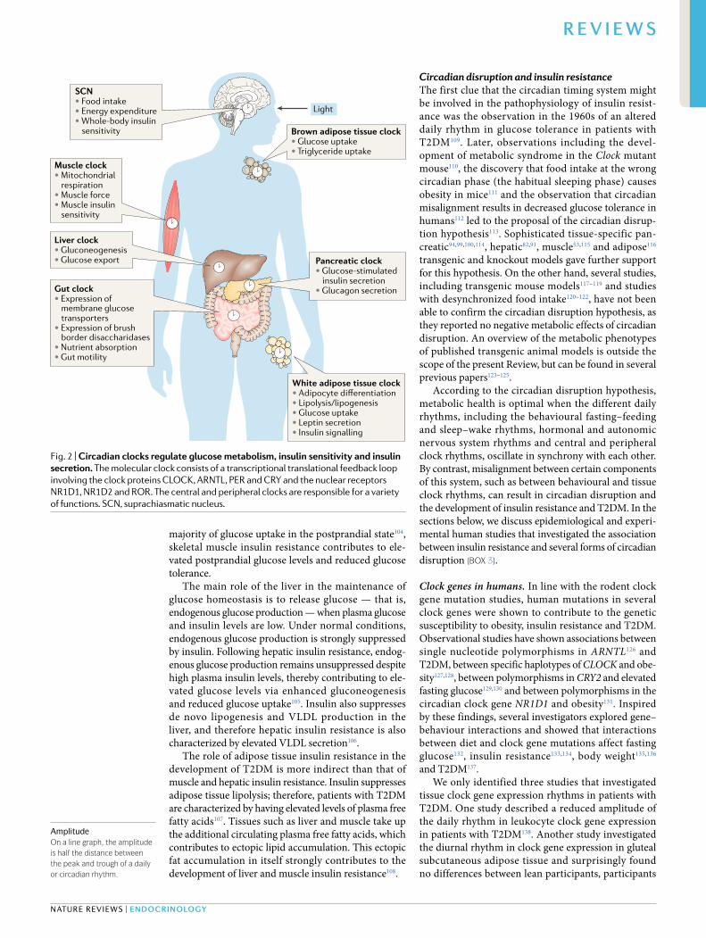
Circadian control of insulin sensitivityThe circadian timing systemThe mammalian circadian timing system is composed of a central pacemaker in the bilateral SCN of the ante-rior hypothalamus and a multitude of peripheral clocks in other brain areas and peripheral tissues (Fig. 2). The
*e- mail: a.kalsbeek@ nin.knaw.nl
https://doi.org/10.1038/ s41574-018-0122-1
Circadian clocks and insulin resistanceDirk Jan Stenvers 1, Frank A. J. L. Scheer2,3, Patrick Schrauwen4, Susanne E. la Fleur1,5,6 and Andries Kalsbeek 1,5,6*
Abstract | Insulin resistance is a main determinant in the development of type 2 diabetes mellitus and a major cause of morbidity and mortality. The circadian timing system consists of a central brain clock in the hypothalamic suprachiasmatic nucleus and various peripheral tissue clocks. The circadian timing system is responsible for the coordination of many daily processes, including the daily rhythm in human glucose metabolism. The central clock regulates food intake, energy expenditure and whole- body insulin sensitivity , and these actions are further fine- tuned by local peripheral clocks. For instance, the peripheral clock in the gut regulates glucose absorption, peripheral clocks in muscle, adipose tissue and liver regulate local insulin sensitivity , and the peripheral clock in the pancreas regulates insulin secretion. Misalignment between different components of the circadian timing system and daily rhythms of sleep–wake behaviour or food intake as a result of genetic, environmental or behavioural factors might be an important contributor to the development of insulin resistance. Specifically , clock gene mutations, exposure to artificial light–dark cycles, disturbed sleep, shift work and social jet lag are factors that might contribute to circadian disruption. Here, we review the physiological links between circadian clocks, glucose metabolism and insulin sensitivity, and present current evidence for a relationship between circadian disruption and insulin resistance. We conclude by proposing several strategies that aim to use chronobiological knowledge to improve human metabolic health.
C I R C A D I A N R H Y T H M S I N E N D O C R I N O LO GY A N D M E TA B O L I S M
RevIews
Nature reviews | Endocrinology

circadian timing system serves to prepare an organ-ism for the alternating opportunities and challenges that go along with the rhythmic changes of the daily light–dark cycle. This concept is illustrated by the evo-lutionary advantage of an internal clock that matches the period duration of the environment, as demonstrated in cyanobacteria3,4.
The discovery of the molecular mechanism that keeps these clocks functioning was awarded the 2017 Nobel Prize in Physiology or Medicine. The central fea-ture of this molecular mechanism is the transcriptional–translational feedback loop (TTFL) involving the core clock genes: the period genes (PER1, PER2 and PER3), cryptochrome genes (CRY1 and CRY2), ARNTL (also known as BMAL1), CLOCK (or its orthologue NPAS2) and the genes encoding the nuclear receptors REV-ERB (NR1D1 and NR1D2) and ROR (RORA, RORB and RORC). The rhythmic signal produced by this molecu-lar clock has a period of approximately 24 h, which is a circadian period.
The period of the endogenous circadian timing system does not match the exact 24 h rhythm of the outside world and, therefore, has to be reset every day. Environmental light is the most important Zeitgeber for resetting the cen-tral pacemaker, reaching the SCN through a direct con-nection from intrinsically photosensitive retinal ganglion cells through the retinohypothalamic tract. The remain-ing clocks in the circadian timing system therefore depend predominantly on the SCN for entrainment to the light–dark cycle of the outside world.
The SCN sends its entrained timing signal to the peripheral tissue clocks through the autonomic nerv-ous system, hormonal signals (including melatonin and cortisol), modulation of body temperature, and behavioural signals, such as physical activity and food
intake. As most peripheral clocks do not receive direct light information, they are also sensitive to these other Zeitgebers. This scenario is especially true for peripheral clocks in metabolic tissues such as liver, white adipose tissue (WAT), brown adipose tissue (BAT), pancreas and muscle, which use the metabolic signals resulting from food intake for their entrainment5–8 (Fig. 1).
Circadian rhythm in glucose metabolismIn healthy humans, plasma glucose tolerance depends on the time of day of glucose ingestion, with glucose toler-ance being higher in the morning than in the evening9,10. This diurnal rhythm in glucose tolerance is partly medi-ated by the diurnal rhythm in whole- body insulin sensi-tivity11. Moreover, the time- dependent glucose tolerance in healthy individuals is strongly mediated by the rhythm in pancreatic β- cell glucose sensitivity (that is, pancre-atic glucose- induced insulin secretion), as demonstrated by studies that use hyperglycaemic clamping12 and a triple- tracer mixed- meal technique13. However, because participants in these studies were sleeping at night and were awake during the day, it remains unclear to what extent these morning–evening differences are the result of behavioural and environmental differences or are caused by a direct influence of the circadian system. A 2015 study using a circadian misalignment protocol demonstrated that the diurnal rhythm in glucose toler-ance is robustly regulated by the circadian timing system, separate from influences of behavioural and environmen-tal changes14. Consistent with the results from diurnal studies, the endogenous circadian influence on glucose tolerance results from a stronger β- cell response in the circadian morning14.
Clock control of insulin sensitivityIn this section, we discuss the clocks in the different tissues and organs that are involved in the control of glucose metabolism and explain their role in the regulation of insulin sensitivity and insulin secretion.
The central clock. The central clock in the SCN not only synchronizes the peripheral clocks described in the sections below, but also affects multiple processes that influence the diurnal rhythm in glucose metabo-lism, including the physiological daily rhythms in sleep–wake behaviour, food intake, hormone secretion, insulin sensitivity and energy expenditure (Fig. 3).
The SCN controls the daily rhythm in sleep–wake behaviour15,16 via its connections with hypothalamic areas such as the subparaventricular zone, the ventrolat-eral preoptic area and the dorsomedial hypothalamus17. The SCN presumably also has a direct role in the control of food intake18,19, which is enhanced via the regulation of the sleep–wake cycle since food intake requires a waking state. The circadian control of food intake might be medi-ated via direct neuroanatomical connections between the SCN and the hypothalamic arcuate nucleus, which is involved in the regulation of food intake20, but indirect connections between the SCN and areas involved in the rewarding aspects of food could also have a role21.
In addition to the aforementioned roles, the SCN controls the daily rhythm of release of several hormones
Key points
•Thecircadiantimingsystemconsistsofacentralbrainclockinthehypothalamicsuprachiasmaticnucleusandperipheralclocksintissues,includingtheliver,muscle,adiposetissueandpancreas.
•Misalignmentbetweendifferentcomponentsofthecircadiantimingsystemanddailyrhythmsofsleep–wakebehaviourandfoodintakemightcontributetothedevelopmentofinsulinresistance.
•Strategiestoimprovemetabolichealthbycircadiansynchronyincludemodulatinglightexposure,modulatingrhythmicbehaviourandchronotherapy.
•Circadianmoleculesareapromisingnewtreatmentoptionforinsulinresistance.
Author addresses
1DepartmentofEndocrinologyandMetabolism,AmsterdamUniversityMedicalCenters,UniversityofAmsterdam,Amsterdam,Netherlands.2DivisionofSleepMedicine,HarvardMedicalSchool,Boston,MA,USA.3MedicalChronobiologyProgram,DivisionofSleepandCircadianDisorders,BrighamandWomen’sHospital,Boston,MA,USA.4DepartmentofNutritionandMovementSciences,NUTRIM,SchoolforNutritionandTranslationalResearchinMetabolism,MaastrichtUniversityMedicalCenter,Maastricht,Netherlands.5LaboratoryforEndocrinology,DepartmentofClinicalChemistry,AmsterdamUniversityMedicalCenters,UniversityofAmsterdam,Amsterdam,Netherlands.6NetherlandsInstituteforNeuroscience(NIN),RoyalDutchAcademyofArtsandSciences(KNAW),Amsterdam,Netherlands.
PeriodThe time difference between two consecutive peaks or troughs, or any other fixed point in the rhythm. in the case of daily or circadian rhythms, this period is exactly or approximately 24 h, respectively. The period of the rhythm in constant conditions is called the free- running period and is denoted by the greek letter τ.
www.nature.com/nrendo
R e v i e w s

affecting glucose tolerance. Firstly, the activity of the hypothalamic–pituitary–adrenal axis is regulated via connections from the SCN to the paraventricu-lar nucleus, resulting in a diurnal rhythm of cortisol
secretion, with a peak before the onset of the active period22. The glucocorticoid cortisol affects insulin signalling and reduces insulin secretion23. Secondly, the circadian rhythm of melatonin (also known as the hormone of darkness, as it is exclusively released dur-ing the dark phase in diurnal and nocturnal species alike), which affects insulin secretion24,25, is orches-trated by output from the SCN, via the paraventricular nucleus and the intermediolateral column to the pineal gland26. Thirdly, the diurnal rhythm in growth hormone, which antagonizes insulin action in liver and muscle27, is partly regulated by the SCN via its control of the sleep–wake cycle27–29.
Furthermore, SCN lesion studies in rodents demon-strated that the SCN controls the diurnal rhythm in whole- body insulin sensitivity30 and reported that within 8 weeks of an SCN lesion being created, rodents are insulin resistant31. In humans, a role for the SCN in the control of insulin sensitivity is suggested by mis-alignment protocols demonstrating the endogenous circadian control of glucose tolerance, independent of behavioural rhythms14,32.
Finally, the central clock is responsible for the cir-cadian regulation of multiple components of energy expenditure, such as the sleep–wake cycle15,16, diet- induced thermogenesis33, resting energy expenditure34 and (at least in rodents) BAT activity35–37.
The gut clock. Glucose enters the body via the gastro-intestinal tract. Intestinal cells throughout the intestinal tract contain a molecular clock38,39 and this gut clock is synchronized by signals resulting from food intake38. The gut clock regulates intestinal motility40 and nutri-ent absorption (Fig. 4). ARNTL regulates the expression of membrane glucose transporters and thus matches the timing of maximal monosaccharide uptake to the habitual feeding period41. Brush border disacchari-dases, including sucrase, display a circadian rhythm in activity42,43, but the mechanism regulating this circadian activity remains to be elucidated (Fig. 4).
The muscle clock. Human skeletal muscle has an auton-omous molecular clock44,45 (Fig. 5). Rodent data showed that the SCN synchronizes the skeletal muscle clock46,47, but signals resulting from physical exercise48,49 and food intake have also been shown to be involved in synchronization49–51.
Cultured rodent myotubes express circadian rhyth-micity in insulin sensitivity52. CLOCK and ARNTL regulate muscle insulin sensitivity via changes in pro-tein levels and membrane translocation of the insulin- sensitive glucose transporter GLUT4 (reF.53), as well as through the modulation of the insulin signalling path-way via expression of the deacetylase SIRT1 (reF.54). Furthermore, a 2017 study showed that the muscle clock regulates muscle insulin sensitivity via histone deacetylation of metabolic genes by HDAC3 (reF.55) (Fig. 5). Consistently, human muscle tissue shows a diur-nal rhythm in insulin sensitivity with higher insulin sen-sitivity in the morning than in the evening56, as well as a diurnal rhythm in mitochondrial oxidative capacity, which peaks in the evening57.
Eye SCNLight
Sleep–wake behaviour
Feeding behaviour
Autonomic nervous system
• Cortisol• Melatonin
Body temperature
Peripheral clocks in gut, muscle, liver, WAT, BAT and pancreas
Fig. 1 | The circadian timing system. The circadian timing system is composed of a central clock in the suprachiasmatic nucleus (SCN) located in the hypothalamus of the brain and peripheral clocks in other brain areas and peripheral tissues. The circadian rhythms in these clocks are generated by a molecular transcriptional- translational feedback loop. The light signal, reaching the SCN via the retina and the retinohypothalamic tract, is the most important Zeitgeber for the SCN. The SCN synchronizes peripheral clocks through neural, endocrine, temperature and behavioural signals. BAT, brown adipose tissue; WAT, white adipose tissue.
Box 1 | concepts in circadian studies
chronobiologyThestudyofbiologicalrhythmssuchasdaily,tidal,weekly,monthlyandseasonalrhythms.
chronotypeHumanscanbecharacterizedaccordingtotheirpreferredsleeptimes;latechronotypes(owls)prefertosleeplaterthanearlychronotypes(larks).
circadian rhythmArhythmwithaperiodof~24hthatpersistsinconstantconditions.CircadiancomesfromtheLatinwordscirca,whichmeansaround,anddies,whichmeansoneday.
daily (diurnal) rhythmPhysiological,hormonalandbehaviouralrhythmsthataremeasuredunderregularlight–darkandsleep–wakecycles,andthereforeshouldbedescribedasdailyrhythms,insteadofcircadianrhythms.
Entrainment and ZeitgeberThenon-24hperiodoftheendogenouscircadianrhythmcanbeadjusted,alignedorsynchronizedtotheexact24hperiodoftheoutsideworldbyaprocesscalledentrainment.TheexternalstimulusresponsibleforthisentrainmentiscalledaZeitgeber.InmammalsthestrongestZeitgeberfortheendogenouscentralbrainclockisenvironmentallight,butfoodintake,locomotoractivityandtemperaturecanalsoserveasZeitgebers.
nocturnal speciesSpeciesthataremainlyawakeandactiveduringthedarkperiod,suchasmostrodents.
diurnal speciesSpeciesthataremostlyawakeandactiveduringthelightperiod,suchashumans.
chronotherapyThespecifictimingofadministrationofdrugclassesbasedonthediurnalrhythmsinpharmacodynamicsandpharmacokineticsoftherapeuticdrugs.
Suprachiasmatic nucleus lesionInrodents,athermalorelectrolyticcompletelesionofthesuprachiasmaticnucleusneuronscausesalossofallcircadianrhythmicity(thatis,theabsenceofdailyrhythmsinlocomotoractivityandfoodintake,butalsoinhormonerelease,bodytemperatureandmetabolicfluxes).
Misalignment protocolAnexperimentalprotocolusingarecurringnon-24hbehaviouralcycle(forexample,a 28h cycle).Thisprotocolcanbeusedtoinvestigatetherelativecontributionsoftheendogenouscircadiancycleandthebehaviouralcycletoaparticularphysiologicalrhythm.
Nature reviews | Endocrinology
R e v i e w s

The adipose tissue clock. WAT contains an autono-mous circadian clock as shown in both rodent58,59 and human60,61 in vitro models (Fig. 6). Similar to the muscle clock, the WAT clock is synchronized by the SCN62 and by signals resulting from food intake63,64.
Adipocytes from rodents have circadian rhythmic-ity in glucose uptake52. In line with this observation, in human WAT ~25% of the transcriptome shows diurnal variation, including pathways involved in the regulation of glucose uptake65. Subcutaneous WAT explants from humans who are obese show an intrinsic diurnal rhythm in insulin signalling as determined by AKT phosphoryl-ation, with peak insulin sensitivity at noon66. Rodent data indicated that this diurnal rhythm in adipose tissue insu-lin sensitivity could be the result of circadian regulation of the retinol- binding protein receptor STRA6 (reF.67). In addition, CLOCK and ARNTL regulate the expression of key enzymes in the regulation of lipolysis such as adipose triglyceride lipase (ATGL), lipoprotein lipase (LPL) and hormone- sensitive lipase (HSL)68,69 (Fig. 6).
BAT from mice also shows a diurnal rhythm in glu-cose uptake70. A 2016 human study confirmed circadian rhythmicity of glucose uptake in BAT, with peak activity just before waking up71.
The liver clock. The liver contains an autonomous clock that is synchronized by the SCN72,73 via a combination of autonomic signals and endocrine signals64 (Fig. 7). The liver clock also responds strongly to the timing of food intake, as the liver clock can be uncoupled from the SCN clock by
inverting the daily feeding rhythm74. The liver clock reg-ulates several pathways involved in the control of glucose and lipid metabolism, as indicated by microarray73,75,76, proteomic77,78 and metabolomic79–81 studies.
By synchronizing the diurnal rhythms in gluconeo-genesis and glucose export to the habitual fasting period, the liver clock in rodents is essential to maintain eug-lycaemia82,83. The repression of gluconeogenesis during the feeding period is mediated by the interaction of CRY (which has its diurnal peak of expression during the feeding period) with the glucocorticoid receptor84 and with G protein-coupled receptor signalling85. The overall result of these interactions is the repression of the expression of rate- limiting gluconeogenetic genes. In addition, insulin- mediated suppression of gluconeo-genesis is partly dependent upon CRY- mediated FOXO1 degradation86,87 (Fig. 7). In view of this information, it is tempting to speculate that the liver clock contributes to the diurnal rhythms in hepatic glycogen content88 and in hepatic insulin sensitivity13 that are observed in healthy individuals.
In addition to the regulation of gluconeogenesis, the liver clock regulates the diurnal rhythm in mito-chondrial dynamics (Fig. 7). Therefore, the liver clock is involved in regulating mitochondrial glucose oxidation and fatty acid oxidation89,90, which protects the liver against oxidative stress during fasting91.
The pancreatic clock. The presence of an autonomous circadian pancreatic clock92 has been demonstrated not only in rodents93–95, but also in human islets and dispersed human islet cells (that is, the cells were cul-tured as seperate or single cells, not as an intact islet)96,97. The pancreatic clock is synchronized to the light–dark cycle95 via signals derived from the central brain clock in the SCN that include autonomic neuronal signals98, melatonin release93, glucocorticoid release96 and changes in body temperature96. The amplitude of oscillations in the expression of clock genes in cultured rat islets is dependent on the glucose concentration in the culture medium95.
Pancreatic islets isolated from rats show a circadian rhythm in insulin secretion93. CLOCK and BMAL1 activate the transcription of genes involved in insulin biosynthesis, insulin transport and glucose- stimulated insulin secretion99 (Fig. 8). In line with this observation, disruption of the pancreatic clock causes defective insulin secretion94,100,101. Similarly, in human pancreatic islets one group has confirmed that the pancreatic clock controls insulin secretion97.
Circadian disruptionInsulin resistanceInsulin resistance of liver, muscle and adipose tissue, which is initially compensated for by increased insulin secretion, is an early characteristic in the development of T2DM. Of note, in addition to insulin resistance, β-cell failure contributes to the development of T2DM102.
Insulin resistance in skeletal muscle is characterized by a reduced insulin- stimulated glucose uptake as a result of reduced insulin signalling and GLUT4 trans-location103. As skeletal muscle is responsible for the
Box 2 | Metabolic definitions
insulin resistance240
Resistancetothephysiologicaleffectsofinsulinatthetissuelevel.Thegoldstandardtomeasureinsulinsensitivityisthehyperinsulinaemiceuglycaemicclamp.
HoMA-ir241
Homeostaticmodelassessmentofinsulinresistance,basedonasinglecombinationoffastingglucoseandinsulinlevels.
glucose tolerancePlasmaglucoseexcursionafterafixedoralorintravenousglucoseload,withhigherglucoseexcursionsbeingindicativeofreducedglucosetolerance.
Prediabetes2
Fastingplasmaglucose5.6–6.9mmol/l(100–125mg/dl),2hplasmaglucoseafteroralglucosetolerancetest(75gglucose)7.8–11.0mmol/l(140–199mg/dl)orHbA1c39–47mmol/mol(5.7-6.4%).
diabetes mellitus2
Fastingplasmaglucose≥7mmol/l(126mg/dl),2hplasmaglucoseafteroralglucosetolerancetest(75gglucose)≥11.1mmol/l(200mg/dl)orHbA1c≥48mmol/mol(≥6.5%)orrandomplasmaglucose≥11.1mmol/l(200mg/dl)withhyperglycaemicsymptoms.
Type 2 diabetes mellitus2
Diabetesmellitusduetoperipheralinsulinresistance,combinedwithrelativeinsulindeficiency.
The metabolic syndrome242
Threeormoreofthefollowing:
•Waistcircumference>102cminmenor>88cminwomen•Triglycerides≥1.69mmol/l(150mg/dl)•HDL-cholesterol<1.04mmol/l(40mg/dl)inmenor<1.30mmol/l(50mg/dl)inwomen•Bloodpressure≥130/85mmHg•Fastingplasmaglucose≥5.6mmol/l(100mg/dl)
www.nature.com/nrendo
R e v i e w s
majority of glucose uptake in the postprandial state104, skeletal muscle insulin resistance contributes to ele-vated postprandial glucose levels and reduced glucose tolerance.
The main role of the liver in the maintenance of glucose homeostasis is to release glucose — that is, endogenous glucose production — when plasma glucose and insulin levels are low. Under normal conditions, endogenous glucose production is strongly suppressed by insulin. Following hepatic insulin resistance, endog-enous glucose production remains unsuppressed despite high plasma insulin levels, thereby contributing to ele-vated glucose levels via enhanced gluconeogenesis and reduced glucose uptake105. Insulin also suppresses de novo lipogenesis and VLDL production in the liver, and therefore hepatic insulin resistance is also characterized by elevated VLDL secretion106.
The role of adipose tissue insulin resistance in the development of T2DM is more indirect than that of muscle and hepatic insulin resistance. Insulin suppresses adipose tissue lipolysis; therefore, patients with T2DM are characterized by having elevated levels of plasma free fatty acids107. Tissues such as liver and muscle take up the additional circulating plasma free fatty acids, which contributes to ectopic lipid accumulation. This ectopic fat accumulation in itself strongly contributes to the development of liver and muscle insulin resistance108.
Circadian disruption and insulin resistanceThe first clue that the circadian timing system might be involved in the pathophysiology of insulin resist-ance was the observation in the 1960s of an altered daily rhythm in glucose tolerance in patients with T2DM109. Later, observations including the devel-opment of metabolic syndrome in the Clock mutant mouse110, the discovery that food intake at the wrong circadian phase (the habitual sleeping phase) causes obesity in mice111 and the observation that circadian misalignment results in decreased glucose tolerance in humans112 led to the proposal of the circadian disrup-tion hypothesis113. Sophisticated tissue- specific pan-creatic94,99,100,114, hepatic82,91, muscle53,115 and adipose116 transgenic and knockout models gave further support for this hypothesis. On the other hand, several studies, including transgenic mouse models117–119 and studies with desynchronized food intake120–122, have not been able to confirm the circadian disruption hypothesis, as they reported no negative metabolic effects of circadian disruption. An overview of the metabolic phenotypes of published transgenic animal models is outside the scope of the present Review, but can be found in several previous papers123–125.
According to the circadian disruption hypothesis, metabolic health is optimal when the different daily rhythms, including the behavioural fasting–feeding and sleep–wake rhythms, hormonal and autonomic nervous system rhythms and central and peripheral clock rhythms, oscillate in synchrony with each other. By contrast, misalignment between certain components of this system, such as between behavioural and tissue clock rhythms, can result in circadian disruption and the development of insulin resistance and T2DM. In the sections below, we discuss epidemiological and experi-mental human studies that investigated the association between insulin resistance and several forms of circadian disruption (Box 3).
Clock genes in humans. In line with the rodent clock gene mutation studies, human mutations in several clock genes were shown to contribute to the genetic susceptibility to obesity, insulin resistance and T2DM. Observational studies have shown associations between single nucleotide polymorphisms in ARNTL126 and T2DM, between specific haplotypes of CLOCK and obe-sity127,128, between polymorphisms in CRY2 and elevated fasting glucose129,130 and between polymorphisms in the circadian clock gene NR1D1 and obesity131. Inspired by these findings, several investigators explored gene–behaviour interactions and showed that interactions between diet and clock gene mutations affect fasting glucose132, insulin resistance133,134, body weight135,136 and T2DM137.
We only identified three studies that investigated tissue clock gene expression rhythms in patients with T2DM. One study described a reduced amplitude of the daily rhythm in leukocyte clock gene expression in patients with T2DM138. Another study investigated the diurnal rhythm in clock gene expression in gluteal subcutaneous adipose tissue and surprisingly found no differences between lean participants, participants
Light
SCN• Food intake• Energy expenditure• Whole-body insulin
sensitivity Brown adipose tissue clock• Glucose uptake• Triglyceride uptake
Pancreatic clock• Glucose-stimulated
insulin secretion• Glucagon secretion
Muscle clock• Mitochondrial
respiration• Muscle force• Muscle insulin
sensitivity
White adipose tissue clock• Adipocyte differentiation• Lipolysis/lipogenesis• Glucose uptake• Leptin secretion• Insulin signalling
Liver clock• Gluconeogenesis• Glucose export
Gut clock• Expression of
membrane glucosetransporters
• Expression of brush border disaccharidases
• Nutrient absorption• Gut motility
Fig. 2 | circadian clocks regulate glucose metabolism, insulin sensitivity and insulin secretion. The molecular clock consists of a transcriptional translational feedback loop involving the clock proteins CLOCK , ARNTL , PER and CRY and the nuclear receptors NR1D1, NR1D2 and ROR . The central and peripheral clocks are responsible for a variety of functions. SCN, suprachiasmatic nucleus.
Amplitudeon a line graph, the amplitude is half the distance between the peak and trough of a daily or circadian rhythm.
Nature reviews | Endocrinology
R e v i e w s

with obesity and patients with T2DM139. A study in circadian myotube explants described unaltered clock gene expression rhythms, but a decreased amplitude in NR1D1 expression in patients with T2DM44. In sum, indications of altered tissue clock rhythms in patients with T2DM are very limited.
Effects of light. Daylight is the main synchronizer of the central clock. Our modern lifestyle, however, is charac-terized by reduced light exposure during the day and increased light exposure during the night. These lifestyle changes have a substantial effect on the alignment of our circadian timing system to the solar day, as illustrated by an elegant study that investigated the effects of camp-ing in natural light–dark conditions on human daily sleep–wake behaviour140.
In several animal models, investigators have shown that dim light at night disturbs diurnal rhythms of food intake and locomotor behaviour120,125,141, causing obesity and reduced glucose tolerance in mice125,141 but not in rats120. In line with these findings, observational studies in humans have shown correlations of exposure to light at night with obesity142,143 and T2DM144.
Under conditions of controlled food intake and phys-ical activity, bright ambient light directly reduces insu-lin sensitivity in a time- dependent manner in healthy individuals145. When healthy participants are kept awake during the night, bright light causes increased levels of glucose in plasma146. In patients with T2DM, bright morning light increases fasting and postprandial levels of glucose147. A 2017 study in rats reported wave-length- dependent effects of ambient light on glucose
tolerance, with white and green light but not blue and red light reducing glucose tolerance148, but whether these observations translate to humans remains to be determined.
Melatonin. Melatonin is secreted by the pineal gland and shows a pronounced diurnal rhythm. During the dark period, plasma levels of melatonin are high149, and melatonin secretion is acutely suppressed by light expo-sure26. Melatonin acutely increases insulin secretion in cultured human islets24. By contrast, melatonin admin-istration in healthy women acutely decreases glucose tolerance150,151, an effect that is dependent on a common gain- of-function variant of the melatonin receptor gene MTNR1B152.
The role of melatonin signalling in the pathophysio-logy of T2DM remains a topic of lively debate153. On the one hand an association exists between reduced mel-atonin levels and the incidence of T2DM154, and rare loss-of-function mutations in MTNR1B are associated with an increase in the risk of T2DM155. On the other hand, one publication suggests that increased pancreatic β-cell melatonin signalling might reduce insulin secre-tion in humans25.
Sleep–wake rhythms. Accumulating evidence from both epidemiological and experimental studies shows that behavioural sleep–wake rhythms affect the risk of developing insulin resistance. A 2015 meta- analysis of prospective studies showed that both individuals who sleep for short periods and those who sleep for long periods are at increased risk of developing T2DM,
↓ Insulin sensitivity↓ Insulin secretion
SCN
↑ Insulin sensitivity↑ Insulin secretion
↓ Insulin sensitivity↓ Insulin secretion
PVN ARC
Food intake
Dopaminergic reward system
• SPZ• DMH • VLPO
Pituitary gland
HPA axis Sympathetic nervous system Sleep–wake rhythm
Pineal
Physical activity SleepGrowth hormone
MelatoninCortisol
? Insulin secretion
↓ Insulin sensitivity
↑ Insulin sensitivity • In the short term: ↑ insulin secretion
• In the long term (excess energy intake): ↓ insulin sensitivity
Parasympathetic nervous system
Sympathetic nervous system
Fig. 3 | The central clock. The suprachiasmatic nucleus (SCN), which contains the central clock , controls the daily rhythms of sleep–wake behaviour and food intake via hypothalamic connections. The central clock controls the circadian rhythm in the secretion of hormones affecting glucose tolerance, including cortisol, melatonin and growth hormone. ARC, arcuate nucleus; DMH, dorsomedial hypothalamus; HPA axis, hypothalamus– pituitary–adrenal axis; PVN, paraventricular nucleus; SPZ, subparaventricular zone; VLPO, ventrolateral preoptic nucleus.
www.nature.com/nrendo
R e v i e w s

with a proposed ‘optimal’ sleep duration of 7–8 h per night156.
The interpretation of epidemiological studies, how-ever, should be made with caution. It has been suggested that the relationship between long sleep duration and adverse health could be the result of reversed causality, undiagnosed disease, residual confounding and the sub-jective reporting on sleep duration possibly represent-ing time in bed157. On the other hand, investigators are in general agreement that poor sleep quality increases the risk of obesity and T2DM. A meta- analysis showed that people with reduced subjective sleep quality are at increased risk of developing T2DM158. In line with this finding, patients with obstructive sleep apnoea are at increased risk of developing T2DM, which could be
mediated by increased food intake and/or decreased physical activity159,160, among other mechanisms, owing to disturbed sleep161.
Several well- controlled human experimental stud-ies shed further light on the relationship between sleep deprivation and insulin sensitivity. The seminal exper-imental study showed reduced glucose tolerance after five nights of chronic partial sleep loss (4 h per night) compared with five well- rested nights (12 h per night) in healthy human participants, under conditions of con-trolled food intake and physical activity162. Subsequent experimental studies under controlled conditions con-firmed reduced liver163, adipose164 and whole- body163–168 insulin sensitivity as a result of sleep restriction to 4–6 h per night for 1–14 nights in healthy individuals. By con-trast, other studies under controlled conditions found only short- term effects169 or no effect170 of sleep restric-tion on insulin sensitivity, which could be the result of milder sleep restriction169 or a mitigating effect of the negative energy balance owing to the experimental design170. Further to these observations, studies have shown that experimental slow- wave sleep suppression resulted in reduced whole- body insulin sensitivity in healthy individuals171–173.
The proposed mechanisms for the effects of sleep restriction and sleep disturbance on insulin sensitiv-ity include an altered sympatho–vagal balance162,171,172 and increased circulating levels of catecholamines167 or cortisol167,172. In conditions of ad libitum food intake, increased food intake as a result of sleep restriction or disturbance probably contributes to decreased insulin sensitivity159,160.
A systematic review and meta- analysis showed that in patients with established T2DM, individuals who sleep for a short duration or a long duration and individuals with lower sleep quality have reduced glycaemic control compared with individuals who get adequate sleep174. Although several of the studies included in the system-atic review and meta- analysis corrected for physical activity, the meta- analysis did not correct for food intake or physical activity, so it is possible that these correla-tions are partly confounded by increased food intake or decreased physical activity174.
The incidence of obstructive sleep apnoea is high in patients with T2DM. Furthermore, in patients with comorbid obstructive sleep apnoea and T2DM, poor glycaemic control correlates with the severity of obstruc-tive sleep apnoea, a finding that could again be partly confounded by increased food intake and/or decreased physical activity161.
Chronotype and social jet lag. An individual’s chrono-type could also be a risk factor for insulin resistance. Observational studies show that evening chronotypes are at increased risk of developing T2DM compared with morning chronotypes175, even when results are corrected for sleep duration and physical activity (food intake was not corrected for in this study)176. Some evi-dence suggests that this increased risk could be the result of increased social jet lag — the discrepancy between the social (behavioural) and endogenous (circadian) time. Individuals with an evening chronotype who are
BloodLumen
Glucose
Galactose
SGLT1
GLUT5
GLUT2
Fructose
Sucrase
Sucrose
CLOCK
?
ARNTL
PER CRY
Gut cell
Fig. 4 | The gut clock. The molecular clock consists of a transcriptional–translational feedback loop involving the clock proteins CLOCK , ARNTL , PER and CRY and the nuclear receptors NR1D1, NR1D2 and ROR . The gut clock regulates the expression of membrane glucose transporters and brush border disaccharidases. GLUT, glucose transporter ; SGLT1, sodium–glucose cotransporter 1.
Glucose
CLOCK ARNTL
PER CRY
Mitochondrion
ATP
HDAC3
SIRT1
Insulin receptor
Insulin
P PAKT2 IRS1
GLUT4
Muscle cell
Fig. 5 | The muscle clock. In muscle, the muscle clock regulates muscle insulin sensitivity via protein levels and membrane translocation of the insulin-sensitive glucose transporter 4 (GLUT4) and through modulation of the insulin signalling pathway via expression of the deacetylase sirtuin 1 (SIRT1). In addition, the muscle clock regulates muscle insulin sensitivity via histone deacetylation of metabolic genes by histone deacetylase 3 (HDAC3). IRS1, insulin receptor substrate; P, phosphate.
Nature reviews | Endocrinology
R e v i e w s

working regular daytime hours are at increased risk of social jet lag.
Social jet lag is associated with the development of T2DM, independently of sleep duration177–179, even when results are corrected for food intake and physi-cal activity177. Patients with T2DM who are evening chronotypes show worse glycaemic control compared with patients who are morning chronotypes, a find-ing that might, in part, be mediated by an increase in evening food intake180; however, an association between poor glycaemic control and chronotype, independent of sleep duration, total food intake and physical activity, does also exist180,181.
Shift work and jet lag. Shift workers are at increased risk of developing T2DM, as shown by a 2015 meta- analysis of observational studies182; the degree of increased risk relates to the number of night shifts per month183. This increased risk of T2DM might be mediated by a combination of acute and chronic effects.
Experimental circadian misalignment under strictly controlled conditions acutely decreases glucose toler-ance and insulin sensitivity both in non-shift workers and in chronic shift workers14,32,112,184–186. To our knowl-edge, the chronic effects of repeated phase shifts on food intake, physical activity and insulin sensitivity have not been studied experimentally in humans, but several animal studies show that repeated phase shifts
cause increased food intake, increased body weight and disturbed glucose metabolism187. A 2014 trans-lational study showed that repeated jet lag in mice causes reduced glucose tolerance via disturbance of the intestinal microbiome. Fascinatingly, faecal trans-fer from jet- lagged humans into germ- free mice also reduced glucose tolerance in mice, suggesting that the microbiome clock might have an important role in the development of insulin resistance owing to repeated phase shifts188.
Does circadian disruption contribute to insulin resistance? Taken together, the hypothesis that circadian disrup-tion contributes to the development of insulin resist-ance in humans is supported by the following findings: decreased glucose tolerance caused by experimental circadian misalignment in humans; the association between human clock gene polymorphisms and insu-lin resistance; the experimentally observed effects of night- time light exposure and sleep disturbance on glucose metabolism; and the association of short sleep duration, long sleep duration, low sleep quality, late chronotype, social jet lag and shift work with insulin resistance. Therefore, it seems probable that disturbance of the central and/or tissue clock rhythms (Fig. 9) con-tributes to the pathophysiology of insulin resistance at the tissue level. Furthermore, circadian disruption might cause misalignment of nutrient fluxes. For instance, a mismatch between hepatic glucose production, mus-cle glucose uptake and carbohydrate intake could con-tribute to elevated levels of glucose and an imbalance between lipid storage in WAT, lipid oxidation in BAT and hepatic lipid production might contribute to ectopic lipid accumulation.
Circadian synchrony and metabolic healthModulating light exposureLight provides the main input for the SCN, and optimization of daily light exposure can therefore increase circadian synchrony140. To our knowledge, however, no published randomized controlled tri-als (RCTs) have investigated the effects of long- term natural light exposure on insulin sensitivity or glycaemia.
One potential strategy is the adaptation of architec-ture and/or indoor lighting conditions. For example, one RCT showed that supplementing daytime indoor light conditions with bright artificial light in homes for the elderly improves cognitive functioning, sleep qual-ity and the diurnal rhythm of locomotor activity189,190. Another study found that increasing blue light intensity in the morning with a system of wavelength- controlled light bulbs and LEDs at home improved subjective sleep quality in elderly women compared with low morning blue light intensity191.
A second potential strategy is to limit the use of screens from computers, tablets and smartphones in the evening, or to use blue light filters with these devices. An experimental study showed that reading a paper book in the evening reduced sleep- onset latency and improved daytime alertness the next day compared with reading a book on a light- emitting tablet192.
Triglyceride
CLOCK ARNTL
ATGL HSL
PER CRY
P
Glucose
GLUT4
Insulin receptor
Insulin
Adipocyte
AKT
PJAK2
LPLFree fatty acid
Diacylglycerol Monoacylglycerol
NR1D1
STRA6
RBP4
Fig. 6 | The white adipose tissue clock. In white adipose tissue, the circadian clock probably regulates the diurnal rhythm in insulin sensitivity via the circadian regulation of the retinol- binding protein receptor stimulated by retinoic acid 6 (STRA6). CLOCK and ARNTL regulate the expression of key enzymes in the regulation of lipolysis. ATGL , adipose triglyceride lipase; GLUT, glucose transporter ; HSL , hormone- sensitive lipase; JAK2, Janus kinase 2; LPL, lipoprotein lipase; P, phosphate; RBP4, retinol–binding protein 4.
www.nature.com/nrendo
R e v i e w s

Modulating rhythmic behaviourSleep–wake behaviour. In view of the strong association between disturbed sleep and impaired insulin sensitivity, sleep improvement could be a sensible approach in the prevention and treatment of insulin resistance193,194, but high- quality intervention studies are currently lacking (Fig. 9). One study in chronically sleep- restricted healthy individuals showed a correlation between improved indices of insulin sensitivity and increased sleep dura-tion after 40 days of sleep extension (~45 min extra each night)195. A study on the metabolic effects of sleep extension in sleep- restricted individuals who are obese is ongoing196.
Evidence- based strategies to treat insomnia include cognitive behavioural therapy (high- quality evidence according to the GRADE criteria), light therapy (low-quality evidence) and exercise (low- quality evi-dence)197. A short course (<4 weeks) of benzodiazepines or benzodiazepine receptor agonists can be considered if these strategies are not successful197, but concerns exist of negative effects of these hypnotic drugs on glucose tolerance, possibly owing to a reduction in slow- wave sleep after hypnotic drug use193.
Individuals with obstructive sleep apnoea represent a unique population regarding sleep–wake behaviour. A 2017 meta- analysis of four RCTs assessed the effect of continuous positive airway pressure (CPAP) treat-ment on insulin resistance in patients with obstructive
sleep apnoea who had either normal glucose values or prediabetes. The authors found no effect on HOMA- IR, but reported a small reduction of fasting insu-lin198. A 2017 meta- analysis showed that treatment of obstructive sleep apnoea with CPAP does not improve levels of haemoglobin A1c (HbA1c) or fasting levels of glucose in patients with obstructive sleep apnoea who have established T2DM, despite reduced daytime sleepiness199. The surprising lack of effect of CPAP treatment on glycaemia in patients with obstructive sleep apnoea could be related to treatment adher-ence199, as two studies with good adherence did show a decrease in HbA1c (reF.200) or mean 24 h glucose concentrations201.
Physical activity. Regular physical activity is one of the cornerstones of the lifestyle changes prescribed to patients with T2DM (Fig. 9). Regular physical exercise decreases insulin resistance and reduces HbA1c (reF.202). As physical activity also shifts the central circadian pacemaker in humans203, improves sleep duration and quality204 and affects the muscle clock48,49, it is possible that some of the beneficial metabolic effects of (day-time) physical activity are mediated through the cir-cadian timing system. To our knowledge, however, no studies have proved this idea. We are also not aware of any studies investigating the optimal timing of physical exercise for the reduction of body weight and insulin resistance.
Feeding behaviour. Individualized nutrition therapy is another core intervention in the prevention and treat-ment of insulin resistance and T2DM2. Classically, the main focus points for feeding behaviour are calorie reduction and healthy macronutrient distribution. An approach with a focus on the timing of food intake can also be of great value for people with insulin resistance205. A 2017 systematic review on meal tim-ing and frequency in the prevention of cardiovascular disease proposed an approach to eating that included the recommendations to eat a greater share of calories early in the day and to use consistent overnight fast periods206 (Fig. 9).
The advice to consume a greater share of calories early in the day mainly results from RCTs showing that breakfast consumption (compared with breakfast skip-ping) improves insulin sensitivity207–210, although not all studies agree211,212. A hallmark study on the overnight fast in 156 North American individuals showed that most people do not consume the ‘normal’ three meals per day within 12 h, but instead showed an irregular eating pattern spread over a >15 hr period. In that same study it was reported that a small group of eight people classed as obese who were treated with ‘time- restricted feeding’ (that is, they were asked to restrict eating to a 10 h period for 16 weeks) lost 3 kg in weight, which persisted over 1 year213. In line with this finding, a 2018 randomized crossover trial in eight individuals with prediabetes showed that isocaloric early time- restricted feeding (that is, a 6 h feeding period with dinner before 15:00 h) reduces insulin resistance compared with a 12 h feeding period214.
Glucose
GLUT2
CLOCK
ARNTL PER
CRY
Mitochondrion
ATP
Insulin receptor
Insulin
PAKT2
PIRS1
Liver cell
SREBP1c
Fission
Fusion
FOXO1
↑G6PC
Glucocorticoid receptor
Pyruvate
Adenylate cyclase
Glucagon
GPCR
cAMP
↑PEPCK
Gluconeogenesis
Adrenaline
Fig. 7 | The liver clock. In the liver, the circadian repression of gluconeogenesis during the habitual feeding period is mediated by the interaction of CRY with the glucocorticoid receptor, G protein-coupled receptor (GPCR) signalling and FOXO1 degradation. The liver clock also regulates the diurnal rhythm in mitochondrial dynamics. FOXO1, forkhead box protein O1; G6PC, glucose 6 phosphatase; GLUT, glucose transporter ; IRS1, insulin receptor substrate 1; P, phosphate; SREBP1c, sterol regulatory element-binding protein 1c.
Nature reviews | Endocrinology
R e v i e w s

Chronotherapy in patients with T2DMIn patients with T2DM, glycaemic control with oral glucose- lowering drugs and/or insulin reduces micro-vascular and cardiovascular complications2,215. To our knowledge and despite preclinical observations on the effects of metformin on the molecular clock216,217, the time- dependent effects of metformin on blood levels of glucose218 and the time- dependent effects of the sul-phonylurea tolbutamide on insulin secretion219, no trials have assessed the chronotherapeutic effects of these fre-quently prescribed glucose- lowering drugs on clinically relevant outcomes.
The only potential example of evidence- based chronotherapy in T2DM is the dopamine agonist bro-mocriptine (Fig. 9). Dopaminergic activity shows a diurnal rhythm220 and dopamine signalling increases insulin sensitvity221. When administered in the morning, a quick-release bromocriptine formulation reduces
HbA1c and fasting levels of glucose in patients with T2DM2,222. To our knowledge, however, no human trials have compared the effects of different adminis-tration times, which would be the ultimate proof that bromocriptine treatment is actually a chronotherapy.
The effects of timed melatonin administration in patients with T2DM have been investigated in one small RCT with 36 participants, which compared 3 weeks of melatonin administration with placebo223. The inves-tigators reported no convincing evidence of beneficial metabolic effects of melatonin.
The insulin requirements of patients on insulin therapy vary over the diurnal cycle owing to the diurnal rhythms of sleep–wake behaviour, physical activity, food intake and insulin sensitivity. The research community has made tremendous efforts to improve insulin phar-macokinetics with the aim of matching them to the individual patient’s diurnal pattern in insulin require-ments224,225. The question of whether the beneficial effects of insulin are (partly) mediated through central or peripheral clock modulation, however, remains to be resolved.
The artificial pancreas — which consists of an insu-lin pump controlled by a control algorithm coupled to a continuous glucose sensor226 — was in 2016 shown to increase the length of time spent in target glucose ranges in patients with T2DM who had been admitted to hospital227. One possible approach to further improve the algorithm controlling the artificial pancreas for patients with T2DM would be to incorporate information on the diurnal rhythm of insulin sensitivity.
The administration of exogenous glucocorticoids causes insulin resistance23. Data from a subgroup anal-ysis of a small open- label randomized trial228 and two prospective cohort studies229,230 suggest once- daily modified-release hydrocortisone formulations might be beneficial for metabolic health compared with thrice- daily immediate-release hydrocortisone. Analysis of the small subgroup of patients with comorbid adrenal insuf-ficiency and diabetes mellitus showed that replacement therapy with once- daily modified- release hydrocortisone formulations (which mimic the physiological diurnal rhythm of cortisol levels) might lead to improvements in body weight and HbA1c compared with thrice- daily immediate- release hydrocortisone.
Circadian moleculesNew circadian therapies might arise from large- scale chemical screens looking for clock- improving mole-cules231–233 (Fig. 9). Promising candidates include the REV- ERBα agonist SR9011 and the REV- ERBβ agonist SR9009, both of which directly target the molecular clock and were shown to decrease obesity and hyper-glycaemia in diet- induced obese mice. Timed twice- daily administration of these REV- ERB agonists alters metabolic gene expression patterns in muscle and WAT, leading to increased muscle glucose and fatty acid oxi-dation (increased energy expenditure), in combination with decreased WAT triglyceride synthesis234.
Another promising candidate is the natural cit-rus compound nobiletin, which has been shown to reduce body weight and improve insulin sensitivity
CLOCK ARNTL
PER CRY
Mitochondrion
ATP
Glucose
GLUT1/GLUT2
ATP-sensitive potassium channel
K+ Ca2+
VOCC
Endoplasmic reticulum
Insulin secretion
Insulin granule
Insulin
Pancreatic β-cell
Fig. 8 | The pancreas clock. In the pancreas, CLOCK and BMAL1 activate the transcription of genes involved in insulin biosynthesis, insulin transport and glucose-stimulated insulin secretion. All depicted processes show circadian rhythmicity. GLUT, glucose transporter ; VOCC, voltage- dependent calcium channel.
Box 3 | circadian disruption and insulin resistance
Epidemiological studies in humans
•Clockgenepolymorphisms(ARNTL126,CLOCK127,128,CRY2(reFs129,130),NR1D1(reF.131)
•Lightatnight125,142,143
•Reducedorincreasedmelatoninsignalling25,153,154,155
•Shortorlongsleep(optimal:7–8h)156,158
•Reducedsleepquality158
•Eveningchronotype175,176,181
•Socialjetlag177–179
•Shiftwork182,183
Experimental studies in humans
•Diet-clockgenemutationinteractions132–137
•Ambientlight145–147
•Melatonin150–152
•Sleeprestriction162–170andsleepdisruption171–173
•Circadianmisalignment14,32,112,184–186
www.nature.com/nrendo
R e v i e w s

in two different mouse models of the metabolic syn-drome (diet- induced obese mice and db/db mice). Nobiletin directly targets the molecular clock by acti-vating RORα and RORγ, thus increasing the amplitude of circadian locomotor behaviour, the rhythm of tissue clock gene expression and the rhythm of hepatic meta-bolic gene expression. As a result, energy expenditure increases, adiposity decreases and hepatic steatosis
decreases235. Finally, in preliminary reports, two different CRY stabilizers were shown to improve glucose tolerance in diet- induced obese mice236 and db/db mice237. The exact mechanism responsible for these metabolic benefits, however, remains to be elucidated.
In conclusion, REV- ERB agonists, ROR agonists and CRY stabilizers are promising circadian molecules for the treatment of T2DM, and human phase I studies of these compounds are to be expected.
ConclusionsDespite the large body of evidence from animal studies, the exact mechanisms mediating the metabolic derange-ments resulting from circadian disruption remain to be resolved. For example, does circadian misalignment cause a mismatch of glucose and lipid fluxes between the various organs or do disrupted tissue clocks cause insu-lin resistance at the tissue level, or are both mechanisms involved?
Currently, the clinical utility of the knowledge on circadian clock regulation of insulin sensitivity is only beginning to be explored. A clear need exists for RCTs that investigate the metabolic effects of natural light–dark exposure, sleep improvement, time- restricted feeding and the daily timing of exercise. Clinical trials are needed that investigate methods to prevent metabolic complica-tions in shift workers. With regard to biomarkers, evi-dence suggests that circadian phase biomarkers can help to optimally synchronize the circadian timing of behav-ioural or pharmacological interventions238. We are in no doubt that new circadian molecules targeting the mole-cular clock will be identified within the next 10 years. Furthermore, mathematical models could be an impor-tant aid to predict the effects of timed administration of clock agonists239. We expect the further development of promising candidate circadian molecules, including nobiletin, in phase I human trials in the coming years.
Published online xx xx xxxx
Circadian disruption Circadian synchrony
Light–dark exposure• Increase daytime light• Decrease light at night
and screen use
Circadian molecules• REV-ERB agonists• CRY stabilizers• ROR agonists
Daytime physical activity
Feeding behaviour• Eat a greater share of calories early in the day• Consistent overnight fast
Sleep–wake behaviour• Regular sleep–wake cycle• Refrain from shift work• Improve sleep quality• Identify and treat obstructive
sleep apnoea
Chronotherapy for type 2 diabetes mellitus• Bromocriptine quick release• Modified-release hydrocortisone
in patients with adrenal insufficiency
Fig. 9 | Potential interventions promoting metabolic health through circadian synchrony. Improving the synchrony between behavioural fasting–feeding and sleep–wake rhythms, hormonal and autonomic nervous system rhythms, and central and peripheral clock rhythms, might prove a valuable approach to prevent and/or treat insulin resistance and type 2 diabetes mellitus. Therapeutic interventions to improve circadian synchrony are possible at several levels: the light input to the circadian timing system; the behavioural level (sleep–wake behaviour, physical activity and food intake), directly targeting the molecular clock and the timing of medication (chronotherapy). Dark blue boxes show information that has some clinical human evidence supporting an effect on insulin sensitivity. Light blue boxes show information that has no clinical evidence supporting an effect on insulin sensitivity.
1. World Health Organization. Global Report on Diabetes (WHO, 2016).
2. American Diabetes Association. Standards of medical care in diabetes-2018. Diabetes Care 41, S1–S159 (2018). 2018 Current guideline of the American Diabetes Association for the diagnosis and treatment of diabetes mellitus.
3. Ouyang, Y., Andersson, C. R., Kondo, T., Golden, S. S. & Johnson, C. H. Resonating circadian clocks enhance fitness in cyanobacteria. Proc. Natl Acad. Sci. USA 95, 8660–8664 (1998).
4. Woelfle, M. A., Ouyang, Y., Phanvijhitsiri, K. & Johnson, C. H. The adaptive value of circadian clocks: an experimental assessment in cyanobacteria. Curr. Biol. 14, 1481–1486 (2004).
5. Dibner, C., Schibler, U. & Albrecht, U. The mammalian circadian timing system: organization and coordination of central and peripheral clocks. Annu. Rev. Physiol. 72, 517–549 (2010).
6. Stenvers, D. J., Jonkers, C. F., Fliers, E., Bisschop, P. H. & Kalsbeek, A. Nutrition and the circadian timing system. Progress Brain Res. 199, 359–376 (2012).
7. Lowrey, P. L. & Takahashi, J. S. Mammalian circadian biology: elucidating genome- wide levels of temporal organization. Annu. Rev. Genom. Hum. Genet. 5, 407–441 (2004).
8. Reppert, S. M. & Weaver, D. R. Coordination of circadian timing in mammals. Nature 418, 935–941 (2002).
9. Roberts, H. J. Afternoon glucose tolerance testing: a key to the pathogenesis, early diagnosis and prognosis
of diabetogenic hyperinsulinism. J. Am. Geriatr. Soc. 12, 423–472 (1964).
10. Van Cauter, E., Polonsky, K. S. & Scheen, A. J. Roles of circadian rhythmicity and sleep in human glucose regulation. Endocr. Rev. 18, 716–738 (1997). Seminal review on the diurnal rhythms of human glucose metabolism.
11. Gibson, T. & Jarrett, R. J. Diurnal variation in insulin sensitivity. Lancet 2, 947–948 (1972).
12. Boden, G., Ruiz, J., Urbain, J. L. & Chen, X. Evidence for a circadian rhythm of insulin secretion. Am. J. Physiol. 271, E246–E252 (1996).
13. Saad, A. et al. Diurnal pattern to insulin secretion and insulin action in healthy individuals. Diabetes 61, 2691–2700 (2012).
14. Morris, C. J. et al. Endogenous circadian system and circadian misalignment impact glucose tolerance via separate mechanisms in humans. Proc. Natl Acad. Sci. USA 112, E2225–E2234 (2015).
15. Stephan, F. K. & Zucker, I. Circadian rhythms in drinking behavior and locomotor activity of rats are eliminated by hypothalamic lesions. Proc. Natl Acad. Sci. USA 69, 1583–1586 (1972).
16. Czeisler, C. A., Weitzman, E., Moore- Ede, M. C., Zimmerman, J. C. & Knauer, R. S. Human sleep: its duration and organization depend on its circadian phase. Science 210, 1264–1267 (1980).
17. Saper, C. B., Scammell, T. E. & Lu, J. Hypothalamic regulation of sleep and circadian rhythms. Nature 437, 1257–1263 (2005).
18. Strubbe, J. H. & van Dijk, G. The temporal organization of ingestive behaviour and its interaction with regulation of energy balance. Neurosci. Biobehav. Rev. 26, 485–498 (2002).
19. Scheer, F. A., Morris, C. J. & Shea, S. A. The internal circadian clock increases hunger and appetite in the evening independent of food intake and other behaviors. Obesity 21, 421–423 (2013). First study showing the control of human appetite by the endogenous circadian timing system.
20. Herrera- Moro Chao, D. et al. The suprachiasmatic nucleus modulates the sensitivity of arcuate nucleus to hypoglycemia in the male rat. Endocrinology 157, 3439–3451 (2016).
21. Blancas- Velazquez, A., Mendoza, J., Garcia, A. N. & la Fleur, S. E. Diet- induced obesity and circadian disruption of feeding behavior. Front. Neurosci. 11, 23 (2017).
22. Buckley, T. M. & Schatzberg, A. F. On the interactions of the hypothalamic- pituitary-adrenal (HPA) axis and sleep: normal HPA axis activity and circadian rhythm, exemplary sleep disorders. J. Clin. Endocrinol. Metab. 90, 3106–3114 (2005).
23. van Raalte, D. H. & Diamant, M. Steroid diabetes: from mechanism to treatment? Neth. J. Med. 72, 62–72 (2014).
24. Ramracheya, R. D. et al. Function and expression of melatonin receptors on human pancreatic islets. J. Pineal Res. 44, 273–279 (2008).
25. Tuomi, T. et al. Increased melatonin signaling is a risk factor for type 2 diabetes. Cell Metab. 23, 1067–1077 (2016).
Nature reviews | Endocrinology
R e v i e w s
26. Claustrat, B., Brun, J. & Chazot, G. The basic physiology and pathophysiology of melatonin. Sleep Med. Rev. 9, 11–24 (2005).
27. Moller, N. & Jorgensen, J. O. Effects of growth hormone on glucose, lipid, and protein metabolism in human subjects. Endocr. Rev. 30, 152–177 (2009).
28. Morris, C. J., Aeschbach, D. & Scheer, F. A. Circadian system, sleep and endocrinology. Mol. Cell. Endocrinol. 349, 91–104 (2012).
29. Willoughby, J. O. & Martin, J. B. The suprachiasmatic nucleus synchronizes growth hormone secretory rhythms with the light- dark cycle. Brain Res. 151, 413–417 (1978).
30. La Fleur, S. E., Kalsbeek, A., Wortel, J., Fekkes, M. L. & Buijs, R. M. A daily rhythm in glucose tolerance: a role for the suprachiasmatic nucleus. Diabetes 50, 1237–1243 (2001).
31. Coomans, C. P. et al. The suprachiasmatic nucleus controls circadian energy metabolism and hepatic insulin sensitivity. Diabetes 62, 1102–1108 (2013).
32. Morris, C. J., Purvis, T. E., Mistretta, J. & Scheer, F. A. Effects of the internal circadian system and circadian misalignment on glucose tolerance in chronic shift workers. J. Clin. Endocrinol. Metab. 101, 1066–1074 (2016).
33. Morris, C. J. et al. The human circadian system has a dominating role in causing the morning/evening difference in diet- induced thermogenesis. Obesity 23, 2053–2058 (2015).
34. Krauchi, K. & Wirz- Justice, A. Circadian rhythm of heat production, heart rate, and skin and core temperature under unmasking conditions in men. Am. J. Physiol. 267, R819–R829 (1994).
35. Moran- Ramos, S. et al. The suprachiasmatic nucleus drives day- night variations in postprandial triglyceride uptake into skeletal muscle and brown adipose tissue. Exp. Physiol. 102, 1584–1595 (2017).
36. Amir, S., Shizgal, P. & Rompre, P. P. Glutamate injection into the suprachiasmatic nucleus stimulates brown fat thermogenesis in the rat. Brain Res. 498, 140–144 (1989).
37. Bamshad, M., Song, C. K. & Bartness, T. J. CNS origins of the sympathetic nervous system outflow to brown adipose tissue. Am. J. Physiol. 276, R1569–R1578 (1999).
38. Hussain, M. M. & Pan, X. Circadian regulation of macronutrient absorption. J. Biol. Rhythms 30, 459–469 (2015).
39. Scheving, L. A. Biological clocks and the digestive system. Gastroenterology 119, 536–549 (2000).
40. Hoogerwerf, W. A. Role of clock genes in gastrointestinal motility. Am. J. Physiol. Gastrointest. Liver Physiol. 299, G549–G555 (2010).
41. Iwashina, I., Mochizuki, K., Inamochi, Y. & Goda, T. Clock genes regulate the feeding schedule- dependent diurnal rhythm changes in hexose transporter gene expressions through the binding of BMAL1 to the promoter/enhancer and transcribed regions. J. Nutr. Biochem. 22, 334–343 (2011).
42. Nishida, T., Saito, M. & Suda, M. Parallel between circadian rhythms of intestinal disaccharidases and food intake of rats under constant lighting conditions. Gastroenterology 74, 224–227 (1978).
43. Saito, M., Kato, H. & Suda, M. Circadian rhythm of intestinal disaccharidases of rats fed with adiurnal periodicity. Am. J. Physiol. 238, G97–G101 (1980).
44. Hansen, J. et al. Synchronized human skeletal myotubes of lean, obese and type 2 diabetic patients maintain circadian oscillation of clock genes. Sci. Rep. 6, 35047 (2016).
45. Perrin, L. et al. Human skeletal myotubes display a cell- autonomous circadian clock implicated in basal myokine secretion. Mol. Metab. 4, 834–845 (2015).
46. Guo, H., Brewer, J. M., Lehman, M. N. & Bittman, E. L. Suprachiasmatic regulation of circadian rhythms of gene expression in hamster peripheral organs: effects of transplanting the pacemaker. J. Neurosci. 26, 6406–6412 (2006).
47. Guo, H., Brewer, J. M., Champhekar, A., Harris, R. B. & Bittman, E. L. Differential control of peripheral circadian rhythms by suprachiasmatic- dependent neural signals. Proc. Natl Acad. Sci. USA 102, 3111–3116 (2005).
48. Yamanaka, Y., Honma, S. & Honma, K. Scheduled exposures to a novel environment with a running- wheel differentially accelerate re- entrainment of mice peripheral clocks to new light- dark cycles. Genes Cells 13, 497–507 (2008).
49. Wolff, G. & Esser, K. A. Scheduled exercise phase shifts the circadian clock in skeletal muscle. Med. Sci. Sports Exercise 44, 1663–1670 (2012).
50. Reznick, J. et al. Altered feeding differentially regulates circadian rhythms and energy metabolism in liver and muscle of rats. Biochim. Biophys. Acta 1832, 228–238 (2013).
51. Opperhuizen, A. L. et al. Feeding during the resting phase causes profound changes in physiology and desynchronization between liver and muscle rhythms of rats. Eur. J. Neurosci. 44, 2795–2806 (2016).
52. Feneberg, R. & Lemmer, B. Circadian rhythm of glucose uptake in cultures of skeletal muscle cells and adipocytes in Wistar- Kyoto, Wistar, Goto- Kakizaki, and spontaneously hypertensive rats. Chronobiol. Int. 21, 521–538 (2004).
53. Dyar, K. A. et al. Muscle insulin sensitivity and glucose metabolism are controlled by the intrinsic muscle clock. Mol. Metab. 3, 29–41 (2014).
54. Liu, J. et al. CLOCK and BMAL1 regulate muscle insulin sensitivity via SIRT1 in male mice. Endocrinology 157, 2259–2269 (2016).
55. Hong, S. et al. Dissociation of muscle insulin sensitivity from exercise endurance in mice by HDAC3 depletion. Nat. Med. 23, 223–234 (2017).
56. Verrillo, A. et al. Differential roles of splanchnic and peripheral tissues in determining diurnal fluctuation of glucose tolerance. Am. J. Physiol. 257, E459–E465 (1989).
57. van Moorsel, D. et al. Demonstration of a day- night rhythm in human skeletal muscle oxidative capacity. Mol. Metab. 5, 635–645 (2016).
58. Otway, D. T., Frost, G. & Johnston, J. D. Circadian rhythmicity in murine pre- adipocyte and adipocyte cells. Chronobiol. Int. 26, 1340–1354 (2009).
59. Ramanathan, C. et al. Cell type- specific functions of period genes revealed by novel adipocyte and hepatocyte circadian clock models. PLOS Genet. 10, e1004244 (2014).
60. Huang, T. S. et al. Induction of circadian rhythm in cultured human mesenchymal stem cells by serum shock and cAMP analogs in vitro. Chronobiol. Int. 26, 242–257 (2009).
61. Gomez- Santos, C. et al. Circadian rhythm of clock genes in human adipose explants. Obesity 17, 1481–1485 (2009).
62. Kolbe, I. et al. The SCN clock governs circadian transcription rhythms in murine epididymal white adipose tissue. J. Biol. Rhythms 31, 577–587 (2016).
63. Wehrens, S. M. T. et al. Meal timing regulates the human circadian system. Curr. Biol. 27, 1768–1775 (2017). Study demonstrating the effect of meal timing on adipose tissue clock gene expression in humans.
64. Su, Y., Foppen, E., Zhang, Z., Fliers, E. & Kalsbeek, A. Effects of 6-meals- a-day feeding and 6-meals- a-day feeding combined with adrenalectomy on daily gene expression rhythms in rat epididymal white adipose tissue. Genes Cells 21, 6–24 (2016).
65. Loboda, A. et al. Diurnal variation of the human adipose transcriptome and the link to metabolic disease. BMC. Med. Genom. 2, 7 (2009).
66. Carrasco- Benso, M. P. et al. Human adipose tissue expresses intrinsic circadian rhythm in insulin sensitivity. FASEB J. 30, 3117–3123 (2016). First study showing a circadian rhythm in insulin signalling in cultured white adipose tissue explants from humans.
67. Gliniak, C. M., Brown, J. M. & Noy, N. The retinol- binding protein receptor STRA6 regulates diurnal insulin responses. J. Biol. Chem. 292, 15080–15093 (2017).
68. Delezie, J. et al. The nuclear receptor REV- ERBalpha is required for the daily balance of carbohydrate and lipid metabolism. FASEB J. 26, 3321–3335 (2012).
69. Shostak, A., Meyer- Kovac, J. & Oster, H. Circadian regulation of lipid mobilization in white adipose tissues. Diabetes 62, 2195–2203 (2013).
70. van der Veen, D. R., Shao, J., Chapman, S., Leevy, W. M. & Duffield, G. E. A diurnal rhythm in glucose uptake in brown adipose tissue revealed by in vivo PET- FDG imaging. Obesity 20, 1527–1529 (2012).
71. Lee, P. et al. Brown adipose tissue exhibits a glucose- responsive thermogenic biorhythm in humans. Cell Metab. 23, 602–609 (2016).
72. Sakamoto, K. et al. Multitissue circadian expression of rat period homolog (rPer2) mRNA is governed by the mammalian circadian clock, the suprachiasmatic nucleus in the brain. J. Biol. Chem. 273, 27039–27042 (1998).
73. Akhtar, R. A. et al. Circadian cycling of the mouse liver transcriptome, as revealed by cDNA microarray, is driven by the suprachiasmatic nucleus. Curr. Biol. 12, 540–550 (2002).
74. Stokkan, K. A., Yamazaki, S., Tei, H., Sakaki, Y. & Menaker, M. Entrainment of the circadian clock in the liver by feeding. Science 291, 490–493 (2001).
75. Panda, S. et al. Coordinated transcription of key pathways in the mouse by the circadian clock. Cell 109, 307–320 (2002).
76. Kornmann, B., Schaad, O., Bujard, H., Takahashi, J. S. & Schibler, U. System- driven and oscillator- dependent circadian transcription in mice with a conditionally active liver clock. PLOS Biol. 5, e34 (2007).
77. Robles, M. S., Cox, J. & Mann, M. In- vivo quantitative proteomics reveals a key contribution of post- transcriptional mechanisms to the circadian regulation of liver metabolism. PLOS Genet. 10, e1004047 (2014).
78. Mauvoisin, D. et al. Circadian clock- dependent and -independent rhythmic proteomes implement distinct diurnal functions in mouse liver. Proc. Natl Acad. Sci. USA 111, 167–172 (2014).
79. Gooley, J. J. & Chua, E. C. Diurnal regulation of lipid metabolism and applications of circadian lipidomics. J. Genet. Genomics 41, 231–250 (2014).
80. Abbondante, S., Eckel- Mahan, K. L., Ceglia, N. J., Baldi, P. & Sassone- Corsi, P. Comparative circadian metabolomics reveal differential effects of nutritional challenge in the serum and liver. J. Biol. Chem. 291, 2812–2828 (2016).
81. Krishnaiah, S. Y. et al. Clock regulation of metabolites reveals coupling between transcription and metabolism. Cell Metab. 25, 1206 (2017).
82. Lamia, K. A., Storch, K. F. & Weitz, C. J. Physiological significance of a peripheral tissue circadian clock. Proc. Natl Acad. Sci. USA 105, 15172–15177 (2008). Key study showing that the hepatic clock is essential to maintain euglycemia during the fasting period in mice.
83. Rudic, R. D. et al. BMAL1 and CLOCK, two essential components of the circadian clock, are involved in glucose homeostasis. PLOS Biol. 2, e377 (2004).
84. Lamia, K. A. et al. Cryptochromes mediate rhythmic repression of the glucocorticoid receptor. Nature 480, 552–556 (2011).
85. Zhang, E. E. et al. Cryptochrome mediates circadian regulation of cAMP signaling and hepatic gluconeogenesis. Nat. Med. 16, 1152–1156 (2010).
86. Jang, H. et al. SREBP1c- CRY1 signalling represses hepatic glucose production by promoting FOXO1 degradation during refeeding. Nat. Commun. 7, 12180 (2016).
87. Tong, X. et al. DDB1-mediated CRY1 degradation promotes FOXO1-driven gluconeogenesis in liver. Diabetes 66, 2571–2582 (2017).
88. Macauley, M., Smith, F. E., Thelwall, P. E., Hollingsworth, K. G. & Taylor, R. Diurnal variation in skeletal muscle and liver glycogen in humans with normal health and Type 2 diabetes. Clin. Sci. 128, 707–713 (2015).
89. Peek, C. B. et al. Circadian clock interaction with HIF1α mediates oxygenic metabolism and anaerobic glycolysis in skeletal muscle. Cell Metab. 25, 86–92 (2017).
90. Neufeld- Cohen, A. et al. Circadian control of oscillations in mitochondrial rate- limiting enzymes and nutrient utilization by PERIOD proteins. Proc. Natl Acad. Sci. USA 113, E1673–E1682 (2016).
91. Jacobi, D. et al. Hepatic Bmal1 regulates rhythmic mitochondrial dynamics and promotes metabolic fitness. Cell Metab. 22, 709–720 (2015).
92. Vieira, E., Burris, T. P. & Quesada, I. Clock genes, pancreatic function, and diabetes. Trends Mol. Med. 20, 685–693 (2014).
93. Peschke, E. & Peschke, D. Evidence for a circadian rhythm of insulin release from perifused rat pancreatic islets. Diabetologia 41, 1085–1092 (1998).
94. Marcheva, B. et al. Disruption of the clock components CLOCK and BMAL1 leads to hypoinsulinaemia and diabetes. Nature 466, 627–631 (2010). Pivotal study showing that specific ablation of the pancreatic clock disrupts insulin secretion in mice.
95. Qian, J., Block, G. D., Colwell, C. S. & Matveyenko, A. V. Consequences of exposure to light at night on the pancreatic islet circadian clock and function in rats. Diabetes 62, 3469–3478 (2013).
96. Pulimeno, P. et al. Autonomous and self- sustained circadian oscillators displayed in human islet cells. Diabetologia 56, 497–507 (2013).
97. Saini, C. et al. A functional circadian clock is required for proper insulin secretion by human pancreatic islet cells. Diabetes Obes. Metab. 18, 355–365 (2016). Study demonstrating that CLOCK inhibition with small interfering RNA disrupts insulin secretion in cultured human pancreatic islet cells.
www.nature.com/nrendo
R e v i e w s

98. Buijs, R. M., Chun, S. J., Niijima, A., Romijn, H. J. & Nagai, K. Parasympathetic and sympathetic control of the pancreas: a role for the suprachiasmatic nucleus and other hypothalamic centers that are involved in the regulation of food intake. J. Comp. Neurol. 431, 405–423 (2001).
99. Perelis, M. et al. Pancreatic beta cell enhancers regulate rhythmic transcription of genes controlling insulin secretion. Science 350, aac4250 (2015).
100. Sadacca, L. A., Lamia, K. A., deLemos, A. S., Blum, B. & Weitz, C. J. An intrinsic circadian clock of the pancreas is required for normal insulin release and glucose homeostasis in mice. Diabetologia 54, 120–124 (2011).
101. Lee, J. et al. Loss of Bmal1 leads to uncoupling and impaired glucose- stimulated insulin secretion in β- cells. Islets 3, 381–388 (2011).
102. Defronzo, R. A. Banting lecture. From the triumvirate to the ominous octet: a new paradigm for the treatment of type 2 diabetes mellitus. Diabetes 58, 773–795 (2009).
103. Shulman, G. I. Cellular mechanisms of insulin resistance. J. Clin. Invest. 106, 171–176 (2000).
104. Shulman, G. I. et al. Quantitation of muscle glycogen synthesis in normal subjects and subjects with non- insulin-dependent diabetes by 13C nuclear magnetic resonance spectroscopy. N. Engl. J. Med. 322, 223–228 (1990).
105. Reaven, G. M., Chen, Y. D., Donner, C. C., Fraze, E. & Hollenbeck, C. B. How insulin resistant are patients with noninsulin- dependent diabetes mellitus? J. Clin. Endocrinol. Metab. 61, 32–36 (1985).
106. Winocour, P. H. et al. A randomized cross- over study of the effects of proinsulin on lipid metabolism in type 2 diabetes. Diabet. Med. 8, 22–27 (1991).
107. Meek, S. E., Nair, K. S. & Jensen, M. D. Insulin regulation of regional free fatty acid metabolism. Diabetes 48, 10–14 (1999).
108. Unger, R. H. Minireview: weapons of lean body mass destruction: the role of ectopic lipids in the metabolic syndrome. Endocrinology 144, 5159–5165 (2003).
109. Jarrett, R. J. & Keen, H. Diurnal variation of oral glucose tolerance: a possible pointer to the evolution of diabetes mellitus. Br. Med. J. 2, 341–344 (1969). First study showing an altered daily rhythm in glucose tolerance in patients with type 2 diabetes mellitus.
110. Turek, F. W. et al. Obesity and metabolic syndrome in circadian Clock mutant mice. Science 308, 1043–1045 (2005). First study to show metabolic disease in a clock gene knockout mouse.
111. Arble, D. M., Bass, J., Laposky, A. D., Vitaterna, M. H. & Turek, F. W. Circadian timing of food intake contributes to weight gain. Obes.(Silver. Spring) 17, 2100–2102 (2009).
112. Scheer, F. A., Hilton, M. F., Mantzoros, C. S. & Shea, S. A. Adverse metabolic and cardiovascular consequences of circadian misalignment. Proc. Natl Acad. Sci. USA 106, 4453–4458 (2009). First study demonstrating that humans develop reduced glucose tolerance when subjected to conditions of circadian misalignment.
113. Bass, J. & Takahashi, J. S. Circadian integration of metabolism and energetics. Science 330, 1349–1354 (2010).
114. Lee, J. et al. Bmal1 and β- cell clock are required for adaptation to circadian disruption, and their loss of function leads to oxidative stress- induced beta- cell failure in mice. Mol. Cell. Biol. 33, 2327–2338 (2013).
115. Harfmann, B. D. et al. Muscle- specific loss of Bmal1 leads to disrupted tissue glucose metabolism and systemic glucose homeostasis. Skelet. Muscle 6, 12 (2016).
116. Paschos, G. K. et al. Obesity in mice with adipocyte- specific deletion of clock component Arntl. Nat. Med. 18, 1768–1777 (2012).
117. Oishi, K. et al. Disrupted fat absorption attenuates obesity induced by a high- fat diet in Clock mutant mice. FEBS Lett. 580, 127–130 (2006).
118. Zani, F. et al. PER2 promotes glucose storage to liver glycogen during feeding and acute fasting by inducing Gys2 PTG and G L expression. Mol. Metab. 2, 292–305 (2013).
119. Grimaldi, B. et al. PER2 controls lipid metabolism by direct regulation of PPARgamma. Cell Metab. 12, 509–520 (2010).
120. Stenvers, D. J. et al. Dim light at night disturbs the daily sleep- wake cycle in the rat. Sci. Rep. 6, 35662 (2016).
121. Shamsi, N. A. et al. Metabolic consequences of timed feeding in mice. Physiol. Behav. 128, 188–201 (2014).
122. Sherman, H. et al. Timed high- fat diet resets circadian metabolism and prevents obesity. FASEB J. 26, 3493–3502 (2012).
123. Kalsbeek, A., la Fleur, S. & Fliers, E. Circadian control of glucose metabolism. Mol. Metab. 3, 372–383 (2014).
124. Eckel- Mahan, K. & Sassone- Corsi, P. Metabolism and the circadian clock converge. Physiol. Rev. 93, 107–135 (2013).
125. Fonken, L. K. & Nelson, R. J. The effects of light at night on circadian clocks and metabolism. Endocr. Rev. 35, 648–670 (2014).
126. Woon, P. Y. et al. Aryl hydrocarbon receptor nuclear translocator- like (BMAL1) is associated with susceptibility to hypertension and type 2 diabetes. Proc. Natl Acad. Sci. USA 104, 14412–14417 (2007). First study showing an association between a clock gene single nucleotide polymorphism and type 2 diabetes mellitus.
127. Scott, E. M., Carter, A. M. & Grant, P. J. Association between polymorphisms in the Clock gene, obesity and the metabolic syndrome in man. Int. J. Obes. 32, 658–662 (2008).
128. Sookoian, S. et al. Genetic variants of Clock transcription factor are associated with individual susceptibility to obesity. Am. J. Clin. Nutr. 87, 1606–1615 (2008).
129. Dupuis, J. et al. New genetic loci implicated in fasting glucose homeostasis and their impact on type 2 diabetes risk. Nat. Genet. 42, 105–116 (2010).
130. Barker, A. et al. Association of genetic Loci with glucose levels in childhood and adolescence: a meta-analysis of over 6,000 children. Diabetes 60, 1805–1812 (2011).
131. Ruano, E. G., Canivell, S. & Vieira, E. REV- ERB ALPHA polymorphism is associated with obesity in the Spanish obese male population. PLOS ONE 9, e104065 (2014).
132. Dashti, H. S. et al. Gene- environment interactions of circadian- related genes for cardiometabolic traits. Diabetes Care 38, 1456–1466 (2015).
133. Dashti, H. S. et al. CRY1 circadian gene variant interacts with carbohydrate intake for insulin resistance in two independent populations: Mediterranean and North American. Chronobiol. Int. 31, 660–667 (2014).
134. Garcia- Rios, A. et al. Beneficial effect of CLOCK gene polymorphism rs1801260 in combination with low- fat diet on insulin metabolism in the patients with metabolic syndrome. Chronobiol. Int. 31, 401–408 (2014).
135. Garaulet, M. et al. CLOCK genetic variation and metabolic syndrome risk: modulation by monounsaturated fatty acids. Am. J. Clin. Nutr. 90, 1466–1475 (2009).
136. Loria- Kohen, V. et al. Polymorphism in the CLOCK gene may influence the effect of fat intake reduction on weight loss. Nutrition 32, 453–460 (2016).
137. Corella, D. et al. CLOCK gene variation is associated with incidence of type-2 diabetes and cardiovascular diseases in type-2 diabetic subjects: dietary modulation in the PREDIMED randomized trial. Cardiovasc. Diabetol. 15, 4 (2016).
138. Ando, H. et al. Clock gene expression in peripheral leucocytes of patients with type 2 diabetes. Diabetologia 52, 329–335 (2009).
139. Otway, D. T. et al. Rhythmic diurnal gene expression in human adipose tissue from individuals who are lean, overweight, and type 2 diabetic. Diabetes 60, 1577–1581 (2011). First study comparing in vivo clock gene expression rhythms between healthy participants, participants with obesity and patients with type 2 diabetes mellitus.
140. Wright, K. P. Jr. et al. Entrainment of the human circadian clock to the natural light- dark cycle. Curr. Biol. 23, 1554–1558 (2013).
141. Fonken, L. K. et al. Light at night increases body mass by shifting the time of food intake. Proc. Natl Acad. Sci. USA 107, 18664–18669 (2010).
142. Obayashi, K. et al. Exposure to light at night, nocturnal urinary melatonin excretion, and obesity/dyslipidemia in the elderly: a cross- sectional analysis of the HEIJO- KYO study. J. Clin. Endocrinol. Metab. 98, 337–344 (2013).
143. McFadden, E., Jones, M. E., Schoemaker, M. J., Ashworth, A. & Swerdlow, A. J. The relationship between obesity and exposure to light at night: cross- sectional analyses of over 100,000 women in the Breakthrough Generations study. Am. J. Epidemiol. 180, 245–250 (2014).
144. Obayashi, K., Saeki, K., Iwamoto, J., Ikada, Y. & Kurumatani, N. Independent associations of exposure
to evening light and nocturnal urinary melatonin excretion with diabetes in the elderly. Chronobiol. Int. 31, 394–400 (2014).
145. Cheung, I. N. et al. Morning and evening blue- enriched light exposure alters metabolic function in normal weight adults. PLOS ONE 11, e0155601 (2016).
146. Albreiki, M. S., Middleton, B. & Hampton, S. M. A single night light exposure acutely alters hormonal and metabolic responses in healthy participants. Endocr. Connect. 6, 100–110 (2017).
147. Versteeg, R. I. et al. Acute effects of morning light on plasma glucose and triglycerides in healthy men and men with type 2 diabetes. J. Biol. Rhythms 32, 130–142 (2017).
148. Opperhuizen, A. L. et al. Light at night acutely impairs glucose tolerance in a time-, intensity- and wavelength- dependent manner in rats. Diabetologia 60, 1333–1343 (2017).
149. Lewy, A. J. & Sack, R. L. The dim light melatonin onset as a marker for circadian phase position. Chronobiol. Int. 6, 93–102 (1989).
150. Rubio- Sastre, P., Scheer, F. A., Gomez- Abellan, P., Madrid, J. A. & Garaulet, M. Acute melatonin administration in humans impairs glucose tolerance in both the morning and evening. Sleep 37, 1715–1719 (2014).
151. Cagnacci, A. et al. Influence of melatonin administration on glucose tolerance and insulin sensitivity of postmenopausal women. Clin. Endocrinol. (Oxf.) 54, 339–346 (2001).
152. Garaulet, M. et al. Common type 2 diabetes risk variant in MTNR1B worsens the deleterious effect of melatonin on glucose tolerance in humans. Metabolism 64, 1650–1657 (2015).
153. Bonnefond, A. & Froguel, P. The case for too little melatonin signalling in increased diabetes risk. Diabetologia 60, 823–825 (2017).
154. McMullan, C. J., Schernhammer, E. S., Rimm, E. B., Hu, F. B. & Forman, J. P. Melatonin secretion and the incidence of type 2 diabetes. JAMA 309, 1388–1396 (2013).
155. Bonnefond, A. et al. Rare MTNR1B variants impairing melatonin receptor 1B function contribute to type 2 diabetes. Nat. Genet. 44, 297–301 (2012).
156. Shan, Z. et al. Sleep duration and risk of type 2 diabetes: a meta- analysis of prospective studies. Diabetes Care 38, 529–537 (2015). Meta- analysis demonstrating an association between sleep duration and the incidence of type 2 diabetes mellitus.
157. Stamatakis, K. A. & Punjabi, N. M. Long sleep duration: a risk to health or a marker of risk? Sleep Med. Rev. 11, 337–339 (2007).
158. Cappuccio, F. P., D’Elia, L., Strazzullo, P. & Miller, M. A. Quantity and quality of sleep and incidence of type 2 diabetes: a systematic review and meta- analysis. Diabetes Care 33, 414–420 (2010).
159. Bosy- Westphal, A. et al. Influence of partial sleep deprivation on energy balance and insulin sensitivity in healthy women. Obes. Facts 1, 266–273 (2008).
160. Nedeltcheva, A. V. et al. Sleep curtailment is accompanied by increased intake of calories from snacks. Am. J. Clin. Nutr. 89, 126–133 (2009).
161. Reutrakul, S. & Mokhlesi, B. Obstructive sleep apnea and diabetes: a state of the art review. Chest 152, 1070–1086 (2017).
162. Spiegel, K., Leproult, R. & Van Cauter, E. Impact of sleep debt on metabolic and endocrine function. Lancet 354, 1435–1439 (1999). Seminal experimental study showing the detrimental effect of sleep debt on glucose metabolism.
163. Donga, E. et al. A single night of partial sleep deprivation induces insulin resistance in multiple metabolic pathways in healthy subjects. J. Clin. Endocrinol. Metab. 95, 2963–2968 (2010).
164. Broussard, J. L., Ehrmann, D. A., Van Cauter, E., Tasali, E. & Brady, M. J. Impaired insulin signaling in human adipocytes after experimental sleep restriction: a randomized, crossover study. Ann. Intern. Med. 157, 549–557 (2012).
165. Buxton, O. M. et al. Sleep restriction for 1 week reduces insulin sensitivity in healthy men. Diabetes 59, 2126–2133 (2010).
166. Cedernaes, J. et al. A single night of partial sleep loss impairs fasting insulin sensitivity but does not affect cephalic phase insulin release in young men. J. Sleep Res. 25, 5–10 (2016).
167. Rao, M. N. et al. Subchronic sleep restriction causes tissue- specific insulin resistance. J. Clin. Endocrinol. Metab. 100, 1664–1671 (2015).
Nature reviews | Endocrinology
R e v i e w s

168. van Leeuwen, W. M. et al. Prolonged sleep restriction affects glucose metabolism in healthy young men. Int. J. Endocrinol. 2010, 108641 (2010).
169. Robertson, M. D., Russell- Jones, D., Umpleby, A. M. & Dijk, D. J. Effects of three weeks of mild sleep restriction implemented in the home environment on multiple metabolic and endocrine markers in healthy young men. Metabolism 62, 204–211 (2013).
170. St- Onge, M. P., O’Keeffe, M., Roberts, A. L., RoyChoudhury, A. & Laferrere, B. Short sleep duration, glucose dysregulation and hormonal regulation of appetite in men and women. Sleep 35, 1503–1510 (2012).
171. Tasali, E., Leproult, R., Ehrmann, D. A. & Van Cauter, E. Slow- wave sleep and the risk of type 2 diabetes in humans. Proc. Natl Acad. Sci. USA 105, 1044–1049 (2008).
172. Stamatakis, K. A. & Punjabi, N. M. Effects of sleep fragmentation on glucose metabolism in normal subjects. Chest 137, 95–101 (2010).
173. Herzog, N. et al. Selective slow wave sleep but not rapid eye movement sleep suppression impairs morning glucose tolerance in healthy men. Psychoneuroendocrinology 38, 2075–2082 (2013).
174. Lee, S. W. H., Ng, K. Y. & Chin, W. K. The impact of sleep amount and sleep quality on glycemic control in type 2 diabetes: A systematic review and meta- analysis. Sleep Med. Rev. 31, 91–101 (2017). Systematic review showing the relationship between sleep and glycaemic control in patients with type 2 diabetes mellitus.
175. Merikanto, I. et al. Associations of chronotype and sleep with cardiovascular diseases and type 2 diabetes. Chronobiol. Int. 30, 470–477 (2013).
176. Yu, J. H. et al. Evening chronotype is associated with metabolic disorders and body composition in middle-aged adults. J. Clin. Endocrinol. Metab. 100, 1494–1502 (2015).
177. Vetter, C. et al. Mismatch of sleep and work timing and risk of type 2 diabetes. Diabetes Care 38, 1707–1713 (2015). Study demonstrating the association between social jet lag and the risk of type 2 diabetes mellitus.
178. Parsons, M. J. et al. Social jetlag, obesity and metabolic disorder: investigation in a cohort study. Int. J. Obes. 39, 842–848 (2015).
179. Koopman, A. D. M. et al. The association between social jetlag, the metabolic syndrome, and type 2 diabetes mellitus in the general population: the New Hoorn study. J. Biol. Rhythms 32, 359–368 (2017).
180. Reutrakul, S. et al. Chronotype is independently associated with glycemic control in type 2 diabetes. Diabetes Care 36, 2523–2529 (2013).
181. Osonoi, Y. et al. Morningness- eveningness questionnaire score and metabolic parameters in patients with type 2 diabetes mellitus. Chronobiol. Int. 31, 1017–1023 (2014).
182. Gan, Y. et al. Shift work and diabetes mellitus: a meta- analysis of observational studies. Occupat. Environ. Med. 72, 72–78 (2015). Meta- analysis showing the association between shift work and diabetes mellitus.
183. Vetter, C. et al. Night shift work, genetic risk, and type 2 diabetes in the UK biobank. Diabetes Care 41, 762–769 (2018).
184. Leproult, R., Holmback, U. & Van Cauter, E. Circadian misalignment augments markers of insulin resistance and inflammation, independently of sleep loss. Diabetes 63, 1860–1869 (2014).
185. Qian, J., Dalla Man, C., Morris, C. J., Cobelli, C. & Scheer, F. A. Differential effects of the circadian system and circadian misalignment on insulin sensitivity and insulin secretion in humans. Diabetes Obes. Metab. 20, 2481–2485 (2018).
186. Wefers, J. et al. Circadian misalignment induces fatty acid metabolism gene profiles and compromises insulin sensitivity in human skeletal muscle. Proc. Natl Acad. Sci. USA 115, 7789–7794 (2018).
187. Opperhuizen, A. L., van Kerkhof, L. W., Proper, K. I., Rodenburg, W. & Kalsbeek, A. Rodent models to study the metabolic effects of shiftwork in humans. Front. Pharmacol. 6, 50 (2015).
188. Thaiss, C. A. et al. Transkingdom control of microbiota diurnal oscillations promotes metabolic homeostasis. Cell 159, 514–529 (2014).
189. Riemersma- van der Lek, R. F. et al. Effect of bright light and melatonin on cognitive and noncognitive function in elderly residents of group care facilities: a randomized controlled trial. JAMA 299, 2642–2655 (2008).
190. Hu, K. et al. Progression of dementia assessed by temporal correlations of physical activity: results from
a 3.5-Year, longitudinal randomized controlled trial. Sci. Rep. 6, 27742 (2016).
191. Sander, B., Markvart, J., Kessel, L., Argyraki, A. & Johnsen, K. Can sleep quality and wellbeing be improved by changing the indoor lighting in the homes of healthy, elderly citizens? Chronobiol. Int. 32, 1049–1060 (2015).
192. Chang, A.-M., Aeschbach, D., Duffy, J. F. & Czeisler, C. A. Evening use of light- emitting eReaders negatively affects sleep, circadian timing, and next- morning alertness. Proc. Natl Acad. Sci. 112, 1232–1237 (2015).
193. Tan, X., van Egmond, L., Chapman, C. D., Cedernaes, J. & Benedict, C. Aiding sleep in type 2 diabetes: therapeutic considerations. Lancet Diabetes Endocrinol. 6, 60–68 (2018).
194. Young, T. Increasing sleep duration for a healthier (and less obese?) population tomorrow. Sleep 31, 593–594 (2008).
195. Leproult, R., Deliens, G., Gilson, M. & Peigneux, P. Beneficial impact of sleep extension on fasting insulin sensitivity in adults with habitual sleep restriction. Sleep 38, 707–715 (2015).
196. Cizza, G., Piaggi, P., Rother, K. I. & Csako, G. & Sleep Extension Study, G. Hawthorne effect with transient behavioral and biochemical changes in a randomized controlled sleep extension trial of chronically short- sleeping obese adults: implications for the design and interpretation of clinical studies. PLOS ONE 9, e104176 (2014).
197. Riemann, D. et al. European guideline for the diagnosis and treatment of insomnia. J. Sleep Res. 26, 675–700 (2017).
198. Chen, L. et al. Continuous positive airway pressure and diabetes risk in sleep apnea patients: a systemic review and meta- analysis. Eur. J. Intern. Med. 39, 39–50 (2017). Meta- analysis investigating a potential relation between obstructive sleep apnoea treatment and the risk of developing type 2 diabetes mellitus.
199. Zhu, B., Ma, C., Chaiard, J. & Shi, C. Effect of continuous positive airway pressure on glucose metabolism in adults with type 2 diabetes: a systematic review and meta- analysis of randomized controlled trials. Sleep Breath. 22, 287–295 (2017).
200. Martinez- Ceron, E. et al. Effect of continuous positive airway pressure on glycemic control in patients with obstructive sleep apnea and type 2 diabetes. A randomized clinical trial. Am. J. Respir. Crit. Care Med. 194, 476–485 (2016).
201. Mokhlesi, B., Grimaldi, D., Beccuti, G. & Van Cauter, E. Effect of one week of CPAP treatment of obstructive sleep apnoea on 24-hour profiles of glucose, insulin and counter- regulatory hormones in type 2 diabetes. Diabetes Obes. Metab. 19, 452–456 (2017).
202. Colberg, S. R. et al. Physical activity/exercise and diabetes: a position statement of the American Diabetes Association. Diabetes Care 39, 2065–2079 (2016).
203. Buxton, O. M., Lee, C. W., L’Hermite- Baleriaux, M., Turek, F. W. & Van Cauter, E. Exercise elicits phase shifts and acute alterations of melatonin that vary with circadian phase. Am. J. Physiol. Regul. Integr. Comp. Physiol. 284, R714–R724 (2003).
204. Kredlow, M. A., Capozzoli, M. C., Hearon, B. A., Calkins, A. W. & Otto, M. W. The effects of physical activity on sleep: a meta- analytic review. J. Behav. Med. 38, 427–449 (2015).
205. Mattson, M. P. et al. Meal frequency and timing in health and disease. Proc. Natl Acad. Sci. USA 111, 16647–16653 (2014).
206. St- Onge, M. P. et al. Meal timing and frequency: implications for cardiovascular disease prevention: a scientific statement from the American Heart Association. Circulation 135, e96–e121 (2017). Statement from the American Heart Association on the effects of meal timing and frequency for metabolic health.
207. Jakubowicz, D., Barnea, M., Wainstein, J. & Froy, O. High caloric intake at breakfast versus dinner differentially influences weight loss of overweight and obese women. Obesity 21, 2504–2512 (2013).
208. Farshchi, H. R., Taylor, M. A. & Macdonald, I. A. Beneficial metabolic effects of regular meal frequency on dietary thermogenesis, insulin sensitivity, and fasting lipid profiles in healthy obese women. Am. J. Clin. Nutr. 81, 16–24 (2005).
209. Farshchi, H. R., Taylor, M. A. & Macdonald, I. A. Deleterious effects of omitting breakfast on insulin sensitivity and fasting lipid profiles in healthy lean women. Am. J. Clin. Nutr. 81, 388–396 (2005).
210. Chowdhury, E. A. et al. The causal role of breakfast in energy balance and health: a randomized controlled trial in obese adults. Am. J. Clin. Nutr. 103, 747–756 (2016).
211. Betts, J. A. et al. The causal role of breakfast in energy balance and health: a randomized controlled trial in lean adults. Am. J. Clin. Nutr. 100, 539–547 (2014).
212. Versteeg, R. I. et al. Meal timing effects on insulin sensitivity and intrahepatic triglycerides during weight loss. Int. J. Obes. 42, 156–162 (2017).
213. Gill, S. & Panda, S. A. Smartphone app reveals erratic diurnal eating patterns in humans that can be modulated for health benefits. Cell Metab. 22, 789–798 (2015). Study demonstrating chaotic diurnal eating patterns in humans, as well as the potential clinical benefits of time- restricted feeding.
214. Sutton, E. F. et al. Early time- restricted feeding improves insulin sensitivity, blood pressure, and oxidative stress even without weight loss in men with prediabetes. Cell Metab. 27, 1212–1221 (2018).
215. Holman, R. R., Sourij, H. & Califf, R. M. Cardiovascular outcome trials of glucose- lowering drugs or strategies in type 2 diabetes. Lancet 383, 2008–2017 (2014).
216. Um, J. H. et al. Activation of 5′-AMP- activated kinase with diabetes drug metformin induces casein kinase Iepsilon (CKIepsilon)-dependent degradation of clock protein mPer2. J. Biol. Chem. 282, 20794–20798 (2007).
217. Barnea, M. et al. Metformin affects the circadian clock and metabolic rhythms in a tissue- specific manner. Biochim. Biophys. Acta 1822, 1796–1806 (2012).
218. Henriksson, E. et al. The liver circadian clock modulates biochemical and physiological responses to metformin. J. Biol. Rhythms 32, 345–358 (2017).
219. Baker, I. A. & Jarrett, R. J. Diurnal variation in the blood- sugar and plasma- insulin response to tolbutamide. Lancet 2, 945–947 (1972).
220. Webb, I. C., Lehman, M. N. & Coolen, L. M. Diurnal and circadian regulation of reward- related neurophysiology and behavior. Physiol. Behav. 143, 58–69 (2015).
221. Ter Horst, K. W. et al. Striatal dopamine regulates systemic glucose metabolism in humans and mice. Sci. Transl Med. 10, eaar3752 (2018).
222. Liang, W. et al. Efficacy and safety of bromocriptine- QR in type 2 diabetes: a systematic review and meta- analysis. Horm. Metab. Res. 47, 805–812 (2015).
223. Garfinkel, D. et al. Efficacy and safety of prolonged- release melatonin in insomnia patients with diabetes: a randomized, double- blind, crossover study. Diabetes Metab. Syndr. Obes. 4, 307–313 (2011).
224. Holleman, F. & Gale, E. A. Nice insulins, pity about the evidence. Diabetologia 50, 1783–1790 (2007).
225. Wallia, A. & Molitch, M. E. Insulin therapy for type 2 diabetes mellitus. JAMA 311, 2315–2325 (2014).
226. Stenvers, D. J., DeVries, J. H. & la Fleur, S. E. What’s the time? Does the artificial pancreas need to know? Diabetes 62, 2173–2174 (2013).
227. Thabit, H. et al. Closed- loop insulin delivery in inpatients with type 2 diabetes: a randomised, parallel- group trial. Lancet Diabetes Endocrinol. 5, 117–124 (2016).
228. Johannsson, G. et al. Improved cortisol exposure- time profile and outcome in patients with adrenal insufficiency: a prospective randomized trial of a novel hydrocortisone dual- release formulation. J. Clin. Endocrinol. Metab. 97, 473–481 (2012).
229. Quinkler, M., Miodini Nilsen, R., Zopf, K., Ventz, M. & Oksnes, M. Modified- release hydrocortisone decreases BMI and HbA1c in patients with primary and secondary adrenal insufficiency. Eur. J. Endocrinol. 172, 619–626 (2015).
230. Giordano, R. et al. Improvement of anthropometric and metabolic parameters, and quality of life following treatment with dual- release hydrocortisone in patients with Addison’s disease. Endocr 51, 360–368 (2016).
231. Zhang, E. E. et al. A genome- wide RNAi screen for modifiers of the circadian clock in human cells. Cell 139, 199–210 (2009).
232. Chen, Z. et al. Identification of diverse modulators of central and peripheral circadian clocks by high- throughput chemical screening. Proc. Natl Acad. Sci. USA 109, 101–106 (2012).
233. Chen, Z., Yoo, S. H. & Takahashi, J. S. Development and therapeutic potential of small- molecule modulators of circadian systems. Annu. Rev. Pharmacol. Toxicol. 58, 231–252 (2018).
234. Solt, L. A. et al. Regulation of circadian behaviour and metabolism by synthetic REV- ERB agonists. Nature 485, 62–68 (2012). Study demonstrating beneficial metabolic effects of targeting the molecular clock with REV- ERB agonists in mice.
www.nature.com/nrendo
R e v i e w s

235. He, B. et al. The small molecule nobiletin targets the molecular oscillator to enhance circadian rhythms and protect against metabolic syndrome. Cell Metab. 23, 610–621 (2016). Study demonstrating that nobiletin improves metabolic health in mouse models of the metabolic syndrome by strengthening the molecular clock.
236. Humphries, P. S. et al. Carbazole- containing sulfonamides and sulfamides: discovery of cryptochrome modulators as antidiabetic agents. Bioorg. Med. Chem. Lett. 26, 757–760 (2016).
237. Humphries, P. S. et al. Carbazole- containing amides and ureas: discovery of cryptochrome modulators as antihyperglycemic agents. Bioorg. Med. Chem. Lett. 28, 293–297 (2017).
238. Laing, E. E. et al. Blood transcriptome based biomarkers for human circadian phase. eLife 6, e20214 (2017).
239. Woller, A., Duez, H., Staels, B. & Lefranc, M. A. Mathematical model of the liver circadian clock linking feeding and fasting cycles to clock function. Cell Rep. 17, 1087–1097 (2016).
240. Samuel, V. T. & Shulman, G. I. The pathogenesis of insulin resistance: integrating signaling pathways and substrate flux. J. Clin. Invest. 126, 12–22 (2016).
241. Wallace, T. M., Levy, J. C. & Matthews, D. R. Use and abuse of HOMA modeling. Diabetes Care 27, 1487–1495 (2004).
242. Sperling, L. S. et al. The CardioMetabolic Health Alliance: working toward a new care model for the metabolic syndrome. J. Am. College Cardiol. 66, 1050–1067 (2015).
Author contributionsD.J.S. researched data for article, all authors provided a sub-stantial contribution to discussion of content and wrote the article, F.A.J.L.S., P.S., S.E.L.F. and A.K. reviewed and edited the manuscript before submission.
Competing interestsF.A.J.L.S. received speaker fees from Bayer Healthcare, Kellogg Company, Philips, Pfizer, Sentara Healthcare and Vanda Pharmaceuticals. F.A.J.L.S. was supported in part by NIH grants R01DK099512, R01HL118601, R01DK102696, R01DK105072 and R01HL140574. The other authors declare no competing interests.
Publisher’s noteSpringer Nature remains neutral with regard to jurisdictional claims in published maps and institutional affiliations.
Nature reviews | Endocrinology
R e v i e w s