cine-angiocardiography with image...
TRANSCRIPT
SELECTIVE CINE-ANGIOCARDIOGRAPHY WITH IMAGEINTENSIFICATION
BY
HAMISH WATSON, C. PICKARD, K. G. LOWE, AND IAN HILLFrom the Departments of Medicine and Radiology, University of St. Andrews
Received February 12, 1958
There has been steady progress in angiocardiographic technique since Castellanos et al. (1937)showed that satisfactory opacification of the heart chambers was possible in children, and Robband Steinberg (1938) confirmed that equally successful results could be obtained in adults. Thoughthe earliest attempts to visualize the interior of the heart had been unsuccessful, it is interesting tonote that in these the contrast medium had been introduced directly into the heart (Forssmann,1931; Perez Ara, 1931; and Moniz et al., 1931). Most of the subsequent work was done usingintravenous injection, and it was not until 1946 that a reliable method of direct intracardiac angio-cardiography was described by Chavez et al. (1947). Since then much thought has been given tomethods for rapid and safe intracardiac injection (Jonsson et al., 1949; Dorbecker et al., 1954;Gidlund, 1956; and Rodriguez-Alvarez and de Rodriguez, 1957) and, though they are not yetperfect, many of their earlier dangers and disadvantages have been overcome.
The-introduction ofimage intensification by Teves and Feddema (1953 and 1955) started a new erain diagnostic radiology and the combination of the intensifier with a cine camera has provided an
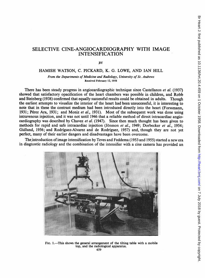
FIG. 1.-This shows the general arrangement of the tilting table with a mobiletop, and the radiological apparatus.
459
on 7 July 2018 by guest. Protected by copyright.
http://heart.bmj.com
/B
r Heart J: first published as 10.1136/hrt.20.4.459 on 1 O
ctober 1958. Dow
nloaded from

WATSON, PICKARD, LOWE, AND HILL
instrument that is particularly well suited to the field of angiocardiography (Lind and Wegelius,1955; Astley, 1955 and 1956; and Stauffer et al., 1955). When it is used with a good technique ofselective injection, the films made can be studied either in motion, at normal or slow speeds, orprojected as serial stills, and this results not only in more accurate diagnosis but allows detailed studyof the actual intracardiac circulation. In addition the comparatively small radiation rates used(Feddema, 1953; and Lind et al., 1955) mean much greater safety for both patients and staff-a factornow rightly considered of the utmost importance.
EQUIPMENT AND METHODSThe installation is illustrated in Fig. 1 and some results in Fig. 2, 3, and 4. It consists of a
tilting table with a mobile top. The undercouch tube is a Super Dynamax with a 1 mm. focal spot,and the focus is 24 inches (60 cm.) from the skin surface. The operator controls the beam delinea-tion by a double diaphragm which affords full protection. Power is supplied to the tube from aWatson cine-generator and, as the H.T. cables are long, no further stabilization is required withlow continuous tube currents. The explorator has been replaced by a Philips 5-inch (12-5 cm.)intensifier which can be used with a fluorescent tunnel, a direct lens system, or a 35-mm. cinecamera. Standard fluorescent screen examination is also possible over a 12-inch (30-cm.) squarearea, and spot films can be taken if required.
GREAT ARTERIAL TRUNKS
ASCENDING AORTAINFUNDIBULUM OF PD.A
RIGHT PULMONARY ARTERY PATENT DUCTUS ARTERIOSUS
DESCENDING AORTA
PULMONARY TRUNK LEFT PULMONARY ARTERY
RIGHTVENTRICLE LEFT VENTRICLE
FIG. 2.-The relationship of the intensifier field to the heart and great vessels (left anterior oblique view).
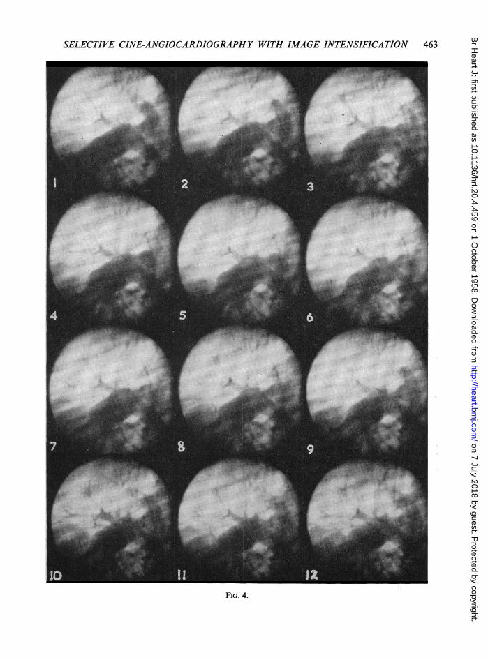
FIG. 3 and 4 illustrate two sets of twelve consecutive pictures taken at 50 frames a second, from a selective cine-angiocardiogram done on an 8-year-old girl, who presented as a case ef Eisenmenger's syndrome (Wood, 1952).The investigation was carried out after clinical examination and cardiac catheterization had failed to differentiatebetween a ventricular septal defect and patency of the ductus arteriosus.
FIG. 3.-In pictures 1-5 the initial opacification of the patent ductus arteriosus is seen developing and its infundibulum(Steinberg et al., 1943; J6nsson and Saltzman, 1952) is clearly defined. The contrast medium outlines thedescending aorta (6 and 7), and thereafter retrograde opacification of the first part of the descending aorta andthe aortic arch takes place (8-12). See page 461, opposite.
FIG. 4.-The first picture, at the end of diastole, shows that the innominate artery and the distal half of the aorticarch are opacified. In the second picture systole begins and the non-opaque blood from the left ventricle " washesout" the contrast medium down to the level of ductal entry (2-7); with diastole the retrograde opacificationbegins again and progresses upwards (8-12). See page 463.
460
on 7 July 2018 by guest. Protected by copyright.
http://heart.bmj.com
/B
r Heart J: first published as 10.1136/hrt.20.4.459 on 1 O
ctober 1958. Dow
nloaded from

SELECTIVE CINE-ANGIOCARDIOGRAPHY WITH IMAGE INTENSIFICATION 461
I'
I
I
I
FIG. 3.
on 7 July 2018 by guest. Protected by copyright.
http://heart.bmj.com
/B
r Heart J: first published as 10.1136/hrt.20.4.459 on 1 O
ctober 1958. Dow
nloaded from

WATSON, PICKARD, LOWE, AND HILL
The control of the exposure for cine-radiography is now standard. A micro-voltmeter is incircuit and arbitrary figures have been established that correlate the size and thickness of patient,the speed of the camera, and the radiographic output from the tube. This system has been satis-factory for all the speeds so far used. At lower speeds radiographic factors of the order of 65 KVand 1 MA are utilized, but at higher speeds, 100 KV and 5 MA are required. A stationary grid hasbeen found an advantage and the filtration of the tube varies from 0 5 to 3 mm. depending upon theKV applied. As the period during which cine-radiography is in operation does not usually exceed12 seconds, it has not been found necessary to involve complicated switching devices. During thepreparation of the installation, much detailed work was carried out on the photographic aspectsof this technique, and after experimenting with a variety of methods, HPX or Scopix G.35-mm.film, processed under standard conditions with standard solutions, has been found satisfactory formost cases, and the master film is available for preliminary examination within a few hours.
There is no doubt that at present the main disadvantage of image intensification is the small sizeof the screen, and like others we have found that this rather limits its usefulness. But as the investi-gation of congenital heart disease is largely confined to children, the 5-inch (12-5-cm.) field is lessrestrictive than would at first sight be imagined. It does, however, mean that, as we can at presentonly film in one plane, the greatest care must be taken with the positioning of the patient if themaximum information is to be obtained. In our preliminary studies we have used variations ofthe left anterior oblique view, depending on the nature of the lesions present, and have foundthat this gives good visualization of the heart chambers and great vessels. In infants and youngchildren, where the size of the screen is no problem, this represents a good compromise in theabsence of the usual biplane photography and gives satisfactory results. When it is not possibleto get the whole heart on the screen, the field is centred on the particular area that will give mosthelp in the diagnosis and future surgical treatment.
If in addition to diagnosis and pre-surgical evaluation, the intracardiac circulation is to bestudied, it is essential that the injection be truly selective. To make certain of this is by no meanseasy because the action of the heart, the rotation of the patient into the desired position, and theforce of the injection itself tend to produce movement of the catheter tip, and may cause it eitherto change its position within the cavity or to move out of it altogether. If, for example, the rightventricular outflow tract and pulmonary valve are to be studied, the contrast medium is injectedinto the lower part of the right ventricle with the intention that ventricular contraction will push itup into the infundibulum. If the catheter moves up or points up into the infundibulum during theinjection, the result is a pulmonary arteriogram and neither the filling mechanism nor the anatomyof the outflow tract will be defined. On the other hand should the force of the injection push thetip of the catheter back into the right atrium, only the initial opacification takes place in the ventricle,and very quickly the unwanted right atrial shadow obscures the detail of the outflow tract andpulmonary valve.
The size of the catheter used varies and is not necessarily the largest one possible. Whenselecting it, the size of the heart as well as the size and age of the patient have to be considered care-fully and in the light of previous experience, if good pictures are to be obtained. For babies a No. 5catheter is used and the injection given as rapidly as possible, but with larger sizes in older childrenthe force of the injection is varied to suit the problem that is being investigated, and the tip of thecatheter is shaped and fixed to match the size of the heart and the site of injection in each case. Aleg vein is used for the catheterization for two reasons: firstly because in babies and very smallchildren it is easier, and secondly because we have found that if an arm vein is used and films aretaken in oblique positions, the catheter may obscure structures to be visualized. When the catheteris in the desired position, it must be securely anchored to the skin of the limb if the force of theinjection is not to cause it to change position, and the use of catheters with lateral holes near thetip does much to reduce recoil during the injection.
Not only the choice of the site of the injection, but also the means used to ensure that the con-trast substance is in fact delivered there, have an important effect on the value of what is eventually
462
on 7 July 2018 by guest. Protected by copyright.
http://heart.bmj.com
/B
r Heart J: first published as 10.1136/hrt.20.4.459 on 1 O
ctober 1958. Dow
nloaded from
SELECTIVE CINE-ANGIOCARDIOGRAPHY WITH IMAGE INTENSIFICATION
FIG. 4.
463
on 7 July 2018 by guest. Protected by copyright.
http://heart.bmj.com
/B
r Heart J: first published as 10.1136/hrt.20.4.459 on 1 O
ctober 1958. Dow
nloaded from

WATSON, PICKARD, LOWE, AND HILL
seen on the cinematograph screen. As the field to be photographed can be seen through a lens ortunnel in full lighting, it is a simple matter to check the position of the patient and the site of thecatheter tip right up to the moment of injection.
When angiocardiograms are studied in motion, opacification is much more obvious than it ison still pictures, and can be seen where, on stills, there would be some doubt as to whether or notfaint delineation is present. With our usual technique ultra-rapid injection is not required, and theaim is to produce adequate opacification over several cardiac cycles. However, since at rapidfilm speeds faint delineation is fairly obvious, we have been trying two or more smaller injections indifferent planes, though still keeping the total dose of contrast medium within normal limits. Forthis it is essential that the small volume of contrast substance enters the heart as a bolus, and theobject is to be sure of catching at least one complete cardiac cycle showing the initial opacification.Though there are obvious drawbacks we hope that some such modification in technique will provesatisfactory for selected cases and that in this way still more information will be obtained.
The problem of correlating angiocardiographic films with events in the cardiac cycle is onethat has in the past received surprisingly little attention, perhaps because it is not vitally importantin many cases with slower film speeds. With cine-photography accurate timing is of first import-ance if the intracardiac circulation is to be studied in detail. A simple device that produces auto-matic exposure marking on a synchronous electrocardiogram (Lind et al., 1954) has been successfullymodified for this purpose and the time relationship of single pictures can be determined. Ifin addition the duration of the injection is shown on the recording paper, an exact correlationbetween electrocardiogram, X-ray picture, and injection of contrast medium is produced. Thistime relationship can be further checked, if by means of a simple circuit a given electrocardiographicsignal, say the top of the R wave, produces a light spot on the film.
One of the major difficulties encountered has been in the presentation of the results for analysis.This has been tackled in various ways (Campeti et al., 1955; Greenwood, 1956) but the generalprinciple that we have now adopted is to have the master film, approximately 80-100 ft., processed,examined for possible faults, and then sent away for professional copying. As it is difficult toproject 35-mm. film in this country because of the existing safety regulations, the prints are reducedto 16 mm. and there is little or no loss of quality or contrast in the process. We have found thedefinition on the 35-mm. film quite satisfactory, and because we think analysis is easier when thecontrast medium appears black, a positive 16-mm. film is made from the original 35-mm. negative.Detailed examination is not carried out until the prints are available, and though this means a certainamount of delay, the master film is protected from wear and tear and filed for future reference.
It takes time to adapt to this new method of presentation, and the eye accustomed to conven-tional angiocardiography finds that the speed of events is a little confusing. When running thefilm through as serial stills it has to be constantly borne in mind that the time interval betweenpictures is very small: otherwise, a false impression of persistent opacification in this or that cham-ber or vessel will be created. We are convinced that slow motion projection is essential for detailedstudies, but at present this is not easy to achieve. The maximum camera speed commonly in useis about 50 frames a second, and is too slow for a genuine slow motion effect. This can, however,be simulated by using skip-frame optical projection in printing, which though very effective is alsovery expensive. The aim must therefore be to increase the camera speeds so that slow motion pro-jection is possible, and with this in mind we have now produced satisfactory films with more than70 frames a second. It has been suggested that image persistence may arise at faster speeds, andthough this may well be a problem in the future, we have not been troubled by it so far.
DIsCUSSIONAngiocardiography, while presenting a factual record for analysis, is very much subject to the
personal factor in interpretation. Nowhere perhaps is this more true than in the study of theopacification of the right ventricular outflow tract in pulmonary stenosis: here the greatest difficulty
464
on 7 July 2018 by guest. Protected by copyright.
http://heart.bmj.com
/B
r Heart J: first published as 10.1136/hrt.20.4.459 on 1 O
ctober 1958. Dow
nloaded from

SELECTIVE CINE-ANGIOCARDIOGRAPHY WITH IMAGE INTENSIFICATION 465
may be experienced in distinguishing between the normal infundibulum in varying stages of con-traction and infundibular stenosis. The healthy outflow tract is funnel-shaped and of much greaterdiameter than the adjacent pulmonary trunk, but in our experience this fully dilated phase is a verymomentary event, and in a child with a fairly rapid heart rate the chances of catching it on singlefilms are very small. The main problem facing those interested in angiocardiography has thereforebeen to get increased film speeds without raising the already high radiation rates. The use of imageintensification and cine-photography not only helps to solve this problem, but makes it possible to takefive times as many pictures per second as the fastest conventional machines with a considerablereduction in the dose of X-rays received by the patient. Though the methods of its applicationare still in a developmental stage it is already obvious that this is an advance in diagnostictechnique. It is, for example, always possible at these film speeds to see the right ventricular out-flow tract fully dilated and to decide whether narrowing is present or not, and this kind of detailedstudy of the intracardiac circulation in patients with congenital heart disease will lead to a muchclearer understanding of the abnormal mechanisms involved (Fig. 2, 3, and 4).
SUMMARYSelective cine-angiocardiography using a Philips image intensifier is described, and the technical
problems of production and presentation of the films for analysis are discussed. The futurepossibilities of this method in the investigation of congenital heart disease are emphasized.
REFERENCESAstley, R. (1955). Brit. J. Radiol., 28, 221.- (1956). Brit. J. Radiol., 29, 556.Campeti, F., Gramiak, R., Watson, J. S., Ramsey, G. H., and Weinberg, S. (1955). Circulation, 12, 199.Castellanos, A., Pereira, R., and Garcia, A. (1937). Arch. Soc. estudios clin. Habana, 31, 523.Chavez, I., Dorbecker, N., and Celis, A. (1947). Amer. Heart J., 33, 560.Dorbecker, N., Rodriguez-Alvarez, A., and Volnie, B. (1954). II Congreso de Cardiologia SIBIC-Acapulco, Mexico.Feddema, J. (1953). Annual Congress, Brit. Institute of Radiology.- (1955). Brit. J. Radiol., 28, 217.
Forssmann, W. (1931). Munch. med. Wchnschr., 78, 489.Gidlund, A. (1956). Acta Radiol., Supplement, 130.Greenwood, F. G. (1956). Brit. J. Radiol., 29, 544.Jonsson, G., Broden, B., and Kamell, J. (1949). Acta Radiol., 32, 486.-, and Saltzman, G. F. (1952). Acta Radiol., 37, 445.Lind, J., Wegelius, C., and Lichtenstein, H. (1954). Circulation, 10, 195.-- , (1955). Nord. Med., 22, 914.
and Boesen, I. (1955). Henry Ford Symposium on Cardiovascular Surgery, Detroit.Moniz, E., de Carvalho, L., and Lima, A. (1931). Presse mid., 39, 996.Perez Ara, A. (1931). Rev. me'd. circ. Habania, 36, 7.Robb, G. P., and Steinberg, I. (1938). J. clin. Invest., 17, 507.Rodriguez-Alvarez, A., and de Rodriguez, G. M. (1957). Amer. Heart J., 53, 481.Stauffer, H. M., Morton, J., Oppenheimer, M. J., Stewart, G. H., and Blackstone, A. W. (1955). Radiology, 65,784.Steinberg, M. F., Grishman, A., and Sussman, M. L. (1943). Amer. J. Roentgenol., 50, 306.Teves, M. C. (1953). Annual Congress, British Institute of Radiology.- (1955). Brit. J. Radiol., 28, 216.
Wood, P. (1952). Brit. med. Bull., 8, 348.
on 7 July 2018 by guest. Protected by copyright.
http://heart.bmj.com
/B
r Heart J: first published as 10.1136/hrt.20.4.459 on 1 O
ctober 1958. Dow
nloaded from