ciliogenesis and the dna damage response: a stressful
TRANSCRIPT

Johnson and Collis Cilia (2016) 5:19 DOI 10.1186/s13630-016-0040-6
REVIEW
Ciliogenesis and the DNA damage response: a stressful relationshipColin A. Johnson1* and Spencer J. Collis2*
Abstract
Both inherited and sporadic mutations can give rise to a plethora of human diseases. Through myriad diverse cellular processes, sporadic mutations can arise through a failure to accurately replicate the genetic code or by inaccurate separation of duplicated chromosomes into daughter cells. The human genome has therefore evolved to encode a large number of proteins that work together with regulators of the cell cycle to ensure that it remains error-free. This is collectively known as the DNA damage response (DDR), and genome stability mechanisms involve a complex net-work of signalling and processing factors that ensure redundancy and adaptability of these systems. The importance of genome stability mechanisms is best illustrated by the dramatic increased risk of cancer in individuals with underly-ing disruption to genome maintenance mechanisms. Cilia are microtubule-based sensory organelles present on most vertebrate cells, where they facilitate transduction of external signals into the cell. When not embedded within the specialised ciliary membrane, components of the primary cilium’s basal body help form the microtubule organising centre that controls cellular trafficking and the mitotic segregation of chromosomes. Ciliopathies are a collection of diseases associated with functional disruption to cilia function through a variety of different mechanisms. Ciliopathy phenotypes can vary widely, and although some cellular overgrowth phenotypes are prevalent in a subset of cili-opathies, an increased risk of cancer is not noted as a clinical feature. However, recent studies have identified surpris-ing genetic and functional links between cilia-associated proteins and genome maintenance factors. The purpose of this mini-review is to therefore highlight some of these discoveries and discuss their implications with regards to functional crosstalk between the DDR and ciliogenesis pathways, and how this may impact on the development of human disease.
Keywords: Cilia, DNA damage response, Cell cycle, Stress signalling, Ciliopathies, Cancer
© 2016 Johnson and Collis. This article is distributed under the terms of the Creative Commons Attribution 4.0 International License (http://creativecommons.org/licenses/by/4.0/), which permits unrestricted use, distribution, and reproduction in any medium, provided you give appropriate credit to the original author(s) and the source, provide a link to the Creative Commons license, and indicate if changes were made. The Creative Commons Public Domain Dedication waiver (http://creativecommons.org/publicdomain/zero/1.0/) applies to the data made available in this article, unless otherwise stated.
BackgroundRecent work from several groups has strengthened the ever-expanding functional links between the DNA dam-age response (DDR) and ciliogenesis. Given that both the DDR and primary ciliogenesis are stress response mechanisms that are inextricably linked to the cell cycle (see below), then these findings are perhaps not too unexpected in the context of their biological function.
Furthermore, centrioles, which can help govern genome stability in proliferating cells through correct microtu-bule organisation and accurate chromosome segregation, also form the basal body of primary cilia within quiescent cells. However, defects in DDR/genome stability fac-tors are traditionally associated with inherited cancer-predisposing diseases syndromes, whereas patients with ciliopathies do not have an increased risk of cancer devel-opment. This makes recent findings that mutations in some DDR proteins are causal for a subset of human cili-opathies all the more intriguing. The following sections will therefore give a brief overview of the recently discov-ered genetic and functional links between DDR and cili-ogenesis. We highlight key proteins identified to date that have dual roles in these biological processes.
Open Access
Cilia
*Correspondence: [email protected]; [email protected] 1 Section of Ophthalmology and Neurosciences, Wellcome Trust Brenner Building, Leeds Institute of Molecular Medicine, St. James’s University Hospital, Leeds LS9 7TF, UK2 Genome Stability Group, Department of Oncology and Metabolism, Academic Unit of Molecular Oncology, Medical School, University of Sheffield, Beech Hill Road, Sheffield S10 2RX, UKFull list of author information is available at the end of the article

Page 2 of 13Johnson and Collis Cilia (2016) 5:19
The DNA damage response and genome stabilityDNA within cells is damaged on a daily basis from both exogenous sources e.g. UV radiation from the sun or car-cinogens within tobacco smoke, and from endogenous sources e.g. metabolic by-products, errors introduced during DNA replication, or by chromosome segregation defects during mitosis [1]. In order to maintain genomic integrity and to minimise the accumulation of poten-tially pro-mutagenic lesions within the genome, sophis-ticated molecular mechanisms have evolved to resolve the numerous daily lesions that can occur within a cell, e.g. DNA breaks (single and double-stranded), base and sugar damage to the DNA backbone, DNA and DNA-protein cross-links, base-pair mismatches incorporated during DNA replication and alkylation lesions on various sites of the DNA [1, 2]. These processes involve highly specialised sets of proteins and pathways that mediate the detection and repair of specific lesions, but often possess overlapping functions between the many differ-ent DNA repair pathways [1, 2]. The detection and sub-sequent repair of DNA damage are coordinated with the cell cycle through a series of complex regulatory and feedback mechanisms known collectively as cell cycle checkpoints [3–5]. Such checkpoints can be activated at various stages of the cell cycle process to allow time for DNA lesions to be resolved before progressing to the next stage of the cell cycle [5]. This is vital for maintaining the sequence integrity of the genome, as failure to carry out these process can lead to potential pro-mutagenic lesions being ‘fixed’ during replication and passed on to daughter cells during mitotic segregation of the chromosomes [4, 6]. If the damage to the genome is beyond a cell’s abil-ity to adequately repair it, then cell death mechanisms are triggered that act as a final fail-safe to prevent the propa-gation and passage of potentially pro-mutagenic lesions to daughter cells [3–5]. The collective term for the detec-tion and subsequent repair of potentially pro-mutagenic DNA lesions is the “DNA damage response” (DDR), which, together with pro-apoptotic mechanisms, acts as a critical barrier to the development of cancer [1, 7–9]. The importance of an intact DDR in combating tumouri-genesis is perhaps best demonstrated by the numerous human cancer-predisposing disease syndromes that are a consequence of underlying mutations in DDR factors [1, 10, 11]. Additionally, it is well established that there is an increased risk of either breast or colorectal cancer in individuals with mutations in specific DDR factors e.g. BRCA1/2 and MSH2, MSH6 etc. [1, 10, 11]. Muta-tions in genes encoding a plethora of DDR factors can also lead to a range of other human inherited or spo-radic disorders with several overlapping clinical pheno-types [1, 10]. The most common overlapping clinical trait associated with mutations in such factors is congenital
microcephaly, potentially due to defects in neurogenesis during the developing embryo [12]. The rapid cell expan-sion that takes place during this process is susceptible to DNA damage [13], and also requires accurate asym-metric cell division. As such, mutations in proteins that have important functions in DNA replication, DNA repair, centrosome maintenance, microtubule regulation, and mitosis have all been shown to be causal for several human microcephalic disorders [12] (see Table 1 for some examples).
DDR factors and centrosomesThe centrosome acts as the major site of microtubule nucleation and organisation in both interphasic and mitotic cells, and forms the basis of the basal body dur-ing ciliogenesis (see below). It consists of two orthogo-nally positioned, cylindrically shaped structures known as centrioles, which are surrounded by an electron-dense matrix termed the pericentriolar material (PCM) and acts as an organised scaffold that facilitates protein recruitment to the centrosome. Associated with the PCM are numerous particles termed centriolar satellites, which contain many components of the PCM and other centro-somal proteins [14–17]. The formation, maturation and duplication of centrosomes are regulated in unison with the cell cycle [16]. As such, defects in cell cycle progres-sion, e.g. following the induction of DNA damage, can lead to changes in the composition and architecture of centriolar satellites and give rise to centrosome duplica-tion errors [18–21]. As the duplication of centrosomes occurs during the G1/S-phases of the cell cycle, cells experiencing persistent DNA damage and checkpoint activation and/or replication stress which prolongs the time spent within S-phase, can give rise to abnormal cen-trosome duplication called supernumerary centrosomes [21–23]. Additionally, it was recently shown that some centriolar satellites form an interactome together with centrosomal proteins to promote CDK2 activity and effi-cient centriolar duplication [24].
Given the important roles of the centrosome within the cell and functional overlap with DDR (see above), it is perhaps not too surprising that defects in centrosome-associated factors that function in DDR processes give rise to a range of human inherited disorders [11, 25, 26] that include several microcephalic disorders and ciliopa-thies (Table 1). This includes examples of clinicopatho-logical overlap between ciliopathy and microcephaly patients [27], as well as mutations in the microtubule-regulating protein CENPF that are associated with both ciliopathy and microcephaly disorders [28]. In addition, there is a long-standing connection between supernu-merary centrosomes, genome instability and cancer development and/or progression, since supernumerary

Page 3 of 13Johnson and Collis Cilia (2016) 5:19
Tabl
e 1
Exam
ples
of c
entr
osom
al p
rote
ins
that
are
mut
ated
in h
uman
mic
roce
phal
ic o
r cili
opat
hy d
isor
ders
, and
hav
e kn
own
func
tion
al ro
les
in D
DR
or g
enom
e in
tegr
ity
Prot
ein
Prot
ein
func
tion
Ass
ocia
ted
dise
ase/
clin
ical
feat
ures
Poss
ible
role
in th
e D
DR?
CEP
63Pr
omot
es a
ccur
ate
dupl
icat
ion
of c
entr
iole
s an
d as
sem
bly/
inte
grity
of t
he m
itotic
spi
ndle
Seck
el s
yndr
ome
(SC
KL6)
; mic
roce
phal
y, s
hort
sta
ture
/dw
arf-
ism
and
del
ayed
spe
ech
deve
lopm
ent
A s
ubst
rate
of D
DR
kina
ses
and
over
-exp
ress
ion
can
lead
to
supe
rnum
erar
y ce
ntro
som
es a
nd D
NA
dam
age.
MRN
A o
ver-
expr
essi
on in
a c
ohor
t of n
euro
blas
tom
as w
as a
ssoc
iate
d w
ith M
YCN
am
plifi
catio
n an
d po
or p
atie
nt p
rogn
osis
CEP
152
Coop
erat
es w
ith C
ep63
to p
rom
ote
accu
rate
cen
trio
le
dupl
icat
ion
Prim
ary
mic
roce
phal
y (M
CPH
9) a
nd S
ecke
l syn
drom
e (S
CKL
5);
mic
roce
phal
y, m
oder
ate
cogn
itive
impa
irmen
t and
mild
be
havi
oura
l dis
orde
rs
Chr
omos
omal
def
ects
obs
erve
d in
SC
KL5
patie
nts
with
po
tent
ial h
eigh
tene
d re
plic
ativ
e st
ress
and
DN
A d
amag
e,
alth
ough
this
has
not
bee
n ex
tens
ivel
y st
udie
d to
dat
e
CEP
164
Loca
lises
to th
e ba
sal b
ody
to p
rom
ote
cilio
gene
sis
Nep
hron
opht
hisi
s-re
late
d ci
liopa
thie
s; re
tinal
deg
ener
atio
n (in
clud
ing
blin
dnes
s) a
nd s
eizu
res
Is a
sub
stra
te o
f DD
R ki
nase
s, lo
calis
es to
nuc
lear
foci
in
resp
onse
DN
A d
amag
e an
d fa
cilit
ates
cel
lula
r res
pons
es to
U
V irr
adia
tion
CEP
290
Requ
ired
for t
he in
tegr
ity o
f man
y ce
ntrio
lar s
atel
lites
, mic
ro-
tubu
le o
rgan
isat
ion
and
cilio
gene
sis
Bard
et–B
iedl
(BBS
14),
Joub
ert (
JBTS
5), M
ecke
l (M
KS4)
and
Se
nior
–Løk
en s
yndr
ome
(SLS
N6)
syn
drom
es; c
linic
al
feat
ures
incl
ude
retin
al a
nd re
nal a
bnor
mal
ities
, bra
in
mal
form
atio
n, d
yspl
astic
kid
neys
and
nep
hron
opht
hisi
s, re
spec
tivel
y
No
over
t inc
reas
ed c
ance
r ris
k as
soci
ated
with
BBS
14, J
BTS5
, M
KS4
or S
LSN
6. M
any
iden
tified
fram
eshi
ft m
utat
ions
iden
ti-fie
d in
var
ious
can
cers
, with
a 1
0 %
mut
atio
n fre
quen
cy in
en
dom
etria
l can
cers
(TCG
A)
MC
PH1
Requ
ired
for c
ell c
ycle
che
ckpo
ints
in re
spon
se to
DN
A d
am-
age,
and
pre
vent
ing
chro
mos
ome
cond
ensa
tion
(PCC
)Pr
imar
y m
icro
ceph
aly;
mic
roce
phal
y, g
row
th re
tard
atio
n/dw
arfis
m, c
ogni
tive
impa
irmen
t, ra
diat
ion
sens
itivi
ty a
nd
PCC
evi
dent
in p
atie
nt c
ells
Prev
ents
gen
ome
inst
abili
ty b
y fa
cilit
atin
g ce
ll cy
cle
chec
k-po
ints
in re
spon
se to
DN
A d
amag
e. D
elet
ed in
sev
eral
can
-ce
rs in
clud
ing
brea
st a
nd o
varia
n ca
ncer
(TCG
A).
Incr
ease
d in
cide
nce
of ly
mph
omas
in M
CPH
1−/-
mou
se m
odel
s
NEK
8Pr
omot
es c
ell c
ycle
pro
gres
sion
from
G2-
M a
nd c
iliog
enes
is
by re
gula
ting
Cycl
in A
–CD
K ac
tivity
Nep
hron
opht
hisi
s (N
PHP9
) and
rena
l-hep
atic
-pan
crea
tic
dysp
lasi
a (R
HPD
2); d
yspl
asia
in th
e ki
dney
as
wel
l as
in th
e liv
er a
nd p
ancr
eas
Prom
otes
ATR
-med
iate
d ce
llula
r res
pons
es to
repl
icat
ion
stre
ss
to p
reve
nt to
acc
umul
atio
n of
DN
A d
amag
e. O
ver-
expr
esse
d in
som
e br
east
tum
ours
PCN
TM
icro
tubu
le o
rgan
isat
ion,
acc
urat
e ch
rom
osom
e se
greg
atio
n an
d ci
lioge
nesi
sM
icro
ceph
alic
ost
eody
spla
stic
prim
ordi
al d
war
fism
type
II
(MO
PDII)
; mic
roce
phal
y, s
hort
sta
ture
and
bon
e ab
norm
ali-
ties
Faci
litat
es c
ellu
lar r
espo
nses
to D
NA
dam
age
thro
ugh
inte
rac-
tion
with
the
DD
R eff
ecto
r kin
ase
CH
K1

Page 4 of 13Johnson and Collis Cilia (2016) 5:19
centrosomes are a common hallmark of cancer cells [25, 29–34]. A functional consequence of abnormal centro-some number within the context of cancer was recently highlighted by the demonstration that centrosome ampli-fication can lead to cell adhesion changes that can help drive the invasive phenotypes associated with metastatic cancer cells [35]. However, it is interesting to note that even given the dual role of many centrosome-associated proteins within ciliogenesis (Table 1), and that cilia-asso-ciated signalling pathways are often dysregulated in can-cers, there is not an overt association between ciliopathy and cancer risk (discussed below).
Functional links between the DDR and centrosomes have been previously inferred by the centrosomal locali-sation of several DDR factors including the DNA repair proteins BRCA1, BRCA2, PARP1 and NBS1; the DDR signalling kinases ATM, CHK1 and CHK2; and the cell cycle checkpoint and transcriptional regulator TP53 [36, 37]. However, it must be noted that antibody cross-reactivity in these studies cannot be excluded without thorough reagent validation [38, 39]. More convincing mechanistic insights into biological function come from the observation that the E3 ubiquitin ligase BRCA1 ubiq-uitylates gamma-tubulin at centrosomes, which is impor-tant for restricting centrosome over-duplication during S and G2 phases of the cell cycle [40] that, in turn, is regu-lated by NBS1 and the upstream DDR-associated kinase ATR [41]. The DDR effector kinase CHK1 was initially reported to also localise to the centrosome [36, 42], but this was subsequently determined to be through a non-specific interaction of the CHK1 antibody cross-reacting with the centrosomal protein CCDC151 [39]. It is there-fore not currently clear how CHK1 may contribute to the mechanism of centrosome amplification by NBS1 and BRCA1 functions which are both capable of activat-ing CHK1 in response to DNA damage and/or replica-tion stress [43, 44]. However, CHK1 function has since been shown to be important for regulating expansion of the PCM [45], a process that has been shown to affect the growth of daughter centrioles [46]. Additionally, CHK1 together with the centrosomal protein MCPH1 (Table 1) can control mitotic entry [39, 47]. Interestingly, changes in MCPH1 expression have been associated with both breast and ovarian cancer grade, which may be a consequence of increased cell division in higher-grade tumours [48, 49]. Changes in either centriole duplication in S-phase due to PCM expansion or inappropriate cell cycle timing could therefore be mechanisms by which alterations in CHK1 function could impact centrosome integrity, although further studies to address these issues are clearly needed.
Interactions between centrosome-associated and DDR proteins can also occur in response to exogenous stress.
For example, the centrosomal and ciliogenesis-pro-moting protein CEP164 (Table 1) is phosphorylated by the DDR-associated kinases ATM and ATR in response to several genotoxic stresses where it helps establish a G2/M damage checkpoint and regulate cell division pro-cesses [50]. CEP164 has also been shown to re-localise to sites of UV-induced damage, and is required for effi-cient cellular responses to UV-induced DNA damage [51]. However, it is presently not clear if this is a specific response to UV, or a more general response to replica-tion-blocking lesions and/or induction of p38-mediated stress signalling pathways. It is interesting to note that the core centriolar factor centrin 2 has both centriolar localisation and a major nuclear component. The latter functionally responds to UV-induced DNA damage and physically interacts with XPC to promote efficient repair of UV-induced DNA lesions [52–54]. Recent studies sug-gest that ATM can also act as a versatile protein kinase during cytoplasmic signalling processes [55], and ATM may therefore have a “non-canonical DDR” ciliary role that maintains genome stability and mediates cellular responses to various other cellular stresses. Indeed, there are a number of centrosome-associated proteins that are known or predicted in vivo substrates of the DDR-associated kinases ATM, ATR and DNA-PKcs, which include centrosomal and ciliary proteins such as ninein, PCM1 and INPP5E [56]. Another example of a centro-some protein that is a direct substrate of DDR kinases is CEP63 (Table 1), which is phosphorylated by ATM and ATR to promote mitotic spindle assembly [57], and has been shown to regulate centriole duplication [58, 59], potentially through centrosomal CDK activity [60]. How-ever, unlike CEP164, a direct role for CEP63 in cellular response to DNA damage is yet to be elucidated. Addi-tionally, although not a directly associated DDR kinase, the kinase Aurora A regulates mitotic entry and exit as well as cilium disassembly [61]. One of Aurora A’s sub-strates is the mitotic kinase PLK1 which can also pro-mote cilia disassembly and has been shown to function in cell cycle checkpoint recovery following DNA damage [62, 63]. Consistent with these findings is work from sev-eral groups linking the APC, which co-ordinates mitotic progression in response to DNA damage and replication stress, to ciliogenesis [64, 65]. Finally, we have recently demonstrated that some centriolar satellite proteins have dual roles in promoting ciliogenesis and preventing the accumulation of DNA damage within the cell [20, 66].
The examples highlighted here (see Table 1 for addi-tional examples) demonstrate both physical and func-tional interactions between DDR centrosomal proteins, many of which control ciliogenesis. The majority of inter-play between the DDR and centrosome proteins involves either regulating centrosome duplication through the

Page 5 of 13Johnson and Collis Cilia (2016) 5:19
cell cycle, or regulating accurate timing of mitotic entry through the spindle pole body. Such crosstalk between these processes may therefore be important for driving faithful cell division during early development, as shown by the example of microcephalic disorders, and may also be linked to uncontrolled cell division during tumour progression and/or development. Further elucidation of the functional connectivity between these cellular pro-cesses should provide new insights into a number of human inherited and sporadic disorders (Table 1).
The cellular role of mammalian ciliaPrimary cilia are microtubule-based organelles that sense and transduce extracellular signals on many cell types during the G1/G0 phase of the cell cycle [67, 68]. Cilia have a complex ultrastructure with compartmen-talisation of molecular components that combine in functional modules. The loss or mutation of these com-ponents can disrupt ciliary functions such as the control of protein entry and exit from the cilium, regulation of signalling cascades and control of the cell cycle. In par-ticular, the ciliary transition zone has been suggested as a hub that mediates and integrates paracrine signal-ling during embryonic development and tissue morpho-genesis, including the SHH, WNT and Notch signalling pathways [69–72]. A common mechanism for regulating these pathways appears to be the discrete compartmen-talisation of signalling components to the cilium. As a paradigm for other pathways, Smo, the co-receptor and transducer for SHH, translocates into and then activates GLI transcription factors within the cilium [73]. Canoni-cal WNT/β-catenin signalling is also constrained by compartmentalisation of the WNT signalling component Jouberin, ensuring the translocation of β-catenin away from the nucleus and into the cilium [74]. In turn, Notch signalling is proposed to be a modulator of ciliary SHH signalling by regulating the ciliary translocation of Smo [75]. More recently, the mTOR [76, 77], Hippo [78–80], TGFβ [81] and PDGF [82] signalling pathways have all been shown to be regulated through ciliary-dependent mechanisms, with diverse consequences on cell prolifera-tion and size, differentiation, autophagy, apoptosis and tumourigenesis. It is presently unclear to what extent any of the ciliary-related signalling pathways modulate DDR, although a recent study has suggested that the Notch1 receptor binds to and negatively regulates the activity of the DDR-associated kinase ATM [83], and may be part of an interactome with other DDR-associated factors [84]. It will therefore be interesting to determine what effect any further connections between the Notch1 receptor and ATM have on ciliogenesis. From these studies, the reported connections between centrosomal and ciliary
proteins with DDR link the processes of cilium biogen-esis and disassembly with the mitotic and S-phase check-point pathways that monitor failures in DNA replication and chromosome transmission. The disruption of these ciliary processes may therefore permit dysregulated cell proliferation, a hallmark of all cancers. Conversely, recent work has led to the growing recognition that alterations of replication timing and progression, leading to replica-tion stress and activation of DDR, are features of some renal ciliopathies [85, 86].
Systems biology approaches have revealed a wide-spread role for spliceosome proteins and other mRNA-processing factors in preventing DNA damage, which in some cases was caused by aberrant RNA–DNA struc-tures [87]. Many of the same spliceosome and mRNA-processing components, including those mutated in inherited forms of the retinal degeneration condition ret-initis pigmentosa, were also identified in a recent reverse genetics screen for genes and pathways regulating cilio-genesis [88]. Loss of primary cilia has also been observed in tumours of many cancers, including breast cancer [89] and renal cell carcinomas [90], prompting suggestions that the cilium may be a “tumour suppressor organelle”. For example, familial adenomatous polyposis (FAP or Gardner syndrome), an inherited Wnt-dependent can-cer, may be mediated by a ciliary-dependent mechanism [91]. However, the mechanistic details to explain these observations remain unknown, so it is unclear if cilia loss contributes to or is merely a consequence of the nuclear events of replication stress and activated DDR.
It is also important to appreciate that signalling path-ways have multiple roles in maintaining normal adult tissue homeostasis that are distinct to developmental sig-nalling during embryogenesis. The role of primary cilia in developmental SHH signalling is well established, but this pathway also regulates the survival and proliferation of tissue progenitor and stem cell populations [92]. These mitogenic roles can explain why abnormal activation of the canonical SHH signalling pathway, either through activating mutations in pathway components or by ligand production in an autocrine mechanism, predisposes to cancer in many different tissues, including medulloblas-toma, glioblastoma and basal cell carcinoma [93–95]. Whether primary cilia are essential for the mitogenic roles of SHH is presently unclear. For example, tumour-igenesis caused by activating mutations in the SHH co-receptor Smo is decreased if cilia are ablated, whereas cilia loss increased tumourigenesis caused by activated GLI2, a transcriptional effector of SHH signalling [96]. However, the complex mitogenic roles of SHH provide one explanation of why there is no apparent increase in cancer incidence in ciliopathy patients.

Page 6 of 13Johnson and Collis Cilia (2016) 5:19
Emerging genetic and functional links between the DDR and primary ciliaIt has recently been shown that in proliferating cells, sev-eral centriolar satellite proteins are re-structured follow-ing exogenous stresses such as UV that, in turn, repress inhibitory signals and facilitate ciliogenesis [97]. Simi-larly, stress-induced autophagy can affect the composi-tion of centriolar satellites to promote ciliogenesis [98]. Conversely, stress signalling through the primary cilium helps regulate autophagy by promoting the formation of the autophagosome [99]. We have also demonstrated that some centriolar satellite proteins act to promote cili-ogenesis as well as genome stability [20, 66], which may in part be through regulation of the composition of the centrosome and centriole duplication through CDK2 activity [24]. Stress signals emanating from DNA damage can be either intra- or intercellular through a variety of mechanisms involving cell–cell contacts and/or extracel-lular signalling collectively known as ‘bystander effects’ [100]. Interplay between the DDR and primary cilia may therefore involve both internal functional interactions between DDR and centriolar/basal body proteins, as well as external signals from neighbouring cells. The last few years have seen emerging functional links between autophagy and DDR, where autophagy facilitates cell fate following DNA damage and also helps prevent genome instability to combat tumourigenesis [101, 102]. Inter-estingly, autophagy processes may also be responsive to DNA damage-induced bystander effects, facilitating both intra- and intercellular stress signalling. This complex interplay between these cellular stress-responsive mech-anisms has potential implications for ciliopathies and microcephalic disorders, as well as for cancer [24, 101].
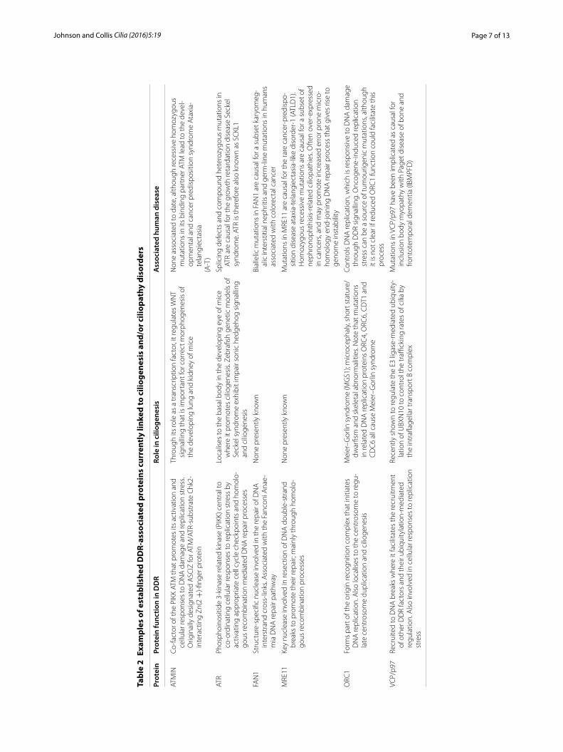
In addition to the examples given above that demon-strate physical and functional connections between DDR and centrosomal proteins, work from several groups has revealed direct genetic and functional links between DDR and ciliogenesis (Tables 1, 2). As mentioned above, the pro-ciliogenesis centrosomal protein CEP164 is reg-ulated by DDR kinases and promotes cellular responses to UV-induced DNA damage [50, 51]. More recently, homozygous recessive mutations in CEP164 were shown to be causal for a subset of nephronophthisis-related cili-opathies, with mutant zebrafish models exhibiting both ciliopathy phenotypes and inefficient responses to DNA damage [103]. Furthermore, this study also showed that NPHP10 (also known as SDCCAG8), which usually resides at centrosomes, re-localised to nuclear foci in response to DNA damage [103], and a subsequent study has suggested that deficiency in NPHP10 (either in cell models or in cells derived from knock-out mice) leads to elevated levels of DNA damage and cell cycle check-point activation [104]. Consistent with an established
functional role for some of the NEK kinase family mem-bers in both DDR and ciliogenesis [105], it was recently reported that the ciliopathy-associated kinase NEK8 (Table 1) is important in controlling cellular responses to replication stress through the DDR kinase ATR and limit-ing CDK activity to suppress DNA break formation [106]. What is more surprising, given the non-overlapping clinical phenotypes of NEK8-associated ciliopathies and ATR-associated Seckel syndrome patients, is that cells expressing a ciliopathy-associated kinase mutant NEK8 had an increase in DNA damage and cell cycle defects, and that the kidneys of NEK8 mutant mice accumulated DNA damage [106]. Furthermore, the centrosomal pro-tein CEP290, mutated in a range of ciliopathies including Joubert syndrome, has also been implicated in the regula-tion of DNA replication stress and DDR (Table 1), sug-gesting that chronic replication stress may be a key driver in the development of some ciliopathies [85, 86]. Similar to the NEK8 study, cells expressing mutant CEP290 also had inappropriate CDK activity. Tissue-specific replica-tion stress in certain genetic backgrounds may therefore be a common mechanism that drives the development of a subset of ciliopathies, and suggests that CDK may be a potential therapeutic target for such diseases [85, 86].
It is intriguing that the same study identifying CEP164 mutations as causative for a subset of nephronophthisis-related ciliopathies also identified causative mutations in MRE11 (Table 2). MRE11 interacts stoichiometrically with RAD50 and NBS1 (forming the so-called MRN complex) to facilitate key functions of DNA repair pro-cesses [103]. Specifically, germ-line mutations in either NBS1 or MRE11 give rise to the cancer-predisposing inherited disorders Nijmegen breakage syndrome and ataxia-telangiectasia-like disorder (ALTD), respectively [107, 108]. Furthermore, MRE11 has been shown to func-tion as a barrier to tumourigenesis [109, 110], and inher-ited heterozygous mutations in MRE11, NBS1 or RAD50 are associated with a low-intermediate penetrance risk of breast cancer [111–113]. It is presently unclear how or why specific mutations in MRE11 in particular can give rise to ciliopathies. This raises interesting questions about whether mutations in other members of the key DDR-associated MRN complex (MRE11-RAD50-NBS1), mutations which cause inherited cancer syndromes [114], may also be causative for other renal-retinal ciliopathies. Perhaps even more surprising was the recent discov-ery that mutations in the Fanconi Anaemia and cancer-associated nuclease FAN1 (Table 2; [115–119]) could be causative for a subset of karyomegalic interstitial nephri-tis-type ciliopathies [120]. As this enzyme is involved in the repair of DNA lesions that block DNA replica-tion, the study suggested that defective nuclease activ-ity within certain organs could drive cellular senescence
Page 7 of 13Johnson and Collis Cilia (2016) 5:19
Tabl
e 2
Exam
ples
of e
stab
lishe
d D
DR-
asso
ciat
ed p
rote
ins
curr
entl
y lin
ked
to c
iliog
enes
is a
nd/o
r cili
opat
hy d
isor
ders
Prot
ein
Prot
ein
func
tion
in D
DR
Role
in c
iliog
enes
isA
ssoc
iate
d hu
man
dis
ease
ATM
INCo
-fact
or o
f the
PIK
K AT
M th
at p
rom
otes
its
activ
atio
n an
d ce
llula
r res
pons
es to
DN
A d
amag
e an
d re
plic
atio
n st
ress
. O
rigin
ally
des
igna
ted
ASC
IZ fo
r ATM
/ATR
-sub
stra
te C
hk2-
inte
ract
ing
Zn(2
+)-fi
nger
pro
tein
Thro
ugh
its ro
le a
s a
tran
scrip
tion
fact
or, i
t reg
ulat
es W
NT
sign
allin
g th
at is
impo
rtan
t for
cor
rect
mor
phog
enes
is o
f th
e de
velo
ping
lung
and
kid
ney
of m
ice
Non
e as
soci
ated
to d
ate,
alth
ough
rece
ssiv
e ho
moz
ygou
s m
utat
ions
in it
s bi
ndin
g pa
rtne
r ATM
lead
to th
e de
vel-
opm
enta
l and
can
cer p
redi
spos
ition
syn
drom
e A
taxi
a-te
lang
iect
asia
(A-T
)
ATR
Phos
phoi
nosi
tide
3-ki
nase
rela
ted
kina
se (P
IKK)
cen
tral
to
co-o
rdin
atin
g ce
llula
r res
pons
es to
repl
icat
ion
stre
ss b
y ac
tivat
ing
appr
opria
te c
ell c
ycle
che
ckpo
ints
and
hom
olo-
gous
reco
mbi
natio
n m
edia
ted
DN
A re
pair
proc
esse
s
Loca
lises
to th
e ba
sal b
ody
in th
e de
velo
ping
eye
of m
ice
whe
re it
pro
mot
es c
iliog
enes
is. Z
ebra
fish
gene
tic m
odel
s of
Se
ckel
syn
drom
e ex
hibi
t im
pair
soni
c he
dgeh
og s
igna
lling
an
d ci
lioge
nesi
s
Splic
ing
defe
cts
and
com
poun
d he
tero
zygo
us m
utat
ions
in
ATR
are
caus
al fo
r the
gro
wth
reta
rdat
ion
dise
ase
Seck
el
synd
rom
e. A
TR is
ther
efor
e al
so k
now
n as
SC
KL1
FAN
1St
ruct
ure-
spec
ific
nucl
ease
invo
lved
in th
e re
pair
of D
NA
in
ters
tran
d cr
oss-
links
. Ass
ocia
ted
with
the
Fanc
oni A
nae-
mia
DN
A re
pair
path
way
Non
e pr
esen
tly k
now
nBi
alle
lic m
utat
ions
in F
AN
1 ar
e ca
usal
for a
sub
set k
aryo
meg
-al
ic in
ters
titia
l nep
hriti
s an
d ge
rm-li
ne m
utat
ions
in h
uman
s as
soci
ated
with
col
orec
tal c
ance
r
MRE
11Ke
y nu
clea
se in
volv
ed in
rese
ctio
n of
DN
A d
oubl
e-st
rand
br
eaks
to p
rom
ote
thei
r rep
air;
mai
nly
thro
ugh
hom
olo-
gous
reco
mbi
natio
n pr
oces
ses
Non
e pr
esen
tly k
now
nM
utat
ions
in M
RE11
are
cau
sal f
or th
e ra
re c
ance
r-pr
edis
po-
sitio
n di
seas
e at
axia
-tel
angi
ecta
sia-
like
diso
rder
-1 (A
TLD
1).
Hom
ozyg
ous
rece
ssiv
e m
utat
ions
are
cau
sal f
or a
sub
set o
f ne
phro
noph
this
is-r
elat
ed c
iliop
athi
es. O
ften
ove
r-ex
pres
sed
in c
ance
rs, a
nd m
ay p
rom
ote
incr
ease
d er
ror p
rone
mic
ro-
hom
olog
y en
d-jo
inin
g D
NA
repa
ir pr
oces
s th
at g
ives
rise
to
geno
me
inst
abili
ty
ORC
1Fo
rms
part
of t
he o
rigin
reco
gniti
on c
ompl
ex th
at in
itiat
es
DN
A re
plic
atio
n. A
lso
loca
lises
to th
e ce
ntro
som
e to
regu
-la
te c
entr
osom
e du
plic
atio
n an
d ci
lioge
nesi
s
Mei
er–G
orlin
syn
drom
e (M
GS1
); m
icro
ceph
aly,
sho
rt s
tatu
re/
dwar
fism
and
ske
leta
l abn
orm
aliti
es. N
ote
that
mut
atio
ns
in re
late
d D
NA
repl
icat
ion
prot
eins
ORC
4, O
RC6,
CD
T1 a
nd
CD
C6
all c
ause
Mei
er–G
orlin
syn
drom
e
Cont
rols
DN
A re
plic
atio
n, w
hich
is re
spon
sive
to D
NA
dam
age
thro
ugh
DD
R si
gnal
ling.
Onc
ogen
e-in
duce
d re
plic
atio
n st
ress
can
be
a so
urce
of t
umou
rigen
ic m
utat
ions
, alth
ough
it
is n
ot c
lear
if re
duce
d O
RC1
func
tion
coul
d fa
cilit
ate
this
pr
oces
s
VCP/
p97
Recr
uite
d to
DN
A b
reak
s w
here
it fa
cilit
ates
the
recr
uitm
ent
of o
ther
DD
R fa
ctor
s an
d th
eir u
biqu
ityla
tion-
med
iate
d re
gula
tion.
Als
o in
volv
ed in
cel
lula
r res
pons
es to
repl
icat
ion
stre
ss
Rece
ntly
sho
wn
to re
gula
te th
e E3
liga
se-m
edia
ted
ubiq
uity
-la
tion
of U
BXN
10 to
con
trol
the
traffi
ckin
g ra
tes
of c
ilia
by
the
intr
aflag
ella
r tra
nspo
rt B
com
plex
Mut
atio
ns in
VC
P/p9
7 ha
ve b
een
impl
icat
ed a
s ca
usal
for
incl
usio
n bo
dy m
yopa
thy
with
Pag
et d
isea
se o
f bon
e an
d fro
ntot
empo
ral d
emen
tia (I
BMPF
D)
Page 8 of 13Johnson and Collis Cilia (2016) 5:19
following increased exposure to genotoxins (perhaps arising from a heightened active metabolism). This may be a similar scenario to the proposed heightened replica-tion stress observed in the kidneys of both CEP290 and NEK8-deficient mice (see above). Although this may be a mechanism by which FAN1 mutations can give rise to ciliopathies, the underlying biology may be more compli-cated, especially given that phenotypes associated with karyomegalic interstitial nephritis-type ciliopathies are not evident in patients with Fanconi anaemia (FA). Such phenotypic discrepancy may also be, in part, due to the redundancy within the pathways that function to resolve DNA replication-impeding lesions [121].
In addition to these genetic studies, several groups have also uncovered functional links to ciliogenesis for proteins traditionally associated with the DDR. An example of this is the recent finding that ATR localises to the basal body in mouse photoreceptor cells (Table 2), and is important for ciliogenesis during the developing eye [122]. ATR is also required for ciliary-related Sonic hedgehog signalling in vitro and in vivo, but appears to be largely dispensable for ciliogenesis, in a role that is distinct from its function in DDR and replication [123]. Another finding is that mutations in DNA replication licensing factors such as ORC1 (Table 2), were causative for the microcephalic disorder Meier–Gorlin syndrome (MGS) and were also shown to affect ciliogenesis through impaired SHH signalling [124]. The AAA-ATPase protein VCP/p97, which regulates the localisation of several DDR factors at DNA damage sites [125], has been shown to be required for ciliogenesis (Table 2), when it may carry out similar functions in regulating E3 ligase-mediated ubiq-uitylation of proteins at the basal body [126]. Finally, the protein ATMIN, a binding partner of the key DDR kinase ATM and also important for cellular responses to replica-tion stress [127, 128], has also been shown to be impor-tant for ciliogenesis during morphogenesis of both the lungs and kidneys in developing mice through its abil-ity as a transcription factor to regulate WNT signalling [129, 130]. Collectively, these studies demonstrate both genetic and functional links between DDR and ciliogen-esis (Table 2).
The human primary cilium and cancerContrary to these recent discoveries involving DDR-asso-ciated factors in human ciliopathies is the general obser-vation that an increased risk or incidence of cancer is not generally associated with human ciliopathies. Exceptions include Birt–Hogg–Dubé syndrome and Von Hippel–Lindau syndrome that are both inherited renal cancer disorders with some clinical features of ciliopathies [131, 132]. Furthermore, although patients with polycystic kid-ney disease have benign renal cysts as a consequence of a
cell overgrowth phenotype, they do not have an increased risk of developing cancer, and may in fact have an overall reduced cancer risk compared with non-affected individ-uals [133, 134]. It is not clear why this may be the case, but it has been suggested that a coincident increased rate of cell death through either apoptotic and/or autophagy mechanisms might help reduce cancer risk in affected individuals. A similar phenomenon has been reported for genetic reduction of ATR activity limiting the tumour growth of P53-deficient tumours in mice [135], although an increased risk of cancer in some Seckel syndrome patients has been reported, with at least one of these having a causative genetic defect in the ATR gene [136, 137]. Interestingly, it has recently been suggested that increased replication stress, similar to that often seen in cancers due to oncogene activation, is a phenotype asso-ciated with a subset of ciliopathies, such as CEP290-asso-ciated Joubert syndrome [85, 86]. Thus, it may be that a certain level of tolerance to heightened replication stress is needed in order to drive more tumourigenic pheno-types associated with DDR-related diseases, which is not selected during the development of the majority of human ciliopathies.
The studies briefly highlighted here provide compel-ling evidence of ever-expanding genetic and functional links between DDR and ciliogenesis pathways. However, the discrepancies between the phenotypes of DDR-asso-ciated cancer-predisposing syndromes and ciliopathies (Tables 1, 2) do not fit with our current limited knowl-edge of how these two pathways could be connected. This may reflect the functional impact each pathway has within both developing and differentiated tissue, as well as how normal or aberrant pathway function may affect both pre-cancerous lesions and transformed cells.
Concluding remarksThe purpose of this mini-review is to highlight emerg-ing links between cellular responses to DNA damage and ciliogenesis. Although some of these studies provide more mechanistic insight into this functional overlap than others, we are still some way from fully understand-ing the intricate interplay between DDR and ciliogenesis factors. Such links were initially striking given the estab-lished role the DDR plays in preventing tumourigenesis and the lack of any increased cancer risk in the major-ity of human ciliopathy patients. However, it is becoming clear from recent genetic and functional-based studies that a subset of DDR and ciliogenesis factors have dual roles in maintaining genomic integrity and primary cilia biology. The majority of this duality appears to stem from the necessity of a cell to regulate centrosome duplication and mitotic spindle integrity, with several DDR proteins localising to the centrosome and/or regulating cell cycle

Page 9 of 13Johnson and Collis Cilia (2016) 5:19
progression and, in turn, centriole duplication events. Additionally, several centriolar satellites help maintain appropriate centrosome structures and microtubule integrity to limit the accumulation of post-mitotic DNA damage. Finally, aberrant mitogenic signals (potentially through a common mechanism of inappropriate CDK activity) can give rise to replication stress which can, in turn, lead to aberrant centrosome duplication and matu-ration processes. As such, heightened replication stress may be a common source of disrupted centrosome func-tion in cancer, and aberrant cilia function in ciliopathies.
The majority of human cells are ciliated with the cil-ium acting as a signalling hub for several interconnected stress response pathways, which are in constant com-munication with the DNA damage response pathways and cell cycle regulators. Recent discoveries demon-strating that autophagy and ciliogenesis can co-regulate each other, and that autophagy is responsive to oxida-tive stress/DNA damage and can regulate DNA repair processes, further draw links between primary cilia and the DDR. Such functional interplay has implications for human disease, which is highlighted by the recent discov-eries of mutations in proteins, traditionally thought to be solely involved in DNA repair processes, being causative for a subset of human ciliopathies with degenerative dis-eases of the kidney and retina. With the advent of next-generation sequencing of larger clinical cohorts, it will also be interesting to see if additional DDR factors and autophagy factors are implicated in ciliopathies, and if dysregulation in any cilia-associated factors is associated with an increased risk of cancer development and pro-gression. Indeed, given the young age and small cohort of current ciliopathy patients with causative mutations in either FAN1 or MRE11, it is too early to determine if these patients have an increased risk of developing can-cer. Given that mutations in both these proteins can give rise to various cancers (see above), one may predict that these ciliopathy patients may have a heightened risk of developing cancer compared with the general population and some other ciliopathy cohorts. For these conditions, pathogenic mechanisms of replication stress leading to DNA damage, concomitant with or upstream of primary cilia function, are an area of exciting future research. Finally, since ciliogenesis and replication stress are poten-tially reversible with small molecule approaches, these findings also reveal new therapeutic intervention oppor-tunities as possible treatment regimes for these diseases.
AbbreviationsAPC: anaphase-promoting complex; ATM: ataxia-telangiectasia mutated; ATMIN: ATM interactor; ATR: ATM-related; BRCA1: breast and ovarian cancer susceptibility protein 1; BRCA2: breast and ovarian cancer susceptibility pro-tein 1; CDK: cyclin-dependent kinase; CEP164: centrosomal protein 164KDa; CHK1: checkpoint kinase 1; DDR: DNA damage response; FA: Fanconi anaemia;
FAN1: FANCD2/FANCI-associated nuclease; G1: growth phase 1 of the cell cycle; G2: growth phase 2 of the cell cycle; M: mitotic phase of the cell cycle; MGS: Meier–Gorlin syndrome; MRE11: meiotic recombination 11 homolog A; mTOR: mammalian target of rapamycin; NEK8: NimA-related kinase 8; NPHP10: nephronophthisis-related ciliopathy protein 10; NBS: Nijmegen breakage syndrome; PARP1: poly (ADP-Ribose) polymerase 1; PCM1: pericentriolar mate-rial 1; PDGF: platelet-derived growth factor; S: DNA synthesis phase of the cell cycle; SHH: sonic hedgehog signalling pathway; SMC1: structural maintenance of chromosomes 1; Smo: smoothened; TGFβ: transforming growth factor β; TP53: tumour suppressor protein 53 kDa; VCP: valosin-containing protein; WNT: wingless-related integration site.
Authors’ contributionsCAJ and SJC co-wrote and edited the manuscript. Both authors read and approved the final manuscript.
Author details1 Section of Ophthalmology and Neurosciences, Wellcome Trust Brenner Building, Leeds Institute of Molecular Medicine, St. James’s University Hospital, Leeds LS9 7TF, UK. 2 Genome Stability Group, Department of Oncology and Metabolism, Academic Unit of Molecular Oncology, Medical School, University of Sheffield, Beech Hill Road, Sheffield S10 2RX, UK.
Authors’ informationProf. Colin Johnson is a Professor of Medical and Molecular Genetics at the University of Leeds. His research focuses on understanding the biologi-cal roles of cilia and basal body-associated proteins, and discovering new ciliopathy genes. Dr. Spencer Collis is a Reader in Genome Stability and a Cancer Research UK Senior Cancer Research Fellow within the Department of Oncology & Metabolism at the University of Sheffield. His research focuses on understanding mechanisms of maintaining genome stability and how disruption to these processes can lead to human diseases as well as potential therapeutic exploitation in the clinical management of cancer.
Acknowledgements and fundingCAJ acknowledges funding from the UK Medical Research Council (project Grant MR/M000532/1) and the European Community’s Seventh Framework Programme FP7/2009 under Grant Agreement No. 241955 SYSCILIA. SJC is funded by a Cancer Research UK (CR-UK) Senior Cancer Research Fellowship (SCaRF; #C36435/A12102) and acknowledges additional related funding from Yorkshire Cancer Research (S312) and Weston Park Hospital Cancer Charity (CA146).
Competing interestsThe authors declare that they have no competing interests.
Received: 16 September 2015 Accepted: 22 March 2016
References 1. Ciccia A, Elledge SJ. The DNA damage response: making it safe
to play with knives. Mol Cell. 2010;40(2):179–204. doi:10.1016/j.molcel.2010.09.019.
2. Hoeijmakers JH. Genome maintenance mechanisms for preventing cancer. Nature. 2001;411(6835):366–74. doi:10.1038/35077232.
3. Lukas J, Lukas C, Bartek J. Mammalian cell cycle checkpoints: signalling pathways and their organization in space and time. DNA Repair (Amst). 2004;3(8–9):997–1007.
4. Malumbres M, Barbacid M. Cell cycle, CDKs and cancer: a changing paradigm. Nat Rev Cancer. 2009;9(3):153–66. doi:10.1038/nrc2602.
5. Shaltiel IA, Krenning L, Bruinsma W, Medema RH. The same, only differ-ent—DNA damage checkpoints and their reversal throughout the cell cycle. J Cell Sci. 2015;128(4):607–20. doi:10.1242/jcs.163766.
6. Kastan MB, Bartek J. Cell-cycle checkpoints and cancer. Nature. 2004;432(7015):316–23.
7. Bartkova J, Horejsi Z, Koed K, Kramer A, Tort F, Zieger K, et al. DNA damage response as a candidate anti-cancer barrier in early human tumorigenesis. Nature. 2005;434(7035):864–70.

Page 10 of 13Johnson and Collis Cilia (2016) 5:19
8. Gorgoulis VG, Vassiliou LV, Karakaidos P, Zacharatos P, Kotsinas A, Lilo-glou T, et al. Activation of the DNA damage checkpoint and genomic instability in human precancerous lesions. Nature. 2005;434(7035):907–13. doi:10.1038/nature03485.
9. Hanahan D, Weinberg RA. Hallmarks of cancer: the next generation. Cell. 2011;144(5):646–74. doi:10.1016/j.cell.2011.02.013.
10. Jackson SP, Bartek J. The DNA-damage response in human biology and disease. Nature. 2009;461(7267):1071–8. doi:10.1038/nature08467.
11. O’Driscoll M. Diseases associated with defective responses to DNA damage. Cold Spring Harb Perspect Biol. 2012;4(12):a012773. doi:10.1101/cshperspect.a012773.
12. Alcantara D, O’Driscoll M. Congenital microcephaly. Am J Med Genet C Semin Med Genet. 2014;166C(2):124–39. doi:10.1002/ajmg.c.31397.
13. Barazzuol L, Rickett N, Ju L, Jeggo PA. Endogenous and X-ray-induced DNA double strand breaks sensitively activate apoptosis in adult neural stem cells. J Cell Sci. 2015. doi:10.1242/jcs.171223.
14. Dictenberg JB, Zimmerman W, Sparks CA, Young A, Vidair C, Zheng Y, et al. Pericentrin and gamma-tubulin form a protein complex and are organized into a novel lattice at the centrosome. J Cell Biol. 1998;141(1):163–74.
15. Gould RR, Borisy GG. The pericentriolar material in Chinese ham-ster ovary cells nucleates microtubule formation. J Cell Biol. 1977;73(3):601–15.
16. Nigg EA, Stearns T. The centrosome cycle: centriole biogenesis, duplica-tion and inherent asymmetries. Nat Cell Biol. 2011;13(10):1154–60. doi:10.1038/ncb2345.
17. Gupta GD, Coyaud E, Goncalves J, Mojarad BA, Liu Y, Wu Q, et al. A dynamic protein interaction landscape of the human centrosome-cil-ium interface. Cell. 2015;163(6):1484–99. doi:10.1016/j.cell.2015.10.065.
18. Dodson H, Bourke E, Jeffers LJ, Vagnarelli P, Sonoda E, Takeda S, et al. Centrosome amplification induced by DNA damage occurs during a prolonged G2 phase and involves ATM. EMBO J. 2004;23(19):3864–73. doi:10.1038/sj.emboj.7600393.
19. Loffler H, Fechter A, Liu FY, Poppelreuther S, Kramer A. DNA damage-induced centrosome amplification occurs via excessive formation of centri-olar satellites. Oncogene. 2013;32(24):2963–72. doi:10.1038/onc.2012.310.
20. Staples CJ, Myers KN, Beveridge RD, Patil AA, Lee AJ, Swanton C, et al. The centriolar satellite protein Cep131 is important for genome stabil-ity. J Cell Sci. 2012;125(Pt 20):4770–9. doi:10.1242/jcs.104059.
21. Mullee LI, Morrison CG. Centrosomes in the DNA damage response-the hub outside the centre. Chromosome Res. 2015. doi:10.1007/s10577-015-9503-7.
22. Balczon R, Bao L, Zimmer WE, Brown K, Zinkowski RP, Brinkley BR. Disso-ciation of centrosome replication events from cycles of DNA synthesis and mitotic division in hydroxyurea-arrested Chinese hamster ovary cells. J Cell Biol. 1995;130(1):105–15.
23. Meraldi P, Lukas J, Fry AM, Bartek J, Nigg EA. Centrosome duplication in mammalian somatic cells requires E2F and Cdk2-cyclin A. Nat Cell Biol. 1999;1(2):88–93. doi:10.1038/10054.
24. Kodani A, Yu TW, Johnson JR, Jayaraman D, Johnson TL, Al-Gazali L, et al. Centriolar satellites assemble centrosomal microcephaly proteins to recruit CDK2 and promote centriole duplication. Elife. 2015;4:e07519. doi:10.7554/eLife.07519.
25. Nigg EA, Cajanek L, Arquint C. The centrosome duplication cycle in health and disease. FEBS Lett. 2014;588(15):2366–72. doi:10.1016/j.febslet.2014.06.030.
26. Nigg EA, Raff JW. Centrioles, centrosomes, and cilia in health and disease. Cell. 2009;139(4):663–78. doi:10.1016/j.cell.2009.10.036.
27. Adly N, Alhashem A, Ammari A, Alkuraya FS. Ciliary genes TBC1D32/C6orf170 and SCLT1 are mutated in patients with OFD type IX. Hum Mutat. 2014;35(1):36–40. doi:10.1002/humu.22477.
28. Waters AM, Asfahani R, Carroll P, Bicknell L, Lescai F, Bright A, et al. The kinetochore protein, CENPF, is mutated in human ciliopathy and micro-cephaly phenotypes. J Med Genet. 2015;52(3):147–56. doi:10.1136/jmedgenet-2014-102691.
29. Godinho SA, Kwon M, Pellman D. Centrosomes and cancer: how cancer cells divide with too many centrosomes. Cancer Metastasis Rev. 2009;28(1–2):85–98. doi:10.1007/s10555-008-9163-6.
30. Fukasawa K. Oncogenes and tumour suppressors take on centrosomes. Nat Rev Cancer. 2007;7(12):911–24. doi:10.1038/nrc2249.
31. Fukasawa K. Centrosome amplification, chromosome instability and cancer development. Cancer Lett. 2005;230(1):6–19. doi:10.1016/j.canlet.2004.12.028.
32. Ganem NJ, Godinho SA, Pellman D. A mechanism linking extra cen-trosomes to chromosomal instability. Nature. 2009;460(7252):278–82. doi:10.1038/nature08136.
33. Silkworth WT, Nardi IK, Scholl LM, Cimini D. Multipolar spindle pole coalescence is a major source of kinetochore mis-attachment and chro-mosome mis-segregation in cancer cells. PLoS ONE. 2009;4(8):e6564. doi:10.1371/journal.pone.0006564.
34. Godinho SA, Pellman D. Causes and consequences of centro-some abnormalities in cancer. Philos Trans R Soc Lond B Biol Sci. 2014;369(1650):20130467. doi:10.1098/rstb.2013.0467.
35. Godinho SA, Picone R, Burute M, Dagher R, Su Y, Leung CT, et al. Onco-gene-like induction of cellular invasion from centrosome amplification. Nature. 2014;510(7503):167–71. doi:10.1038/nature13277.
36. Kramer A, Lukas J, Bartek J. Checking out the centrosome. Cell Cycle. 2004;3(11):1390–3.
37. Shimada M, Komatsu K. Emerging connection between centrosome and DNA repair machinery. J Radiat Res. 2009;50(4):295–301.
38. Lukinavicius G, Lavogina D, Gonczy P, Johnsson K. Commercial Cdk1 antibodies recognize the centrosomal protein Cep152. Biotechniques. 2013;55(3):111–4.
39. Matsuyama M, Goto H, Kasahara K, Kawakami Y, Nakanishi M, Kiyono T, et al. Nuclear Chk1 prevents premature mitotic entry. J Cell Sci. 2011;124(Pt 13):2113–9. doi:10.1242/jcs.086488.
40. Sankaran S, Starita LM, Simons AM, Parvin JD. Identification of domains of BRCA1 critical for the ubiquitin-dependent inhibition of centro-some function. Cancer Res. 2006;66(8):4100–7. doi:10.1158/0008-5472.CAN-05-4430.
41. Shimada M, Sagae R, Kobayashi J, Habu T, Komatsu K. Inactivation of the Nijmegen breakage syndrome gene leads to excess centrosome dupli-cation via the ATR/BRCA1 pathway. Cancer Res. 2009;69(5):1768–75. doi:10.1158/0008-5472.CAN-08-3016.
42. Kramer A, Mailand N, Lukas C, Syljuasen RG, Wilkinson CJ, Nigg EA, et al. Centrosome-associated Chk1 prevents premature activation of cyclin-B-Cdk1 kinase. Nat Cell Biol. 2004;6(9):884–91. doi:10.1038/ncb1165.
43. Lee J, Dunphy WG. The Mre11-Rad50-Nbs1 (MRN) complex has a specific role in the activation of Chk1 in response to stalled replica-tion forks. Mol Biol Cell. 2013;24(9):1343–53. doi:10.1091/mbc.E13-01-0025.
44. Sato K, Sundaramoorthy E, Rajendra E, Hattori H, Jeyasekharan AD, Ayoub N, et al. A DNA-damage selective role for BRCA1 E3 ligase in claspin ubiquitylation, CHK1 activation, and DNA repair. Curr Biol. 2012;22(18):1659–66. doi:10.1016/j.cub.2012.07.034.
45. Antonczak AK, Mullee LI, Wang Y, Comartin D, Inoue T, Pelletier L, et al. Opposing effects of pericentrin and microcephalin on the pericentri-olar material regulate CHK1 activation in the DNA damage response. Oncogene. 2015. doi:10.1038/onc.2015.257.
46. Loncarek J, Hergert P, Magidson V, Khodjakov A. Control of daugh-ter centriole formation by the pericentriolar material. Nat Cell Biol. 2008;10(3):322–8. doi:10.1038/ncb1694.
47. Alderton GK, Galbiati L, Griffith E, Surinya KH, Neitzel H, Jackson AP, et al. Regulation of mitotic entry by microcephalin and its overlap with ATR signalling. Nat Cell Biol. 2006;8(7):725–33. doi:10.1038/ncb1431.
48. Bruning-Richardson A, Bond J, Alsiary R, Richardson J, Cairns DA, McCormack L, et al. ASPM and microcephalin expression in epithelial ovarian cancer correlates with tumour grade and survival. Br J Cancer. 2011;104(10):1602–10. doi:10.1038/bjc.2011.117.
49. Richardson J, Shaaban AM, Kamal M, Alisary R, Walker C, Ellis IO, et al. Microcephalin is a new novel prognostic indicator in breast cancer associated with BRCA1 inactivation. Breast Cancer Res Treat. 2011;127(3):639–48. doi:10.1007/s10549-010-1019-4.
50. Sivasubramaniam S, Sun X, Pan YR, Wang S, Lee EY. Cep164 is a media-tor protein required for the maintenance of genomic stability through modulation of MDC1, RPA, and CHK1. Genes Dev. 2008;22(5):587–600. doi:10.1101/gad.1627708.
51. Pan YR, Lee EY. UV-dependent interaction between Cep164 and XPA mediates localization of Cep164 at sites of DNA damage and UV sensi-tivity. Cell Cycle. 2009;8(4):655–64.

Page 11 of 13Johnson and Collis Cilia (2016) 5:19
52. Renaud E, Miccoli L, Zacal N, Biard DS, Craescu CT, Rainbow AJ, et al. Differential contribution of XPC, RAD23A, RAD23B and CENTRIN 2 to the UV-response in human cells. DNA Repair (Amst). 2011;10(8):835–47. doi:10.1016/j.dnarep.2011.05.003.
53. Palomera-Sanchez Z, Zurita M. Open, repair and close again: chromatin dynamics and the response to UV-induced DNA damage. DNA Repair (Amst). 2011;10(2):119–25. doi:10.1016/j.dnarep.2010.10.010.
54. Nishi R, Sakai W, Tone D, Hanaoka F, Sugasawa K. Structure-function analysis of the EF-hand protein centrin-2 for its intracellular localization and nucleotide excision repair. Nucleic Acids Res. 2013;41(14):6917–29. doi:10.1093/nar/gkt434.
55. Shiloh Y, Ziv Y. The ATM protein kinase: regulating the cellular response to genotoxic stress, and more. Nat Rev Mol Cell Biol. 2013;14(4):197–210.
56. Matsuoka S, Ballif BA, Smogorzewska A, McDonald ER 3rd, Hurov KE, Luo J, et al. ATM and ATR substrate analysis reveals extensive protein networks responsive to DNA damage. Science. 2007;316(5828):1160–6. doi:10.1126/science.1140321.
57. Smith E, Dejsuphong D, Balestrini A, Hampel M, Lenz C, Takeda S, et al. An ATM- and ATR-dependent checkpoint inactivates spindle assembly by targeting CEP63. Nat Cell Biol. 2009;11(3):278–85. doi:10.1038/ncb1835.
58. Brown NJ, Marjanovic M, Luders J, Stracker TH, Costanzo V. Cep63 and cep152 cooperate to ensure centriole duplication. PLoS ONE. 2013;8(7):e69986. doi:10.1371/journal.pone.0069986.
59. Sir JH, Barr AR, Nicholas AK, Carvalho OP, Khurshid M, Sossick A, et al. A primary microcephaly protein complex forms a ring around parental centrioles. Nat Genet. 2011;43(11):1147–53. doi:10.1038/ng.971.
60. Loffler H, Fechter A, Matuszewska M, Saffrich R, Mistrik M, Marhold J, et al. Cep63 recruits Cdk1 to the centrosome: implications for regulation of mitotic entry, centrosome amplification, and genome maintenance. Cancer Res. 2011;71(6):2129–39. doi:10.1158/0008-5472.CAN-10-2684.
61. Marumoto T, Zhang D, Saya H. Aurora-A—a guardian of poles. Nat Rev Cancer. 2005;5(1):42–50. doi:10.1038/nrc1526.
62. Macurek L, Lindqvist A, Lim D, Lampson MA, Klompmaker R, Freire R, et al. Polo-like kinase-1 is activated by aurora A to promote checkpoint recovery. Nature. 2008;455(7209):119–23. doi:10.1038/nature07185.
63. van Vugt MA, Bras A, Medema RH. Polo-like kinase-1 controls recovery from a G2 DNA damage-induced arrest in mammalian cells. Mol Cell. 2004;15(5):799–811. doi:10.1016/j.molcel.2004.07.015.
64. Ganner A, Lienkamp S, Schafer T, Romaker D, Wegierski T, Park TJ, et al. Regulation of ciliary polarity by the APC/C. Proc Natl Acad Sci USA. 2009;106(42):17799–804. doi:10.1073/pnas.0909465106.
65. Wang W, Wu T, Kirschner MW. The master cell cycle regulator APC-Cdc20 regulates ciliary length and disassembly of the primary cilium. Elife. 2014;3:e03083. doi:10.7554/eLife.03083.
66. Staples CJ, Myers KN, Beveridge RD, Patil AA, Howard AE, Barone G, et al. Ccdc13 is a novel human centriolar satellite protein required for ciliogenesis and genome stability. J Cell Sci. 2014;127(Pt 13):2910–9. doi:10.1242/jcs.147785.
67. Berbari NF, O’Connor AK, Haycraft CJ, Yoder BK. The primary cilium as a complex signaling center. Curr Biol. 2009;19(13):R526–35. doi:10.1016/j.cub.2009.05.025.
68. Lancaster MA, Gleeson JG. The primary cilium as a cellular signaling center: lessons from disease. Curr Opin Genet Dev. 2009;19(3):220–9. doi:10.1016/j.gde.2009.04.008.
69. Huangfu D, Liu A, Rakeman AS, Murcia NS, Niswander L, Anderson KV. Hedgehog signalling in the mouse requires intraflagellar transport proteins. Nature. 2003;426(6962):83–7. doi:10.1038/nature02061.
70. Goetz SC, Anderson KV. The primary cilium: a signalling centre dur-ing vertebrate development. Nat Rev Genet. 2010;11(5):331–44. doi:10.1038/nrg2774.
71. Angers S, Moon RT. Proximal events in Wnt signal transduction. Nat Rev Mol Cell Biol. 2009;10(7):468–77. doi:10.1038/nrm2717.
72. Ezratty EJ, Stokes N, Chai S, Shah AS, Williams SE, Fuchs E. A role for the primary cilium in Notch signaling and epidermal differentiation during skin development. Cell. 2011;145(7):1129–41. doi:10.1016/j.cell.2011.05.030.
73. Corbit KC, Aanstad P, Singla V, Norman AR, Stainier DY, Reiter JF. Vertebrate smoothened functions at the primary cilium. Nature. 2005;437(7061):1018–21. doi:10.1038/nature04117.
74. Lancaster MA, Schroth J, Gleeson JG. Subcellular spatial regula-tion of canonical Wnt signalling at the primary cilium. Nat Cell Biol. 2011;13(6):700–7. doi:10.1038/ncb2259.
75. Stasiulewicz M, Gray SD, Mastromina I, Silva JC, Bjorklund M, Seymour PA, et al. A conserved role for Notch signaling in priming the cellular response to Shh through ciliary localisation of the key Shh transducer Smo. Development. 2015;142(13):2291–303. doi:10.1242/dev.125237.
76. Boehlke C, Kotsis F, Patel V, Braeg S, Voelker H, Bredt S, et al. Primary cilia regulate mTORC1 activity and cell size through Lkb1. Nat Cell Biol. 2010;12(11):1115–22. doi:10.1038/ncb2117.
77. Bell PD, Fitzgibbon W, Sas K, Stenbit AE, Amria M, Houston A, et al. Loss of primary cilia upregulates renal hypertrophic signaling and promotes cystogenesis. J Am Soc Nephrol. 2011;22(5):839–48. doi:10.1681/ASN.2010050526.
78. Habbig S, Bartram MP, Muller RU, Schwarz R, Andriopoulos N, Chen S, et al. NPHP4, a cilia-associated protein, negatively regulates the hippo pathway. J Cell Biol. 2011;193(4):633–42. doi:10.1083/jcb.201009069.
79. Habbig S, Bartram MP, Sagmuller JG, Griessmann A, Franke M, Muller RU, et al. The ciliopathy disease protein NPHP9 promotes nuclear delivery and activation of the oncogenic transcriptional regulator TAZ. Hum Mol Genet. 2012;21(26):5528–38. doi:10.1093/hmg/dds408.
80. Frank V, Habbig S, Bartram MP, Eisenberger T, Veenstra-Knol HE, Decker C, et al. Mutations in NEK8 link multiple organ dysplasia with altered Hippo signalling and increased c-MYC expression. Hum Mol Genet. 2013;22(11):2177–85. doi:10.1093/hmg/ddt070.
81. Clement CA, Ajbro KD, Koefoed K, Vestergaard ML, Veland IR, Henriques de Jesus MP, et al. TGF-beta signaling is associated with endocytosis at the pocket region of the primary cilium. Cell Rep. 2013;3(6):1806–14. doi:10.1016/j.celrep.2013.05.020.
82. Clement DL, Mally S, Stock C, Lethan M, Satir P, Schwab A, et al. PDG-FRalpha signaling in the primary cilium regulates NHE1-dependent fibroblast migration via coordinated differential activity of MEK1/2-ERK1/2-p90RSK and AKT signaling pathways. J Cell Sci. 2013;126(Pt 4):953–65. doi:10.1242/jcs.116426.
83. Vermezovic J, Adamowicz M, Santarpia L, Rustighi A, Forcato M, Lucano C, et al. Notch is a direct negative regulator of the DNA-damage response. Nat Struct Mol Biol. 2015;22(5):417–24. doi:10.1038/nsmb.3013.
84. Yatim A, Benne C, Sobhian B, Laurent-Chabalier S, Deas O, Judde JG, et al. NOTCH1 nuclear interactome reveals key regulators of its tran-scriptional activity and oncogenic function. Mol Cell. 2012;48(3):445–58. doi:10.1016/j.molcel.2012.08.022.
85. Slaats GG, Saldivar JC, Bacal J, Zeman MK, Kile AC, Hynes AM, et al. DNA replication stress underlies renal phenotypes in CEP290-associated Joubert syndrome. J Clin Invest. 2015;125(9):3657–66. doi:10.1172/JCI80657.
86. Slaats GG, Giles RH. Are renal ciliopathies (replication) stressed out? Trends Cell Biol. 2015;25(6):317–9. doi:10.1016/j.tcb.2015.03.005.
87. Paulsen RD, Soni DV, Wollman R, Hahn AT, Yee MC, Guan A, et al. A genome-wide siRNA screen reveals diverse cellular processes and pathways that mediate genome stability. Mol Cell. 2009;35(2):228–39. doi:10.1016/j.molcel.2009.06.021.
88. Wheway G, Schmidts M, Mans DA, Szymanska K, Nguyen TM, Racher H, et al. An siRNA-based functional genomics screen for the identifica-tion of regulators of ciliogenesis and ciliopathy genes. Nat Cell Biol. 2015;17(8):1074–87. doi:10.1038/ncb3201.
89. Menzl I, Lebeau L, Pandey R, Hassounah NB, Li FW, Nagle R, et al. Loss of primary cilia occurs early in breast cancer development. Cilia. 2014;3:7. doi:10.1186/2046-2530-3-7.
90. Basten SG, Willekers S, Vermaat JS, Slaats GG, Voest EE, van Diest PJ, et al. Reduced cilia frequencies in human renal cell carcino-mas versus neighboring parenchymal tissue. Cilia. 2013;2(1):2. doi:10.1186/2046-2530-2-2.
91. Gomez Garcia EB, Knoers NV. Gardner’s syndrome (familial adenoma-tous polyposis): a cilia-related disorder. Lancet Oncol. 2009;10(7):727–35. doi:10.1016/S1470-2045(09)70167-6.

Page 12 of 13Johnson and Collis Cilia (2016) 5:19
92. Beachy PA, Karhadkar SS, Berman DM. Tissue repair and stem cell renewal in carcinogenesis. Nature. 2004;432(7015):324–31. doi:10.1038/nature03100.
93. Bale AE, Yu KP. The hedgehog pathway and basal cell carcinomas. Hum Mol Genet. 2001;10(7):757–62.
94. Berman DM, Karhadkar SS, Hallahan AR, Pritchard JI, Eberhart CG, Watkins DN, et al. Medulloblastoma growth inhibition by hedgehog pathway blockade. Science. 2002;297(5586):1559–61. doi:10.1126/science.1073733.
95. Teglund S, Toftgard R. Hedgehog beyond medulloblastoma and basal cell carcinoma. Biochim Biophys Acta. 2010;1805(2):181–208. doi:10.1016/j.bbcan.2010.01.003.
96. Wong SY, Seol AD, So PL, Ermilov AN, Bichakjian CK, Epstein EH Jr, et al. Primary cilia can both mediate and suppress Hedgehog pathway-dependent tumorigenesis. Nat Med. 2009;15(9):1055–61. doi:10.1038/nm.2011.
97. Villumsen BH, Danielsen JR, Povlsen L, Sylvestersen KB, Merdes A, Beli P, et al. A new cellular stress response that triggers centriolar satellite reor-ganization and ciliogenesis. EMBO J. 2013;32(23):3029–40. doi:10.1038/emboj.2013.223.
98. Tang Z, Lin MG, Stowe TR, Chen S, Zhu M, Stearns T, et al. Autophagy promotes primary ciliogenesis by removing OFD1 from centriolar satel-lites. Nature. 2013;502(7470):254–7. doi:10.1038/nature12606.
99. Pampliega O, Orhon I, Patel B, Sridhar S, Diaz-Carretero A, Beau I, et al. Functional interaction between autophagy and ciliogenesis. Nature. 2013;502(7470):194–200. doi:10.1038/nature12639.
100. Malaquin N, Carrier-Leclerc A, Dessureault M, Rodier F. DDR-mediated crosstalk between DNA-damaged cells and their microenvironment. Front Genet. 2015;6:94. doi:10.3389/fgene.2015.00094.
101. Vessoni AT, Filippi-Chiela EC, Menck CF, Lenz G. Autophagy and genomic integrity. Cell Death Differ. 2013;20(11):1444–54. doi:10.1038/cdd.2013.103.
102. Liu EY, Xu N, O’Prey J, Lao LY, Joshi S, Long JS, et al. Loss of autophagy causes a synthetic lethal deficiency in DNA repair. Proc Natl Acad Sci USA. 2015;112(3):773–8. doi:10.1073/pnas.1409563112.
103. Chaki M, Airik R, Ghosh AK, Giles RH, Chen R, Slaats GG, et al. Exome capture reveals ZNF423 and CEP164 mutations, linking renal ciliopa-thies to DNA damage response signaling. Cell. 2012;150(3):533–48. doi:10.1016/j.cell.2012.06.028.
104. Airik R, Slaats GG, Guo Z, Weiss AC, Khan N, Ghosh A, et al. Renal-retinal ciliopathy gene Sdccag8 regulates DNA damage response signaling. J Am Soc Nephrol. 2014;25(11):2573–83. doi:10.1681/ASN.2013050565.
105. Fry AM, O’Regan L, Sabir SR, Bayliss R. Cell cycle regulation by the NEK family of protein kinases. J Cell Sci. 2012;125(Pt 19):4423–33. doi:10.1242/jcs.111195.
106. Choi HJ, Lin JR, Vannier JB, Slaats GG, Kile AC, Paulsen RD, et al. NEK8 links the ATR-regulated replication stress response and S phase CDK activity to renal ciliopathies. Mol Cell. 2013;51(4):423–39. doi:10.1016/j.molcel.2013.08.006.
107. Williams RS, Williams JS, Tainer JA. Mre11-Rad50-Nbs1 is a keystone complex connecting DNA repair machinery, double-strand break sign-aling, and the chromatin template. Biochem Cell Biol. 2007;85(4):509–20. doi:10.1139/O07-069.
108. Uchisaka N, Takahashi N, Sato M, Kikuchi A, Mochizuki S, Imai K, et al. Two brothers with ataxia-telangiectasia-like disorder with lung adeno-carcinoma. J Pediatr. 2009;155(3):435–8. doi:10.1016/j.jpeds.2009.02.037.
109. Gupta GP, Vanness K, Barlas A, Manova-Todorova KO, Wen YH, Petrini JH. The Mre11 complex suppresses oncogene-driven breast tumori-genesis and metastasis. Mol Cell. 2013;52(3):353–65. doi:10.1016/j.molcel.2013.09.001.
110. Reddy JP, Peddibhotla S, Bu W, Zhao J, Haricharan S, Du YC, et al. Defining the ATM-mediated barrier to tumorigenesis in somatic mammary cells following ErbB2 activation. Proc Natl Acad Sci USA. 2010;107(8):3728–33. doi:10.1073/pnas.0910665107.
111. Ripperger T, Gadzicki D, Meindl A, Schlegelberger B. Breast cancer sus-ceptibility: current knowledge and implications for genetic counselling. Eur J Hum Genet. 2009;17(6):722–31. doi:10.1038/ejhg.2008.212.
112. Couch FJ, Hart SN, Sharma P, Toland AE, Wang X, Miron P, et al. Inherited mutations in 17 breast cancer susceptibility genes among a large triple-negative breast cancer cohort unselected for family history of breast cancer. J Clin Oncol. 2015;33(4):304–11. doi:10.1200/JCO.2014.57.1414.
113. Damiola F, Pertesi M, Oliver J, Le Calvez-Kelm F, Voegele C, Young EL, et al. Rare key functional domain missense substitutions in MRE11A, RAD50, and NBN contribute to breast cancer susceptibility: results from a breast cancer family registry case-control mutation-screening study. Breast Cancer Res. 2014;16(3):R58. doi:10.1186/bcr3669.
114. Stracker TH, Petrini JH. The MRE11 complex: starting from the ends. Nat Rev Mol Cell Biol. 2011;12(2):90–103. doi:10.1038/nrm3047.
115. MacKay C, Declais AC, Lundin C, Agostinho A, Deans AJ, MacArtney TJ, et al. Identification of KIAA1018/FAN1, a DNA repair nuclease recruited to DNA damage by monoubiquitinated FANCD2. Cell. 2010;142(1):65–76. doi:10.1016/j.cell.2010.06.021.
116. Wang R, Persky NS, Yoo B, Ouerfelli O, Smogorzewska A, Elledge SJ, et al. DNA repair. Mechanism of DNA interstrand cross-link processing by repair nuclease FAN1. Science. 2014;346(6213):1127–30. doi:10.1126/science.1258973.
117. Smith AL, Alirezaie N, Connor A, Chan-Seng-Yue M, Grant R, Selander I, et al. Candidate DNA repair susceptibility genes identified by exome sequencing in high-risk pancreatic cancer. Cancer Lett. 2016;370(2):302–12. doi:10.1016/j.canlet.2015.10.030.
118. Segui N, Mina LB, Lazaro C, Sanz-Pamplona R, Pons T, Navarro M, et al. Germline mutations in FAN1 cause hereditary colorectal cancer by impairing DNA repair. Gastroenterology. 2015;149(3):563–6. doi:10.1053/j.gastro.2015.05.056.
119. Lachaud C, Moreno A, Marchesi F, Toth R, Blow JJ, Rouse J. Ubiquitinated Fancd2 recruits Fan1 to stalled replication forks to prevent genome instability. Science. 2016;351(6275):846–9. doi:10.1126/science.aad5634.
120. Zhou W, Otto EA, Cluckey A, Airik R, Hurd TW, Chaki M, et al. FAN1 muta-tions cause karyomegalic interstitial nephritis, linking chronic kidney failure to defective DNA damage repair. Nat Genet. 2012;44(8):910–5. doi:10.1038/ng.2347.
121. Wang AT, Smogorzewska A. SnapShot: Fanconi anemia and associated proteins. Cell. 2015;160(1–2):354. doi:10.1016/j.cell.2014.12.031.
122. Valdes-Sanchez L, De la Cerda B, Diaz-Corrales FJ, Massalini S, Chakarova CF, Wright AF, et al. ATR localizes to the photoreceptor connecting cilium and deficiency leads to severe photoreceptor degeneration in mice. Hum Mol Genet. 2013;22(8):1507–15. doi:10.1093/hmg/dds563.
123. Stiff T, Casar Tena T, O’Driscoll M, Jeggo PA, Philipp M. ATR promotes cilia signalling; links to developmental impacts. Hum Mol Genet. 2016. doi:10.1093/hmg/ddw034.
124. Stiff T, Alagoz M, Alcantara D, Outwin E, Brunner HG, Bongers EM, et al. Deficiency in origin licensing proteins impairs cilia formation: implications for the aetiology of Meier–Gorlin syndrome. PLoS Genet. 2013;9(3):e1003360. doi:10.1371/journal.pgen.1003360.
125. Dantuma NP, Acs K, Luijsterburg MS. Should i stay or should I go: vCP/p97-mediated chromatin extraction in the DNA damage response. Exp Cell Res. 2014;329(1):9–17. doi:10.1016/j.yexcr.2014.08.025.
126. Raman M, Sergeev M, Garnaas M, Lydeard JR, Huttlin EL, Goessling W, et al. Systematic proteomics of the VCP-UBXD adaptor network identifies a role for UBXN10 in regulating ciliogenesis. Nat Cell Biol. 2015;17(10):1356–69. doi:10.1038/ncb3238.
127. Kanu N, Zhang T, Burrell RA, Chakraborty A, Cronshaw J, Costa CD, et al. RAD18, WRNIP1 and ATMIN promote ATM signalling in response to replication stress. Oncogene. 2015. doi:10.1038/onc.2015.427.
128. Schmidt L, Wiedner M, Velimezi G, Prochazkova J, Owusu M, Bauer S, et al. ATMIN is required for the ATM-mediated signaling and recruit-ment of 53BP1 to DNA damage sites upon replication stress. DNA Repair (Amst). 2014;24:122–30. doi:10.1016/j.dnarep.2014.09.001.
129. Goggolidou P, Hadjirin NF, Bak A, Papakrivopoulou E, Hilton H, Norris DP, et al. Atmin mediates kidney morphogenesis by modulating Wnt sign-aling. Hum Mol Genet. 2014;23(20):5303–16. doi:10.1093/hmg/ddu246.
130. Goggolidou P, Stevens JL, Agueci F, Keynton J, Wheway G, Grimes DT, et al. ATMIN is a transcriptional regulator of both lung morphogenesis and ciliogenesis. Development. 2014;141(20):3966–77. doi:10.1242/dev.107755.
131. Schmidt LS, Linehan WM. Molecular genetics and clinical features of Birt–Hogg–Dube syndrome. Nat Rev Urol. 2015. doi:10.1038/nrurol.2015.206.
132. Kuehn EW, Walz G, Benzing T. von Hippel–Lindau: a tumor suppressor links microtubules to ciliogenesis and cancer development. Cancer Res. 2007;67(10):4537–40. doi:10.1158/0008-5472.CAN-07-0391.

Page 13 of 13Johnson and Collis Cilia (2016) 5:19
• We accept pre-submission inquiries
• Our selector tool helps you to find the most relevant journal
• We provide round the clock customer support
• Convenient online submission
• Thorough peer review
• Inclusion in PubMed and all major indexing services
• Maximum visibility for your research
Submit your manuscript atwww.biomedcentral.com/submit
Submit your next manuscript to BioMed Central and we will help you at every step:
133. Ward CJ, Wu Y, Johnson RA, Woollard JR, Bergstralh EJ, Cicek MS, et al. Germline PKHD1 mutations are protective against colorectal cancer. Hum Genet. 2011;129(3):345–9. doi:10.1007/s00439-011-0950-8.
134. Wetmore JB, Calvet JP, Yu AS, Lynch CF, Wang CJ, Kasiske BL, et al. Poly-cystic kidney disease and cancer after renal transplantation. J Am Soc Nephrol. 2014;25(10):2335–41. doi:10.1681/ASN.2013101122.
135. Schoppy DW, Ragland RL, Gilad O, Shastri N, Peters AA, Murga M, et al. Oncogenic stress sensitizes murine cancers to hypomorphic suppres-sion of ATR. J Clin Invest. 2012;122(1):241–52. doi:10.1172/JCI58928.
136. Hayani A, Suarez CR, Molnar Z, LeBeau M, Godwin J. Acute myeloid leukaemia in a patient with Seckel syndrome. J Med Genet. 1994;31(2):148–9.
137. Tanaka A, Weinel S, Nagy N, O’Driscoll M, Lai-Cheong JE, Kulp-Shorten CL, et al. Germline mutation in ATR in autosomal-dominant oro-pharyngeal cancer syndrome. Am J Hum Genet. 2012;90(3):511–7. doi:10.1016/j.ajhg.2012.01.007.