chromophore conformation and the evolution of tertiary structural changes in photoactive yellow...
TRANSCRIPT

Structure, Vol. 12, 1039–1045, June, 2004, 2004 Elsevier Ltd. All rights reserved. DOI 10.1016/j .str .2004.04.008
Chromophore Conformationand the Evolution of Tertiary StructuralChanges in Photoactive Yellow Protein
goes a fully reversible, light-induced modification of itschromophore (Cusanovich and Meyer, 2003; Hellingwerfet al., 2003). PYP is a bacterial photoreceptor initiallyisolated from Halorhodospira halophila (Meyer, 1985),which is believed to be involved in a negative phototactic
Spencer Anderson,1,3,* Vukica Srajer,1,3
Reinhard Pahl,3 Sudarshan Rajagopal,1
Friedrich Schotte,4 Philip Anfinrud,4
Michael Wulff,5 and Keith Moffat1,2,3
1Department of Biochemistry and Molecular Biology2 Institute for Biophysical Dynamics response to blue light (Sprenger et al., 1993). After ab-
sorbing a blue light photon, the covalently attached3 Consortium for Advanced Radiation SourcesUniversity of Chicago coumaric acid chromophore undergoes trans to cis
isomerization, which initiates a fully reversible photocy-Chicago, Illinois 606374 National Institutes of Health cle that lasts on the order of 1 s (Figure 1). The photon
energy is transduced into a structural signal as the mole-Bethesda, Maryland 208925 European Synchrotron Radiation Facility cule thermally relaxes through a series of spectroscopi-
cally distinguishable intermediates, in which the finalGrenoble Cedex 9France two intermediates are denoted pR and pB (alternatively,
I1 and I2). The lifetimes of successive intermediatesprogressively increase throughout the photocycle, withthat of pR being �250 �s and of the final intermediate,SummarypB, �150 ms (Hoff et al., 1994). Although these interme-diates are spectroscopically homogeneous, biphasicWe use time-resolved crystallography to observe thetransition kinetics between them have been observedstructural progression of a bacterial blue light photo-using both visible and FTIR spectroscopy (Hoff et al.,receptor throughout its photocycle. Data were col-1994; Xie et al., 2001). Thus, they may not be structurallylected from 10 ns to 100 ms after photoactivation ofhomogeneous, and the reaction may progress via mul-the E46Q mutant of photoactive yellow protein. Refine-tiple pathways. Direct interpretation of earlier time-ment of transient chromophore conformations showsresolved crystallographic data on PYP was hamperedthat the spectroscopically distinct intermediates areby this complex photocycle, in which multiple structuralformed via progressive disruption of the hydrogenintermediates may overlap in time (Perman et al., 1998;bond network to the chromophore. Although struc-Ren et al., 2001). We focus our study on the E46Q mutanttural change occurs within a few nanoseconds on andof PYP, an isosteric mutant which exhibits a faster pho-around the chromophore, it takes milliseconds for atocycle than wild-type PYP, but one that is simplified indistinct pattern of tertiary structural change to fullythat it apparently follows a single decay pathway (Genickprogress through the entire molecule, thus generatinget al., 1997b; Mihara et al., 1997). The mutation replacesthe putative signaling state. Remarkably, the couplinga protonated glutamic acid in the chromophore pocketbetween the chromophore conformation and the ter-of PYP with glutamine and thereby slightly modifies atiary structure of this small protein is not tight: therekey hydrogen bond to the chromophore (Figure 2)are leads and lags between changes in the conforma-(Anderson et al., 2004).tion of the chromophore and the protein tertiary
Although the pR intermediate has been extensivelystructure.studied by multiple techniques, the chromophore con-formation of this spectroscopic intermediate has never
Introduction been accurately refined (Perman et al., 1998; Ren etal., 2001). Time-resolved FTIR data on wild-type PYP
Although the static, ground-state, atomic structures of predicts the presence of at least two pR chromophoreproteins may be readily characterized both experimen- conformations (Brudler et al., 2001), one of which retainstally and computationally, much less is known about the the ground-state hydrogen bond network among Tyr42,protein dynamics that follow activation of the molecule. Glu46, and the chromophore (Borgstahl et al., 1995).Individual static structures designed to mimic each of The crystal structure of a photostationary state in PYPthe dynamic, transient intermediates may be trapped related to the pB intermediate revealed the chromo-in the crystal, but only time-resolved crystallography phore in a cis conformation, fully exposed to solventallows visualization of the entire structural progression, (Genick et al., 1997a), but did not reveal large changes infrom the reactant ground state via transient intermedi- tertiary structure distant from the chromophore bindingates to a final product state. Chemical or physical events pocket. Subsequent NMR experiments on a relatedlike transient binding of a ligand, absorption of a photon, photostationary state in solution, however, revealed sig-or covalent phosphorylation initiate such a progression, nificant changes throughout much of the moleculepopulate intermediate states, and ultimately modulate (Rubinstenn et al., 1998, 1999), most notably in thethe signaling activity of the molecule. N-terminal cap, helices A, B, and C, the first turn of helix
PYP is a very attractive system in which to study the D, parts of � strands I and VI, and the 310 helix to whichmolecular basis of signal transduction since it under- the chromophore is covalently linked.
We are interested in the high-energy chromophoreconformations that harness much of the absorbed pho-*Correspondence: [email protected]

Structure1040
Figure 1. The Room Temperature PYP Photocycle
Absorption maxima and intermediate lifetimes are shown for PYPin solution at neutral pH (Zhou et al., 2001); the values for wild-type(Hoff et al., 1994; Ujj et al., 1998) are listed above those for theE46Q mutant (Genick et al., 1997b; Imamoto et al., 2001b), shownin parentheses. ND, not determined. Figure 2. The Ground-State Coumaric Acid Chromophore and Its
Binding Pocket in E46Q Mutant PYP
Hydrogen bonds are shown as dashed lines.
ton energy and the global structural changes that occurabsorbed by the crystal while limiting laser-inducedduring the photocycle that might modify interactionscrystal damage. Experiments were performed near roomof PYP with its putative binding partner(s). In order totemperature to avoid freezing out the pattern of transientdevelop a comprehensive structural model of the PYPstructural change.photocycle, significant effort was taken to collect accu-
The extent of photoactivation in individual datasetsrate, high-resolution, highly redundant crystallographicvaried from �6% to 34%, which made it difficult todata throughout the photocycle that could be used bothinterpret the progression of structural change through-to clearly visualize very short-lived intermediates and toout the data series. We circumvented this problem byreveal any small changes in tertiary structure. We reportaveraging datasets in reciprocal space at adjacent timehere an extensive time-resolved crystallographic studydelays to increase the signal-to-noise ratio, effectivelyon the E46Q mutant of PYP as it executes its photocycle,increasing data quality at the expense of a reduction inand identify both intermediate chromophore conforma-time resolution. This extensive averaging reduced thetions and an unusual pattern of tertiary structural change.impact of systematic and random errors, increasedoverall completeness, and led to difference electrondensity maps that are clearly more interpretable thanResults and Discussionany single map (see Supplemental Data). We divided thedatasets into eight time regimes (Table 1); the number ofDirectly Visualizing Structural Change
A total of 54 time-resolved crystallographic Laue data time regimes and their temporal extent were chosen toproduce a series of maps of comparable quality. Sincesets (Srajer et al., 1996) were collected at 30 time delays
spaced evenly in the logarithm of time from 10 ns to the the data exhibited only a slow evolution of structuralchange and relatively constant difference electron den-end of the E46Q mutant photocycle (Table 1). All data
were collected on crystals of comparable dimensions sity, the averaging procedure was particularly advanta-geous. This data series is also being analyzed by SVDand extend to an equivalent resolution, 1.6 A (see Sup-
plemental Data at http://www.structure.org/cgi/content/ in order to identify intermediate protein structures andtheir rates of interconversion (S.R. et al., unpublishedfull/12/6/1039/DC1). The photocycle was initiated by illu-
mination with a 5–7 ns laser pulse; the laser energy was data). Although SVD has many advantages for interpre-tation of a time-resolved data series (Schmidt et al.,titrated in order to maximize the number of photons
Table 1. Crystallographic Data for the Eight Averaged Datasets
Individual Datasets (#) 5 8 10 10 5 5 5 5Time range 10 ns 100 ns 300 ns 700 ns 10 �s 75 �s 900 �s 7 ms
40 ns 150 ns 500 ns 5 �s 30 �s 300 �s 3 ms 30 msTotal observations 456,340 820,816 1,050,181 1,084,273 503,343 470,122 485,793 502,110Unique observations 13,028 13,628 13,535 13,748 13,107 13,251 13,252 13,225Redundancy 35 60 78 79 38 35 37 38Completeness (%)
100 to 1.60 A 93.3 97.6 96.9 98.5 93.9 94.9 94.9 94.71.67 to 1.60 A 74.6 89.6 86.2 92.0 74.9 81.3 81.1 79.5

Structural Changes in Photoactive Yellow Protein1041
Figure 3. Difference Electron Density Maps of the Chromophore Binding Pocket
Maps show the difference between the illuminated and dark state, superimposed on the ground-state model. Red contours denote negativedifference electron density, and blue, positive. The density is contoured at �2.5� and �3.5�, where � is the rms deviation of differenceelectron density in the asymmetric unit. Averaged time ranges of (A) 10 to 40 ns, (B) 100 to 150 ns, (C) 300 to 500 ns, (D) 700 ns to 5 �s, (E)10 to 30 �s, (F) 75 to 300 �s, (G) 900 �s to 3 ms, and (H) 7 to 30 ms.
2003, 2004), the weighted averaging in reciprocal space this extensive averaging produced chemically plausiblechromophore conformations with continuous electronpresented here produced maps that were more detailed
and easier to interpret. density supports the conclusion that there are only twopredominant conformations, one in each of the timedomains. Any population with an alternate chromophoreIntermediate Chromophore Conformationsconformation would be minor and require data of higherOnly two intermediate chromophore conformations arequality to visualize it. The pR intermediate chromophoreapparent from 10 ns to the end of the photocycle (Figureconformation was refined against a dataset generated3). We refer to them as the pR and pB chromophoreby averaging the 23 individual datasets from 10 to 500conformations, since they have population dynamicsns, and the pB conformation was refined against thevery similar to the corresponding spectroscopic inter-average of 15 datasets from 10 �s to 3 ms. Although themediates in solution (Meyer et al., 1987). The pR chromo-
phore conformation is the sole conformation present inthe first three averaged maps, from 10 to 500 ns. Al-
Table 2. Crystallographic Data and Refinement Statistics forthough there are slight differences in these maps (com-Highly Averaged Datasetspare Figures 3A, 3B, and 3C), the differences are very
subtle and cannot be clearly differentiated from noise. Chromophore conformation pR pBThis pR conformation coexists with the later pB chromo- Individual datasets (#) 23 15
Time range 10 ns 10 �sphore conformation in the fourth averaged map, from500 ns 3 ms700 ns to 5 �s. The pB chromophore conformation is
Total observations 2,214,726 1,519,242then present in the final four averaged maps, from 10Unique observations 13,701 13,371
�s to 30 ms. Nearly all molecules have returned to the Redundancy 162 114ground-state conformation in the final dataset at 100 Completeness (%)ms, which was therefore omitted from the averaging. 100 to 1.60 A 98.1 95.8
1.67 to 1.60 A 92.4 83.8Since we could not detect any significant evolutionDifference Rcryst
a (%) 5.14 6.55of the pR or pB chromophore conformations during theDifference Rfree
b (%) 5.96 7.02first three (pR) or last four (pB) averaged maps (FigurePDB accession code 1S1Z 1S1Y
3), we further averaged the data in order to generatea Difference Rcryst � �hkl ||(Fo
light � Fodark) � (Fc
light � Fcdark) ||/�hkl |Fo
light �two extremely high-quality datasets (Table 2) againstFo
dark| includes all data.which the intermediate chromophore conformationsb Difference Rfree uses 5% of the data for the test set.
could be directly visualized and refined. The fact that
Structure1042
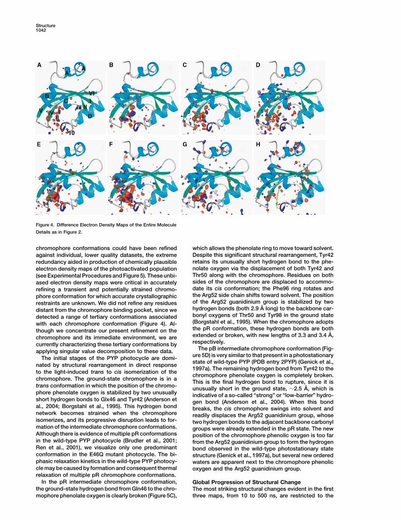
Figure 4. Difference Electron Density Maps of the Entire Molecule
Details as in Figure 2.
chromophore conformations could have been refined which allows the phenolate ring to move toward solvent.Despite this significant structural rearrangement, Tyr42against individual, lower quality datasets, the extremeretains its unusually short hydrogen bond to the phe-redundancy aided in production of chemically plausiblenolate oxygen via the displacement of both Tyr42 andelectron density maps of the photoactivated populationThr50 along with the chromophore. Residues on both(see Experimental Procedures and Figure 5). These unbi-sides of the chromophore are displaced to accommo-ased electron density maps were critical in accuratelydate its cis conformation; the Phe96 ring rotates andrefining a transient and potentially strained chromo-the Arg52 side chain shifts toward solvent. The positionphore conformation for which accurate crystallographicof the Arg52 guanidinium group is stabilized by tworestraints are unknown. We did not refine any residueshydrogen bonds (both 2.9 A long) to the backbone car-distant from the chromophore binding pocket, since webonyl oxygens of Thr50 and Tyr98 in the ground statedetected a range of tertiary conformations associated(Borgstahl et al., 1995). When the chromophore adoptswith each chromophore conformation (Figure 4). Al-the pR conformation, these hydrogen bonds are boththough we concentrate our present refinement on theextended or broken, with new lengths of 3.3 and 3.4 A,chromophore and its immediate environment, we arerespectively.currently characterizing these tertiary conformations by
The pB intermediate chromophore conformation (Fig-applying singular value decomposition to these data.ure 5D) is very similar to that present in a photostationaryThe initial stages of the PYP photocycle are domi-state of wild-type PYP (PDB entry 2PYP) (Genick et al.,nated by structural rearrangement in direct response1997a). The remaining hydrogen bond from Tyr42 to the
to the light-induced trans to cis isomerization of thechromophore phenolate oxygen is completely broken.
chromophore. The ground-state chromophore is in a This is the final hydrogen bond to rupture, since it istrans conformation in which the position of the chromo- unusually short in the ground state, �2.5 A, which isphore phenolate oxygen is stabilized by two unusually indicative of a so-called “strong” or “low-barrier” hydro-short hydrogen bonds to Glx46 and Tyr42 (Anderson et gen bond (Anderson et al., 2004). When this bondal., 2004; Borgstahl et al., 1995). This hydrogen bond breaks, the cis chromophore swings into solvent andnetwork becomes strained when the chromophore readily displaces the Arg52 guanidinium group, whoseisomerizes, and its progressive disruption leads to for- two hydrogen bonds to the adjacent backbone carbonylmation of the intermediate chromophore conformations. groups were already extended in the pR state. The newAlthough there is evidence of multiple pR conformations position of the chromophore phenolic oxygen is too farin the wild-type PYP photocycle (Brudler et al., 2001; from the Arg52 guanidinium group to form the hydrogenRen et al., 2001), we visualize only one predominant bond observed in the wild-type photostationary stateconformation in the E46Q mutant photocycle. The bi- structure (Genick et al., 1997a), but several new orderedphasic relaxation kinetics in the wild-type PYP photocy- waters are apparent next to the chromophore phenoliccle may be caused by formation and consequent thermal oxygen and the Arg52 guanidinium group.relaxation of multiple pR chromophore conformations.
In the pR intermediate chromophore conformation, Global Progression of Structural Changethe ground-state hydrogen bond from Gln46 to the chro- The most striking structural changes evident in the first
three maps, from 10 to 500 ns, are restricted to themophore phenolate oxygen is clearly broken (Figure 5C),

Structural Changes in Photoactive Yellow Protein1043
distant from the chromophore. Changes in the chromo-phore conformation thus induce long-range changes inthe protein tertiary structure, but our results show thatthey are not tightly coupled. The trans to cis isomeriza-tion of the PYP chromophore upon photon absorptionoccurs in much less than one nanosecond, a rapid mo-tion that forces neighboring residues like Tyr42, Glu46,Thr50, Arg52, and Phe96 to move on a similar time scale.In contrast, we see that the indirect rearrangementsinitiated by rupture of the hydrogen bond network aremuch slower and follow a microsecond to millisecondtime scale (akin to rates observed in protein folding[Englander, 2000]). PYP therefore uses a rapid, light-driven motion of a cofactor to introduce strain andthereby initiate much slower, but spatially more exten-sive, rearrangement of the surrounding protein.
Surprisingly, we observe the greatest spatial extentof tertiary structural change after the chromophore hasreisomerized to trans and moved to its ground-statelocation. A few small difference electron density featuresremain in the chromophore binding pocket in the finalmap (Figure 4H). Since these features overlay the strong-est difference electron density peaks created by the pBconformation (Figure 4G), they are likely to arise from asmall, residual population of molecules whose chromo-phore remains in the pB conformation. The fact thattertiary structural changes significantly lag behind thoseat the chromophore implies it takes some time for theFigure 5. Chromophore Conformations in the pR and pB Interme-protein to fully sense the pB chromophore conformationdiatesand shift accordingly. This same quality is observed inDifference electron density maps (A and B) and electron densitywild-type PYP in solution: the chromophore first as-maps (C and D) for the pR and pB chromophore conformations,
derived from the two highly averaged data sets (Table 2). Difference sumes its protonated pB conformation before subse-electron density contoured at �2� and �3.5�, for the (A) pR and quent global structural changes expose a region that is(B) pB states. Electron density maps contoured at �1� and �3�; capable of binding a hydrophobic dye (Hendriks et al.,yellow atomic model, ground-state conformation; orange models,
2002). This loose coupling between the chromophorechromophore conformations for the (C) pR and (D) pB states.conformation and tertiary structure is especially appar-ent in the E46Q mutant because of its faster photocycle
chromophore binding pocket (Figures 4A–4C). There is compared to wild-type. The lifetime of the pB chromo-a slight progression in the extent of structural change phore conformation is much shorter in the mutant (Fig-throughout this pR time domain, but these changes be- ure 1), perhaps because the mutant does not have tocome more apparent in the fourth map (Figure 4D), when reprotonate residue 46 in order to regain the groundthe pB chromophore conformation begins to form. Al- state. Thus, the tertiary structural changes are still devel-though the concentration of the pB chromophore con- oping in the mutant when the chromophore reisomerizesformation first waxes then wanes in the final four maps to the trans ground state. This accounts for the observa-from 10 �s to 30 ms, the spatial extent of tertiary struc- tion that the E46Q mutant has a reduced extent of ter-tural change steadily increases (Figures 4E–4H). Differ- tiary structural change in solution (Xie et al., 2001): theence electron density features are initially evident in chromophore does not remain in its pB conformationhelix B (Figures 4E and 4F), subsequently on the long enough for the tertiary structural changes to fullyN-terminal helices (Figures 4G and 4H), and finally on the develop.region of the � sheet most distant from the chromophore The pB conformation is the putative signaling state(Figure 4H). The locations of these features are fully of PYP, in which the most prominent structural changesconsistent with that visualized in solution using NMR include exposure of hydrophobic regions and tertiarytechniques on wild-type PYP (compare Figure 4H in this structural changes that have been described as partialarticle with Figure 4 in Rubinstenn et al. [1998]). Although unfolding (Lee et al., 2001; Rubinstenn et al., 1998; Xieboth the E46Q mutation and the protein environment in et al., 2001). As observed in solution on both wild-typethe crystal reduce the magnitude of tertiary structural PYP and the E46Q mutant (Brudler et al., 2001; Xie etchanges (Genick et al., 1997a; Xie et al., 2001), our re- al., 2001), only very limited tertiary structural changessults imply that neither factor qualitatively alters the occur prior to formation of the pB intermediate (Figuresnature of structural changes occurring within the PYP 4A–4C). The chromophore conformation regulates ac-photocycle. quisition of this protein conformation by stabilizing the
Although the chromophore has almost completely re- protein in its ground state; the protein can only developturned to its trans ground-state conformation in the final into the biological signaling state upon photon absorp-map from 7 to 30 ms (Figure 4H), this map displays tion. The trans to cis isomerization of the chromophore
provides a means to transiently rupture these stabilizingthe most prominent difference electron density features

Structure1044
Diffraction data were collected either at the BioCARS beamlineelements, of which the most notable is the hydrogen14ID-B, the Advanced Photon Source, or the European Synchrotronbond network to the chromophore phenolate oxygen.Radiation Facility beamline ID09. Crystal temperature was kept atIndeed, structural instability similar to that found in the297 K for all experiments. The exact data collection protocol de-
tertiary pB conformation can be created artificially in pended on the time delay and the synchrotron bunch mode (seethe ground state by significantly weakening the links Supplemental Data).between the chromophore and rest of the protein. Forexample, the PYP apoprotein lacks a chromophore and Photoactivation
Crystals were photoactivated using an Nd:YAG laser to pump eitheris substantially unstable and prone to denaturation,a dye laser with Coumarin 500 dye or an optical parametric oscillator,while removing the unusually short hydrogen bond toproducing a 7 or 5 ns FWHM pulse, respectively (see Supplementalthe chromophore, as in the Y42F and Y42A mutants,Data). The laser spot profile was approximately 1 mm in diameter atgreatly destabilizes the ground state (Brudler et al., 2000;the crystal for all data sets. Laser pulse energy was varied between
Imamoto et al., 2001a). datasets to ensure maximum photoactivation. The pulse energy wasThe initial stages of light-driven signal transduction titrated upwards to the maximum value at which the images could be
observed in PYP may be analogous to that in another processed. Above this value, crystal twitching between exposurescaused elongated diffraction spots that could not be accuratelyPAS domain protein, the FMN-containing LOV2 domainquantitated.from the plant blue light photoreceptor, phototropin
(Crosson et al., 2003). Despite very different chromo-Data Averagingphores, photoactivation of both PYP and LOV2 leads toRedundant datasets were merged in reciprocal space using
a similar tertiary structural change in which one or more weighted averaging of �F values. The weight was related to the helices flanking the central � sheet are destabilized standard error, �, of �F: weight � 1/(1 �2/��2�) (Ursby and Bour-(Harper et al., 2003; Rubinstenn et al., 1998). The struc- geois, 1997). The unique reflections present only in a single dataset
were retained in order to increase completeness. The influence oftural changes in PYP are initiated by chromophore isom-poorly measured �F values on the Fourier series can be minimizederization, while those in phototropin LOV2 by formationby weighting (Ren et al., 2001), but after sufficient averaging, theof a transiently stable covalent bond between the FMNweighting of �F values was no longer necessary or helpful in visualiz-chromophore and a nearby cysteine side chain (Crossoning the difference electron density signal.
and Moffat, 2002). Photoactivation has opposite effectsin the chromophore binding pocket of the two proteins. Generating Maps of the Photoactivated PopulationChromophore isomerization in PYP ruptures the hydro- Electron density maps of the photoactivated population were gener-gen bond network, a critical, preexisting structural link- ated to assist in model building and assessment of refinement.
These maps were generated by multiplying �F by a factor relatedage, but a completely new structural linkage is formedto occupancy of the photoactivated state before adding to Fc
dark:in LOV2 via the covalent bond between FMN and the(2/photoactive occupancy) (Fo
light � Fodark) Fc
dark. The differenceprotein. Despite this large difference in the underlyingbetween Fo
dark and Fcdark is the residual of the ground-state refine-
chemistry, both initiate a pattern of structural change ment; the final map thus represents the electron density of thein the protein that ultimately propagates to the surface photoactivated population minus the ground-state residual. Theof the molecule (Crosson et al., 2003), where it can be phases were taken from the simulated annealing, 1.10 A room tem-sensed by a downstream partner. perature E46Q structure with the chromophore omitted (Anderson
et al., 2004).
ConclusionData Processing and RefinementOur time-resolved structural study on the PYP photore-All indexing, integration, scaling, and merging was performed usingceptor has detailed a specific mode of signal transduc-LaueView (Ren and Moffat, 1995). Model building was performed
tion. The high resolution and quality of the extensively using XtalView (McRee, 1999), and refinement with SHELX97 (Shel-averaged, time-resolved crystallographic data allow us drick, 1997). All refinement was performed as difference refinementto unambiguously assign the structure of the chromo- in reciprocal space (Terwilliger and Berendzen, 1995). The intermedi-
ate chromophore coordinates were initially refined with occupancyphore conformation in the two principal spectroscopicand B values fixed. After convergence of the intermediate structure,intermediates in the PYP photocycle. We directly ob-the occupancy was refined along with the intermediate atomic posi-serve the progression of global structural changes astions. Distance, angle, and planarity restraints in the chromophorethe protein evolves from its ground to signaling statetail were gradually relaxed, and finally the isotropic B value was
and demonstrate that the nature of these changes in the refined.crystal are consistent with those in solution. Although The value of R free was monitored to assess the progress ofprevious studies in both solution and crystal (Brudler et refinement, but the absolute value of this parameter was of limited
usefulness in difference refinement. The same set of reflections wasal., 2001; Perman et al., 1998; Ren et al., 2001) endeav-used for all intermediate refinements. Progress was also criticallyored to identify the conformation of the chromophoreassessed using residual electron density maps; we aimed to accountin the pR intermediate, this is the first time such a novelfor all significant electron density features.species with a submicrosecond lifetime has been clearly
visualized and accurately refined at room temperature. AcknowledgmentsThe ability to visualize detailed structural changes as aprotein progresses from its ground to signaling state We thank Hyotcherl Ihee, Jason Key, and Marius Schmidt for theirand back again broadens our understanding of how support during data collection and technical advice. This work was
supported by National Institutes of Health Grant GM36452 (K.M.);proteins are able to transmit environmental stimuli intothe BioCARS facility at the Advanced Photon Source is supporteda biological response.by National Institutes of Health Grant RR07707 (K.M.).
Experimental ProceduresReceived: February 26, 2004Revised: April 1, 2004Laue Data CollectionAccepted: April 5, 2004All crystals were prepared as described (Anderson et al., 2004). The
crystals chosen for data collection were �180 180 500 �m. Published: June: 8, 2004

Structural Changes in Photoactive Yellow Protein1045
References Meyer, T.E., Yakali, E., Cusanovich, M.A., and Tollin, G. (1987). Prop-erties of a water-soluble, yellow protein isolated from a halophilicphototrophic bacterium that has photochemical activity analogousAnderson, S., Crosson, S., and Moffat, K. (2004). Short hydrogento sensory rhodopsin. Biochemistry 26, 418–423.bonds in photoactive yellow protein. Acta Crystallogr. D Biol. Crys-
tallogr., 60, 1008–1016. Mihara, K., Hisatomi, O., Imamoto, Y., Kataoka, M., and Tokunaga,F. (1997). Functional expression and site-directed mutagenesis ofBorgstahl, G.E., Williams, D.R., and Getzoff, E.D. (1995). 1.4 A struc-photoactive yellow protein. J. Biochem. (Tokyo) 121, 876–880.ture of photoactive yellow protein, a cytosolic photoreceptor: un-
usual fold, active site, and chromophore. Biochemistry 34, 6278– Perman, B., Srajer, V., Ren, Z., Teng, T., Pradervand, C., Ursby, T.,6287. Bourgeois, D., Schotte, F., Wulff, M., Kort, R., et al. (1998). Energy
transduction on the nanosecond time scale: early structural eventsBrudler, R., Meyer, T.E., Genick, U.K., Devanathan, S., Woo, T.T.,in a xanthopsin photocycle. Science 279, 1946–1950.Millar, D.P., Gerwert, K., Cusanovich, M.A., Tollin, G., and Getzoff,
E.D. (2000). Coupling of hydrogen bonding to chromophore confor- Ren, Z., and Moffat, K. (1995). Quantitative-analysis of synchrotronmation and function in photoactive yellow protein. Biochemistry 39, Laue diffraction patterns in macromolecular crystallography. J.13478–13486. Appl. Crystallogr. 28, 461–481.Brudler, R., Rammelsberg, R., Woo, T.T., Getzoff, E.D., and Gerwert, Ren, Z., Perman, B., Srajer, V., Teng, T.Y., Pradervand, C., Bourgeois,K. (2001). Structure of the I1 early intermediate of photoactive yellow D., Schotte, F., Ursby, T., Kort, R., Wulff, M., and Moffat, K. (2001).protein by FTIR spectroscopy. Nat. Struct. Biol. 8, 265–270. A molecular movie at 1.8 A resolution displays the photocycle of
photoactive yellow protein, a eubacterial blue-light receptor, fromCrosson, S., and Moffat, K. (2002). Photoexcited structure of a plantnanoseconds to seconds. Biochemistry 40, 13788–13801.photoreceptor domain reveals a light-driven molecular switch. Plant
Cell 14, 1067–1075. Rubinstenn, G., Vuister, G.W., Mulder, F.A.A., Dux, P.E., Boelens,R., Hellingwerf, K.J., and Kaptein, R. (1998). Structural and dynamicCrosson, S., Rajagopal, S., and Moffat, K. (2003). The LOV domainchanges of photoactive yellow protein during its photocycle in solu-family: photoresponsive signaling modules coupled to diverse out-tion. Nat. Struct. Biol. 5, 568–570.put domains. Biochemistry 42, 2–10.Rubinstenn, G., Vuister, G.W., Zwanenburg, N., Hellingwerf, K.J.,Cusanovich, M.A., and Meyer, T.E. (2003). Photoactive Yellow pro-Boelens, R., and Kaptein, R. (1999). NMR experiments for the studytein: a prototypic PAS domain sensory protein and development ofof photointermediates: application to the photoactive yellow protein.a common signaling mechanism. Biochemistry 42, 965–970.J. Magn. Reson. 137, 443–447.Englander, S. (2000). Protein folding intermediates and pathwaysSchmidt, M., Rajagopal, S., Ren, Z., and Moffat, K. (2003). Applica-studied by hydrogen exchange. Annu. Rev. Biophys. Biomol. Struct.tion of singular value decomposition to the analysis of time-resolved29, 213–238.macromolecular X-ray data. Biophys. J. 84, 2112–2129.Genick, U.K., Borgstahl, G.E., Ng, K., Ren, Z., Pradervand, C., Burke,Schmidt, M., Pahl, R., Srajer, V., Anderson, S., Ren, Z., Ihee, H.,P.M., Srajer, V., Teng, T.Y., Schildkamp, W., McRee, D.E., et al.Rajagopal, S., and Moffat, K. (2004). Protein kinetics: structure of(1997a). Structure of a protein photocycle intermediate by millisec-intermediates and reaction mechanism from time-resolved X-rayond time-resolved crystallography. Science 275, 1471–1475.data. Proc. Natl. Acad. Sci. USA, in press.Genick, U.K., Devanathan, S., Meyer, T.E., Canestrelli, I.L., Williams,Sheldrick, G. (1997). SHELXL97 (Gottingen, Germany: University ofE., Cusanovich, M.A., Tollin, G., and Getzoff, E.D. (1997b). ActiveGottingen).site mutants implicate key residues for control of color and light
cycle kinetics of photoactive yellow protein. Biochemistry 36, 8–14. Sprenger, W.W., Hoff, W.D., Armitage, J.P., and Hellingwerf, K.J.(1993). The eubacterium Ectothiorhodospira halophila is negativelyHarper, S., Neil, L., and Gardner, K. (2003). Structural basis of aphototactic, with a wavelength dependence that fits the absorptionphototropin light switch. Science 301, 1541–1544.spectrum of the photoactive yellow protein. J. Bacteriol. 175, 3096–
Hellingwerf, K.J., Hendriks, J., and Gensch, T. (2003). Photoactive3104.
yellow protein, a new type of photoreceptor protein: will this “yellowSrajer, V., Teng, T.Y., Ursby, T., Pradervand, C., Ren, Z., Adachi, S.,lab” bring us where we want to go? J. Phys. Chem. A 107, 1082–1094.Schildkamp, W., Bourgeois, D., Wulff, M., and Moffat, K. (1996).
Hendriks, J., Gensch, T., Hviid, L., van der Horst, M.A., Hellingwerf,Photolysis of the carbon monoxide complex of myoglobin: nanosec-
K.J., and van Thor, J.J. (2002). Transient exposure of hydrophobicond time-resolved crystallography. Science 274, 1726–1729.
surface in the photoactive yellow protein monitored with Nile red.Terwilliger, T.C., and Berendzen, J. (1995). Difference refinement:Biophys. J. 82, 1632–1643.obtaining differences between 2 related structures. Acta Crystallogr
Hoff, W.D., van Stokkum, I.H., van Ramesdonk, H.J., van Brederode, D Biol. Crystallogr. 51, 609–618.M.E., Brouwer, A.M., Fitch, J.C., Meyer, T.E., van Grondelle, R., and
Ujj, L., Devanathan, S., Meyer, T.E., Cusanovich, M.A., Tollin, G.,Hellingwerf, K.J. (1994). Measurement and global analysis of theand Atkinson, G.H. (1998). New photocycle intermediates in theabsorbance changes in the photocycle of the photoactive yellowphotoactive yellow protein from Ectothiorhodospira halophila: pico-protein from Ectothiorhodospira halophila. Biophys. J. 67, 1691–second transient absorption spectroscopy. Biophys. J. 75, 406–412.1705.Ursby, T., and Bourgeois, D. (1997). Improved estimation of struc-Imamoto, Y., Koshimizu, H., Mihara, K., Hisatomi, O., Mizukami, T.,ture-factor difference amplitudes from poorly accurate data. ActaTsujimoto, K., Kataoka, M., and Tokunaga, F. (2001a). Roles of aminoCrystallogr. A 53, 564–575.acid residues near the chromophore of photoactive yellow protein.Xie, A., Kelemen, L., Hendriks, J., White, B.J., Hellingwerf, K.J., andBiochemistry 40, 4679–4685.Hoff, W.D. (2001). Formation of a new buried charge drives a large-Imamoto, Y., Mihara, K., Tokunaga, F., and Kataoka, M. (2001b).amplitude protein quake in photoreceptor activation. BiochemistrySpectroscopic characterization of the photocycle intermediates of40, 1510–1517.photoactive yellow protein. Biochemistry 40, 14336–14343.Zhou, Y., Ujj, L., Meyer, T.E., Cusanovich, M.A., and Atkinson, G.H.Lee, B.C., Croonquist, P.A., Sosnick, T.R., and Hoff, W.D. (2001).(2001). Photocycle dynamics and vibrational spectroscopy of thePAS domain receptor photoactive yellow protein is converted to aE46Q mutant of photoactive yellow protein. J. Phys. Chem. A 105,molten globule state upon activation. J. Biol. Chem. 276, 20821–5719–5726.20823.
McRee, D.E. (1999). XtalView Xfit: a versatile program for manipulat-Accession Numbers
ing atomic coordinates and electron density. J. Struct. Biol. 125,156–165.
Coordinates for both structures have been deposited in the ProteinMeyer, T.E. (1985). Isolation and characterization of soluble cyto- Data Bank under accession codes 1S1Z and 1S1Y.chromes, ferredoxins and other chromophoric proteins from thehalophilic phototrophic bacterium Ectothiorhodospira-Halophila.Biochim. Biophys. Acta 806, 175–183.