chris jacobsen dept. physics & astronomy stony … the cold, soft truth: cryo diffraction...
TRANSCRIPT

1
The cold, soft truth:cryo diffraction microscopy
Chris Jacobsen
Dept. Physics & Astronomy
Stony Brook University

Cryo x-ray diffraction microscopy: why?
• X rays: best probe for samples thicker than ~1 μm.
• Diffraction microscopy (aka CXDI):– Get the most out of the exposure to the sample.
– Freedom from depth of focus limits.
• Cryo: essential for soft and/or wet specimens.
2

Electron interactions
3
These plots: Jacobsen, Medenwaldt, and Williams, in X-ray Microscopy & Spectromicroscopy (Springer, 1998)

Erbium atom columns (10-15 atoms each) in 8-20 nm thick silicon (001; lattice spacing 0.252 nm). Kaiser et al., Nature Materials 1, 102 (2002).
4
TEM of dopant atoms

If dose doesn’t matter, electrons can go pretty thick!
5
Voids in Cu interconnects: slices from a tomographic reconstruction.Ercius et al., Appl. Phys. Lett. 88, 243116 (2006)

6
X rays and thick specimensX-rays: better for thicker specimens. Sayre et al., Science 196, 1339 (1977); Schmahl & Rudolph in X-ray Microscopy: Instrumentation and Biological Applications (Springer, 1987)
These plots: based on Jacobsen, Medenwaldt, and Williams, in X-ray Microscopy & Spectromicroscopy (Springer, 1998)
Phase contrast is important!

Cryo x-ray diffraction microscopy: why?
• X rays: best probe for samples thicker than ~1 μm.
• Diffraction microscopy (aka CXDI):– Get the most out of the exposure to the sample.
– Freedom from depth of focus limits.
• Cryo: essential for soft and/or wet specimens.
7

8
• For many specimens, radiation damage sets the ultimate limit on achievable resolution.
• Lenses phase the signal, but lose the signal. Example: 20 nm zone plate with 10% efficiency, 50% window transmission, 20% modulation transfer function (MTF) for 15 nm half-period:
net transfer of 1% for high spatial frequencies• Can we avoid this ~100x signal loss, and also go beyond numerical
aperture limit of available optics?
Radiation damage sets the ultimate resolution limit

Can one recover phase from noisy data? Yes!
9
• Simulation: exit wave from thick cell (X. Huang et al.)• Poisson noise on intensities• Zone plate: 20 nm, 10% efficiency, incoherent bright field• Diffraction: reconstruction from noisy intensity• Direct test of low photon count builds upon earlier results by Fienup,
Optics Lett. 3, 27 (1978); and Williams et al., Acta Cryst. A 63, 36 (2007).

3D imaging with lenses
10
Transverse:
Longitudinal:
20 nm resolution at 520 eV: depth of field ~1 µm
Through-focus deconvolution with lenses:• Confocal: fully incoherent (fluorescence)• EM: phase only, coherent• TXM: partially coherent, equal absorption and
phase contrast, need for experimental CTF
1
10
100
1000
Tra
nsvers
e r
esolu
tion (
nm
)
1 10Energy (keV)
10 nm
0.2 20
0.1 µm
1 µm
10 µm
100 µm
DOF=1 mm
!t !!
4"=
!rN
2
!! !!
"2= 4!rN
!rN
!

11
Bragg gratings that diffract to a certain angle represent a specific transverse and longitudinal periodicity (Ewald sphere)
Diffraction microscopy in 3D
Ewald sphereData collection over a series of rotations about an axis fills in 3D Fourier space for phasing

Pure projections from phased 3D data
12
Chapman, Barty, Marchesini, Noy, Hau-Riege, Cui, Howells, Rosen, He, Spence, Weierstall, Beetz, Jacobsen, Shapiro, J. Opt. Soc. Am. A 23, 1179 (2006)

qx
qz
Experimental realization
Chapman, Barty, Marchesini, Noy, Hau-Riege, Cui, Howells, Rosen, He, Spence, Weierstall, Beetz, Jacobsen, Shapiro, J. Opt. Soc. Am. A 23, 1179 (2006)

Cryo x-ray diffraction microscopy: why?
• X rays: best probe for samples thicker than ~1 μm.
• Diffraction microscopy (aka CXDI):– Get the most out of the exposure to the sample.
– Freedom from depth of focus limits.
• Cryo: essential for soft and/or wet specimens.
14

15
Dried yeast: Stony Brook/ALS
D. Shapiro et al., Proc. Nat. Acad. Sci. 102, 15343 (2005).
J. Nelson, X. Huang, J. Steinbrener et al.
Visible
X ray

• Human blood platelets
• 1 MeV transmission electron microscope (JEOL-1000)
• O’Toole, Wray, Kremer, and McIntosh, J. Struct. Bio. 110, 55 (1993)
16
Froz
en h
ydra
ted
2% g
luta
rald
ehyd
e fix
1% O
sO4
post
fixcr
itica
l-po
int d
ry
1 µm
1 µm
100 nm
500 nm
100 nm

17
Frozen hydrated: stable specimens!
Frozen hydrated specimens don’t shrink in the beam (freeze-dried specimens do)
Scattering power is linear with dose thus far in both cases
David Shapiro, PhD dissertation, Stony Brook, 2004

18
Stony Brook cryo chamber at ALS 9.0.1Incorporates JEOL goniometer, Gatan cryo holder. Beetz et al., Nucl. Inst. Meth. A 545, 459 (2005).

Upgrade at ALS 9.0.1
19
Positioning platform for aligning “birdhouse” relative to the rest of the chamber
Overall support for entire structure
Rail mounting
Rail mounting
Bellows
Better stability, higher resolution positioning (curved beam, ptychography), better visible light positioning, better anticontamination... With T. Warwick et al., ALS.
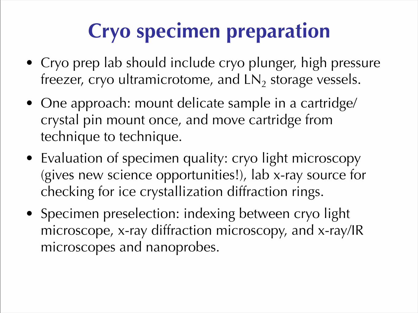
Cryo specimen preparation• Cryo prep lab should include cryo plunger, high pressure
freezer, cryo ultramicrotome, and LN2 storage vessels.
• One approach: mount delicate sample in a cartridge/crystal pin mount once, and move cartridge from technique to technique.
• Evaluation of specimen quality: cryo light microscopy (gives new science opportunities!), lab x-ray source for checking for ice crystallization diffraction rings.
• Specimen preselection: indexing between cryo light microscope, x-ray diffraction microscopy, and x-ray/IR microscopes and nanoprobes.

21
Sample freezing
Ken Downing, Bjorg Larson, and Andrew Stewart. Thanks also to Eva Nogales and her lab!
Plunge freezing: FEI Vitrobot (hopping between Donner Lab and ALS)


23Visible light phase contrast Estimating ice thickness

FEI Titan Krios TEM
• Being introduced at MPI Martinsried on April 15.
• 300 keV, field emission gun, energy filter.
• “Maximize Sample Integrity - achieve contamination-free sample transfer and imaging”
• “Increase Productivity - The AutoLoader™ allows for fully automated and contamination-free loading and analysis of up to 12 samples contained in specially designed AutoGrid™ sample holder”
24

Cryo system: Xradia example
• Mount fragile grid in cartridge once; grids, capillaries, ...
• Transfer cartridge between visible light and various X-ray microscopes (including scanning, tomography).
• Robotic sample insertion in microscope.
Xradia cryo team: C. Jacobsen, D. Trapp, et al.

Cryo x-ray diffraction microscopy: why?
• X rays: best probe for samples thicker than ~1 μm.
• Diffraction microscopy (aka CXDI):– Get the most out of the exposure to the sample.
– Freedom from depth of focus limits.
• Cryo: essential for soft and/or wet specimens.
26
NSLS II should include cryo for soft and/or wet materials!