chitosan/bioactive glass nanoparticle composite membranes...
TRANSCRIPT
Acta Biomaterialia 8 (2012) 4173–4180
Contents lists available at SciVerse ScienceDirect
Acta Biomaterialia
journal homepage: www.elsevier .com/locate /actabiomat
Chitosan/bioactive glass nanoparticle composite membranesfor periodontal regeneration
Joana Mota a,b, Na Yu c, Sofia G. Caridade a,b, Gisela M. Luz a,b, Manuela E. Gomes a,b, Rui L. Reis a,b,John A. Jansen c, X. Frank Walboomers c, João F. Mano a,b,⇑a 3B’s Research Group – Biomaterials, Biodegradables and Biomimetics, University of Minho, Headquarters of the European Institute of Excellence on Tissue Engineeringand Regenerative Medicine, AvePark, 4806-909 Taipas, Guimarães, Portugalb ICVS/3B’s – PT Government Associate Laboratory, Braga/Guimarães, Portugalc Radboud University Nijmegen Medical Centre, Department of Biomaterials, P.O. Box 9101, 6500HB Nijmegen, The Netherlands
a r t i c l e i n f o
Article history:Received 13 April 2012Received in revised form 27 June 2012Accepted 29 June 2012Available online 5 July 2012
Keywords:BiomaterialsTissue engineeringGuided tissue regenerationChitosanNanoparticles
1742-7061/$ - see front matter � 2012 Acta Materialhttp://dx.doi.org/10.1016/j.actbio.2012.06.040
⇑ Corresponding author at: 3B’s Research Group –and Biomimetics, University of Minho, HeadquartersExcellence on Tissue Engineering and Regenerative MTaipas, Guimarães, Portugal. Tel.: +351 253510900; fa
E-mail address: [email protected] (J.F. Mano)
a b s t r a c t
Barrier membranes are used in periodontal applications with the aim of supporting periodontal regener-ation by physically blocking migration of epithelial cells. The present work proposes a combination ofchitosan (CHT) with bioactive glass nanoparticles (BG-NPs) in order to produce a novel guided tissueand bone regeneration membrane, fabricated by solvent casting. The CHT/BG-NP nanocomposite mem-branes are characterized in terms of water uptake, in mechanical tests, under simulated physiologicalconditions and in in vitro bioactivity tests. The addition of BG-NPs to CHT membranes decreased themechanical potential of these membranes, but on the other hand the bioactivity improved. The mem-branes containing the BG-NPs induced the precipitation of bone-like apatite in simulated body fluid(SBF). Biological tests were carried out using human periodontal ligament cells and human bone marrowstromal cells. CHT/BG-NP composite membranes promoted cell metabolic activity and mineralization.The results indicate that the CHT/BG-NP composite membrane could potentially be used as a temporaryguided tissue regeneration membrane in periodontal regeneration, with the possibility to induce boneregeneration.
� 2012 Acta Materialia Inc. Published by Elsevier Ltd. All rights reserved.
1. Introduction
Periodontitis is an inflammatory disease of the periodontal tis-sues, caused by microorganisms and calculus accumulation on thebacterial biofilm, leading to degradation of the connective tissuesand alveolar bone and subsequent formation of soft tissue pocketsaround the root surface [1]. Regeneration of the periodontal regionis challenged by complex inflammatory processes and events re-lated to healing, resulting in unsatisfactory results in clinical casesusing the currently available therapies. Ideal strategies should beable to regenerate all damaged structures including the cementum,periodontal ligament and alveolar bone. The number of cells occu-pying the treated area upon surgery as well as the size of the defectdetermine the type of connection formed between the cementumand bone. Epithelial cells are the first cells to migrate to the siteof injury becoming a problem as they prevent bone formation.
ia Inc. Published by Elsevier Ltd. A
Biomaterials, Biodegradablesof the European Institute ofedicine, AvePark, 4806-909
x: +351 253510909..
The guided tissue regeneration (GTR) technique uses a membrane,which acts as a barrier to prevent epithelial cells and gingival tis-sue reaching the area of injured tissue [2]. This procedure favorsthe regeneration of lost and damaged tissue, as it promotes cellrepopulation of the periodontal ligament and adjacent alveolarbone, allowing the necessary time for osteoblast proliferation andbone regeneration [3,4]. Cells derived from adult human bone mar-row stromal cells (hBMSC) are capable of contributing to the for-mation of new bone completely surrounding the tooth, whichhas to be able to anchor itself to the jaw through its roots and peri-odontal ligament [5]. The continuing process of periodontal tissuerepair is followed by the formation of granulation tissue as a sourceof future periodontal connective tissue cells, such as osteoblasts,periodontal ligament fibroblasts and cementoblasts [5]. Thus hu-man periodontal ligament cells (hPDL) and hBMSCs are ideal cellsources to be used in biological tests to evaluate the performanceof GTR membranes.
When selecting an ideal biomaterial for GTR membranes thefollowing requirements must be considered: wound stabilization,space creation and maintenance, protection of the underlyingblood clot, and the ability to exclude unwanted tissues or cells(connective tissue and gingival epithelium). Collagen has been
ll rights reserved.
4174 J. Mota et al. / Acta Biomaterialia 8 (2012) 4173–4180
one of the most common materials used to produce GTR mem-branes. However, this material is derived from animal sourcesand has an associated risk of disease transmission and associatedethical and cultural issues. Moreover, the fast resorption rate ofthis material and its poor mechanical strength are a concern tomany clinicians. In addition, rapid collagen biodegradation, bythe enzymatic activity of macrophages, polymorphonuclear leuco-cytes and several pathogens capable of producing collagenase,results in a limited membrane resistance to collapse. This eventallows unwanted cell types to enter the wound area [6]. Sincecollagen membranes have shown such limited properties, newmaterials with better properties are required.
Chitosan (CHT) is obtained by alkaline deacetylation of chitinand presents excellent biological properties, such as biodegradabil-ity, biocompatibility, and immunogenicity, as well as antibacterial,antifungal and wound-healing activity. Its degradation productsare non-toxic, non-antigenic, non-immunogenic and non-carcino-genic. CHT also evokes minimal foreign body reaction [7–12]. Thepositive surface charge of this biomaterial and its biocompatibilityenable it to effectively support cell growth, while the hydrophilicsurface facilitates cell adhesion, proliferation, and differentiation[7,11,13]. When placed in hydrated environments CHT is a flexiblematerial. This is an advantage over more rigid synthetic materialslike polylactic acid (PLA) and polyglycolic acid (PGA), facilitatinghandling during the implantation process [14–16]. Because ofthese properties CHT has been widely used in biomedical applica-tions [9,12,13,17]. It can accelerate wound healing and enhancebone formation both in vivo and in vitro. It has been reported thatwhen implanted in vertebrates it may enhance the migration anddifferentiation of specific types of progenitor cells (e.g. osteoblastdifferentiation) [10,18,19]. Lahiji et al. found that CHT may serveas an effective template in the repair of osseous defects, becauseit has unique material properties and the ability to support viableand functioning human osteoblasts. [9] Thus CHT is an attractivecandidate for future use in GTR membranes. However, CHT is notan ideal material for bone regeneration, as its osteoconductivityneeds to be improved. Zhang et al. [20] verified that no apatitewas formed on the surface of pure CHT scaffolds. To improve thebioactivity of CHT it is necessary to combine it with other bioactivematerials [21]. Recent studies have highlighted the importance ofthe development of composite materials based on biodegradablepolymers containing bioactive glasses [22]. Bioactive glasses oftenshow excellent bioactivity and biocompatibility both in vitro andin vivo: when implanted at the site of bone defects they can di-rectly bond to the surrounding tissues by chemical linkage. As wellas the GTR function, acting as a barrier, the addition of bioactiveglass particles to CHT membranes should promote guided boneregeneration (GBR) activity, giving a dual function, producing aGTR/GBR membrane. The biomineralization capability of bioactiveglasses is related to its composition, but is also influenced by phys-ical properties such as particle size, porosity, surface area and mor-phology. In recent years a growing consensus has been reached toconcentrate on the development of nanostructural bioactive glass/ceramic materials, due to their high biomineralization capability.Large surface area bioactive glass materials should facilitate bonehealing and tooth defects, as well as blood clotting. Consequently,the preparation and application of nanoscale bioactive glass mate-rials has received increasing attention from biological materialresearchers. The sol–gel method has been used to prepare highsurface area bioactive glass particles, including bioactive glassnanoparticles (BG-NPs) [10,23–26].
In this study CHT/BG-NP nanocomposite membranes were pre-pared by solvent casting, and the physical, bioactive and biologicalproperties of these biomaterials were characterized, to evaluatethe possible performance in periodontal defect applications, usinghPDL and hBMSC cells in biological tests.
2. Materials and methods
Chitosan (CHT) (Mw 190,000–310,000, 75–85% deacetylated,viscosity 200–800 cps) was obtained from Sigma and was purifiedbefore use. Tetraethyl orthosilicate (TEO) (99.90% pure), ammo-nium phosphate dibasic, calcium nitrate tetrahydrate (99%), citricacid monohydrate (99–100%), ammonium hydroxide (maximum33% NH3) and all chemicals for SBF preparation were purchasedfrom Sigma–Aldrich. All other reagents and solvents used were ofreagent grade.
2.1. Bioactive glass nanoparticle preparation
To prepare the BG-NPs a protocol based on previous work wasfollowed [27,28]. The procedure to obtain nanoparticles with thecomposition SiO2:CaO:P2O5 (mol.%) = 55:40:5 consisted of sequen-tial reagent dissolution that resulted in hydrolysis and polyconden-sation reactions. TEOS (99.90% pure) was used as the siliconprecursor, ammonium phosphate dibasic as the phosphorus pre-cursor, calcium nitrate tetrahydrate (99%) as the calcium precursor,citric acid monohydrate (99–100%) to promote hydrolysis, absoluteethanol, ammonium hydroxide (maximum 33% NH3) as the gellingagent and polyethylene glycol 20,000 (PEG) as the surfactant. TheBG-NPs were sintered at 700 �C for 5 h. Spherical particles withsizes below 50 nm were obtained (see Fig. 3).
2.2. Membrane preparation
Composite membranes were obtained by the dissolution of 0.7%(w/v) purified CHT and 0.3% (w/v) BG-NPs in a solution of 2 vol.%acid acetic. Pure CHT membranes were obtained by dissolution of1% (w/v) CHT in a solution of 2 vol.% acid acetic. After complete sol-vent evaporation the membranes were neutralized with a solutionof 0.4% (w/v) NaOH and left to dry at room temperature.
2.3. Water uptake
1 � 1 cm samples cut from the membranes were weighted(Wini) and then immersed at 37 �C in 2 ml of phosphate-bufferedsaline (PBS), pH 7.4, containing 0.8% (w/v) lysozyme (hen egg-white) and 0.02% (w/v) sodium azide. The concentration oflysozyme was similar to the concentration in human serum [29].Membranes of known dry weight (n = 3 samples of each set ofmembranes) were incubated in the lysozyme solution for the per-iod of study. At each time point samples were removed from thesolution and rinsed with distilled water. The superficial waterwas removed and samples were weighted (Wfin). The water uptakewas calculated using the equation:
ðW fin �W iniÞ=W ini � 100 ð%Þ
2.4. Mechanical tests
The ultimate tensile strength (UTS) and tensile modulus of themembranes were determined using an Instron 4505 Universal Ma-chine tensile testing machine in tensile mode. The tests were per-formed at room temperature at a rate of 5 mm min�1. Wetmembranes were immersed in PBS for 3 h before the test and werefixed with sandpaper, to prevent slipping. Tests were performed ona single membrane with a gauge length of 10 mm. For each condi-tion a minimum of five specimens were tested.
2.5. In vitro bioactivity tests
In vitro bioactivity tests were carried out by soaking the mem-branes in 50 ml of SBF for 5 days at 37 �C. The SBF composition and
J. Mota et al. / Acta Biomaterialia 8 (2012) 4173–4180 4175
preparation was previously described by Kokubo and Takadama[30]. Upon removal from the SBF the samples were rinsed withdistilled water and left to dry.
2.6. Scanning electron microscopy (SEM) and energy dispersivespectroscopy (EDX) experiments
A NanoSEM FEI Nova 200 (FEG/SEM) scanning electron micro-scope was used to study the surface and the morphology of thesamples. A conductive gold coating was applied to the samplesprior to observation. A Pegasus X4M instrument was used to pre-form the energy dispersive X-ray spectroscopy (EDX) experiments,in a low vaccum and without coating.
2.7. Cell culture
Periodontal ligament cells were obtained from human thirdmolars according to the following procedure. After extraction theteeth were washed three times for 10 min in PBS with100 units ml�1 penicillin/streptomycin. Periodontal ligament tis-sue was scraped from the middle third of the root with a scalpelblade, to avoid contamination by epithelial or pulp cells. The freedportions of the periodontal ligament were minced and transferredto a small culture flask, which was filled with alpha minimal essen-tial medium (a-MEM) (Gibco) containing 10 vol.% fetal calf serum(FCS) (Gibco), 50 mg ml�1 ascorbic acid (Sigma), 10.8 M dexameth-asone (Sigma), 50 mg ml�1 gentamycin (Gibco) and 10 mM sodiumb-glycerophosphate (Sigma). Cells were cultured at 37 �C in ahumidified atmosphere with 5% CO2. The medium was replacedevery 2–3 days. Upon reaching 100% confluency the cells were re-leased with trypsin/EDTA (0.25% w/v crude trypsin and 1 mMEDTA, pH 7.2) and sub-cultured for two passages in standard cul-ture flasks. The cells were then frozen in liquid nitrogen until use.
hBMSCs were isolated from bone blocks of human iliac crestbiopsies. The biopsies were discarded tissue from standard surgicalprocedures at Radboud University Nijmegen Medical Centre(Nijmegen, The Netherlands). The bone blocks were cut into smallpieces and subsequently placed in a 50 ml tube to which 20 ml ofa-MEM was added. Then the tubes were shaken vigorously andthe medium with cells collected. This procedure was repeated sev-eral times. The collected medium with cells was plated in T25 cul-ture flasks (Greiner Bio-one), which were filled with 4 ml of a-MEMcontaining 10 vol.% FCS and 100 units ml-1 penicillin/streptomycin(all from Gibco BRL Life Technologies BV, Breda, The Netherlands).The cells were cultured at 37 �C in a humid atmosphere with 5%CO2 and passaged at 80% confluency using trypsin/EDTA (Gibco).
Two kinds of proliferation medium were used (hereafter re-ferred to as O� culture media). The hPDL O� culture medium wascomposed of a-MEM (Gibco) with 10% fetal bovine serum (FBS)(Greiner Bio-one) and 100 units ml�1 penicillin/streptomycin (Gib-co BRL). The hBMSC O� culture medium was composed of a-MEM(Gibco) with 15% FBS (Greiner Bio-one), 1% L-glutamine, 1% ascor-bic acid (Sigma), 100 units ml�1 penicillin/streptomycin (GibcoBRL) and 1%, by volume added to each cell culture flask, basic fibro-blast growth factor (bFGF).
After one generation the cells were plated at a density of5000 cells cm�2 in T75 culture flasks and expanded in O� culturemedium. The culture medium was changed twice a week. Cellsfrom passage 3 were used in this experiment.
2.8. Cell seeding
The membranes were glued to metal rings (1.5 cm diameter)with RTV silicone adhesive (Nusil Technology, Carpinteria, CA), inorder to prevent membrane fluctuation and to fix them on the cul-ture well bottom. The samples were sterilized by immersion in 70%
ethanol for 1 h, rinsed in PBS and soaked in medium overnight.Cells, either hPDL or hBMSC, were seeded on the samples and after4 h (at 37 �C and 5% of CO2) osteogenic differentiation culture med-ium (O+ culture medium) was added. As controls cells were seededdirectly on the bottom of the plate and O+ culture medium wasadded immediately.
Two types of O+ culture medium were used. The hPDL O+ cul-ture medium was composed of a-MEM (Gibco) with 10% FBS (Gre-iner Bio-one), 1% ascorbic acid (Sigma), 1% b-glycerophosphate(Sigma), 1% dexamethasone (Sigma) and 100 units ml�1 penicil-lin/streptomycin (Gibco BRL). The hBMSC O+ culture medium wascomposed of a-MEM (Gibco) with 15% FBS (Greiner Bio-one), 1%L-glutamine, 1% ascorbic acid (Sigma), 1% b-glycerophosphate (Sig-ma), 1% dexamethasone (Sigma) and 100 units ml�1 penicillin/streptomycin (Gibco BRL).
2.9. Biological tests
2.9.1. Immunofluorescence and image analysesAfter 3 days in culture on the membrane surface cells were
fixed for 10 min in 3% paraformaldehyde (Fluka AG), and perme-abilized with 1% Triton X-100 for 5 min. Then filamentous actinwas stained with AlexaFluor 568 phalloidin (Molecular ProbesInc., Eugene, OR) diluted 1:200 in PBS containing 1% bovine serumalbumin (BSA) for 2 h. The cells nuclei were stained with 4,6-diamidino-2phenylindole (DAPI) diluted 1:2500 in PBS for10 min. Finally, the specimens were examined with a Zeiss auto-mated fluorescence microscope (Imager Z1) at a magnification of10�.
2.9.2. Proliferation assayAfter the various experimental periods the medium was re-
moved from the chambers and the wells were washed with PBS.The cells were lysed using milliQ filtered water with subsequentsonication for 10 min between two freeze–thaw cycles at �80 �C.The supernatant was stored at �20 �C until further analysis. A Pico-Green dsDNA Quantification Kit (Molecular Probes, Eugene, OR)was used according to the manufacturer’s instructions. The analy-sis was performed on the supernatants from days 1, 3, 7, 14 and 28.100 ll of PicoGreen working solution was added to 100 ll of eachsupernatant sample. The samples were incubated for 2–5 min atroom temperature in the dark. After incubation fluorescence wasmeasured in a fluorescence cuvette reader (microplate fluores-cence reader, Bio-Tek Instruments, Winooski, VT) with a 485 nmexcitation filter and a 530 nm emission filter.
2.9.3. Alamar blueCell metabolic activity was measured using the Alamar blue as-
say (Invitrogen) according to the manufacturer’s instructions. Asolution was made of Alamar blue in culture medium in the pro-portions 1:9 and held at 37 �C for 5 min. Culture medium wasflushed from wells containing samples with cells and replaced withthe test solution. Culture plates were incubated at 37 �C in 5% CO2
for 4 h. After incubation 200 ll of each sample solution were trans-ferred to 96-well plates (Greiner Bio-one). Fluorescence was mea-sured using a microplate reader (FL 600, Bio-Tek Instruments) at570 nm. The assay was performed on days 1, 3, 7, 14 and 28.
2.9.4. Calcium content measurementCalcium content was assessed 28 days after cell seeding to
obtain information on mineralized matrix formation. The sampleswere rinsed with milliQ filtered water and 1 ml of acetic acid wasadded. The samples were incubated overnight under vigorousconstant shaking and the acetic acid with dissolved calciumwas frozen and kept at �20 �C until further investigation.After thawing the calcium content was determined using the OCPC
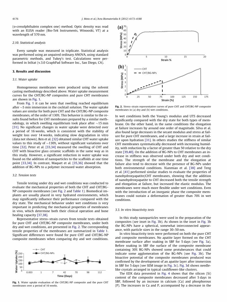
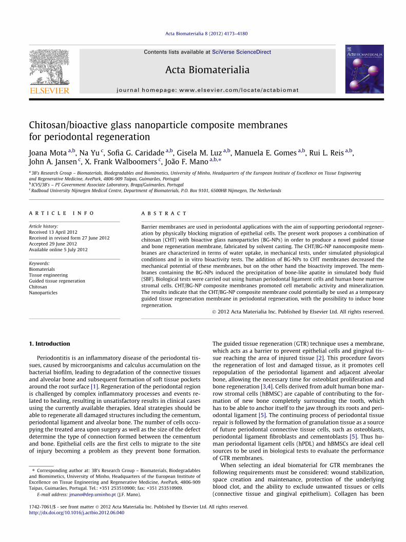
Fig. 2. Stress–strain representative curves of pure CHT and CHT/BG-NP compositemembranes in (a) dry and (b) wet conditions.
4176 J. Mota et al. / Acta Biomaterialia 8 (2012) 4173–4180
(o-cresolphthalein complex one) method. Optic density was readwith an ELISA reader (Bio-Tek Instruments, Winooski, VT) at awavelength of 570 nm.
2.10. Statistical analysis
Every sample was measured in triplicate. Statistical analysiswas performed using an unpaired ordinary ANOVA, using standardparametric methods, and Tukey’s test. Calculations were per-formed in InStat (v.3.0 GraphPad Software Inc., San Diego, CA).
3. Results and discussion
3.1. Water uptake
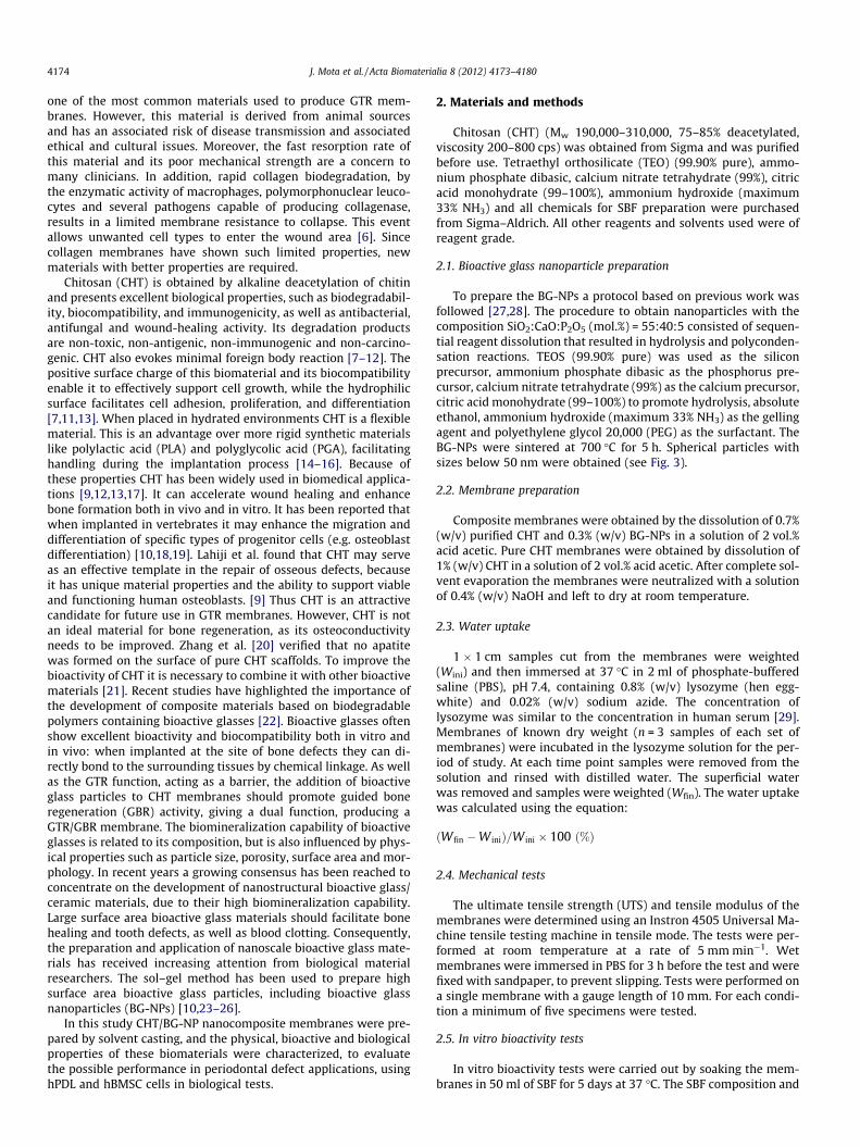
Homogeneous membranes were produced using the solventcasting methodology described above. Water uptake measurementcurves for the CHT/BG-NP composites and pure CHT membranesare shown in Fig. 1.
From Fig. 1 it can be seen that swelling reached equilibriumafter �5 min immersion in the cocktail solution. The water uptakevalues are similar for both pure CHT and the CHT/BG-NP compositemembranes, of the order of 130%. This behavior is similar to the re-sults found before for CHT membranes prepared by a similar meth-odology, in which swelling equilibrium took place after �15 min[31]. No significant changes in water uptake were detected overa period of 16 weeks, which is consistent with the stability ofweight loss over 14 weeks, indicating slow degradation in vitro(data not shown). Ren et al. [32] obtained similar CHT water uptakevalues to this study of �130%, without significant variations overtime [32]. Peter et al. [33,34] measured the swelling of CHT andCHT with bioactive glass ceramic scaffolds in the same way as inthis study. However, a significant reduction in water uptake wasfound on the addition of nanoparticles to the scaffolds at one timepoint [33,34]. In contrast, Maquet et al. [35,36] showed that theaddition of BG-NPs to a polymer increased water absorption.
3.2. Tension tests
Tensile testing under dry and wet conditions was conducted toevaluate the mechanical properties of both the CHT and CHT/BG-NP composite membranes (see Fig. 2 and Table 1). Biomedical im-plants are usually placed in very hydrated environments, whichmay significantly influence their performance compared with thedry state. The mechanical behavior under wet conditions is veryimportant in predicting the mechanical properties of membranesin vivo, which determine both their clinical operation and bonehealing capacity [37,38].
Representative stress–strain curves from tensile tests obtainedfor pure CHT and CHT/BG-NP composite membranes, under bothdry and wet conditions, are presented in Fig. 2. The correspondingtensile properties of the membranes are summarized in Table 1.Significant differences were found for pure CHT and CHT/BG-NPcomposite membranes when comparing dry and wet conditions.
Fig. 1. Water uptake evaluation of the CHT/BG-NP composite and the pure CHTmembranes over a period of 16 weeks.
In wet conditions both the Young’s modulus and UTS decreasedsignificantly compared with the dry state for both types of mem-brane. On the other hand, in the same conditions the elongationat failure increases by around one order of magnitude. Silva et al.also found large decreases in the secant modulus and stress at fail-ure for pure CHT membranes, and a large increase in strain at fail-ure upon hydration [31]. In others studies the stiffness of similarCHT membranes systematically decreased with increasing humid-ity, with reduction by a factor of greater than 50 relative to the drystate [39,40]. On the addition of BG-NPs to CHT membranes an in-crease in stiffness was observed under both dry and wet condi-tions. The strength of the membrane and the elongation atfailure also tend to decrease with the presence of BG-NPs underboth environmental conditions. Xianmiao et al. [38] and Tenget al. [41] performed similar studies to evaluate the properties ofnanohydroxyapatite/CHT membranes, showing that the additionof nanohydroxyapatite to CHT decreased both the tensile strengthand elongation at failure, but increased the elastic modulus. Themembranes were much more flexible under wet conditions. Evenwith the introduction of an inorganic phase the composite mem-branes could sustain a deformation of greater than 70% in wetconditions.
3.3. In vitro bioactivity tests
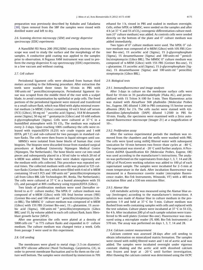
In this study nanoparticles were used in the preparation of thecomposites (see inset in Fig. 3b). As shown in the inset in Fig. 3bthe BG-NPs have a spherical, somewhat interconnected appear-ance, with particle sizes in the range 30–50 nm.
In vitro bioactivity tests were performed on both the pure CHTand composite membranes. No apatite layer formed on the CHTmembrane surface after soaking in SBF for 5 days (see Fig. 3a).Before soaking in SBF the surface of the composite membranecontaining 30% BG-NPs showed some protuberances that couldindicate some agglomeration of the BG-NPs (see Fig. 3b). Thebioactive potential of the composite membranes produced wasconfirmed by the development of an apatite layer after immersionin SBF for 5 days (see SEM image in Fig. 3c). Fig. 3d shows needle-like crystals arranged in typical cauliflower-like clusters.
The EDX data presented in Fig. 4 shows that the silicon (Si)content of the composite membranes decreased after 5 days inSBF, followed by an increase in calcium (Ca) and phosphorous(P). The increases in Ca and P, accompanied by a decrease in the
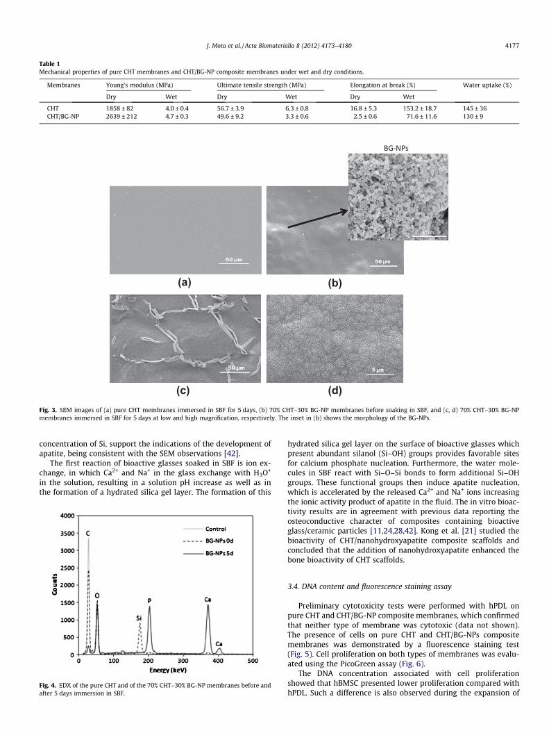
Table 1Mechanical properties of pure CHT membranes and CHT/BG-NP composite membranes under wet and dry conditions.
Membranes Young’s modulus (MPa) Ultimate tensile strength (MPa) Elongation at break (%) Water uptake (%)
Dry Wet Dry Wet Dry Wet
CHT 1858 ± 82 4.0 ± 0.4 56.7 ± 3.9 6.3 ± 0.8 16.8 ± 5.3 153.2 ± 18.7 145 ± 36CHT/BG-NP 2639 ± 212 4.7 ± 0.3 49.6 ± 9.2 3.3 ± 0.6 2.5 ± 0.6 71.6 ± 11.6 130 ± 9
(a) (b)
(c) (d)
µm
Fig. 3. SEM images of (a) pure CHT membranes immersed in SBF for 5 days, (b) 70% CHT–30% BG-NP membranes before soaking in SBF, and (c, d) 70% CHT–30% BG-NPmembranes immersed in SBF for 5 days at low and high magnification, respectively. The inset in (b) shows the morphology of the BG-NPs.
J. Mota et al. / Acta Biomaterialia 8 (2012) 4173–4180 4177
concentration of Si, support the indications of the development ofapatite, being consistent with the SEM observations [42].
The first reaction of bioactive glasses soaked in SBF is ion ex-change, in which Ca2+ and Na+ in the glass exchange with H3O+
in the solution, resulting in a solution pH increase as well as inthe formation of a hydrated silica gel layer. The formation of this
Fig. 4. EDX of the pure CHT and of the 70% CHT–30% BG-NP membranes before andafter 5 days immersion in SBF.
hydrated silica gel layer on the surface of bioactive glasses whichpresent abundant silanol (Si–OH) groups provides favorable sitesfor calcium phosphate nucleation. Furthermore, the water mole-cules in SBF react with Si–O–Si bonds to form additional Si–OHgroups. These functional groups then induce apatite nucleation,which is accelerated by the released Ca2+ and Na+ ions increasingthe ionic activity product of apatite in the fluid. The in vitro bioac-tivity results are in agreement with previous data reporting theosteoconductive character of composites containing bioactiveglass/ceramic particles [11,24,28,42]. Kong et al. [21] studied thebioactivity of CHT/nanohydroxyapatite composite scaffolds andconcluded that the addition of nanohydroxyapatite enhanced thebone bioactivity of CHT scaffolds.
3.4. DNA content and fluorescence staining assay
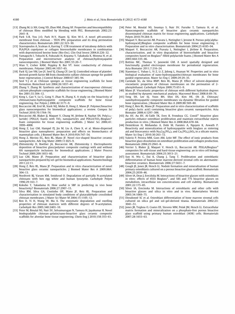
Preliminary cytotoxicity tests were performed with hPDL onpure CHT and CHT/BG-NP composite membranes, which confirmedthat neither type of membrane was cytotoxic (data not shown).The presence of cells on pure CHT and CHT/BG-NPs compositemembranes was demonstrated by a fluorescence staining test(Fig. 5). Cell proliferation on both types of membranes was evalu-ated using the PicoGreen assay (Fig. 6).
The DNA concentration associated with cell proliferationshowed that hBMSC presented lower proliferation compared withhPDL. Such a difference is also observed during the expansion of
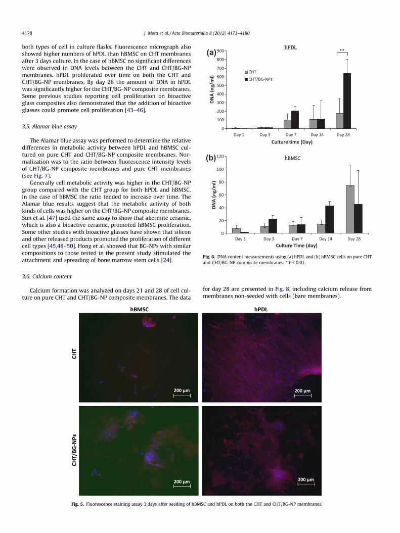
Fig. 6. DNA content measurements using (a) hPDL and (b) hBMSC cells on pure CHTand CHT/BG-NP composite membranes. ⁄⁄P < 0.01.
4178 J. Mota et al. / Acta Biomaterialia 8 (2012) 4173–4180
both types of cell in culture flasks. Fluorescence micrograph alsoshowed higher numbers of hPDL than hBMSC on CHT membranesafter 3 days culture. In the case of hBMSC no significant differenceswere observed in DNA levels between the CHT and CHT/BG-NPmembranes. hPDL proliferated over time on both the CHT andCHT/BG-NP membranes. By day 28 the amount of DNA in hPDLwas significantly higher for the CHT/BG-NP composite membranes.Some previous studies reporting cell proliferation on bioactiveglass composites also demonstrated that the addition of bioactiveglasses could promote cell proliferation [43–46].
3.5. Alamar blue assay
The Alamar blue assay was performed to determine the relativedifferences in metabolic activity between hPDL and hBMSC cul-tured on pure CHT and CHT/BG-NP composite membranes. Nor-malization was to the ratio between fluorescence intensity levelsof CHT/BG-NP composite membranes and pure CHT membranes(see Fig. 7).
Generally cell metabolic activity was higher in the CHT/BG-NPgroup compared with the CHT group for both hPDL and hBMSC.In the case of hBMSC the ratio tended to increase over time. TheAlamar blue results suggest that the metabolic activity of bothkinds of cells was higher on the CHT/BG-NP composite membranes.Sun et al. [47] used the same assay to show that akermite ceramic,which is also a bioactive ceramic, promoted hBMSC proliferation.Some other studies with bioactive glasses have shown that siliconand other released products promoted the proliferation of differentcell types [45,48–50]. Hong et al. showed that BG-NPs with similarcompositions to those tested in the present study stimulated theattachment and spreading of bone marrow stem cells [24].
3.6. Calcium content
Calcium formation was analyzed on days 21 and 28 of cell cul-ture on pure CHT and CHT/BG-NP composite membranes. The data
Fig. 5. Fluorescence staining assay 3 days after seeding of hBMS
for day 28 are presented in Fig. 8, including calcium release frommembranes non-seeded with cells (bare membranes).
C and hPDL on both the CHT and CHT/BG-NP membranes.

Fig. 7. Ratio of fluorescence intensity between CHT/BG-NP composite membranesand CHT membranes due to (a) hPDL and (b) hBMSC, obtained from Alamar blueassays.
J. Mota et al. / Acta Biomaterialia 8 (2012) 4173–4180 4179
The calcium concentration in the presence of hPDL and hBMSCcells on CHT/BG-NP composite membranes was significantly high-er than on pure CHT membranes and control groups, indicating apositive effect of the presence of BG-NPs on cell mineralization.The highest calcium contents were found for membranes withBG-NPs, which could be attributed to the release of calcium fromthe inorganic nanoparticles. Dieudonné et al. [51] concluded thatthe high base calcium levels of bioactive glasses (in the absenceof cells) could be responsible for the increased mineralization.However, in the present study the calcium levels on day 28 onthe CHT/BG-NP composite membranes in the presence of cellswere significantly higher than on bare composite membranes. Thisresult indicates that the increased calcium formation could be a re-sult of cell matrix mineralization. Others studies have demon-strated that the presence of bioactive glasses can improve cellmineralization [11,48,52]. On the CHT/BG-NP composite mem-branes both type of cell showed an increase in calcium levels be-tween day 21 (data not shown) and day 28, indicating thatmineralized matrix formation occurred over time, which meansthat the cells continued to mineralize after 21 days.
Fig. 8. Calcium content measurements of hPDL and hBMSC on CHT membranes andCHT/BG-NP composite membranes, and bare membranes. ⁄⁄⁄P < 0.001; ⁄⁄P < 0.01.
4. Conclusion
CHT/BG-NP composite membranes were studied for possibleapplication as GTR membranes and for GBR, provided by the addi-tion of BG-NPs to the polymer, promoting the possibility of peri-odontal regeneration. The introduction of BG-NPs into the CHTincreased the stiffness of the membrane and the composite mem-branes showed adequate extensibility in wet conditions. Uponimmersion in SBF the composite membranes, but not pure CHTmembranes, were able to promote the deposition of an apatitelayer, evidence of osteoconductive potential. The present studycompared the in vitro biological performance of pure CHT mem-branes with CHT/BG-NP composite membranes. The metabolicactivity of hPDL and hBMSC was enhanced on the addition of BG-NPs to CHT membranes. An increase in cell proliferation of hPDLon CHT/BG-NP composite membranes was verified compared withpure CHT membranes. The incorporation of BG-NPs into the mem-brane promoted greater cell matrix mineralization by both types ofcells.
The CHT/BG-NP composite membranes studied could be used asbarrier membranes to prevent the invasion of periodontal defectsby soft tissues, since these membranes did not show early degrada-tion and were not cytotoxic. The results obtained suggest the pos-sible applicability of these composite membranes in GBR and,consequently, periodontal regeneration.
Acknowledgement
This work was financially supported by the Foundation for Sci-ence and Technology (FCT) within the project PTDC/CTM-BPC/112774/2009.
Appendix A. Figures with essential colour discrimination
Certain figures in this article, particularly Fig. 5, are difficult tointerpret in black and white. The full colour images can be foundin the on-line version, at http://dx.doi.org/10.1016/j.actbio.2012.06.040.
References
[1] Friedrich D. Treatment of extended periodontal defects using a b-TCPcomposite material. Dent Implantol 2009;4:258–62.
[2] Buser D, Brägger U, Lang NP, Nyman S. Regeneration and enlargement of jawbone using guided tissue regeneration. Clin Oral Implan Res 1990;1:22–32.
[3] Kay SA, Wisner-Lynch L, Marxer M, Lynch SE. Guided bone regeneration:integration of a resorbable membrane and a bone graft material. PractPeriodontics Aesthet Dent 1997;9:185–94.
[4] Zitzmann N, Naef R, Schärer P, Schüpbach P. Guided bone regeneration andaugmentation in implant surgery, using Bio-Oss together with the membranetechnique. Dtsch Zahnarztl Z 1996;6:51.
[5] Lanza RP, Langer RS, Vacanti J. Principles of tissue engineering. NewYork: Elsevier Academic Press; 2007.
[6] Bunyaratavej P, Wang HL. Collagen membranes: a review. J Periodontol2001;72:215–29.
[7] Alves NM, Mano JF. Chitosan derivatives obtained by chemical modificationsfor biomedical and environmental applications. Int J Biol Macromol2008;43:401–14.
[8] Chatelet C, Damour O, Domard A. Influence of the degree of acetylation onsome biological properties of chitosan films. Biomaterials 2001;22:261–8.
[9] Lahiji A, Sohrabi A, Hungerford DS, Frondoza CG. Chitosan supports theexpression of extracellular matrix proteins in human osteoblasts andchondrocytes. J Biomed Mater Res 2000;51:586–95.
[10] Li Z, Ramay HR, Hauch KD, Xiao D, Zhang M. Chitosan–alginate hybrid scaffoldsfor bone tissue engineering. Biomaterials 2005;26:3919–28.
[11] Lu HH, El-Amin SF, Scott KD, Laurencin CT. Three-dimensional, bioactive,biodegradable, polymer–bioactive glass composite scaffolds with improvedmechanical properties support collagen synthesis and mineralization ofhuman osteoblast-like cells in vitro. J Biomed Mater Res A 2003;64:465–74.
[12] Mi F-L, Shyu S-S, Wu Y-B, Lee S-T, Shyong J-Y, Huang R-N. Fabrication andcharacterization of a sponge-like asymmetric chitosan membrane as a wounddressing. Biomaterials 2001;22:165–73.
4180 J. Mota et al. / Acta Biomaterialia 8 (2012) 4173–4180
[13] Zhang M, Li XH, Gong YD, Zhao NM, Zhang XF. Properties and biocompatibilityof chitosan films modified by blending with PEG. Biomaterials 2002;23:2641–8.
[14] Park S-B, You J-O, Park H-Y, Haam SJ, Kim W-S. A novel pH-sensitivemembrane from chitosan – TEOS IPN; preparation and its drug permeationcharacteristics. Biomaterials 2001;22:323–30.
[15] Stavropoulos A, Sculean A, Karring T. GTR treatment of intrabony defects withPLA/PGA copolymer or collagen bioresorbable membranes in combinationwith deproteinized bovine bone (Bio-Oss). Clin Oral Invest 2004;8:226–32.
[16] Yamaguchi I, Tokuchi K, Fukuzaki H, Koyama Y, Takakuda K, Monma H, et al.Preparation and microstructure analysis of chitosan/hydroxyapatitenanocomposites. J Biomed Mater Res 2001;55:20–7.
[17] Wan Y, Creber KAM, Peppley B, Bui VT. Ionic conductivity of chitosanmembranes. Polymer 2003;44:1057–65.
[18] Park YJ, Lee YM, Lee JY, Seol YJ, Chung CP, Lee SJ. Controlled release of platelet-derived growth factor-BB from chondroitin sulfate-chitosan sponge for guidedbone regeneration. J Control Release 2000;67:385–94.
[19] Seol Y-J et al. Chitosan sponges as tissue engineering scaffolds for boneformation. Biotechnol Lett 2004;26:1037–41.
[20] Zhang Y, Zhang M. Synthesis and characterization of macroporous chitosan/calcium phosphate composite scaffolds for tissue engineering. J Biomed MaterRes 2001;55:304–12.
[21] Kong L, Gao Y, Lu G, Gong Y, Zhao N, Zhang X. A study on the bioactivity ofchitosan/nano-hydroxyapatite composite scaffolds for bone tissueengineering. Eur Polym J 2006;42:3171–9.
[22] Boccaccini AR, Erol M, Stark WJ, Mohn D, Hong Z, Mano JF. Polymer/bioactiveglass nanocomposites for biomedical applications: a review. Compos SciTechnol 2010;70:1764–76.
[23] Boccaccini AR, Blaker JJ, Maquet V, Chung W, Jérôme R, Nazhat SN. Poly(D,L-lactide) (PDLLA) foams with TiO2 nanoparticles and PDLLA/TiO2-Bioglass�
foam composites for tissue engineering scaffolds. J Mater Sci 2006;41:3999–4008.
[24] Hong Z, Luz GM, Hampel PJ, Jin M, Liu A, Chen X, et al. Mono-dispersedbioactive glass nanospheres: preparation and effects on biomechanics ofmammalian cells. J Biomed Mater Res A 2010;95A:747–54.
[25] Hong Z, Merino EG, Reis RL, Mano JF. Novel rice-shaped bioactive ceramicnanoparticles. Adv Eng Mater 2009;11:B25–9.
[26] Zhitomirsky D, Roether JA, Boccaccini AR, Zhitomirsky I. Electrophoreticdeposition of bioactive glass/polymer composite coatings with and withoutHA nanoparticle inclusions for biomedical applications. J Mater ProcessTechnol 2009;209:1853–60.
[27] Luz GM, Mano JF. Preparation and characterization of bioactive glassnanoparticles prepared by sol–gel for biomedical applications. Nanotechnology2011;22:11.
[28] Hong Z, Reis RL, Mano JF. Preparation and in vitro characterization of novelbioactive glass ceramic nanoparticles. J Biomed Mater Res A 2009;88A:304–13.
[29] Nordtveit RJ, Varum KM, Smidsrod O. Degradation of partially N-acetylatedchitosans with hen egg white and human lysozyme. Carbohydr Polym1996;29:163–7.
[30] Kokubo T, Takadama H. How useful is SBF in predicting in vivo bonebioactivity? Biomaterials 2006;27:2907–15.
[31] Silva RM, Silva GA, Coutinho OP, Mano JF, Reis RL. Preparation andcharacterisation in simulated body conditions of glutaraldehyde crosslinkedchitosan membranes. J Mater Sci Mater M 2004;15:1105–12.
[32] Ren D, Yi H, Wang W, Ma X. The enzymatic degradation and swellingproperties of chitosan matrices with different degrees of N-acetylation.Carbohydr Res 2005;340:2403–10.
[33] Peter M, Binulal NS, Nair SV, Selvamurugan N, Tamura H, Jayakumar R. Novelbiodegradable chitosan–gelatin/nano-bioactive glass ceramic compositescaffolds for alveolar bone tissue engineering. Chem Eng J 2010;158:353–61.
[34] Peter M, Binulal NS, Soumya S, Nair SV, Furuike T, Tamura H, et al.Nanocomposite scaffolds of bioactive glass ceramic nanoparticlesdisseminated chitosan matrix for tissue engineering applications. CarbohydrPolym 2010;79:284–9.
[35] Maquet V, Boccaccini AR, Pravata L, Notingher I, Jerome R. Porous poly(alpha-hydroxyacid)/Bioglass composite scaffolds for bone tissue engineering. I:Preparation and in vitro characterisation. Biomaterials 2004;25:4185–94.
[36] Maquet V, Boccaccini AR, Pravata L, Notingher I, Jérôme R. Preparation,characterization, and in vitro degradation of bioresorbable and bioactivecomposites based on Bioglass�-filled polylactide foams. J Biomed Mater Res A2003;66A:335–46.
[37] Bottino MC, Thomas V, Janowski GM. A novel spatially designed andfunctionally graded electrospun membrane for periodontal regeneration.Acta Biomater 2011;7:216–24.
[38] Xianmiao C, Yubao L, Yi Z, Li Z, Jidong L, Huanan W. Properties and in vitrobiological evaluation of nano-hydroxyapatite/chitosan membranes for boneguided regeneration. Mater Sci Eng C 2009;29:29–35.
[39] Caridade SG, da Silva RMP, Reis RL, Mano JF. Effect of solvent-dependentviscoelastic properties of chitosan membranes on the permeation of 2-phenylethanol. Carbohydr Polym 2009;75:651–9.
[40] Mano JF. Viscoelastic properties of chitosan with different hydration degreesas studied by dynamic mechanical analysis. Macromol Biosci 2008;8:69–76.
[41] Teng SH, Lee EJ, Yoon BH, Shin DS, Kim HE, Oh JS. Chitosan/nanohydroxyapatite composite membranes via dynamic filtration for guidedbone regeneration. J Biomed Mater Res A 2009;88:569–80.
[42] Hong Z, Reis RL, Mano JF. Preparation and in vitro characterization of scaffoldsof poly(L-lactic acid) containing bioactive glass ceramic nanoparticles. ActaBiomater 2008;4:1297–306.
[43] Au AY, Au RY, Al-Talib TK, Eves B, Frondoza CG. Consil� bioactive glassparticles enhance osteoblast proliferation and maintain extracellular matrixproduction in vitro. J Biomed Mater Res A 2008;86A:678–84.
[44] Demirkiran H, Mohandas A, Dohi M, Fuentes A, Nguyen K, Aswath P.Bioactivity and mineralization of hydroxyapatite with bioglass as sinteringaid and bioceramics with Na3Ca6(PO4)5 and Ca5(PO4)2SiO4 in a silicate matrix.Mater Sci Eng C 2010;30:263–72.
[45] Valerio P, Pereira MM, Goes AM, Leite MF. The effect of ionic products frombioactive glass dissolution on osteoblast proliferation and collagen production.Biomaterials 2004;25:2941–8.
[46] Verrier S, Blaker JJ, Maquet V, Hench LL, Boccaccini AR. PDLLA/Bioglass�
composites for soft-tissue and hard-tissue engineering: an in vitro cell biologyassessment. Biomaterials 2004;25:3013–21.
[47] Sun H, Wu C, Dai K, Chang J, Tang T. Proliferation and osteoblasticdifferentiation of human bone marrow-derived stromal cells on akermanite-bioactive ceramics. Biomaterials 2006;27:5651–7.
[48] Gough JE, Jones JR, Hench LL. Nodule formation and mineralisation of humanprimary osteoblasts cultured on a porous bioactive glass scaffold. Biomaterials2004;25:2039–46.
[49] Silver IA, Deas J, Erecinska M. Interactions of bioactive glasses with osteoblastsin vitro: effects of 45S5 Bioglass�, and 58S and 77S bioactive glasses onmetabolism, intracellular ion concentrations and cell viability. Biomaterials2001;22:175–85.
[50] Silver IA, Erecinska M. Interactions of osteoblastic and other cells withbioactive glasses and silica in vitro and in vivo. Materialwiss Werkst2003;34:1069–75.
[51] Dieudonné SC et al. Osteoblast differentiation of bone marrow stromal cellscultured on silica gel and sol–gel-derived titania. Biomaterials 2002;23:3041–51.
[52] Jones JR, Tsigkou O, Coates EE, Stevens MM, Polak JM, Hench LL. Extracellularmatrix formation and mineralization on a phosphate-free porous bioactiveglass scaffold using primary human osteoblast (HOB) cells. Biomaterials2007;28:1653–63.