chemical sensing with solid state devices || biosensor principles
TRANSCRIPT

7
Biosensor Principles
In this chapter we cover only the most elementary principles involved in biosensors. We are not trying to review the recent literature in this area, since that would constitute a book in itself. The purpose is to provide an introduction to this fascinating field and to highlight the potential of biosensors to nonbiologists.
Biosensors are defined here as small analytical devices that combine a transducer with a biologically active substance. In the broader sense it is also any sensor that measures the concentration of a biological substance. Many biosubstances are very good catalysts, and both selectivity and sensitivity of biosensors are frequently substantially better than for previously discussed sensor systems. The major disadvantage of biosensors is often the limited stability of the biosubstances in an environment different than their natural one.
Biochemical amplification schemes coupled with electronic amplification lead to unprecedented sensitivities in biosensors. The ultimate sensor in this respect is a living cell, and with the use of a bilayer lipid membrane (BLM) such cells and their sensing functions can be simulated. Biochemical amplification and BLMs will be treated in detail in this chapter, since they are the most important building blocks for the biosensors of the future.
Because of the extensive terminology used in this field, we have provided the reader with a glossary of the most important recurring terms at the end of this chapter.
277
278 Biosensor Principles 7
7.1 Biosensor Characteristics
Definition
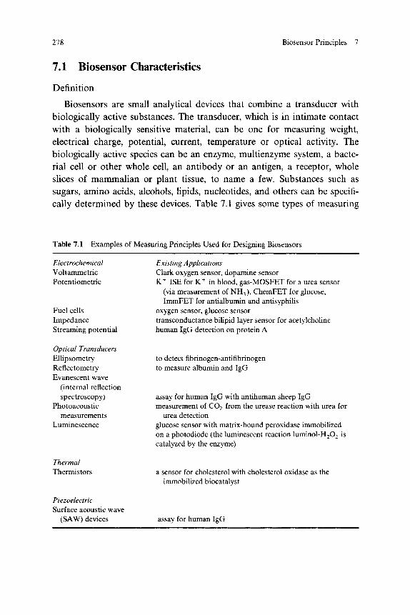
Biosensors are small analytical devices that combine a transducer with biologically active substances. The transducer, which is in intimate contact with a biologically sensitive material, can be one for measuring weight, electrical charge, potential, current, temperature or optical activity. The biologically active species can be an enzyme, multienzyme system, a bacterial cell or other whole cell, an antibody or an antigen, a receptor, whole slices of mammalian or plant tissue, to name a few. Substances such as sugars, amino acids, alcohols, lipids, nucleotides, and others can be specifically determined by these devices. Table 7.1 gives some types of measuring
Table 7.1 Examples of Measuring Principles Used for Designing Biosensors
Electrochemical Voltammetric Potentiometric
Fuel cells Impedance Streaming potential
Existing Applications Clark oxygen sensor, dopamine sensor K
+ ISE for K
+ in blood, gas-MOSFET for a urea sensor
(via measurement of NH3), ChemFET for glucose, ImmFET for antialbumin and antisyphilis
oxygen sensor, glucose sensor transconductance bilipid layer sensor for acetylcholine human IgG detection on protein A
Optical Transducers Ellipsometry Reflectometry Evanescent wave
(internal reflection spectroscopy)
Photoacoustic measurements
Luminescence
to detect fibrinogen-antifibrinogen to measure albumin and IgG
assay for human IgG with antihuman sheep IgG measurement of C 02 from the urease reaction with urea for
urea detection glucose sensor with matrix-bound peroxidase immobilized on a photodiode (the luminescent reaction luminol-H202 is catalyzed by the enzyme)
Thermal Thermistors a sensor for cholesterol with cholesterol oxidase as the
immobilized biocatalyst
Piezoelectric Surface acoustic wave
(SAW) devices assay for human IgG

7.1 Biosensor Characteristics 279
SUBSTANCE TO BE
DETECTED
PHYSICAL OR
RECEPTOR CHEMICAL CHANGE
TRANSDUCER RECEPTOR TRANSDUCER . READOUT
BIORECOGNITION LAYER
(RECEPTOR)
SIGNAL CONVERSION
BIOSUBSTANCE
MEMBRANE
Figure 7.1 Schematic presentation of a Biosensor
principles employed to detect a variety of biologically important substances. In this table we also include sensors that are used to detect biologically important substances but which do not necessarily contain a biological substance themselves. Also included in this table are measuring techniques that are, because of the bulky apparatus involved, not typically classified as sensor techniques (e.g., ellipsometry).
A schematic representation of a biosensor is shown in Fig. 7.1. It consists of a biosubstance—that is, a molecule-discriminating section held physically together over a transducer surface by a membrane or otherwise coupled (fixed) to such a transducer. The biosubstance is an organism-related material also called a receptor. The word "receptor" is used here in the general meaning of the word, as opposed to its specific use in Table 7.2, where it refers to a specific class of molecules with bioaffinity.
The subject matter of biosensors is very broad. In this chapter we will limit ourselves to covering electrochemically based biosensors, and we will concentrate on the detection principles involved rather than discussing

280 Biosensor Principles 7
Table 7.2 Molecular Recognition Affinity Pairs
Enzyme
Lectin
Antibody
Receptor DNA probes
Binding protein
Porphyrins as in heme proteins (e.g., hemoglobin and myoglobin) or as in heme enzymes, etc.
Substrate," KA « 102-10
6 L M"
1 6
Inhibitor Effector Carbohydrates, KA « 10
3-10
4 L M"
1 (simple sugars),
KA « 106-10
7 L M
-1 (with multipoint attachment)
Glycoproteins Glycolipids Antigen, KA « 10
3-10
16 L M
-1
Hapten, KA « 1 05- 1 0
u L M"
1
Complement (induces lysis of target cells in the presence of an antigen-antibody pair)
Molecules such as hormones binding to a receptor DNA or RNA sequence with complementary base
sequence Biotin, KA « 10
15 L M
_1 for biotin-avidin pair
Retinal, protein A-Fc region of IgG Fe(II), Fe(III), Mg(II), Zn(II), Cd(II), Hg(II), Cu(II), Ni(II)
and Co(II)
a N o t t o b e confused wi th t he solid s u b s t r a t e of a sensor . * T h e affinity c o n s t a n t KA (L M
_ 1) is d i scussed fur ther in c o n n e c t i o n w i t h E q . (7.1).
specific devices. We will repeatedly direct the reader to the literature for more details.
Affinity Pairs
Frequently, concentrations of biological analytes are very low. For example, the concentration of certain proteins in blood serum may be as low as micrograms per liter or less, compared with a total protein content of 70 g L
1 therefore requiring a discrimination ratio (i.e., selectivity of
107-10
8) in order to specifically estimate the concentration of the desired
protein.1 Because of these very stringent requirements the best sensing
devices used are based on schemes provided by nature itself. These are "affinity pairs," molecules or particles designed by nature to react selectively with the other member of the pair. For example, for molecular recognition tremendous use can be made of the inherent selectivity afforded by the tertiary structure of proteins and polypeptides, and by nucleic acid complementary pairing. Examples of molecular recognition based affinity pairs are given in Table 7.2.

7.1 Biosensor Characteristics 281
Immobilization
One of the most active study domains in the biosensor area is the immobilization of various biologically active substances on a transducer and the study of the influence that immobilization has on the activity of these substances. Various designs for immobilizing biosubstances are shown in Fig. 7.2 (based on Aizawa
2). When discussing enzyme sensors, we
examine more specific immobilization schemes. The demands on a biosensor are very high. They include, in the case of a
drug monitoring system, for example, high specificity to the drug or metabolite being monitored, sensitivity in the range of 1 to 100 mg L"
1,
ON BEADS CONDUCTIVE POLYMER SUCH AS POLYANILINE OR POLYPYRROLE)
Figure 7.2 Immobilization schemes for biosubstances (Réf. 2)

282 Biosensor Principles 7
fast response time (e.g., 1 to 60 sec), possibility for miniaturization, biocom-patibility, sterilizability, compensation for adverse conditional affects (e.g., temperature) and high accuracy.
3 At this stage it is difficult to meet all these
requirements simultaneously. Fortunately some other demands are more relaxed. Biosensors do not usually require a long lifetime, for example.
Biocompatibility
In addition to all the engineering problems associated with chemical sensors (e.g., encapsulation, reliability, durability, reproducibility, need for recalibration) biosensors have some unique problems. Inserting sensors into the human body, for example, does not only introduce problems with the sensor's reliability and durability, it also introduces the need for prevention of unwanted effects in the body. The encapsulation material has to be chosen with great care so as not to cause toxication, thrombosis or inflammatory effects due to irritation of blood vessels or tissue. Electric shock, needless to say, should also be avoided. Microbial fouling-resisting layers have been identified,
4 and, although these materials can be used in the
structure of the sensor, it is difficult to use them as part of the sensitive membrane itself. For instance, surface modification of plastics can significantly change the spectrum of organisms and biopolymers that will adhere to the surface. However, it is difficult to predict what effect these will have on the specificity, sensitivity and response time of the sensor. Both blood and tissue compatibility are still major hurdles to be overcome before any type of invasive biosensor (be it electrochemical, fiber optic, or, for that matter, any kind of biosensor) will be accepted commercially for use on humans. Some further reading on the topic of implantable sensors is suggested, for example W. Ko et al.
5 and McKinley et al.
6
Sterilization
Biosensors for bioreactors (e.g., the human body or fermentors) must be nonfouling and sterilizable, which make the task of developing biosensors even more complex. Steam treatment remains the most acceptable, sometimes the only means of achieving reliable sterilization. Unfortunately, in many instances steam treatment will adversely affect a biosensor. Alternative radiation treatment (e.g., gamma rays) or chemical treatment (e.g., ethylene oxide) of sensors offers a less suitable solution (gamma rays may degrade the organic sensor components, and ethylene oxide is toxic).
Closely related to the sterilization issue is the question of in-bioreactor or off-bioreactor employment of sensors. Some pros and cons for in-bioreactor

7.1 Biosensor Characteristics 283
In-Bioreactor Off-Bioreactor
Pros Measures in situ Real time
Easy to make many measurements Easy to use many different tests Easy to use biochemical
Cons Need to make several macromolecules
Delayed measurements holes in the reactor
Restricted number of Difficulties in taking a representative sample
Growth of cells onto sampling device
possible tests Sterility problems
Autoclaveable equipment required
Growth of cells on the sensor will cause false readings
and off-bioreactor analysis are summarized in Table 7.3. The importance of the various factors in this table largely depends on whether the bioreactor is a human body, an animal body or a fermentor.
In general, substantial work is still needed in the area of sterility of biosensors. If manufacturing technologies can be developed so that the sensors are produced under sterile conditions and the biological macro-molecules are added in sterile form, it might be possible to produce disposable biosensors. Such an approach, for example, would be attractive in conjunction with Si-based microsensors, as discussed in Chapters 8 and 9. The above-mentioned limited lifetime requirement (disposable use) represents an advantage in developing biomedical-type sensors.
On the topic of sterilization some further reading is advised. We recommend Clarke et al.,
4 Mullen and Vadgama,
7 and Enfors and Nilsson.
8
Specificity in Biological Membranes (Biocatalysts)
When certain foreign molecules enter a living organism, the defense system (the immune system) is alerted. Specific antibodies (Ab) are formed that bind to the foreign molecule (the antigen, Ag). Whereupon antigen-antibody complexes are removed from circulation by phagocytosis. Antigen-antibody complexes are only one example of the many affinity pairs one can rely on to make highly specific biosensors (see Table 7.2). In principle, it should be possible to construct biosensors for any organic molecule capable
Table 7.3 Reasons for In-Bioreactor or Off-Bioreactor Analysis

284 Biosensor Principles 7
of interaction with a biological species. This generic aspect of biosensors is one of the major reasons for the big interest in them.
One of the most desired biosensor products would be an immunosensor to detect the presence of antibodies or antigens. Immunodiagnostics has become a major area in clinical diagnostics, and the potential of im-munosensors is enormous, since immunoassays can be used to monitor almost any reaction (i.e., the method is very generic). Immunochemical analysis is based on the exploitation of the specific interaction between antibodies and their antigens. At this time an immunoassay is often slow, takes many different wet-chemistry steps (labor intensive) and is quite expensive. Huge savings in time and labor are expected if reagent-free, inexpensive, disposable immunosensors could be developed.
Affinity Constant
Biological molecules immobilized on a surface or in a membrane and in intimate contact with a suitable transducing system (see Fig. 7.1) interact specifically and reversibly according to the scheme
A + Β ^ AB, (7.1) kr
where the equilibrium reaction constant KA (in this field often called the affinity constant) is given by the ratio of rate constants ki/kl with a value encompassing the range 1 0
+2 to 10
+ 16 L M
- 1, depending on the interact
ing system. For example, the affinity constants of enzyme-substrate, inhibitor, effector or coenzyme complexes generally he within the range 1 0
2- 1 0
6 L M "
1, whereas immunological complexes display KA values in
the range 106-10
12 L M
- 1. The lower affinity displayed between enzymes
and their substrates may be utilized in fully reversible sensors for the assay of their complementary substrates (detection limit is about 10 ~
6 L M
- 1) .
In contrast, the higher affinity and specificity of immunological systems may be utilized in disposable devices for the estimation of trace metabolites present at very low concentrations and with even higher discrimination (detection limit is 10~
12 L M "
1 and below).
1 By changing the pH, the ionic
strength or other solution parameters, the AgAb complex can sometimes be dissociated again and a new analysis can be undertaken. However, for a sensor the need for such wet-chemistry steps is undesirable; consequently the extremely high selectivity of an immunochemical reaction is a mixed blessing.
7.2 Enzyme Electrodes 285
7.2 Enzyme Electrodes
Enzymes are high-molecular-weight biocatalysts (proteins) that increase the rate of numerous reactions critical to life itself. Enzyme electrodes are devices in which the analyte is either a substrate, also called reactant, or a product of the enzyme reaction, detected potentiometrically or amperomet-rically. They act according to the scheme
E n z y m e . .
Substrate + Cofactor > Products. (7.2)
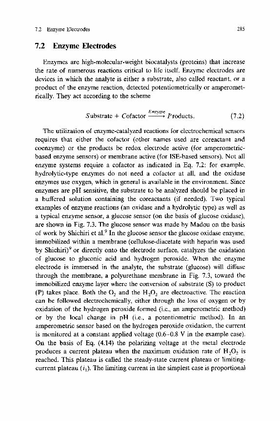
The utilization of enzyme-catalyzed reactions for electrochemical sensors requires that either the cofactor (other names used are coreactant and coenzyme) or the products be redox electrode active (for amperometric-based enzyme sensors) or membrane active (for ISE-based sensors). Not all enzyme systems require a cofactor as indicated in Eq. 7.2; for example, hydrolytic-type enzymes do not need a cofactor at all, and the oxidase enzymes use oxygen, which in general is available in the environment. Since enzymes are pH sensitive, the substrate to be analyzed should be placed in a buffered solution containing the coreactants (if needed). Two typical examples of enzyme reactions (an oxidase and a hydrolytic type) as well as a typical enzyme sensor, a glucose sensor (on the basis of glucose oxidase), are shown in Fig. 7.3. The glucose sensor was made by Madou on the basis of work by Shichiri et al.
9 In the glucose sensor the glucose oxidase enzyme,
immobilized within a membrane (cellulose-diacetate with heparin was used by Shichiri)
9 or directly onto the electrode surface, catalyzes the oxidation
of glucose to gluconic acid and hydrogen peroxide. When the enzyme electrode is immersed in the analyte, the substrate (glucose) will diffuse through the membrane, a polyurethane membrane in Fig. 7.3, toward the immobilized enzyme layer where the conversion of substrate (S) to product (P) takes place. Both the 0 2 and the H 20 2 are electroactive. The reaction can be followed electrochemically, either through the loss of oxygen or by oxidation of the hydrogen peroxide formed (i.e., an amperometric method) or by the local change in pH (i.e., a potentiometric method). In an amperometric sensor based on the hydrogen peroxide oxidation, the current is monitored at a constant applied voltage (0.6-0.8 V in the example case). On the basis of Eq. (4.14) the polarizing voltage at the metal electrode produces a current plateau when the maximum oxidation rate of H 20 2 is reached. This plateau is called the steady-state current plateau or limiting-current plateau (jj). The limiting current in the simplest case is proportional

286 Biosensor Principles 7
GLUCOSE OXIDASE β-Θ11ΚΧ)5Ε+ 0 2 -> GLUCONIC ACID + H 20 2 (OXIDASE TYPE)
UREASE CO (NH2) 2- > C 02 + 2NH3 (HYDROLYTIC TYPE)
H 20
(+) R ANODE
(-) Ag CATHODE
HEAT-SHRINK TEFLON
GLASS BEAD
Pt BEAD
IMMOBILIZED GLUCOSE OXIDASE (CELLULOSE-DIACETATE WITH HEPARIN)
POLYURETHANE OUTER MEMBRANE
| ^ * 0.4 mm ^ |
Figure 7.3 Two typical enzyme-reactions and a glucose sensor
to the amount of H 20 2, which in turn is proportional to the amount of glucose in the analyte.
Primary and Secondary Membranes
The polyurethane outer membrane (primary membrane) on the glucose sensor in Fig. 7.3 serves to selectively allow the passage of glucose and oxygen while keeping out interférants (e.g., proteins, cellular components). Besides polyurethane one could use plasma-etched polycarbonates designed to have a pore size of about 300 A for this function. The H 20 2 detector in a glucose sensor (the Pt electrode in Fig. 7.3) often is covered with a

7.2 Enzyme Electrodes 287
secondary membrane for extra selectivity. The Pt electrode unfortunately is capable of oxidizing other reducing agents such as ascorbic acid, uric acid, bilirubin, and amino acids, which are common components in biological fluids and can penetrate the primary membrane. A secondary membrane to prevent such penetration could be a cellulose acetate membrane with a molecular cutoff at about 100.
10 Such a membrane is permeable to small
molecules, such as H 20 2, but not to larger reducing agents. No secondary membrane was provided in the sensor in Fig. 7.3, except that the cellulose diacetate with heparin might play to some extent such a role. Another material used for such a secondary membrane is a silicone rubber layer that is relatively impermeable to glucose, uric acid and ascorbic acid, but also passes H 20 2 readily.
The enzyme in a sensor incorporating a primary membrane and a secondary membrane is trapped between those two layers. The enzyme catalyzes the reaction between glucose and oxygen, which both diffuse through the primary layer, to produce hydrogen peroxide, which, after diffusing through the secondary membrane, is exclusively oxidized at the Pt electrode.
A glucose sensor for whole blood has been developed with only one membrane that combines the properties of primary and secondary membranes, thereby simplifying the sensor structure dramatically.
11 The mem
brane in this sensor is basically a modified silicone rubber membrane. Silicone rubber membranes are whole blood compatible and oxygen permeable, but they are not usually glucose permeable, an essential function of a primary membrane. In contrast, a water-based silicone elastomer was shown to exhibit an unexpected combination of both glucose and oxygen permeability while screening out interférants such as ascorbic acid and uric acid. To make the membrane, DOW Corning 3-5035, silicone latex (40% by weight solids) is diluted with an equal volume of distilled water; the membrane is produced by simply letting the water-based silicone latex dry.
Another method12 of making a glucose sensor is to modify the platinum
electrode surface with glucose oxidase entrapped in a polyaniline thin layer. This modification was accomplished by the electropolymerization of aniline in the presence of glucose oxidase in a neutral aqueous solution. The polyaniline membrane rejects the permeation of chemicals, except for gas molecules such as dissolved oxygen. The oxygen gas can then be monitored to follow the enzyme reaction and consequently to determine its substrate concentration in the analyte. Since only the enzyme molecules embedded on the outside of the polyaniline film are expected to react with the substrate
288 Biosensor Principles 7
molecules, one can expect a somewhat reduced dynamic range for the current type of sensors. Shinohara et al.
12 used the polyaniline film in its
reduced form (i.e., in its nonconducting form), and the polymer in such a case does not take part in the enzyme reaction itself; its only role is as a molecular sieve. Electropolymerizable polymers (such as polypyrrole) can be used also as electron shuttles (see below); in the latter case the conductive polymer does participate in the enzyme reaction, and the conductive film couples the substrate activity in the analyte to the underlying metal electrode.
The transducer underlying the enzyme can also be an ISE. In this case one monitors, for example, the pH change associated with the enzyme reaction (formation of gluconic acid in the example case). In the urease reaction (see Fig. 7.3) the hydrolytic enzyme generates ammonia, which can be detected by a composite-type gas sensor as shown in Fig. 6.22.
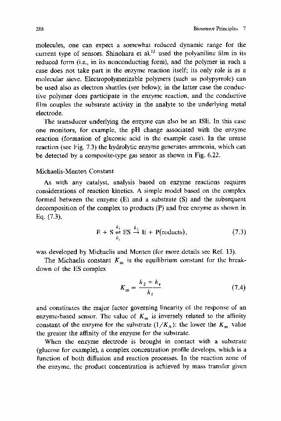
Michaelis-Menten Constant
As with any catalyst, analysis based on enzyme reactions requires considerations of reaction kinetics. A simple model based on the complex formed between the enzyme (E) and a substrate (S) and the subsequent decomposition of the complex to products (P) and free enzyme as shown in
was developed by Michaelis and Menten (for more details see Ref. 13). The Michaelis constant Km is the equilibrium constant for the break
down of the ES complex
and constitutes the major factor governing linearity of the response of an enzyme-based sensor. The value of Km is inversely related to the affinity constant of the enzyme for the substrate (l/KA): the lower the Km value the greater the affinity of the enzyme for the substrate.
When the enzyme electrode is brought in contact with a substrate (glucose for example), a complex concentration profile develops, which is a function of both diffusion and reaction processes. In the reaction zone of the enzyme, the product concentration is achieved by mass transfer given
Eq. (7.3),
E + S ^ E S ^ E + P(roducts), (7.3)
(7.4)

7.2 Enzyme Electrodes 289
by Fick's second law as well as by enzyme kinetics. Fick's second law is
„ 82P(x,t) SP(x,t)
D>-sS- = — « Γ (7·5)
and pertains to the change of product Ρ with time and distance x, Dp is the product diffusion coefficient. Diffusion and reaction have opposing effects on product concentration, and a steady state is reached when the rate of change of product formation becomes zero. When the substrate concentration is so high that all the enzyme is present as ES, then the rate of the reaction is maximal, and this velocity, F m a x, has the value A:2[E]. After some simple manipulations one can then derive that
9*P(x,t) -Vmax[S] P - £ ^ ~ " ^ ~ M ' ( 7 - 6 )
where V is the reaction rate (d[P]/dt), [S] is the substrate concentration, and χ is the distance from the reaction front. This equation relates product formation rate (K), substrate concentration ([S]) and the Michaelis constant Km.
Because of the steady state, enzyme electrodes can achieve stable, pseu-doequilibrium, readings despite an ongoing enzymatic reaction. The key point to remember from Eq. (7.6) is that at low substrate concentrations, where [S] Km, the reaction rate, and consequently the measured reaction products on an electrode, is linearly related to the substrate concentration.
Equation (7.6) was actually derived for enzyme reactions in a solution. But in a sensor the situation is somewhat different. In this case the only means by which enzyme and substrate molecules can interact in a sensor is by diffusion of the latter into the insoluble matrix containing the enzyme. Consequently, with an excess of enzyme present, diffusion will be the rate-limiting step. This has the beneficial effect of increasing the linear range of the relation between substrate concentration and reaction rate
14 in
Eq. (7.6). For a potentiometric sensor, assuming high enzyme loading and a substrate concentration below or about equal to the Km of the enzyme, the product concentration at the sensor surface will be proportional to the bulk concentration. With an amperometric sensor, product Ρ (e.g. H 20 2 in the case of glucose) is electrochemically consumed, and although the surface concentration is thereby reduced to zero, the product flux (which determines the response) can be similarly related to the bulk concentration of S (glucose in the example). So in order to achieve operational stability, one

290 Biosensor Principles 7
uses an excess of enzyme. Under these conditions the response is observed to be independent of enzyme concentration, less susceptible to variables such as pH, temperature and inhibitors, the sensor lifetime is longer and the response is linear as long as [S] < Km (rather than [S] < Km on the basis of (7.6)). However, for [S] > Km response is independent of bulk substrate concentration. With low enzyme loading, the rate of the reaction is governed by enzyme kinetics (see Eq. (7.6), and the situation would resemble the solution case. Under these circumstances the response would depend on the enzyme concentration, and a linear response would only be possible when [S] < 0.1Km.
Enzyme Immobilization
Historically enzyme electrodes have been based on enzymes immobilized in thick layers, and this leads to sluggishness in response; 5-10 minutes to reach equilibrium in a stagnant solution is not unusual. The most rapid response is obtained with maximal enzyme activity (pH optimum, zero-order kinetics), a thin layer (to avoid diffusion restrictions) and rapid stirring of the solution. The way by which the enzymes are immobilized is crucial. Stability of enzymes is closely related to the way they are immobilized. A thin layer, ideally a monolayer, of active enzyme molecules will be able to respond quickly, but will not give a high sensitivity. With such a thin layer there will be very little diffusion restriction, but not having a large amount of enzyme will reduce the sensitivity. There is thus a trade-off between thickness of layer and response time on one side and sensitivity on the other.
A commonly employed procedure for immobilizing the enzyme is to physically restrain the enzyme at the transducer surface by entrapment in polymer matrices such as polyacrylamide or agarose, or by retention with a polymer membrane comprising cellophane, cellulose acetate/nitrate, polyvinyl-alcohol or polyurethane. Chemically cross-linking the biocatalyst with an inert, generally proteinaceous, material with a bifunctional reagent to form intermolecular bonds between the catalyst and the inert protein is another possible means of immobilizing the biocatalyst. In another example, enzymes have been attached with gluteraldehyde to a nylon mesh. The nylon mesh with the immobilized glucose oxidase gave a robust membrane for covering a standard platinum electrode.
15 As discussed earlier, yet
another novel way of immobilizing enzymes on an electrode is to elec-tropolymerize polymers such as polyanaline or polypyrrole from the monomers in the presence of the enzyme.
12

7.2 Enzyme Electrodes 291
Finally, a preferred procedure in some instances is to covalently attach the biologically sensitive material directly to the surface of the transducer and thereby achieve intimate contact without incurring diffusional limitations in a membrane. The covalent procedure will, as indicated above, lead to improved response times but also, in many cases, a more limited sensitivity due to the smaller concentration of bioactive substance. Continuing along those Unes, in Section 8.6 we will briefly discuss that one monolayer of active sites might constitute a problem for the dynamic range of the sensor. Moreover, if one works in a regime other than diffusion limited, the analytical results are often very complicated and irreproducible. For example, in Chapter 11 we will see that for solid electrolyte gas sensors operated in the current mode no diffusion regime is normally established (in air D is typically « 0.1 cm
2 sec
- 1) . A diffusion barrier is created on
purpose in this solid electrolyte sensor by interposing a porous ceramic in order to get simple analytical results (i.e., a limiting current proportional to the gas to be analyzed).
With potentiometric as well as amperometric sensors the response time is thus dependent on membrane thickness. In fact, with membranes 0.3 to 0.6 mm thick, response times range from 3 to 6 min. With enzymes directly immobilized on the electrode and protected with a thin protective outer membrane, the time required to reach steady state can be reduced to about 25 sec. In the latter case the thin protective membrane constitutes the main diffusion barrier (D « 10"
7 cm
2 s e c
-1 is a typical value for a diffusion
constant in a membrane, compared with 10"5 cm
2 s e c
-1 in an aqueous
solution). Some of the outlined immobilization schemes were schematically shown
in Fig. 7.2.
Cofactor Dependence
Oxidase and hydrolytic-type enzyme reactions (see Fig.7.3) are relatively simple systems, since there is no cofactor dependence. Actually, in the case of oxidases, 0 2 is considered the cofactor (also called the coreactant) and is usually supplied by the environment. Approximately 50% of all known enzymes are cofactor dependent. It is difficult to coimmobilize low-molecular-weight cofactors at an electrode surface with the primary enzyme or enzymes.
16 There are a few possible solutions to avoid the problem of the
need for coimmobilization of the cofactor. One is to covalently bind the cofactor to the enzyme. This has proven to be quite difficult in practice. Also, direct coupling of the enzyme to the electrode can be attempted so

292 Biosensor Principles 7
that electron transfer between the immobilized enzyme and the electrode can proceed directly.
17 In such a system the enzyme is regenerated by the
electrode rather than by the cofactor. Living cells offer a complete system for retention and regeneration of coenzymes, which means living cells can also be exploited in coenzyme-dependent analyses (see Section 7.3). Finally, mediators (electron shuttles) can be used, which we will discuss in more detail below.
Although the future of enzyme-based biosensors is very promising, three major problems remain: cofactor dependence, stability and commercial availability of enzymes. Consequently, the number of enzymes used today in constructing biosensors is rather limited. This is mainly due to the fact that few enzymes are commercially available in a purified state.
We will now examine one way of solving the problem of cofactor dependency, namely, by the use of electron shuttles.
Electron Shuttles
Electron shuttles can be used to avoid cofactor dependency. As an example, we will use the glucose oxidase where the cofactor is basically oxygen. Therefore, the reaction in Fig. 7.3 can also be written as
(see also Fig. 7.4a). In this reaction the enzyme is regenerated by oxygen from the environment. Amperometric biosensors such as the glucose sensor frequently are based on the consumption of oxygen. This makes these particular sensors sensitive to the oxygen content in the sample. The glucose sensor in Fig. 7.3, for example, can only be used to detect glucose in concentration ranges from 10"
5 to 10 ~
2 M L
1. But the limiting factor
with the oxidase-catalyzed reaction in this sensor, as with most oxidase-catalyzed reactions, is the oxygen availability (in a conventional assay only 0.25 mM oxygen is present). The lack of oxygen often necessitates sample dilution.
One solution to this problem is to generate oxygen through the electrolysis of water so that enough oxygen can be produced for the enzyme-catalyzed reaction to proceed.
1 8 1 9 , 20 Also by a clever choice of an outer
membrane, the uptake of glucose and oxygen can be regulated so that the 0 2 supply is always in relative excess.
21
The approach we want to discuss here, however, is the use of electron shuttles, also called acceptors or redox mediators, which are chemical
Glucose 4- GOD( o x) -> gluconic acid + GOD( GOD( r e d) + 0 2- G O D ( o x ),
' ( r e d ) '
(7.7)

7.2 Enzyme Electrodes 293
species that are reduced more rapidly than oxygen. Because they often show low solubility in aqueous media, they can be retained in close proximity to the electrode for extended periods of time. Moreover, they can be recycled on the electrode: for example, with bis(cyclopentadienyl)iron as the electron shuttle (replacing the enzyme cofactor, which is oxygen in this case) we can write the following reac t ion :
2 2 2 3 24
Glucose + GOD( o x) -> gluconic acid 4- GOD( r e d ), ( l ) .
G O D ( r e d) + 2Feep2R+-+ GOD( o x) + 2Fecp2R + 2 H
+, (2)
2Fecp2R ^ 2Fecp2R++ 2e~. (3) (7.8)
The enzyme is regenerated in reaction (2) by the Fecp2R+ rather than by
the oxygen. The protons freed in the same reaction are neutralized by the buffering capacity of the medium. So, the above constitutes an oxygen independent glucose sensor. Such sensors have already been used as subcutaneous glucose sensors in pigs.
2 5'26 Hence, when the glucose concentration
is very high, redox mediator-based glucose sensors might present a solution. Figure 7.4 illustrates the operation of a classical glucose sensor and
various modes of mediated electron transfer to an electrode. For example, mediators such as benzoquinone, polyviologen, chloranil, methylene blue and ferrocene derivatives have all been utilized to shuttle electrons from the enzyme to a suitable electrode material.
1 Detector surfaces may therefore be
modified with redox mediators that communicate chemical oxidation changes at the sensor/analyte interface. Especially mediators such as polyvinyl-ferrocene and methylviologen, which may be applied as a film or as electroactive molecules chemically linked to the surface, are desirable. The use of conductive polymers, such as polypyrrole, as electron shuttles was commented upon earlier. The coupling of these various types of films to an enzyme or enzyme-labeled bacterial antigen (see below) results in changes in the film that are related to the substrate activity in the analyte. J. J. Kulys
27 studied enzyme electrodes with organic metals as possible
candidates for direct electron transfer enzyme electrodes. Such mediator-free mechanisms involve electron exchange in the absence of kinetically mobile mediators at organic compounds possessing metallic conductivity at room temperature (e.g., TTF
+TCNQ and TTT
+TCNQ~ complexes of tetrathia-
fulvalene and tetrathiatetracene with 7,7,8,8-tetracyano-/?-quinodimethane with room temperature conductivities > 200 (Ω c m )
- 1) . Besides being very
good electronic conductors, these electrodes can be made catalytically active, which enables such a direct electron transfer.

294 Biosensor Principles 7
WE
Λ
H 20 2
WE
i Θ
.0 1 Red
Λ
"Ox
(a) GLUCOSE SENSOR (b) ' R e d '
FORM OF FREE MEDIATOR
WE
I 1Θ
Θ
WE
Θ I I Θ
MEDIATOR IMMOBILIZED IN OR ON A POLYMER MATRIX (e.g. POLYVINYLFERROCENE)
(c) IMMOBILIZED MEDIATOR
CATALYTIC WE ELECTRODE (e.g. ORGANIC METAL)
(d) DIRECT METAL COUPLING
Figure 7.4 Operation of a classical glucose sensor and various modes of mediated electron transfer to an electrode. WE = working electrode; E(nzyme) = enzyme catalyst, e.g. glucose oxidase; S(ubstrate) = e.g. glucose; P(roduct) = e.g. gluconic acid and hydrogen peroxide; M(embrane)
Enzyme sensors have been developed for a wide variety of chemicals ranging from C O
28 to acetylsalicylic acid (aspirin).
29 Because we mainly
focus on mechanisms, we leave it to the reader to consult the literature for other examples.
7.3 Cell-Based Sensors (Microbial Sensors)
The microbial sensor consists of immobilized, living, whole cells and an indicator electrode (see Fig. 7.1). When the microbial sensor is inserted in a sample solution containing growth-promoting organic compound (the analyte), the organic compound (S) diffuses toward the microbial cells, where it

7.3 Cell-Based Sensors (Microbial Sensors) 295
is taken up. Consequently, the respiration (oxygen uptake during growth) of the microbial cells rises, causing the amount of oxygen diffusing toward an underlying oxygen probe (typically a Clark oxygen probe) to decrease. Metabolites (e.g., C 0 2 or NH3) can also be monitored.
In Fig. 7.5 a possible assembly of a microbial sensor is shown. In this particular case a differential-type setup is used. Instead of isolating enzymes from bacteria, the whole culture is immobilized on an electrode surface in a buffered medium, and is isolated from the analyte by a permeable membrane (see Fig. 7.5). The response time tends to be longer (10-20 min) than for enzyme electrodes (typically 1-2 min or shorter) and the sensor lifetime is between 2 and 3 weeks. With cold storage (4°C) a shelf life of 20-30 days can be achieved. Interferences from intermediate metabolic products are
Ag/AgCl
MATRIX WITH CELLS MATRIX WITHOUT CELLS
Figure 7.5 A possible assembly of a microbial sensor for respiratory monitoring Clark oxygen electrodes are used as shown here and for metabolite monitoring one can use, for example, an ammonia-selective electrode

296 Biosensor Principles 7
frequent because of the presence of a multitude of metabolites and enzymes.
30 When one is trying to avoid the cofactor dependency of enzymes,
using whole cells is a good solution, as we have seen in the preceding sections, since cells already contain cofactors and other necessary ingredients in an optimized environment. Intact cells therefore offer optimal conditions for the maintenance of biological activity of enzymes. Cells can also be used where appropriate enzymes are not known or not available. Moreover, cells are very low priced compared with isolated purified enzymes. Tissue-slice electrodes and membranes sectioned from various plants or organs are similar to bacterial electrodes in construction. In the tissue electrodes a thin layer of a tissue is fixed to the internal indicating system, which senses the products of enzymatic reactions of the substrate taking place in the tissue layer.
Another important benefit of microbial sensors is that often the dynamic range (see Section 8.6) is much larger when the bioaffinity molecule used is within a whole cell structure. For example, it has been found that an isolated receptor responds in a concentration range of only about two or three orders of magnitude. With a microbial sensor, where the receptor is in a cell, the dynamic range of a chemoreceptor is much larger. Several theories of receptor action state that the rather limited dynamic range of a single receptor is a result of the saturation of the receptor binding sites. As we will discuss in Section 8.6, a possible explanation for a large dynamic range despite a limited number of active sites is that there might be a range of available sites with different KAs that are active in different concentration ranges. Belli et al.
31 give another explanation for receptors. Their
model is based on chemoreceptors with various shapes within one cell. The sensitivity of receptors, they argue, is dominated by the extent of dentritic branching of the receptor, and within one organism there will be a population of chemoreceptors with differing branching and sensitivities. The higher the number of branches the higher the probability for an excitation of the chemoreceptor, and with more branches the analytical region will consequently be shifted to a lower concentration region (fewer molecules are needed to give a signal).
Several microbial sensors, such as alcohol, acetic acid, and BOD (biochemical oxygen demand), are being commercialized in Japan and applied to fermentation processes and environmental analyses. Some examples of microbial sensors are listed in Table 7.4. Often they are based on an amalgamation of an enzyme reaction and a bacterial metabolism (so-called hybrid systems).

7.4 Immunosensors 297
Analyte Biorecognition System Transducer Electrode
Biochemical oxygen demand Trichosporon cutaneum 0 2 (BOD) (immobilized yeast)
Creatinine in serum and Creatinase + Nitrosomonas 0 2 urine sp. and Nitrobacter sp.
Urea (NH3) Urease + Nitrosomonas sp. o2 and Nicrobacter sp. Glutamine Escherichia coli co2 Aspartase Bacillus subtilis 0 2 α-amylase Glucoamylase and o2 Bacillus subtilis CH4 Methylomonas flagellata o2 Glucose P. fluorescens 0 2 Ethanol T. Brassicae 0 2 Vitamin Lactobacillus arabinosus 0 2 Cepharosepollin C. fruendii H
+
Mutagen Bacillus subtilis Rec~ o2
7.4 Immunosensors
Immunoassay
In an immunoassay one measures the concentration of either an antibody or an antigen. Homogeneous immunoassays were developed in the 1970s and quickly constituted a large market. Various antigens including proteins, peptides, drugs, and microorganisms are identified by immunoassays. Test kits for analyzing hormones, proteins, pharmaceuticals and others are used as standard tests in today's clinical laboratory. In the near future, the cost of monoclonal antibodies should decline to a point where their use in sensors becomes attractive.
32 Hybridoma technology has stabi
lized the reagent source (i.e., the monoclonal antibodies), thereby ensuring the uninterrupted generation of antibodies by continuous cell culture.
33
Although the discussion in this section is on immunosensors, the same reasoning can be used for any of the affinity pairs in Table 7.2.
Immunoreactions result in the formation of antigen-antibody complexes. The amount of agglutinated complexes is measured by turbidimetry (some antigens are as large as several micrometers) or by weighing after centrifu-gation. When the amounts are too small to be detected by these methods, labels are used. Labels can be radioisotopes, electrochemical active species
Table 7.4 Some Examples of Microbial Sensors

298 Biosensor Principles 7
(ions, redox molecules, ionophores), fluorophores, chromophores, particles, liposomes (vesicles with a BLM) and enzymes (catalysts). Both the enzyme method and the liposome label, as we will explain later, can lead to significant amplification of the signal. On the basis of those two types of immunoassays, nonlabeled and labeled, a variety of immunosensors have been proposed, which are classified as follows:
34 nonlabeled immunosen
sors: transmembrane conductance, electrode potential, piezoelectric, po-larographic, FET, surface plasmon, capacitance; labeled immunosensors: enzyme-linked amperometry, enzyme-linked potentiometry, enzyme-linked luminescence, liposome-linked luminescence, electrochemical luminescence, other optical immunosensors.
In a classical (homogeneous) immunoassay the sample containing the antigen is first mixed with a fixed amount of labeled antigen (e.g., a radioisotope). Since a fixed amount of labeled antigen is competing with varying amounts of native antigen for the same antibody, it is possible to construct a calibration curve, which is used to evaluate the content of native antigen in unknown samples. The experimental procedure involves several additions of reagents, incubation times and washing steps. It is important to be able to separate bound from free antigen before reading. The label facilitates the detection of a binding event.
The agglutination reaction proceeds slowly in certain cases. Sometimes it requires 1-2 days to complete the reaction. Therefore, the acceleration of the agglutination reaction is sometimes required for more rapid immunoassays and practical sensing devices. The more frequently that the antibody and antigen encounter each other, the higher is the complex formation rate. Electric pulses are considered promising for the increase of contact frequency between antigen and antibody and, consequently, for increasing agglutination rate. The idea was tested successfully for the measurement of Candida albicans (a pathogenic yeast).
35 In some cases it is possible to run
an assay, using a flow system, without any other acceleration means of the complex formation rate in about 6 min. The same times are expected to be needed in an immunosensor. The long time most current immunosensors require is, in general, still a major disadvantage compared with enzyme sensors.
Frequently antibodies are immobilized to a solid support in order to make washing easier. The washing is needed to get rid of the unbound labeled antigen, which would otherwise also lead to a signal but not in proportion to the amount of native antigen. Immobilization on a solid substrate (e.g., a metal electrode) is really the only practical way to develop

7.4 Immunosensors 299
ANTIGEN (DETERMINANT
H 20 2
Figure 7.6 An immunoelectrode based on catalase-labeled antigen (Ref. 2)
an immunosensor. But the immunosensors developed at the present time often are not much more than the traditional immunoassays, the new feature being that the reaction is localized at the tip of a transducer. Such a sensor is shown in Fig. 7.6. The exact mode of operation of this particular sensor is explained later. It still involves the addition of labeled antigen and washing of the sensor before introducing it in a substrate solution.
The ultimate goal for a "true" immunosensor is to have a device in which the binding event is read directly, and which is capable of continuous monitoring without the need for added reagents, transfer of the probe from one to another solution or washing. Because of the need of washing away the unbound labeled antigen, a nonlabeled system would be preferred. Some examples of direct measuring methods with nonlabeled immunosensors (i.e., methods with no need for added reagents or washing) that have been attempted are ellipsometry, reflectometry, evanescent wave, streaming potential measurements, ImmFET and piezoelectric systems. None of these recently attempted analytical techniques have yet led to commercial products. The ImmFET device will be described in Chapter 9. Janata and Blackburn
36 calculated that if 10
12 molecules c m
-2 of antibody
with a binding constant of KA = 108 L M
-1 could be immobilized on an
insulated gate of a FET, a theoretical detection limit should be around

300 Biosensor Principles 7
10~12 mol L "
1. Unfortunately, the device does not work properly because
the macromolecules are not the only ones setting up a signal at the gate of the FET. Continuous sensors require that the AbAg complex formation be reversible. However, this may not always be feasible, especially with a very high KA (see Table 7.2). Recall that as the affinity constant decreases, the sensitivity is sacrificed.
We will discuss some electrochemical immunosensors involving interesting amplification schemes next. Amplification is not limited to electronic devices; nature provides some fascinating chemical amplification schemes that can be used to enhance sensitivity in sensors. There are several ways of amplification of biosensor response. We discuss three of them: enzyme, liposome and BLM. Because of its emerging importance, the BLM amplification scheme is treated separately in Section 7.5.
Enzyme Immunoassay
The first type of chemical amplification occurs in an electrode-based enzyme immunoassay, analogous to the well-known radio immunoassay. Here enzymes replace the radioisotopes as labels. The enzyme label on the antigen must have a high turnover rate and, therefore, must be a good catalyst, influencing a reaction that produces or consumes an ion or molecule that can be measured by an electrochemical sensor. The principle can be understood with the help of Fig. 7.6, representing an immunosensor developed by Aizawa.
2 Typically an antigen (the same antigen identified in
the sample solution) is labeled with an enzyme (catalase in this case) and added to the unknown sample in which the sensor is placed. The labeled antigen competes with native (unlabeled antigen) for reaction with the antibody, which is immobilized on an electrode surface (here a polymeric membrane). The ratio of labeled versus unlabeled antigen binding to the membrane surface is determined by the original concentration of native antigen; this means that a calibration curve can be made up if the concentration of the labeled antigen on the surface can be determined. This determination is done as follows. After the adsorbed but unbound labeled antigen is washed off, the probe is brought in contact with the substrate for the enzyme ( H 20 2 in this case). The catalase enzyme decomposes the H 20 2, and the oxygen formed diffuses through the membrane to the internal Clark oxygen probe (see Fig. 7.6). The oxygen current decreases with increasing concentration of the nonlabeled native antigen in the sample solution. The enzyme reaction will produce many detectable species per bound antigen-antibody pair, hence the name "enzyme amplification." The method just

7.4 Immunosensors 301
described is called the competitive method, as opposed to the sandwich method. We leave it up to the reader to compare the details of these two techniques with the help of Fig. 7.7.
Chemical amplification as described here is especially effective in ultra-trace analysis—analysis where there are extremely low concentrations of the species to be detected. Theophylline (a drug used in the treatment of asthma), for instance, was determined to concentrations of 5 X 1 0
- 8M with
an immunosensor37 and, as indicated earlier, theoretically 10~
1 2M is possi
ble. Also, an iodide sensor that detects a decrease in iodide concentration resulting from oxidation by peroxide (a peroxidase was used as the enzyme label) was used to detect hepatitis B.
38 Another example is determination of
cyclic adenosine monophosphate (C-AMP) and bovine serum albumin (BSA) using urease as the enzyme label with an ammonia gas-sensing electrode.
39
Drug electrodes can be made on the basis of enzyme-linked immunoassays as well. Measurements of drug concentration in the blood is important because therapeutic efficacy and toxic side effects are linked to dose levels. Drugs are usually small molecular species that tend to be therapeutically active in the micromolar to nanomolar concentration range. An amperometric immunoelectrode is capable of combining the sensitivity of an enzyme electrode with the specificity of an AgAb interaction. For further description we follow the work by Hill et al .
2 2'23 A ferrocene drug (lidocaine)
conjugate was first synthesized (Fig. 7.8). The modified ferrocene retained its ability to act as an electron acceptor for glucose oxidase, and the conjugate was recognized by antibodies raised against that drug. The binding of antibody to the ferrocene drug conjugate specifically inhibits its ability to act as a mediator. This inhibition can be reversed by addition of a free drug. In the presence of a free drug in the sample, a competition will occur between the ferrocene drug complex and the drug for the antibody. The more drug that is present in the sample, the less the ferrocene drug complex will be bound and the greater the catalytic current will become. As a result, a competition assay for the drug arises, with the difference in current reflecting the amount of drug present. Figure 7.8 schematically depicts how the drug sensor operates.
Liposome Amplification
A second type of chemical amplification uses direct chemical release from liposomes rather than indirect enzyme reactions. Liposomes are artificial vesicles that consist of concentric shells of lipid bilayers whose aqueous interspaces can contain trapped markers. Liposomes are loaded

302 Biosensor Principles
ANTIGEN
(a) COMPETITIVE METHOD
ANTIBODY
(b) SANDWICH METHOD
Figure 7.7 Principle of Labeled Immunosensors

7.4 Immunosensors 303
N H C O C H 2N ( C 2H 5) 2
FERROCENE DRUG CONJUGATE
Ag.
Ab
ELECTRODE
2 e _ J
Fc-Ag
Fc
+-Ag
GOD
• AgAb
Fc-AgAb
> GLUCOSE
k GLUCONOLACTONE
Figure 7.8 Schematic representation of a homogeneous amperometric immunoassay. Ag refers to the antigen (in this case, lidocaine); Fc-Ag is the ferrocene-drug conjugate; Ab is antibody to lidocaine (Ref. 22, 23)
with an easily detectable, nonphysiologic ion (e.g., trimethylphenylammo-nium, marker ions). The liposomes naturally possess specific antigenic determinants on their surfaces. In the presence of a protein complement, the adduct AgAb evokes a series of reactions resulting in the lysis of the liposome (see Fig. 7.9). A complement is a system of nine serum proteins that react in a specific order to produce lysis of target cells. In this way the markers are released in high concentration and, when trimethylphenylam-monium marker ions are used,
40 are detected by an NH4-selective elec
trode. Alternatively the liposome could also be loaded with other markers such
as chromophores and enzymes. In the case of enzyme loading, one really

304 Biosensor Principles 7
^ - A N T I G E N
(a) I M M U N O L O G I C A L R E A C T I O N (b) L Y S I S BY C O M P L E M E N T (c) E N Z Y M A T I C R E A C T I O N
Figure 7.9 Principle of Liposome Immunosensor
has a double amplification scheme (i.e., liposome and enzyme amplification). Haga et al .40 relied on such a scheme to detect theophylline; a detection of 4 X 1 0 - 9M theophylline was possible with this method compared with 5 X 10~8M with an enzyme immunoassay method. If both complement and liposomes were easier to stabilize, this technique would be very promising for the development of immunosensors.
Another way for chemical amplification is the chemical gating of BLM channels as in nerve synapses. In the latter case, reaction of acetylcholine with a receptor causes a temporary opening of a channel, with a resulting flux of ions through the membrane. A detailed discussion follows next.
7.5 Very Thin Membranes—Bilayer Lipid Membranes—Langmuir-Blodgett Films
Our previous discussions involving membranes (Chapter 6) were concerned with rather thick (e.g., 40-500 /xm) artificial membranes. Naturally occurring biological membranes are usually much thinner (e.g., 6-9 nm) and consist of three types of substances: lipids, proteins and carbohydrates. Lipid molecules are by far the most numerous and are responsible for the structural integrity of biological membranes. Two types of lipid molecules

7.5 Very Thin Membranes—Bilayer Lipid Membranes—Langmuir-Blodgett Films 305
LIPID MOLECULE
Ο HYDROPHILIC HYDROPHOBIC
END END
Figure 7.10 Lipid mosaic model of a cell membrane
are present: cholesterol and phospholipids. Both are amphiphilic (i.e., molecules possessing both a hydrophobic and a hydrophilic end; see Fig. 7.10).
Organisms with such membranes are able to sense a large variety of biochemicals even at the picomolar level. They employ specialized, chemically selective binding sites, called receptors, which can interact with particular stimulants at the cell surface. This interaction can generate a sudden measurable inorganic ion flux across the cell membrane. Attempts to mimic the ability of natural cells to monitor chemicals were reported as early as the 1960s.
41 And Thompson et a l .
4 2 , 43 have been guided by the
natural processes involved in olfaction (smell) and gustation (taste) to develop very thin membrane biosensors. The electrophysiology of olfaction and gustation is thought to involve a change (increase) of ionic conduction across a sensory membrane following a chemoreceptive selective binding event at the membrane surface. So Thompson and co-workers and other research groups have been making artificial BLMs for incorporation in electrochemical sensors, as shown in Fig. 7.11.
All the biorecognition schemes listed in Table 7.2 are part of the arsenal at the disposal of living cells. Since a living cell obviously is the ultimate sensor, there is a lot of interest in making artificial systems that resemble the structure of cell membranes and in studying the electrochemical pro-

306 Biosensor Principles 7
(b)
Figure 7.11 Schematic diagram of a conductometric LB sensor (Based on Refs. 41, 49, and 50)
cesses on those membranes to provide models for the development of new electrochemical sensors.
43
The most convenient way of artificially making thin lipid films is by using the Langmuir-Blodgett (LB) technique.
Langmuir-Blodgett Technique
The LB method enables the layering of organic films layer by layer onto solid substrates. Amphiphilic molecule layers are formed on a water surface and then transferred onto a substrate (see Fig. 7.12). Molecules stick to the hydrophilic substrate via their hydrophilic head and make the surface hydrophobic. When the substrate is dipped down in the Langmuir trough (see Fig. 7.12), a second layer attaches, with its hydrophilic head sticking

7.5 Very Thin Membranes—Bilayer Lipid Membranes—Langmuir-Blodgett Films 307
DEPOSITION (1st LAYER)
DEPOSITION (2nd LAYER)
DEPOSITION (3rd LAYER)
Figure 7.12 The Langmuir sequence
out, turning the surface hydrophilic again. The two layers form a bilayer. The repetitive procedure gives rise to a multilayered structure in which the polar heads of two adjacent bilayers are paired to form hydrophilic double planes. For an excellent review on all type of applications of LB films see Roberts,44 and for the theory and practice of BLMs, see Tien.45 For a more specific paper on chemical sensor applications of LB films, see Reichert

308 Biosensor Principles 7
et al .46 Here, we will concentrate on sensor applications of LB films. A
schematic diagram of a Langmuir trough is shown in Fig. 7.13. Langmuir-Blodgett films exhibit very good adherence, and they are very
regular, with a very low density of defects and holes. Despite the mechanical fragility of the LB films, they have many projected sensor applications. The biggest redeeming factors are the very fast response time expected for these thin organic films, the versatility in electronic properties (from insulating to semiconductive, depending on the types of molecules used) and the wide choice of applicable molecules (biological and nonbiological in nature). There is also the fact, as pointed out earlier, that many sensing schemes of living cells might be mimicked with such films. Finally LB-based sensors are not subject to the competitive effects that limit ISE (see Chapter 6), since the development of an equilibrium interfacial potential is not the source of the signal in this case,
47 so better selectivities can also be
expected. Various means to detect chemical changes on LB films are feasible, and most of them have been proven to function in the lab; they involve optical changes induced in the films, dielectric changes (in FET configurations; see Chapters 8 and 9) and electrical changes such as ac impedance, dc voltage, dc current and dc resistance as well as weight changes (e.g., as measured on a surface acoustic wave device, SAW devices). The electrical changes can be measured across or in the plane of the LB film.
We will concentrate in this section on electrical changes induced by chemical reactions on thin lipid films. Not all of the thin films mentioned in this section are made by LB techniques, but all of them are either bilayer lipid membranes (BLMs) or are very thin compared to the membranes discussed in Chapter 6.
One often studied system with biological-type thin phospholipid films is shown in Fig. 7.11a. In this configuration the transverse electrical impedance of thin phospholipid films suspended over an orifice in a hydrophobic support separating two aqueous solutions, is monitored. The technique by Mueller et al .
48 is used for the formation of the BLMs over the orifice. In
this technique lipid molecules are dissolved in an organic solvent and introduced in the orifice by using a fine-hair brush. The solvent diffuses into the aqueous electrolyte, leaving a thin lipid membrane that appears black. The membrane has, because of its black appearance, appropriately been given the name of black lipid membrane (with the same abbreviation BLM as for bilipid membrane).
Immunological or enzymatic reactions involving proteins affixed to or embedded in BLMs have been shown to induce temporarily electrical

Very Thin M
embranes—
Bilayer Lipid Mem
branes—Langm
uir-Blodgett Films

310 Biosensor Principles 7
κ κ
+
(a)
Κ
+κ
+
N a
+
N a
+ N a
+ iill (b)
Figure 7.14 Schematic illustration of transmitter-receptor interaction mechanism
changes in the lipid membrane.41 Supposedly the interaction between a
membrane-bound receptor and a stimulant or transmitter results in a transmembrane ionic flux. This mechanism is illustrated schematically in Fig. 7.14. Here we show how the binding of a transmitter to a receptor embedded in the lipid membrane causes a channel, next to the receptor/transmitter pair, to open up. The channel allows small cations to cross the membrane layer. Because of this ionic flux, the impedance of the membrane changes dramatically. The described effect is usually transient, though. There is a wide range of substances (see Table 7.2) that will induce conductance changes in BLMs, but to date the detailed mechanisms are poorly understood:
(a) In the case of antigen and antibody additions to the electrolyte on one side of the cell shown in Fig. l ia , a dramatic, but transient, impedance reduction of the membrane is observed regardless of the order in which the two substances are applied. If the antigen and antibody are mixed before
7.5 Very Thin Membranes—Bilayer Lipid Membranes—Langmuir-Blodgett Films 311
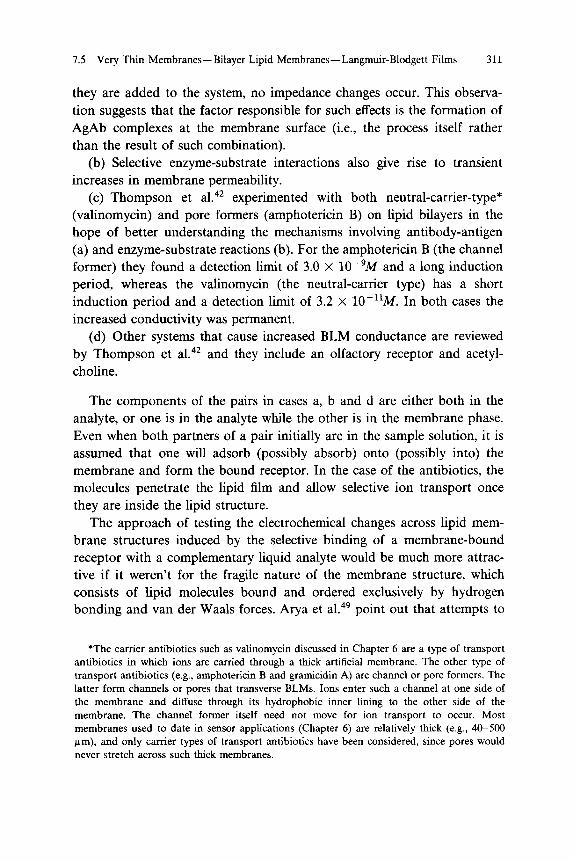
they are added to the system, no impedance changes occur. This observation suggests that the factor responsible for such effects is the formation of AgAb complexes at the membrane surface (i.e., the process itself rather than the result of such combination).
(b) Selective enzyme-substrate interactions also give rise to transient increases in membrane permeability.
(c) Thompson et al .42 experimented with both neutral-carrier-type*
(valinomycin) and pore formers (amphotericin B) on lipid bilayers in the hope of better understanding the mechanisms involving antibody-antigen (a) and enzyme-substrate reactions (b). For the amphotericin Β (the channel former) they found a detection limit of 3.0 X 10~
9M and a long induction
period, whereas the valinomycin (the neutral-carrier type) has a short induction period and a detection limit of 3.2 X 1 0
- 1 1M . In both cases the
increased conductivity was permanent. (d) Other systems that cause increased BLM conductance are reviewed
by Thompson et al .42 and they include an olfactory receptor and acetyl
choline.
The components of the pairs in cases a, b and d are either both in the analyte, or one is in the analyte while the other is in the membrane phase. Even when both partners of a pair initially are in the sample solution, it is assumed that one will adsorb (possibly absorb) onto (possibly into) the membrane and form the bound receptor. In the case of the antibiotics, the molecules penetrate the lipid film and allow selective ion transport once they are inside the lipid structure.
The approach of testing the electrochemical changes across lipid membrane structures induced by the selective binding of a membrane-bound receptor with a complementary liquid analyte would be much more attractive if it weren't for the fragile nature of the membrane structure, which consists of lipid molecules bound and ordered exclusively by hydrogen bonding and van der Waals forces. Arya et al.
49 point out that attempts to
T h e carrier antibiotics such as valinomycin discussed in Chapter 6 are a type of transport antibiotics in which ions are carried through a thick artificial membrane. The other type of transport antibiotics (e.g., amphotericin Β and gramicidin A) are channel or pore formers. The latter form channels or pores that transverse BLMs. Ions enter such a channel at one side of the membrane and diffuse through its hydrophobic inner lining to the other side of the membrane. The channel former itself need not move for ion transport to occur. Most membranes used to date in sensor applications (Chapter 6) are relatively thick (e.g., 40-500 μιή), and only carrier types of transport antibiotics have been considered, since pores would never stretch across such thick membranes.

312 Biosensor Principles 7
overcome the mechanical fragility of BLMs have resulted in limited success so far. Some such attempts mentioned by these authors include membrane miniaturization to maximize surface effects by incorporation of lipid membrane systems in microfiltration paper and incorporation of stabilizing agents such as surfactants and polymers. Although these methods improve the membrane strength, the structural integrity and reproducibility required of a practical device still have not been achieved. The continuing improvements in LB technology now allow for films to be polymerized in situ, and one would think this is a logical way to improve the mechanical stability of BLMs. Unfortunately, as indicated before, the operation of the BLM is based on an variation of the inorganic ion current through the structure as a function of membrane perturbation caused by receptor complexation. It is not clear if polymerization of the LB film would compromise the desirable electrochemical properties because of the increased rigidity in the lipid film and/or because of the perturbation of the receptor incorporation.
For sensing on an LB film, a reservoir of ions with the appropriate indicator electrodes must exist on either face of the membrane, as indicated in Fig. 7.11a. A slightly more advanced structure with a functional BLM on a rigid ion-conducting hydrated polymeric substrate (e.g., on an agar hydrogel), which can act as one of the ion reservoirs, would take a significant step to a more stable "solid-state" chemical sensor.
4 9 , 50 Such a
structure is represented in Fig. 7.11b. Some positive results with such hydrogel-based BLM sensors were obtained by Arya et al.,
49 but the results
were not yet convincing enough to warrant confidence in this type of approach.
Another approach would be to deposit LB films on a solid substrate (e.g., on a mixed conductor such as I r02) , which should be easier to do without breaking the membrane. The I r 0 2 could be soaked with ions before deposition of the membrane.
A major problem in depositing LB films is the heterogeneity of surfaces in terms of hydrophobic and hydrophilic patches. To avoid heterogeneity and contamination, extremely clean surfaces and working environments are required during the LB depositions.
Thompson47 achieved selectivity for ammonia gas with a BLM incorpo
rating the antibiotic nonactin. The detection limit was found to be comparable to that of conventional ammonia gas-sensing electrodes, but the selectivity, as expected (see above), was much better (the lipid film was made with the hairbrush method). Langmuir-Blodgett gas sensors were also made on the basis of porphyrin and phthalocyanine films. These films,

Table 7.5 Comparison of Different Sensor Technologies
Detection Response Type of Limit Time Cost Multiple Who& Development
Method Analyte mol/L Selectivity min Sensor Instrument Use Where Time
D i r e c t
Potentiometric I Ions 1 0 "
6 Good < 1 Low Low Yes Corning, Exists
(ISE, CHEMFET) I Immunochemicals H T
7 Poor 1-20 Low Low Yes
Beckman, SRI Univ. Osaka Medium
A mperometric ( gases, ions \ neutral molecules H T
9 Fair < 0.5 Low Low Yes SRI Exists
Capacitive Immunochemicals μ g/ml Poor < 5 Low Low Yes Biotronics Medium Conductimetric ( Immunochemicals SRI (BLM) \ enzyme-substrates Ι Ο "
10 Fair < 2 Low Low Yes Univ of Utah Long
Chemiresistors enzyme-substrates Ι Ο "
6 Good < 5 Low Low Yes Ohmicron Medium
Optical ( Fiber Based ) Absorption Scattering
Fluorescence Luminescence
Evanescent
Surface Plasmon
ions, gases
ions, gases
1 0 "
7
10"
Interference"
Immunochemicals 10
9
Immunochemicals 10
9
Immunochemicals 1 0 "
9
Good
Poor
Poor
Poor
Poor
1-5
1-5
2 -10
2 - 5
2 -20
Medium High
Medium High
Medium High
Medium High
Low Low
Yes
Yes
Yes
Yes
Yes
NIH
CDI
Geneva Research Center
Short
Short
Long
Linkôping Institute of Technology, Sweden Long
Biostar SRI
Exists
Gravimetric Piezoelectric ( Gases Integrated Chemical
\ Immunochemicals 1 0 "
9 Poor 2-20 Low Low Yes Sensors Medium
SAW f gases Integrated Chemical \ Immunochemicals 1 0 "
9 Poor 0.5-10 Low Low Yes Sensors Medium
Nonfiber based sensor. hValues depend on the analyte, especially in the case of antigen-antibody reactions.
7.5 V
ery Thin M
embranes—
Bilayer Lipid Mem
branes—Langm
uir-Blodgett Films
313

Table 7.5 Continued
Detection'' Response'' Type of Limit Time Cost Multiple Who & Development
Method Analyte mol/L Selectivity min Sensor Instrument Use Where Time
I n d i r e c t
Potentiometric Enzyme Amplification Liposome Amplification
Gases ( Enzyme Substrate \ Antigen-Antibody
Antigen-Antigody ο ο
ο ο
Good Good Good
Good
1-2 1-4 5-10
< 10
Low Low Low
Low
Low Low Low
Low
Yes Yes No
No
Orion, Beckman Universal Sensors Univ. Delaware
Univ. Tokyo
Exists Exists Medium
Long
A mperometric
Enzyme 1 Enzyme Substrate ΙΟ"
5 Good 1-3 Low Low Yes YSI, Fuji
SRI Exists
Amplification
Liposome Amplification
I Antigen-Antibody
Antigen-Antibody
ΙΟ"
11
< 1(Γ
12 Good
Good
< 5
< 5
Low
Low
Low
Low
No
No
Univ. Arizona Univ. Tokyo
Univ. Tohoku
Short
Long
Optical ( Fiber Based ) Absorbance with
enzyme amplification Fluorescence with
no amplification Evanescent with
no amplification (Fluorescence)
' Enzyme Substrate , Antigen-Antibody ' Enzyme Substrate , Antigen-Antibody
Antigen-Antibody
io-
10"
Good
Good
Good
2-10
4-20
4-20
Medium High
Medium High
Medium High
No
No
No
Univ. Iowa
Univ. Tennessee
Geneva Research Center
Medium
Medium
Medium
Thermal Thermocouple/ Thermopiles Enzyme Substrate 10"
6 Poor
Thermistors ( Enzyme Substrate 10
6 Poor
\ Antigen-Antibody 10~
10 Poor
Pyroelectric'
1-5 Low Low Yes Univ. Arizona Long 1-5 Low Low Yes Univ. Lund Long < 10 Low Low No Univ. Lund Long
' No biosensor application known.
314 Biosensor Principles
7

Glossary of Biosensor Terminology 315
which were several molecules thick, exhibit resistance variations on exposure to gases such as N 0 2, CO and H 2S ,
51 often with quite fast response
times. Wohltjen et al.52 reported a response time for ammonia detection of
less than 30 sec for 45 layers of copper tetracumylphenoxy phthalocyanine (concentrations significantly below 0.5 ppm could be detected). In the structure used by Wohltjen et al.
52 planar electrodes microfabricated on a
substrate make ohmic contact to the semiconducting thin films and facilitate the measurement of very low conductances. Such a structure, discussed in Chapter 10, is called a chemiresistor. Roberts
53 used LB films on LEDs
and FETs. In FETs a phthalocyanine film was shown to respond to NOx at room temperature. The response time was reversible at room temperature and was less than 1 min.
In conclusion we show in Table 7.5 a comparison of different biosensor technologies.
Glossary of Biosensor Terminology
Acetylcholine: An important neurotransmitter produced by the vagus nerve.
Adenosine: A nucleoside composed of one molecule of adenine and one molecule of D-ribose, a product of hydrolysis of adenylic acid.
Affinity pair: The property of a substance that makes it more readily combine with or take up some substances than others.
Agarose: A polysaccharide present in agar and responsible for its gelling. It consists of residues of 3,6-anhydro-L-galactose (an ether oxygen links C-3 and C-6) and D-galactose. It is used as a medium for gel chromatography.
Albumin: Any of a class of water-soluble proteins. Antibody: A protein molecule formed by the immune system that
reacts specifically with the antigen that induced its synthesis. All antibodies are immunoglobulins.
Antigen: A substance that stimulates production of an antibody when introduced into a living organism.
Ascorbic acid: Also known as vitamin C. It can be synthesized by

316 Biosensor Principles 7
most mammals. It is easily destroyed by oxidation, heat and light. Biologically it acts as a coreductant in several oxidations that use molecular oxygen.
Aspartase: Aspartate ammonia-lyase: a bacterial enzyme that catalyzes the removal of ammonia from L-aspartate to form fumarate.
Avidin: A protein in egg white. It can bind a biotin molecule tightly (dissociation constant 1 0
- 1 5M ) to each of its
four subunits. Since it also binds biotin groups in enzymes, its ability to inhibit carboxylases and trans-carboxylases is used to detect their dependence on biotin.
Bilipid membrane: The structure found in most biologic membranes, in which two layers of lipid molecules are so arranged that their hydrophobic parts interpenetrate, whereas their hydrophilic parts form the two surfaces of the bilayer.
Biotin: A substance whose molecules consist of two five-membered rings, one a thiolane ring, the other formed by fusion of the side opposite the sulfur of this ring to an — NH —CO—NH— group. It reacts with ATP and carbon dioxide to convert one of its — N H — groups into —N(—COO—)—, and the carbon dioxide thus "fixed" is subsequently transferred to an appropriate recipient. It is a vitamin for many animals, including man.
BLM: Bilipid layer membranes or black lipid membrane. BOD: Biochemical oxygen demand. B SA: Bovine serum albumin. C-AMP: Cyclic adenosine monophosphate. Catalase: A heme-containing enzyme found in the microbodies
(peroxisomes) of animal cells that catalyzes the reaction 2 H 20 2 -> 2 H 20 + 0 2. Its iron central element is normally in the III state.
Chemoreceptor: A sense organ excited by specific chemical substances and changes in their concentrations (e.g., gustatory, olfactory, and carotid body receptors). Also called "chemoceptor" and "chemical ceptor."
Chloranil: C6H204C12. A red, crystalline benzoquinone that forms

Glossary of Biosensor Terminology 317
various colored compounds used as end points for spectrophotometry. In histologic staining it precipitates with calcium as a biréfringent yellow-brown salt.
Chromophores: The groups in a molecule that absorb visible or ultraviolet radiation. They are usually unsaturated groups like — C H = C H — , — N = N — , or — C ( = 0 ) — , and are particularly likely to give color when conjugated.
Coenzyme: An organic substance of relatively low molecular mass (usually less than 1 kDa) required for an enzymatic reaction. It often proves to be a substrate for one enzyme, which converts it into a form that is reconverted to the first by a second enzyme. It thus links two reactions, usually by transferring a group from one substance to another.
Cofactor: A heat-stable, nonprotein substance necessary for optimal activity of some enzymes. It may be an inorganic ion or a more complex organic material.
Creatinine: The product of cyclization of creatine by lactam formation. Creatinine output is proportional to muscle amount and very constant. Urinary creatinine is measured to check completeness of urine collection.
Dopamine: 3,4-Dihydroxyphenethylamine. An intermediate in the biosynthesis of epinephrine. It is produced by decarboxylation of dopa and can be converted into norepinephrine by dopamine /3-monooxygenase.
Effector: An organ that responds to nervous impulses either by movement (muscle, chromatophore), secretion (exocrine and endocrine glands), or release of an electrical discharge (electro-organ), as of an electric eel. Also called "effector organ."
Elastomer: A man-made material with elastic properties resembling those of rubber.
Enzyme: High-molecular-weight biocatalyst (protein) that increases the rate of numerous reactions critical to life.
Glycolipids: Any lipid that contains a carbohydrate group. Glycoproteins: A conjugated protein containing one or more carbohy
drate residue. The glycoproteins are important components of the plasma membrane, as well as of mucin and chondroitin.

Biosensor Principles 7
A substance that is unable to induce antibody formation but that can react with an antibody: applied especially to organic chemicals of low molecular weight. To raise antibodies to haptens, one must couple them to an immunogenic "carrier" molecule that can recruit a helper T-cell response. Also called "partial antigen" and "incomplete antigen." Also known as hap tin and haptene. (contraction of heamtin) Any iron-porphyrin coordination complex. Porphyrins bind iron(II) and iron(III) very tightly. Also called "oxyheme," "oxyhemoch-romogen" and "ferriheme." A heme protein of approximately 64,000 MW that transports oxygen and carbon dioxide and constitutes approximately 99% of the protein content of mammalian erythrocytes. Hemoglobin is a tetramer of four subunits, each consisting of a globin chain of amino acids and a heme group. A substance present in liver and some other tissues, which inhibits formation or action of thrombin. A cell type formed by fusion of two or more different types of cells. In biochemistry, any substance that diminishes the rate of an enzymatic reaction, such as competitive inhibitors, noncompetitive inhibitors or allosteric inhibitors. Also called "inhibiter." A hemagglutinating protein substance present in the saline extracts of the seeds of certain plants. Lectins bind specifically to certain sugars and oligopolysaccha-rides, including certain glycoproteins present on the surface of many mammalian cells. Their reaction usually requires divalent cations. Heterogeneous compounds soluble in fats and their solvents, including fats, waxes, chromolipids, sterols, glycolipides, phospholipids, etc. A small vesicular structure that forms spontaneously when phospholipids are placed in water. The phospholipids form bimolecular layers, with the hydrophobic portion of the molecules facing toward the middle of

Glossary of Biosensor Terminology 319
Lysis
Metabolism:
Metabolite:
Microbial:
Monoclonal:
Myoglobin:
Nitrobacter:
Nitrosomonas:
Nonactin:
Nucleic acid:
Nucleotide:
Olfaction: Peptide:
the layer and the hydrophilic portions of the molecules facing outward. The bilipid layers form multiple-layered spheres, the phospholipid bilayers being separated by an aqueous phase. Any form of dissolution, particularly the breaking of membrane-bound structures such as cells. The totality of the chemical processes occurring in a living organism, especially those associated with the exchange of matter and energy between a cell and its environment. A substance taking part in, or produced by, metabolic activity. Pertaining to, or caused by, a microbe or microbes. Also called "microbic." Pertaining to, or originating in, a single clone of cells. All of the cells in such a clone would have identical products, such as specific antibodies or proteins. A respiratory pigment seen in muscle cells. It binds reversibly with molecular oxygen, thus functioning in oxygen storage. Myoglobin contains a globular protein of molecular weight 16,900, containing 153 amino acid residues and a prosthetic (heme) group. The major genus of chemolithotrophic bacteria that oxidize nitrite to nitrate. The major genus of chemolithotrophic bacteria that oxidize ammonia to nitrite. An antibiotic ionophore containing a tetralactone ring of 32 atoms. Any of a group of complex organic substances present in all living cells. A molecule formed from the combination of one nitrogenous base (purine or pyrimidine), a sugar (ribose or deoxyribose), and a phosphate group. It is a hydrolysis product of a nucleic acid. The process of smelling. The sense of smell. Any substance composed of amino acid residues joined by amide bonds. Some natural peptides are the products of enzymic synthesis, whereas others are derived by hydrolysis of proteins synthesized on ribosomes.

Biosensor Principles 7
The enzyme that catalyzes the oxidation of a wide range of substances using hydrogen peroxide as oxidant. It is a heme protein. Similar forms occur in both plants and animals. The process by which a cell, as a leukocyte, surrounds and engulfs a foreign particle, a bacterium or other microorganism, or another cell. The ingested particles are moved into the cytoplasm of the cell surrounded by a segment of membrane derived from the plasma membrane. The cytoplasmic vesicle, the phagosome, then combines with a lysosome, forming a secondary lyso-some in which the ingested particle is digested. A lipid containing phosphoric acid as a monoester or diester. Phospholipids are major components of biologic membranes. Also called plasmagene and cytogene. Any genetic locus that occurs on nonnuclear DNA, and DNA that is taken into the cytoplasm from outside the cell but is not yet incorporated into nuclear DNA or degraded. A substance whose molecule consists of a single chain as amino acid residues joined by amide bonds. Denoting a molecule small enough so that its different foldings are interconvertible. A macrocyclic ring system of four pyrrole rings joined by methine bridges. It is the parent compound of many substituted porphyrins found naturally as their chelates with metal ions, such as iron in heme and magnesium in chlorophyll. Two of the four nitrogen atoms carry hydrogen atoms, which are displaced as hydrogen ions on replacement by metal ions. A substance whose molecules are composed largely of amino acid residues linked by peptide bonds and containing more than about 50 such residues. Proteins have diverse functions in living organisms, some being structural, some being enzymes, and some being hormones. A surface protein of many strains of Staphylococcus aureus that binds the F portion of many immunoglobulins and hence is useful in collecting AgAb com-

Glossary of Biosensor Terminology 321
Subcutaneous: Surfactants:
Synapse:
Thrombosis:
Urea:
Valinomycin :
Vesicle:
plexes. The interaction elicits various immunologic effects in animals. Beneath the skin. A substance that reduces the interfacial or surface tension of a liquid. Such substances include detergents, dispersing agents, emulsifiers, surface tension depressants, and wetting agents. Also called surface active agent. A phospholipid material present in the fluid lining the alveoli of the lung, which helps to prevent collapse and aids reinflation of deflated alveoli. A region of structural specialization constituting a junctional site between two or more neurons or between a neuron and a muscle cell or gland cell. They permit the unidirectional transmission of nerve impulses from the presynaptic neuron to the postsynaptic neuron or effector cell. Formation of a clot of blood in a blood vessel or organ of the body. A soluble, colorless crystalline compound contained especially in the urine of mammals. A depsipeptide antibiotic, consisting of the sequence of L-valine, 2-hydroxy-3-methylbutanoic acid, D-valine, and lactic acid, occurring twice to make an eight-residue cyclic molecule. It is an ionophore for potassium ions. It has been used in the study of the pumping of hydrogen ions in the electron transport chain, since by allowing potassium transport it discharges the electric field across mitochondrial membranes and therefore allows hydrogen-ion pumping to continue until a measurable pH gradient is established. A small hollow structure in a plant or animal body.
References
1. C. R. Lowe, Biosensors 1, 3-16 (1985). 2. M. Aizawa in Rl, 683 (1983). 3. T. C. Pinkerton and B. L. Lawson, Clin. Chem. 28/9, 194 (1982). 4. D. J. Clarke, M. R. Calder, R. J. G. Carr, B. C. Blake-Coleman, S. C. Moody and T. A .
Collinge, Biosensors 1, 213 (1985).

322 Biosensor Principles 7
5. W. H. Ko, J. M. Mugica and A. Ripart, Implantable Sensors for Closed-Loop Prosthetic Systems, Futura, Mount Kisco, N.Y. (1985).
6. B. A. McKinley, B. A. Houtchens and J. Janata, Ion-Selective Electrode Rev. 6,173 (1984). 7. W. H. Mullen and P. M. Vadgama, / . Appl. Bacteriology 61, 181 (1986). 8. S. O. Enfors and H. Nilsson, Enzyme Microbe Technol. 1, 260 (1979). 9. M. Shichiri, R. Kawamori, Y. Yamasaki, N. Hakui and H. Abe, The Lancet 2,1129 (1982).
10. Mat H. Ho, Biomedical Sci. Instr. 20, 85 (1984). 11. J. E. Jones, European Patent Appl. 207, 370, A2. 12. H. Shinohara, T. Chiba and M. Aizawa, Sensors and Actuators 13, 79 (1987). 13. A. White, P. Handler, E. L. Smith and D. Stetten, in Principles of Biochemistry, McGraw-
Hill, New York, 1954. 14. L. Bowers, Trends in Anal. Chem. 1(8), 191 (1982). 15. G. J. Moody and G. S. Sanghera, Analyst 111, 605 (1986). 16. B. Mattiasson, Studies on Immobilized Multi-Step-Enzyme Systems, Thesis, University of
Lund, Sweden (1974). 17. L. B. Wingard, Jr. Trends Anal. Chem. 3, 235 (1984). 18. N. Cleland and S. O. Enfors, Anal. Chim. Acta 163, 281 (1984). 19. S. O. Enfors, Enzyme Microb. Technol. 3, 29 (1981). 20. S. O. Enfors, Appl. Biochem. Biotechnol. 7(1-2), 113 (1982). 21. J. L. Romette, B. Froment and D. Thomas, Clinica Chim. Acta 95, 249 (1979). 22. M. J. Green and H. A. O. Hill, / . Chem. Soc, Faraday Trans. 1, 82, 1237 (1986). 23. J. E. Frew, M. J. Green and H. A. O. Hill, / . Chem. Tech. Biotechnol. 36, 357 (1986). 24. L. D. Scott and A. P. F. Turner, Anal. Chem. 56, 667 (1984). 25. D. J. Claremont, I. E. Sambrook, C. Penton and J. C. Pickup, Diabetologia 29, 817 (1986). 26 . D. J. Clarmont, J. C. Pickup and I. E. Sambrook, Life Support Systems 4, 369 (1986). 27. J. J. Kulys, Biosensors 2, 3-13 (1986). 28. A. P. F. Turner, W. J. Aston, I. J. Higgins, J. M. Bell, J. Colby, G. Davis and H. A. O.
Hill, Anal. Chim. Acta 163, 161 (1984). 29. T. Fonong and G. A. Rechnitz, Anal. Chim. Acta 158, 357 (1984). 30. G. A. Rechnitz, Trends Anal. Chem. 5, (7) (1986). 31. S. L. Belli, R. M. Buch and G. A. Rechnitz, Analytical Letters 20(2), 327 (1987). 32. G. A. Rechnitz, Trends Anal. Chem. 5, (7) (1986). 33. S. J. Pace, Med. Instrum. 19(4), 168 (1985). 34. M. Aizawa, IFCC, Clinical Chemistry, The Hague (1987). 35. H. Matsuoka, E. Tamiya and I. Karube, Anal. Chem. 57, 1998 (1985). 36. J. Janata and G. F. Blackburn, Ann. N.Y. Acad. Sci. 428, 286 (1984). 37. H. Itagaki, Y. Hakoda, Y. Suzuki and M. Haga, Chem. Pharm. Bull. 31, 1283 (1983). 38. J. L. Boitieux, G. Desmet and D. Thomas, Clin. Chem. 25, 318 (1979). 39. K. Shiba, Y. Umezawa, T. Watanabe, S. Ogawa and S. Fujiwara, Anal. Chem. 52, 1610
(1980). 40. M. Haga, S. Sugawara and H. Itagaki, Anal. Biochem. 118, 286 (1981). 41. J. del Castillo, A. Rodriguez, C. A. Romero and V. Sanchez, Science 153, 185 (1986). 42. M. Thompson, U. J. Krull and P. J. Worsfold, Anal. Chim. Acta 117, 121 (1980). 43. M. Thompson and U. J. Krull, Trends Anal. Chem. 3(7), 173 (1984). 44. G. G. Roberts, Advances in Physics 34, 475-512 (1985). 45. H. Ti Tien, in Bilayer Lipid Membranes (BLM), Dekker, New York, 1974. 46. W. M. Reichert, C. J. Bruckner and J. Joseph, Thin Solid Films 152, 345 (1987). 47. M. Thompson, U. J. Krull and L. I. Bendell-Young, Talanta 30(12), 919 (1983). 48. P. Mueller, D. O. Rudin, H. Ti Tien and W. C. Wescott, Nature (London) 194, 979 (1962). 49. A. Arya, U. J. Krull, M. Thompson and H. E. Wong, Anal. Chim. Acta 173, 331 (1985).
Glossary of Biosensor Terminology 323
50. K. Hongyo, R. J. Huber, J. Janata, J. Joseph and W. Wlodarski, Electrochem. Soc. Proc. San Diego, Abstract 579 (1986).
51. R. H. Tredgold, M. C. J. Young, P. Hodge and A. Hoorfar, IEEE Proc. 132(3), 151 (1985). 52. H. Wohltjen, W. R. Barger, A. W. Snow and N. L. Jarvis, IEEE Trans. Elec. Dev. 32,
1170 (1985). 53. G. G. Roberts, Sensors and Actuators 4, 131 (1983).