chemical regulating systems hormones: cell to cell communication molecules made in gland(s) or...
TRANSCRIPT

Chemical Regulating Systems
Hormones: cell to cell communication molecules Made in gland(s) or cells
Transported by blood
Distant target tissue receptors
Activates physiological response
Pheromones: organism to organism communication

Mechanisms of cell-to-cell signaling via hormones

Anatomy Summary: Hormones
Figure 7-2 (1 of 4)

Figure 7-2 (2 of 4)
Anatomy Summary: Hormones

Anatomy Summary: Hormones
Figure 7-2 (3 of 4)
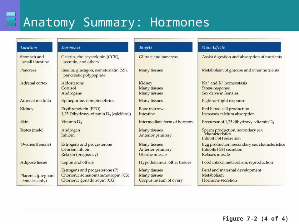
Figure 7-2 (4 of 4)
Anatomy Summary: Hormones

Hormones: Function
Control of Rates of enzymatic reactions
Transport of ions or molecules across cell membranes
Gene expression and protein synthesis
Exert effects at very low concentrations
Bind to target cell receptors
Half-life indicates length of activity

Enzyme-linked immunosorbent assay (ELISA)

Hormone dose response curve

Hormones: Classification
Peptide or protein hormones
Steroid hormones
Amine hormones

Peptide Hormone Synthesis, Packaging, and Release
Figure 7-3, step 1
ECFCytoplasm Plasma
Capillaryendothelium
Messenger RNA on the ribosomes binds amino acids into a peptide chain called a preprohormone.The chain is directed into the ER lumen by a signal sequence of amino acids.
mRNA
Ribosome
Endoplasmic reticulum (ER)
Preprohormone
1
1

Peptide Hormone Synthesis, Packaging, and Release
Figure 7-3, steps 1–2
ECFCytoplasm Plasma
Capillaryendothelium
Messenger RNA on the ribosomes binds amino acids into a peptide chain called a preprohormone.The chain is directed into the ER lumen by a signal sequence of amino acids.
Enzymes in the ER chop off the signal sequence, creating an inactiveprohormone.
mRNA
Ribosome
Prohormone
Signalsequence
Endoplasmic reticulum (ER)
Preprohormone
1 2
1
2

Peptide Hormone Synthesis, Packaging, and Release
Figure 7-3, steps 1–3
Golgi complex
ECFCytoplasm Plasma
Capillaryendothelium
Messenger RNA on the ribosomes binds amino acids into a peptide chain called a preprohormone.The chain is directed into the ER lumen by a signal sequence of amino acids.
Enzymes in the ER chop off the signal sequence, creating an inactiveprohormone.
The prohormone passes from theER through the Golgi complex.
mRNA
Ribosome
Prohormone
Signalsequence
Transportvesicle
Endoplasmic reticulum (ER)
Preprohormone
1 2 3
1
2
3

Peptide Hormone Synthesis, Packaging, and Release
Figure 7-3, steps 1–4
4
Active hormone
Golgi complex
Secretoryvesicle
ECFCytoplasm Plasma
Peptidefragment
Capillaryendothelium
Messenger RNA on the ribosomes binds amino acids into a peptide chain called a preprohormone.The chain is directed into the ER lumen by a signal sequence of amino acids.
Enzymes in the ER chop off the signal sequence, creating an inactiveprohormone.
The prohormone passes from theER through the Golgi complex.
Secretory vesicles containing enzymes and prohormone bud off the Golgi. The enzymes chop the prohormone into one or more active peptides plus additional peptide fragments.
mRNA
Ribosome
Prohormone
Signalsequence
Transportvesicle
Endoplasmic reticulum (ER)
Preprohormone
1 2 3
1
2
3
4

Peptide Hormone Synthesis, Packaging, and Release
Figure 7-3, steps 1–5
4 5
Active hormone
Golgi complex
Secretoryvesicle
ECFCytoplasm Plasma
Peptidefragment
Releasesignal
Capillaryendothelium
Messenger RNA on the ribosomes binds amino acids into a peptide chain called a preprohormone.The chain is directed into the ER lumen by a signal sequence of amino acids.
The secretory vesicle releases its contents by exocytosis into the extracellular space.
Enzymes in the ER chop off the signal sequence, creating an inactiveprohormone.
The prohormone passes from theER through the Golgi complex.
Secretory vesicles containing enzymes and prohormone bud off the Golgi. The enzymes chop the prohormone into one or more active peptides plus additional peptide fragments.
mRNA
Ribosome
Prohormone
Signalsequence
Transportvesicle
Endoplasmic reticulum (ER)
Preprohormone
1 2 3
1
2
3
4
5

Peptide Hormone Synthesis, Packaging, and Release
Figure 7-3, steps 1–6
4 5
To target
Active hormone
Golgi complex
Secretoryvesicle
ECFCytoplasm Plasma
Peptidefragment
Releasesignal
Capillaryendothelium
Messenger RNA on the ribosomes binds amino acids into a peptide chain called a preprohormone.The chain is directed into the ER lumen by a signal sequence of amino acids.
The secretory vesicle releases its contents by exocytosis into the extracellular space.
The hormone moves into the circulation for transport to its target.
Enzymes in the ER chop off the signal sequence, creating an inactiveprohormone.
The prohormone passes from theER through the Golgi complex.
Secretory vesicles containing enzymes and prohormone bud off the Golgi. The enzymes chop the prohormone into one or more active peptides plus additional peptide fragments.
mRNA
Ribosome
Prohormone
Signalsequence
Transportvesicle
Endoplasmic reticulum (ER)
Preprohormone
1 2 3 6
1
2
3
4
5
6

Activation of a G-protein coupled receptor (GPCR)

Peptide Hormone-Receptor Complex
Membrane receptors and signal transduction for peptide hormones
Figure 7-5

Steroid Hormones: Features
Cholesterol-derived Lipophilic and can enter target cell
Cytoplasmic or nuclear receptors (mostly)
Activate DNA for protein synthesis
Slower acting, longer half-life
Examples Cortisol, estrogen, and testosterone

Steroid Hormones: Structure
Steroid hormones are derived from cholesterol
Figure 7-6

Steroid Hormones: Action
Figure 7-7, step 1
Most hydrophobic steroids are bound to plasma protein carriers. Only unbound hormones can diffuse into the target cell.
Cellmembrane
Interstitialfluid
Nucleus
Bloodvessel
Proteincarrier
1
1

Steroid Hormones: Action
Figure 7-7, steps 1–2
Most hydrophobic steroids are bound to plasma protein carriers. Only unbound hormones can diffuse into the target cell.
Steroid hormone receptors are typically in the cytoplasm or nucleus.
Cellmembrane
Interstitialfluid
Cytoplasmicreceptor
Nucleus
Nuclear receptor
Steroidhormone
Bloodvessel
Proteincarrier2
1
1
2

Steroid Hormones: Action
Figure 7-7, steps 1–2a
Most hydrophobic steroids are bound to plasma protein carriers. Only unbound hormones can diffuse into the target cell.
Some steroid hormones also bind to mem-brane receptors that use second messenger systems to create rapid cellular responses.
Steroid hormone receptors are typically in the cytoplasm or nucleus.
Cellmembrane
Interstitialfluid
Cytoplasmicreceptor
Nucleus
Nuclear receptor
Rapid responses
Steroidhormone
Bloodvessel
Proteincarrier
Cell surface receptor
2
1
2a
1
2
2a

Steroid Hormones: Action
Figure 7-7, steps 1–3
Most hydrophobic steroids are bound to plasma protein carriers. Only unbound hormones can diffuse into the target cell.
Some steroid hormones also bind to mem-brane receptors that use second messenger systems to create rapid cellular responses.
Steroid hormone receptors are typically in the cytoplasm or nucleus.
The receptor-hormone complex binds to DNA and activates or represses one or more genes.
Cellmembrane
Interstitialfluid
Cytoplasmicreceptor
Nucleus
Nuclear receptor
DNA
Rapid responses
Steroidhormone
Bloodvessel
Proteincarrier
Cell surface receptor
2
3
1
2a
1
2
2a
3

Steroid Hormones: Action
Figure 7-7, steps 1–4
Most hydrophobic steroids are bound to plasma protein carriers. Only unbound hormones can diffuse into the target cell.
Some steroid hormones also bind to mem-brane receptors that use second messenger systems to create rapid cellular responses.
Steroid hormone receptors are typically in the cytoplasm or nucleus.
The receptor-hormone complex binds to DNA and activates or represses one or more genes.
Activated genes create new mRNA that moves into the cytoplasm.
Cellmembrane
Interstitialfluid
Cytoplasmicreceptor
Nucleus
Nuclear receptor
DNA
Rapid responses
Transcriptionproduces mRNA
Steroidhormone
Bloodvessel
Proteincarrier
Cell surface receptor
2
3
1
4
2a
1
2
2a
3
4

Steroid Hormones: Action
Figure 7-7, steps 1–5
Most hydrophobic steroids are bound to plasma protein carriers. Only unbound hormones can diffuse into the target cell.
Translation produces new proteins for cell processes.
Some steroid hormones also bind to mem-brane receptors that use second messenger systems to create rapid cellular responses.
Steroid hormone receptors are typically in the cytoplasm or nucleus.
The receptor-hormone complex binds to DNA and activates or represses one or more genes.
Activated genes create new mRNA that moves into the cytoplasm.
Cellmembrane
Interstitialfluid
Cytoplasmicreceptor
Endoplasmicreticulum
Nucleus
Nuclear receptor
DNA
Translation
Rapid responses
Transcriptionproduces mRNA
Steroidhormone
Bloodvessel
Proteincarrier
Newproteins
Cell surface receptor
2
3
1
4 5
2a
1
2
2a
3
45
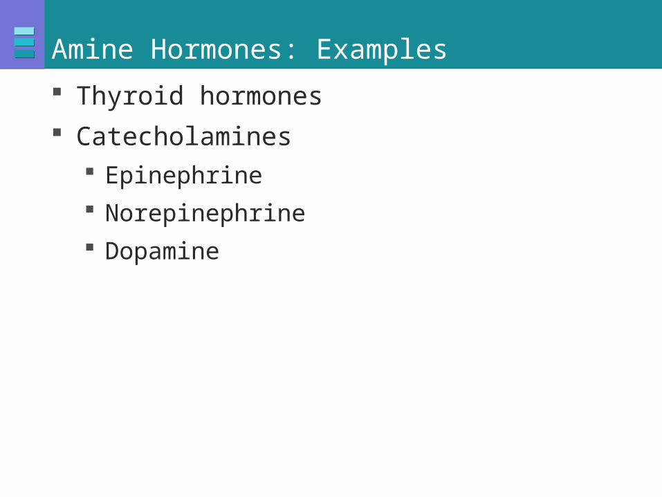
Amine Hormones: Examples
Thyroid hormones
Catecholamines Epinephrine
Norepinephrine
Dopamine

Amine Hormones: Structure
Tyrosine-derived amine hormones
Figure 7-8

Endocrine Reflex Pathways
Hormones may have multiple stimuli for their release
Figure 7-9

Figure 7-10
Simple Endocrine Reflex: Parathyroid Hormone

Neurohormones: Major Groups
Adrenal medulla Catecholamines
Hypothalamus Anterior pituitary
Posterior pituitary

The Pituitary Gland Anatomy
Figure 7-11

The Pituitary Gland: Two Fused
Posterior pituitary Vasopressin
Oxytocin
Figure 7-12 (1 of 4)

Hypothalamic-Hypophyseal Portal System
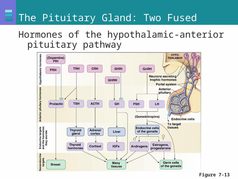
The Pituitary Gland: Two Fused
Hormones of the hypothalamic-anterior pituitary pathway
Figure 7-13

Endocrine Control
Three levels Hypothalamic stimulation—from CNS
Pituitary stimulation—from hypothalamic trophic hormones
Endocrine gland stimulation—from pituitary trophic hormones

Negative Feedback Controls
Long-loop feedback
Short-loop feedback
Figure 7-14

Control Pathway for Cortisol Secretion
Figure 7-15

Figure 7-16
The Hypothalamic-Hypophyseal Portal System

Example of Synergism
Figure 7-18

Figure 7-19
Endocrine Pathologies
Exogenous medication Replaces and
exceeds normal
Cause atrophy of gland

Excess cortisol
QuickTime™ and aTIFF (Uncompressed) decompressor
are needed to see this picture.
QuickTime™ and aTIFF (Uncompressed) decompressor
are needed to see this picture.QuickTime™ and a
TIFF (Uncompressed) decompressorare needed to see this picture.

Endocrine Pathologies
Hypersecretion: excess hormone Tumors or cancer
Grave’s disease—thyroxin
Hyposecretion: deficient hormone Goiter—thyroxin
Diabetes—insulin

Pathologies: Due to Receptors
Downregulation Hyperinsulinemia
Transduction abnormalities Testicular feminization syndrome
Pseudohypothyroidism
Abnormalities of control mechanisms