chemical approaches toward preparation of water-soluble
TRANSCRIPT

1
Chemical Approaches Toward Preparation of
Water-soluble Curcumin Derivatives
Thesis
Submitted to University of Mysore For the award of the degree of
DOCTOR OF PHILOSOPHY
in
CHEMISTRY
by
K. S. Parvathy, M. Sc.
Department of Plantation Products, Spices and Flavor Technology
CENTRAL FOOD TECHNOLOGICAL RESEARCH INSTITUTE
Mysore, INDIA
JULY 2009

2
DECLARATION
I hereby declare that the thesis entitled “CHEMICAL
APPROACHES TOWARD PREPARATION OF WATER-SOLUBLE
CURCUMIN DERIVATIVES” submitted to UNIVERSITY OF MYSORE for
the award of the degree of DOCTOR OF PHILOSOPHY IN CHEMISTRY
is the work carried out by me at laboratories of Plantation Products
Spices & Flavor Technology department under the guidance of Dr. P.
Srinivas, Head, Department of Plantation Products Spices & Flavor
Technology, CFTRI, Mysore – 570020, India, during the period 2006 –
2009. I further declare that the results are not submitted for the award
of any other degree or fellowship.
Mysore (PARVATHY K.S.) Date:

3
Dr. P. Srinivas Head, Dept. of PPSFT [email protected]
CERTIFICATE
I hereby certify that the thesis entitled “CHEMICAL APPROACHES
TOWARD PREPARATION OF WATER-SOLUBLE CURCUMIN
DERIVATIVES” submitted by Ms. K. S. Parvathy for the degree of
DOCTOR OF PHILOSOPHY IN CHEMISTRY to the UNIVERSITY OF
MYSORE for the result research work carried out by her at laboratories
of Plantation Products Spices & Flavor Technology department, CFTRI,
Mysore – 570020, India, under my guidance and supervision during the
period 2006 – 2009.
Mysore (P. SRINIVAS) Date:

4
DEDICATED TO MY BELOVED PARENTS
& DEEPU

5
ACKNOWLEDGEMENTS
I owe my sincere gratitude to my guide Dr. P. Srinivas, Head, Dept.
of Plantation Products Spices & Flavor Technology for conceptualizing and
evolving the synthetic strategies for the various curcumin derivatives and
for his constant advice and guidance throughout my research work. I
thank Dr. P.S. Negi, Scientist, Dept. of Human Resources Development for
his valuable guidance and help in evaluation and interpretation of the
antimicrobial and antimutagenic attributes of the curcumin derivatives.
My special thanks to J. R. Manjunatha for the help provided for the
NMR studies and Dr. H. H. Pattekhan for his assistance in the initial
stages of my work. I also express my sincere thanks to Padmere Mukund
Lakshman and other staff of Central Instrumentation Facility and Services
for their help in chromatographic and spectroscopic studies.
I express my gratitude to Dr. K.N. Gurudutt, Former Head, Dept. of
FS & AQCL, for all helpful discussions and advice.
I thank Dr. V. Prakash, Director, CFTRI, for his constant support and
infra-structural facilities provided for my research work at Central Food
Technological Research Institute.
I am grateful to Dr. M. C. Varadaraj, Head, HRD for his
encouragement and permission to carry out microbiology experiments in
his department.

6
I thank Dr. B. R. Lokesh, Dr. K. S. Jagannatha Rao, Dr. Lalitha
Gowda and Dr. L. Jagan Mohan Rao, A. G. Appu Rao and Dr. P. V.
Salimath for their valuable suggestions during registration and pre-thesis
submission viva voce deliberations.
I owe my gratitude to Dr. R. N. Tharanathan and Dr. K. Srinivasan
from Biochemistry and nutrition department, Dr. N. S. Susheelamma from
Sensory science department for their encouragement and suggestions.
I am also thankful to former Heads of PPSFT department, Mr. B.
Ragahvan and Mr. S. R. Sampathu for their help.
I thank, Dr. Bharathi, Dr. H. B. Sowbhagya, Mrs. Pushpa S. Murthy
and all other senior colleagues of PPSFT department for their constant
encouragement during my research work.
I thank Desai, S.V. and Divya, K.H. for their help during my
microbiology experiments.
My thanks to all my friends, Raghu Raj Singh Chouhan, Shyamala
Hegde, R.S. Jagdish, and Nandini Jagdish.
My special gratitude to my parents and my brother Deepak for their
encouragement during my research work and Bharati Cariappa for giving
me emotional support.
PARVATHY, K.S

7
CONTENTS
Page
SYNOPSIS i-vi
CHAPTER 1
INTRODUCTION
References
1
28
CHAPTER 2 SYNTHESIS OF GLUCOSIDES OF CURCUMIN
Section 2.1
Section 2.2
Section 2.3
Section 2.4
Introduction
Experimental
Results & discussion
Conclusions
References
45
53
57
75
76
CHAPTER 3 SYNTHESIS OF AMINO ACID CONJUGATES
OF CURCUMIN
Section 3.1
Section 3.2
Section 3.3
Section 3.4
Introduction
Experimental
Results & discussion
Conclusions
References
81
85
90
110
111

8
CHAPTER 4
SYNTHESIS OF GLUCURONIC ACID
& GLUCAL DERIVATIVES OF CURCUMIN
Synthesis of glucuronic acid derivative
Section 4.1.1
Section 4.1.2
Section 4.1.3
Introduction
Experimental
Results & discussion
115
118
122
Synthesis of glucal derivative of curcumin
Section 4.2.1
Section 4.2.2
Section 4.2.3
Section 4.3
Introduction
Experimental
Results & discussion
Conclusions
References
128
130
132
136
137
CHAPTER 5
ANTIOXIDANT, ANTIMUTAGENIC &
ANTIBACTERIAL PROPERTIES
OF CURCUMIN DERIVATIVES
Section 5.1
Section 5.2
Section 5.3
Section 5.4
Introduction
Experimental
Results & discussion
Conclusions
References
143
148
154
189
191 SUMMARY & CONCLUSIONS
199
PUBLICATIONS

9
SYNOPSIS
Curcumin, the major coloring principle of turmeric (Curcuma longa L.,
Zingiberaceae), has a wide array of biological activities. It is soluble in organic solvents
like acetone and dioxane. But, it is insoluble in water at acidic or neutral pH, which is a
disadvantage for its use in water-based food products. Also, the molecule undergoes
degradation on exposure to light. Hence, newer strategies to prepare simple and safe
adducts/derivatives are needed to overcome these drawbacks. In order to make curcumin
water-soluble, it is essential to attach to it a polar group or molecule that can impart
hydrophilic nature to it. The major objectives of the present investigation were
development of alternative, efficient and stereo-specific methods for obtaining specific
derivatives of curcumin, their characterization, analysis and study of their bioactive
attributes. Approaches delineated in literature on curcumin derivatization are wide-
ranging in nature. In the present study, particular emphasis was laid on preparation of
curcumin derivatives with a sugar or an amino acid, as the resultant compounds would on
hydrolysis afford natural molecules. Also, keeping in view the labile nature of curcumin,
strategies evolved needed bringing about the derivatization under milder conditions and
in higher yields. Thesis, describing the research results, includes five chapters. Chapter
one covers literature on curcuminoids and their derivatives. Chapters 2, 3 and 4, describe
the several synthetic methodologies developed in this study for selected curcumin
derivatives. In Chapter 5, the in vitro antioxidant, antibacterial and antimutagenic
properties of all the curcumin derivatives prepared in the current work are presented. The
salient findings of the investigation are summarized.
CHAPTER 1. CURCUMINOIDS AND THEIR DERIVATIVES
This introductory chapter describes the physico-chemical, spectral and stability
aspects of curcuminoids. Analytical protocols for curcuminoids are also briefly
discussed. Biological attributes of curcumin like antioxidant, antimicrobial and
antimutagenic effects in several in vitro as well as animal models along with several other
biological activities of curcumin are detailed in this chapter. This chapter also elaborates
several known synthetic approaches to preparation of selected derivatives of curcumin to

10
improve its solubility and photo-stability. Few important examples of curcumin
derivatives - synthesis and their biological properties - are discussed.
CHAPTER 2. SYNTHESIS OF CURCUMIN GLUCOSIDES
Glycosidation of curcumin is one of several approaches to render curcumin water-
soluble. Reaction of α-acetobromoglucose with potassium salt of curcumin under either
thermal or high pressure conditions results in drastic reduction in the yields of the
glucosides. In the present study, this drawback was effectively overcome by carrying out
reaction of 2, 3, 4, 6-Tetra–O-Acetyl-α–D-glucopyranosyl bromide with potassium salt of
curcumin under biphasic conditions in the presence of phase transfer catalysts and under
the influence of ultrasound as a new approach to Koenigs-Knorr reaction. The reaction
under the sonochemical conditions was faster and resulted in the increased yield of the
glucoside products (> 65%). The reaction, when investigated in detail, clearly
underscored the significance of the nature and quantity of the phase transfer catalyst
employed in the reaction. The amount of potassium hydroxide, employed for the
formation of the salt of curcumin too played a critical role in the reaction. Optimum
yield and selectivity towards preponderant formation of the curcumin di-β-glucoside
tetracetate was obtained when benzyltributylammonium chloride was used as PTC and
the base was used in 10 mmol, wherein the reaction was complete in 4 h. The reaction
was also investigated under mono-phasic conditions with the benzyltributylammonium
salt of curcumin, produced in situ from the potassium salt of curcumin and PTC, in
DMSO and alcoholic solvents. Among these, the reaction in DMSO afforded the
curcumin mono-β-glucoside tetracetate in 50 % yield. The curcumin β-glucoside
tetraacetates were deacetylated to afford pure curcumin β-glucosides. Thus, the study
established a simple synthetic protocol for the glucoside derivatives of curcumin in high
yields and selectivity using ultrasound.
CHAPTER 3. SYNTHESIS OF CURCUMIN AMINO ACID CONJUGATES
Another approach to solublize curcumin is the preparation of its amino acid
conjugates. Recent literature reports preparation of di-O-glycinoyl curcumin and di-O-
glycinoyl-C14-glycylcurcumin by condensation of curcumin with N-phthaloyl glycinoyl

11
chloride in anhydrous pyridine. Also, preparation of derivatives of curcumin viz., 4,4’-di-
(O-glycinoyl) curcumin, 4,4’-di-(O-glycinoyl-di-N-piperoyl) curcumin, 4,4’-di-(O-
piperoyl)curcumin and 4,4’-(O,O-cysteinoyl)-3,3’-dimethoxydi-phenyl-1,6-heptadiene-
3,5-dione has been described. A multi-step synthesis of tetraglycine conjugate of
curcumin too is reported. However, applicability of these methods, to general synthesis
of other amino acid derivatives of curcumin under mild conditions is not demonstrated.
In the present investigation, a general synthetic method with superior yields of
amino-acid conjugates of curcumin was developed. The protocol involved reaction of
curcumin in dry dioxane with t-Boc-amino acids in the presence of a dehydrating agent
like dicyclohexylcarbodiimide with 4-dimethylaminopyidine and triethylamine as
catalysts. Under an inert atmosphere (N2) and at 25 – 30 0C, the reaction afforded
curcumin t-Boc-amino acid conjugates in high yields (50 – 84 %). The reaction was
demonstrated with several t-Boc protected amino acids viz., i) an un-substituted one like
glycine, ii) alkyl substituted ones like alanine, leucine, isoleucine and valine, iii) an
amino acid that is an aryl substituted one like phenylalanine and phenylglycine, iv) an
amino acid containing sulphur like cysteine, v) an amino acid present as an imino acid
like in proline, and vi) an amino acid that contains a hydroxyl group like serine. Also, in
this study, an efficient protocol was developed under milder conditions for the rapid
removal of the t-Boc group with TFA (10 %) using ultrasound. Several new amino-acid
conjugates of curcumin are reported in this study.
CHAPTER 4. GLUCURONIDE AND GLUCAL DERIVATIVES OF CURCUMIN
Glucuronide derivative of curcumin and its reduced forms are reported as major
metabolites of curcumin in the experiments on its metabolism in rat models. In the
present study, a synthetic pathway for this derivative of curcumin was delineated. In this
approach, glucuronolactone was stirred with NaOMe in methanol to cleave the lactone
ring followed by methylation of free carboxyl group. The product formed was acetylated
with acetic anhydride / perchloric acid reagent. The resultant acetylated compound was
brominated at the anomeric position. Reaction of the methyl α-bromoglucuronide
triacetate with curcumin was carried out under biphasic conditions with phase transfer

12
catalyst (PTC) using ultrasound to afford curcumin methyl mono-β-glucuronide
triacetate. The product was deacetylated to yield curcumin carboxymethyl mono-β-
glucuronide. 2,3-Unsaturated glycosides are versatile synthetic intermediates and also
constitute the structural units of several natural products. Allylic rearrangement of
glycals, known as the Ferrier rearrangement, in the presence of a nucleophile leads to the
formation of 2,3-unsaturated glycosides. Ferrier reaction of 3,4,6-tri-O-acetyl-D-glucal
with curcumin in presence of ferric sulfate catalyst at 40 oC in acetonitrile afforded the
mono glucal derivative of curcumin. Deacetylation of the glucal derivative afforded the 2,
3- dideoxysugar derivative of curcumin in 50 % yield.
CHAPTER 5. IN-VITRO ANTIOXIDANT, ANTIBACTERIAL AND
ANTIMUTAGENIC ATTRIBUTES OF CURCUMIN DERIVATIVES
Curcumin derivatives - glucosides, amino acid conjugates, glucuronide and 2,3–
dideoxyglucose compounds of curcumin - synthesized in the present study were soluble
in water. The solubility of curcumin glucoside and glucuronide was 10 mg/ml in water.
The curcumin glucal derivative was soluble at 1 mg/ml concentration. While all
curcumin amino acid conjugates were soluble in water at 1 mg/ml concentration at pH 6,
the conjugates of curcumin with glycine and proline exhibited higher solubility in water
(10 mg/ml).
It was important to determine the effect of derivatization of curcumin on its’ vital
innate biological attributes for which this important nutraceutical molecule is highly
valued. Hence, the in vitro antioxidant, antibacterial and antimutagenic properties of the
various derivatives of curcumin prepared in this study were assessed and compared with
those of curcumin.
The radical scavenging activity by DPPH method as well as antioxidant activity
by β–carotene-bleaching assay showed 67-68% and 100 % activity at 0.15 µM levels for
curcumin and curcumin glucoside respectively. Both curcumin and its glucoside showed
strong antimutagenic activity with Salmonella Typhimurium TA 98 against sodium azide
at all the concentrations tested. In case of TA 1531, curcumin had moderate
antimutagenic activity but its glucoside showed significantly higher activity at 625

13
μg/plate. These results indicated higher antimutagenic potential of curcumin glucoside
compared to curcumin. Curcumin and its glucoside showed 27% and 70% inhibition
against E. coli at 0.14 and 0.15 µM concentrations respectively. In case of Y.
enterocolitica, curcumin and its glucoside showed 100% inhibition at 0.68 and 0.87 µM
respectively. Against B. cereus, curcumin and its glucoside showed 30% and 66%
inhibition at 0.07 µM. In the case of S. aureus curcumin and its glucoside showed 68%
and 100% inhibition at 0.07 and 0.05 µM concentrations. These results demonstrated
that curcumin glucoside possessed superior antibacterial activity against the
microorganisms tested.
Curcumin amino acid conjugates showed lower IC50 values than curcumin in
DPPH method as well as β–carotene-bleaching assay. Conjugates of curcumin with
amino acids like alanine, valine, serine, cysteine and isoleucine were effective even at
~50 % of the concentration of curcumin.
Amino acid conjugates of curcumin showed strong antimutagenic activity against
Salmonella Typhimurium TA 98, with sodium azide and methyl methanesulphonate as
mutagens at 100 as well as 250 µg/plate concentrations. With Salmonella Typhimurium
TA 1531 at 100 µg/plate concentration, against sodium azide as mutagen, conjugates of
curcumin with proline, leucine, serine and valine showed activity similar to curcumin;
other derivatives showed higher activity than curcumin. In the case of MMS as mutagen,
cysteine, glycine and proline conjugates of curcumin showed lower activity than
curcumin while other derivatives showed higher activity than curcumin. However, all the
amino acid conjugates showed higher activity than curcumin against TA 1531 at 250
µg/plate concentration.
Curcumin amino acid conjugates showed lower MIC values compared to
curcumin against the Gram - negative bacteria, E. coli and Y. enterocolitica. Against
S.aureus, all derivatives, except alanine, phenylalanine and alanine conjugates of
curcumin, exhibited higher inhibition. Against B. cereus, isoleucine, phenylalanine,
valine, proline and glycine conjugates of curcumin showed lower inhibition compared to
curcumin. IC50 values of glucuronide and glucal derivatives were lower than the parent

14
molecule in antioxidant assays. Both the derivatives showed strong antimutagenicity as
the parent molecule against sodium azide and MMS at 100 and 250 μg/plate
concentrations. Inhibition against E. coli and Y. enterocolitica was higher compared to
curcumin in the case of glucuronide derivative. Glucal derivative of curcumin also
showed good inhibition against E. Coli but comparable to curcumin in the case of Y.
enterocolitica.
SUMMARY
In the present research study, several curcumin derivatives viz., glucosides, amino
acid conjugates, glucuronide and 2,3-dideoxyglucose derivatives have been synthesized
in moderate to high yields. The binding of the phenolic hydroxyl moiety in curcumin to
various groups rendered the curcumin derivatives soluble in water at 1 to 10 mg/ml
concentrations. The water-soluble derivatives, thus prepared, exhibited more pronounced
in vitro antioxidant, antibacterial and antimutagenic properties compared to curcumin.
Signature of the candidate Signature of the guide
(Parvathy K.S) (P. Srinivas)
Date: Date:

15
CHAPTER 1 INTRODUCTION

16
1.1 Turmeric
Turmeric, a very important spice in India, comes from the root of Curcuma
longa, a leafy plant of the ginger family. The plant is an herbaceous perennial, 60
- 90 cm high, with a short stem and tufted leaf. The yellow turmeric tuberous root
belongs to the group of aromatic spices. The root or rhizome has a tough brown
skin and bright orange flesh. In fresh state, it has a strong and spicy fragrance,
which on drying gives way to a more mellowed aroma. The spice is sometimes
called the 'Indian saffron' due to its brilliant yellow color. Turmeric forms part of
most Indian spice powders. It is widely consumed as a dietary spice and a
natural food colorant. It is a natural antiseptic that finds extensive use in
traditional Indian medicine for the treatment of various ailments. It is also used in
the textile and pharmaceutical industries.
1.1.1 Turmeric Production
Curcuma, a genus in the plant family of Zingiberacea, is the biological
source for curcuminoids with curcumin as the major constituent (Aggarwal, et al.,
2005; Govindarajan, 1980). It is grown in Southeast Asia, China, North Australia,
West Indies and South America and of late also grown in few African countries.
Production of turmeric in India for the year 2005-2006 was estimated to be about
8, 92,180 tons (Spices Board of India, 2006). Around 42,750 tons of turmeric
has been exported from India in the period April 2006 - January 2007. The genus
Curcuma (family of Zingiberacea) contains 49 genera. Among these, Curcuma
Longa L, is of commercial importance. Turmeric is commercialized as whole
spice, powder and oleoresin. Oleoresin contains mixture of volatile oil along with

17
non-fatty and resinous material. Solvents like hexane, acetone and alcohol are
often employed for the extraction of curcumin from turmeric. The high solubility of
curcumin in acetone renders it the solvent of choice for the extraction. The
extraction time is variable but in a soxhlet equipment the yield is about 8-10 %,
containing 42–50 % curcuminoids in 4-5 h (Delgado–Vergas & Paredes – Lopez,
2003).
1.1.2 Varieties of Turmeric and its uses
The Curcuma genus has an estimated 1,000 species. Curcuma
alismatifolia, C. Cordata, C. gracillima, C. longa ‘O’Lena & C. Parviflora are few
to mention. More than 50 cultivars are known in India and are known mostly by
the name of location where they are cultivated. Madras variety, named Alleppy,
has higher curcuminoid content of 5–7 %. Some of the other popular cultivars are
Duggirala, Tekurpeta, Sugandham, Amalapuram, Erode local, Moovattupuzha,
and Lakadong. Turmeric is used for various purposes such as a coloring agent
and a condiment. It is a principal ingredient in Indian curry powder. Turmeric
oleoresin is used in brine pickles, to some extent in mayonnaise, relish
formulations, in nonalcoholic beverages, gelatins, butter and cheese. Turmeric is
also used as a dye in textile industry, in cosmetics, preparation of medicinal oils,
ointments and poultice. It possesses stomachic, carminative, tonic, blood purifier
and antiseptic properties. The aqueous extracts also have bio-pesticide
properties (Delgado–Vergas & Paredes – Lopez, 2003).

18
1.1.3 Constituents of turmeric
Total extracts of turmeric, curcumin and turmeric oil, are all credited with
medicinal properties. Biological activities of these components and their
constituents however, differ considerably. It is reported that the proportions of
curcuminoids play a considerable role in optimum bio-protective activity of
turmeric. The yellow-pigmented fraction of Curcuma longa contains
curcuminoids, which are chemically related to its principal ingredient, curcumin.
Curcuminoids are generally present in the range of 3–5% in turmeric. Curcumin
and its analogs are present in turmeric along with other components like
essential and fixed oils, starch, protein, fiber, bitter principles and moisture.
1.2 Curcuminoids: Analogs, isolation techniques and stability
Curcumin, a phenolic component present in turmeric along with two other
analogs, is responsible for its bioactive properties. Curcumin, an orange yellow
pigment, has major role as colorant and as flavoring for the Indian food
industries. Curcuminoids are extracted from turmeric by extraction with selected
organic solvents. The three main curcuminoids isolated from turmeric are
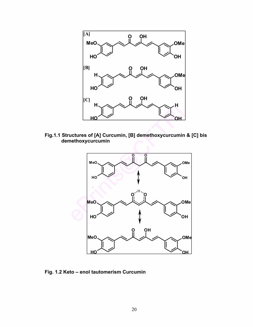
curcumin, demethoxycurcumin, and bisdemethoxycurcumin (Fig. 1.1 a - c). As
the curcuminoid pigments vary in chemical structures, their chemical and color
characteristics as well as the functional properties (Table 1.1) could be different.
Curcumin of very high purity (> 99%), obtained by selective crystallization
process, is highly valued as a nutraceutical. As demethoxycurcumin and bis-
demethoxycurcumin, being minor constituents, are not commercially available,
their separation and characterization assumes significance.

19
1.2.1 Isolation, purification, separation and characterization of curcuminoids
Curcumin was first isolated in 1815 by Vogel, obtained in crystalline form
in 1870 by Daube and identified as (1E, 6E)-1,7-bis(4-hydroxy-3-methoxyphenyl),
1,6-heptadiene-3,5-dione). In 1910, the feruloylmethane skeleton of curcumin
was confirmed. Curcumin is a yellow-orange powder that is insoluble in water,
sparingly soluble in ether and soluble in acetone, dioxane, ethanol and dimethyl
sulfoxide. It has a melting point of 182-183 0C, a molecular formula of C21H20O6,
and a molecular mass of 368.38. The compound exhibits maximum absorption (λ
max) at 430 nm in methanol and at 415–420 nm in acetone (Boong, 2000). A 1 %
solution of curcumin contains 1650 absorbance units (Delgado–Vergas &
Paredes – Lopez, 2003). Curcumin appears in brilliant yellow hue at pH 2.5–7
and red at pH > 7. Curcumin exists in enolic and diketonic forms.
Table 1.1 Physico chemical properties of curcuminoids
Trivial name Curcumin Demethoxy curcumin Bis-Demethoxy
curcumin
Chemical Name Diferuloyl
methane
4– Hydroxycinnamoyl
feruloyl methane
Bis-4-Hydroxy-
cinnamoylmethane
Molecular weight 368.38 338.36 308.33
Chemical formula C21H20O6 C20H18O5 C19H16O4
Melting point (oC) 182-183 172-174 223-224
Absorption maxima (λ max) in ethanol
427 424 418
20
O O
OHHO
OMeMeO
O O
OHHO
OMeMeO
O OH
OHHO
OMeMeO
H
O OH
OHHO
OMeMeO
O OH
OHHO
OMeH
O OH
OHHO
HH
[A]
[B]
[C]
Fig.1.1 Structures of [A] Curcumin, [B] demethoxycurcumin & [C] bis demethoxycurcumin
Fig. 1.2 Keto – enol tautomerism Curcumin

21
Curcuminoids are separated by column chromatography and HPLC. Many
methods are reported for the separation of curcuminoids by HPLC
(Jayaprakasha, et al., 2002; Hiserodt, et al., 1996; Braga, et al., 2003; Peret-
Almeida, et al., 2005), Reports are available on rapid quantitation of curcumin by
NMR and LC-tandem mass spectrometry (Goren, et al., 2009) and HPTLC
(Paramasivam, et al., 2009). Curcumin is a bis-α, β-unsaturated 1,3-diketone. As
such, curcumin exists in equilibrium with its enol tautomer (Fig. 1.2) (Sharma, et
al., 2005).
1.2.2 Stability aspects of curcumin
The degradation kinetics of curcumin under various pH conditions and the
stability of curcumin in physiological matrices have been established (Wang, et
al., 1997). When curcumin is incubated in 0.1 M phosphate buffer and serum-free
medium (pH 7.2 at 37 0C), about 90% decomposes within 30 min. A series of pH
conditions ranging from 3 to 10 are tested and the results show that
decomposition is pH-dependent and occurs faster at neutral or basic conditions.
It is more stable in cell culture medium containing 10% fetal calf serum and in
human blood. Less than 20% of curcumin decomposes within 1 h, and after
incubation for 8 h, about 50% of curcumin still remains. (2Z,5E)-2-hydroxy-6-(4-
hydroxy-3-methoxyphenyl)-4-oxohexa-2,5-dienal is suggested to be the major
degradation product (Fig. 1.3). Vanillin, ferulic acid, and feruloyl methane are
identified as minor degradation products (Wang, et al., 1997). The amount of
vanillin increases with incubation time. Curcumin is insoluble in water and at
acidic pH but soluble under alkaline conditions. Curcumin hue progressively
22
transforms from yellow to intense red above pH 7. Tonnesen, et al., (1985) has
first reported the complex kinetics of pH-dependent degradation of curcumin in
aqueous solution. These studies reveal that curcumin degrades rapidly under
alkaline pH (Tonnsen, et al., 1986) and is extremely unstable at pH above 7 in
aqueous systems.
1.3 Metabolism of curcumin
Water-insolubility and photo-instability of curcumin render it less suitable
for food / pharmacological applications. The enol form ensures its stability to
resonance structures, which give it pseudo-aromatic character and helps
curcumin to form hydrogen bonds with metals to form complexes. Although the
exact mechanism of degradation is not completely understood, curcumin should
be stable in the stomach and small intestines because the pH is between 1 and
6, and degradation of curcumin is slow under these conditions (Wang, et al.,
1997). Biotransformation of curcumin has been studied, its metabolites isolated
after oral i. p. administration in mice and identified. THC is a major metabolite of
curcumin and its stability at different pH has been studied (Pan, et al., 1999). In -
vitro studies show the metabolic steps involve the glucuronidation of curcumin
and its major reduced forms viz., tetrahydrocurcumin, hexahydrocurcumin and
octahydrocurcumin (Fig. 1.4). Glucuronides of curcumin, hexahydrocurcumin,
and other analogs have been synthesized using rat and human liver microsomes
and characterized by LC-MS/MS analysis (Pfeiffer, et al., 2007).

23
O OH
OH OH
OMe OMe
Curcumin
OHC
O
OH
O MeHO(2Z,5E)-2-hydroxy-6-(4-hydroxy-3-methoxyphenyl)-4-oxohexa-2,5-dienal
OH3C OH
O
OH
Feulic acid
Me
O
Me
OH
O
Feruloyl methane
OH
OHC
O
Me
Vanillin
O OH
OHHO
OMeMeOO OH
OHHO
OMeMeO
Curcumin Dihydrocurcumin
Reductase
Reductase
O OH
OHHO
OMeMeO
Tetrahydrocurcumin
O OH
OHHO
OMeMeO
Hexahydrocurcumin
Reductase
OH OH
OHHO
OMeMeO
OctahydrocurcuminReductase
Fig. 1.3 pH dependent degradation of curcumin
Fig. 1.4 Curcumin metabolism

24
1.4 Biological attributes of curcumin
Curcumin shows many pharmacological applications from ancient
ayurveda to modern homeopathy because of its high biological activity (Singh,
2007). Curcumin, a constituent of turmeric, has anti-inflammatory, anti-
carcinogenic, and chemo-preventive effects as evidenced in several animal
tumor models. Curcumin has potent antioxidant, antitumor and anticancer
properties (Goel, et al., 2008). The molecule is known to possess multiple
biological activities like wound healing, antifertility, antibacterial, antifungal,
antiprotozoal, antiviral, antifibrotic and antivenom (Aggarwal et al. 2007).
Curcumin possesses diverse effects on cellular enzymes and on angiogenesis
and cell adhesion. In particular, ability of curcumin to affect gene transcription
and induce apoptosis in pre-clinical models advocates its potential utility in
cancer chemoprevention and chemotherapy. Although curcumin’s low systemic
bio-availability following oral dosing seems to limit the number of tissues that it
can reach at efficacious concentrations to exert beneficial elects, the attainment
of such levels in the gastrointestinal tract, particularly the colon and rectum, has
been demonstrated in animals and humans (Alpers, 2008). In view of the peer-
reviewed reports of the pharmacological properties of curcumin, its phase II
clinical evaluation in individuals at risk of developing cancer, especially of the
gastrointestinal tract, appears opportune (Kunnumakkara & Aggarwal, 2008)
Curcumin inhibits tumorogenesis and also suppresses mammary
carcinogenesis. Curcumin exhibits antiproliferative effects against cancer cells.
Curcumin has been shown to inhibit the proliferation of a wide variety of tumor

25
cells, including B-cell and T-cell leukemia, colon carcinoma, and epidermoid
carcinoma cells. It has also been shown to suppress the proliferation of various
breast carcinoma cell lines in culture. Curcumin administration in initiation and
post initiation period shows inhibition of colon tumorogenesis, curcumin with low
chronic toxicity will be advantageous to combat against cancer. The molecular
basis of anticarcinogenic and chemopreventive effects of curcumin is attributed
to its effect on specific targets which include transcription factors, growth
regulators, adhesion molecules, apoptotic genes, angiogenesis regulators and
signaling molecules (Maheshwari,, et al., 2006; Anand, et al., 2008; Aggarwal, et
al., 2005; Anand, et al., 2008a). It has attracted special attention due to its
pharmacological activities such as protection of cells from β-amyloid insult in
Alzheimer’s disease (Park & Kim, 2002). Antioxidant property of curcumin,
clinical evaluation of curcumin as an anticancer agent in humans, effect of
curcumin on lymphocytes, platelet aggregation, detoxification mechanisms, cell
cycle and apoptosis, diabetes, wound healing, stress responses and antiviral
effects are well established (Joe, et al., 2004).
Few important examples from recent literature that underscore the
importance of curcumin as a therapeutic and nutraceutical compound are
enlisted below.
• Curcumin is a potent antioxidant by virtue of which it has anti-inflammatory
and anti-carcinogenic potential (Srinivasan, 2005).
• Curcumin is also an effective hypolipidemic agent (Srinivasan et al.,
2004). It is useful as an anti-diabetic food adjunct (Srinivasan, 2005a).

26
• Curcumin possesses antilithogenic property (Hussain and
Chandrasekhara, 1993, 1994).
• The pharmacodynamics and pharmacokinetics of curcumin have been
examined in animals and in humans. Curcumin protects against various
forms of stress, curcumin modulates multiple sclerosis, reduces the
formation of cholesterol gallstone formation, suppresses the symptoms of
arthritis, regulates myocardial infarction, inhibits platelet aggregation and
lowers the serum cholesterol levels (Chattopadhyay, et al., 2004).
• Curcumin has various pharmacological and therapeutic properties as
demonstrated in in vitro and in vivo studies (Aggarwal, et al., 2009).
• Curcumin is a good scavenger of reactive oxygen species (Elizabeth and
Rao, 1990).
• Curcumin is a potent singlet oxygen quencher at physiological or
pharmacological concentrations (Das & Das, 2002).
• Curcumin is shown to penetrate the blood - brain barrier and bind to A-
beta in brain cells both in in vivo and in vitro studies (Balasubramanian,
2006).
• Curcumin inhibits proliferation of cancer cells by perturbing microtubule
assembly dynamics and efficacious curcumin analogs have been
developed for trials in cancer chemotherapy (Gupta &
Balasubrahmanyam, 1998).
• Curcumin down-regulates the multi drug-resistance mdr1b gene by
inhibiting the PI3K/Akt/NFkB pathway (Choi, et al., 2008).

27
• Curcumin and its decomposition products vanillin, vanillic acid, ferulic
aldehyde and ferulic acid inhibit human recombinant cytochrome P450s.
(Appiah – Opang & Commandeur, 2007).
• Curcumin induces changes in the expression of genes in cholesterol
homeostasis (Peschel, et al., 2007).
• Curcumin reduces lead-induced neurotoxicity in male wistar rats (Dairam,
et al., 2007).
• Curcumin prevents Fos-Jun-DNA complex formation (Kim & Yang, 2004).
• Tetrahydrocurcumin (THC), a major colorless metabolite of curcumin, is
shown to exhibit a more prominent anti-diabetic and plasma glycoprotein
controlling ability than curcumin (Pari & Muruan, 2007).
• Curcumin has antidepressant effect, which is related to serotonergic
system and mediated by interactions with 5-HT1 and 5-HT2 receptors
(Wang, et al., 2008).
• Curcumin exhibits anti-lymphangiogenic property as shown by its ability to
inhibit the formation of capillary-like tubes by rat lymphatic endothelial
cells (Matsuo, et al., 2007).
• Curcumin attenuates haloperidol-induced oxidative damage in all the
regions of the brain especially in the sub-cortical region containing
striatum, of possible therapeutic relevance for the treatment of tardive
dyskinesia (TD), a hyper-kinetic movement disorder (Bishnoi, et al., 2008).
• Active site binding modes of curcumin in HIV-1 protease and integrase are
reported wherein, symmetrical structure of curcumin seems to play an

28
important role for binding to the PR protein, wherein the keto–enol and
only one side of the terminal O-hydroxyl show tight binding to the IN active
site (Vajragupta, et al., 2005).
• Curcumin prevents and reverses murine cardiac hypertrophy (Li, et al.,
2008).
• Curcumin inhibits lipid peroxidation in rat liver microsomes, erythrocyte
membranes and brain homogenates (Pulla Reddy, et al., 1994).
• Studies show curcumin in aqueous preparations consisting of
cylcodextrin, DMSO, surfactant like triton, sodium aliginate and liposomes
in combination with light affected salivary gland acinar cells. The study
points out to the possible use of curcumin as a photosensitizer in the
treatment of oral lesions (Bruzell, et al., 2005).
• Curcumin displays differential effects on vasoactive factor expression in
the heart and that the tissue microenvironment is of importance in
treatment of diabetic complications as demonstrated in experiments with
Streptozotocin-induced diabetic rats (Farhangkhoee, et al., 2006).
• Curcumin together with EGF-R related protein causes a greater inhibition
of growth of colon cancer cells (Sudha Reddy, et al., 2006).
• Effect of antioxidant activity of turmeric, turmerin and curcumin as anti HIV
drugs has been studied. Water-soluble extract, turmerin, inhibits HIV
infected T–cell proliferation and combination with 3’ azido-3’-
deoxythymidine decreases T-cell infection and increases cell viability
(Cohly, et al., 2003).

29
• Curcumin is an effective chemo-preventive compound. The mechanism of
action of curcumin is complex and likely multi-factorial. It modulates
proteins involved in iron metabolism in cells and tissues and acts as a
good chelator for iron (Jiao, et al., 2006).
• Curcumin and its analogs are shown to induce Phase 2 detoxification
enzymes in murine hepatoma cells underlining the chemo-protective
potential of these compounds. The activities of molecules indicate Keto –
enol moiety plays an important role for the biological activities (Dinkova &
Talalay, 1999).
1.4.1 Antioxidant activity of curcumin
Antioxidant activity of curcumin has been assayed by various in vitro
antioxidant assays and compared against BHA, BHT, α-tocopherol and trolox
standards (Tuba Ak & Gulcin, 2008). These include 1,1-diphenyl-2-picryl-
hydrazyl free radical (DPPH) scavenging activity, 2,2′-azino-bis(3-
ethylbenzthiazoline-6-sulfonic acid) (ABTS) radical scavenging activity, N,N-
dimethyl-p-phenylenediamine (DMPD) radical scavenging activity, total
antioxidant activity determination by ferric thiocyanate, total reducing ability
determination by the Fe3+–Fe2+ transformation method, superoxide anion radical
scavenging by the riboflavin/methionine/illuminate system, hydrogen peroxide
scavenging and ferrous ions (Fe2+) chelating activity. Curcumin,
bisdemethoxycurcumin and demethoxycurcumin are tested for their antioxidant
activities by in vitro model systems, such as the phosphomolybdenum and

30
linoleic acid peroxidation methods. Curcumin shows stronger activity compared
to its analogs (Sandur, et al., 2007, Jayaprakasha, et al., 2006).
1.4.2 Antioxidant mechanisms of curcumin
Curcumin molecule possesses high antioxidant activity. Several
mechanisms are postulated for proton transfer during radical scavenging action.
The phenolic hydroxyls of curcumin are shown to be involved in the route of HAT
(Hydrogen Atom Transfer) mechanism of antioxidant activity. Alternatively
sequential proton loss electron transfer (SPLET) postulates that keto – enol
moiety of curcumin has a more easily dissociable proton, which is supposed to
be faster than the other mechanism, as the enolic proton is more acidic than the
other two hydroxyls of phenol rings in curcumin (Litwinienko & Ingold, 2004). This
hypothesis is also supported by theoretical calculations like Density Functional
Theory (DFT) and Time Dependent Density Functional Theory (TD-DFT), which
also support sequential proton loss electron transfer (SPLET) in curcumin (Shen
& Ji, 2007). Studies reveal that curcumin is significantly more stable in keto-enol
form. This is due to strong internal hydrogen bonding formed in the enol along
with the extended conjugation in the enol chain (Wright, 2002). The studies by
Sun et al., 2004, also show the computational results indicating enol form of
curcumin is more stable than diketo form because of the intra-molecular
hydrogen bonding.
1.5 Curcumin Derivatives
Curcumin’s vast array of biological properties and their molecular
mechanisms has been the subject of several scientific investigations.

31
Derivatization of curcumin is made in order to enhance its biological activity, to
make it hydrophilic or in certain cases to increase lipophilicity. Accordingly,
several curcumin derivatives with specific biological activities as targets have
been prepared and their bio-attributes evaluated. Chemical modifications of this
natural phytochemical are carried out either at its phenolic position or at the keto-
enolic position. The physical modifications are also carried out as a parallel
approach. Methods like glycosylation allow the conversion of water-insoluble
compounds into corresponding water-soluble ones, which improve their
bioavailability, pharmacological properties and assist targeted delivery of drugs.
A brief discussion on these methodologies is presented with relevant examples
from literature.
1.5.1 Physical modifications to make curcumin water-soluble
Curcumin is water-insoluble which limits its usage in water based foods
and pharmaceutical applications. The oral bioavailability of curcumin is reported
to be 1% in rat (Yang, et al., 2007) wherein fast excretion of curcumin is
observed from the living system due to poor absorption after oral administration.
Attempts have been made to enhance the solubility of curcumin using oil-
in-water (O/W) nanoemulsions of different sizes to encapsulate curcumin. Such
preparations also exhibit improved anti-inflammatory activity, as substantiated by
using a mouse-ear inflammation model (Wang, et al., 2008a).
Curcumin forms inclusion complex with β-cyclodextrin. The binding to β-
cyclodextrin is confirmed by stop flow technique. Curcumin - β-cyclodextrin

32
complex renders curcumin water-soluble and shows increased radical
scavenging activity compared to curcumin (Swaroop, et al., 2007).
Curcumin has been rendered water-soluble by use of an emulsifier like
Tween-60. Curcumin formulation thus prepared has been incorporated in
extruded food formulations and its stability studied in comparison with tartrazine
(synthetic yellow color) (Sowbhagya, et al., 2005).
Solid dispersion of curcumin with PVP has been prepared in different
ratios with a view to enhancing the solubility of curcumin. Such a preparation
shows complete dissolution within 30 min, whereas curcumin does not show any
dissolution even after 90 min (Paradkar, et al., 2004).
Synthesis of biodegradable and self-assembling methoxy poly (ethylene
glycol)–palmitate nanocarrier for curcumin delivery to cancer cells is reported.
The encapsulated derivative has been shown to assist in the target delivery of
the drug (Sahu, et al., 2008).
Efforts are made to improve the water solubility, phase distribution,
hydrolytic stability and photo stability of curcumin in cyclodextrin solutions. While
these derivatives prepared show higher solubility in aqueous systems and
superior hydrolytic stability than curcumin, limitations still remain with respect to
its photo stability. This study also indicates that the two halves of the symmetric
curcumin molecule act as two separate units and scavenge one radical each and
underscores the importance of both enol group as well as phenolic moiety for its
antioxidant behavior (Tomren, et al., 2007).

33
1.5.2 Derivatization at keto-enolic position of curcumin
Attempts have been made to synthesize derivatives of curcumin by
reaction at the diketo / keto-enolic group and the α-methylene position as shown
in the examples below.
Copper (II) conjugates (Fig. 1.5) of curcumin and its derivatives are
prepared, structurally characterized and evaluated for their potential of
inhibiting TNF induced NF-kB activation and proliferation in human
leukemic KBM-5 cells (Zambre, et al., 2006).
Binding of Cu (II) to various sites in curcumin and its analogs,
demethoxycurcummin and bisdemethoxycurcumin, have been studied.
The keto-enolic proton has been shown to be one of the sites for the Cu
(II) binding. These derivatives have been shown to exhibit antioxidant and
DNA cleavage potential (Ahsan, et al., 1999).
The copper (II) complexes of two bis-curcuminoids having
diphenylmethane linkage have been synthesized and characterized by
mass and NMR spectroscopy (Sundaryono, et al., 2003).
Electron-rich pyrazole and isoxazole analogs have been prepared by
Michel addition to α, β-unsaturated 1,3–diketone moiety of curcumin.
These derivatives show antiproliferative activity (Amolins, et al., 2009).
Rare earth metal complexes of curcumin are prepared by the reaction of
curcumin with rare earth (Sm, Eu, Dy) (III) nitrates by coordination at the
β-diketone moiety and tested for their antibacterial properties (Song, et al.,
2009).

34
O OH
OHHO
OMeMeO
O O
OHHO
OMeMeO
CH
R
CuCl Cl
Selective hydrogenation of α, β-unsaturated olefinic bonds in curcumin,
affords tetrahydrocurcumin (Fig. 1.6), a colorless derivative of curcumin
(Pattekhan, et al. 2005).
Reaction of tetrahydrocurcumin with chitosan is also reported. The product
exhibits good antioxidant and antibacterial properties (Portes, et al., 2008).
Fig. 1.5 Copper (II) conjugates of curcumin
Fig. 1.6 Tetrahydrocurcumin

35
Isoxazolcurcumin (IOC) is synthesized and its interactions with calf
thymus DNA investigated by UV–Visible spectroscopy, fluorescence,
circular dichroism, viscosity measurements and docking studies. The
binding studies indicate the derivative has the capacity to bind to the minor
grooves of nucleic acids unlike the parent molecule (Bera, et al., 2008).
Binding interactions of bovine serum albumin (BSA) with isoxazolcurcumin
and diacetylcurcumin (Fig.1.7 [A] & [B]) have been studied using FT –IR
and circular dichroism. The results indicate isoxazolcurcumin shows
strong binding towards BSA (Sahoo, et al., 2008).
Cyclic analogs of curcumin are synthesized by using boron trioxide-
mediated aldol condensation using conventional heating and microwave
irradiation. Microwave synthesis provides increased yield of 2-arylidene-6-
(3-arylacryoyl)-cycloalkanone derivatives of curcumin. The derivatives are
found to have significant anticancer activity against representative murine
and human cancer cell lines (Youssef, et al., 2007).
Binuclear and mononuclear ortho-palladated complexes based on a
functionalized 2-phenylquinoline ligand have been synthesized and tested
for their in vitro cytotoxic activity (Pucci, et al., 2007). Conjugating
cyclopalladated fragments to curcumin β-diketone moiety gives two
different functionalities in one single molecule.
Mono-carbonyl analogs of curcumin are synthesized and their structures
confirmed by NMR analysis and tested for anti-inflammatory activities
(Liang, et al., 2007).

36
Curcumin effectively chelates to Cu (II). Cu (II)–curcumin complexes are
shown to prevent A-beta aggregation (Shen, et al., 2005).
Three manganese complexes of curcumin and related compounds,
diacetylcurcumin (AcylCp) and ethylenediamine derivative (CpED), are
synthesized and evaluated in vitro for antilipid peroxidation and
superoxide dismutase activity (Virajagupta, et al., 2003).
Hydrazinocurcumin (Fig. 1.8) is synthesized which is shown to be a potent
inhibitor of endothelial cell proliferation (Shim, et al., 2002).
Hydrazinocurcumin is a very potent multi-activity compound found to have
several pharmaceutical applications (Rathore, et al., 2007).
Curcumin chelated with Mg – Al layered double hydroxides are prepared.
These complexes show enhanced thermal stability and exhibit slow drug
release at pH 6.5 (Ni, et al., 2008).
A large number of curcumin analogs have been synthesized with different
substituents at the α-methylene carbon in the 1,3-diketo systems in
curcumin molecule. These derivatives have been shown to possess anti-
androgen activity with relation to treatment of prostrate cancer (Ohtsu, et
al., 2002), anti tumor activity (Ohtsu, et al., 2003) and inhibition of
cytochrome – P450 (Appiah-Opong, et al., 2007 & 2008).

37
O N
OHHO
OMeMeO
O O
OO
OMeMeO
H
OH3C OH3C
[A]
[B]
N NH
OHHO
OMeMeO
Fig. 1.7 [A] Isoxazolcurcumin [B] Diacetyl curcumin derivatives
Fig. 1.8 Hydrazinocurcumin
Fig. 1.9 Curcumin – polyethylene glycol conjugate
O OH
OHO
OMeMeO
H3O2CCO
NH
O
n

38
1.5.3 Derivatizations at phenolic position of curcumin
Curcumin analogs are prepared by replacing the –OH group of the
phenolic group with methoxyls and few derivatives are synthesized by
replacing methoxyl with all -OH. In this case the derivatives bearing
multiple – OH groups on the phenolic rings of curcumin show anti–
hemolytic activity. These derivatives inhibit free radical induced oxidative
hemolysis of human RBC (Deng, et al., 2006).
The monoesters of curcumin, a symmetric diphenol with valine, glycine
glutamic acid and demethylenated piperic acid have been prepared by a
novel solid phase synthesis and their antimicrobial and antiproliferative
effects demonstrated (Dubey, et al., 2008).
Pyrazole derivative of curcumin is synthesized and its effects on memory
tested (Maher, et al., 2008).
In an attempt to make curcumin water-soluble, it is conjugated to N-
acetylamino PEG-carboxylic acid by a carbodiimide-mediated
esterification at the phenolic hydroxyl group. The reaction is standardized
with different sized polyethylene glycols (Fig. 1.9). With the modification,
enhancement in the cytotoxicity is found due to its aqueous solubility
(Safavy, et al., 2007).
Curcumin boron complexes with phenolic group substituted with nitro,
methyl and hydroxyls (Fig. 1.10) show better inhibition of Jun – Fos – DNA
complexation than curcumin (Kim & Yang, 2004).

39
Catharanthus roseus cell suspension cultures are capable of converting
exogenously supplied curcumin to glucosides (Kaminaga, et al., 2003).
Sugar derivative of curcumin would increase its water solubility as well as
reduces toxicity (Kaminaga, et al., 2004).
Reactions are catalyzed using an amyloglucosidase (glucan 1,4-alpha-
glucosidase) from Rhizopus and a beta-glucosidase with several
carbohydrate moieties (D-glucose, D-mannose, maltose, sucrose and D-
mannitol) in di-isopropyl ether (Vijay Kumar & Divakar, 2007).
Known methods of preparation of curcumin sugar derivatives employ the
reaction of α-D-tetraacetohaloglucose with curcumin under biphasic
conditions in the presence of a phase transfer catalyst at higher
temperatures but give very low yields (Hergenhahn, et al., 2003; Mishra,
et al., 2005).
Condensation of glycosylated arylaldehyde with acetylacetone–B2O3
complex gives a corresponding diglycosyl curcuminoid, and a similar
reaction using a mixture of arylaldehyde and glycosylarylaldehyde affords
an unsymmetrical monoglycosyl curcuminoid, both as boron-complexes.
The boron is removed from the complexes by heating in methanol, thus
achieving the synthesis of di- and mono-glycosyl curcuminoids (Mohri, et
al., 2003).

40
O O
OR1R1O
R2R2
n
M
1.[VO(cur) 2], VO(DAC)2] n = 2; M=VO, R1 = H, Ac; R2 = OCH3
2.[VO(BDC)2], VO(DABC)2] n = 2; M=VO, R1 = H, Ac; R2 = H
3.Ga(cur)3], [Ga(DAC)3] n = 3; M = Ga, R1 = H, Ac; R2 = OCH3
4.[In(cur) 3], [In(DAC)3] n = 3; M = In, R1 = H, Ac; R2 = OCH3
Fig. 1.10 Curcumin boron complexes
Fig. 1.11 Curcumin β-maltooligosaccharide derivatives
Fig. 1.12 Curcumin VO2+, Ga(III) and In(III) complexes
O
H3CO
O
OH
OCH3
OH
OHO
HO
O
OH
OH
O
HOOH
OH
n
O O
bb
aa
O O
aa
bb
O O
OO
Bc c
c c
Derivative 1 a,b,c ; -H, -CH3, -H
Derivative 2 a,b,c ; -NO2, -CH3, -H
Derivative 3 a,b,c ; -NO2, -OH, -

41
4,4’-di-(O-acetyl) curcumin, 4,4’-di-(O-glycinoyl) curcumin, 4,4’-di-(O-
glycinoyl-di-N-piperoyl) curcumin, 4,4’-di-(O-piperoyl) curcumin, and 4,4’-
(O,O-cysteinoyl)-3,3’-dimethoxydiphenyl-1,6-heptadiene-3,5-dione are
synthesized by reacting the phenolic hydroxyl of curcumin with N-
phthaloylglycinoyl chloride, p-Nitrophenyl ester of piperic acid and Fmoc-
protected cysteine respectively. These conjugates prepared are tested for
their apoptotic potential on tumor cells. (Mishra, et al., 2005a).
Curcumin β-maltooligosaccharides (Fig. 1.11) are synthesized by
sequential biocatalytic glycosylation of using curcumin β-D-glucoside by
Strophanthus gratus cell culture and cyclodextrin glucanotransferase
(CGTase) (Shimoda, et al., 2007).
Metal complexes of curcumin diacetyl derivatives with VO2+, Ga(III), and
In(III) (Fig.1.12) have been synthesized and characterized. Gallium and
indium tris complexes of curcumin have much lower IC50 (Mohammadi, et
al., 2005).
1.6 Present Study: Objectives and Outline
The solubility of curcumin in aqueous phase is a serious drawback for its
use in water-based food matrices. The different strategies for derivatization made
in order to achieve the solubility of curcumin in water have been discussed in
previous sections. The present investigation deals with achieving the aqueous
solubility of curcumin through the approach of attachment of sugar or amino acid
moieties and study of the in vitro antioxidant, antibacterial and antimutagenic
properties of such derivatives.

42
Results of the present investigation are described in chapters two to five.
In chapter 2 (Sections 2.1 to 2.4) synthesis of curcumin glucosides and the
standardization of the reaction conditions for optimization of yields have been
described. Chapter 3 (Sections 3.1 to 3.4), deals with the development of a
general protocol for the synthesis of curcumin amino acid conjugates. Chapter 4
includes the synthesis of curcumin glucuronide (Sections 4.1.1 to 4.1.3) and
curcumin glucal derivatives (Sections 4.2.1 to 4.2.3). Chapter 5 describes the in
vitro antioxidant, antibacterial and antimutagenic properties of curcumin and its
derivatives mentioned in chapters 2, 3, and 4. The salient findings of this study
are summarized.

43
References
Aggarwal, B. B., & Harikumar, K. B., 2009. Potential therapeutic effects of
curcumin, the anti-inflammatory agent, against neurodegenerative,
cardiovascular, pulmonary, metabolic, autoimmune and neoplastic
diseases. The International Journal of Biochemistry & Cell Biology,
41(1), 40-59.
Aggarwal, B.B., Sundaram, C., Malani, M., & Ichikawa H. 2007. Curcumin: the
Indian solid gold. Advances in Experimental Medicine & Biology. 595, 1–
75.
Aggarwal, B. B., Kumar, A., Aggarwal, M. S., & Shishodia. S. 2005.
Phytopharmaceuticals in Cancer Chemoprevention, Curcumin Derived
from Turmeric (Curcuma longa): a Spice for All Seasons. Published by
CRC Press LLC; 350-379.
Ahsan, H., Parveen, N., Khan, N.U., & Hadi, S.M. 1999. Pro-oxidant, antioxidant
and cleavage activities on DNA of curcumin and its derivatives,
demethoxycurcumin and bisdemethoxycurcumin. Chemico-Biological
Interactions, 121, 161–175.
Alpers, D.H. 2008. The potential use of curcumin in management of chronic
disease: too good to be true? Current Opinion in Gastroenterology. 24,
173–175.
Amolins, M. W., Peterson, L. B., & Blagg, B.S.J., 2009. Synthesis and evaluation
of electron-rich curcumin analogs. Bioorganic & Medicinal Chemistry,
17(1), 360-367.
44
Anand, P., Sundaram, C., Jhurani, S., Kunnumakkara, A. B., & Aggarwal, B. B.,
2008. Curcumin and cancer: An “old-age” disease with an “age-old”
solution. Cancer Letters, 267 (1), 133-164.
Anand, P., Thomas, S. G., Kunnumakkara, A. B., Sundaram, C., Harikumar, K.
B., Sung, B., Tharakan, S.T., Misra, K., Priyadarsini, I. K.,
Rajasekharan, K.N., & Aggarwal, B. B., 2008a. Biological activities of
curcumin and its analogs (Congeners) made by man and Mother Nature.
Biochemical Pharmacology, 76 (11), 1590-1611.
Appiah – Opang, R., & Commandeur, J.N.M., 2007. Inhibition of human
recombinant cytochrome P450s by curcumin and curcumin
decomposition products. Toxicology, 235 (1-2), 83-91.
Appiah-Opong, R., Esch, I., Commandeur, J. N.M., Andarini, M., & Vermeulen,
N.P.E., 2008. Structure–activity relationships for the inhibition of
recombinant human cytochromes P450 by curcumin analogs. European
Journal of Medicinal Chemistry, 43 (8), 1621-1631.
Balasubramanian, K., 2006. Molecular orbital basis for yellow curry spice
curcumin’s prevention of Alzheimer’s disease. Journal of Agricultural &
Food chemistry, 54 (10), 3512 – 3520.
Bera, R., Sahoo, B.K., Ghosh, K. S., & Dasgupta, S. 2008. Studies on the
interaction of isoxazolcurcumin with calf thymus DNA. International
Journal of Biological Macromolecules, 42, 14–21.
Bishnoi, M., Chopra, K., & Kulkarni, S. K., 2008. Protective effect of Curcumin,
the active principle of turmeric (Curcuma longa) in haloperidol-induced

45
orofacial dyskinesia and associated behavioural, biochemical and
neurochemical changes in rat brain. Pharmacology, Biochemistry &
Behavior. 88, 511–522.
Boong, P. 2000. Spectral and photophysical behaviors of curcumin and
curcuminoids. Bulletin Korean Chemical Society, 21 (1), 81-86.
Braga, M. E. M., Leal, P. F., Carvalho, J. E., & Meireles, M. A. A. 2003.
Comparison of Yield, Composition, and Antioxidant Activity of Turmeric
(Curcuma longa L.) Extracts Obtained Using Various Techniques.
Journal of Agricultural & Food Chemistry, 51, 6604-6611.
Bruzell, E. M., Morisbak, E., & Tonnesen, H.H., 2005. Studies on curcumin and
curcuminoids. XXIX. Photoinduced cytotoxicity of curcumin in selected
aqueous preparations. Photochemical Photobiological Science, 4, 523 -
530.
Chattopadhyay, I., Biswas, K., Bandyopadhyay, U., & Banerjee, R.K., 2004.
Turmeric and curcumin: Biological actions and medicinal applications.
Current Science, 87 (1), 44- 50.
Choi, B.H., Kim, C.G., Lim, Y., Shin, S.Y., & Lee, Y.H., 2008. Curcumin down-
regulates the multidrug-resistance mdr1b gene by inhibiting the
PI3K/Akt/NFκB pathway. Cancer Letters, 259, 111–118.
Cohly, H. H. P., Asad, S., Das, S. K., Angel, M. F., & Rao, M., 2003. Effect of
Antioxidant (Turmeric, Turmerin and Curcumin) on Human
Immunodeficiency Virus. International Journal of Molecular Sciences, 4,
22-33.

46
Dairam, A., Limson, J.L., Watkins, G.M., Antunes, E., & Daya, S., 2007.
Curcuminoids, curcumin and demethoxycurcumin reduce lead memory
deficits in male wistar rats. Journal of Agricultural & Food Chemistry, 55
(3), 1039-1044.
Das, K.C., & Das, C.K., 2002. Curcumin (diferuloylmethane), a singlet oxygen
(1O2) quencher. Biochemical & Biophysical Research Communications,
295, 62–66.
Delgado – Vergas & Paredes – Lopez, 2003. Natural colorants for food and
nutraceutical uses. CRC press LLC, Florida, Chapter 9 (239-243);
Chapter 10 (286-289).
Deng., S.L., Chen, W.F., Bo – Zhou, Li-Yang & Liu, Z.L., 2006. Protective effects
of curcumin and its analogs against free radical-induced oxidative
haemolysis of human red blood cells. Food chemistry, 98 (1), 112-119.
Dinkova-Kostova, A. T., & Talalay, P., 1999. Relation of structure of curcumin
analogs to their potencies as inducers of Phase 2 detoxification
enzymes. Carcinogenesis, 20 (5), 911–914.
Dubey, S. K., Sharma, A.K., Narain, U., Misra, K., & Pati, U. 2008. Design,
synthesis and characterization of some bioactive conjugates of crucumin
with glycine, glutamic acid, valine and demethylenated piperic acid and
study of their antimicrobial and antiproliferative properties. European
Journal of Medicinal Chemistry, 43 (9), 1837-1846.
Elizabeth, K., & Rao, M.N.A., 1990. Oxygen radical scavenging activity of
curcumin. International Journal of Pharmacology, 58, 237–240.

47
Farhangkhoee, H., Khan, Z. A., Chen, S., & Chakrabarti, S., 2006. Differential
effects of curcumin on vasoactive factors in the diabetic rat heart.
Nutrition & Metabolism, 3, 7-9.
Goel, A., Kunnumakkara, A.B., & Aggarwal B.B. 2008. Curcumin as
‘‘Curecumin’’: From kitchen to clinic. Biochemical Pharmacology, 75, 787
– 809.
Goren, A. C., Cikrikci, S., Cergel, M., & Bilsel, G., 2009. Rapid quantitation of
curcumin in turmeric via NMR and LC–tandem mass spectrometry.
Food Chemistry, 113 (4), 1239-1242.
Govindarajan, V.S. 1980. Turmeric–chemistry, technology, and quality. Critical
Reviews in Food Science & Nutrition, 12, 199–301.
Gupta, R.K., & Balasubrahmanyam, L., 1998. The turmeric effect. World Patent
Information, 20, 185 –191.
Hergenhahn, M., Bertram, B., Wiessler, M., & Sorg, B. L. 2003. Curcumin
derivatives with improved water solubility compared to curcumin and
medicaments containing the same. United States Patent Application,
20030153512.
Hiserodt, R., Hartman, T.G., Ho, C., & Rosen, R.T., 1996. Characterization of
powdered turmeric by liquid chromatography- mass spectrometry and
gas chromatography -.mass spectrometry. Journal of Chromatography
A, 740, 51-63.
48
Hussain, M.S., Chandrasekhara, N., 1993. Influence of curcumin and capsaicin
on cholesterol gallstone induction in hamsters and mice. Nutrition
Research, 13, 349–357.
Hussain, M.S., Chandrasekhara, N., 1994. Effect of curcumin and capsaicin on
the regression of pre-established cholesterol gallstones in mice.
Nutrition Research, 14, 1561–1574.
Jayaprakasha, G. K., Jaganmohan Rao, L., & Sakariah, K. K. 2002. Improved
HPLC method for the determination of curcumin, demethoxycurcumin,
and bisdemethoxycurcumin. Journal of Agricultural & Food Chemistry,
50, 3668–3672.
Jayaprakasha, G.K., Rao L.J.M., & Sakariah, K.K., 2006. Antioxidant activities of
curcumin, demethoxycurcumin and bisdemethoxycurcumin. Food
Chemistry, 98, 720–724.
Jiao, Y., Wilkinson, J., Pietsch, E. C., Buss, J.L. Wang, W., Planalp, R., Torti,
F.M., & Torti, S. V. 2006. Iron chelation in the biological activity of
curcumin. Free Radical Biology & Medicine, 40, 1152 – 1160.
Joe, B., Vijaykumar, M., & Lokesh, B. R. 2004. Biological properties of curcumin -
cellular and molecular mechanisms of action. Critical Reviews in Food
Science & Nutrition, 44(2), 97-111.
Kaminaga, Y., Nagatsu, A., Akiyama, T., Sugimoto, N., Yamazaki, T., Maitani, T.,
& Mizukami, H. 2003. Production of unnatural glucosides of curcumin
with drastically enhanced water solubility by cell suspension cultures of
Catharanthus roseus, FEBS Letters, 555, 311- 316.

49
Kaminaga, Y., Sahin, F.P., & Mizukami, H. 2004. Molecular cloning and
characterization of a glucosyltransferase catalyzing glucosylation of
curcumin in cultured Catharanthus roseus cells, FEBS Letters, 567,
197– 202.
Kim, H., & Yang, C., 2004. Synthetic Curcumin Derivatives Inhibit Jun-Fos-DNA
Complex Formation. Bull. Korean Chemical Society, 25(12), 1769-1774.
Kunnumakkara A. B., & Aggarwal, B. B., 2008, Chemoprevention of GI Cancers
with Dietary Agents: Are We There Yet? AGA Perspectives, 8-10.
Li, H. L., Liu, C., de Couto, G., Ouzounian, M., Sun, M., Wang, A.B., Huang, Y.,
He, C.W., Shi, Y., Chen, X., Nghiem, M. P., Liu, Y., Chen, M., Dawood,
F., Fukuoka, M., Maekawa, Y., Zhang, L., Leask, A., Ghosh, A. K.,
Kirshenbaum, L. A., & Liu, P. P., 2008. Curcumin prevents and reverses
murine cardiac hypertrophy. The Journal of Clinical Investigation. 118
(3), 879 -893.
Liang, G., Li, X., Chen, L., Yang, S., Wu, X., Studer, E., Gurley, E., Hylemon, P.
B., Ye, F., Li, Y., & Zhou, H. 2007. Synthesis and anti-inflammatory
activities of mono-carbonyl analogs of curcumin. Bioorganic & Medicinal
Chemistry Letters, 18 (4), 1525-1529.
Litwinienko, G., & Ingold, K.U. 2004. Abnormal solvent effects on hydrogen atom
abstraction, Resolution of curcumin antioxidant controversy, the role of
sequential proton loss electron transfer. Journal Organic Chemistry, 69,
5888 – 5896.

50
Maher, P., Akaishi, T., Schubert, D., & Abe, K., 2008. A pyrazole derivative of
curcumin enhances memory. Neurobiology of Aging, (In press).
Maheshwari, R.K., Singh, A.K., Gaddipati, J., & Srimal, R.C., 2006. Multiple
biological activities of curcumin: A short review. Life sciences, 78, 2081-
2087.
Masada, S., Kawase, Y., Nagatoshi, M., Oguchi, Y., Terasaka, K., & Mizukami,
H. 2007. An efficient chemoenzymatic production of small molecule
glucosides with in situ UDP-glucose recycling. FEBS Letters, 581, 2562–
2566.
Matsuo, M., Sakurai, H., Koizumi, K., Saiki, I., 2007. Curcumin inhibits the
formation of capillary-like tubes by rat lymphatic endothelial cells. Cancer
Letters, 251, 288–295.
Mishra, S. Narain, U. Mishra R., & Misra, K. 2005. Design, development and
synthesis of mixed bioconjugates of piperic acid – glycine, curcumin –
glycine / alanine and curcumin – glycine – piperic acid and their
antibacterial and antifungal properties, Bioorganic & Medicinal
Chemistry, 13, 1477-1486.
Mishra, S., Kapoor, N., Ali, A. M., Pardhasaradhi, B. V. V., Leela Kumari, A.,
Khar, A., & Misra, K. 2005a. Differential apoptotic and redox regulatory
activities of curcumin and its derivatives. Free Radical Biology &
Medicine, 38, 1353– 1360.
Mohammadi, K., Thompson, K. H., Patrick, B. O., Storr, T., Martins, C.,
Polishchuk, E., Yuen, V. G., McNeill, J. H., & Orvig, C. 2005. Synthesis

51
and characterization of dual function vanadyl, gallium and indium
curcumin complexes for medicinal applications. Journal of Inorganic
Biochemistry, 99 (11), 2217-2225.
Mohri, K. Watanabe, Y. Yoshida, Y., Satoh, M., Isobe, K, Sugimoto, N, & Tsuda,
Y. 2003. Synthesis of glycosylcurcuminoids, Chemical & Pharmaceutical
Bulletin, 51 (11), 1268-1272.
Ni, Z., Xing, F., Wang, P., and Cao, G. 2008. Synthesis, characterization and
release of curcumin-intercalated Mg–Al layered doublehydroxides.
Applied Clay Science, 40(1-4), 72-80.
Ohtsu, H., Itokaw, H., Xiao, Z., Su, C., Shih, C. C.Y., Chiang, T., Chang, E., Lee,
Y., Chiu, S., Chang, C., & Lee, K., 2003. Antitumor Agents
222.Synthesis and Anti-androgen Activity of New Diarylheptanoids.
Bioorganic & Medicinal Chemistry, 11, 5083–5090.
Ohtsu, H., Xiao, Z., Ishida, J., Nagai, M., Wang, H., Itokawa, H., Ching-Yuan Su,
Shih,C., Chiang, T., Chang, E., Lee, Y., Tsai, M., Chang, C., & Lee. K.
2002. Antitumor Agents. 217. Curcumin Analogs as Novel Androgen
Receptor Antagonists with Potential as Anti-Prostate Cancer Agents.
Journal of Medicinal Chemistry, 45, 5037-5042.
Pan, M.H., Huang, T.M., & Lin J.K. 1999. Biotransformation of curcumin through
reduction and glucuronidation in mice. Drug Metabolism & Disposition,
27(1), 486-494.

52
Paradkar, A., Ambike, A. A., Jadhav, B. K., & Mahadik, K.R., 2004.
Characterization of curcumin–PVP solid dispersion obtained by spray
drying. International Journal of Pharmaceutics, 271, 281–286.
Paramasivam, M., Poi, R., Banerjee, H., & Bandyopadhyay, A., 2009. High-
performance thin layer chromatographic method for quantitative
determination of curcuminoids in Curcuma longa germplasm. Food
Chemistry, 113(2), 640-644.
Pari, L., & Muruan, P., 2007. Changes in glycoprotein components in
streptozotocin–nicotinamide induced type 2 diabetes influence of
tetrahydrocurcumin from Curcuma longa. Plant Foods for Human
Nutrition, 62 (1), 25-29.
Park, S., & Kim, D. S. H. L. 2002. Discovery of natural products from Curcuma
longa that protect cells from β-Amyloid Insult: A drug discovery effort
against Alzheimer’s disease. Journal of Natural Products, 66 (9), 1227-
1231.
Pattekhan H. H. and Srinivas P. 2005. An improved process for the preparation
of tetrahydrocurcumin (786/DEL/05)
Peret-Almeida, Cherubino, A.P.F., Alves, R.J., Dufosse, L.,. Gloria, M.B.A., 2005.
Separation and determination of the physico-chemical characteristics of
curcumin, demethoxycurcumin and bisdemethoxycurcumin. Food
Research International, 38, 1039–1044.

53
Peschel, D., Koerting, R., & Nass, N., 2007. Curcumin induces changes in
expression of genes involved in cholesterol omeostasis. Journal of
Nutritional Biochemistry, 18 (2), 113-119.
Pfeiffer, E., Hoehle, S. I., Walch,S. G., Riess, A., Solyom, A. M., & Metzler, M.
2007. Curcuminoids form reactive glucuronides in-vitro. Journal of
Agricultural and Food Chemistry, 55 (2), 538-544.
Portes, E., Gardrat, C., Castellan, A., & Coma, V., 2008. Environmentally friendly
films based on chitosan and tetrahydrocurcuminoid derivatives exhibiting
antibacterial and antioxidative properties. Carbohydrate Polymers, 76 (4)
578-584.
Pucci, D., Bloise, R., Bellusci, A., Bernardini, S., Ghedini, M., Pirillo, S.,
Valentini, A., Crispini, A., 2007. Curcumin and cyclopalladated
complexes: A recipe for bifunctional biomaterials, Journal of Inorganic
Biochemistry, 101, 1013–1022.
Pulla Reddy, A. C. H., & Lokesh, B. R., 1994. Effect of dietary turmeric (Curcuma
longa) on iron-induced lipid peroxidation in the rat liver. Food & Chemical
Toxicology, 32, 279–283.
Rathore, R., Jain, J.P., Srivastava, A., Jachak, S.M. Kumar, N. 2008.
Simultaneous determination of hydrazinocurcumin and phenol red in
samples from rat intestinal permeability studies: HPLC method
development and validation. Journal of Pharmaceutical & Biomedical
Analysis, 46, 374–380.

54
Safavy, A., Raisch, K. P., Mantena, S., Sanford, L. L., Sham, S. W., Krishna, N.
R., & Bonner, J. A. 2007. Design and Development of Water-Soluble
Curcumin Conjugates as Potential Anticancer Agents, Journal of
Medicinal Chemistry, 56 (24), 6284 – 6288.
Sahoo, B. K., Ghosh, K. S., & Dasgupta, S., 2008. Investigating the binding of
curcumin derivatives to bovine serum albumin. Biophysical Chemistry,
132, 81–88.
Sahu, A., Bora, U., Kasoju, N., & Goswami, P., 2008. Synthesis of novel
biodegradable and self-assembling methoxy poly(ethylene glycol)–
palmitate nanocarrier for curcumin delivery to cancer cells. Acta
Biomaterialia, 4 (6), 1752-1761.
Sandur, S. K., Pandey, M K., Sung, B., Ahn, K. S., Murakami, A., Sethi, G.,
Limtrakul, P., Badmaev, V., & Aggarwal, B.B. 2007. Curcumin,
demethoxycurcumin, bisdemethoxycurcumin, tetrahydrocurcumin and
turmerones differentially regulate anti-inflammatory and anti-proliferative
responses through a ROS-independent mechanism. Carcinogenesis, 28,
1765–1773.
Sharma, R. A., Gescher, A. J., & Steward, W. P. 2005. Curcumin: the story so
far. European Journal of Cancer, 41, 1955–1968.
Shen, L., & Ji, H. 2007. Theoretical study on physicochemical properties of
curcumin. Spectrochimica Acta Part A, 67, 619–623.
Shen, L., Zhang, H., & Ji, H. 2005. A theoretical study on Cu(II)-chelating
properties of curcumin and its implications for curcumin as a multipotent

55
agent to combat Alzheimer’s disease. Journal of Molecular Structure:
THEOCHEM, 728, 159–162.
Shim, J.S., Kim, D.H., Jung, H.J., Kim, J.H., Lim, D., Lee, S.K., Kim, K.W., Ahn,
J.W., Yoo, J.S., Rho, J.R., Shin, J., & Kwon, H.J., 2002.
Hydrazinocurcumin, a novel synthetic curcumin derivative, is a potent
inhibitor of endothelial cell proliferation, Bioorganic & Medicinal
Chemistry, 10 (9), 2987–2992.
Shimoda, K., Hara, T., Hamada, H., & Hamada, H. 2007. Synthesis of curcumin
β-maltooligosaccharides through biocatalytic glycosylation with
Strophanthus gratus cell culture and cyclodextrin glucanotransferase,
Tetrahedron Letters, 48, 4029–4032.
Singh, S. 2007. From exotic spice to modern drug? Cell, 130, 765–768
Song, Y., Xu, J., Ding, L., Hou, Q., Liu, J., & Zhu, Z., 2009. Syntheses,
characterization and biological activities of rare earth metal complexes
with curcumin and 1,10-phenanthroline-5,6-dione. Journal of Inorganic
Biochemistry, 103 (3), 396-400.
Sowbhagya, H.B., Smitha, S., Sampathu, S.R., Krishnamurthy, N., and
Bhattacharya, S. 2005. Stability of water-soluble turmeric colorant in an
extruded food product during storage. Journal of Food Engineering, 67,
367–371.
Spices Board Statistics, 2006. Spices Board, Kochi, India.
Srinivasan, K., 2005. Role of spices beyond food flavouring: Nutraceuticals with
multiple health effects. Food Reviews International, 21, 167–188.

56
Srinivasan, K., 2005a. Plant foods in the management of diabetes mellitus:
spices as potential antidiabetic agents. International Journal of Food
Science and Nutrition, 56, 399–414.
Srinivasan, K., Sambaiah, K., Chandrasekhara, N., 2004. Spices as beneficial
hypolipidemic food adjuncts: A review. Food Reviews International,
20, 187–220.
Sudha Reddy, Rishi, A.K., Hu-Xu,, Levi, E., Sarkar, F.H., & Majumdar, A.P.N.,
2006. Mechanisms of curcumin and EGF receptor related protein
(ERRP) dependent growth inhibition of colon cancer cells. Nutrition &
Cancer, 55 (2), 185-194.
Sun, Wang, Y., Yuan, R., Lin, S., Liu, X., & Cheng-Bu., 2004. Theoretical Study
on the Antioxidant Activity of Curcumin. Chinese Journal of Chemistry,
22, 827 – 830.
Sundaryono, A., Nourmamode, A., Gardrat, C., Fritsch, A., & Castellan, A. 2003.
Synthesis and complexation properties of two new curcuminoids
molecules bearing a diphenylmethane linkage. Journal of Molecular
Structure, 649, 177–190.
Swaroop, S., Mishra, B., & Priyadarsini, K.I. 2007. Cyclodextrin inclusion
complex of curcumin. BARC Newsletter, 285, 208 -212.
Tomren, M.A. Masson, M., Loftsson, T., & Tonnesen, H.H. 2007. Studies on
curcumin and curcuminoids XXXI. Symmetric and asymmetric
curcuminoids: Stability, activity and complexation with cyclodextrin.
International Journal of Pharmaceutics, 338, 27–34.

57
Tonnesen, H.H., & Karlsen, J., 1985. Studies on curcumin and curcuminoids; IV.
Kinetics of curcumin degradation in aqueous solution. Z. Lebensm
Unters Forsch, 180, 402 -404.
Tonnesen, H.H., Karlsen, J., & Henegouwen, G.B.. 1986. Studies on curcumin
and curcuminoids; VIII. Photochemical stability of curcumin. Z. Lebensm
Unters Forsch, 183, 116-122.
Tuba Ak & Gulcin, I., 2008. Antioxidant and radical scavenging properties of
curcumin. Chemico Biological Interactions, 174 (1), 27-37.
Vajragupta, O., Boonchoong, P., Morries, M., & Olson, A.J. 2005. Active site
binding modes of curcumin in HIV-1 protease and integrase. Bioorganic
& Medicinal Chemistry Letters, 15, 3364 – 3368.
Vajragupta, O., Boonchoong, P., Watanabe, H., Tohda, M., Kummasud, N., &
Sumanont, Y. 2003. Manganese complexes of curcumin and its
derivatives: Evaluation for the radical scavenging ability and
Neuroprotective activity. Free Radical Biology & Medicine, 35 (12),
1632–1644.
Vijay Kumar, G. R., & Divakar, S. 2007. Amyloglucosidase – catalysed synthesis
of eugenyl and curcuminyl glycosides. Biotechnology Letters, 29, 575-
584.
Wang, F., & Huang, W. 2007. Determination of curcumin by its quenching effect
on the fluorescence of Eu3+–tryptophan complex. Journal of
Pharmaceutical & Biomedical Analysis, 43, 393–398.

58
Wang, R., Xu, Y., Wu, H., Li, Y., Li, Y., Guo, J., & Li, X. 2008. The
antidepressant effects of curcumin in the forced swimming test involve 5-
HT1 and 5-HT2 receptors. European Journal of Pharmacology, 578, 43–
50.
Wang, X., Jiang, Y., Wang, Y., Huang, M., Chi-Tang Ho., & Huang, Q. 2008a.
Enhancing anti-inflammation activity of curcumin through O/W
nanoemulsions. Food Chemistry, 108 (2), 419-424.
Wang, Y., Pan, M., Cheng, A., Lin, L., Ho, Y., Hsieh, C., & Lin, J. 1997. Stability
of curcumin in buffer solutions and characterization of its degradation
products. Journal of Pharmaceutical & Biomedical Analysis, 15, 1867-
1876.
Wright, J., 2002. Predicting the antioxidant activity of curcumin and curcuminoids.
Journal of molecular structure (Thermochem), 591, 207-217.
Yang, K., Lin, L., Tseng, T., Wang, S., & Tsai, T. 2007.
Oral bioavailability of curcumin in rat and the herbal analysis from
Curcuma longa by LC–MS/MS. Journal of Chromatography B, 853, 183–
189.
Youssef, D., Nichols, C. E., Cameron, T. S., Balzarini, J., Clercq, E., & Jha, A.,
2007. Design, synthesis, and cytostatic activity of novel cyclic curcumin
analogs. Bioorganic & Medicinal Chemistry Letters, 17, 5624–5629.
59
Zambre, A.P., Kulkarni, V. M., Padhye, S., Sandur, S. K., & Aggarwal, B. B.,
2006. Novel curcumin analogs targeting TNF-induced NF-κB activation
and proliferation in human leukemic KBM-5 cells. Bioorganic & Medicinal
Chemistry, 14, 7196–7204.

60
CHAPTER 2
SYNTHESIS OF GLUCOSIDES OF
CURCUMIN
61
2.1 INTRODUCTION 2.1.1 Phase transfer catalysts (PTC) in organic synthesis
Phase transfer catalysis is an efficient methodology often employed for
biphasic reactions in organic synthesis. Selectivity and increased reaction rates
with concomitant rise in yields are its advantages. The rate enhancement of a
reaction between chemical substances of preferential solubility in heterogeneous
phases is effected by the action of a catalyst across the interface between the
two phases. The efficiency of phase transfer catalysis is influenced by the nature
of the groups in the catalyst. The advantage with use of phase transfer catalysts
is their ability to form desired products in good yields, with reduction in the
formation of side products. Examples of phase transfer catalysts are salts of
‘onium ions’ (e.g. tetraalkylammonium salts) or agents that complex inorganic
cations (e.g. crown ethers). The catalyst cation soluble in aqueous phase is not
consumed in the reaction but an anion exchange does occur. The attached
groups are lipophilic and soluble in organic phase, which help to transport the
anion from aqueous to organic phase, where the reaction takes place with the
reactant (Makosza, 2000). The choice of PTC and the careful monitoring of the
reaction conditions are essential features that govern the course of the reaction
and determine the yields of the products. Specific protocols for the
standardization of PTC methodology have been elucidated (Fiamegos & Stalikas,
2005). There are several advantages of the phase transfer catalysis systems
over single-phase systems, such as an increased reaction rate, greater mass
transfer between the two phases, lower reaction temperatures, lesser need for
expensive anhydrous or aprotic solvents and the use of water along with an

62
organic solvent as the reaction medium. Also, elimination of hazardous reactants
(NaH, NaNH2, etc.,), simplicity of the procedures, low energy consumption and
minimization of industrial wastes are few other useful attributes. The advantages
of PTC for industrial processes are discussed and progress in catalysts,
methodology and applications is critically reviewed (Freedman, 1986). Various
trends and applications of commercial interest where PTC is involved are
emphasized. The reactions of aza anions, 2-bromocarboxylic esters and
epoxides have been tried under PTC conditions to yield protected α - amino
acids. The standardization of reduction of allylic nitro derivatives with CS2 to
oximes under solid – liquid and liquid – liquid PTC conditions are reported along
with the synthesis of hydrofluorinating agents for solid – liquid PTC reactions
(Albanese, et al., 1999). The kinetics and mechanism of dichlorocarbene addition
across the double bond under solid – liquid / liquid – liquid PTC conditions and
the effect of physical factors on the reaction have been studied (Sirovski, et al.,
2003)
Polyethylene glycol of different molecular weights are used as
environmentally friendly reaction solvents for the synthesis of dibenz[b,f]-1,4-
oxazepine in the presence of PTC to enhance the yields (Jorapur, et al., 2008).
Kinetic model for phase-transfer catalyzed reactions involving two liquid phases
and a homogeneous catalyst has been developed. The proposed new model
introduces the separation of the contributions of the phase-transfer catalysis
(PTC) enhanced reaction and the non-PTC reaction toward the overall
conversion. The industrially important reaction, synthesis of benzaldehyde from

63
benzyl chloride, has been used to validate the model (Satrio & Doraiswamy,
2002). Reaction of alkyl bromides with aqueous sodium sulfide under phase-
transfer catalysts (PTC) employing ultrasound affords sulfide derivatives.
Synthesis of chiral polyamine based phase transfer catalysts and its role in
synthesizing phenylalanine has been reported (Kano, et al., 2004). The low
power microwave irradiated solid–liquid phase transfer catalysis, (MISL-PTC),
has brought out enhancements of the rates of selective O-alkylation of sodium
salt of O-hydroxyacetophenone with benzyl chloride by using tetra-n-
butylammonium bromide as a catalyst for the synthesis of
benzyloxyacetophenone, a pharmaceutical intermediate in the manufacture of
related drug molecules. Synergistic combination of solid – liquid phase transfer
catalysis in microwave irradiation showed 100% conversion of desired product
(Yadav & Bisht, 2004). Microwave activation is used in most to the organic
reactions where slow reacting phase transfer catalysts are involved. N-
alkylations, C- alkylations, deprotonations and nuclephilic substitution reactions
are also reported under microwave conditions (Deshayes, et al., 1999).
Synthesis of phenylacetyl arylthioureas under microwave irradiation is reported.
The effects of microwave irradiation power, catalyst and solvent been studied
(Bai, et al., 2002).
2.1.2 Ultrasound in organic reactions
The first report of use of ultrasound to describe its influence on reaction
rates was by Richards and Loomis using high frequencies in range of 100 - 500
KHz. High-power ultrasound can generate cavitation in which the formation,

64
growth and implosions of the bubbles occur within a liquid. Cavitation bubbles
are vacuum bubbles created by fast moving surface on one side and an inert
liquid on the other side. Cavitational collapse of the bubbles produces intense
local heating. Such uses of ultrasound for chemical reactions have been grouped
under the general name sonochemistry. Ultrasound does provide a distinct
alternative to other, more traditional, techniques of improving reaction rates and
product yields. Ultrasonic reactors normally employed in organic synthesis, are
bath or probe type. In 1989, Einhorn et al., summarized many reactions irradiated
by ultrasonic waves. Work on non-aqueous reaction systems has been described
by Miethchen, 1992, where Friedel–crafts reactions, functionalization of
carbohydrates, reverse micelle formations demonstrate the applicability of
ultrasonic waves in homogeneous and heterogeneous reactions. Some other
important applications of ultrasonic waves include its use in biomedical
applications, manufacture of protein micro – spheres used for time released drug
delivery and in making amorphous nanoparticles (Suslick, et al., 1999).
Ultrasound accelerated chemical reactions, in fact, are well known and
documented in literature (Mason, 1997). It is demonstrated as an alternative
energy source for organic reactions normally accomplished by employing higher
temperatures. Improved yields and increased selectivity are reported when
ultrasound is employed in reactions of SN1-active alkyl halides with metal
thiocyanates (Bettadaiah, 2003). The use of power ultrasound in synthesis is
reviewed and the cavitational chemistry, factors affecting cavitation and different

65
types of organic reactions under sonic conditions are well documented (Cravotto
& Cintas, 2006).
2.1.3 Effect of ultrasound on phase transfer catalysis
Ultrasound as a technique of synthesis is extensively used in synthesis of
organometallics, in enzyme-catalyzed reactions and many other synthetic
reactions involving heterogeneous reaction mixtures. Ultrasound is employed
increasingly in organic chemistry in recent times in both homogeneous as well as
heterogeneous reactions. Use of ultrasound in biphasic reaction systems helps in
facilitating efficient mass transfer between the two phases. Ultrasound helps in
interfacial mixing of the two phases than the conventional forms of energy. This
form of energy is very suitable for the reactions especially those involving heat-
labile substrates. Also, application of ultrasound accelerates PTC reactions,
which being biphasic can be slow. Kinetics for dichlorocyclopropanation of 1, 7-
octadiene under the influence of ultrasound and phase-transfer catalysis
conditions have been reported (Wang & Rajendran, 2007). Ultrasound is used in
electro - organic synthesis, as it helps in the cleaning of the electrodes by
enhancing mass transfer. Electro oxidation of L- di-ketone-L-sorbose into the
corresponding ketogulonic acid and the direct electroreduction of acetophenone
into pinacol are illustrated under sonic and PTC conditions (Cognet, et al., 2000).
Reactions under ultrasonic conditions are reported to enhance the rate of PTC
reactions. Canizzaro reaction under sonic conditions along with PTC leads to the
greater interfacial mixing and enhanced mass transfer between the phases as
showed by the accelerated reaction rates (Entezari, et al., 2000). The reaction of

66
oximes with dichloromethane is carried out in the presence of sodium hydroxide
by combination of benzyldimethyltetradecylammonium chloride and application of
ultrasonication results in considerable reduction in the reaction time compared to
classical methods (Li & Li, 2007). Ultrasound and phase transfer catalysts
enhances the conversion of benzamide to benzonitrile (Sivakumar, 2001).
2.1.4 Koenigs - Knorr glycosylation
Koenigs-Knorr method for the synthesis of glycosides and alkyl glycosides
probably represents the most widely used procedure in the chemistry of
carbohydrate derivatives. Koenigs-Knorr synthesis of glycosides under classical
conditions involves formation of glycosyl halides followed by the glycosyl transfer
in the presence of heavy metal salts to afford the glycoside. (Koenigs & Knorr,
1901; Goldschmid & Perlin, 1961; Sinay, 1998). Application of the Koenigs-Knorr
method confers a strong glycosyl donor character to the activated species by
exchange of the anomeric hydroxyl functionality by a bromine or chlorine atom in
the activation step. The α-halo-substituted sugar generated in the activation step,
can be readily further activated in the glycosylation step by halophilic promoters,
i.e. heavy metal salts, resulting in an irreversible glycosyl transfer to the acceptor.
This method is the basis of a valuable technique for the synthesis of complex
alkyl glycosides, oligosaccharides or glycoconjugates.
2.1.5 Glycosylation of curcumin
Curcumin is insoluble in water, which limits its usage in many food
products as well as in pharmaceuticals. The oral bioavailability of curcumin is
reported to be only 1% in rat as fast excretion of curcumin is observed from the

67
living systems (Yang, et al., 2007). Higher plants accumulate wide range of
glycosides, as secondary metabolites and are capable of conjugating sugar
residues to the targeted sites. The biological activities of glycosylated
components have become more important because these are widely distributed
in plants and they play a key role in the detoxification and stabilization pathways.
Glycosylation allows the conversion of water-insoluble compounds into
corresponding water-soluble derivatives, which could improve their bioavailability
and pharmacological properties. Although curcumin is insoluble in water at acidic
and neutral pH, it can be made soluble under alkaline conditions, but the color of
the chromophore changes to deep red and it also undergoes degradation. Its
low solubility in aqueous systems is a disadvantage. The alkyl and aryl portion of
the molecule makes it lipophilic and hence, soluble only in fats and organic
media. In order to make it water-soluble, it was envisaged that attachment of a
polar group or molecule would enhance the hydrophilicity of the molecule. This
can be achieved, for example, by making suitable sugar derivatives. Known
methods of preparation of curcumin sugar derivatives employ the reaction of α-D-
halotetraacetoglucose with curcumin under biphasic conditions in the presence of
a phase transfer catalyst at higher temperatures but these give very low yields
(Hergenhahn, et al., 2003; Mishra, et al., 2005). Condensation reaction of
arylaldehyde with acetyl acetone–B2O3 complex also gives curcumin glycoside
boron complex (Mohri, et al., 2003). Attempts are also made to synthesize
curcumin glucoside by enzymatic means using amyloglucosidase (Vijay kumar &
Divakar, 2007) and Catharanthus roseus cell cultures by supplying curcumin

68
exogenously (Kaminaga, et al., 2003). Lower yields, higher temperatures and
longer reaction times are the drawbacks of chemical and enzymatic synthetic
methods reported hitherto. Recent reports are available on synthesis of
oligosaccharide conjugates of curcumin (Shimoda, et al., 2007) and synthesis of
PEG curcumin (Safavy, et al., 2007).
The present study aimed at synthesis of glucoside derivatives of curcumin
with improved yields by addressing to the limitations of methods available in
literature and looking at means to overcome these drawbacks.

69
2.2 EXPERIMENTAL
2.2.1 Apparatus and materials
Acetic anhydride, methanol and dimethylsufoxide were procured from s. d.
fine chemicals Mumbai, India and dried as per the standard protocol (Armarego
& Chai, 2003). Perchloric acid (70%), Benzyltributylammonium chloride,
Cetyltributylammonium bromide, Dowex (IR-120) and Tetrabutylammonium
bromide were procured from Sigma – Aldrich chemical company, USA. Alumina
coated TLC plates were procured from E - Merck India Ltd. Curcumin (97 %) was
procured from Spicex Pvt. Ltd., Mysore, India. Glucose, red phosphorous,
bromine, dichloromethane, sodium bicarbonate, diethylether, hexane, sodium
sulfate, chloroform, potassium hydroxide were procured Qualigens India Ltd.
Mumbai, India. Silica gel was obtained from s. d fine chemicals, Mumbai.
1H NMR spectra for the compounds were recorded on a Bruker Avance
500 MHz spectrometer using deuterated solvents, CDCl3 and DMSO-d6.
Coupling constants (J values) are given in Hz. Mass spectral analyses of the
synthetic compounds were carried out using MS (Waters Q-Tof Ultima) in the ES
positive mode. Ultrasound device used for the reaction was Vibracell with a high
intensity tapered probe of tip diameter - 6.5 mm from Sonics and Materials Inc.,
Newtown, USA. For the reactions, the ultrasound reactor was set at 25%
amplitude and pulse cycle of 25 sec. (on) and 5 sec. (off) with frequency of 40
KHz and an output power of 750 W. Thin-layer chromatographic (TLC) analysis
was performed on silica gel 60 F254 (Merck KgaA, 64271 Darmstadt, Germany)
coated on alumina sheet and 3% methanol in chloroform was used as the

70
developing solvent. Isolation of the products was done by column
chromatography on silica gel (100-200 mesh) with chloroform as the eluent.
High performance liquid chromatography of these samples was carried out on
reverse phase C-18 column with methanol: water (70:30) containing trifluroacetic
acid (0.1%) as the mobile phase at flow rate of 1 ml/min and monochromatic
detection at 423 nm.
2.2.2 Preparation of 2, 3, 4, 6-Tetra–O-Acetyl-α–D-Glucopyranosyl Bromide
(Furaniss, 1989)
To acetic anhydride (145 g, 1.42 mol), cooled to 4 0C, perchloric acid (1 ml)
was added drop-wise maintaining the temperature below 4 0C. Glucose (35 g,
0.194 mol) was then added slowly maintaining the temperature between 30-40
0C. After cooling the reaction mixture to 20 0C, red phosphorus (10.25 g) was
added followed by drop-wise addition of bromine (20 ml) maintaining the
temperature below 20 0C. Water (12 ml) was then added over a period of 1 h
maintaining temperature below 20 0C. The reaction mixture was stirred for 2 h,
diluted with dichloromethane (100 ml) and then filtered through glass wool. The
organic layer was washed with a cold saturated solution of sodium bicarbonate
followed by chilled water. The organic phase was filtered through activated silica
gel and the solvent distilled under reduced pressure. The product was
crystallized from diethyl ether - hexane (1:2) mixture. The product was mixed with
0.1 % of potassium carbonate and stored in an airtight amber colored glass bottle
at 4 0C in a refrigerator. [65 g, 81 %, m. p. 87 0C, 1H NMR (CDCl3): 2.04 (s, 3H);
2.06 (s, 3H); 2.10 (s, 3H); 2.11 (s, 3H); 4.13(dd, 1H, J = 1.5 Hz and 12.5 Hz);

71
4.29-4.35 (m, 2H); 4.85(1H, dd, J = 4 Hz and 10 Hz); 5.17 (t, 1H, J = 9.5 Hz);
5.56 (t, 1H, J = 9.5 Hz); 6.62 (d, 1H, J = 4 Hz).
2.2.3 General procedure for the preparation of Curcumin glucoside tetraacetate (CGTA)
i) Biphasic reaction conditions
To a solution of curcumin (1, Fig. 2.1, 1 g, 2.7 mmol) in chloroform (40 ml),
aqueous KOH (0.6 g, 10.8 mmol in 20 ml water) was added and stirred for 5- 10
min. Transfer of the yellow curcumin compound from organic to the aqueous
phase indicated the formation of the potassium salt of curcumin. A solution of 2,
3, 4, 6-tetra–O-acetyl-α–D-glucopyranosyl bromide (2.2 g, 5.35 mmol) in
chloroform (40 ml) was then added. To the heterogeneous reaction mixture, an
aqueous solution of benzyltributylammonium chloride (0.5 g, 1.6 mmol in 20 ml
water) was added. On stirring for 5-10 min, a homogeneous solution was
obtained. The mixture was sonicated until the completion of the reaction (4 h),
which was indicated, by phase separation, discoloration of the aqueous layer and
TLC analysis of the product, which was devoid of the spot corresponding to
curcumin. The organic layer was washed with water, dried over anhydrous
sodium sulfate and distilled under reduced pressure. The crude sample was
loaded on to silica gel (100-200 mesh) column and eluted out with CHCl3. Pure
fractions were pooled and evaporated to afford curcumin mono and di-β-
glucoside tetraacetates (2´ and 3΄) in 78% yield.
ii) Mono-phasic reaction conditions
Potassium salt of curcumin was prepared by the reaction of curcumin (1 g)
with KOH (0.056 g) in ethanol (50 ml) under stirring for 1 h. The solvent was
72
distilled under reduced pressure and the salt was dried over KOH in a vacuum
desiccator (1 Torr). The potassium salt of curcumin (1.1 g, 2.7 mmol) was taken
along with benzyltributylammonium chloride (0.5 g, 1.6 mmol) and 2, 3, 4, 6-
tetra–O-acetyl-α–D-glucopyranosyl bromide (2.2 g, 5.35 mmol) in DMSO (30 ml)
and was sonicated until the completion of the reaction (20 h). Then, solvent in the
reaction mixture was evaporated under reduced pressure. The residue was
dissolved in chloroform and the organic layer washed with water, dried over
anhydrous sodium sulfate and distilled under vacuum. The crude sample was
loaded on to silica gel (100-200 mesh) column and eluted out with distilled
CHCl3. Pure fractions were pooled and evaporated to afford curcumin mono-β-
glucoside tetraacetate in 50% yield (2´).
2.2.4 General procedure for the preparation of curcumin glucoside (3)
To a solution of curcumin di-β-glucoside tetraacetate (3´, 1 g) in dry
methanol (10 ml), sodium methoxide, prepared by dissolving sodium (45 mg) in
dry methanol (6 ml), was added drop-wise and the mixture was stirred. The
deacetylation reaction was monitored on TLC. At the end of the reaction (0.5 h),
the solution was neutralized by the addition of freshly prepared Dowex (IR-120)
H+ resin. The resin was filtered and the solvent distilled under reduced pressure
to afford pure curcumin di-β-glucoside in 95% yield (3).

73
2.3 RESULTS AND DISCUSSION
2.3.1 Reaction of curcumin with 2, 3, 4, 6-Tetra–O-Acetyl-α–D- glucopyranosyl bromide
Reaction of a solution of curcumin in chloroform with aqueous KOH led to
facile formation of potassium salt of curcumin indicated by the decolorization of
the organic layer with concomitant transfer of the pigment to the aqueous phase.
A solution of 2, 3, 4, 6-tetra–O-acetyl-α–D-glucopyranosyl bromide in chloroform
was then added, which constituted a biphasic system with the glycosyl halide in
the organic phase and the potassium salt of the phenolic substrate, curcumin, in
the aqueous phase. Aqueous solution of benzyltributylammonium chloride was
then added and the contents were stirred which resulted in the formation of a
homogeneous mixture. The reaction mixture when refluxed for 12 h under
stirring resulted in the separation of aqueous and organic phases with
concomitant discoloration of the aqueous layer. The organic layer after work up
and chromatographic separation afforded mixture of mono- and diglucoside
tetracetates of curcumin in an isolated yield of 15 %. The reaction was also
carried out under pressure of 10 bar and with stirring in a pressure vessel for 4 h
and the reaction product purified by chromatography afforded curcumin glucoside
tetracetates in 10 % yield. The results indicated that the reactions employing
higher temperatures and pressures mainly led to formation of intractable
products, apparently due to the labile nature of curcumin, which is prone to
degradation under high temperatures and alkaline pH. This apparently affected
the overall yield of the glucoside products. The use of ultrasound as an
alternative source for providing the activation energy for this reaction was thus

74
envisaged. Also, in our earlier work on reaction of SN1-active halides with zinc
thiocyanate (Bettadaiah, et al., 2003), we showed that ultrasound accelerated the
nucleophilic substitution reaction with the ambident thiocyanate nucleophile and
afforded superior selectivity with respect to preponderant formation of alkyl
thiocyanates. In the present work, reaction of 2, 3, 4, 6-Tetra–O-Acetyl-α–D-
glucopyranosyl bromide with potassium salt of curcumin under biphasic
conditions using phase transfer catalysts was explored under the influence of
ultrasound for the synthesis of curcumin glucosides (Fig. 2.1), as a new approach
to Koenigs-Knorr reaction. The reaction of potassium salt of curcumin with 2, 3,
4, 6-tetra–O-acetyl-α–D-glucopyranosyl bromide, accordingly, when carried out
under sonochemical conditions afforded enhanced yield of the glucosides (> 65
%). This interesting finding prompted us to further probe the reaction in detail
with a view to optimizing the yield and studying the selectivity of the formation of
glucosides.
2.3.2 Reaction employing different phase transfer catalysts
To study the effect of role of the phase transfer catalyst in the reaction and
optimize the reaction conditions, the reaction was carried out with three different
phase transfer catalysts, tetra-substituted ammonium halides. The reaction with
benzyltributylammonium chloride afforded higher yield of the glucosides in a
reaction time of 4 h (Table 2.1, entry 1). In this reaction, the selectivity is also
reflected in the preponderant formation of the diglucoside.
Cetyltributylammonium bromide facilitated the completion of the reaction in 4 h
but the yield of the product was less (Table 2.1, entry 2). Interestingly, here the

75
formation of the mono-β-glucoside was observed. However, tetrabutylammonium
bromide did not catalyze the reaction. The results clearly indicated the vital role
in the catalysis played by the nature of the alkyl or aryl nature of the substituents
on the ternary ammonium salt. The critical requirement of an aryl moiety in the
phase transfer catalyst can also be related to the structural features of curcumin.
Fig. 2.1 Curcumin and its glucoside derivatives
Compound Structure number
R R'
Curcumin 1 OH OH Curcumin mono-β-glucoside tetraacetate
2΄ OH
Curcumin mono-β-glucoside
2
OH
Curcumin di -β-glucoside tetraacetate
3΄
Curcumin di-β- glucoside
3
R'R
OMeO OH
MeO
O
OH
HH
H
H
HOHOH
OH
O
O
OAc
HH
H
H
HAcOAcO
OAc
O
O
OAc
HH
H
H
HAcOAcO
OAc
O
O
OAc
HH
H
H
HAcOAcO
OAc
O
O
OH
HH
H
H
HOHOH
OH
O
O
OH
HH
H
H
HOHOH
OH
O

76
Table 2.1 The effect of phase transfer catalyst on the yields of ultrasound assisted reaction of potassium salt of curcumin with α-2, 3, 4, 6-tetra-O-acetyl-1-bromoglucose.
a Salt of curcumin to catalyst ratio: 1: 0.59; b X and Y - Compounds 2΄ and 3´ (Fig. 2.1)
Fig. 2.2 Ultrasonic reactor used for our studies
Entry Catalysta Time (h) Yield (%) X: Yb
1 Benzyltributylammonium chloride 4 78 0.4: 1
2 Cetyltributylammonium bromide 4 50 1:0
3 Tetrabutylammonium bromide 10 No reaction
Reaction vessel
Probe Tip
Reaction mixture
Probe head

77
2.3.3 Mechanism of the reaction
In heterogeneous reactions containing two immiscible liquid phases, the
limited mass transfer of the reactants between the phases due to their solubility
characteristics is the cause for slow reaction rates. While, phase transfer catalyst
(PTC) causes homogenization of the two phases, ultrasound is known to effect
cavitation and facilitate the mixing of two phases, thereby, providing high
interfacial area with the formation of fine emulsion and enhancing interfacial
contact between the reactants. The probe type ultrasound reactor used in the
present study is depicted in Fig 2.2 and the reaction sequence envisaged is
presented in Fig. 2.3.
Reaction of potassium salt of curcumin (PhO-K+) with the
trialkylarylammonium halide (Q+ X-, PTC) in the aqueous phase results in the
formation of the trialkylarylammonium salt of curcumin (Q+ PhO-). Migration of
the latter to organic phase was indicated by the clarification of the aqueous
phase, which almost turns colorless. In the organic layer it reacts with the
halosugar (RX) to afford curcumin glucoside (PhOR). The trialkylarylammonium
halide is regenerated and the reaction, which is accelerated by ultrasound,
proceeds further along the above pathway. In fact, the reaction at ambient
temperature under stirring without ultrasonic irradiation showed negligible
product formation even after 48 h. Therefore, it is now demonstrated that
Koenigs – Knorr type reaction of 2, 3, 4, 6-tetra-O-acetyl-α-D-1-bromoglucose
with curcumin is facilitated by combined effect of PTC and ultrasound which

78
promote efficient mixing along with superior mass transfer of reactants between
the two phases.
Q - Quaternary ammonium salt (Phase transfer catalyst)
PhOH - Curcumin
RX - 2, 3, 4, 6-Tetra–O-acetyl-α–D-glucopyranosyl bromide
Fig. 2.3 Mechanism of the reaction of potassium curcuminate with α-2, 3, 4, 6-tetra-O-acetyl-1-bromoglucose under the combined effect of ultrasound and PTC.
Q+PhO-+ KX
)))))
AQUEOUS PHASE
ORGANIC PHASE
Q+X- + PhO- K+
Q+X- + PhOR Q+PhO- + RX

79
2.3.4 Spectral Characterization of products Completion of the reaction in these experiments was indicated by
separation of the layers and the discoloration of the aqueous phase and also
confirmed by analysis of the product by TLC. The product was easily isolable
from the reaction mixture by separation of the organic phase, its treatment with
water, followed by drying of the organic layer and distillation of the solvent. The
crude product was purified by column chromatography on silica gel. TLC of the
product showed two spots with RF values of 0.75 and 0.60 respectively. The
compounds were separated by column chromatography of the product on silica
gel with chloroform for elution. HPLC of the products showed (Fig. 2.4) presence
of two compounds with retention times of 11.7 and 15.62. Pure curcumin
showed retention of 14.66 min under similar conditions. Mass spectral analysis
of the product [ESI - MS] exhibited two molecular mass ions at 721.37 [M + Na]+
and m/z 1051.56 [M + Na]+ corresponding to mono- and diglucoside tetracetates
of curcumin (Fig. 2.5).
They were characterized as the tetracetates of mono- and di-β-glucosides
of curcumin by the 1H NMR spectral data (2΄ and 3΄, Table 2.2, Fig. 2.6). The
tetracetate of curcumin di-β-glucoside (3΄) showed signals at 3.97 corresponding
to the methoxyl and those at 6.95, 7.07 and 7.14 to aromatic protons of the
curcumin moiety. While signals corresponding to the olefinic protons appear as
doublets at 6.50 and 7.61 with J value of 16 Hz, the enolic proton appears at
5.82. The four singlets at 2.09 - 2.13 corresponded to the acetyl groups of the
glucoside moiety. Protons on carbons 1 to 6 in the glucose portion of the
molecule appeared at characteristic positions in the 4.25-6.66 range. 1H NMR

80
spectrum of mono β-glucoside tetraacetate (2΄) exhibited similar structural
features. However, the integrals for the signals of methoxyl to acetyl groups in
1:2 and 1:4 ratio and the number of C1-C6 protons of the glucose moieties
characterized 2΄ and 3΄ as the mono and the di-β-glucoside tetraacetate
derivatives of curcumin respectively.
Table 2.2 1H NMR spectral data for the curcumin glucoside tetraacetates
Compound 1H NMR signals from aromatic nucleus and substituents on the aromatic moiety of aglycone
1H NMR signals from hepta-3, 5- dienone moiety of aglycone
1H NMR signals from glucoside moiety
Curcumin (1) 3.90 (s, 6H, OCH3), 6.88 (d, 2H, J = 8 Hz), 7.00(s, 2H), 7. 07 (d, 2 H, J = 8.5 Hz),
6.42 (d, 2H, J = 16 Hz), 7.54 (d, 2H, J = 16 Hz), 5.75 (s, 1H),
-
Curcumin mono-β-glucoside tetraacetate (2΄)
3.95 (s, 6H, OCH3), 6.94 (d, 2H, J = 8.5 Hz), 7.06 (d, 2H, J= 1.5 Hz), 7.13 (dd, 2H, J = 1.5 and 8.25Hz).
6.49 (d, 2H, J = 16 Hz), 7.60 (d, 2H, J = 16 Hz), 5.81 (s, 1H)
2.02 (s, 3H, COCH3), 2.03 (s, 3H, COCH3), 2.07 (s, 3H, COCH3), 2.11 (s, 3H, COCH3), 4.17(dd, 1H, J= 2.5 Hz), 4.3 (dd, 1H, J= 5.5 Hz), 4.45 (d, 1H, J= 8 Hz), 5.01,(t, 1H, J= 8 Hz), 5.18 (t, 1H, J= 9.5Hz), 5.23 (t, 1H, J= 9.5 Hz), 5.97(m, 1H)
Curcumin di -β-glucoside tetraacetate (3΄)
3.97 (s, 6H, OCH3), 6.95 (d, 2H, J = 8 Hz), 7. 07 (d, 2 H, J = 2 Hz), 7.14 (dd, 2H, J = 2 and 8 Hz).
6.50 (d, 2H, J = 16 Hz), 7.61 (d, 2H, J = 16 Hz), 5.82 (s, 1H).
2.09 (s, 6H, COCH3), 2.12 (s, 6H, COCH3), 2.13 (s, 6H, COCH3), 2.13 (s, 6H, COCH3),4.25 (dd, 2H, J = 3 Hz and 7Hz), 4.40 (dd, 2H, J = 3 Hz and 6 Hz), 4.45 (dd, 2H, J = 7 Hz and 12 Hz), 5.25 (dd, 2H, J = 4.5 Hz and 5Hz), 5.58 (d, 2H, J = 4 Hz), 5.92 (br, s, 2H), 6.66 (s, 2H).

81
Minutes0 5 10 1
Vol
ts
0.000
0.025
0.050
0.075
11.7
00
Minutes5 10 15 20
mA
U
0
25
50
15.6
16
Fig. 2.4 HPLC profile of curcumin [A] mono- and [B] di-β-glucoside
tetraacetates
[A]
[B]

82
Fig. 2.5 ESI–MS (+ve) of curcumin [A] mono- and [B] di-β-glucoside
tetraacetates
m/z100 200 300 400 500 600 700 800 900 1000 1100 1200 1300 1400 1500 1600 1700 1800 1900
%
0
100 721.37
721.41
m/zm/z100 200 300 400 500 600 700 800 900 1000 1100 1200 1300 1400 1500 1600 1700 1800 1900
%
0
100 721.37
721.41
m/z100 200 300 400 500 600 700 800 900 1000 1100 1200 1300 1400 1500 1600 1700 1800 1900
%
0
1001051.56
1051.58
m/zm/z100 200 300 400 500 600 700 800 900 1000 1100 1200 1300 1400 1500 1600 1700 1800 1900
%
0
1001051.56
1051.58
[A]
[B]

83
2.02.53.03.54.04.55.05.56.06.57.07.5 ppm
Aromatic region of curcumin
6.95 (d, 2H, J = 8 Hz)
7. 07 (d, 2 H, J = 2 Hz),
7.14 (dd, 2H, J = 2 and 8
Hz).
Olefinic region of curcumin
6.50 (d, 2H, J = 16 Hz),7.61 (d, 2H, J = 16 Hz), 5.82 (s, 1H).
Methoxy group of curcumin
3.97 (s, 6H, OCH3),
Sugar region of glucose4.25 (dd, 2H, J = 3 Hz and 7Hz),4.40 (dd, 2H, J = 3 Hz and 6 Hz), 4.45 (dd, 2H, J = 7 Hz and 12 Hz), 5.25 (dd, 2H, J = 4.5 Hz and 5Hz), 5.58 (d, 2H, J = 4 Hz), 5.92 (br, s, 2H), 6.66 (s, 2H).
Acetyl region of glucose2.09 (s, 6H, COCH3), 2.124 (s, 6H, COCH3), 2.128 (s, 6H, COCH3), 2.13 (s, 6H, COCH3),
Fig. 2.6 1H NMR spectra of curcumin di β-glucoside tetraacetate
Fig. 2.7 Ultrasound-assisted reaction of potassium salt of curcumin with α- 2, 3, 4,6-tetra-O-acetyl-1-bromoglucose under PTC conditions
O H
Br
H
OAcH
OAc
AcO
H
H
AcO2
PhO- K+ O O
H
H
OAcH
OAc
AcO
H
H
AcO
Ph
2
KBrPTC/ CHCl3/ H2O, 25 oC

84
2.3.5 Optimization of quantities of catalyst and base in the reaction
Experiments were carried out to determine the optimum quantity of the
catalyst in the reaction of 2, 3, 4, 6-tetra–O-acetyl-α–D-glucopyranosyl bromide
with potassium salt of curcumin. When benzyltributylammonium chloride was
employed in 0.8 mmol, the reaction did not occur (Table 2.3, entry 1). Only
when used in 1.6 mmol, the reaction did progress smoothly and, afforded the
glucoside tetracetates in 4 h and in more than 75 % yield. Higher quantities of
the catalyst neither facilitated the reaction nor improved the selectivity of the
reaction. These findings suggest the amount of catalyst also is an important
aspect of the reaction conditions.
Interestingly, the amount of the base, potassium hydroxide,
employed for the formation of the salt of curcumin played a critical role in the
reaction. Surprisingly, when the base was employed in 5 mmol (entry 1, Table
2.4), the reaction did not occur. Use of base in more than 15 mmol (entry 3,
Table 2.4) led to obtaining good yields of the glucosides but the selectivity shifted
more towards the formation of the mono-glucoside. Further increase of base to
21 mmol (entry 4, Table 2.4) slowed the reaction and also decreased the yield.
Optimum yield and selectivity towards preponderant formation of the di-
glucoside was obtained when the base was used in 10 mmol (entry 2, Table 2.4),
wherein the reaction was complete in 4 h.

85
Table 2.3 The effect of amount of catalyst on product yields of ultrasound assisted reaction of potassium salt of curcumin (2.7 mmol) with α- 2, 3, 4, 6-tetra-O-acetyl- 1-bromoglucose (5.35 mmol)
Table 2.4 Ultrasound-assisted reaction of curcumin (2.7 mmol) with α-2, 3, 4,
6-tetra-O-acetyl-1-bromoglucose (5.35 mmol) in presence of benzyltributylammonium chloride (1.6 mmol) and KOH in chloroform (80 ml) and water (40 ml)
Entry KOH
(mmol) Reaction time (h)
Product (X: Y)
Isolated yield (%)
1 5.4 10 No reaction
2 10.8 4 0.4:1 78
3 16.2 10 0.76:1 74.9
4 21.6 12 0.3:1 72.3
Table 2.5 Ultrasound-assisted reaction of potassium salt of curcumin (2.7
mmol) with α- 2, 3, 4, 6-tetra-O-acetyl 1-bromoglucose (5.35 mmol) and benzyltributylammonium chloride (1.6 mmol) in organic solvents (30 ml)
Entry Solvent Reaction time (h) Product
(X: Y)Isolated yield (%)
1 Ethanol 2 1:0 21.7
2 DMSO 20 1:0.3 50.6
3 Methanol 2 1:0 36.2
X and Y - Compounds 2΄ and 3´ (Fig. 2.1)
Entry Catalyst (mmol)
Time (h)
Yield (%)
X: Y
1 0.8 NO REACTION
2 1.6 4 78 0.4:1
3 3.2 4 50 1:0.85
4 3.8 12 43 0.3:1
86
2.3.6 Reaction under mono-phasic conditions
The reaction was also investigated under mono-phasic conditions with the
aryltrialkylammonium salt of curcumin produced in situ from the potassium salt of
curcumin and the phase transfer catalyst. The reaction was carried out under the
influence of ultrasound in alcoholic solvents and DMSO. Among these, the
reaction in DMSO afforded the monoglucoside tetraacetate in twenty hours in 50
% yield (Table 2.5, entry 2). Interestingly, the selective formation of
monoglucoside tetraacetate was obtained in these reactions.
2.3.7 Deacetylation of tetracetates to glucoside derivatives
Tetraacetates of mono- and di-β-glucosides of curcumin could be
conveniently deacetylated using sodium methoxide in dry methanol at room
temperature. HPLC analysis showed two peaks of retention time 6.25 and 5.54
(Fig. 2.8 (a) & (b)). The MS analysis of the products [ESI-MS] exhibited two
molecular mass ions (Fig. 2.9) at 553.28 [M + Na] + and m/z 715.34 [M + Na] +,
which were further characterized as mono- and di-β-glucosides of curcumin by
NMR spectral data (2 and 3, Table 2.6, Fig. 2.10).
The di-β-glucoside of curcumin (3) showed signals at 3.77 corresponding
to the methoxyl and those at 7.05, 7.18 and 7.32 to aromatic protons of the
curcumin moiety. While signals corresponding to the olefinic protons appear as
doublets at 6.81 and 7.52 with J value of 16 Hz, the enolic proton appears at
6.04. The sugar protons appeared in the range 3.10-5.25 accounting for protons
from two glucose moieties. Deacetylation was indicated by the absence of the
four singlets at 2.09 - 2.13 corresponding to the acetyl groups seen in the

87
spectrum for the corresponding tetraacetate. Also, the chemical shift value of
anomeric proton at 5.06, compared well with the literature value of 5.09 in
curcumin β-glucoside (Kaminaga et al., 2003). The nucleophilic substitution takes
place by inversion of the configuration at the anomeric position of the sugar
moiety resulting in the formation of β-glucoside as shown in the reaction in Fig.
2.7.
Table 2.6 1H NMR spectral data for the glucoside derivatives of curcumin
Compound 1H NMR signals from aromatic nucleus and substituents on the aromatic moiety of aglycone
1H NMR signals from hepta-3, 5- dienone moiety of aglycone
1H NMR signals from glucoside moiety
Curcumin (1) 3.90 (s, 6H, OCH3), 6.88 (d, 2H, J = 8 Hz), 7.00(s, 2H), 7. 07 (d, 2 H, J = 8.5 Hz).
6.42 (d, 2H, J = 16 Hz), 7.54 (d, 2H, J = 16 Hz), 5.75 (s, 1H).
-
Curcumin mono-β- glucoside (2)
3.77 (s, 6H, OCH3), 7.05 (d, 2H, J= 8.5 Hz), 7.18 (d, 2H, J= 8Hz), 7.32 (s, 2H).
6.81(d, 2H, J=16 Hz), 7.52 (d, 2H, J= 16 Hz), 6.04 (s, 1H).
3.6 (m, 1H), 4.31 (br, s, 1H), 4.5 (t, 1H, J = 5.5 Hz), 4.93 (d, 1H, J = 7 Hz), 4.98 (d, 1 H, J = 5 Hz), 5.06 (br, s, 1H), 5.25 (d, 1H, J = 4 Hz), 3.1-3.4 (m, 4H).
Curcumin di-β- glucoside (3)
3.77 (s, 6H, OCH3), 7.05 (d, 2H, J= 8.5 Hz), 7.18 (d, 2H, J= 8Hz), 7.32 (s, 2H).
6.81(d, 2H, J=16 Hz), 7.52 (d, 2H, J= 16 Hz), 6.04 (s, 1H).
3.6 (m, 3H), 4.31 (br, s, 1H), 4.5 (t, 2H, J = 5.5 Hz), 4.93 (d, 2H, J = 7 Hz), 4.98 (d, 2 H, J = 5 Hz), 5.06 (br, s, 2H), 5.25 (d, 2H, J = 4 Hz), 3.1-3.4 (m, 8H).

88
Minutes2 4 6
Vol
ts
0.00
0.05
0.10
0.15
6.25
0
Minutes4 6
Vol
ts
0.0
0.5
1.0
1.5
5.54
2
Fig. 2.8 HPLC profile of curcumin mono- and di-β-glucosides
(a)
(b)

89
m /z6 9 0 7 0 0 7 1 0 7 2 0 7 3 0 7 4 0 7 5 0 7 6 0 7 7 0 7 8 0 7 9 0 8 0 0
%
0
1 0 0 7 1 5 .3 4
(b)
m/z100 200 300 400 500 600 700 800 900 1000
%
0
100
553.28553.33
(a)
Fig. 2.9 ESI-MS of curcumin mono- and di-β-glucosides

90
3.54.04.55.05.56.06.57.07.5 ppm
Aromatic region of curcumin
7.05 (d, 2H, J= 8.5 Hz),7.18 (d, 2H, J= 8Hz),7.32 (s, 2H)
Olefinic region of curcumin
6.81(d, 2H, J=16 Hz),7.52 (d, 2H, J= 16 Hz), 6.04 (s, 1H)
Sugar protons of glucose
3.6 (m, 3H)
4.31 (br, s, 1H)
4.5 (t, 2H, J = 5.5Hz)
4.93 (d, 2H, J = 7Hz)
4.98 (d, 2 H, J = 5 Hz),
5.06 (br, s, 2H)
5.25 (d, 2H, J = 4 Hz)
3.1-3.4 (m, 8H)
Methoxy group of curcumin3.77 (s, 6H, OCH3)
Fig. 2.10 1H NMR spectra of curcumin di-β-glucoside

91
2.4 CONCLUSIONS
Ultrasound brought about acceleration and increase in yields of the
curcumin glucosides in the Koenigs – Knorr type reaction of 2, 3, 4, 6-tetra-O-
acetyl-α-D-1-bromoglucose with the potassium salt of curcumin [bis-1, 7-(3’-
methoxy-4’-hydroxy)phenyl-5-hydroxy-1,4,6-heptatrien-3-one] under the biphasic
reaction conditions in the presence of benzyltributylammonium chloride as a
phase transfer catalyst. The reaction was carried out conveniently at ambient
temperature overcoming the drawbacks of the earlier methods, employing heat in
presence of a base, which led to decomposition of the labile curcumin substrate
and resultant lower yields. The importance of the role of the nature and quantity
of the phase transfer catalyst in the reaction has been underscored in this study.
The reaction was stereo-selective leading to the preponderant formation of either
mono- or di-β-glucoside tetracetates of curcumin under controlled conditions in
mono- and biphasic reactions respectively. This work establishes a simple
synthetic methodology for the glucoside derivatives of curcumin in high yields
and selectivity using ultrasonic waves.

92
References
Albanese, D., Landini, D., Maia, A., & Penso, M. 1999. Phase transfer catalysis:
some recent applications in organic synthesis. Journal of Molecular
Catalysis A: Chemical, 150,113–131.
Armarego, W.L.F., & Chai, C.L.L., 2003. Purification of Laboratory chemicals.
Butterworth-Heinernann Publications, 5Th edition.
Bai, L., Li, K., Li, S., & Wang, J. 2002. Phase transfer catalytic synthesis of
phenylacetyl arylthioureas under microwave irradiation conditions.
Synthetic Communications, 32 (7), 1001 – 1007.
Bettadaiah, B. K., Gurudutt, K. N. & Srinivas, P. 2003. Ultrasound-assisted
nucleophilic substitution reaction of t-alkyl halides with zinc and
titanium thiocyanate. Synthetic communications, 33, 2293.
Cognet, P., Wilhelm, A.M., Delmas, H., H. Ait Lyazidi, Fabre, P.L. 2000.
Ultrasound in organic electrosynthesis. Ultrasonics Sonochemistry, 7,
163–167.
Cravotto, G. & Cintas, P. 2006. Power ultrasound in organic synthesis: moving
cavitational chemistry from academia to innovative and large scale
applications. Chemical Society Reviews, 35, 180 -196.
Deshayes, S., Liagre, M., Loupy, A., Luche, J., & Petit, A. 1999. Microwave
Activation in Phase Transfer Catalysis. Tetrahedron, 55, 10851-10870.
Einhorn, C., Einhorn, J. & Luche, J. L. 1989. Sonochemistry- the use of ultrasonic
waves in synthetic organic chemistry. Synthesis, 787 – 813.

93
Entezari, M.H. & Shameli, A.A. 2000. Phase-transfer catalysis and ultrasonic
waves I. Cannizzaro reaction. Ultrasonics Sonochemistry, 7,169–172.
Fiamegos, Y.C., & Stalikas, C.D. 2005. Phase-transfer catalysis in analytical
chemistry. Analytica Chimica Acta, 550, 1–12.
Freedman, H. H. 1986. Industrial applications of phase transfer catalysis (PTC):
past, present and future. Pure & Applied Chemistry, 58 (6), 857—868.
Furaniss, B. S. Hannaford, A. J. Smith, P. W. G. & Tatchell, A. R. 1989. Vogel’s
text book of practical organic chemistry, 5th Edition, Longman Scientific
& Technical, UK, Chapter 5, 694.
Goldschmid, H.R., & Perlin, A.S. 1961. Some factors affecting the koenigs -knorr
synthesis of glycosides. Canadian Journal of Chemistry, 39, 2025-
2034.
Hergenhahn, M., Bertram, B., Wiessler, M., & Sorg, B. L. 2003. Curcumin
derivatives with improved water solubility compared to curcumin and
medicaments containing the same. United States Patent Application,
20030153512.
Jorapur, Y.R., Rajagopal, G., Saikia, P. J. & Pal, R.R. 2008. Poly (ethylene
glycol) (PEG) as an efficient and recyclable reaction medium for the
synthesis of dibenz[b,f]-1,4-oxazepine. Tetrahedron Letters 49, 1495–
1497.
Kaminaga, Y., Nagatsu, A., Akiyama, T., Sugimoto, N., Yamazaki, T., Maitani,
T., & Mizukami, H. 2003. Production of unnatural glucosides of
94
curcumin with drastically enhanced water solubility by cell suspension
cultures of Catharanthus roseus, FEBS Letters, 555, 311- 316.
Kano, T., Konishi, S., Shirakawa, S., & Maruoka, K. 2004. Design of new
polyamine-based chiral phase-transfer catalysts for the
enantioselective synthesis of phenylalanine. Tetrahedron: Asymmetry,
15, 1243–1245.
Koenigs, W., & Knorr, E. 1901. Ueber einige Derivate des Traubenzuckers und
der Galactose, Berichte der Deutschen Chemischen Gesellschaft, 34,
957- 981.
Li, J.T., & Li, X.L., 2007. An efficient and practical synthesis of methylene
dioximes by combination of ultrasound and phase transfer catalyst.
Ultrasonics Sonochemistry, 14, 677–679.
Makosza, M. 2000. Phase-transfer catalysis. A general green methodology in
organic synthesis. Pure Applied Chemistry, 72 (7) 1399 -1403.
Mason, T.J. 1997. Ultrasound in synthetic organic chemistry. Chemical Society
Reviews, 26, 443 -451.
Miethchen, R. 1992. Selected applications of sonochemistry in organic chemistry.
Ultrasonics, 30 (3), 173 – 179.
Mishra, S. Narain, U. Mishra R., & Misra, K. 2005. Design, development and
synthesis of mixed bioconjugates of piperic acid – glycine, curcumin –
glycine / alanine and curcumin – glycine – piperic acid and their
antibacterial and antifungal properties, Bioorganic & Medicinal
Chemistry, 13, 1477-1486.
95
Mohri, K. Watanabe, Y. Yoshida, Y., Satoh, M., Isobe, K, Sugimoto, N, & Tsuda,
Y. 2003. Synthesis of glycosylcurcuminoids, Chemical &
Pharmaceutical Bulletin, 51 (11), 1268-1272.
Safavy, A., Raisch, K. P., Mantena, S., Sanford, L. L., Sham, S. W., Krishna, N.
R., & Bonner, J. A. 2007. Design and Development of Water-Soluble
Curcumin Conjugates as Potential Anticancer Agents, Journal of
Medicinal Chemistry, 56 (24), 6284 – 6288.
Satrio, J. A. B., & Doraiswamy, L. K. 2002. Phase-transfer catalysis: a new
rigorous mechanistic model for liquid–liquid systems. Chemical
Engineering Science, 57, 1355 – 1377.
Shimoda, K., Hara, T., Hamada, H., & Hamada, H. 2007. Synthesis of curcumin
β-maltooligosaccharides through biocatalytic glycosylation with
Strophanthus gratus cell culture and cyclodextrin glucanotransferase,
Tetrahedron Letters, 48, 4029–4032.
Sinay, P. 1998. Recent advances in the synthesis of carbohydrate mimics. Pure
& Applied Chemistry, 70 (8), 1495-1499.
Sirovski, F., Gorokhova, M., & Ruban, S. 2003. Phase-transfer catalysis: kinetics
and mechanism of dichlorocyclopropane formation in liquid/liquid and
solid/liquid systems. Journal of Molecular Catalysis A: Chemical, 197,
213–222.
Sivakumar, M., Senthilkumar, P & Pandit, A. B. 2001. Ultrasound enhanced PTC
conversion of Benzamide to benzonitrile. Synthetic communications,
31(17), 2583–2587.

96
Suslick, K.S., Didenko, Y., Feng, M.F., Hyeon, T., Kolbeck, K.J., McNarama,
W.B., Mdleleni, M. M., & Wong, M. 1999. Acoustic cavitation and its
chemical consequences. Philosophical Transactions of the Royal
Society A, 357, 335-353.
Vijay Kumar, G. R., & Divakar, S. 2007. Amyloglucosidase – catalysed synthesis
of eugenyl and curcuminyl glycosides. Biotechnology Letters, 29, 575-
584.
Wang, M., & Rajendran, V. 2007. Kinetics for dichlorocyclopropanation of 1,7-
octadiene under the influence of ultrasound assisted phase-transfer
catalysis conditions. Journal of Molecular Catalysis A: Chemical, 273,
5–13.
Yadav, G. D., & Bisht, P. M. 2004. Novelties of microwave irradiated solid–liquid
phase transfer catalysis (MISL-PTC) in synthesis of 2’-
benzyloxyacetophenone. Journal of Molecular Catalysis A: Chemical,
221, 59–69.
Yang, K., Lin, L., Tseng, T., Wang, S., & Tsai, T. 2007.
Oral bioavailability of curcumin in rat and the herbal analysis from
Curcuma longa by LC–MS/MS. Journal of Chromatography B, 853,
183–189.

97
CHAPTER 3
SYNTHESIS OF AMINO ACID
CONJUGATES OF CURCUMIN

98
3.1 INTRODUCTION
Curcumin (diferuloyl methane), the natural yellow pigment in turmeric, is
isolated from the rhizomes of the plant Curcuma longa. It constitutes about 3–4%
of the composition of turmeric, used in Indian cuisine mainly for its coloring and
flavoring characteristics. Curcumin is the major coloring constituent with several
important biological attributes. It is soluble in organic solvents like acetone and
dioxane but is practically insoluble in water at acidic or neutral pH. The pKa
values for the dissociation of the three acidic protons in curcumin are 7.8, 8.5 and
9.0. Curcumin is used worldwide for food, medicinal and nutraceutical purposes
(Aggarwal, et al., 2005; Joe, et al., 2004; Sharma, et al., 2005). It has also potent
pharmacological activities such as to protect cells from β-amyloid insult in
Alzheimer’s disease and cancer preventive properties (Park & Kim, 2002; Ishida,
et al., 2004). Biological activities of curcumin chelated to metal ions as well as
antioxidant effects of curcumin are also reported (Jiao, et al., 2006). Anti-
inflammatory properties of curcumin are reported in literature (Chainani, 2003).
The antioxidant and anti-inflammatory properties of curcumin and their mode of
action are described (Menon & Sudheer, 2007). Curcumin exhibits great promise
as a therapeutic agent, and is currently used in human clinical trials for a variety
of conditions, including multiple myeloma, pancreatic cancer, myelodysplastic
syndromes, colon cancer, psoriasis and Alzheimer's disease (Hatcher, et al.,
2008). Pharmacological attributes of curcumin in animal models are discussed
(Ammon & Wahl., 1991).

99
Curcumin and its derivatives have been shown to possess a number of
bioactive attributes, especially antioxidant, anti-inflammatory and anti-
carcinogenic properties (Srimal, 1997). Antioxidant properties of a number of
ring-substituted analogs of curcumin show that the highest antioxidant activity is
obtained when the phenolic group is hindered sterically viz., by the introduction of
two methyl groups at the ortho position. This and several other compounds are
more active than the standard antioxidants, α-tocopherol and “trolox”
(Venkatesan & Rao, 2000). Curcumin analogs prepared by replacing the –OH
group of the phenolic group with methoxyls and few are synthesized by replacing
methoxyl with -OH. Derivatives bearing multiple – OH groups on the phenolic
rings of curcumin show high anti – hemolysis activity (Deng, et al., 2006). A
study also reports comparison of the relative electrophilicity of diketone, pyrazole
and isooxazole derivatives of curcumin (Amolins, et al., 2009).
The drawback of curcumin is related to its poor solubility in aqueous
systems. Though it is soluble under aqueous alkaline conditions, the color of the
chromophore not only changes to deep red but it also undergoes degradation.
The alkyl and aryl portion of the molecule renders it lipophilic and hence, soluble
only in fats and organic media. Attachment of a polar group or molecule, for
example by making suitable sugar derivatives, would enhance the hydrophilic
nature of the molecule. Basically, the Koenigs-Knorr synthesis of phenolic
glycosides under classical conditions involves formation of glycosyl halides
followed by the glycosyl transfer to a phenol in the presence of heavy metal salts.
Reaction of α-D-tetraacetohaloglucose with curcumin under biphasic conditions

100
in the presence of a phase transfer catalyst at higher temperatures is reported,
but these reactions afford very low yields (Hergenhahn, et al., 2003; Mishra, et
al., 2005). Synthesis of mono- and di-β-glucosides of curcumin in high yields by
ultrasound-assisted reaction of 2,3,4,6-tetra-O-acetyl-α-D-glucopyranosyl
bromide with potassium salt of curcumin under phase transfer catalytic conditions
is reported (Parvathy & Srinivas, 2008).
Synthesis of selected amino acid conjugates of curcumin are reported in
literature. Curcumin bioconjugates, viz. di-O-glycinoylcurcumin, di-O-glycinoyl-C4-
glycylcurcumin, 5'-deoxy-5'-curcuminylthymidine and 2'-deoxy-2'-
curcuminyluridine are synthesized and characterized by 1H NMR. Derivatives are
tested in comparison with curcumin particularly against β-lactamase-producing
microorganisms. Study proves the efficacy of curcumin and its bioconjugates and
as antibacterial compounds. (Kumar, et al., 2001; Kumar, et al., 2000).
Curcumin bioconjugates viz. 4,4’-di-O-glycinoyl-curcumin, 4,4’-di-O-D-
alaninoylcurcumin, 4,4’-di-O-(glycinoyl-di-N-piperoyl)-curcumin, 4,4’-di-O-piperoyl
curcumin, curcumin-4,4’-di-O-β-D-glucopyranoside, 4,4’-di-O-acetyl-curcumin
along with piperoyl glycine, have been synthesized by condensation of curcumin
with N-phthaloyl glycinoyl chloride in anhydrous pyridine and characterized by 1H
NMR. The enhanced activity of these bioconjugates against different bacteria
and fungi compared to curcumin may be due to improved cellular uptake or
reduced metabolism of these bioconjugates resulting in building up of
concentration inside the infected cells (Mishra, et al., 2005). Formation of an

101
active intermediate like p-nitrophenyl ester has been envisaged for preparation of
the mixed conjugates.
Derivatives of curcumin, i.e., 4,4’-di-(O-glycinoyl) curcumin, 4,4’-di-(O-
glycinoyl-di-N-piperoyl) curcumin, 4,4’-di-(O-piperoyl) curcumin, and 4,4’-(O,O-
cysteinoyl)-3,3’-dimethoxydiphenyl-1,6-heptadiene-3,5-dione have been
described and their apoptotic potential on tumor cells demonstrated. The results
indicate the activities are attributed to the cellular uptake and enzymatic
degradation of curcumin derivatives to yield free curcumin and also to the
chemical nature of the derivative and length of the attached moiety (Mishra, et
al., 2005a). These conjugates with ester, peptide, thiol and disulfide links show
enhanced bioactivities (Kapoor, et al., 2007). Synthesis of telomerase targeted
anticancer bioactive pro-drug DNA-curcumin-tetraglycine by antisense-based
approach is reported (Kapoor, et al., 2007a).
However, applicability of these methods, to general synthesis of other
amino acid conjugates of curcumin are not demonstrated. In the present
investigation, synthesis of several amino acid conjugates was studied with
emphasis on the development of a general method with superior yields.

102
3.2 EXPERIMENTAL 3.2.1 Materials and equipment Dimethylsulfoxide (DMSO), procured from s. d. fine chemicals Mumbai, India,
was dried as per the standard protocol (Armarego & Chai, 2003). All the t-Boc
amino acids, dicyclohexylcarbodiimide, dimethylaminopyridine and trifluoroacetic
acid were procured from Sigma –Aldrich chemical company, USA. Alumina
coated TLC plates were procured from E - Merck India Ltd. Curcumin (97 %) was
procured from Spicex Pvt. Ltd., Mysore, India. Dichloromethane, triethylamine,
hexane, sodium sulfate, chloroform, potassium hydroxide and potassium
carbonate were procured from Qualigens India Ltd. Mumbai, India. Silica gel was
obtained from s. d. Fine chemicals, Mumbai, India.
Ultrasound reactor used was Vibracell-750 with tapered probe of tip
diameter 6.5 mm from Sonics and Materials Inc., Newtown, USA and was set at
25% amplitude and pulse cycle of 25 s (on) and 5 s (off) with frequency of 40
KHz and an output power of 750 W. Ultrasound bath used was Bransonics-
1510E-DTH from Branson Ultrasonics Corporation, Daburry, USA and with
frequency of 20 KHz. 1H NMR spectra for the compounds were recorded on a
500 MHz NMR spectrometer (Bruker Avance, Reinstetten, Germany) using,
CDCl3 and CD3OD solvents. Chemical shift values and coupling constants are
given in δ and Hz respectively. Mass spectral analyses of compounds were
carried out using MS (Waters Q-Tof Ultima, Manchester, UK) in the ESI positive
mode. Thin-layer chromatographic (TLC) analysis was performed on silica gel 60
F254 (Merck, Germany) coated on alumina sheet with 3% methanol in chloroform
as the developing solvent. Samples were analyzed on HPLC (Waters, LC 10 A,

103
USA) using a reverse phase C-18 column with methanol : water (70:30)
containing trifluroacetic acid (0.1%) as the mobile phase at flow rate of 1 ml/min
and monochromatic detection at 423 nm.
3.2.2 General method for the synthesis of curcumin t -Boc amino acid Conjugates
To the solution of curcumin (2.71 mmol) in dry dioxane (10 ml), a t -Boc
amino acid (Table 3.1) (5.41 mmol) was added followed by the addition of 4-
dimethylaminopyridine (2.70 mmol) and triethylamine (1 ml) and stirred under
nitrogen atmosphere for 5 min; to the reaction mixture N,N’-
dicyclohexylcarbodiimide (4.85 mmol) was added. The mixture was stirred at 25
0C-30 0C until the completion of the reaction (8-12 h) as indicated by TLC, where
the disappearance of curcumin was observed. The organic layer was filtered to
remove dicyclohexylurea formed as a byproduct of the reaction. The filter paper
was washed with chloroform and the filtrate was diluted with chloroform (50 ml)
followed by washing with water (100 ml). The organic layer thus collected was
dried over anhydrous sodium sulfate and evaporated to dryness under vacuum.
The residue was dissolved in chloroform (10 ml), to this hexane (20 ml) was
added to remove residual dicyclohexylurea (DHU) by decantation. The
compound, separated as a precipitate during the process, was purified on
column chromatography on silica gel (200-400 mesh) with chloroform as the
eluent. Initially, TLC checked the isolated product for its purity. The pure
fractions were pooled. Yields of the various curcumin t -Boc amino acid
conjugates synthesized are presented in Table 3.1.

104
3.2.3. General method for deprotection of curcumin-t -Boc amino acid conjugates
The deprotection of curcumin t -Boc amino acid conjugate (1 g) was done
with 10% TFA in dichloromethane (5 ml) under ultrasound (10 min). The mixture
in dichloromethane was neutralized by potassium carbonate. The product
separated as a precipitate as it was insoluble in dichloromethane. It was filtered
and dried over KOH in a vacuum desiccator under reduced pressure (1 torr).
3.2.4 Synthesis of amino acid conjugates- Representative examples
(i) 1,7-Bis(4-O-L-leucinoyl-3-methoxyphenyl)-1,4,6-heptatriene-5-ol-3-one (4)
To a solution of 1 (1 g, 2.71 mmol) in dioxane (10 ml), t -Boc leucine (1.25
g, 5.41 mmol) was added followed by the addition of dimethylaminopyridine (0.33
g, 2.70 mmol) and triethylamine (1 ml). The mixture was stirred under nitrogen
atmosphere for 5 min; dicyclohexylcarbodiimide (1 g, 4.85 mmol) was then
added. The reactants were stirred until the completion of the reaction (8 h), which
was checked by TLC for the disappearance of curcumin. The organic layer was
filtered to remove dicyclohexylurea (DHU) formed as a byproduct in the reaction.
The filter paper was washed with chloroform (10 ml) and the filtrate was diluted
with chloroform (50 ml) followed by washing with water. The organic layer thus
collected was evaporated to dryness under vacuum. The residue (2.5 g) after
drying was redissolved in chloroform (10 ml). To this hexane (20 ml) was added
to remove residual DHU in the product by decantation. The compound that
precipitated out (2 g) during the process was loaded on to the column
chromatography on silica gel (200-400 mesh) with chloroform as the eluent. TLC
checked the isolated product for its purity. The pure fractions were pooled and

105
evaporated to dryness. Isolated product yield was 1.7 g (79 %), Rf 0.58 (CHCl3 –
MeOH: 95:5), m. p. 83 – 85oC, HPLC retention time -13.16.
Curcumin t-Boc lecuine conjugate (1 g) was dissolved in dry
dichloromethane (10 ml). To it trifluoroacetic acid (10 %) in dichloromethane (5
ml) was added. The solution was sonicated until the completion of the reaction
(10 min) as indicated by the TLC analysis of the product. The product (4, Table
3.3), being insoluble in dichloromethane, precipitated out of the solution of
dichloromethane after the removal of t-Boc group. Isolated product yield was 0.9
g, Rf 0.5 (CHCl3 – MeOH - 50:50), m. p. 161 – 163 oC. HPLC retention time -
6.33.
ii) 1,7-Bis(4-O-L-alaninoyl-3-methoxyphenyl)-1,4,6-heptatriene-5-ol-3-one (6)
To curcumin (1 g, 2.71 mmol) in dry dioxane (10 ml), t-Boc alanine (1.026 g,
5.42 mmol) was added followed by the addition of dimethylaminopyridine (0.33 g,
2.70 mmol) and triethylamine (1 ml). The contents were stirred under nitrogen
atmosphere for 5 min; to the reaction mixture dicyclohexylcarbodiimide (1 g, 4.85
mmol) was added. The mixture was stirred until the completion of the reaction (8
h), which was indicated by TLC. The organic layer was filtered to remove
dicyclohexylurea formed as a byproduct of the reaction. The filter paper was
washed with chloroform and the filtrate was diluted with chloroform followed by
washing with water. The organic layer thus collected was evaporated to dryness
under vacuum. The residue (2.56 g) after drying was re-dissolved in chloroform
(10 ml). To this hexane (20 ml) was added to remove residual DHU in the
product by decantation. The compound that precipitated out (2.16 g) during the

106
process was loaded on to the column chromatography on silica gel (200-400
mesh) with chloroform as the eluent. TLC checked the isolated product for its
purity. The pure fractions were pooled and evaporated to dryness. The isolated
yield was 1.6 g (84 %), Rf 0.61 (CHCl3 – MeOH: 95:5), m. p. 78 – 80 oC. HPLC
retention time- 8.05.
The t -Boc-alanine conjugate of curcumin (6΄, Table 3.1, 1g) was dissolved
in dry dichloromethane (10 ml) and to this 10% trifluoroacetic acid in
dichloromethane was added (5 ml). The solution was sonicated and the reaction
was monitored on TLC for the completion. After the deprotection of t -Boc group,
the conjugate (6) exhibited insolubility in dichloromethane and precipitated out.
The product was filtered and washed with dichloromethane (20 ml). Product yield
was 0.9 g, m.p. 133 – 135oC. HPLC retention time-5.06.

107
3.3 RESULTS AND DISCUSSION
3.3.1 Reaction of curcumin with t-Boc amino acids
Protection of amino group in amino acids by t-Boc moiety was envisaged in
their reaction with curcumin. The t-butoxycarbonyl (Boc) is one of the most
frequently used protective groups used in organic synthesis due to its chemical
stability to basic and mildly acidic conditions and its ease of removal under
specific conditions. The α-amino group of the amino acid, after protection by a t-
butoxycarbonyl (BOC) group, reduces the nucleophilicity of the nitrogen (Green &
Wuts, 1999). In case of serine, it was taken with t-Boc protection of the amino
moiety, but the –OH group was unprotected. Reaction of curcumin with t-Boc
protected amino acids was carried out in dry dioxane in the presence of a
dehydrating agent like N,N’-dicyclohexylcarbodiimide with 4-
dimethylaminopyridine (DMAP) and triethylamine (TEA) as catalysts. The
reaction of –COOH with Ph-OH group assisted by dicyclohexylcarbodiimide
(DCC) is shown in Fig. 3.1
The reaction was tried in methanol, DMSO and dichloromethane solvents
but the product formation was very low. This was apparently due to low solubility
of curcumin in these solvents. The high solubility of curcumin in dry dioxane
helped in overcoming this limitation. The reaction was carried out in presence of
individual catalysts DMAP and triethylamine, but was found to be very slow.
However, when the reaction was carried out in presence of both DMAP and
triethylamine, the reaction was found to be faster and could go to completion.
The reaction was carried out under nitrogen atmosphere at ambient temperature

108
as employing higher reaction temperatures resulted in very low product yields.
Accordingly, the reaction of curcumin with different N–protected amino acids was
carried out at 25 0C –30 0C under stirring until completion of the reaction (8-12 h).
Facile reaction work up protocol included initial removal of dicyclohexylurea
(DHU) formed in the reaction by filtration. The reaction mixture was washed with
water and the compounds were extracted in chloroform. The chloroform layer
was washed with water (2-3 times), dried and distilled under reduced pressure.
The crude conjugates thus obtained were purified by column chromatography on
silica gel of 200-400 mesh with CHCl3 as eluent. The pure fractions were
collected and dried.
3.3.2 Curcumin-t-Boc amino acid conjugates
HPLC and MS analyzed all the curcumin-amino acid conjugates
synthesized with t-Boc N-protected amino acids. Isolated yields, their melting
points, HPLC retention times and molecular masses are presented in Table 3.1.
The product yields were in the range of 70-84% in case of several amino acids
(4´, 5´, 6´, 7´, 8´, 10´ & 13´, Table 3.1). Only in case of serine, glycine and
phenylglycine (9´, 11´ & 12´, Table 3.1) the yields of the conjugates were in 50–
66% range.
Further, NMR characterized the compounds and the 1H spectral data is
presented in Table 3.2. The t-Boc group showed characteristic –CH3 chemical
shifts between 1.4-1.5 corresponding to eighteen protons of tertiary methyl
groups of two t-Boc moieties indicating the coupling at both the phenolic –OH
positions of curcumin. In the case of conjugates of curcumin with amino acids

109
such as leucine (4´), isoleucine (7´) and valine (8´) signals corresponding to
twelve protons were seen for four methyl groups. In the spectrum of curcumin-
alanine conjugate, signals for six protons corresponding to two methyl groups
were present. The compounds showed characteristic signals for two –OCH3
groups of curcumin between 3.7 - 3.9. The conjugates showed distinct signals
pertaining to aromatic region of curcumin at 6.8 – 7.64. The enolic proton of
curcumin was seen in the region from 5.75 – 6.22. All amino acids exhibited
characteristic signals for other protons related to the different functional groups.
In the case of 5´ and 11´, the conjugates of curcumin with amino acids having
phenyl substitution, the aromatic protons were seen in the 7.26 – 7.55 range
(Table 3.4).
Fig. 3.1 Synthesis of curcumin amino acid conjugates
O OHOMeMeO
HO OH
NH CH COO OOC CHR
NH
O OHOMeMeO
NH CHt-Boc COOH+
(I) (II)
Dicyclohexylcarbodiimide / Dimethylaminopyridine/ TriethylamineDioxane (dry)
N2 , 25 - 30 0C
+Dicyclohexylurea
TFA (10% solution in dry dichloromethane), 10 min
H2N CHR
COO CHOOC
O OHOMeMeO
t-Boc t-Boc
NH2R
(III)
(IV)
NH CC6H11 NH C6H11
O
R
R

110
Table 3.1 t-Boc Amino acid conjugates of curcumin
(III)
Compound
Structure III, R =
Isolated Yield (%)
Retention time (Min)
Mass (M++23)
m p. oC
4´ CH CH2
CH3
CH3
79 13.16 817 83-85
5´ CH2
75 7.59 885 69-70
6´ CH3 84 8.05 733 78-80
7´ CH
CH3
CH2CH3
70 18.38 817 79-80
8´ CH3
CHCH3
72 13.25 789 66-68
9´ CH2OH
50 10.57 765 58-60 10´ CH2SH
78 6.30 973 72–74
11´
66 16.26 857 90–92
12´ H 63 13.08 705 82–85
13´ CH2 CH2 CH2
( as part of a pyrolidine ring)
78 8.06 786 85-87
NH CH COO OOC CHR
NH
O OHOMeMeO
t-Boc t-BocR
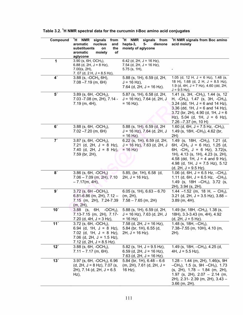
111
Table 3.2. 1H NMR spectral data for the curcumin t-Boc amino acid conjugates
Compound 1H NMR signals from aromatic nucleus and substituents on the aromatic moiety of aglycone
1H NMR signals from hepta-3, 5- dienone moiety of aglycone
1H NMR signals from Boc amino acid moiety
1 3.90 (s, 6H, OCH3), 6.88 (d, 2H, J = 8 Hz), 7.00(s, 2H), 7. 07 (d, 2 H, J = 8.5 Hz),
6.42 (d, 2H, J = 16 Hz), 7.54 (d, 2H, J = 16 Hz), 5.75 (s, 1H),
-
4´ 3.88 (s, -OCH3, 6H), 7.08 –7.19 (m, 6H)
5.88 (s, 1H), 6.59 (d, 2H, J = 16 Hz), 7.64 (d, 2H, J = 16 Hz).
1.05 (d, 12 H, J = 6 Hz), 1.48 (s, 18 H), 1.68 (d, 2 H, J = 8.5 Hz), 1.9 (d, 4H, J = 7 Hz), 4.60 (dd, 2H, J = 9.5 Hz),
5´ 3.89 (s, 6H, -OCH3), 7.03 -7.08 (m, 2H), 7.14 -7.19 (m, 4H),
5.87 (s, 1H), 6.58 (d, 2H, J = 16 Hz), 7.64 (d, 2H, J = 16 Hz).
1.41 (s, 3H, -CH3), 1.44 (s, 12 H, -CH3), 1.47 (s, 3H, -CH3), 3.24 (dd, 1H, J = 6 and 14 Hz), 3.36 (dd, 1H, J = 6 and 14 Hz), 3.72 (br, 2H), 4.90 (d, 1H, J = 6 Hz), 5.04 (d, 1H, J = 6 Hz), 7.26 –7.37 (m, 10 H)
6´ 3.88 (s, 6H, -OCH3), 7.02 –7.20 (m, 6H)
5.88 (s, 1H), 6.59 (d, 2H J = 16 Hz), 7.64 (d, 2H, J = 16 Hz).
1.60 (d, 6H, J = 7.5 Hz, -CH3), 1.49 (s, 18H, -CH3), 4.62 (br, 2H)
7´ 3.87 (s, 6H, -OCH3), 7.21 (d, 2H, J = 8 Hz), 7.40 (d, 2H, J = 8 Hz), 7.59 (br, 2H),
6.22 (s, 1H), 6.59 (d, 2H, J = 16 Hz), 7.63 (d, 2H, J = 16 Hz)
1.49 (s, 18H, -CH3), 1.21 (d, 6H, -CH3, J = 6 Hz), 1.25 (d, 6H, -CH3, J = 6 Hz), 3.72(s, 1H), 4.13 (s, 1H), 4.23 (s, 2H), 4.58 (dd, 1H, J = 4 and 9 Hz), 4.98 (d, 1H, J = 7.5 Hz), 5.12 (d, 2H, J = 9.5 Hz).
8´ 3.86 (s, 6H, -OCH3) 7.06 – 7.09 (m, 2H), 7.10 – 7.17(m, 4H),
5.85, (br, 1H), 6.58 (d, 2H, J = 16 Hz),
1.06 (d, 6H, J = 6.5 Hz, –CH3), 1.11 (d, 6H, J = 6.5 Hz, -CH3), 1.49 (s, 18H –CH3), 3.72 (s, 2H), 3.94 (s, 2H).
9´ 3.72 (s, 6H –OCH3), 6.81-6.86 (m, 2H), 7.12 –7.15 (m, 2H), 7.24-7.39 (m, 2H),
6.05 (s, 1H), 6.63 – 6.70 (m, 2H), 7.58 – 7.65 (m, 2H)
1.44 –1.52 (m, 18 H, – CH3), 3.21 (d, 2H, J = 3.5 Hz), 3.88 – 3.89 (m, 4H).
10´ 3.88 (s, 6H, -OCH3), 7.13-7.15 (m, 2H), 7.17-7.20 (d, 4H, J = 3 Hz),
5.88 (s, 1H), 6.59 (d, 2H, J = 16 Hz), 7.63 (d, 2H, J = 16 Hz)
1.49 (br, 18H, -CH3), 1.38 (s, 18H), 3.3-3.43 (m, 4H), 4.92 (d, 2H, J = 5 Hz).
11´ 3.72 (s, 6H, -OCH3), 6.94 (d, 1H, J = 8 Hz), 7.02 (d, 1H, J = 8 Hz), 7.06 (d, 2H, J = 1.5 Hz), 7.12 (d, 2H, J = 8.5 Hz).
7.58 (d, 2H, J = 16 Hz), 5.84 (br, 1H), 6.55 (d, 2H, J = 16 Hz).
1.48 (s, 18H, –CH3), 7.38–7.55 (m, 10H), 4.10 (m, 2H).
12´ 3.88 (s, 6H, -OCH3), 7.11 – 7.17 (m, 6H).
5.82 (s, 1H, J = 9.5 Hz), 6.59 (d, 2H, J = 16 Hz), 7.63 (d, 2H, J = 16 Hz).
1.49 (s, 18H, –CH3), 4.25 (d, 4H, J = 5.5 Hz),
13´ 3.97 (s, 6H, -OCH3), 6.96 (d, 2H, J = 8 Hz), 7.07 (s, 2H), 7.14 (d, 2H, J = 6.5 Hz),
5.84 (br, 1H), 6.48 – 6.6 (m, 2H), 7.61 (d, 2H, J = 16 Hz).
1.28 – 1.44 (m, 2H), 1.46(s, 9H –CH3), 1.5 (s, 9H –CH3), 1.73 (s, 2H), 1.78 – 1.84 (m, 2H), 1.97 (s, 2H), 2.07 – 2.14 (m, 2H), 2.31- 2.39 (m, 2H), 3.43 – 3.66 (m, 2H).

112
3.3.3 Reaction mechanism
The reaction of carboxylic acid of t-Boc protected amino acid with phenolic
hydroxyl of curcumin was carried out in the presence of dehydrating agent like
dicyclohexylcarbodiimide (DCC) in the presence of dimethylaminopyridine and
triethylamine as catalysts. The reaction mechanism envisaged is presented in
Fig. 3.2. Carboxylic acids capable of forming ketene intermediates undergo
esterification upon treatment with carbodiimides (Steps 1 & 2, Fig. 3.2).
Accordingly, protection of NH2 group in amino acids by t-Boc moiety led to
smooth reaction of the carboxyl group of amino acid with DCC for the formation
of reactive intermediates.
Further, selective acylation of phenols, lacking strong electron-withdrawing
groups, need acylation catalysts, mainly pyridine derivatives, to increase the
reactivity of electrophilic species for the preparation of esters through
carbodiimide couplings (Shelkov, et al., 2004). In the present reaction, the
relatively less reactive nature of phenolic group in curcumin warranted the use of
triethylamine along with DMAP for the smooth progress of the reaction. The
reaction of phenolic group with activated carboxyl moiety leading to the formation
of curcumin t-Boc amino acid conjugate and dicyclohexylurea is shown in Step 3.
Also, it was observed that in case of amino acids containing other groups, such
as –OH and –SH as in serine and cysteine respectively, no interference was
observed from these moieties.

113
R =
R΄ = alkyl / aryl / imino / substituted alkyl groups from amino acids
ArOH =
Fig. 3.2 Mechanism of t-BOC amino acids with curcumin
CH
NH
R'
t Boc
O OH
OHHO
OMeMeO
R C OH
O
R C O
O
N CC6H11 N C6H11 NH
CC6H11 N C6H11
STEP 1
STEP 2
R C O
O
R C O
O
C N
HN
C6H11
C6H11
STEP 3
Ar OH R C O
O
C N
HN
C6H11
C6H11
R C O
O
Ar
NH
CC6H11 N C6H11
HN CC6H11
HN C6H11
R C O
O
C N
HN
C6H11
C6H11
Ar OH
O

114
3.3.4 Curcumin-amino acid conjugates
The deprotection of t-Boc group was carried out using the conventional
method, where 50% TFA was used at RT for 1-2 h. The low product yield
observed was apparently due to exposure of the compounds to highly acidic
environment for long periods. An improvement in the protocol for the
deprotection of t-Boc amino acid-curcumin conjugates was envisaged with TFA
in dry dichloromethane under ultrasonic conditions. Use of ultrasound for the
deprotection was studied at different concentrations of trifluoroacetic acid (Table
3.3). The deprotection reaction was carried out with 5, 10, 15 and 20% TFA in
dichloromethane at 20 and 40 KHz frequencies. The reactions at 5 % level of
TFA (entry 1, Table 3.3) showed complete deprotection in 40 and 30 min under
20 and 40 KHz frequencies respectively, whereas the same reaction under
stirring took 48 h for completion of the reaction. Similarly reactions with 10 %
TFA in dichloromethane (entry 1, Table 3.3) took 30 and 15 min for the
completion. The deprotection rate increased as the TFA level was increased
(entries 3 and 4, Table 3.3); the time taken for the deprotection was as low as 5
min.
Table 3.3 Deprotection of t-Boc group under ultrasonic and normal conditions
Reaction time
Entry
Concentration of TFA Ultrasonic bath
(20KHz), min
Ultrasonic reactor (40KHz),
min
Stirring at RT, h
1 5% TFA in DCM 40 30 48 2 10% TFA in DCM 30 15 36 3 15% TFA in DCM 25 10 24 4 20% TFA in DCM 15 5 20

115
For the deprotection of all the conjugates of curcumin, 10% TFA in
dichloromethane was the reagent of choice and sonication (40 KHz) was
employed for removal of t-Boc group. The curcumin amino acid conjugates
formed were insoluble in dichloromethane as the removal of t-Boc moiety made
the compounds less lipophilic due to the free –NH2 group in the final product.
This facilitated the easy isolation of the curcumin-amino acid conjugates.
The method developed was milder and faster than the general protocol for
t-Boc deprotection. The compounds thus prepared were obtained in pure form
with an overall yield of 45-76%. The purity of the samples was assessed by
HPLC (Table 3.4). The removal of the t-Boc groups made these derivatives
hydrophilic. The retention time of the all the derivatives were lesser compared to
the lipophilic curcumin (14.66 min, retention time). The t-Boc derivatives showed
the retention times ranging from 6.3 – 18.38, while the deprotected derivatives
showed retention times ranging from 4.04 – 6.33. Deprotection of t-Boc group
was confirmed by the absence of eighteen protons of tertiary methyl groups of
two t-Boc moieties in the 1H NMR spectra of curcumin- amino acid conjugate
(Table 3.5). The deprotection of t-Boc group indicated shift in the positions of
signals pertaining to α– methine protons in amino acids. However, the methoxy
protons of curcumin showed very slight shift. Earlier studies reported glycine and
alanine conjugates of curcumin using corresponding N-phthaloyl amino acids
wherein carboxyl group was in activated acid chloride or p-nitrophenyl ester form.
In the present study, a simple and general reaction protocol has been delineated

116
for synthesis of curcumin-amino acid conjugates, several of them being reported
for the first time, in high yields.
Table 3.4 Curcumin amino acid conjugates
NH2 CH
R
COO OOC CH
R
NH2
O OH
OMeMeO
Structure No.
Compound Structure IV, R =
Overall Yield (%)
Retention time (Min)
4 1,7-Bis (4-O-L-leucinoyl-3-methoxyphenyl)-1,4,6-heptatriene-5-ol-3-one
CH CH2
CH3
CH3
71 6.33
5 1,7-Bis (4-O-L-phenylalaninoyl-3-methoxyphenyl)-1,4,6-heptatriene-5-ol-3-one
CH2
68 6.01
6 1,7-Bis (4-O-L-alaninoyl-3-methoxyphenyl)-1,4,6-heptatriene-5-ol-3-one
CH3 76 5.06
7 1,7-Bis (4-O-L- isoleucinoyl -3-methoxyphenyl)-1,4,6-heptatriene-5-ol-3-one
CH
CH3
CH2CH3
63 6.14
8 1,7-Bis (4-O-L- valinoyl -3-methoxyphenyl)-1,4,6-heptatriene-5-ol-3-one
CH3
CHCH3
65 4.86
9 1,7-Bis (4-O-L- serinoyl -3-methoxyphenyl)-1,4,6-heptatriene-5-ol-3-one
CH2OH
45 5.06
10 1,7-Bis (4-O-L- cysteinoyl -3-methoxyphenyl)-1,4,6-heptatriene-5-ol-3-one
CH2SH
70 4.76
11 1,7-Bis (4-O-L- phenylglycinoyl -3-methoxyphenyl)-1,4,6-heptatriene-5-ol-3-one
59 4.78
12 1,7-Bis (4-O-L- glycinoyl -3-methoxyphenyl)-1,4,6-heptatriene-5-ol-3-one
H 57 4.04
13 1,7-Bis (4-O-L- prolinoyl -3-methoxyphenyl)-1,4,6-heptatriene-5-ol-3-one
CH2 CH2 CH2
( as part of a pyrolidine ring)
70 5.00
(IV)
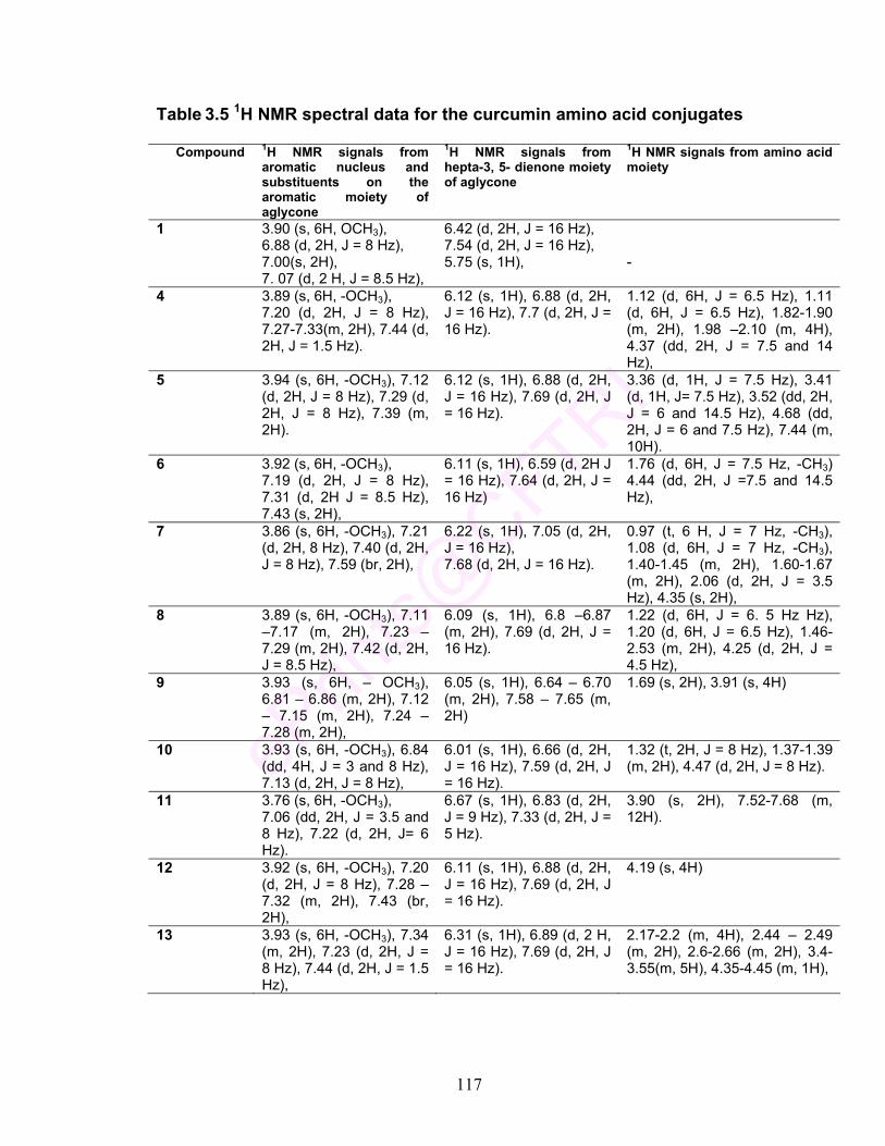
117
Table 3.5 1H NMR spectral data for the curcumin amino acid conjugates
Compound 1H NMR signals from aromatic nucleus and substituents on the aromatic moiety of aglycone
1H NMR signals from hepta-3, 5- dienone moiety of aglycone
1H NMR signals from amino acid moiety
1 3.90 (s, 6H, OCH3), 6.88 (d, 2H, J = 8 Hz), 7.00(s, 2H), 7. 07 (d, 2 H, J = 8.5 Hz),
6.42 (d, 2H, J = 16 Hz), 7.54 (d, 2H, J = 16 Hz), 5.75 (s, 1H),
-
4 3.89 (s, 6H, -OCH3), 7.20 (d, 2H, J = 8 Hz), 7.27-7.33(m, 2H), 7.44 (d, 2H, J = 1.5 Hz).
6.12 (s, 1H), 6.88 (d, 2H, J = 16 Hz), 7.7 (d, 2H, J = 16 Hz).
1.12 (d, 6H, J = 6.5 Hz), 1.11 (d, 6H, J = 6.5 Hz), 1.82-1.90 (m, 2H), 1.98 –2.10 (m, 4H), 4.37 (dd, 2H, J = 7.5 and 14 Hz),
5 3.94 (s, 6H, -OCH3), 7.12 (d, 2H, J = 8 Hz), 7.29 (d, 2H, J = 8 Hz), 7.39 (m, 2H).
6.12 (s, 1H), 6.88 (d, 2H, J = 16 Hz), 7.69 (d, 2H, J = 16 Hz).
3.36 (d, 1H, J = 7.5 Hz), 3.41 (d, 1H, J= 7.5 Hz), 3.52 (dd, 2H, J = 6 and 14.5 Hz), 4.68 (dd, 2H, J = 6 and 7.5 Hz), 7.44 (m, 10H).
6 3.92 (s, 6H, -OCH3), 7.19 (d, 2H, J = 8 Hz), 7.31 (d, 2H J = 8.5 Hz), 7.43 (s, 2H),
6.11 (s, 1H), 6.59 (d, 2H J = 16 Hz), 7.64 (d, 2H, J = 16 Hz)
1.76 (d, 6H, J = 7.5 Hz, -CH3) 4.44 (dd, 2H, J =7.5 and 14.5 Hz),
7 3.86 (s, 6H, -OCH3), 7.21 (d, 2H, 8 Hz), 7.40 (d, 2H, J = 8 Hz), 7.59 (br, 2H),
6.22 (s, 1H), 7.05 (d, 2H, J = 16 Hz), 7.68 (d, 2H, J = 16 Hz).
0.97 (t, 6 H, J = 7 Hz, -CH3), 1.08 (d, 6H, J = 7 Hz, -CH3), 1.40-1.45 (m, 2H), 1.60-1.67 (m, 2H), 2.06 (d, 2H, J = 3.5 Hz), 4.35 (s, 2H),
8 3.89 (s, 6H, -OCH3), 7.11 –7.17 (m, 2H), 7.23 – 7.29 (m, 2H), 7.42 (d, 2H, J = 8.5 Hz),
6.09 (s, 1H), 6.8 –6.87 (m, 2H), 7.69 (d, 2H, J = 16 Hz).
1.22 (d, 6H, J = 6. 5 Hz Hz), 1.20 (d, 6H, J = 6.5 Hz), 1.46-2.53 (m, 2H), 4.25 (d, 2H, J = 4.5 Hz),
9 3.93 (s, 6H, – OCH3), 6.81 – 6.86 (m, 2H), 7.12 – 7.15 (m, 2H), 7.24 –7.28 (m, 2H),
6.05 (s, 1H), 6.64 – 6.70 (m, 2H), 7.58 – 7.65 (m, 2H)
1.69 (s, 2H), 3.91 (s, 4H)
10 3.93 (s, 6H, -OCH3), 6.84 (dd, 4H, J = 3 and 8 Hz), 7.13 (d, 2H, J = 8 Hz),
6.01 (s, 1H), 6.66 (d, 2H, J = 16 Hz), 7.59 (d, 2H, J = 16 Hz).
1.32 (t, 2H, J = 8 Hz), 1.37-1.39 (m, 2H), 4.47 (d, 2H, J = 8 Hz).
11 3.76 (s, 6H, -OCH3), 7.06 (dd, 2H, J = 3.5 and 8 Hz), 7.22 (d, 2H, J= 6 Hz).
6.67 (s, 1H), 6.83 (d, 2H, J = 9 Hz), 7.33 (d, 2H, J = 5 Hz).
3.90 (s, 2H), 7.52-7.68 (m, 12H).
12 3.92 (s, 6H, -OCH3), 7.20 (d, 2H, J = 8 Hz), 7.28 – 7.32 (m, 2H), 7.43 (br, 2H),
6.11 (s, 1H), 6.88 (d, 2H, J = 16 Hz), 7.69 (d, 2H, J = 16 Hz).
4.19 (s, 4H)
13 3.93 (s, 6H, -OCH3), 7.34 (m, 2H), 7.23 (d, 2H, J = 8 Hz), 7.44 (d, 2H, J = 1.5 Hz),
6.31 (s, 1H), 6.89 (d, 2 H, J = 16 Hz), 7.69 (d, 2H, J = 16 Hz).
2.17-2.2 (m, 4H), 2.44 – 2.49 (m, 2H), 2.6-2.66 (m, 2H), 3.4-3.55(m, 5H), 4.35-4.45 (m, 1H),
118
3.3.5 Characterization of amino acid conjugates- Representative examples (i) 1,7-Bis(4-O-L-leucinoyl-3-methoxyphenyl)-1,4,6-heptatriene-5-ol-3-one (4)
The purity of the sample was assessed by HPLC where a single peak at
13.16 min was seen for 4΄ (Table 3.1, Fig. 3.3). Pure curcumin showed retention
of 14.66 min under similar conditions.
The mass spectral analysis showed that 4΄ had a M+Na adduct at 817
corresponding to the di-Boc leucinoyl derivative of curcumin (Table 3.1, Fig. 3.4).
The 1H NMR of 4΄ showed characteristic tert-butylcarbamate associated –CH3
signals for eighteen protons at a chemical shift of 1.48. The two –CH3 group
protons of the leucine moiety attached to curcumin were seen at 1.05 as a
doublet corresponding to twelve protons indicating the two Boc–leucine groups
were attached to the two phenolic hydroxyls of curcumin (Table 3.2, Fig. 3.5).
The –OCH3 signals of curcumin were seen at 3.88 corresponding to 6H. The
characteristic –CH proton of the t-Boc leucine linked to curcumin signals were
seen at 4.60 corresponding to 2H compared to t-Boc leucine standard which
shows the –CH linked to carboxyl at 4.29. This indicated the conjugation of two
moieties of t-Boc leucine to curcumin. The methylene group of the amino acid
moiety showed signal at 1.9 corresponding to 4H. The methine moiety α to two
methyls showed signal at 1.68 (2H).

119
700 750 800 850
%
0
100 817.55
817.52
817.58
817.62
Minutes8 10 12 14 16
Vol
ts
0.0
0.5
1.0
13.1
58
Fig. 3.3 HPLC profile of 1,7-Bis (4-O-L-t-Boc-leucinoyl- 3-methoxyphenyl)-
1,4,6- heptatriene-5-ol-3-one (4΄ )
Fig. 3.4 ESI-MS of 1,7-Bis (4-O-L-t-Boc-leucinoyl-3-methoxyphenyl)-1,4,6- heptatriene-5-ol-3-one (4΄)
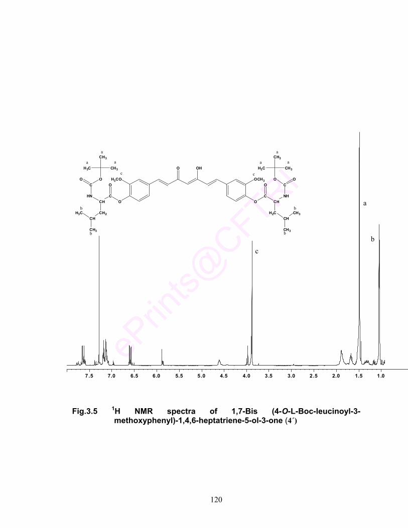
120
Fig.3.5 1H NMR spectra of 1,7-Bis (4-O-L-Boc-leucinoyl-3- methoxyphenyl)-1,4,6-heptatriene-5-ol-3-one (4΄)
O OH
H3CO OCH3
HNCH
C
CH2
O
CH
CH3
H3C
CO
H3C
CH3
CH3
O
ONH
CHC
H2C
O
CH
CH3
CH3
CO
CH3
CH3
H3C
O
O
a
a a
a
a a
b
b
b
b
c c
a
bc
7.5 7.0 6.5 6.0 5.5 5.0 4.5 4.0 3.5 3.0 2.5 2.0 1.5 1.0
121
After deacetylation the product was checked for its purity by HPLC. The
compound showed retention at 6.33 (Fig. 3.6). The removal of t-Boc groups
showed shift in the retention time from 13.16 for curcumin-t-Boc leucine
conjugate to 6.33 for curcumin-leucine conjugate. Further, the compound was
characterized by 1H NMR (Table 3.5). Disappearance of –CH3 (18H) signals
from the t-Boc groups in the conjugate showed completion of deprotection (Fig.
3.7). The -CH3 group signals of the leucine moiety were seen at a chemical shift
value of 1.11-1.12 (12 H). The –OCH3 of curcumin corresponding to six protons
was seen at 3.89. The α–methine protons showed signal at 4.37. Signals for the
other methine group of leucine linked to –CH3 was observed at 1.82-1.90 (m, 2H)
and the methylene group was observed at 1.98 - 2.1 (m, 4H). The aromatic
protons of curcumin in the curcumin – leucine conjugate now appeared at 7.20
(2H), 7.27-7.33 (2H) and 7.44 (2H).
(ii) 1,7-Bis (4-O-L-alaninoyl-3-methoxyphenyl)-1,4,6- heptatriene-5-ol-3-one (6)
The purity of the compound was assessed by HPLC where the single peak
at 8.06 for 6΄ (Table 3.1, Fig 3.8). NMR and ESI MS characterized the compound
6΄. The ESI MS (+ve mode) spectral analysis showed that 6΄ had a M+Na adduct
at 733 corresponding to di-Boc alaninoyl derivative curcumin the of (Table 3.1,
Fig. 3.9). 1H NMR further confirmed the t-Boc derivative of curcumin alanine
conjugate. The spectra showed chemical shift values at 1.66 and 1.59
corresponding to the six protons of two –CH3 groups of alanine. The chemical
shift values of 1.49 were shown for t-Boc moiety corresponding to eighteen
protons. The –OCH3 signal of curcumin was at 3.88 a singlet showing integration

122
Minutes2 4 6 8
Vol
ts
0.0
0.1
6.33
for 6 protons. The α–methine protons of Boc alanine linked to curcumin showed
signal at 4.62 corresponding to 2H, while the standard t-Boc alanine showed
signal at 4.37 for the same. The spectra confirmed the formation of t-Boc alanine
linked at both the phenolic hydroxyls of curcumin (Fig. 3.10).
After deprotection of t-Boc group, the product was checked for its purity by
HPLC. The compound 6 showed retention time of 5.06 as a single peak
confirming its purity (Fig. 3.11). 1H NMR using CD3OD as a solvent further
characterized the product 6. The spectra showed the absence of t-Boc methyl
groups (18H) confirming the deprotection (Fig. 3.12). Chemical shift value of 1.76
(d, 6H, J = 7.5 Hz, -CH3) indicated the presence of the two methyl groups of
alanine moiety, the –OCH3 showed singlet at 3.92 from curcumin moiety of 6.
The α–methine protons of curcumin-alanine conjugate was seen at 4.44 (2H).
The aromatic protons of curcumin in the curcumin–alanine conjugate now
appeared at 7.19 (2H), 7.31 (2H) and 7.43 (2H).
Fig. 3.6 HPLC profile of 1,7-Bis (4-O-L-leucinoyl-3-methoxyphenyl)-1,4,6-
heptatriene-5-ol-3-one (4)

123
Fig. 3.7 1H NMR spectra of 1,7-Bis (4-O-L-leucinoyl-3-methoxyphenyl)-1,4,6- heptatriene-5-ol-3-one (4)
7.5 7.0 6.5 6.0 5.5 5.0 4.5 4.0 3.5 3.0 2 .5 2.0 1.5 1.0 0.5 ppm
O OH
H3CO OCH3
H2NCH
C
CH2
O
CH
CH3
H3C
ONH2
CHC
H2C
O
CH
CH3
CH3
O
b
b
b
b
cc
bc

124
Minutes2 4 6 8
Vol
ts
0.0
0.5
1.0
8.05
8
m/695 700 705 710 715 720 725 730 735 740 745
%
0
100 1733.54733.48 733.56
Fig. 3.8 HPLC profile of 1,7-Bis (4-O-L-t-Boc alaninoyl -3-methoxyphenyl)- 1,4,6- heptatriene-5-ol-3-one (6΄)
Fig. 3.9 ESI-MS of 1,7-Bis (4-O-L-Boc-alaninoyl-3-methoxyphenyl)-1,4,6-
heptatriene-5-ol-3-one (6΄)

125
Fig. 3.10 1H NMR spectra of 1,7-Bis (4-O-L-Boc-alaninoyl-3-methoxyphenyl)-
1,4,6-heptatriene-5-ol-3-one (6´)
1.52.02.53.03.54.04.55.05.56.06.57.07.5 ppm
b
a
c
O OH
OMe OMe
CHNH CH COO OOC
CH3 CH3b b
NH
ca
a
CO
H3CCH3
CH3
COH3C
CH3
CH3c

126
Minutes0 2 4 6 8 10
Vol
ts
0.0
0.5
1.0
5.05
8
Fig. 3.11 HPLC profile of 1,7-Bis (4-O-L- alaninoyl -3-methoxyphenyl)-1,4,6- heptatriene-5-ol-3-one (6)
Fig. 3.12 1H NMR spectra of 1,7-Bis (4-O-L- alaninoyl -3-methoxyphenyl)- 1,4,6-heptatriene-5-ol-3-one (6)
1.52.02.53.03.54.04.55.05.56.06.57.07.5 ppm
O OH
OMe OMe
CHNH2 CH COO OOC
CH3 CH3b b
NH2
c c
bc

127
3.4 CONCLUSIONS
In this present study, a synthetic protocol was developed for the preparation
of several curcumin-amino acid conjugates in high yields. In the reaction of
curcumin with amino acids, the amino functionality in amino acids was protected
by a N-tert-butoxycarbonyl (t-Boc) group. Selective dehydration reaction between
the phenolic groups in curcumin and the carboxyl functional group of the amino
acids as effected by N, N’-dicylohexylcarbodiimide (DCC) and catalyzed by
dimethylaminopyridine and triethylamine led to the formation of the t-Boc
protected amino acid conjugates of curcumin. The synthetic methodology
developed envisaged facile reaction of the N-protected (t-Boc) amino acids with
curcumin and included an improved accelerated protocol for the deprotection of t-
Boc group to afford the formation of amino-acid conjugates of curcumin in high
yields. Accordingly, several amino acid conjugates of curcumin were synthesized
in 45–76% yields. Covalent binding of amino acids to curcumin at phenolic
positions rendered the curcumin derivatives water-soluble.

128
References
Aggarwal, B. B., Kumar, A., Aggarwal, M. S., & Shishodia. S. 2005. Curcumin
Derived from Turmeric (Curcuma longa): a Spice for All Seasons.
Published by CRC Press LLC; 350-379.
Ammon, H.P., & Wahl, M.A. 1991. Pharmacology of Curcuma longa. Planta
Medica, 57(1), 1-7.
Amolins, M. W., Peterson, L. B., & Blagg, B. S. J. 2009. Synthesis and evaluation
of electron-rich curcumin analogs. Bioorganic & Medicinal Chemistry, 17
(1), 360-367.
Armarego, W.L.F., & Chai, C.L.L., 2003. Purification of Laboratory chemicals.
Butterworth-Heinernann Publications, 5Th edition.
Chainani. Wu N. 2003. Safety and anti-inflammatory activity of curcumin: a
component of turmeric (curcuma longa). Journal of Alternative &
Complementary Medicine, 9(1), 161-168.
Deng., S.L., Chen, W.F., Bo –Zhou, Li-Yang & Liu, Z.L. 2006. Protective effects
of curcumin and its analogs against free radical-induced oxidative
haemolysis of human red blood cells. Food chemistry, 98 (1), 112-119.
Green, T. W. & Wuts, P. G. M. 1999. Protective Groups in Organic Synthesis,
Wiley-Interscience, New York, 518-525 & 736-739.
Hatcher, H., Planalp, R., Cho, J., Torti, F.M., & Torti, S.V. 2008. Curcumin: from
ancient medicine to current clinical trials. Cellular & Molecular Life
Sciences, 65(11), 1631-1652.

129
Hergenhahn, M., Bertram, B., Wiessler, M., & Sorg, B. L. 2003. Curcumin
derivatives with improved water solubility compared to curcumin and
medicaments containing the same. United States Patent Application,
20030153512.
Ishida, J., Ohtsu, H., Tachibana, Y., Nakanishi, Y., Bastow, K. F., Nagai, M., Hui-
Kang, W., Itokawa, H., & Kuo-Hsiung, L. 2002. Antitumor Agents. Part
214: Synthesis and Evaluation of Curcumin Analogs as Cytotoxic
Agents. Bioorganic & Medicinal Chemistry, 10, 3481–3487.
Jiao, Y., Wilkinson, J., Pietsch, E. C., Buss, J. L., Wang, W., Planalp , R., Torti,
F. M., & Torti, S. V. 2006. Iron chelation in the biological activity of
curcumin. Free Radical Biology & Medicine, 40, 1152 – 1160.
Joe, B., Vijaykumar, M., & Lokesh, B. R. 2004. Biological properties of curcumin -
cellular and molecular mechanisms of action. Critical Reviews in Food
Science & Nutrition, 44(2), 97-111.
Kapoor, N., Narain, U., & Misra, K. 2007. Bioactive conjugates of curcumin
having ester, peptide, thiol and disulphide links. Journal of Scientific &
Industrial Research, 66 (8), 647-650.
Kapoor, N., Sharma, A.K., Dwivedi, V., Kumar, A., Pati, U., & Misra, K. 2007a.
Telomerase targeted anticancer bioactive prodrug by antisense-based
approach. Cancer Letters, 248, 245–250.
Kumar, S., Dubey, K. K., Tripathi, S., Fujii, M., & Misra, K. 2000. Design and
synthesis of curcumin-bioconjugates to improve systemic delivery.
Nucleic Acids Symposium Series, 44, 75-76.

130
Kumar, S., Narain, U., Tripathi, S., & Misra, K. 2001. Synthesis of curcumin
bioconjugates and study of their antibacterial activities against β-
Lactamase-producing microorganisms. Bioconjugate Chemistry, 12, 464
-469.
Menon, V.P., & Sudheer, A. R. 2007. Antioxidant and anti-inflammatory
properties of curcumin. Advances in Experimental Medicine & Biology,
595, 105-125.
Mishra, S., Kapoor, N., Ali, A. M., Pardhasaradhi, B. V. V., Leela Kumari, A.,
Khar, A., & Misra, K. 2005. Differential apoptotic and redox regulatory
activities of curcumin and its derivatives. Free Radical Biology &
Medicine, 38, 1353– 1360.
Mishra, S., Narain, U., Mishra R., & Misra, K. 2005a. Design, development and
synthesis of mixed bioconjugates of piperic acid–glycine, curcumin–
glycine/alanine and curcumin–glycine–piperic acid and their antibacterial
and antifungal properties. Bioorganic & Medicinal Chemistry, 13, 1477-
1486.
Park, S., & Kim, D. S. H. L. 2002. Discovery of Natural Products from Curcuma
longa that Protect Cells from Beta- amyloid Insult: A Drug Discovery
Effort against Alzheimer’s Disease. Journal of Natural Products, 66 (9),
1227 -1231.
Parvathy, K.S., & Srinivas, P. 2008. Ultrasound-assisted reaction of 2,3,4,6-tetra-
O-acetyl-a-D-glucopyranosyl bromide with potassium salt of curcumin
under PTC conditions. Ultrasonics Sonochemistry, 15, 571–577.
131
Sharma, R. A., Gescher, A. J., & Steward, W. P. 2005. Curcumin: the story so
far, European Journal of Cancer, 41, 1955–1968.
Shelkov, R., Nahmany, M., & Melman, A. 2004. Selective esterifications of
alcohols and phenols through carbodiimide couplings. Organic
Biomoleular Chemistry, 2, 397- 401.
Srimal, R. C., 1997. Turmeric: a brief review of medicinal properties. Fitoterapia,
68, 483-493.
Venkatesan, P., & Rao, M. N. A. 2000. Structure-activity relationships for the
inhibition of lipid peroxidation and the scavenging of free radicals by
synthetic symmetrical curcumin analogs. Journal of Pharmacy &
Pharmacology, 52(9), 1123-1128.

132
CHAPTER 4 SYNTHESIS OF GLUCURONIC ACID &
GLUCAL DERIVATIVES OF CURCUMIN
133
4.1 GLUCURONIC ACID DERIVATIVE OF CURCUMIN
4.1.1 INTRODUCTION
Biotransformation of curcumin has been studied and the structure of its
metabolites after oral or i. p. administration in mice characterized. THC is a major
metabolite of curcumin and its stability at different pH is also studied (Pan, et al.,
1999). These studies show the metabolic steps of curcumin involve the
glucuronidation of curcumin and its major reduced forms viz.,
tetrahydrocurcumin, hexahydrocurcumin and octahydrocurcumin. Glucuronides
of curcumin, hexahydrocurcumin, and other analogs have been synthesized
using rat and human liver microsomes and characterized by LC-MS/MS analysis
(Pfeiffer, et al., 2007). Biotransformation of curcumin shows that curcumin is first
bio-transformed to dihydrocurcumin and tetrahydrocurcumin and that these
compounds subsequently are converted to monoglucuronide conjugates. Thus,
curcumin glucuronide, dihydrocurcumin glucuronide, tetrahydrocurcumin
glucuronide and tetrahydrocurcumin are major metabolites of curcumin in mice
(Lin, et al., 2000). As the glucuronidation is a part of metabolic pathway of
curcumin, the synthesis of curcumin glucuronide has been taken up in this
present work, as this derivative would be water-soluble.
Curcumin metabolism and its absorption are studied after its oral
administration in rats at different dosages of [3H] curcumin. The radioactivity
studies have shown that curcumin gets poorly absorbed in gut and transforms
into more polar and colorless compounds. These experiments indicate that
curcumin undergoes transformation during absorption from the intestine and the

134
studies on the tissue distribution are also studied. Rat intestines when incubated
with 50-750 μg of curcumin in incubation medium, 30-80% of the added curcumin
disappears from the mucosal side. Curcumin is not detectable in the serosal fluid.
Less than 3% of the added curcumin is found in the tissue at the highest
concentration, which shows curcumin gets transformed in the tissues
(Ravindranath & Chandrasekhara, 1980; 1981; 1982). Curcumin metabolism is
studied in human and rat hepatocytes and also in rats in vivo. Analysis by HPLC
with detection at 420 and 280 nm show metabolites with both intact
diferuoylmethane and increased saturation of the heptatrienone chain. Results
are confirmed by mass spectral analysis where, hexahydrocurcumin and
hexahydrocurcuminol are identified as major metabolites of curcumin. In rats, in
vivo, curcumin administered (40 mg/kg) disappears from the plasma within 1 h of
dosing. After administration (500 mg/kg), parent drug is present in plasma at
levels near the detection limit. The major products of curcumin biotransformation
identified in rat plasma are curcumin glucuronide and curcumin sulfate, whereas
hexahydrocurcumin, hexahydrocurcuminol, and hexahydrocurcumin glucuronide
are present in small amounts. (Iresson, et al., 2002). The effect of curcumin on
lipid metabolism in rats fed on a control, moderately high-fat diet and those given
supplements of 0.2 g curcuminoids/100 g diet is studied. Triacylglycerol and
cholesterol contents are lower in rats fed curcumin than in control rats. Results
indicate that dietary curcuminoids have lipid-lowering potency in vivo, probably
due to alterations in fatty acid metabolism (Asai & Miyazawa, 2001).

135
Methods are reported in literature where various strategies are adopted for
the covalent linking of glucuronic acid to other molecules. Acetylated glucuronic
acid moiety is made to react with alcohols to give glycosides (Tosin & Murphy,
2004). The glycosidation of glucuronic acid with 4-hydroxycinnamic acid has
been reported with the anomeric activation (Pearson, et al., 2005). 2,3,4-Tri-O-
acetyl-D-glucurono-6,1-lactone is obtained in ‘one-pot’ synthesis from D-
glucuronic acid. The lactone opening with different alcohols in the presence of a
variety of catalysts has been studied. All reactions have been performed under
microwave irradiation in solvent-free conditions (Rat, et al., 2007). Different
synthetic routes to differentially protected D-glucuronic acid glycols that may
serve as intermediates en route to a variety of natural products are reported
(Schell, et al., 2001). Glucuronidation of opium drug like thienorphine is carried
out and its synthetic protocol developed (Liu, et al., 2005). Synthesis of some
phenolic glycosides (urine drug metabolites) is reported (Arewang, et al., 2007).
Glucuronide prodrugs of 9-aminocamptothecin have been synthesized and
shown to possess the necessary prerequisites to be considered as candidates
for cancer prodrug monotherapy and antibody directed enzyme prodrug therapy
of cancer (Leu, et al., 1999).
In the present study, a synthetic pathway to curcumin glucuronide was
envisaged starting from glucuronolactone, a readily available substrate. The
synthetic route involves opening of the lactone ring with concomitant protection of
the carboxyl group and activation at the anomeric carbon for facile bonding of the
phenolic groups of curcumin with the sugar moiety.
136
4.1.2 EXPERIMENTAL
(a) Materials and equipment
Acetic anhydride, acetonitrile and methanol were procured from s. d. fine
chemicals Mumbai, India and dried as per the standard protocol (Armarego &
Chai, 2003). Glucuronic acid γ-lactone, Titanium tetrabromide, Perchloric acid
(70%), Benzyltributylammonium chloride, Cetyltributylammonium bromide,
Dowex (IR-120), Tetrabutylammonium bromide and 3,4,6-tri-O-acetyl-D-glucal
were procured from Sigma–Aldrich chemical company, USA. Alumina coated
TLC plates were procured from E-Merck India Ltd. Curcumin (97 %) was
procured from Spicex Pvt. Ltd., Mysore, India. Fe2 (SO4)3 xH2O,
Dichloromethane, triethylamine, hexane, sodium sulfate, chloroform, potassium
hydroxide and potassium carbonate were procured from Qualigens India Ltd.
Mumbai, India. Silica gel was obtained from s. d. Fine chemicals, Mumbai, India.
Ultrasound reactor used was Vibracell-750 with tapered probe of tip
diameter 6.5 mm from Sonics and Materials Inc., Newtown, USA and was set at
25% amplitude and pulse cycle of 25 s (on) and 5 s (off) with frequency of 40
KHz and an output power of 750 W. 1H NMR spectra for the compounds were
recorded on a 500 MHz NMR spectrometer (Bruker Avance, Reinstetten,
Germany) using, CDCl3 and CD3OD solvents. Chemical shift values and coupling
constants are given in δ and Hz respectively. Mass spectral analyses of
compounds were carried out using MS (Waters Q-Tof Ultima, Manchester, UK) in
the ESI positive mode. Thin-layer chromatographic (TLC) analysis was
performed on silica gel 60 F254 (Merck, Germany) coated on alumina sheet with

137
3% methanol in chloroform as the developing solvent. Samples were analyzed
on HPLC (Waters, LC 10 A, USA) using a reverse phase C-18 column with
methanol : water (70:30) containing trifluroacetic acid (0.1%) as the mobile phase
at flow rate of 1 ml/min and monochromatic detection at 423 nm.
(b) Synthesis methyl 1,2,3,4-Tetra-O-acetyl-β-D-glucopyranuronate (Leu, et al., 1999; Liu, et al., 2005)
Glucuronic acid γ-lactone, [(2R)-2-[(2S, 3R, 4S)-3,4-Dihydroxy-5-oxo-
tetrahydrofuran-2-yl]-2-hydroxy-acetaldehyde (25 g)], was added to MeOH (50
ml) that contained sodium (75 mg). The mixture was stirred at room temperature
for 60 min after which the solvent was distilled under reduced pressure. The
syrupy product was diluted with acetic anhydride (10 ml). This solution was
added drop wise to a mixture of acetic anhydride (30 ml) and HClO4 (0.15 ml),
prepared below 4 °C. During this addition, the reaction temperature was
maintained below 40 °C. The mixture was stirred for 24 h at room temperature
and the solution stored overnight at 4 °C to yield 12 g of crystalline product. For
further work up of the product, the mother liquor was poured onto crushed ice
and neutralized with NaHCO3 and the filtrate was extracted with CHCl3. The
CHCl3 extract was dried over anhydrous Na2SO4 and concentrated to syrup.
Upon storage at 4°C an additional 5 g of crude crystalline material was obtained.
The product was re-crystallized from MeOH: 62%, mp 140 °C. 2.05 (s, 3H,
OCOCH3), 2.06 (s, 3H, OCOCH3), 2.07 (s, 3H, OCOCH3), 2.14 (s, 3H, OCOCH3),
3.77 (s, 3H, -CH3), 4.20 (1H, d, J = 9.5 Hz), 5.16 (1H, dd, J = 8 Hz), 5.26 (1H, t, J
= 9 Hz), 5.33 (1H, t, J = 9 Hz) & 5.78 (1H, d, J = 8 Hz).

138
(c) Synthesis methyl 1-bromo-2, 3, 4-tri-O-acetyl-α-D-glucopyranuronate
A solution of compound methyl 1, 2, 3, 4-Tetra-O-acetyl-α-D-
glucopyranuronate (10 g, 2.66 mmol) and TiBr4 (11 g, 3.19 mmol) in CH2Cl2 (25
ml) was stirred at room temperature for 24 h. The mixture was washed with ice
water and NaHCO3 solution, dried over Na2SO4, and evaporated to dryness to
give 4 g of white solid. m. p. 151-153 oC, ESI – MS (M+ Na)+ 410, 1H NMR (500
MHz, CDCl3); 2.07 (s, 3H, OCOCH3), 2.08 (s, 3H, OCOCH3), 2.12 (s, 3H,
OCOCH3), 3.78 (s, 3H, -COOCH3), 4.60 (d, 1H, J = 10.5 Hz), 4.87 (dd, 1H, J =
10 Hz), 5.26 (t, 1H, J = 10 Hz), 5.63 (t, 1H, J= 10 Hz), 6.66 (d, 1H, J = 4Hz).
(d)Synthesis of methyl 1-curcuminyl-2,3,4-tri-O-acetyl-β-D-gluco-pyranuronate (14´)
To a solution of curcumin (1, Fig. 1, 1 g, 2.7 mmol) in chloroform (40 ml),
aqueous KOH (0.6 g, 10.8 mmol in 20 ml water) was added and stirred for 5 min.
A solution of methyl 1-bromo-2,3,4-tri-O-acetyl-α-D-glucopyranuronate (2.2 g,
5.35 mmol) in chloroform (40 ml) was added. To the reaction mixture an aqueous
solution of benzyltributylammonium chloride (0.5 g, 1.6 mmol in 20 ml water) was
added. The mixture was sonicated until the completion of the reaction (4 - 6 h),
which was indicated by the formation of clear aqueous layer. TLC analyzed the
reaction product for the disappearance of curcumin. Then the chloroform layer
was washed with water, dried over anhydrous sodium sulfate and distilled under
reduced pressure. The crude sample was loaded on to silica gel (200-400 mesh)
column and eluted out with CHCl3. Pure fractions were pooled and evaporated to
afford curcumin mono β-glucuronide triacetate derivative in 50 % yield (14΄).

139
1H NMR (CDCl3): 2.07 (s, 3H, -OCOCH3), 2.08 (s, 3H, -OCOCH3), 2.12 (s, 3H,
OCOCH3), 3.79 (s, 3H, -COOCH3), 3.99 (s, 6H, –OCH3), 4.50 (1H, d, J = 10.5
Hz), 4.87 (dd, 1H, J = 4 Hz), 5.26 (t, 1H, J = 10 Hz), 5.62 (t, 1H, J = 10 Hz), 5.84
(t, 1H, J = 8 Hz), 5.91 (s, 1H), 6.80 (d, 2H, J = 15.5 Hz), 7.15 (d, 2H, J = 8 Hz),
7.28 (d, 2H, J = 3.5 Hz), 7.42 (s, 2H), 7.60 (d, 2H, J = 15.5 Hz).
(e) Methyl 1-curcuminyl-β-D-glucopyranuronate (14)
To methyl 1-curcuminyl-2,3,4-tri-O-acetyl-β-D-gluco-pyranuronate (14΄, 1
g) in dry methanol (10 ml), a solution of LiOH (2.2 mmol) in methanol (8 ml) was
added and the mixture was stirred. The deacetylation reaction was monitored on
TLC. At the end of the reaction (0.5 h), the solution was neutralized by the
addition of freshly regenerated Dowex (IR-120) H+ resin. The resin was filtered
and washed with methanol. The solvent distilled under reduced pressure to
afford pure methyl 1-curcuminyl-β-D-glucopyranuronate in 95% yield (14). m.p.
65 oC; 1H NMR (CD3OD): 3.17 (s, 3H, -COOCH3), 3.85 (s, 6H, –OCH3), 4.10 (1H,
d, J = 7.5 Hz), 5.11 (m, 2H, J = 5 Hz), 5.76 (1H, s), 6.04 (s, 1H), 6.10 (s, 1H),
6.78 (d, 2H, J = 15.5 Hz), 7.13 (d, 2H, J = 8 Hz), 7.26 (d, 2H, J = 3.5 Hz), 7.39 (s,
2H), 7.57 (d, 2H, J = 15.5 Hz).

140
4.1.3 RESULTS AND DISCUSSION
Reaction of curcumin with methyl 1-bromo-2, 3, 4-tri-O-acetyl-α-D-
glucopyranuronate with potassium salt of curcumin under biphasic conditions
using phase transfer catalysts was studied under the influence of ultrasound for
the synthesis of glucuronide derivative of curcumin (Fig. 4.1). Curcumin is prone
to rapid degradation in the presence of alkali and high temperature. Reaction of a
solution of curcumin in chloroform with aqueous KOH led to facile formation of
potassium salt of curcumin indicated by the decolorization of the organic layer
with concomitant transfer of the pigment to the aqueous phase. A solution of
methyl 1-bromo-2, 3, 4-tri-O-acetyl- α -D-glucopyranuronate in chloroform was
added, which constituted a biphasic system with the sugar halide in the organic
phase and the potassium salt of the phenolic substrate, curcumin in the aqueous
phase. Aqueous solution of benzyltributylammonium chloride was then added
and the contents were stirred which resulted in the formation of a homogeneous
mixture. The use of ultrasound as an alternative source for providing the
activation energy for this reaction was envisaged. The reaction mixture was
sonicated for 4 h until the aqueous and organic phases got separated and the
aqueous layer became colorless. The organic layer after work up and
chromatographic separation afforded methyl 1-curcuminyl-2,3,4-tri-O-acetyl-β-D-
glucopyranuronate (>50 %).

141
Fig 4.1 Synthesis of methyl 1-curcuminyl-β-D-glucopyranuronate
i, NaoMeii, Acetic anhydride, Perchloric acid i, TiBr4 , DCM
O OH
H3CO
HO
OCH3
OH
O OH
H3CO
O
OCH3
OH
O OH
H3CO
O
OCH3
OH
DeacetylationLiOH, Methanol
PTC, KOHH2O : CHCl31:2
1
14
14'
O
OO
HO
OH
OH
OAcO
AcOOAc
OAc
H3CO O
OAcO
AcO
OAc
H3CO O
Br
OAcO
AcO
OAc
H3CO O
Br
OAcO
AcO
OAc
H3CO O
OHO
HO
OH
H3CO O

142
Minutes5 10 15 20
mA
U
0
25
50
15.6
16
Fig. 4.2 HPLC profile of methyl 1-curcuminyl-2,3,4-tri-O-acetyl-β-D-gluco- pyranuronate (14΄)
Fig. 4.3 ESI MS of methyl 1-curcuminyl-2,3,4-tri-O-acetyl-β-D-gluco- pyranuronate (14΄)
143
(a) Spectral characterization of the products
The reactions were carried out with different phase transfer catalysts like
cetyltributylammonium bromide, tetrabutylammonium bromide and
benzyltributylammonium chloride. The reaction was found to occur efficiently in
the presence of benzyltributylammonium chloride. TLC of the product showed a
spot with RF value of 0.65. HPLC of the product showed retention time of 15.6
(Fig. 4.2), MS analysis of the product [ESI-MS] exhibited molecular mass ion at
723.72 [M + K]+ corresponding 14΄ (Fig. 4.3). The 1H NMR characterized the
compound as the methyl 1-curcuminyl-2,3,4-tri-O-acetyl-β-D-glucopyranuronate
of curcumin. While, the signals at 3.99, 5.91, 7.15, 7.28 and 7.42 correspond to
the methoxyl, enolic and aromatic protons of the curcumin moiety, signals
corresponding to the olefinic protons appear as doublets at 6.80 and 7.60 with J
value of 15.5 Hz. The characteristic methyl group of –COOCH3 was observed at
3.79. The three singlets at 2.07-2.12 corresponded to the acetyl groups. Signals
corresponding to the sugar protons appeared in the range 4.5–5.84 accounting
for five protons. The product was deacetylated with LiOH in methanol to yield
methyl 1-curcuminyl-β-D-glucopyranuronate. HPLC showed retention at 4.76 min
(Fig. 4.4). MS analysis of the product [ESI-MS] exhibited molecular mass ion at
581.71 [M + Na] + corresponding to curcumin conjugate of methyl mono-β-
glucuronide 14 (Fig. 4.5, Table 4.1). The 1H NMR characterized the compound
as methyl 1-curcuminyl-β-D-glucopyranuronate. The signals at 3.85, 6.10, 7.39,
7.26 and 7.13 corresponded to the methoxyl, enolic and aromatic protons of the
curcumin moiety. While signals corresponding to the olefinic protons appear as

144
doublets at 6.78 and 7.57 with J value of 15.5 Hz, the characteristic methyl group
of –COOCH3 was observed at 3.17. 1H NMR showed the absence of acetyl
groups, which confirmed the deacetylation. The sugar protons after deacetylation
appeared in the range 4.1-6.04.
Minutes0 2 4 6 8
Vol
ts
0.0
0.5
1.0
4.75
8
Fig. 4.4 HPLC profile of methyl 1-curcuminyl-β-D-glucopyranuronate (14)
Fig. 4.5 ESI MS of methyl 1-curcuminyl-β-D-glucopyranuronate (14)

145
Table 4.1 Curcumin and its glucuronide derivative
Compound Structure
number
R R'
Curcumin 1 OH OH
Methyl 1-curcuminyl-2,3,4-tri-O-acetyl-β-D-gluco-pyranuronate
14΄ OH
Methyl 1-curcuminyl-β-D-glucopyranuronate
14 OH
R'R
OMeO OH
MeO
OHO
HO
OH
O
H3CO O
OAcO
AcO O
H3CO O
OAc

146
4.2 GLUCAL DERIVATIVE OF CURCUMIN
4.2.1 INTRODUCTION
2,3-Unsaturated glycosides are versatile synthetic intermediates and also
constitute the structural units of several natural products. Allylic rearrangement of
3,4,6-tri-O-acetyl-D-glucal, known as the Ferrier rearrangement, in the presence
of a nucleophile leads to the formation of 2,3-unsaturated glycosides (Ferrier
1969; Ferrier 2001). This stereo-selective reaction is one of the most important
methods for the preparation of glycosides utilized for synthesis of antibiotics. Its
application to synthesis of natural products is well established (Marzabadi &
Franck, 2000). Among glycosides, the 2, 3-unsaturated glycosides have a unique
place and importance in carbohydrate chemistry since this double bond can be
further functionalized. The reactions of glucal with phenolics are well established
to yield bioactive natural products (Noshita, et al., 1994; Tosima & Tatsuta,
1993).
Several Lewis acids are employed for the Ferrier reaction. Reactions of
glucal with various alcohols and thiols are reported in the presence of BiCl3
(Swamy & Venkateswaralu, 2002). Different reagents such as, boron trifluoride
etherate (Kunz & Sager, 1985), tin (IV) chloride, montmorillonite K-10, DDQ,
BiCl3 and N-iodosuccinimide have been employed for the Ferrier rearrangement.
Glucosides are synthesized from glucose and fatty acid alcohols using Ferrier
reaction catalyzed by BF3·Et2O (Konstantinovic, et al., 2001). Ferrier
rearrangement reaction is reported using acid zeolite as catalyst (Rauter, et al.,
2007). A reagent trimethylsilylnitrate is employed for direct synthesis of 2-deoxy-

147
O-glycosides from glycols (Reddy & Vankar, 2004). Reports are available on
microwave-induced, Montmorillonite K10-catalyzed Ferrier rearrangement of tri-
O-acetyl-D-galactal (Shanmugasundaram, et al., 2002). Zinc chloride has been
employed for the synthesis of 2,3-unsaturated 1-O-glucopyranosides of allylic,
benzylic and tertiary alcohols (Bettadaiah & Srinivas, 2003).
Ferrier rearrangement for the synthesis of PEG-bound 2,3-unsaturated
glycopyranosyl-amino acids (Bhat, et al., 2007), by a mild, efficient and selective
method using potassium dodecatungstocobaltate trihydrate as catalyst, is
reported (Rafiee, et al., 2004). Use of Indium chloride (Frost & Hartley, 2004),
Ferric sulfate hydrate (Zhang, et al., 2008), Ceric ammonium nitrate (CAN),
Bi(OTf)3 and SiO2-Bi(OTf)3 (Babu, et al., 2005) as efficient catalysts is
documented. Metal-catalyzed transformation of d-glucal to optically active furan
diol (Hayashi, et al., 1999) is reported in literature. Other examples include,
synthesis of 2,3-unsaturated C-glycosides by HClO4–SiO2 catalysis, (Tiwari, et
al., 2005), Ferrier sulfonamidoglycosylation of D-glycals (Colinas, et al., 2007),
Metal nitrates catalyzed O-glucosylation using acetyl glucal in organic solvents
and ionic liquids (Naik, et al., 2005), Iodine catalyzed C-glycosidation of D-glucal
with silylacetylene, (Saeeng, et al., 2003) and Niobium (V) chloride catalyzed
microwave assisted synthesis of 2,3-unsaturated O-glycosides by the Ferrier
reaction (Hotha & Tripathi, 2005).
In this present study, Ferrier reaction of D-glucal with curcumin was
investigated with several catalysts with a view to synthesizing 2,3–dideoxy sugar
derivative of curcumin.
148
4.2.2 EXPERIMENTAL (a) Synthesis of curcuminyl 4,6-di-O-acetyl-hex-2-enopyranoside (15΄)
To the solution of anhydrous acetonitrile (20 ml) at 40 oC, curcumin (2.7
mmol, 1 g) was added. To the clear solution, 3, 4, 6-tri-O-acetyl-D-glucal (2 mmol
x 3) in acetonitrile was added. To the mixture Fe2 (SO4)3 x H2O (1 mol %) was
added and stirred at 40 oC. TLC monitored the reaction for the disappearance of
curcumin (6 h). The organic layer was washed with water, dried over anhydrous
sodium sulfate and distilled under reduced pressure. The crude sample was
loaded on to silica gel (200-400 mesh) column and eluted out with distilled
CHCl3. Pure fractions were pooled and evaporated to afford curcumin acetyl
mono glucal derivative at 50 % yield.
1H NMR (500 MHz, CDCl3); 2.03 (s, 3H, -OCOCH3) 2.07 (s, 3H, -OCOCH3), 3.95
(s, 6H, -OCH3), 4.28 (m, 1H), 4.41 (t, 2H, J = 6.5 Hz & J= 5.5 Hz), 4.85 (dd, 1H, J
= 3 Hz), 5.19 (m, 1H), 5.99 (s, 2H), 6.15 (s, 1H), 6.64 (d, 2H, J = 15 Hz), 7.01 (d,
2H, J = 5 Hz), 7.11 (d, 2H, J = 7.5 Hz), 7.21(s, 2H), 7.58 (d, 2H, J = 15 Hz).
(b) Deacetlyation of curcuminyl 4,6-di-O-acetyl-hex-2-enopyranoside
To a solution of curcumin acetyl mono glucal derivative (15΄, 1 g) in dry
methanol (10 ml), sodium methoxide, prepared by dissolving sodium (45 mg) in
dry methanol (6 ml), was added drop-wise and the mixture was stirred. The
deacetylation reaction was monitored on TLC. At the end of the reaction (0.5 h),
the solution was neutralized by the addition of freshly regenerated Dowex (IR-
120) H+ resin. The resin was filtered and the solvent distilled under reduced
pressure to afford curcuminyl hex-2-enopyranoside (15) in 90 % yield.

149
O OHH3CO
HO
OCH3
OH
O OH
H3CO
O OH
OCH3
O
OAc
AcO
AcO
O
AcO
AcO
O OH
H3CO
O OH
OCH3
O
HO
HO
Dry Acetonitrile40 oC, 6h
Fe2(SO4)3xH2O
1H NMR (500 MHz, CD3OD); 3.70 (s, 6H, -OCH3), 4.03 (m, 1H), 4.16 (t, 2H, J =
6.5 Hz & J= 5.5 Hz), 4.6 (dd, 1H, J = 3 Hz), 4.94 (m, 1H), 5.74 (s, 2H), 5.9 (s,
1H), 6.71 (d, 2H, J = 15 Hz), 7.04 (d, 2H, J = 5 Hz), 7.16 (d, 2H, J = 7.5 Hz), 7.30
(s, 2H), 7.50 (d, 2H, J = 15 Hz).
Fig. 4.6 Synthesis of Curcuminyl 4,6-di-O-acetyl-hex-2-enopyranoside

150
4.2.3 RESULTS AND DISCUSSION
Ferrier reaction is a well-known methodology for the synthesis of O-
glycosides. Ferrier arrangement involves the rearrangement of the double bond
from 1, 2 position with the removal of an acetyl group at the 3 position leading to
the formation of 2,3-dideoxy derivatives. In this present study, reaction of a
phenolic substrate, curcumin, with 3,4,6-tri-O-acetyl-D-glucal was studied. The
reaction was explored in presence of several Lewis acid catalysts like Bi (NO3)3,
InCl3, BF3-EtO2, ZnBr2, ZnCl2 and Iodine. It was observed that the reaction was
very slow in these cases. Curcumin mainly remained intact but the glucal was
expended in the reactions. The latter is a competitive pathway reported in the
Ferrier reaction, wherein the glucal gets dimerized leading to eventual formation
of polymeric products. However, in presence of Fe2 (SO4)3 x H2O as catalyst, the
reaction progressed satisfactorily indicating the formation of a product. Reaction
was investigated under different solvents viz., methanol, ethanol,
dichloromethane, DMSO, chloroform, acetone and dioxane. The reaction in
acetonitrile was found to provide optimum results. Curcumin showed moderate
solubility in acetonitrile at RT and the reaction at ambient temperature was found
to be very slow. However, curcumin showed good solubility at 40 oC and hence,
the reaction could be conveniently carried out at this temperature. Accordingly,
Ferrier reaction of 3,4,6-tri-O-acetyl-D-glucal with curcumin was carried out in
presence of ferrous sulfate hydrate as catalyst under stirring at 40 oC to afford
curcuminyl 4,6-di-O-acetyl-hex-2-enopyranoside in over 50 % yield (Fig. 4.6).
The reaction carried out with different amounts of catalysts in acetonitrile solvent

151
showed varied yields (Table 4.2). 0.5 mol % of Fe2 (SO4)3 x H2O showed little
reaction. The results of 1 mol % of Fe2 (SO4)3 x H2O showed 50 % yield. Further
increase in the amount of catalyst amount did not result in improvement in the
yield of the product.
(a) Spectral Characterization of products
Completion of the reaction in these experiments was confirmed by
analysis of the product by TLC. The product was easily isolable from the reaction
mixture by separation of the organic phase, its treatment with water, followed by
drying of the organic layer and distillation. The crude product was purified by
column chromatography on silica gel. TLC of the product showed a spot with RF
value of 0.62. HPLC of the product showed presence of one compound with
retention time of 11.7(Fig. 4.7). MS analysis of the product [ESI - MS] exhibited
molecular mass ion at 603.95 [M + Na]+ corresponding to diacetyl mono-glucal
derivative of curcumin (Fig. 4.8). It was characterized as curcuminyl 4,6-di-O-
acetyl-hex-2-enopyranoside by the 1H NMR spectral data (15΄). The signals at
3.95 correspond to the methoxyl and those at 6.85, 7.11 and 7.21 to aromatic
protons of the curcumin moiety. While signals corresponding to the olefinic
protons appear as doublets at 6.64 and 7.58 with J value of 15 Hz, the two
singlets at 2.03 and 2.07 correspond to the acetyl groups of the glucal moiety.
The anomeric proton of glucal shows a shift from 6.5 to 5.19 after getting
attached to curcumin. The glucal sugar protons appereard in the range 4.28-
5.99.

152
Table 4.2 Effect of amount of catalyst on the reaction
Entry Catalyst (mol %)
Yield %
1 0.5 No reaction
2 1 50
3 2 35
Minutes0 5 10 15
Volts
0.000
0.025
0.050
0.075
11.7
00
Fig. 4.7 HPLC profile of curcuminyl 4,6-di-O-acetyl-hex-2-enopyranoside (15΄)
Fig. 4.8 ESI MS of curcuminyl 4,6-di-O-acetyl-hex-2-enopyranoside

153
Minutes1 2 3 4 5 6
Vol
ts
0.0
0.5
1.0
5.53
3
Fig. 4.9 ESI MS of curcuminyl hex-2-enopyranoside (15)
Fig. 4.10 ESI MS of curcuminyl hex-2-enopyranoside
154
(b) Deacetylation of curcumin acetyl mono glucal derivative
Curcuminyl 4,6-di-O-acetyl-hex-2-enopyranoside could be conveniently
deacetylated using sodium methoxide in dry methanol at room temperature.
HPLC analysis showed one peak of retention time 5.53 (Fig. 4.9). The MS
analysis of the products [ESI-MS] exhibited molecular mass ion (Fig. 4.10) at
519.87 [M + Na] + which were further characterized as curcuminyl hex-2-
enopyranoside by NMR spectral data (15). The absence of acetyl groups in the
region of 2.03-2.09 indicated complete deacetylation. The spectrum showed
characteristic signals for methoxyl and enolic protons of curcumin at 3.70 and
5.90 respectively. The aromatic protons of curcumin apperared at 7.04, 7.16 and
7.30. The olefinic protons were seen at 6.71 and 7.50. The sugar protons were
in the region of 4.41-5.99.
4.3 CONCLUSIONS
A synthetic methodology was developed for the synthesis of methyl 1-
curcuminyl-β-D-glucopyranuronate starting from glucuronolactone. Ferrier
reaction of 3,4,6-tri-O-acetyl-D-glucal with curcumin in presence of ferric sulfate
catalyst at 40 oC in acetonitrile, followed by deacetylation afforded curcuminyl
hex-2-enopyranoside in 45 % yield.

155
References
Arewang, C.J., Lahmann, M., Oscarson, S., & Tiden, A. 2007. Synthesis of urine
drug metabolites: glucuronic acid glycosides of phenol intermediates.
Carbohydrate Research, 342, 970–974.
Armarego, W.L.F., & Chai, C.L.L., 2003. Purification of Laboratory chemicals.
Butterworth-Heinernann Publications, 5Th edition.
Asai, A. and Miyazawa, T. 2001. Dietary curcuminoids prevent high-fat diet-
induced lipid accumulation in rat liver and epididymal adipose tissue,
Journal of Nutrition, 131 (11), 2932–2935.
Babu, J.L., Khare, A., & Vankar, Y. D. 2005. Bi(OTf)3 and SiO2-Bi(OTf)3 as
Effective Catalysts for the Ferrier Rearrangement, Molecules, 10, 884-
892.
Bettadaiah B. K., & P. Srinivas, 2003, ZnCl2-catalyzed Ferrier reaction; synthesis
of 2,3-unsaturated 1-O-glucopyranosides of allylic, benzylic and tertiary
alcohols, Tetrahedron Letters, 44, 7257-7259.
Bhat, B.A., Shafi, S., Purnima, B., Banday, A.B., & Sampath Kumar, H.M., 2007.
Ferrier rearrangement for the synthesis of PEG-bound 2,3-unsaturated
glycopyranosyl-aminoacids, Tetrahedron Letters, 48(6), 1041-1043.
Colinas, P.A., & Bravo, R.D. 2007. Ferrier sulfonamidoglycosylation of D-glycals.
Carbohydrate Research, 342, 2297–2302.
Ferrier, R. J., 1969. Unsaturated Sugars, Advance in Carbohydrate Chemistry &
Biochemistry, 24, 199-266.

156
Ferrier, R. J., 2001. Topics in Current Chemistry, 215, (Ed. A . E. Stutz),Springer-
Verlag, Berlin, 153-175.
Frost, C.G. & Hartley, J.P. 2004. New Applications of Indium Catalysts in Organic
Synthesis. Mini-Reviews in Organic Chemistry, 1, 1-7.
Hayashi, M., Kawabata, H., & Yamada, K. 1999. Metal-catalyzed transformation
of d-glucal to optically active furan diol. Chemical Communications, 965–
966.
Hotha, S., & Tripathi, A. 2005. Niobium(V) chloride catalyzed microwave assisted
synthesis of 2,3-unsaturated O-glycosides by the Ferrier reaction.
Tetrahedron Letters, 46, 4555–4558.
Ireson, C.R., Jones, D.J., Orr, S., Coughtrie, M.W., Boocock, D.J., Williams, M.L.,
Farmer, P.B., Steward, W.P., & Gescher, A.J. 2002. Metabolism of the
cancer chemopreventive agent curcumin in human and rat intestine,
Cancer Epidemiology Biomarkers & Prevention, 11 (1), 105–111.
Konstantinovic, S., Predojevic, J., Gojkovic, S., Ratkovic, Z., Mojsilovic, B., &
Pavlovic, V. 2001. Synthesis of C7-C16-alkyl 2,3-dideoxy glucosides from
glucose and fatty acids. Indian Journal of Chemistry, 40B, 1242-1244.
Kunz, H., & Sager, W. 1985. Stereoselective glycosylation of alcohols and silyl
ethers using glycosyl fluorides and boron trifluoride etherate. Helvetica
Chemica Acta, 68, 283-287.
Leu, Y., Roffler, S. R., & Chern, J. 1999. Design and Synthesis of water-soluble
glucuronide derivatives of camptothecin for cancer prodrug monotherapy
157
and antibody-directed enzyme prodrug therapy (ADEPT). Journal of
Medicinal Chemistry, 42, 3623-3628.
Lin, J.K., Pan, M.H., and Lin-Shiau, S.Y. 2000. Recent studies on the
Biofunctions and biotransformations of curcumin, Biofactors, 13 (1–4),
153–158.
Liu, C., Liu, H., Han, X., Wu, B., Zhong, B., & Gong, Z. 2005. Synthesis and
Characterization of Thienorphine and Its Glucuronide Conjugate. Synthetic
Communications, 35, 701–710.
Marzabadi, C., & Franck, R.W. 2000. The synthesis of 2-Deoxyglucosides: 1988-
1999. Tetrahedron, 56, 8385-8417.
Naik, P.U., Nara, S.J., Harjani, J.R., & Salunkhe, M.M. 2005. Metal nitrates
catalysed O-glucosylation using acetyl glucal in organic solvents and ionic
liquids: A comparative investigation. Journal of Molecular Catalysis A:
Chemical, 234, 35–43.
Noshita, T., Sugiyama, T., Kitazumi, Y., & Oritani, T. 1994. Phenolic Ferrier
reaction and its apllication to the natural product synthesis. Tetrahedron
Letters, 35 (44), 8259-8262.
Pan, M., Huang, T., & Lin, J. 1999. Biotransformation of curcumin through
reduction and Glucuronidation in mice. Drug Metabolism & Disposition,
27(1), 486-494.
Pearson, A. G., Kiefel, M. J., Ferrob, V., & Itzsteina, M., 2005. Towards the
synthesis of aryl glucuronides as potential heparanase probes. An

158
interesting outcome in the glycosidation of glucuronic acid with 4-
hydroxycinnamic acid. Carbohydrate Research, 340, 2077–2085.
Pfeiffer, E., Hoehle, S. I., Walch,S. G., Riess, A., Solyom, A. M., & Metzler, M.
2007. Curcuminoids form reactive glucuronides in-vitro. Journal of
Agricultural & Food Chemistry, 55 (2), 538-544.
Rafiee, E., Tangestaninejad, S., Habibi, M.H., & Mirkhani, V. 2004. A mild,
efficient and a-selective glycosidation by using potassium
dodecatungstocobaltate trihydrate as catalyst. Bioorganic & Medicinal
Chemistry Letters, 14, 3611–3614.
Rat, S., Mathiron, D., Michaud, P., Kovensky, J. & Wadouachi, A. 2007.
Tetrahedron, 63, 12424–12428. Efficient glycosydation and/or
esterification of d-glucuronic acid and its 6,1-lactone under solvent-free
microwave irradiation.
Rauter, A. P., Almeida, T., Xavier, N. M., Siopa, F., Vicente, A. I., Lucas, S.D.,
Marques, J. P., Ribeiro, F. R., Guisnet, M., & Ferreira, M.J. Acid zeolites
as efficient catalysts for O- and S-glycosylation. 2007. Journal of
Molecular Catalysis A: Chemical, 275, 206–213.
Ravindranath, V., & Chandrasekhara, N. 1980. Absorption and tissue distribution
of curcumin in rats. Toxicology, 16, 259–265.
Ravindranath, V., & Chandrasekhara, N. 1981. In vitro studies on the intestinal
absorption of curcumin in rats. Toxicology, 20, 251–257.
Ravindranath, V., & Chandrasekhara, N. 1982. Metabolism of curcumin-studies
with [3H] curcumin. Toxicology, 22, 337–344.

159
Reddy, B.G & Vankar, Y.D. 2004. Trimethylsilylnitrate: a useful reagent for direct
synthesis of 2-deoxy-O-glycosides from glycols. ARKIVOC (viii) 12-19.
Saeeng, R., Sirion, U., Sahakitpichan, P., & Isobec, M. 2003. Iodine catalyzes C-
glycosidation of d-glucal with silylacetylene. Tetrahedron Letters, 44,
6211–6215.
Schell, P., Orgueira, H. A., Roehrig, S., & Seeberger, P.H. 2001. Synthesis and
transformations of D-glucuronic and L-iduronic acid glycols. Tetrahedron
Letters, 42, 3811–3814.
Shanmugasundaram, B., Bose, A.K & Balasubramaniana, K.K. 2002. Microwave-
induced, Montmorillonite K10-catalyzed Ferrier rearrangement of tri-O-
acetyl-d-galactal: mild, eco-friendly, rapid glycosidation with allylic
rearrangement. Tetrahedron Letters, 43, 6795–6798.
Suresh, D., & Srinivasan, K. 2007. Studies on the in vitro absorption of spice
principles –Curcumin, capsaicin and piperine in rat intestines, Food &
Chemical Toxicology, 45, 1437–1442
Swamy, N.R., & Venkateswarlu, Y. 2002. An efficient method for the synthesis of
2,3-unsaturated glycopyranosides catalyzed by bismuth trichloride in
Ferrier rearrangement. Synthesis, 5, 598-599.
Tiwari, P., Agnihotri, G., & Misra, A.K. 2005. Synthesis of 2,3-unsaturated C-
glycosides by HClO4–SiO2 catalyzed Ferrier rearrangement of glycals.
Carbohydrate Research, 340, 749–752.

160
Tosima, K., & Tatsuta, K. 1993. Recent progress in O-Glycosylation methods
and its application to natural product synthesis. Chemical Reviews, 83,
1503-1531.
Tosin, M., & Murphy, P.V. 2002. Synthesis of α-Glucuronic Acid and Amide
Derivatives in the Presence of a Participating 2-Acyl Protecting Group.
Organic Letters, 4, 21, 3675-3678.
Zhang, G., Liu, Q., Shi, L., & Wang, J. 2008. Ferric sulfate hydrate-catalyzed O-
glycosylation using glycols with or without microwave irradiation.
Tetrahedron, 64, 339-344.

161
CHAPTER 5 ANTIOXIDANT, ANTIMUTAGENIC AND
ANTIBACTERIAL PROPERTIES OF
CURCUMIN DERIVATIVES
162
5.1 INTRODUCTION
Curcumin is the nutraceutical component of turmeric. Its medicinal
properties and its use in food are well studied. Antioxidant and wound-healing
properties of curcumin, its’ anti-cancer and anti-viral attributes, its’ effect on
lymphocytes, platelet aggregation, detoxification mechanisms, cell cycle and
apoptosis and its’ ameliorating role in diabetes and stress responses are well
established (Joe, et al., 2004; Goel, et al., 2008). The antioxidant properties,
antimutagenicity and antimicrobial studies of curcumin are well recognized
(Araujo & Leon, 2001; Jayaprakasha, et al., 2006). Antioxidant activity of
turmeric, turmerin and curcumin has been studied. Water-soluble extract
turmerin, inhibits HIV infected T–cell proliferation and combination with 3’ azido-
3’-deoxythymidine decreases T-cell infection and increases cell viability (Cohly,
et al., 2003). The effect of curcumin on lipid peroxidation has also been studied in
various models by several authors. Curcumin is a good antioxidant and inhibits
lipid peroxidation in rat liver microsomes, erythrocyte membranes and brain
homogenates (Pulla Reddy, et al., 1994). The antioxidant capacities of curcumin
extracts from fresh and dried rhizomes from different clones of turmeric grown in
vitro are compared with their capacity to scavenge DPPH (Cousins, et al., 2007).
Curcumin, bisdemethoxycurcumin and demethoxycurcumin, are tested for
their antioxidant activities by in vitro model systems, such as the
phosphomolybdenum and linoleic acid peroxidation methods (Sun et al., 2004).
Several theoretical calculations are also reported wherein DFT and TD-DFT
calculations help to find out keto-enol equilibrium of curcumin in solution state

163
and low pKa value for the dissociation of enol proton. This study shows the
importance of proton-transfer/dissociation-associated radical-scavenging
mechanisms of curcumin (Shen & Ji, 2007). Reports are available wherein,
curcumin structure containing enol forms suppose to be more stable than diketo
form. Attempts are made to understand the antioxidant action and mechanism of
curcumin (Wright, 2002; Sun, et al., 2004). The antioxidant activities and
antioxidant capacities of curcumin, dimethoxycurcumin and
bisdemthoxycurcumin have been studied with in vitro model systems
(Jayaprakasha, et al., 2006, Naidu et al., 2009). In order to understand the
chemical basis of various biological properties of curcumin, the structural activity
studies on curcumin, dimethoxycurcumin and bisdemthoxycurcumin are carried
out. Curcumin is found to be more effective than its analogs in its cleavage
reaction with DNA. The relative antioxidant activity has been examined by
studying the effect of these curcuminoids on cleavage of plasmid DNA by Fe(II)-
EDTA system (hydroxyl radicals) and the generation of singlet oxygen by
riboflavin. The DNA cleavage activity is the consequence of binding of Cu(II) to
various sites on the curcumin molecule and also anti-inflammatory and
proapoptotic activities assigned to curcumin are mediated through its
prooxidant/antioxidant mechanism. (Ahsan, et al., 1999; Sandur, et al., 2007).
Antioxidant properties of curcumin using EPR spectroscopic technique are
investigated. Study reveals that curcumin is a potent singlet oxygen quencher at
low physiological or pharmacological concentration. In aqueous solutions, this
physiologically relevant property of this compound, can explain its effect in

164
protecting skin against UV light. Singlet molecular oxygen is an electronically
excited species of oxygen that is produced in mammalian cells under normal and
patho-physiological conditions (Das & Das, 2002). Antioxidant activities of
curcumin extracted from turmeric by different techniques like hydro-distillation,
low-pressure solvent extraction, soxhlet, and supercritical extraction using carbon
dioxide and co-solvents have been compared. The solvents and co-solvents
tested are ethanol, isopropyl alcohol, and their mixture in equal proportions. The
soxhlet and low-pressure extracts exhibit the strongest antioxidant activities
(Braga, et al., 2003). Curcumin protects islets against streptozotocin-induced
oxidative stress by scavenging free radicals. Studies show curcumin prevents
reduction in levels of cellular free radical scavenging enzymes (Meghana, et al.,
2007). Antimutagenic activity and structure-activity relationship between
curcumin and its naturally-occurring analogs, namely demethoxycurcumin and
bisdemethoxycurcumin, and other structurally-related natural and synthetic
analogs of curcumin, namely tetrahydrocurcumin, dibenzoylmethane,
dibenzoylpropane, vanillin, ferulic acid, isoferulic acid and caffeic acid, by Ames
test, against different classes of cooked food mutagens are studied. The results
indicate that unsaturation in the side chain, a methoxy group on the benzene ring
and keto-enol moiety in the curcumin molecule are the important structural
requirements responsible for high antimutagenic potential of curcumin against
mutagenicity of several food derived heterocyclic amines (Shishu & Kaur, 2008).
The antimicrobial activities of turmeric extracts are reported. Activites of ar-
turmerone from the rhizome and curcumin show similar activity (Hoi-Seon-Lee,

165
2006). Antibacterial and antifungal properties of curcumin and its derivatives are
reported wherein it is shown that synthetic derivatives of curcumin with piperine,
glycine and alanine exhibit activities superior to curcumin (Mishra, et al., 2005).
Curcuma oil when tested against cultures of Staphylococcus albus,
Staphylococcus aureus and Bacillus typhosus show inhibition of the growth of S.
Albus and S. Aureus at different concentrations (Chopra et al., 1941). The oil
extracted from the spent turmeric oleoresin and its purified fractions are tested
for antibacterial activity by pour plate method fraction eluted out with 5% ethyl
acetate in hexane is the most active fraction (Negi, et al., 1999). GC and GC-MS
analysis establish ar-turmerone, turmerone, and curlone are the major
compounds present in these fractions along with other oxygenated compounds.
The antifungal activity of turmeric oil, which is isolated from mother liquor after
isolation of curcumin has also been studied. The turmeric oil is fractionated using
fractional distillation under vacuum to get two fractions. These fractions tested for
antifungal activity against different organisms by spore germination method
(Jayaprakasha, et al., 2001).
Several derivatives of curcumin are synthesized with an aim to increasing
the potency of biological activities of curcumin. Antioxidant properties of three
curcumin derivatives, in which the 1,3-diketone is linked with nitrogen and sulfur
moieties and their copper conjugates have been studied. Metal conjugation to
curcumin imparts distinct advantage reflected in radical scavenging activities of
curcumin compounds (Dutta, et al., 2001). Water-soluble cyclodextrin inclusion
complex of curcumin shows enhanced radical scavenging activity compared to

166
curcumin. Kinetics of binding is studied by stopped flow technique, from which
the binding constants for inclusion complex are estimated. The influence of such
inclusion complex on change in superoxide radical scavenging property of
curcumin is examined using xanthine/ xanthine oxidase assay (Swaroop, et al.,
2007). Manganese complexes of curcumin, diacetylcurcumin and
ethylenediamine derivative are synthesized and evaluated in-vitro for antilipid
peroxidation and superoxide dismutase activity. The results show enhanced
radical scavenging activity (Vajragupta, et al., 2003).
In this present chapter, results of study of in vitro antioxidant, antibacterial
and antimutagenic attributes of water-soluble derivatives of curcumin viz.,
curumin glucoside, amino acid conjugates of curcumin, curcumin glucuronide
conjugate and curcumin glucal derivative, whose synthesis is reported in
Chapters 2, 3 and 4, are presented and discussed.

167
5.2 EXPERIMENTAL
5.2.1 Materials and equipments
All the solvents and reagents used for the studies were of analytical grade.
2,2-Diphenyl-1-picrylhydrazyl (DPPH) and β-carotene were procured from Sigma
Chemical Co. (St. Louis, MO, USA). Spectrophotometric studies were done on a
UV-visible spectrophotometer (GBC Cintra 10, Australia). All the chemicals and
petri plates used for microbial studies were procured from Hi Media Ltd.,
Mumbai, India.
5.2.2 Radical-scavenging activity by DPPH method
Radical-scavenging activity of the derivatives was analyzed by DPPH
method (Moon & Terao, 1998). DPPH is a free radical (I), stabilized by virtue of
the delocalization of the spare electron over the whole molecule. The
delocalization also gives rise to the deep violet color, characterized by an
absorption band at about 515 -520 nm when measured in alcohol solution. When
a solution of DPPH is mixed with that of a substance that can donate a hydrogen
atom, then it gives rise to the reduced form (II) with the loss of the violet color
(Fig. 5.1). The reaction in Fig. 5.1 shows the formation of free radical. Radical
will then undergo further reactions which control the overall stoichiometry, that is,
the number of molecules of DPPH reduced (decolorized) by one molecule of the
reductant.
Individual samples were prepared by dissolving each compound in DMSO.
The samples were assayed at different concentrations, making them up to 1 ml
with 100 mM Tris-HCl buffer (pH 7.4), followed by addition of 4 ml of 2,2-

168
diphenyl-1-picrylhydrazyl (0.1 mM solution in methanol) and mixing of the
contents by vigorous shaking. Control in these experiments was prepared by
same protocol wherein the compound was not included. The tubes were
incubated in dark at room temperature for 20 min. The absorbance was then
recorded at 517 nm using DMSO for baseline correction. The experiments were
carried out in triplicates.
The % radical-scavenging activity was calculated using the following
formula,
Radical-scavenging activity (%) = [(Control OD – Sample OD)/ Control OD] X 100
5.2.3 Antioxidant activity by β-carotene bleaching method
In this assay, antioxidant activity was determined by measuring the
inhibition of the formation of lipophilic organic compounds and the conjugated
diene hydroperoxides arising from linoleic acid oxidation (Adegako, et al., 1998).
The mechanism of bleaching of β-carotene is a free radical mediated
phenomenon resulting from the hydroperoxides formed from linoleic acid. β-
carotene undergoes rapid discoloration in the absence of an antioxidant. The
linoleic acid free radical formed upon the abstraction of a hydrogen atom from
one of its diallylic methylene groups attacks the highly unsaturated β-carotene
molecules. As β-carotene loses its double bonds by oxidation, its characteristic
orange color is lost. The change is monitored spectrophotometrically.
A stock solution of β-carotene-linoleic acid mixture was prepared as
follows: 0.2 mg β-carotene was dissolved in 0. 2 ml of chloroform (HPLC grade),
to which 20 mg linoleic acid and 200 mg Tween 40 were added. Chloroform was

169
completely evaporated using a vacuum evaporator. Then, 40 ml of distilled water
saturated with oxygen (30 min, 100 ml/min) was added with vigorous shaking. 4
ml of this stock was dispensed into test tubes and 0.2 ml portions of the samples
were added and the emulsion system was incubated for 2 h at 50 oC
temperature. The same procedure was repeated for curcumin as positive control
and a blank. After this incubation period, absorbance of the mixtures was
measured at 470 nm at zero time (t = 0) and continued, at an interval of 15 min,
till the color of β–carotene in the control tubes disappeared (180 min) and the
antioxidant activity was determined with the formula,
Antioxidant property = 100 [1-(AO - At)/ (AoO - Ao
t)] where Ao and AoO are
the absorbance measured at zero time for the incubation of the test sample and
control respectively and At and Aot are the absorbance measured in the test
sample and control after incubation for 180 min.
5.2.4 Antimutagenicity by Ames test
The standard plate incorporation test was carried out according to Maron
and Ames (1983). In the antimutagenicity test, the inhibition of mutagenic activity
of the sodium azide/MMS by the test samples was determined. Two milliliters of
top agar (0.6% agar & 0.25 ml of 0.5 mM Histidine–Biotin mixture) was
distributed to 13 X 100 mm capped culture tubes held at 45 oC in a water bath
and to it different concentrations of the test sample made in DMSO and 0.1 ml of
10 h old culture of either Salmonella typhimurium TA 1531 (MTCC 1254,
Microbial Type Culture Collection, Institute of Microbial Technology, Chandigarh,
India) or Salmonella typhimurium TA 98 (MTCC 1251, Microbial Type Culture

170
Collection, Institute of Microbial Technology, Chandigarh, India) were added. It
was mixed by vortexing at low speed and then poured on to the minimal glucose
agar (1.5% agar with 40% glucose and VB salt) plate to form a uniform thin layer.
Positive and negative controls were included in each assay. Sodium azide/MMS
was used as a diagnostic mutagen (1.5 μg/plate) in positive control plates.
Negative control plates were prepared with equivalent amounts of DMSO without
sodium azide and the test sample, which was used as a check for the number of
colonies that arise spontaneously. Only sample without mutagen was also taken
as control. The number of histidine+ (His+) revertant colonies was counted after
incubation of the plates at 37 oC for 48 h. Each sample was assayed using
duplicate plates and the data presented as mean ± SD of the three independent
assays. The mutagenicity of sodium azide/MMS in the absence of test samples
was defined as 100% inhibition. The calculation of percent inhibition was done
according to the formula given
% Inhibition = [1-T/M] X 100,
Where T is number of revertants per plate in presence of mutagen
(sodium azide/MMS) and test samples and M is number of revertants per plate in
positive control (sodium azide/MMS) (Ong, et al., 1986). The number of
spontaneous revertants was subtracted from numerator and denominator. The
antimutagenic effect was considered moderate when the inhibitory effect was 25
– 40% and strong where, it was more than 40%. Inhibitory effects of less than
25% were considered as weak and were not recognized as a positive result
(Ilken, et al., 1999).

171
5.2.5 Antibacterial activity
The antibacterial assay of curcumin and its derivatives was tested by pour
plate method against Bacillus cereus (F 4810, Public Health Laboratory, London,
UK), Staphylococcus aureus (FRI 722, Public Health Laboratory, The
Netherland), E.Coli (MTCC 108, Microbial Type Culture Collection, Institute of
Microbial Technology, Chandigarh, India) and Yersinia enterocolitica (MTCC 859,
Microbial Type Culture Collection, Institute of Microbial Technology, Chandigarh,
India) by the method of Negi et al., (1999). Samples were prepared by dissolving
compounds in acetone. To flasks containing 20 ml melted agar, different
concentrations of samples were added. Equivalent amounts of acetone were
used as controls. One hundred μl (about 103 cfu/ml) of culture was inoculated
into the flasks under aseptic conditions. The media was then poured into
sterilized petri plates and incubated at 37 oC for 24 h for growth studies. Each
experiment was done in duplicates and repeated three times.
The inhibitory effect was calculated using the following formula:
% Inhibition = (1 – T/C) x 100, Where, T is cfu/ml of test sample and C is cfu/ml
of control. The minimum inhibitory concentration (MIC) was reported as the
lowest concentration of the compound capable of inhibiting the complete growth
of the bacterium being tested.
5.2.6 Statistical analysis
All the experiments were repeated three times and the data were
calculated as mean ± SD and analyzed by one–way ANOVA. The Duncan’s
multiple range test (DMRT), which involves the computation of numerical

172
boundaries that allow for the classification of the difference between any two
treatments means as significant or non- significant. DMRT requires computation
of series of values each corresponding to a specific set of pair comparisons
(Gomez & Gomez, 1984). In all the plots and figures, the treatment means of the
derivatives were calculated by Duncan’s multiple range test (DMRT), the graph
followed by different letters differs significantly (p<0.05).

173
5.3 RESULTS AND DISCUSSION 5.3.1 Curcumin glucoside
Curcumin di-β-glucoside tetracetate (Table 5.1, structure no. 3’) was
synthesized in high yields by Koeings-Knorr reaction of curcumin (1) with 2, 3, 4,
6-Tetra–O-acetyl-α–D-glucopyranosyl bromide in presence of
benzyltributylammonium chloride as the phase transfer catalyst under biphasic
conditions and under the influence of ultrasound. Deacetylation of 3 afforded the
corresponding curcumin di-β-glucoside (3) (Discussed in detail in Chapter 2).
i) Solubility
The solubility of curcumin (1) in water was always a matter of concern
because of its insolubility at neutral and acidic pH. The modification of 1 by
attaching sugar moiety to its phenolic hydroxyl group rendered it water-soluble.
The solubility of curcumin di-β-glucoside (3) was 10 mg/ml. In the present study,
the antioxidant, antimutagenic and antibacterial activities of 3 were evaluated by
in vitro methods and compared with those of 1.
ii) Radical-scavenging activities (RSA) and antioxidant activities
Radical-scavenging assay was carried out with curcumin (1) as a
reference for comparison with the water-soluble curcumin di-β-glucoside (3). Both
1 and 3 were dissolved in DMSO at 0.05-0.25 µM concentrations. The radical-
scavenging activity (RSA) by DPPH method showed 40.65% and 49.5% activity
at 0.05 µM of 1 and 3 respectively (Fig. 5.2). At 0.15 µM, RSA of 1 and 3 was
67% and 100% respectively. However, RSA of 1 and 3 at both these
concentrations were significantly different from each other (p < 0.05). In the

174
R'R
OMeO OH
MeO
antioxidant activity assay by β–carotene bleaching method (Fig. 5.3), 41% and
55% activity was observed at 0.05 µM for 1 and 3, which were statistically (p <
0.05) different. At 0.15 µM concentration, 3 showed significantly (p < 0.05) higher
activity (100%) than 1 (68%). These results clearly showed that 3 exhibited
higher antioxidant activity than 1 at all concentrations.
Fig. 5.1 Radical scavenging activity of DPPH
Table 5.1 Curcumin and its glucoside derivatives
Compound Structure number
RR’
Curcumin 1 OH
Curcumin di-β-glucoside tetra
acetate 3΄
Curcumin di-β-glucoside 3
O
OH
HH
H
H
HOHOH
OH
O
O
OAc
HH
H
H
HAcOAcO
OAc
O
O2N
NO2
NO2
N N
(I) DIPHENYLPICRYLHYDRAZYL(FREE RADICAL)
O2N
NO2
NO2
HN N
(II) DIPHENYLPICRYLHYDRAZINE(NON RADICAL)
AH A

175
0 1 2 3 4 5 6 7 8 9 10 11 120
20
40
60
80
100
fde
d
c
b
aaaaa
0.250.20 0.250.200.150.15 0.100.10 0.050.05
CURCUMIN GLUCOSIDECURCUMIN Concentration (µM)
% R
adic
al s
cave
ngin
g ac
tivity
0 2 4 6 8 10
0
2
4
6
8
1
0 1 2 3 4 5 6 7 8 9 10 11 120
20
40
60
80
100
d
c
0.250.200.150.100.050.250.200.150.100.05
bb
aaaa
CURCUMIN CURCUMIN GLUCOSIDE Concentration (µM)
% A
ntio
xida
nt a
citiv
ity
0 2 4 6 8 10
0
2
4
6
8
1
f
e
Fig 5.2 Radical-scavenging activity of 1 and 3 by DPPH method
Fig. 5.3 Antioxidant activity of 1 and 3 by β-carotene bleaching method

176
0 625 1250 1875 2500 3125 3750 4375 50000
50
100[A]
Inhi
bitio
n (%
)
Concentration (µg / plate)
CURCUMIN CURCUMIN GLUCOSIDE
0 2 4 6 8 10
0
2
4
6
8
10
c
a
a
a
b
b
d
0 625 1250 1875 2500 3125 3750 4375 50000
50
100[B]
ba aa
cd
Inhi
bitio
n (%
)
Concentration (µg / plate)
CURCUMIN CURCUMIN GLUCOSIDE
0
2
4
6
8
10
Fig. 5.4 Inhibitory effect of 1 and 3 against the mutagenicity of sodium azide to[A] S. typhimurium TA 1531 and [B] S. typhimurium TA 98

177
iii) Antimutagenicity of curcumin and curcumin glucoside by ames test
Antimutagenicity studies of curcumin (1) and curcumin di-β-glucoside (3)
were carried out with Salmonella typhimurium TA 1531 as well as TA 98 by
Ames test against sodium azide as a mutagen. The antimutagenic effect was
considered moderate when the inhibitory effect was 25 – 40% and strong where
it was more than 40%. Inhibitory effects of less than 25% were considered as
weak and were not recognized as a positive result (Ilken, et al., 1999). 1 is known
to be a good antimutagen (Goud, et al., 1993). The results of the present study
are shown in Fig. 5.4. In case of TA 1531 (Fig.5.4 [A]), 1 had moderate activity at
625 μg/plate, whereas 3 showed strong and significantly higher (p < 0.05)
antimutagenic activity at similar concentration. 1 had strong antimutagenic
activity at higher concentrations, which were statistically similar to 3. In case of
TA 98 (Fig. 5.4[B]) 3 had significantly higher (p < 0.05) antimutagenic activity at
625 and 1250 μg/plate concentration than 1, but at higher concentrations both
showed similar activity. These results indicated higher antimutagenic potential of
3 compared to 1. Free radicals are implicated in many physiological disorders
such as inflammation, aging and carcinogenicity (Namiki, 1990), but oxygen
scavenging can reduce these effects (Surh, 1999). Hochstein and Atallah (1988)
suggested that compounds having antioxidant potential could also inhibit
mutagenicity and cancer. Results in this study supported these observations, as
reflected by antioxidant and antimutagenic activities of 1 and 3.
The antioxidant activity and antimutagenicity studies showed, 3 was more
potent as 1. In the former, the phenolic hydroxyl of 1 was substituted with
178
glucose blocking the route of HAT (Hydrogen Atom Transfer) mechanism of
antioxidant activity. 3 was found to have high antioxidant and antimutagenic
properties after its modifications at phenolic hydroxylic positions. Here in this
case, the mechanism involved may be sequential proton loss electron transfer
(SPLET), which is supposed to be faster than other mechanisms, where it
postulates that keto–enol moiety of 1 has a more easily dissociable proton, the
enolic proton being more acidic than the other two hydroxyls of phenol rings in 1
(Litwinienko & Ingold, 2004). This hypothesis of sequential proton loss electron
transfer (SPLET) in 1 was also supported by specific theoretical calculations like
Density Functional Theory (DFT) and Time Dependent Density Functional
Theory (TD-DFT) (Shen & Ji, 2007).
iv) Antibacterial activity
Further, curcumin (1) and curcumin di-β-glucoside (3) were tested for their
antibacterial potential against B. Cereus, S. Aureus, E. Coli and Y. Enterocolitica.
MIC values of the compounds are presented in Table 5.2. Fig. 5.5 (A -D) show
the inhibition of bacteria at different concentrations of 1 and 3.
1 and 3 showed 27% and 70% inhibition in the case of E. Coli at 0.14 µM
and 0.15 µM concentrations respectively, which were statistically different (p <
0.05). 3 showed 100% inhibition at 0.47 µM but for 1, 100% inhibition was
obtained only at 0.68 µM. In case of Y. Enterocolitica, 0.68 µM of 1 showed
100% inhibition whereas similar inhibition with 3 was observed at 0.87 µM.
In case of S. Aureus, 1 showed 68 % inhibition at 0.07 µM (Fig 5.5, C),
while 3 showed complete inhibition at 0.05 µM (Table 5.2). The growth inhibition
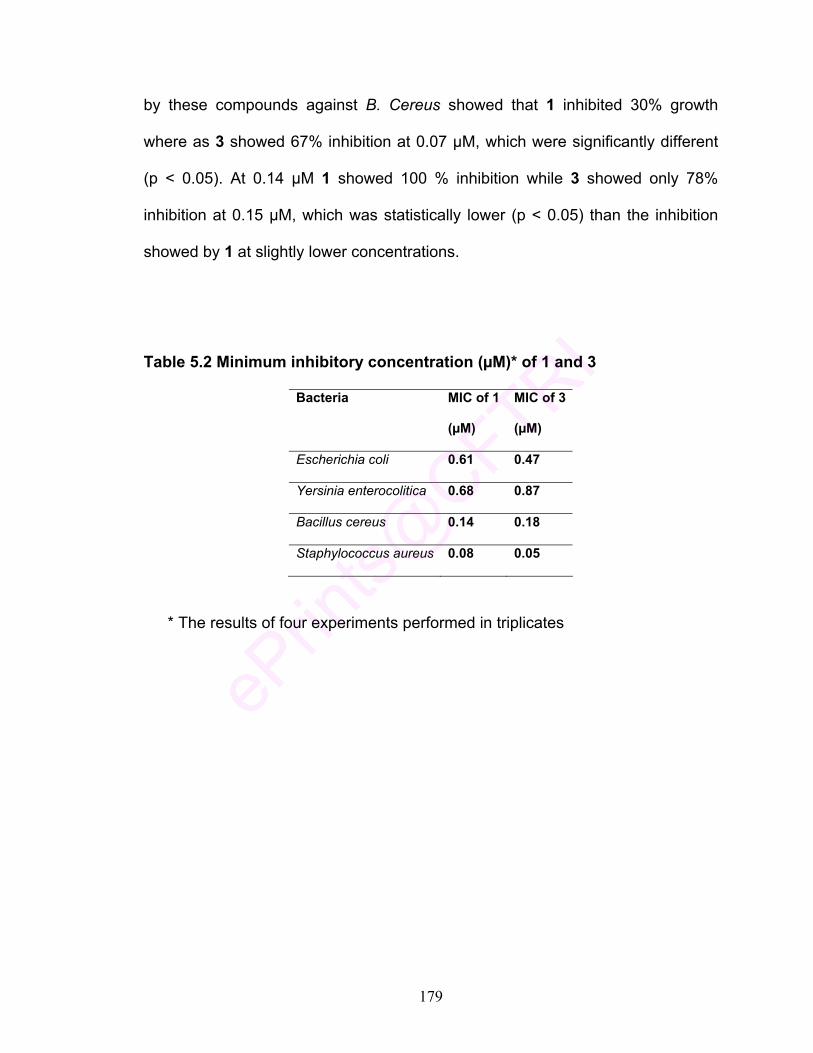
179
by these compounds against B. Cereus showed that 1 inhibited 30% growth
where as 3 showed 67% inhibition at 0.07 µM, which were significantly different
(p < 0.05). At 0.14 µM 1 showed 100 % inhibition while 3 showed only 78%
inhibition at 0.15 µM, which was statistically lower (p < 0.05) than the inhibition
showed by 1 at slightly lower concentrations.
Table 5.2 Minimum inhibitory concentration (µM)* of 1 and 3
Bacteria MIC of 1
(µM)
MIC of 3
(µM)
Escherichia coli 0.61 0.47
Yersinia enterocolitica 0.68 0.87
Bacillus cereus 0.14 0.18
Staphylococcus aureus 0.08 0.05
* The results of four experiments performed in triplicates

180
0 2 4 6 8 10
0
2
4
6
8
10
0 1 2 3 4 5 6 7 8 90
20
40
60
80
100[A]
(ppm)600500400300250100 20050(µM)
CURCUMIN GLUCOSIDECURCUMIN0.870.720.580.430.680.540.270.14
cd
c
a
aa
b
cde
f
% In
hibi
tion
Concentration
0
2
4
6
8
10
0 1 2 3 4 5 6 7 8 9 1
0
20
40
60
80
100
(ppm)3502501005035025010050(µM)
CURCUMIN GLUCOSIDECURCUMIN
0.470.360.150.07
a
a
b
c
aa
c
c
0.950.680.270.14
% In
hibi
tion
Concentration
[B]
Fig. 5.5 Antibacterial activities of 1 and 3 against different bacteria [A] Y. enterocolitica ; [B] E. Coli; [C] S.Aureus & [D] B. Cereus

181
0 2 4 6 8 10
0
2
4
6
8
10
0 1 2 3 4 5 6 7 8 9 100
20
40
60
80
100
(ppm)12510050251251005025
(µM)0.180.34
aa aa
b
aa
c
CURCUMIN GLUCOSIDECURCUMIN0.150.070.040.270.140.07
% In
hibi
tion
Concentration
[C]
0
2
4
6
8
10
B C D E F G H I0
20
40
60
80
100
(ppm)(µM)
12510050251251005025
a
b
bc
c
aaa
CURCUMIN GLUCOSIDECURCUMIN
0.180.150.070.040.340.270.140.07
% In
hibi
tion
Concentration
[D]
d
182
Certain curcumin bio-conjugates containing esters and peptides show
enhancement in antifungal and antibacterial activities, which is attributed to better
cellular uptake, increased cellular concentration and better receptor binding
(Kapoor, et al., 2007), as observed in higher antibacterial activity of 3 against S.
Aureus and E. Coli. However, in a certain study an enhancement in bacterial
growth in the case of Bacillus cereus and Y. Enterocolitica was observed when
the growth medium was supplemented with glucose (Rhee, et al., 1994). The
high MIC value in the case of B. Cereus and Y. Enterocolitica may be possibly
due to the fact that bacterial enzyme system may cleave the glycosidic linkage
and the bacteria utilize the released glucose.
The higher antimicrobial activity observed for these curcumin bioconjugates may
be due to,
Enhanced metabolic stability due to masking of phenolic hydroxyl groups
and delay in their glucuronide formation during metabolism.
Better cellular uptake due to the transportation of the conjugate via the
amino acid carrier protein, i.e. drug transport, and
Superior aqueous solubility of the conjugates because of enhancement in
polar character (hydrophilicity).
This makes these derivatives as potent prodrugs, which can get
hydrolysed at the target sites. The amino acids are known to be transported
through carrier proteins associated with cellular uptake and linked with 1 in order
to facilitate the cellular uptake through receptor mediated endocytosis (Dubey, et
al., 2008).

183
5.3.2 Curcumin Amino Acid Conjugates
A synthetic protocol was developed for the preparation of several
curcumin-amino acid conjugates in high yields. Reaction of curcumin (1) with t-
Boc–protected amino acids with dicylcohexylcarbodiimide in presence of
dimethylaminopyridine and triethylamine as catalysts afforded t-Boc-amino acid
conjugates of 1. These were deprotected to afford the corresponding curcumin-
amino acid conjugates (Table 5.3) in 45–76% yields (Discussed in detail in
Chapter 3).
Table 5.3 Curcumin amino acid conjugates synthesized
NH 2 C H
R
C O O O O C C H
R
N H 2
O O H
O M eM e O
Structure No.
Compound Structure IV, R =
4 1,7-Bis (4-O-L-leucinoyl-3-methoxyphenyl)-1,4,6-heptatriene-5-ol-3-one CH CH2
CH3
CH3 5 1,7-Bis (4-O-L-phenylalaninoyl-3-
methoxyphenyl)-1,4,6-heptatriene-5-ol-3-one CH2
6 1,7-Bis (4-O-L-alaninoyl-3-methoxyphenyl)-
1,4,6-heptatriene-5-ol-3-one CH3
7 1,7-Bis (4-O-L- isoleucinoyl -3-methoxyphenyl)-1,4,6-heptatriene-5-ol-3-one CH
CH3
CH2CH3 8 1,7-Bis (4-O-L- valinoyl -3-methoxyphenyl)-
1,4,6-heptatriene-5-ol-3-one CH3
CHCH3
9 1,7-Bis (4-O-L- serinoyl -3-methoxyphenyl)-1,4,6-heptatriene-5-ol-3-one
CH2OH
10 1,7-Bis (4-O-L- cysteinoyl -3-methoxyphenyl)-1,4,6-heptatriene-5-ol-3-one
CH2SH
11 1,7-Bis (4-O-L- phenylglycinoyl -3-methoxyphenyl)-1,4,6-heptatriene-5-ol-3-one
12 1,7-Bis (4-O-L- glycinoyl -3-methoxyphenyl)-1,4,6-heptatriene-5-ol-3-one
H 13 1,7-Bis (4-O-L- prolinoyl -3-methoxyphenyl)-
1,4,6-heptatriene-5-ol-3-one CH2 CH2 CH2
( as part of a pyrolidine ring)
(IV)

184
i) Solubility
All the amino acid-curcumin conjugates were tested for their solubility in
aqueous systems. While all these derivatives were soluble in water at 1 mg / ml
concentration pH 6, the conjugates of 1 with glycine (12) and proline (13)
exhibited higher solubility in water (10 mg/ml).
ii) Radical-scavenging activity
These compounds were evaluated for their antioxidant behavior in
comparison with 1 and the results are presented in Table 5.4. The antioxidant
methods of DPPH clearly indicated that all the curcumin amino acid conjugates
had lower IC50 values than 1. The conjugates showed differences in their
activities, which could be attributed to the various amino acids attached to 1.
Especially, alkyl substituted amino acids attached to 1 showed higher activity.
Conjugates of curcumin with leucine (4, Table 5.4), alanine (6), isoleucine (7),
valine (8), serine (9) and cysteine (10) exhibited 42%, 53%, 51%, 59%, 59% and
70% lesser IC50 values than curcumin (1). Among these, 10 showed highest
activity, which could be attributed to the sulphur moiety present in it acting as a
good radical-scavenging agent. Conjugates of curcumin with phenylalanine (5),
phenylglycine (11) and proline (13) derivatives exhibited comparatively less
radical scavenging activity than the other conjugates but still showed 31%, 22%
and 17% lesser IC 50 value than 1.

185
Table 5.4 IC 50 value of curcumin-amino acid conjugates
Structure
No.
IC 50, (μmol) by DPPH
method
IC 50, (μmol), by β - carotene
bleaching method
1 0.118a 0.109a
4 0.068f 0.064f
5 0.082d 0.077d
6 0.055h 0.047h
7 0.058g 0.06g
8 0.049i 0.037j
9 0.048j 0.043i
10 0.036k 0.03k
11 0.092c 0.086c
12 0.076e 0.066e
13 0.098b 0.091b

186
iii) Antioxidant activity
Antioxidant activity by β – carotene-bleaching assays also showed that all
these derivatives had lower IC50 values than the parent molecule. The conjugates
6, 7, 8, 9 and 10 showed IC 50 values 57%, 45%, 66%, 61% and 73% lower than
1. The proline, phenylglycine and phenylalanine conjugates of curcumin (13, 11
and 5) also showed IC50 values 17%, 21% and 29% lower than 1. Also in this
assay the compound 10 showed highest activity substantiating the role of sulphur
moiety in scavenging free radicals. Thus both the DPPH and β – carotene-
bleaching assays established that curcumin - amino acid conjugates exhibited
radical – scavenging and antioxidant properties superior than curcumin.
The enhancement in the antioxidant activities of curcumin amino acid
conjugates could be attributed to two factors, firstly the radical scavenging
activity of keto – enol group in 1. The phenolic hydroxyl of 1 was substituted with
amino acids blocking the route of HAT (Hydrogen Atom Transfer) mechanism of
antioxidant activity. Here in this case, the mechanism involved may be sequential
proton loss electron transfer (SPLET) where it postulates that keto – enol moiety
of 1 has more easily dissociable proton, which is supposed to be faster than
other mechanisms. The enolic proton being more acidic than the other two
hydroxyls of phenol rings in 1 (Litwinienko, & Ingold, 2004). This hypothesis was
also supported by some theoretical calculations like Density Functional Theory
(DFT) and Time Dependent Density Functional Theory (TD - DFT), which also
support sequential proton loss electron transfer (SPLET) in 1 (Shen & Ji, 2007).
187
Another reason may be attributed to the radical-scavenging activity of
amino group with the hydrazyl moiety to enhance the total antioxidant activity
(Ionita, et al., 2000). Amine group absorbs a hydrogen ion from the solution to
form NH3+ (Kim & Thomas, 2007). Free radicals can react with the hydrogen ion
from the ammonium ions (NH3+) to form a stable molecule. Reports are available
on the functionality of the amino acid affecting the activity. The activity of basic
and neutral amino acids supposed to be higher compared to others because of
their functionality (Triantis, et al., 2007). In the present study, it was observed
that curcumin conjugates of alanine, valine, serine, isoleucine and cysteine (6, 8,
9, 7 and 10) showed enhanced activity while leucine and glycine conjugates of
curcumin (4 and 12) exhibited slightly better activity than 1 and proline,
phenylalanine and phenylglycine conjugates of curcumin (13, 5 and 11) showed
almost similar activity compared to 1. The lower activity of the other amino acids
may be due to the steric hindrance during radical-scavenging action because of
the presence of bulky groups.
iv) Antimutagenic activity
Antimutagenicity studies of 1 and curcumin amino acid conjugates were
carried out with Salmonella typhimurium TA 1531 as well as TA 98 by Ames test
against sodium azide and MMS as a mutagen. The results indicated that
curcumin amino acid conjugates had strong antimutagenic property similar to 1
against Salmonella typhimurium TA 98 when tested with sodium azide (Fig.5.6 [A
& B]). Both 1 and curcumin amino acid conjugates showed strong antimutagenic
activity against TA 98 with sodium azide at 100 and 250 µg / plate

188
concentrations. Alanine, isoleucine, valine and phenylglycine conjugates (6, 7, 8
and 11) showed the activities statistically similar (p<0.05) to 1 at 100 µg / plate
concentration with sodium azide as mutagen. At 100 and 250 µg / plate
concentration with sodium azide as mutagen 13, showed strong antimutagenicity
compared to 1. Leucine, serine and glycine conjugated to curcumin (4, 9 and 12)
showed statistically similar antimutagenic activity but it was significantly lower
than 1. Overall the conjugates as well 1 showed strong antimutagenicity at 100
and 250 µg / plate concentrations. In the case of MMS the conjugates showed
strong antimutagenic activity against TA 98 at 100 µg / plate concentration for
phenylalanine and cysteine conjugates of curcumin (5 and 10) and it was
statistically similar to 1 (p < 0.05). 13 showed strong antimutagenic activity
against TA 98 at 100 µg / plate concentration compared to 1 but showed almost
similar activity at 250 µg / plate concentration. Alanine and glycine conjugates of
curcumin (6 and 12) were statistically similar in their activity but had higher
antimutagenic activity than 1. Valine and phenylglycine conjugates of (8 and 11)
also showed statistical similarity but significantly lower antimutagenic activity than
1. At 250 µg / plate concentration all the conjugates showed strong
antimutagenic activity which was statistically similar to 1 (Fig 5.6. [C & D]).

189
0 1 2 3 4 5 6 7 8 9 10 110
10
20
30
40
50
60
70
80
121110987654 131
CURCUMIN AMINO ACID CONJUGATES
% IN
HIB
ITIO
N
[A]
0
2
4
6
8
1a
ed
fgf
dccd
b
f
cde
0 1 2 3 4 5 6 7 8 9 10 110
20
40
60
80
100
121110987654 131
% IN
HIB
ITIO
N
CURCUMIN AMINO ACID CONJUGATES
de
aa
fed
h
bc
g
0 2 4 6 8 10
0
2
4
6
8
1[B]
Fig. 5.6 Antimutagenicity of 1 and its amino acid conjugates against Salmonella Typhimurium TA 98;
[A] & [B] against sodium azide as mutagen at 100 & 250 μg/ plate concentrations

190
2 3 4 5 6 7 8 9 10 11
0
10
20
30
40
50
60
70
121110987654 131
[C]
0
2
4
6
8
10
CURCUMIN AMINO ACID CONJUGATES
a
b
g
c
fge
bcd
c
0 1 2 3 4 5 6 7 8 9 10 110
20
40
60
80
100
121110987654 131
[D]
0 2 4 6 8 10
0
2
4
6
8
1
% IN
HIB
ITIO
N
CURCUMIN AMINO ACID CONJUGATES
b
gh
fe
j
d
ih
ca
[C] & [D] against MMS as mutagen at 100 & 250 μg/ plate concentrations

191
1 and curcumin amino acid conjugates were also tested for their
antimutagenic activity against TA 1531 with sodium azide at 100 and 250 µg /
plate concentrations (Fig. 5.7 [A & B]). Results indicated that at 100 µg / plate
concentration against sodium azide as mutagen the activity of curcumin proline
conjugate (13) was statistically similar to 1. Leucine and serine conjugates of
curcumin (4 and 9) showed similar activity but it was statistically lower than
curcumin. 5 and 7 strong antimutagenicity compared to 1. Other derivatives
showed significantly higher activity than 1. 8 and 12 showed moderate activity
compared to curcumin. At 250 µg / plate concentration 1 showed lower activity
than all the amino acid conjugates. In the case of MMS as mutagen cysteine,
glycine and proline conjugates of curcumin (10, 12 and 13) showed lower activity
than 1 while other derivatives showed significantly higher activity than 1 at 100
µg / plate concentration. At 250 µg / plate concentration curcumin-amino acid
conjugates showed significantly higher activity than 1 except in the case of
proline (13) derivative (Fig. 5.7 [C & D]).
1 is known to be a good anti-mutagen (Goud, et al, 1999). In the present
study antimutagenicity studies of curcumin amino acid conjugates against
Salmonella typhimurium TA 1531 as well as TA 98 by Ames test using sodium
azide and MMS as mutagens were evaluated. The results indicated that
curcumin amino acid conjugates exhibited strong antimutagenic property. 1
showed strong antimutagenic activity against sodium azide and MMS at both the
concentrations tested, but the derivatives, in general, comparable and even
superior antimutagenic activities at the concentrations tested. Strong

192
antimutagenicity observed for curcumin amino acid conjugates can be attributed
to their conversion to 1 in the bacterial cell. Earlier, it has been shown that even
the traces of curcuminoids in 1, curcumin-removed turmeric oil were found to
impart strong antimutagenicity (Jayaprakasha, et al., 2002). Free radicals are
implicated in many physiological disorders such as inflammation, aging and
carcinogenicity (Namiki, 1990), but oxygen scavenging can reduce these effects
(Surh, 1999). It was reported that compounds having antioxidant potential could
also inhibit mutagenicity and cancer (Hochstein, & Atallah, 1988). Amino acids
being the component of bacterial cellular environment, these conjugates may
help to increase the intracellular delivery of 1 (Mishra, et al., 2005). Amino acids
may cross the cell membranes gaining facile entry to the bacterial cells (Mishra,
et al., 2005). The blocking of phenolic – OH group by amino acids in 1 helps in
better endocytosis and their retention in the cell after the enzymatic cleavages
(Kapoor, et al., 2007).

193
0 1 2 3 4 5 6 7 8 9 10 110
10
20
30
40
121110987654 131
f
h
cd
a
fe
[A]
0
2
4
6
8
10
% IN
HIB
ITIO
N
CURCUMIN AMINO ACID CONJUGATES
g
ed
b
0 1 2 3 4 5 6 7 8 9 10 110
10
20
30
40
50
60
70
80
121110987654 131
hi
d
g
bcfe
a
i
j
[B]
0 2 4 6 8 10
2
4
% IN
HIB
ITIO
N
CURCUMIN AMINO ACID CONJUGATES
Fig. 5.7 Antimutagenicity of 1 and its amino acid conjugates against Salmonella Typhimurium TA 1531;
[A] & [B] against sodium azide as mutagen at 100 & 250 μg/ plate concentrations

194
0 1 2 3 4 5 6 7 8 9 10 110
10
20
30
40
50
60
70
1211107 8 9654 131
d
bb
e
[C]
0
2
4
6
8
10
% IN
HIB
ITIO
N
CURCUMIN AMINO ACID CONJUGATES
gh
c
gf
c
a
0 1 2 3 4 5 6 7 8 9 10 110
20
40
60
80
100
121110987654 131
h
aeed
bf
caa
g
[D]
0 2 4 6 8 10
% IN
HIB
ITIO
N
CURCUMIN AMINO ACID CONJUGATES
[C] & [D] against MMS as mutagen at 100 & 250 μg/ plate concentrations

195
v) Antibacterial activity
Further, 1 and conjugates were tested for their antibacterial potential
against B. Cereus, Staphylococcus aureus, E.Coli and Y. Enterocolitica. MIC
values of the compounds are presented in Table. 5.5. Results showed the
conjugates synthesized were more effective in inhibiting the growth of Gram –ve
bacteri\a compared to the parent molecule. The MIC values of curcumin amino
acid conjugates were lower compared to 1. Against E. Coli, curcumin conjugates
4, 5, 6, 8, 10 and 11 (Table 5.5) showed 72%, 75%, 84%, 71%, 78% and 74%
lower MIC values respectively compared to 1. Conjugation of curcumin with
isoleucine (7), glycine (12) and proline (13) showed moderate activity but
curcumin conjugated to serine (9) showed lesser inhibition. Against Y.
Enterocolitica lower MIC values were observed for all the curcumin amino acid
conjugates compared to curcumin. Against B. cereus the conjugates 4, 6, 9, 10
and 11 (Table 5.5) showed 63%, 42%, 45%, 70% and 53% lower MIC compared
to 1. Conjugates such as 5, 7, 8, 12 and 13 showed higher MIC values than
curcumin. Against S. Aureus, curcumin amino acid conjugates 7, 8, 9, 10, 11 and
13 exhibited lower MIC values compared to curcumin. Conjugation of 1 with
phenylalanine (5), alanine (6) and glycine (12) showed higher MIC value
compared to the parent molecule 1.
The enhancement in antimicrobial activity of these curcumin amino acid
conjugates may be due to, enhanced metabolic stability due to masking of
phenolic hydroxyl groups and delay in their glucoronide formation during
metabolism, better cellular uptake due to the transportation of the conjugate via

196
the amino acid carrier protein, i.e. drug smuggling, and more solubility of the
conjugates because of enhancement in polar character (hydrophilicity). This
makes these derivatives as potent prodrugs, which can get hydrolysed at the
target sites and enhance the cellular uptake (Dubey, et al., 2008).
Table 5.5 MIC Values (µM*) of amino-acid conjugates of 1 against different bacteria
Structure No. E.Coli
(µM) Y. Enterocolitica
(µM)
B. Cereus
(µM)
S. Aureus
(µM)
1 0.61 1.16 0.14 0.081
4 0.17 0.55 0.05 0.050
5 0.15 0.26 0.53 0.11
6 0.10 0.10 0.08 0.20
7 0.55 0.17 1.35 0.05
8 0.18 0.49 0.27 0.05
9 0.65 0.19 0.07 0.06
10 0.13 0.10 0.04 0.03
11 0.16 0.08 0.06 0.05
12 0.31 0.42 0.21 0.21
13 0.44 0.09 1.95 0.07
* The results of four experiments performed in duplicates

197
O OH
H3CO
O
OCH3
OH14
OHO
HO
OH
H3CO O
5.3.3 Glucuronide derivative of curcumin Ultrasound-assisted reaction of 1 with methyl 1-bromo-2,3,4-tri-O-acetyl-α-
D- glucopyranuronate in the presence of phase transfer catalyst under biphasic
conditions followed by deacetylation to afforded methyl 1-curcuminyl-β-D-
glucopyranuronate (Fig.5.8, Discussed in detail in Chapter 4)
i) Solubility: 14 was tested for its solubility in aqueous systems. It was soluble
in water at 10 mg/ml concentration compared to the water-insoluble parent
molecule.
Fig. 5.8 Methyl 1-curcuminyl-β-D-glucopyranuronate (14)
ii) Radical-scavenging activity
Radical-scavenging assay was carried out with 1 as a reference for
comparison with the water-soluble 14. 1 and 14 were dissolved in DMSO at
0.05–0.25 µM concentrations. The radical-scavenging activity (RSA) by DPPH
method showed 41 and 46% activity at 0.05 µM for 1 and 14, respectively (Fig.
5.9). At 0.1 µM, RSA of 1 and 14 was 47 and 56%, respectively However, RSA of
1 and 14 at both these concentrations were significantly different (p < 0.05). At
0.25 µM 1 showed 100% RSA but 14 showed the same result at 0.2 µM.

198
iii) Antioxidant activity
The antioxidant activity by β–carotene bleaching method (Fig. 5.10) was
carried out wherein, 1 and 14 were dissolved in DMSO at 0.05–0.25µM
concentrations. 1 showed 41% and 14 had 46% activity at 0.05 µM, which were
statistically (p<0.05) different. At 0.1 µM concentration 14 showed significantly
(p<0.05) higher activity (56%) than 1 (50%). Similarly at 0.2 µM concentrations
14 showed significantly higher antioxidant activity (100%) than 1 (90%). These
results clearly showed that 14 exhibited higher antioxidant activity than 1 at all
the concentrations.
iv) Antimutagenic activity
Antimutagenicity studies of 1 and 14 were carried out with Salmonella
typhimurium TA 1531 as well as TA 98 by Ames test against sodium azide/MMS
as a mutagen (Table 5.5). The results indicated that 14 possessed as strong
antimutagenic property as 1. Both 1 and 14 showed strong antimutagenic activity
with TA 98 against sodium azide at all the concentrations tested. With MMS as
diagnostic mutagen the derivative showed higher antimutagenicity compared to
1, which were statistically different at the tested concentrations.
In case of TA 1531 1 had 36% activity at 100 μg/plate, against sodium
azide as mutagen, where as 14 showed strong 61% and strong antimutagenic
activity at similar concentration. In case of TA 98, 14 had 74% and 76% activity
for sodium azide and MMS respectively at 100 μg/plate concentration higher than
1, which showed 63 % and 49% activity for sodium azide and MMS respectively.
At 250 μg/plate concentration 1 showed strong antimutagenicity compared to 14.

199
Fig 5.9 Radical-scavenging activity of 1 and 14 by DPPH method
Fig. 5.10 Antioxidant activity of 1 and 14 by β-carotene bleaching method
0 2 4 6 8 100
20
40
60
80
100
fe
d
g
c
bb
a aa
CURCUMIN GLUCURONIDECURCUMIN
0.250.20.1 0.150.050.250.200.150.100.05
% R
adic
al s
cave
ngin
g ac
tivity
Concentration (µM)
0 2 4 6 8 10
0
2
4
6
8
10
0 2 4 6 8 100
20
40
60
80
100
CURCUMIN GLUCURONIDECURCUMIN0.250.200.150.100.050.250.200.150.100.05
Concentration (µM)
% A
ntio
xida
nt a
ctiv
ity
0 2 4 6 8 10
0
2
4
6
8
10
g
h
f
e
d
cb
aaa

200
Table 5.5 Antimutagenic activity of 1 and 14 against sodium azide and MMS as diagnostic mutagens
TA -1531 TA - 98 Compounds
Sodium azide MMS Sodium azide MMS
100 µg/ plate
1 36 ± 0.5 53 ± 1.8 63 ± 1 49 ± 1
14 61 ± 0.8 76 ± 0.5 74 ± 0.8 76 ± 0.5
250 µg/ plate
1 44 ± 1 77 ± 0.8 83 ± 0.6 96 ± 0.4
14 61 ± 0.6 74 ± 0.5 79 ± 0.6 88 ± 0.5
v) Antibacterial activity
1 and 14 were tested for their antibacterial potential against B. Cereus, S.
Aureus, E. Coli and Y. Enterocolitica. MIC values of the compounds presented
in Table 5.6 showed the inhibition of bacteria at different concentrations of 14.
MIC values of 14 were higher than 1 for inhibition against B. Cereus, S. Aureus
but lower than 1 in the case of E. Coli and Y. Enterocolitica.
The 1 and 14 showed 75% and 93% inhibition at 0.54 µM, which were
statistically different (p < 0.05). 100 % inhibition was observed at 0.82 and 0.72
µM for 1 and 14 respectively against Y. enterocolitica (Fig. 5.11 [A]).
In the case of E. coli, 0.14 µM of 1 showed 25% inhibition, while 0.09 µM
of 14 showed 36% inhibition, which was statistically different (p < 0.05). 100%
growth inhibition for E. coli was observed at 0.68 µM for 1 whereas, 14 showed it

201
at 0.45 µM (Fig. 5.11 [B]). In the case of S. Aureus the 0.07 µM concentration of
1 showed 65 % while 14 showed 57 % inhibition at 0.05 µM level (Fig. 5.11[C]).
Complete growth inhibition for S. Aureus observed at 0.13 and 0.14 µM
concentrations for 1 and 14 respectively, which were statistically similar (p <
0.05). Against B. cereus, 1 showed 30% inhibition at 0.07 µM level, while 14 had
significantly higher inhibition (52%) at 0.05 µM concentration (Fig. 5.11 [D]).
Complete inhibition of B. cereus was observed at 0.14 µM level for both the
compounds and were statistically similar (p < 0.05).
Table 5.6 Minimum inhibitory concentration (µM)* of 1 and 14
Bacteria MIC of 1(µM)
MIC of 14 (µM)
Escherichia coli 0.61 0.27
Yersinia enterocolitica 0.68 0.63
Staphylococcus aureus 0.08 0.13
Bacillus cereus 0.14 0.18

202
0 1 2 3 4 5 6 7 8 90
20
40
60
80
100 [A]
0.720.540.36
a
0.54 1.090.82CURCUMIN GLUCURONIDECURCUMIN
Concentration (µmol)
% In
hibi
tion
0 2 4 6 8 10
0
2
4
6
8
1
dc
baa
0 1 2 3 4 5 6 7 8 9 100
20
40
60
80
100
0.630.450.180.090.950.680.270.14CURCUMIN GLUCURONIDE
e
dc
b
aaaa
% In
hhib
ition
Concentration (µmol)
0 2 4 6 8 10
[B]
CURCUMIN
0 1 2 3 4 5 6 7 8 9 10
0
20
40
60
80
100
c
d
b
aaaaa
0.180.130.090.050.270.200.140.07CURCUMIN GLUCURONIDECURCUMIN
% In
hibi
tion
0 2 4 6 8 10
0
2
4
6
8
10[C]
Concentration (µmol)
1 2 3 4 5 6 7 8 90
20
40
60
80
100 [D]
0.270.200.140.07 0.180.130.09CURCUMIN GLUCURONIDECURCUMIN
d
c
b
aaaaa
% In
hibi
tion
Concentration (µmol)
0.05
Fig. 5.11 Antibacterial activities of 1 and 14 against different bacteria [A] Y. enterocolitica ; [B] E. Coli; [C] S.Aureus & [D] B. Cereus

203
5.3.4 Curcumin Glucal Derivative
In an another approach to render 1 water-soluble Ferrier reaction of 1 was
with tri-O-acetyl-D-glucal was carried out in the presence of Fe2 (SO4)3 x H2O as
catalyst under stirring at 40 oC in acetonitrile. The product formed was
deacetylated to yield curcuminyl hex-2-enopyranoside (Fig. 5.12, Discussed in
detail in Chapter 4).
i) Solubility
15 was tested for its solubility in aqueous systems. The derivative was
soluble in water at 1 mg/ml concentration.
Fig. 5.12 Structure of curcuminyl hex-2-enopyranoside (15) ii) Radical-scavenging activity
The radical-scavenging activity of 1 was compared to 15. Both the
samples were dissolved in DMSO at a concentration range of 0.05 -0.25 μM (Fig.
5.13). At 0.05 μM concentration the compounds 1 and 15 showed 47% and 53%
radical-scavenging activity respectively, which were statistically different
(p<0.05). Complete inhibition was observed at 0.25 and 0.20 μM concentrations
for 1 and 15 respectively. An IC50 value 0.067 μM of 15 was very low compared
to the parent molecule (0.118 μM).
O OH
H3CO
O OH
OCH3
O
HO
HO (15)

204
iii) Antioxidant activity
Antioxidant activity by β-carotene bleaching method (Fig. 5.14) was
carried out at 0.05-0.25 μM concentration in DMSO for 15 and compared to 1.
The antioxidant activity of 41 and 46 % was observed at 0.05 μM for 1 and 15
respectively, which were statistically different (p < 0.05). 100 % activity inhibition
was observed for 1 at (0.20 μM) and 15 (0.25 μM) concentrations respectively.
iv) Antimutagenic activity
Antimutagenicity studies of 1 and 15 were carried out with Salmonella
typhimurium TA 1531 as well as TA 98 by Ames test against sodium azide and
MMS as mutagens. In the case of TA –1531, 1 had moderate and 15 had
strong activity at 100 µg/ plate as well as 250 µg/plate concentrations against
both sodium azide and MMS as mutagens (Table 5.7). In the case of TA 98, both
1 and 15 moderate antimutagenicity at 100 µg/plate but much superior activity at
250 µg/plate concentration.
Table 5.7 Antimutagenic activity of 1 and 15 against sodium azide and MMS
as diagnostic mutagens.
TA -1531 TA - 98
Sodium azide MMS Sodium azide MMS
100 µg/ plate
1 36 ± 0.5 53 ± 1.8 63 ± 1 49 ± 1
15 55 ± 0.7 59 ± 1.2 71 ± 0.7 55 ± 0.7
250 µg/ plate
1 44 ± 1 77 ± 0.8 83 ± 0.6 96 ± 0.4
15 77 ± 0.8 82± 0.5 90 ± 0.4 99 ± 0.1

205
Fig. 5.13 Radical-scavenging activity of 1 and 15 by DPPH method
Fig. 5.14 Antioxidant activity of 1 and 15 by β-carotene bleaching method
0 1 2 3 4 5 6 7 8 9 10 11 1
0
20
40
60
80
100
% R
adic
al s
cave
ngin
g ac
tivity
0.250.200.150.100.050.250.200.150.100.05
CURCUMIN GLUCALCURCUMINConcentration (µM)
0
2
4
6
8
10
f f
ed
c
bb
aaa
0 1 2 3 4 5 6 7 8 9 10 11 120
20
40
60
80
100
0.05 0.250.200.150.100.250.200.150.100.05
hg
fe
d
cb
aaa
% A
ntio
xida
nt a
ctiv
ity
CURCUMIN GLUCALCURCUMINConcentration (µM)
0 2 4 6 8 10
0
2
4
6
8
10
206
v) Antibacterial activity
Compounds 1 and 15 were tested for their antibacterial potential against
B. Cereus, S. Aureus, E. Coli and Y. Enterocolitica. MIC of the compounds is
presented in Table 5.8. Fig. 5.15 (a-d) shows the inhibition of these bacteria at
different concentrations of both the compounds.
In the case of Y. Enterocolitica, 1 showed 75 % inhibition at 0.54 μM and
15 showed 66 % inhibition at 0.40 μM, which were statistically different (p < 0.05)
[Fig 5.15 (A)]. Complete inhibition was observed at an MIC of 0.68 and 0.70 μM
of 1 and 15 respectively (Table 5.8). Against E. Coli similar inhibition was
observed at 0.27 and 0.10 μM respectively [Fig 5.15(B)] which were statistically
similar (p < 0.05). The inhibition was 100% at a MIC of 0.61 and 0.40 μM
concentrations of 1 and 15 respectively (Table 5.8).
The antibacterial studies on the 15 was carried out against S. Aureus.
Inhibition was 65% and 41% at 0.07 and 0.05 μM for 1 and 15 respectively,
which were statistically different (p<0.05). Inhibition was 100% at 0.08 and 0.20
μM (MIC) for 1 and 15 respectively (Fig. 5.15 [C]). Against B. Cereus 1 and 15,
showed 32 and 37 % at 0.07 and 0.05 μM concentrations, which were statistically
different (p<0.05). Complete inhibition was observed at 0.14 and 0.20 μM (MIC)
for 1 and 15 respectively (Fig. 5.15 [D]). Against gram +ve bacteria, MIC values
of 15 were high compared to the parent molecule. Against E. Coli, the derivative
15 showed MIC value lesser than 1 but in the case of Y. Enterocolitica the
derivative 15 showed MIC value comparable to the parent molecule.

207
0 1 2 3 4 5 6 7 8 9 100
20
40
60
80
100
Concentration (µmol)
[B]
0.700.500.200.100.950.680.270.14CURCUMIN
f
ed d
c
b
aaa
0
2
4
6
8
10
% In
hhib
ition
CURCUMIN GLUCAL
Table 5.8 Minimum inhibitory concentration (µM)* of 1 and 15
Bacteria MIC of 1(µM)
MIC of 15 (µM)
Escherichia coli 0.61 0.40
Yersinia enterocolitica 0.68 0.71
Bacillus cereus 0.14 0.20
Staphylococcus aureus 0.08 0.20
Fig. 5.15 Antibacterial activity of 1 and 15 conjugate against, [A] Y.
enterocolitica ; [B] E. Coli; [C] S.Aureus & [D] B. Cereus
0 1 2 3 4 5 6 7 80
20
40
60
80
100 [A]
0.800.600.401.090.820.54
d
c
baaa
CURCUMIN GLUCALCURCUMIN
% In
hibi
tion
Concentration (µmol)
0 2 4 6 8 10
0 1 2 3 4 5 6 7 8 9 100
20
40
60
80
100
Concentration (µmol)
[C]
e
dc
baaaa
0.200.150.100.050.270.200.140.07CURCUMIN GLUCALCURCUMIN
% In
hibi
tion
0 2 4 6 8 10
0 1 2 3 4 5 6 7 8 9 100
20
40
60
80
100
Concentration (µmol)
[D]
db
c
b
aaaa
0.200.150.100.050.270.200.140.07
% In
hibi
tion
CURCUMIN GLUCALCURCUMIN
0 2 4 6 8 10
0
2
4
6
8
1

208
5.4 CONCLUSIONS
The antioxidant, antimutagenicity and antibacterial properties were
evaluated for all the derivatives whose synthesis is discussed in Chapters 2, 3
and 4. Antioxidant activities of glucoside, glucuronide and glucal derivatives of
curcumin, as determined by DPPH and β–carotene-bleaching assays, were
higher than that of curcumin. Conjugates of curcumin with amino acids like
alanine, valine, serine, cysteine and isoleucine exhibited even higher antioxidant
potential as these were effective even at ~ 50 % of the concentration of
curcumin.
Antimutagenicity studies were conducted for all the derivatives by Ame’s
test against Salmonella Typhimurium TA 98 and 1531 with Sodium azide and
MMS as mutagens at 100 and 250 μg/plate concentrations. Results indicated
higher antimutagenic potential for glucoside, glucuronide and glucal derivatives
of curcumin compared to curcumin. Amino acid conjugates of curcumin showed
strong antimutagenic activity against S. Typhimurium TA 98, with sodium azide
and methyl methanesulphonate as mutagens at both the concentrations. With S.
Typhimurium TA 1531 at 100 µg/plate concentration, against sodium azide as
mutagen, conjugates of curcumin with proline, leucine, serine and valine showed
activity similar to curcumin; other derivatives showed higher activity than
curcumin. In the case of MMS as mutagen, cysteine, glycine and proline
conjugates of curcumin showed lower activity than curcumin while other
derivatives showed higher activity than curcumin. However, all the amino acid

209
conjugates showed higher activity than curcumin against TA 1531 at 250
µg/plate concentration.
Antibacterial potential of all the derivatives were evaluated against two
gram +ve and two gram –ve bacteria by pour plate method. Results indicated
curcumin glucoside possessed superior antibacterial activity against the tested
bacteria. Curcumin amino acid conjugates, showed good inhibition against Gram
–ve bacteria compared to Gram +ve bacteria. Inhibition potential was higher for
these conjugates against E. coli and Y. enterocolitica compared to curcumin in
the case of glucuronide derivative. Glucal derivative of curcumin also showed
good inhibition against E. coli but inhibition was comparable to that of curcumin in
the case of Y. enterocolitica.
The studies demonstrated that all the sugar and amino acid derivatives of
curcumin, synthesized in the present study, were soluble in water and displayed
in vitro antioxidant, antimutagenic and antibacterial properties superior than
curcumin.

210
References
Adegako, G. O., Vijay Kumar, M., Gopal Krishna, A. G., Varadaraj, M.C.,
Sambaiah, K., & Lokesh, B. R. 1998. Antioxidants and lipid oxidation in
food. Journal of Food Science & Technology, 35, 283-298.
Ahsan, H., Parveen, N., Khan, N.U., & Hadi, S.M. 1999. Prooxidant, antioxidant
and cleavage activities on DNA of curcumin and its derivatives,
demethoxycurcumin and bisdemethoxycurcumin, Chemico Biological
Interactions, 121,161–175.
Araujo, C. A. C., & Leon, L. 2001. Biological Activities of Curcuma longa L., Mem
Inst Oswaldo Cruz, Rio de Janeiro, 96(5), 723-728.
Braga, M. E. M., Leal, P. F., Carvalho, J.E., & Meireles, M. A. A. 2003.
Comparison of Yield, Composition, and Antioxidant Activity of Turmeric
(Curcuma longa L.) Extracts Obtained Using Various Techniques.
Journal of Agricultural & Food Chemistry, 51, 6604-6611.
Chopra, R. N., Gupta, J. C., & Chopra, G. S. 1941. Pharmacological action of the
essential oil of Curcuma longa. The Indian Journal of Medical Research,
29, 769–772.
Cohly, H. H. P., Asad, S., Das, S. K., Angel, M. F., & Rao, M. 2003. Effect of
Antioxidant (Turmeric, Turmerin and Curcumin) on Human
Immunodeficiency Virus. International Journal of Molecular Sciences, 4,
22-33.
Cole, G.M., Teter, B., & Frautschy S.A. 2007. Neuroprotective effects of
curcumin, Advances in Experimental Medicine & Biology, 595, 197-212.

211
Cousins, M., Adelberg, J., Chen, F., & Rieck, J. 2007. Antioxidant capacity of
fresh and dried rhizomes from four clones of turmeric (Curcuma longa
L.) grown in vitro. Industrial Crops & Products, 25, 129-135.
Das, K. C. & Das, C.K. 2002. Curcumin (diferuloylmethane), a singlet oxygen
(1O2) quencher. Biochemical & Biophysical Research Communications,
295, 62–66.
Dubey, S. K., Sharma, A.K., Narain, U., Misra, K., & Pati, U. 2008. Design,
synthesis and characterization of some bioactive conjugates of curcumin
with glycine, glutamic acid, valine and demethylenated piperic acid and
study of their antimicrobial and antiproliferative properties. European
Journal of Medicinal Chemistry, 43 (9), 1837-1846.
Dutta, S., Murugkar, A., Gandhe, N & Padhye, S. 2001. Enhanced antioxidant
activities of metal conjugates of Curcumin derivatives. Metal Based
Drugs, 8 (4), 183-188.
Goel, A., Kunnumakkara, A.B., & Aggarwal B.B. 2008. Curcumin as
‘‘Curecumin’’: From kitchen to clinic. Biochemical Pharmacology, 75, 787
– 809.
Gomez, K.A., & Gomez, A.A. 1984. Comparison between treatment means. In
K.A. Gomez, & A.A. Gomez (Eds) New York: Wiley. Statistical
Procedures For Agricultural Research, 207 - 240.
Goud, V. K., Polasa, K., & Krishnaswamy, K. 1993. Effect of turmeric on
xenobiotic metabolizing enzymes. Plant Food & Human Nutrition, 44, 87-
92.

212
Hochstein, P., & Atallah, A.S. 1988. The nature of oxidant and antioxidant
systems in inhibition of mutation and cancer. Mutation Research, 202,
363–375.
Hoi-Seon-Lee, 2006. Antimicrobial properties of turmeric (Curcuma longa L.)
rhizome-derived ar-turmerone and curcumin. Food Science &
Biotechnology, 15(4), 559-563.
Ilken, Y., Morales, P., Martinez, A., Marin, M.L., Haza, A. I., & Cambero, M. I.
1999. Antimutagenic effect of fruit and vegetable ethanolic extracts
against N-nitrosoamines evaluated by the Ames test. Journal of
Agricultural & Food Chemistry, 47, 3257-3264.
Ionita, G., Sahini, V., Semenescu, G., & Ionita, P. 2000. Kinetics of oxidation of
amino acids by some free stable hydrazyl radicals. Acta Chimica
Slovenica, 47, 111-119.
Jayaprakasha, G. K., Jena, B. S., Negi, P. S., & Sakariah, K. K. 2002. Evaluation
of antioxidant activities and antimutagenicity of turmeric oil: a byproduct
from curcumin production. Zeitschrift fur Naturforschung, 57, (9-10), 828-
835.
Jayaprakasha, G. K., Negi, P. S., Anandha Ramakrishanan, C., & Sakariah, K. K.
2001. Chemical composition of turmeric oil—a byproduct from turmeric
oleoresin industry and its inhibitory activity against different fungi.
Zeitschrift fur Naturforschung, 56c, 40–44.

213
Jayaprakasha, G.K., Rao, L. J. M., & Sakariah, K.K. 2006. Antioxidant activities
of curcumin, demethoxycurcumin and bisdemethoxycurcumin. Food
Chemistry 98, 720–724
Joe, B., Vijaykumar, M., & Lokesh, B. R. 2004. Biological properties of curcumin -
cellular and molecular mechanisms of action, Critical Reviews in Food
Science & Nutrition, 44(2), 97-111.
Kapoor, N., Narain, U., & Misra, K. 2007. Bioactive conjugates of curcumin
having ester, peptide, thiol and disulphide links. Journal of Scientific &
Industrial Research, 66 (8), 647-650.
Kapoor, N., Sharma, A.K., Dwivedi, V., Kumar, A., Pati, U., & Misra, K. 2007.
Telomerase targeted anticancer bioactive prodrug by antisense-based
approach. Cancer Letters, 248, 245–250.
Kim, K.W., & Thomas, R.L. 2007. Antioxidative activity of chitosans with varying
molecular weights. Food Chemistry, 101, 308–313.
Kumar, S., Dubey, K. K, Tripathi, S., Fujii, M., & Misra, K. 2000. Design and
synthesis of curcumin-bioconjugates to improve systemic delivery.
Nucleic Acids Symposium Series, 44, 75-76.
Kumar, S., Narain, U., Tripathi, S., & Misra, K. 2001. Synthesis of curcumin
bioconjugates and study of their antibacterial activities against β-
Lactamase-producing microorganisms. Bioconjugate Chemistry, 12, 464
-469.
Litwinienko, G., & Ingold, K.U. 2004. Abnormal solvent effects on hydrogen atom
abstraction, resolution of curcumin antioxidant controversy, the role of

214
sequential proton loss electron transfer. Journal of Organic Chemistry,
69, 5888 – 5896.
Maron, D.M., & Ames, B.N. 1983. Revised methods for the salmonella
mutagenicity test, Mutation Research, 113, 173-815.
Meghana, K. Sanjeev, G. & Ramesh, B. 2007. Curcumin prevents streptozotocin-
induced islet damage by scavenging free radicals: A prophylactic and
protective role European Journal of Pharmacology, 577, 183–191.
Mishra, S., Kapoor, N., Ali, A. M., Pardhasaradhi, B. V. V., Leela Kumari, A.,
Khar, A., & Misra, K. 2005. Differential apoptotic and redox regulatory
activities of curcumin and its derivatives. Free Radical Biology &
Medicine, 38, 1353– 1360.
Mishra, S., Narain, U., Mishra R., & Misra, K. 2005. Design, development and
synthesis of mixed bioconjugates of piperic acid–glycine, curcumin–
glycine/alanine and curcumin–glycine–piperic acid and their antibacterial
and antifungal properties. Bioorganic & Medicinal Chemistry, 13, 1477-
1486.
Mohri, K. Watanabe, Y. Yoshida, Y., Satoh, M., Isobe, K, Sugimoto, N, & Tsuda,
Y 2003. Synthesis of glycosylcurcuminoids, Chemical & Pharmaceutical
Bulletin, 51 (11), 1268-1272.
Moon, J., & Terao J. 1998. Antioxidant Activity of Caffeic Acid and Dihydrocaffeic
Acid in Lard and Human low-density lipoprotein, Journal of Agricultural &
Food Chemistry, 29, 273 -300.

215
Naidu, M. M., Shyamala, B.N. Manjunatha, J. R., Sulochanamma, G & Srinivas,
P. 2009. A simple HPLC method for separation of curcuminoids with
potential radical scavenging activity. Journal of Food Science, 74 (4),
C312-318.
Namiki, M. 1990. Antioxidants/anti-mutagens in food. Critical Reviews in Food Science
& Nutrition, 44(2), 97-111.
Negi, P.S., Jayaprakasha, G. K., Rao, L.J.M., & Sakariah, K.K. 1999.
Antibacterial activity of turmeric oil – a byproduct from curcumin
manufacturers, Journal of Agricultural & Food Chemistry, 47, 4297-4300.
Ong, T., Wang, W. Z., Stewart, J. D., & Brockman, H.E. 1986. Chlorophylin a
potent anti-mutagen against environmental and dietary complex mixture.
Mutation Research, 173, 111-115.
Park, S., & Kim, D. S. H. L. 2002. Discovery of natural products from Curcuma
longa that protect cells from β -Amyloid Insult: A drug discovery effort
against Alzheimer’s disease. Journal of Natural Products, 66 (9), 1227 -
1231.
Rhee, J., Diaz Ricci, J.C., Bode, J., & Schugerl, K. 1994. Metabolic enhancement
due to plasmid maintenance, Biotechnology Letters, 16 (9), 881 – 884.
Sandur, K., Ichikawa, H., Pandey, M.K., Kunnumakkara, A. B., Sung, B., Sethi,
G., & Aggarwal, B.B. 2007. Role of pro-oxidants and antioxidants in the
anti-inflammatory and apoptotic effects of curcumin (diferuloylmethane).
Free Radical Biology & Medicine, 43, 568–580.
Sharma, R. A., Gescher, A. J., & Steward, W. P. 2005. Curcumin: the story so
far, European Journal of Cancer, 41, 1955–1968.

216
Shen, L., & Ji, H. 2007. Theoretical study on physicochemical properties of
curcumin. Spectrochimica Acta Part A, 67, 619–623.
Shimoda, K., Hara, T., Hamada, H., & Hamada, H. 2007. Synthesis of curcumin
β-maltooligosaccharides through biocatalytic glycosylation with
Strophanthus gratus cell culture and cyclodextrin glucanotransferase,
Tetrahedron Letters, 48, 4029–4032.
Shishu & Kaur, I. 2008 Antimutagenicity of curcumin and related compounds
against genotoxic heterocyclic amines from cooked food: The structural
requirement. Food Chemistry, 111(3), 573-579.
Sun, Y., Wang, R., Yuan, S., Lin, X., & Liu, C. 2004. Theoretical study on the
antioxidant activity of curcumin. Chinese Journal of Chemistry, 22, 827-
830.
Surh, Y. J. 1999. Molecular mechanisms of chemopreventive effects of selected
dietary and medicinal phenolic substances, Mutation Research 428,
305– 327.
Swaroop, S., Mishra, B., & Priyadarsini, K.I. 2007. Cyclodextrin inclusion
complex of curcumin. BARC Newsletter, 285, 208 -212.
Triantis, T.M., Yannakopoulou, E., Nikokavoura A., Dimotikali, D., &
Papadopoulos, K., 2007. Chemiluminescent studies on the antioxidant
activity of amino acids. Analytica Chimica Acta, 591, 106–111.
Vajragupta, O., Boonchoong, P., Watanabe, H., Tohda, M., Kummasud, N., &
Sumanont, Y. 2003. Manganese complexes of curcumin and its
derivatives: Evaluation for the radical scavenging ability and

217
Neuroprotective activity. Free Radical Biology & Medicine, 35(12), 1632–
1644.
Venkatesan, P., & Rao, M. N. A. 2000. Structure-activity relationships for the
inhibition of lipid peroxidation and the scavenging of free radicals by
synthetic symmetrical curcumin analogs. Journal of Pharmacy &
Pharmacology, 52(9), 1123-1128.
Wright, J.S., 2002. Predicting the antioxidant activity of curcumin and
curcuminoids. Journal of Molecular Structure (Theochem), 591, 207-217.

218
SUMMARY
&
CONCLUSIONS

219
Synthetic routes to several curcumin derivatives viz., glucosides, amino
acid conjugates, glucuronide and 2,3-dideoxyglucose derivatives have been
evolved and conditions standardized for obtaining these compounds in optimum
yields. The water-soluble derivatives, thus prepared, exhibited more pronounced
in vitro antioxidant, antimicrobial and antimutagenic properties compared to
curcumin. Salient findings of the study are:
A synthetic protocol was developed for the glucoside derivatives of
curcumin in high yields (>65 %) and selectivity using ultrasonic waves.
Ultrasound-assisted Koenigs – Knorr reaction of 2, 3, 4, 6-tetra-O-acetyl-
α-D-1-bromoglucose with the potassium curcuminate under the biphasic
reaction conditions in the presence of benzyltributylammonium chloride
afforded curcumin di-β-glucoside selectively.
The reaction under monophasic conditions in alcoholic solvents and
DMSO led to the selective formation of curcumin mono-β-glucoside.
This work establishes a simple synthetic methodology for the glucoside
derivatives of curcumin in high yields and selectivity using ultrasonic
conditions.
Amino acid derivatives of curcumin were synthesized by reaction of the N-
protected t-Boc amino acids with curcumin in presence of
dicyclohexylcarbodiimide with dimethylaminopyridine and triethylamine
as catalysts under inert atmosphere at ambient temperature.

220
The reaction was demonstrated with several t-Boc-amino acids with
different functionalities viz., i) an un-substituted one like glycine, ii) alkyl
substituted ones like alanine, isoleucine and valine, iii) an amino acid that
is an aryl substituted one like phenylalanine and phenylglycine, iv) an
amino acid containing sulphur like cysteine, v) an amino acid present as
an imino acid like in proline, and vi) an amino acid that contains a hydroxyl
group like in serine.
An improved accelerated protocol for the deprotection of t-BOC group with
TFA (10%) under ultrasonic conditions has been developed.
Several curcumin-amino acid conjugates were obtained in high yields (45 -
76 %).
A synthetic methodology was developed for the synthesis of methyl 1-
curcuminyl-β-D-glucopyranuronate starting from glucuronolactone.
Curcuminyl hex-2-enopyranoside was obtained in 50 % yield by Ferrier
reaction of 3,4,6-tri-O-acetyl-D-glucal with curcumin in presence of ferric
sulfate catalyst at 40 oC in acetonitrile, followed by deacetylation.
The glucoside, amino acid, glucuronide and 2,3-dideoxy derivatives of
curcumin, thus prepared, exhibited good water solubility (1-10 mg/ml).
The radical scavenging activity by DPPH method as well as antioxidant
activity by β–carotene-bleaching assay showed 67-68% and 100 %
activity at 0.15 µM concentrations for curcumin and curcumin di-β-
glucoside respectively.

221
Curcumin glucoside exhibited higher antimutagenic potential compared to
curcumin in experiments with Salmonella Typhimurium TA 98 and TA
1531 against sodium azide as the mutagen.
Curcumin glucoside showed antibacterial activity superior than curcumin
against E. coli, B. cereus and S. aureus.
IC50 values were found to be lower for curcumin amino acid conjugates
compared to curcumin in both DPPH method and β–carotene-bleaching
assay. Conjugates of curcumin with amino acids like alanine, valine,
serine, cysteine and isoleucine were effective even at ~50 % of the
concentration of curcumin.
Amino acid conjugates of curcumin showed strong antimutagenic activity
against Salmonella Typhimurium TA 98, with sodium azide and methyl
methanesulphonate as mutagens at 100 as well as 250 µg/plate
concentrations.
With Salmonella Typhimurium TA 1531 at 100 µg/plate concentration,
against sodium azide as mutagen, conjugates of curcumin with proline,
leucine, serine and valine showed activity similar to curcumin; other
derivatives showed higher activity than curcumin.
In the case of MMS as mutagen, cysteine, glycine and proline conjugates
of curcumin showed lower activity than curcumin while other derivatives
showed higher activity than curcumin. However, all the amino acid
conjugates showed higher activity than curcumin against TA 1531 at 250
µg/plate concentration.
222
Curcumin amino acid conjugates showed lower MIC values compared to
curcumin against the Gram - negative bacteria, E. coli and Y.
enterocolitica. Against S.aureus, all derivatives, except alanine and
phenylalanine conjugates of curcumin, exhibited higher inhibition. Against
B. cereus, isoleucine, phenylalanine, valine, proline and glycine
conjugates of curcumin showed lower inhibition compared to curcumin.
IC50 values of glucuronide and glucal derivatives of curcumin were lower
than the parent molecule in antioxidant assays.
Both the derivatives showed as strong antimutagenicity as the parent
molecule against sodium azide and MMS at 100 and 250 μg/plate
concentrations.
Inhibition against E. coli and Y. enterocolitica was higher compared to
curcumin in the case of glucuronide derivative. Glucal derivative of
curcumin also showed good inhibition against E. Coli but comparable to
curcumin in the case of Y. enterocolitica.
The studies clearly demonstrated that the binding of the sugar and amino
acid groups to curcumin imparted hydrophilic nature to the molecule and
rendered it water-soluble.
The derivatives possess high potential for use as food additives and for
therapeutic applications in view of their superior biological attributes.