chemical analysis of every constituent
TRANSCRIPT
5/13/2018 Chemical Analysis of Every Constituent - slidepdf.com
http://slidepdf.com/reader/full/chemical-analysis-of-every-constituent 1/35
967 (2002) 21–55Journal of Chromatography A,
www.elsevier.com/locate/chroma
Review
Chemical analysis of Ginkgo biloba leaves and extracts
*Teris A. van Beek Laboratory of Organic Chemistry, Phytochemical Section, Wageningen University, Dreijenplein 8, 6703 HB Wageningen,
The Netherlands
Abstract
The chemical analysis and quality control of Ginkgo leaves and extracts is reviewed. Important constituents present in the
medicinally used leaves are the terpene trilactones, i.e., ginkgolides A, B, C, J and bilobalide, many flavonol glycosides,
biflavones, proanthocyanidins, alkylphenols, simple phenolic acids, 6-hydroxykynurenic acid, 4-O-methylpyridoxine and
polyprenols. In the commercially important Ginkgo extracts some of these compound classes are no longer present. Many
publications deal with the analysis of the unique terpene trilactones. They can be extracted with aqueous acetone or aqueous
methanol but also supercritical fluid extraction is possible. Still somewhat problematic is their sample clean-up. Various
procedures, not all of them validated, employing partitioning or SPE have been proposed. Some further development in this
area can be foreseen. Separation and detection can be routinely carried out by HPLC with RI, ELSD or MS, or with GC–FID
after silylation. TLC is another possibility. No quantitative procedure for flavonol glycosides has been published so far due
their difficult separation and commercial unavailability. Fingerprint analysis by gradient RP-HPLC is possible. After acidic
hydrolysis to the aglycones quercetin, kaempferol and isorhamnetin and separation by HPLC, quantitation is straightforward
and yields by recalculation an estimation of the original total flavonol glycoside content. For biflavones, simple phenols,
6-hydroxykynurenic acid, 4-O-methylpyridoxine and polyprenols analytical procedures have been published but not allassays are yet ideal. Lately a there is a lot of interest in the analysis of the undesired alkylphenols and a few validated
procedures have been published. The analysis of Ginkgo proanthocyanidins is still in its infancy and no reliable assays exist.
2002 Elsevier Science B.V. All rights reserved.
Keywords: Ginkgo biloba; Reviews; Secondary metabolites; Phytochemical analysis; Terpene trilactones; Ginkgolides;
Bilobalide; Flavonol glycosides; Quercetin; Biflavones; Proanthocyanides; Alkylphenols; Ginkgolic acid; Phenolic acids;
6-Hydroxykynurenic acid; 4-O-Methylpyridoxine; Polyprenols
Contents
1. Introduction ............................................................................................................................................................................ 22
2. Analyses of different classes of compounds occurring in Ginkgo biloba leaves and extracts.......................................................... 23
2.1. Terpene trilactones............ .................... ................... .................... .................... ................... .................... .................... .... 23
2.1.1. Extraction ........................................................................................................................................................... 24
2.1.2. Sample clean-up.................................................................................................................................................. 24
2.1.3. Separation and detection..... .................... .................... ................... .................... .................... ................... ........... 28
2.1.3.1. HPLC.................................................................................................................................................. 28
*Tel.: 131-317-482-376; fax: 131-317-484-914.
E -mail address: [email protected] (T.A. van Beek).
0021-9673/02/$ – see front matter
2002 Elsevier Science B.V. All rights reserved.P I I : S0021-9673(02)00172-3

5/13/2018 Chemical Analysis of Every Constituent - slidepdf.com
http://slidepdf.com/reader/full/chemical-analysis-of-every-constituent 2/35
967 (2002) 21–5522 T . A. van Beek / J . Chromatogr . A
2.1.3.2. Thin-layer chromatography......... .................... .................... ................... .................... ................... ........ 31
2.1.3.3. Gas chromatography .................. .................... .................... ................... .................... ................... ........ 32
2.1.3.4. SFC .................................................................................................................................................... 33
2.1.3.5. Capillary electrophoresis ................... ................... .................... .................... .................... ................... . 33
2.2. Flavonoids and proanthocyanidins.......... ................... .................... .................... ................... .................... .................... .... 34
2.2.1. Flavonol glycosides............................................................................................................................................. 352.2.2. Biflavones .......................................................................................................................................................... 39
2.2.3. Proanthocyanidins ............................................................................................................................................... 41
2.3. Alkylphenols .................................................................................................................................................................. 41
2.4. Carboxylic acids .................. .................... .................... ................... .................... .................... ................... .................... . 46
2.5. 4-O-Methylpyridoxine................. .................... ................... .................... .................... ................... .................... .............. 48
2.6. Polyprenols .................................................................................................................................................................... 49
3. Conclusion ............................................................................................................................................................................. 51
4. Abbreviations ......................................................................................................................................................................... 52
Acknowledgements....... .................... .................... .................... ................... .................... .................... .................... ................... . 52
References .................................................................................................................................................................................. 52
1. Introduction for the presence of one or more of the groups givenin Table 1 and a large number of other parameters.
Ginkgo biloba is among the most sold medicinal An example of a list of extract specifications is given
plants of this world with estimates of worldwide in Table 2. Other manufacturers may or may not
annual sales varying from a conservative US M$ 450 check for additional items such as proanthocyanidin
[1] to over 1 billion US $ in 1998 [2]. Most of the content, organic acid content, limited ginkgolic acid
sales concern special extracts from the leaves which content, individual content of bilobalide and gink-
have been standardised for their content of terpene golides A, B, C and J, solubility, qualitative finger-
trilactones and flavonol glycosides. The extracts are prints for terpene trilactones, flavonoid glycosides
mainly used for the improvement of the blood and organic acids, sulphated ash, total residual
circulation, both peripherally and centrally [3]. The organic solvents, separate residual ethanol and
extracts are prepared in a multi-step process which chlorinated solvents, microbiological contamination,
may vary from manufacturer to manufacturer with presence of phosphorous and chlorine containingthe exact details remaining unknown. Most infor- pesticides, positive reaction in test tube assays for
mation can be found in a few patents [4,5]. During the presence of specific functional groups, pH-value
the process some compounds are enriched while and particle size. Many of those tests are well
others are removed. The final extracts contain a large known, described in Pharmacopeias and not specific
number of constituents from various classes. Cur- for Ginkgo. Therefore no attention will paid to them
rently flavonol glycosides and terpene trilactones are in this review. For more information on this topic see
considered the two pharmacologically most impor- the recent overview by Camponovo and Soldati [7].
tant groups present. A summary of the different In recent years draft monographs on Ginkgo folium
classes of compounds present in the firstly developed and extract for the United States Pharmacopeia
and most sold special extract is given in Table 1. (USP) [8,9] and Ginkgo folium and standardised
All the larger manufacturers control their extracts Ginkgo extract for the European Pharmacopeia
Table 1
Different classes of compounds present in the standardised Ginkgo extract EGb 761 [6]
Compound class % Compound class %
Flavonol glycosides 24.0 High molecular mass compounds 4.0
Terpene trilactones 6.0 Inorganic constituents 5.0
Proanthocyanidins 7.0 Water, solvent 3.0
Carboxylic acids 13.0 Various 3.0
Catechins 2.0 Unknown 13.0
Non-flavonol glycosides 20.0 Alkylphenols #5 ppm

5/13/2018 Chemical Analysis of Every Constituent - slidepdf.com
http://slidepdf.com/reader/full/chemical-analysis-of-every-constituent 3/35
967 (2002) 21 –55 23T . A. van Beek / J . Chromatogr . A
Table 2
Example of specifications for a standardised Ginkgo extract
Description Brown powder with characteristic smell
Identity Green-brown colour after adding FeCl to a 0.1%3
solution (g/v) in alcohol–water (1:1)
Heavy metals Not more than 20 ppm
Arsenic Not more than 2 ppm
Ginkgolic acid Not more than 10 ppm
Loss on drying Not more than 5.0% (80 8C, vacuum)
Residue on ignition Not more than 1.0%
Total flavonoid content Not less than 24.0% (HPLC–UV)
Total terpene trilactone content Not less than 6.0% (HPLC–RI)
[10,11] have appeared for the quality control of these uniqueness, their importance in quality control and
products and these publications will be briefly dis- the analytical challenge. Their trivial names are
cussed. ginkgolides A, B, C and J (further abbreviated as
This review will mainly focus on the quantitative G-A, G-B, G-C and G-J) and bilobalide. The gink-chemical analysis of the secondary metabolites oc- golides are diterpenes while bilobalide is a closely
curring in Ginkgo biloba leaves and extracts, i.e., related C compound. The structures of these highly15
terpene trilactones, flavonol glycosides, biflavones, oxidised terpenes are given below. The structures of
proanthocyanidins, alkylphenols, simple phenolic G-A, G-B and G-C were originally elucidated by two
acids, 6-hydroxykynurenic acid, 4-O-methylpyridox- Japanese groups in the 1960s [2,35,36]. The struc-
ine and polyprenols. No attention will be paid to tures of bilobalide [37] and G-J [38] were published
preparative isolations [12–19] of particular con- a few years later. In the 1980s the interest in the
stituents, qualitative tests in quality control like TLC ginkgolides suddenly soared when they were found
[20], quantitative results without methodology [21] to be potent and selective platelet-activating-factor
or extraneous constituents in phytopharmaceuticals antagonists and with the increase of sales of special-
[22] nor to investigations of finished drugs con- ised Ginkgo extracts. A detailed review on their
taining Ginkgo extracts [23]. Papers on constituents chemical analysis has appeared recently [39]. In thefrom other parts of the Ginkgo tree than leaves [24] following only the more interesting papers and best
and papers dealing with the biotransformation of approaches in addition to the most recent papers will
flavonol glycosides [25–28] will not be reported on. be discussed in detail.
Also analyses of Ginkgo leaf compounds which are
neither secondary metabolites nor relevant for the
medicinal activity like an antifungal protein [29],
plant hormones [30] or chlorophyll [31] are not
discussed. Earlier reviews of smaller scope on the
analysis and quality control of Ginkgo leaves and
extracts have been published by Sticher [32] and van
Beek et al. [33,34].
2. Analyses of different classes of compounds
occurring in Ginkgo biloba leaves and extracts
2.1. Terpene trilactones
Of all the compound classes present in Ginkgo
biloba, the terpene trilactones have received by far Technically speaking the chemical analysis of
the most attention. This is due to their chemical Ginkgo terpene trilactones can be divided in three
5/13/2018 Chemical Analysis of Every Constituent - slidepdf.com
http://slidepdf.com/reader/full/chemical-analysis-of-every-constituent 4/35
967 (2002) 21 –5524 T . A. van Beek / J . Chromatogr . A
distinct parts: (1) extraction, (2) sample clean-up and and Wai, should be considered a poor choice [53].
(3) separation and detection. These steps should be They first extracted leaves during 2 min with 100%
described in detail and thoroughly validated but water followed by a second extraction with refluxing
unfortunately this has not always been the case for 0.1% Na HPO (pH 8) during 15 min. The degra-2 4
Ginkgo publications. Several papers from industry dation of bilobalide is obvious from the low valuesand academia are lacking in sufficient experimental reported by them. This unusual extraction procedure
details and proper validation, respectively. In the as well as the ensuing sample clean-up invited a
following the extraction, clean-up and separation– critical comment in the same journal [54].
detection will be discussed separately with some Terpene trilactones can also be extracted super-
conclusions at the end of each section. critically (SFE). Carbon dioxide modified with 10%
methanol at 335 atm and 45 8C can be used for a2.1.1. Extraction selective extraction of Ginkgo terpene trilactones
To avoid too many apolar impurities, in almost all from standardised extracts [55]. Advantages are
approaches water is an important constituent of the reproducibility and automation. Unfortunately SFE
solvent initially used for the extraction of gink- was not successful for Ginkgo leaves due to the high
golides from Ginkgo leaves. Normally an organic amounts of co-extracted apolars like chlorophyll.solvent like methanol or acetone is added to improve The extraction of Ginkgo standardised extracts is less
the rate of extraction because G-A and especially of a problem than Ginkgo leaves because matrix
G-B are poorly soluble in 100% water at room effects and diffusion do not play a role. Ginkgo
temperature. Examples are methanol–water (7:3) extracts can be fully dissolved in 100% methanol for
[40], refluxing or sonicating methanol–water (1:9) example.
[41,42], acetone–water (4:1) after a prior defatting Concluding one can state that there are several
step with trichloroethylene [43,44], acetone–water good solvents available for the extraction of terpene
(1:1) [45,46] and acetone–water (6:4) [10,47]. Pure trilactones from leaves and the exact composition
methanol under ultrasonic agitation has also been will more influence the concentration of other com-
used [48]. A few systematic investigations have been pounds than the terpene trilactone content. When
carried out to compare some of the above solvents. selecting the extraction solvent it is good to keep
Mixtures of ethanol–water and acetone–water (ratios already in mind the sample clean-up step.5:5, 6:4, 7:3, 8:2 and 9:1) all gave satisfactory
extractions of terpene trilactones but the amount of 2.1.2. Sample clean-up
co-extracted components varied significantly [6,49]. The major problem in Ginkgo terpene trilactone
The polar proanthocyanidins were not extracted at all analysis still lies in the sample clean-up of the crude
with 90% organic solvent while the apolar ginkgolic initial leaf extracts or solutions of standardised
acids were poorly extracted at less than 50% organic extracts. Standardised extracts not only contain |6%
solvent content. Similar results were published by terpene trilactones but also |24% flavonol glyco-¨Aye and Muller [50]. Camponovo compared the sides and many other similarly polar constituents
efficiency of methanol, methanol–water (1:1) and (Table 1) which can interfere with the ensuing
refluxing water and reported that all three solvents separation and detection step if not removed. Addi-
extracted the terpene trilactones equally well. How- tionally crude leaf extracts contain significantever methanol–water (1:1) was the preferred choice amounts of more apolar compounds like ginkgolic
because it gave the most clean extract. The addition acids, biflavones and chlorophyll. Many procedures
of a small percentage acid, e.g., 1% acetic acid, to have been published during the last two decades. The
the extraction solvent can be considered. It will first procedures were extremely time-consuming and
reduce the amount of co-extracted chlorophyll and error-prone comprising up to 35 partitioning steps or
will diminish decomposition of the rather labile column chromatography steps and not validated
bilobalide during the extraction. Bilobalide is un- leading to extracts which were either still not analys-
stable above pH 7 [51,52]. Therefore the combina- able [56] or gave wrong values [57,58]. Other early
tion of extraction solvents recently proposed by Lang publications looked very promising but were lacking

5/13/2018 Chemical Analysis of Every Constituent - slidepdf.com
http://slidepdf.com/reader/full/chemical-analysis-of-every-constituent 5/35
967 (2002) 21 –55 25T . A. van Beek / J . Chromatogr . A
Table 3
Quantitative SPE–HPLC–RI method for terpene trilactones in leaves by van Beek et al. [41]
(1) Reflux 600 mg Ginkgo leaves twice with 5 ml MeOH–H O (1:9) during 15 min2
¨(2) Filter over Buchner, quantitatively collect aqueous extracts and apply to a 500-mg polyamide SC6 column
connected in series with a 500-mg C SPE column18
(3) Wash columns with 15 ml and 5 ml of 2 and 5% MeOH in H O, respectively, suck dry with air2
(4) Disconnect polyamide column (discard) and wash C column with 6 ml hexane (discard)18
(5) Elute C SPE column with 7 ml hexane–MeOAc (6:4) and evaporate solvent18
(6) Dissolve in MeOH, add internal standard (benzyl alcohol) and inject into HPLC
(7) HPLC on a 5 mm 25034.6-mm C column, 1 ml/ min MeOH–H O (33:67), RI detection18 2
in sufficient experimental detail for others to re- and more apolar organic solvents like diethyl ether
produce them [43,59]. The first validated method and halogenated hydrocarbons. Additionally there
was published in 1991 [41]. An aqueous leaf extract are considerable differences in polarity between the
was purified over a combination of a polyamide and individual terpene trilactones with G-B being the
C SPE column (see Table 3 for details and Fig. 3 most apolar and G-C being the most polar. If one18
lower trace for a chromatogram). Phenols (flavo- wants to extract 100% of G-C from the aqueous
noids) remained on the polyamide column while phase this necessitates a repeated extraction with a
remaining impurities could be removed on C . rather polar water-inmiscible solvent like ethyl ace-18
Although the method worked it was complicated. An tate. This in turn implies the co-extraction of many
additional disadvantage was that it was not very impurities resulting in a poor clean-up effect. How-
robust. With different batches of C SPE columns ever, this is still to be preferred over a repeated18
breakthrough could occur leading to wrong results extraction with more apolar solvents like diethyl
[60]. Instead of C , alumina has also been proposed ether [45] or worse dichloromethane [53]. On18
as a stationary phase for the SPE clean-up of crude another occasion dichloromethane has even been
Ginkgo extracts [61,62]. Although this material gives used to remove (!) impurities from terpene trilac-
a much better clean-up effect for ginkgolides than tones [68]. The use of such solvents in combinationC or silica gel, others have reported that bilobalide with lack of recovery experiments will produce at
18
decomposes on this material [55]. Perhaps acidic least wrong results for G-C and G-J and possibly
alumina might be useful. Recovery experiments are also bilobalide which remain partially in the aqueous
clearly indicated when this material is to be used. phase [54]. The only usable exception is possibly the
Although perhaps not as reproducible and certain- procedure published recently by Lang et al. [69].
ly more time-consuming than SPE, clean-up methods They performed the partitioning in a 7-ml vial with
making use of partitioning steps by means of separat- the rather polar mixture of EtOAc and THF and an
ory funnels keep being published [10,45,46,53,63– aqueous phase which was made more polar by the
67]. A problem with all of these procedures is the addition of salts. The full procedure is given in Table
limited solubility of terpene trilactones in both water 4.
Table 4
Quantitative partitioning-GC–FID method for terpene trilactones in standardised extracts by Lang et al. [69]
(1) Sonicate 40 mg extract and 20 ml 10% NaH PO (pH|4) in a 25 ml vial during 15 min at |55 8C2 4
(2) Shake the capped vials three to four times during the sonication, afterwards settling during 30 min
(3) Take 1.00 ml clear solution and transfer to a 7-ml sample vial, add 3 ml EtOAc–THF (7:3) and 25 mg
squalane (I.S.), shake the flasks during 1 min and transfer 1–2 ml sample to a 4-ml vial
(4) Evaporate the solvent with N and derivatise with 600 ml BSTFA–TMCS–DMF (99:1:100) during 45 min2
at 120 8C, after cooling down inject 1 ml into a GC
(5) GC analysis on a DB-5 column (15 m30.32 mm30.25 mm), oven temp. 200–280 8C, FID

5/13/2018 Chemical Analysis of Every Constituent - slidepdf.com
http://slidepdf.com/reader/full/chemical-analysis-of-every-constituent 6/35
967 (2002) 21 –5526 T . A. van Beek / J . Chromatogr . A
Table 5
Quantitative partitioning-HPLC–RI method for terpene trilactones in leaves proposed for the European Pharmacopeia [10]
(1) Reflux 3 g powdered leaves with 100 ml Me CO–H O (6:4) for 30 min, filter and collect filtrate2 2
(2) Repeat extraction with 80 ml solvent, filter, combine filtrates and evaporate Me CO2
(3) Transfer to separatory funnel with 10 ml phosphate buffer, pH 5.8, extract 33 with 50 ml EtOAc(4) Combine EtOAc layers, evaporate in vacuo, dissolve residue in 10 ml phosphate buffer, pH 5.8
(5) Transfer quantitatively to a column containing 20 g kieselguhr with an additional 5 ml of buffer
(6) Wait 15 min, elute column with 100 ml EtOAc, evaporate in vacuo
(7) Dissolve in 2.5 ml THF–MeOH–H O (10:20:75) and inject 100 ml into HPLC2
(8) HPLC on a 5 mm 25034 mm C column, 1.0 ml/ min THF–MeOH–H O (10:20:75)18 2
(9) RI detection, calculation by response factors against an external standard of benzyl alcohol
A better approach is to carry out such partitioning
experiments in small SPE-like columns. This elimi-
nates the problematic phase separation. Such pro-cedures have been published [10,11,70–72] although
not all of them have been properly validated. As an
example the procedure proposed for the European
Pharmacopeia is given in Table 5. The advantages of
a sample clean-up by means of partitioning chroma-
tography are that (1) the separation mechanism is
different from the ensuing RP-HPLC or GC sepa-
ration, (2) an aqueous solution can be applied on top
of the column and (3) the ginkgolides are eluted in
an easy to concentrate organic solvent. An almost
identical procedure as the one in Table 5 was Fig. 1. GC–FID profile of an extract of Ginkgo leaves after
sample clean-up on silica gel according to Lolla et al. [47].published in a draft United States PharmacopeiaInternal standard, squalane. Reproduced from Ref. [34] withmonograph for standardised Ginkgo extract [8].permission of the editor.
Still another useful stationary phase for sample
clean-up is silica gel. A problem with silica gel is
that the terpene trilactone extract to be purified needs methanol and subsequently removed the methanol at
to be applied in a very apolar organic solvent 60 8C in vacuo. Then the terpene trilactones could be
otherwise the ginkgolides are not retained. However, eluted with a much more apolar solvent. The entire
in such solvents the ginkgolides are poorly soluble. procedure is given in Table 6. An example chro-
This problem was solved in an elegant way by Lolla matogram is given in Fig. 1. Validation experiments
et al. [47]. They applied their crude extract in were carried out with respect to peak purity (GC–
Table 6
Quantitative SPE–GC–FID method for terpene trilactones in leaves by Lolla et al. [47]
(1) Extract 5 g powdered leaves in a Soxhlet with hexane for 4 h, discard and dry leaves in an oven
(2) Extract leaves with 120 ml Me CO–H O (6:4) overnight at room temperature with stirring2 2
(3) Filter, evaporate in vacuo and dissolve in 5 ml MeOH under sonication
(4) Transfer 0.5 ml to an SPE column containing 1.2 g silica gel, dry column at 60 8C in vacuo for 4 h
(5) Elute column with 10 ml toluene–Me CO (7:3), add internal standard (squalane) to eluate2
(6) Evaporate under N at 60 8C, add pyridine and 1% TMCS in BSTFA and heat at 60 8C for 30 min2
(7) GC analysis on a DB-1 column (15 m30.25 mm30.25 mm), oven temp. 120–300 8C, FID
5/13/2018 Chemical Analysis of Every Constituent - slidepdf.com
http://slidepdf.com/reader/full/chemical-analysis-of-every-constituent 7/35
967 (2002) 21 –55 27T . A. van Beek / J . Chromatogr . A
Table 7
Quantitative SPE–GC–GC–MS method for terpene trilactones in tissue cultures by Balz et al. [44]
(1) Stir 1 g freeze-dried cells with Me CO–H O pH 2.5, (4:1) during 1 h, evaporate Me CO2 2 2
(2) Push aqueous solution through a preactivated RP Seppak column, elute with MeOH
(3) Apply methanolic extract to silica gel column and dry overnight in a vacuum oven at 60 8C(4) Elute with Me CO, derivatise overnight at 1008 with BSTFA–pyridine (1:1)
2
(5) Inject 1 ml on-column into a two-dimensional GC (BP10 and 1) with MS detection
(6) Calibration with reference substances (G-A, G-B, G-C and G-J)
MS), linearity (over a factor 20), reproducibility
(RSD52.5–3.5%) and recovery (99–99.5%). Al-
though the entire procedure is rather lengthy, the
sample clean-up is robust and has been applied
successfully by others [55]. Similar clean-up pro-
cedures were used by Balz and co-workers andPeishan and co-workers for the analysis of Ginkgo
tissue cultures and leaves, respectively [42,44,48,73].
Their procedures are given in Tables 7 and 10.
Silica gel has also been used for on-line sample
clean-up of standardised Ginkgo extracts during SFE
[55]. Some silica gel was placed in the extraction
cell where it retained flavonoids. Silica gel was
further used to trap the extracted terpene trilactones.
After trapping they could be flushed from the trap
with methyl acetate. This is the only procedure so far
which combines extraction and sample clean-up. The
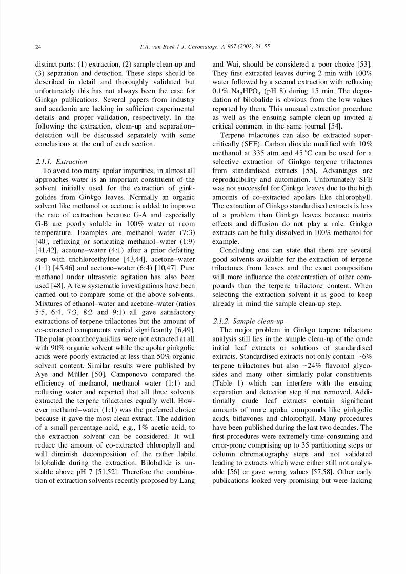
entire procedure and an example chromatogram aregiven in Table 8 and Fig. 2, respectively. The final
extract is also amenable to GC analysis after silyla-
tion, see Fig. 8 for an example chromatogram.Fig. 2. HPLC–ELSD profile of a methanolic solution of aThe most simple sample clean-up is no samplesupercritical fluid extract of a standardised Ginkgo extract.clean-up but just an extraction immediately followedPhenomenex column 25034.6 mm filled with Spherisorb 5
by analysis. An early example of this approach canODS(2), solvent H O–THF–MeOH (68.5:10.5:21) 1.0 ml/min,
2
be seen in Fig. 10 where a liquid Ginkgo drug is Varex ELSD, N as nebulizer gas at 2.06 l /min, drift tube 1078.2
directly analysed by SFC-ELSD. This method has From Ref. [55], reproduced with permission from John Wiley &
Sons Limited.not been validated for quantitative use. Very recently
Table 8
SFE–HPLC–ELSD method for standardised extracts by van Beek and Taylor [55]
(1) Apply |18 mg standardised Ginkgo extract in MeOH on 2 g silica gel and 0.5 g sand in a thimble
(2) Extract supercritically at 335 atm and 45 8C with 10% MeOH in CO , first 5 min static, then 40 min2
dynamic at 1.5 ml/min, nozzle and trap both 80 8C, solid trap consists of 400 mg silica gel
(3) Stop flow, cool nozzle and trap to 50 and 30 8C, respectively, and wash trap with 2.5 ml MeOAc
(4) Collect first 1.7 ml, evaporate, dissolve in MeOH and inject 5 ml into the HPLC
(5) HPLC on a 5 mm 25034.6 mm C column, 1.0 ml/min with H O–THF–MeOH (68.5:10.5:21) with ELS18 2
detection
5/13/2018 Chemical Analysis of Every Constituent - slidepdf.com
http://slidepdf.com/reader/full/chemical-analysis-of-every-constituent 8/35
967 (2002) 21 –5528 T . A. van Beek / J . Chromatogr . A
Table 9
Quantitative HPLC–ELSD method with minimal sample clean-up by Ganzera et al. [75]
(1) Sonicate 500.0 mg sample three times during 10 min with each time 3 ml MeOH
(2) Centrifuge at 3000 rpm during 10 min, combine supernatants in a 10-ml volumetric flask
(3) Add MeOH to 10.0 ml, take sample and filter over a 0.45-mm membrane(4) HPLC on a 4 mm 25034.6 mm C column, 1.0 ml/ min with a 10 m M NH OAc (pH 5) to MeOH–iBuOH
18 4
(9:1) gradient with ELS detection, triplicate injections of 10 ml
(5) Calculation with external standardisation and log–log calibration curves
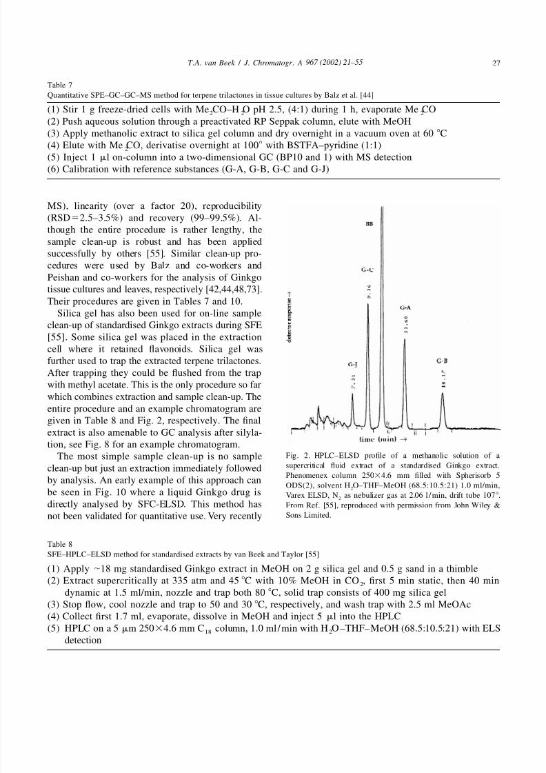
two papers with a validated quantitative procedure procedures are intrinsically more filthy extracts
have appeared. Ganzera et al. [75] investigated a which will lead to a faster degradation of HPLC
Ginkgo extract and several phytopharmaceuticals columns and possibly insufficient peak purity unless
according to the procedure given in Table 9. The an expensive and more complex mass spectrometer
method is relatively fast and cheap but the required is used as detector. Future improvements which can
sample size is rather high. The method was validated be expected are further simplifications, the intro-in terms of recovery, peak purity, limit of detection, duction of internal standards for HPLC methods, a
linearity, extraction efficiency and reproducibility. move towards a smaller scale resulting in a reduction
Some criticisms on the peak purity evaluation could of the required amounts of solvents which are still
be made. A chromatogram can be found in Fig. 4. high and the development of one method which can
The method of Jensen et al. is almost identical to the be used for Ginkgo leaves, extracts and phytophar-
above method except for the use of a mass spec- maceuticals.
trometer as detector [74]. This leads to a much
higher sensitivity and selectivity so the sample size 2.1.3. Separation and detection
could be reduced and peak purity should not pose a
problem. The procedure was validated. RSDs were 2.1.3.1. HPLC
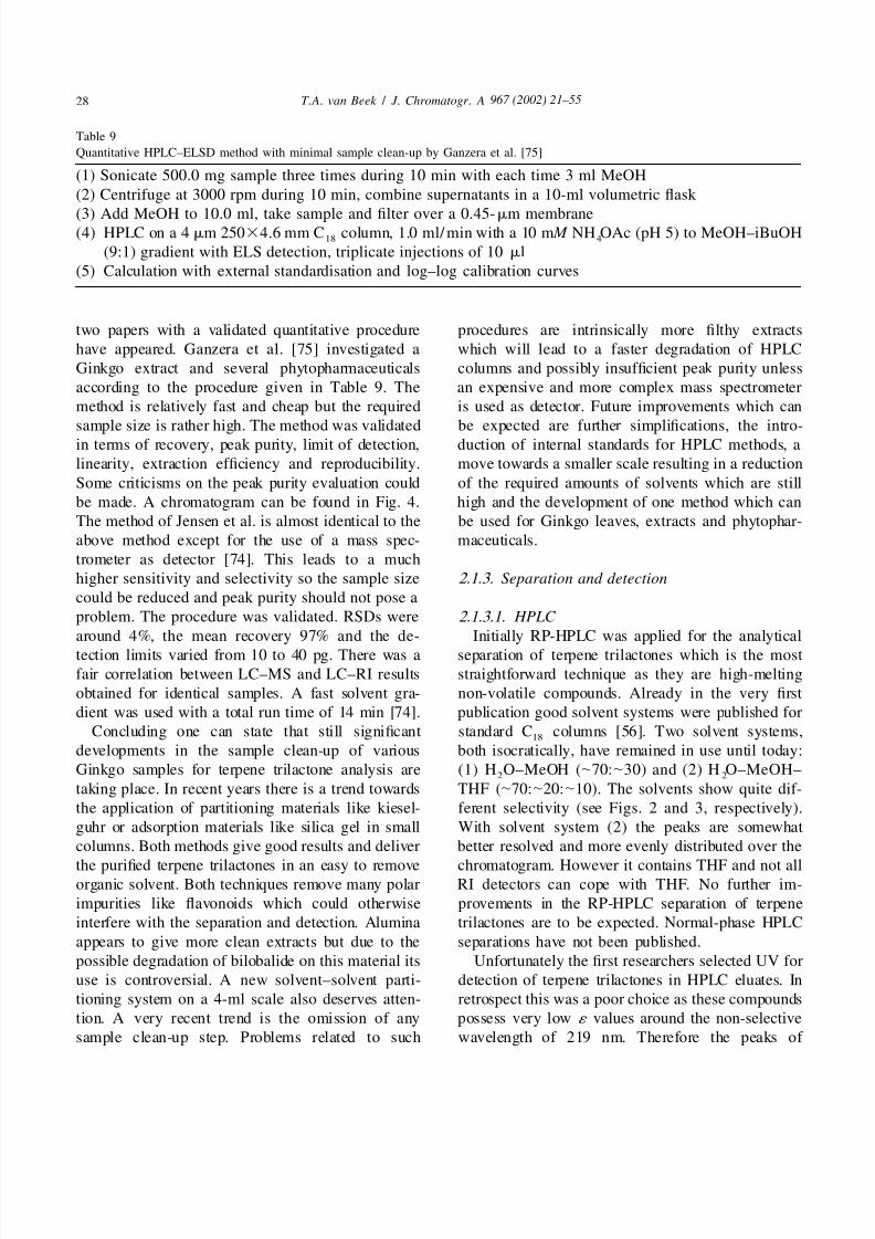
around 4%, the mean recovery 97% and the de- Initially RP-HPLC was applied for the analytical
tection limits varied from 10 to 40 pg. There was a separation of terpene trilactones which is the mostfair correlation between LC–MS and LC–RI results straightforward technique as they are high-melting
obtained for identical samples. A fast solvent gra- non-volatile compounds. Already in the very first
dient was used with a total run time of 14 min [74]. publication good solvent systems were published for
Concluding one can state that still significant standard C columns [56]. Two solvent systems,18
developments in the sample clean-up of various both isocratically, have remained in use until today:
Ginkgo samples for terpene trilactone analysis are (1) H O–MeOH (|70:|30) and (2) H O–MeOH–2 2
taking place. In recent years there is a trend towards THF (|70:|20:|10). The solvents show quite dif-
the application of partitioning materials like kiesel- ferent selectivity (see Figs. 2 and 3, respectively).
guhr or adsorption materials like silica gel in small With solvent system (2) the peaks are somewhat
columns. Both methods give good results and deliver better resolved and more evenly distributed over the
the purified terpene trilactones in an easy to remove chromatogram. However it contains THF and not allorganic solvent. Both techniques remove many polar RI detectors can cope with THF. No further im-
impurities like flavonoids which could otherwise provements in the RP-HPLC separation of terpene
interfere with the separation and detection. Alumina trilactones are to be expected. Normal-phase HPLC
appears to give more clean extracts but due to the separations have not been published.
possible degradation of bilobalide on this material its Unfortunately the first researchers selected UV for
use is controversial. A new solvent–solvent parti- detection of terpene trilactones in HPLC eluates. In
tioning system on a 4-ml scale also deserves atten- retrospect this was a poor choice as these compounds
tion. A very recent trend is the omission of any possess very low ´ values around the non-selective
sample clean-up step. Problems related to such wavelength of 219 nm. Therefore the peaks of

5/13/2018 Chemical Analysis of Every Constituent - slidepdf.com
http://slidepdf.com/reader/full/chemical-analysis-of-every-constituent 9/35
967 (2002) 21 –55 29T . A. van Beek / J . Chromatogr . A
small solvent peak and greater sensitivity. Advan-
tages of RI over ELSD are larger linear range, lower
costs and its broader availability.
The only other LC detection technique used is
mass spectrometry [74,75,79–81]. With a thermo-spray interface and post-column addition of am-
monium acetate strong quasi molecular ions [M11
NH ] could be observed for all terpene trilactones4
and the method could be used for the selective
detection and semi-quantitative analysis of the ter-
pene trilactones after minimal sample clean-up [79].
Disadvantages were the large day to day variation
necessitating recalibration every day and the much
higher cost compared to other LC detectors.
Electrospray ionisation MS (ESI-MS) was used by
Mauri et al. and is more robust than the TSPinterface [80]. Best ESI-MS results were obtained in
the positive mode. All the terpene trilactones gave1
sodiated adducts [M1Na] as the main ion. No
additional sodium needed to be added to the sample
or solvent for the sodiated adducts to be the main
ions. Both direct infusion ESI-MS and on-line ESI-
MS after an isocratic or gradient RP-HPLC run were
possible. An example of the direct infusion technique
showing also many flavonoids is given in Fig. 5. The
detection limit for this technique was |50 mg/ ml for
each terpene when present in standardised extracts.
A much higher sensitivity could be achieved byselected ion monitoring in the LC ESI-MS mode: |1
Fig. 3. HPLC profiles of 1.00 ml of purified Tanakan phytophar-ng. Good linearity was obtained in the range 1–20
maceutical with (upper trace) UV detection at 219 nm and (lowermg/ml for each terpene trilactone. Overall repro-trace) RI detection. See Table 3 for chromatographic details.ducibility was 3.4% (same day) and 5.8% (betweenInternal standard (I.S.) is benzyl alcohol. Reprinted from Ref.
[41], Copyright (1991), with permission from Elsevier Science. days) [80]. Ganzera et al. also used ESI-MS but then
in the negative mode with a 10 m M ammonium2
acetate buffer. [M2H] could be observed for all
interest were overshadowed by the absorbance of five terpene trilactones [75]. A chromatogram is
traces of other compounds remaining in the partially depicted in Fig. 6. In a more recent paper Mauri et
purified extracts [56]. This is clearly demonstrated in al. used atmospheric pressure chemical ionization
Figs. 3 and 4. A much better approach for gink- (LC–APCI-MS) in the negative mode. The highgolides is therefore a detector which shows less sensitivity and specificity of the method (|1 ng/ml)
variation in response factors, e.g., refractive index allowed the quantitation of terpene trilactones in
detection (RI) [7,10,11,40,41,43,44,59,60,66,72,76– plasma samples of volunteers. Due to a fast gradient
78] or evaporative light scattering detection (ELSD) the total separation time was 7 min [81]. A similar
[42,55,65,75]. Examples of UV/RI and UV/ELS LC–MS procedure was used by Jensen et al. [74].
detection are given in Figs. 3 and 4, respectively. They used LC–MS as part of a quantitative method
Both methods are suitable for the routine analysis of for Ginkgo extracts (vide supra).
all terpene trilactones after an RP-HPLC separation. Although the MS detector offers a high selectivity
Advantages of ELSD over RI are better baseline and sensitivity, due to its high price and more
stability, compatibility with THF and gradients, complicated operation and maintenance it remains to

5/13/2018 Chemical Analysis of Every Constituent - slidepdf.com
http://slidepdf.com/reader/full/chemical-analysis-of-every-constituent 10/35
967 (2002) 21 –5530 T . A. van Beek / J . Chromatogr . A
Fig. 4. HPLC–UV and HPLC–ELSD profiles of a filtered methanolic extract of a Ginkgo drug containing standardised extract. Phenomenex˚ Synergi Max-RP 80 A 4 mm column 15034.6 mm, gradient from 10% B to 20% B in 10 min, then to 25% B in 15 min, 1.0 ml/min. (A) 10
m M NH OAc adjusted to pH 5; (B) MeOH–iBuOH (9:1). Sedex 55 ELSD, N as nebulizer gas at 2.4 bar, drift tube 458. Reproduced from4 2
[75] with permission of The Pharmaceutical Society of Japan.
Fig. 5. Positive ion mass spectrum of Ginkgo standardised extract introduced by direct infusion into an ESI-MS. Peaks at m / z 349, 431, 4471
and 463 correspond with [M1Na] for bilobalide, G-A, G-B and G-J, and G-C, respectively. From Ref. [80], reproduced with permission
from John Wiley & Sons Limited.
5/13/2018 Chemical Analysis of Every Constituent - slidepdf.com
http://slidepdf.com/reader/full/chemical-analysis-of-every-constituent 11/35
967 (2002) 21 –55 31T . A. van Beek / J . Chromatogr . A
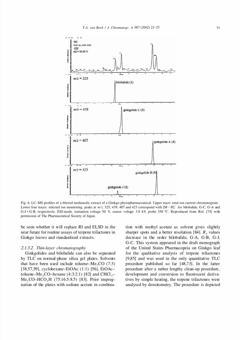
Fig. 6. LC–MS profiles of a filtered methanolic extract of a Ginkgo phytopharmaceutical. Upper trace: total ion current chromatogram.2
Lower four traces: selected ion monitoring, peaks at m / z 325, 439, 407 and 423 correspond with [M2H] for bilobalide, G-C, G-A and
G-J1G-B, respectively. ESI-mode, ionization voltage 50 V, source voltage 3.0 kV, probe 350 8C. Reproduced from Ref. [75] with
permission of The Pharmaceutical Society of Japan.
be seen whether it will replace RI and ELSD in the tion with methyl acetate as solvent gives slightly
near future for routine assays of terpene trilactones in sharper spots and a better resolution [84]. R valuesf
Ginkgo leaves and standardised extracts. decrease in the order bilobalide, G-A, G-B, G-J,G-C. This system appeared in the draft monograph
2.1.3.2. Thin-layer chromatography of the United States Pharmacopeia on Ginkgo leaf
Ginkgolides and bilobalide can also be separated for the qualitative analysis of terpene trilactones
by TLC on normal-phase silica gel plates. Solvents [9,85] and was used in the only quantitative TLC
that have been used include toluene–Me CO (7:3) procedure published so far [48,73]. In the latter2
[38,57,59], cyclohexane–EtOAc (1:1) [56], EtOAc– procedure after a rather lengthy clean-up procedure,
toluene–Me CO–hexane (4:3:2:1) [82] and CHCl – development and conversion to fluorescent deriva-2 3
Me CO–HCO H (75:16.5:8.5) [83]. Prior impreg- tives by simple heating, the terpene trilactones were2 2
nation of the plates with sodium acetate in combina- analysed by densitometry. The procedure is depicted

5/13/2018 Chemical Analysis of Every Constituent - slidepdf.com
http://slidepdf.com/reader/full/chemical-analysis-of-every-constituent 12/35
967 (2002) 21 –5532 T . A. van Beek / J . Chromatogr . A
Table 10
Quantitative SPE–HPTLC-densitometric method for terpene trilactones in leaves by Peishan et al. [48,73]
(1) Extract 3 g Ginkgo leaf powder under sonication 20115 min with 23 80 ml MeOH–H O (1:9)2
(2) Filter, combine filtrates and pass through a polyamide cartridge and elute with 100 ml water
(3) Evaporate eluate, dissolve in 5 ml MeOH, mix with 3 g silica gel, dry 4 h over P O in vacuo2 5
(4) Transfer silica gel to a 5-g activated silica gel SPE column, elute with 200 ml CHCl –MeOH (1:1)3
(5) Evaporate eluate, dissolve in 1.0 ml MeOH
(6) Apply 6–10 ml and reference solutions on a NaOAc impregnated silica gel plate with autosampler
(7) Dry the plate .4 h over P O , condition the plate over aqueous H SO2 5 2 4
(8) Develop HPTLC plate 9 cm with toluene–EtOAc–Me CO–MeOH (10:5:5:0.6)2
(9) Evaporate solvent, heat plate 30 min at 160 8C, scan the chromatogram in a TLC scanner at l5366 nm,
calibrate by second-order polynomial regresssion, calculate results
in Table 10. A chromatogram can be viewed in Fig. stability, ruggedness, costs and sensitivity. The best
7. The method was validated and gave accurate and column is a 30-m capillary one coated with 100%reproducible results for well known standardised dimethyl polysiloxane phase. One of the milestones
extracts. The results were comparable with those in this area is the paper by Hasler and Meier [86].
obtained by HPLC–ELSD or HPLC–RI [73]. They investigated among others the optimal silyla-
tion procedure. BSTFA with 1% TMCS at 120 8C2.1.3.3. Gas chromatography was found to give the best results. Rather mystifying
Instead of HPLC, GC can also be used. However is the recent remark of Balz et al. that bilobalide
prior silylation is necessary because ginkgolides and cannot be derivatised [44]. This is in contradiction
bilobalide are non-volatile. This is the main dis- when many other publications (vide infra). After
advantage in comparison with HPLC. The separation silylation the mixture should be injected directly into
is at least as good and detection by FID surpasses the GC to avoid desilylation problems. Detection
any available LC detector in reproducibility, baseline limits varied from 50 to 100 ng and RSDs were low
(1–2%). Various internal standards have been pro-posed: cholesterol [86], octacosane [55] and squalane
Fig. 8. GC–FID profile of a standardised Ginkgo leaf extract after
supercritical fluid extraction and on-line sample clean-up after
Fig. 7. TLC scanning profile of a Ginkgo leaf extract on a sodium silylation [55]. See Table 8 for details on the sample preparation.
acetate impregnated hand-made silica gel plate. Bilobalide, G-A, GC analysis took place on an Ultra 1 column (25 m30.2 mm3
G-B and G-C can be observed at R values of 0.52, 0.42, 0.33 and 0.33 mm), oven temp. 230–280 8C. Internal standard, octacosane.f
0.18, respectively. Reproduced in modified form from [73] with From Ref. [55], reproduced with permission from John Wiley &
permission of the editor. Sons Limited.

5/13/2018 Chemical Analysis of Every Constituent - slidepdf.com
http://slidepdf.com/reader/full/chemical-analysis-of-every-constituent 13/35
967 (2002) 21 –55 33T . A. van Beek / J . Chromatogr . A
[47,69]. An example chromatogram can be seen in 2.1.3.4. SFC
Fig. 8. Other publications describing silylation of Supercritical fluid chromatography is also capable
terpene trilactones followed by GC are [45– of separating Ginkgo terpene trilactones [91]. On a
47,53,55,63,64,69,83,87,88]. Deltabond deactivated aminopropyl HPLC column
Instead of flame ionisation two other detectors can (15034.6 mm, 5 mm) with 12% methanol in carbonbe used for the detection of silylated terpene trilac- dioxide as fluid (280 atm, 3.5 ml/ min, 40 8C), a
tones: electron capture and mass spectrometry. ECD baseline separation of bilobalide and all four gink-
has been used only once [34]. The slight increase in golides could be achieved within 9 min. Detection
selectivity and sensitivity of ECD compared with occurred through evaporative light scattering detec-
FID is offset by the problems associated with this tion (ELSD). A detection limit of approximately 10
detector: not generally available, limited linear range ng was reported. The selectivity of the system
and its inherent radioactivity. GC–MS is only neces- appears to be higher than that of RP-HPLC. The
sary when the concentrations are very low: in certain explanation given was that the separation mechanism
Ginkgo cell cultures [44,89] and for blood and urine is essentially a normal-phase one. As most impurities
samples in pharmacokinetic studies [51,90]. For the present in Ginkgo extracts are more polar than
procedure used by Balz et al. see Table 7. ECD can terpene trilactones, the latter elute first and thealso be used for a highly sensitive detection of impurities remain on the column. Although a sample
terpene trilactones if the derivatisation takes place clean-up over silica did give cleaner chromatograms,
with halogen-containing reagents, e.g., heptafluoro- some standardised extracts and phytopharmaceuticals
butyric anhydride. Then detection limits of below 1 could be analysed without any clean-up (Fig. 10).
pg (Fig. 9) can be realised although the stability of Thus SFC is an alternative for the analysis of Ginkgo
the bilobalide derivative might be a problem [34]. extracts: quick, low consumption of organic solvent
This derivatisation is of little practical consequence and a more simple sample clean-up. However in the
for the routine analysis of leaves or extracts where near future it is unlikely to replace either HPLC or
such high sensitivity is not needed. For phar- GC in the average quality control laboratory.
macokinetic studies where sensitivity is an issue, this
technique may be more valuable but still GC–MS 2.1.3.5. Capillary electrophoresis
with its inherent higher selectivity is probably more Oerhle has demonstrated that G-A, G-B anduseful and practical. bilobalide can be separated by micellar electrokinetic
capillary electrophoresis (MECC) [92]. The three
compounds were separated in a capillary of 60 cm3
75 mm at 30 8C with a buffer consisting of 25 m M
phosphate and 90 m M SDS (Fig. 11). The voltage
was not given. Detection took place by UV at 185
nm. There was a good separation between bilobalide
(10 min) and the two ginkgolides but G-A and G-B
were not baseline separated (16.5 and 16.7 min,
respectively). No quantitative data or chromatograms
of leaf extracts were included in this paper. Unlessthe separation considerably improves, it is clear that
capillary electrophoresis cannot yet compete with
HPLC, GC or SFC for the separation of Ginkgo
terpene trilactones.
Two less straightforward techniques for the quan-Fig. 9. GC–ECD profile of a toluene extract of bilobalide (BB), titative analysis of terpene trilactones are quantitativeG-C, G-B and G-A reference substances after reaction with NMR [71] and biological standardisation [83,93].heptafluorobutyric anhydride. Each peak corresponds with approx-
Neither of these techniques are likely to be routinelyimately 10 pg of underivatised terpene trilactone. After 14.5 min
used for quality control of Ginkgo leaves or extractsattenuator value is halved. Reproduced from Ref. [34] with
permission of the editor. because of one or more disadvantages. NMR is too
5/13/2018 Chemical Analysis of Every Constituent - slidepdf.com
http://slidepdf.com/reader/full/chemical-analysis-of-every-constituent 14/35
967 (2002) 21 –5534 T . A. van Beek / J . Chromatogr . A
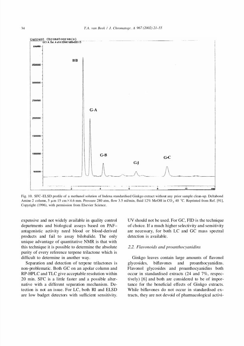
Fig. 10. SFC–ELSD profile of a methanol solution of Indena standardised Ginkgo extract without any prior sample clean-up. Deltabond
Amino 2 column, 5 mm 15 cm34.6 mm. Pressure 280 atm, flow 3.5 ml/min, fluid 12% MeOH in CO , 40 8C. Reprinted from Ref. [91],2
Copyright (1996), with permission from Elsevier Science.
expensive and not widely available in quality control UV should not be used. For GC, FID is the technique
departments and biological assays based on PAF– of choice. If a much higher selectivity and sensitivity
antagonistic activity need blood or blood-derived are necessary, for both LC and GC mass spectral
products and fail to assay bilobalide. The only detection is available.
unique advantage of quantitative NMR is that withthis technique it is possible to determine the absolute 2.2. Flavonoids and proanthocyanidins
purity of every reference terpene trilactone which is
difficult to determine in another way. Ginkgo leaves contain large amounts of flavonol
Separation and detection of terpene trilactones is glycosides, biflavones and proanthocyanidins.
non-problematic. Both GC on an apolar column and Flavonol glycosides and proanthocyanidins both
RP-HPLC and TLC give acceptable resolution within occur in standardised extracts (24 and 7%, respec-
20 min. SFC is a little faster and a possible alter- tively) [6] and both are considered to be of impor-
native with a different separation mechanism. De- tance for the beneficial effects of Ginkgo extracts.
tection is not an issue. For LC, both RI and ELSD While biflavones do not occur in standardised ex-
are low budget detectors with sufficient sensitivity. tracts, they are not devoid of pharmacological activi-

5/13/2018 Chemical Analysis of Every Constituent - slidepdf.com
http://slidepdf.com/reader/full/chemical-analysis-of-every-constituent 15/35
967 (2002) 21 –55 35T . A. van Beek / J . Chromatogr . A
Fig. 11. MECC profile of standards of G-A, G-B and bilobalide. Capillary 60 cm375 mm, buffer 25 m M phosphate and 90 m M SDS,
30 8C. Detection UV at 185 nm. Reprinted from Ref. [92] by courtesy of Marcel Dekker Inc., 1995.
ty. Lately they have been applied in cosmetics [94]. 2.2.1. Flavonol glycosides
In the following these three different groups of In Ginkgo leaves and extracts many different
polyphenolics are discussed. An earlier review on the flavonol glycosides occur most of them being deriva-
chemical analysis of Ginkgo flavonoids was pub- tives of quercetin, kaempferol and isorhamnetin. The
lished by Sticher et al. [95]. aglycones themselves occur only in relatively low
Fig. 12. Upper: HPLC profile of 33 different flavonol glycosides (1– 22), flavonol aglycones (23–28) and biflavones (29–33) occurring in
an alcoholic Ginkgo leaf extract. Lower: HPLC profile of a mixture of 33 reference compounds. For chromatographic details see Table 11,
line 8. Reproduced with permission from Ref. [96].
5/13/2018 Chemical Analysis of Every Constituent - slidepdf.com
http://slidepdf.com/reader/full/chemical-analysis-of-every-constituent 16/35
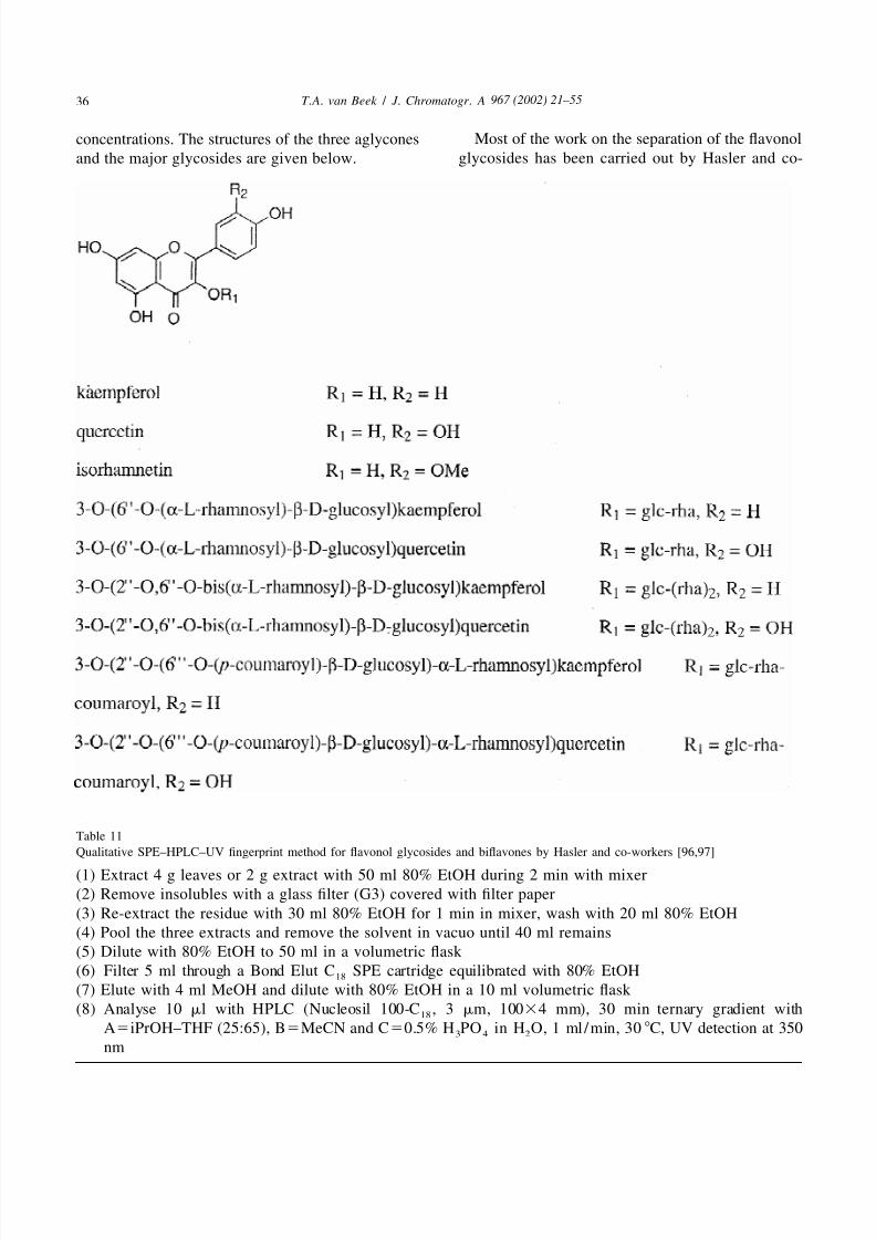
967 (2002) 21 –5536 T . A. van Beek / J . Chromatogr . A
Most of the work on the separation of the flavonolconcentrations. The structures of the three aglyconesglycosides has been carried out by Hasler and co-and the major glycosides are given below.
Table 11
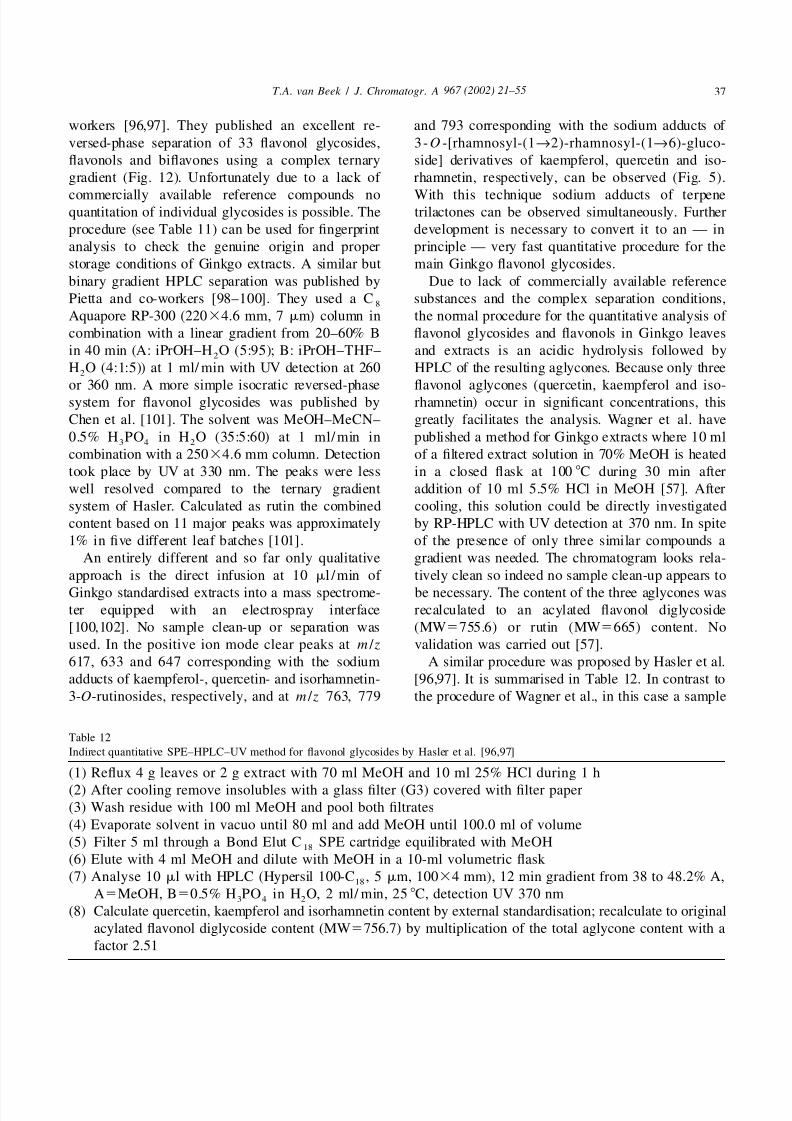
Qualitative SPE–HPLC–UV fingerprint method for flavonol glycosides and biflavones by Hasler and co-workers [96,97]
(1) Extract 4 g leaves or 2 g extract with 50 ml 80% EtOH during 2 min with mixer
(2) Remove insolubles with a glass filter (G3) covered with filter paper
(3) Re-extract the residue with 30 ml 80% EtOH for 1 min in mixer, wash with 20 ml 80% EtOH
(4) Pool the three extracts and remove the solvent in vacuo until 40 ml remains
(5) Dilute with 80% EtOH to 50 ml in a volumetric flask
(6) Filter 5 ml through a Bond Elut C SPE cartridge equilibrated with 80% EtOH18
(7) Elute with 4 ml MeOH and dilute with 80% EtOH in a 10 ml volumetric flask
(8) Analyse 10 ml with HPLC (Nucleosil 100-C , 3 mm, 10034 mm), 30 min ternary gradient with18
A5iPrOH–THF (25:65), B5MeCN and C50.5% H PO in H O, 1 ml /min, 30 8C, UV detection at 3503 4 2
nm
5/13/2018 Chemical Analysis of Every Constituent - slidepdf.com
http://slidepdf.com/reader/full/chemical-analysis-of-every-constituent 17/35
967 (2002) 21 –55 37T . A. van Beek / J . Chromatogr . A
workers [96,97]. They published an excellent re- and 793 corresponding with the sodium adducts of
versed-phase separation of 33 flavonol glycosides, 3 -O -[rhamnosyl-(1→2)-rhamnosyl-(1→6)-gluco-
flavonols and biflavones using a complex ternary side] derivatives of kaempferol, quercetin and iso-
gradient (Fig. 12). Unfortunately due to a lack of rhamnetin, respectively, can be observed (Fig. 5).
commercially available reference compounds no With this technique sodium adducts of terpenequantitation of individual glycosides is possible. The trilactones can be observed simultaneously. Further
procedure (see Table 11) can be used for fingerprint development is necessary to convert it to an — in
analysis to check the genuine origin and proper principle — very fast quantitative procedure for the
storage conditions of Ginkgo extracts. A similar but main Ginkgo flavonol glycosides.
binary gradient HPLC separation was published by Due to lack of commercially available reference
Pietta and co-workers [98–100]. They used a C substances and the complex separation conditions,8
Aquapore RP-300 (22034.6 mm, 7 mm) column in the normal procedure for the quantitative analysis of
combination with a linear gradient from 20–60% B flavonol glycosides and flavonols in Ginkgo leaves
in 40 min (A: iPrOH–H O (5:95); B: iPrOH–THF– and extracts is an acidic hydrolysis followed by2
H O (4:1:5)) at 1 ml/ min with UV detection at 260 HPLC of the resulting aglycones. Because only three2
or 360 nm. A more simple isocratic reversed-phase flavonol aglycones (quercetin, kaempferol and iso-system for flavonol glycosides was published by rhamnetin) occur in significant concentrations, this
Chen et al. [101]. The solvent was MeOH–MeCN– greatly facilitates the analysis. Wagner et al. have
0.5% H PO in H O (35:5:60) at 1 ml/min in published a method for Ginkgo extracts where 10 ml3 4 2
combination with a 25034.6 mm column. Detection of a filtered extract solution in 70% MeOH is heated
took place by UV at 330 nm. The peaks were less in a closed flask at 100 8C during 30 min after
well resolved compared to the ternary gradient addition of 10 ml 5.5% HCl in MeOH [57]. After
system of Hasler. Calculated as rutin the combined cooling, this solution could be directly investigated
content based on 11 major peaks was approximately by RP-HPLC with UV detection at 370 nm. In spite
1% in five different leaf batches [101]. of the presence of only three similar compounds a
An entirely different and so far only qualitative gradient was needed. The chromatogram looks rela-
approach is the direct infusion at 10 ml /min of tively clean so indeed no sample clean-up appears to
Ginkgo standardised extracts into a mass spectrome- be necessary. The content of the three aglycones waster equipped with an electrospray interface recalculated to an acylated flavonol diglycoside
[100,102]. No sample clean-up or separation was (MW5755.6) or rutin (MW5665) content. No
used. In the positive ion mode clear peaks at m / z validation was carried out [57].
617, 633 and 647 corresponding with the sodium A similar procedure was proposed by Hasler et al.
adducts of kaempferol-, quercetin- and isorhamnetin- [96,97]. It is summarised in Table 12. In contrast to
3-O-rutinosides, respectively, and at m / z 763, 779 the procedure of Wagner et al., in this case a sample
Table 12
Indirect quantitative SPE–HPLC–UV method for flavonol glycosides by Hasler et al. [96,97]
(1) Reflux 4 g leaves or 2 g extract with 70 ml MeOH and 10 ml 25% HCl during 1 h
(2) After cooling remove insolubles with a glass filter (G3) covered with filter paper
(3) Wash residue with 100 ml MeOH and pool both filtrates
(4) Evaporate solvent in vacuo until 80 ml and add MeOH until 100.0 ml of volume
(5) Filter 5 ml through a Bond Elut C SPE cartridge equilibrated with MeOH18
(6) Elute with 4 ml MeOH and dilute with MeOH in a 10-ml volumetric flask
(7) Analyse 10 ml with HPLC (Hypersil 100-C , 5 mm, 10034 mm), 12 min gradient from 38 to 48.2% A,18
A5MeOH, B50.5% H PO in H O, 2 ml/ min, 25 8C, detection UV 370 nm3 4 2
(8) Calculate quercetin, kaempferol and isorhamnetin content by external standardisation; recalculate to original
acylated flavonol diglycoside content (MW5756.7) by multiplication of the total aglycone content with a
factor 2.51

5/13/2018 Chemical Analysis of Every Constituent - slidepdf.com
http://slidepdf.com/reader/full/chemical-analysis-of-every-constituent 18/35
967 (2002) 21 –5538 T . A. van Beek / J . Chromatogr . A
clean-up step was part of the procedure. Perhaps this of the acetone and addition of methanol and HCl the
is caused by the fact that the procedure is suitable for glycosides are hydrolysed. Separation takes place
both Ginkgo leaves and extracts. On the other hand, isocratically on a 12534.6-mm C column with18
few leaf constituents eluting around the same time as iPrOH–MeCN–0.6% citric acid in H O (5:47:100)2
flavonol aglycones and which absorb light at the as solvent at 1.0 ml/ min. UV detection occurs at 370selective wavelength of 370 nm occur in Ginkgo nm. This solvent system resolves isorhamnetin and
leaves. The chromatogram (Fig. 13) is of high kaempferol rather poorly but on the other hand is
quality and the separation time is 12 min excluding very simple and fast (7 min). All peaks are first
10 min washing and re-equilibration. Also in this calculated as quercetin by external standardisation
case a mild gradient was used. Kim et al. and Chen and then recalculated to a flavonol glycoside of mass
et al. have both experimented with isocratic systems 756.7 by multiplication with 2.514. Why the kaemp-
but the resulting chromatograms are not as good as ferol–isorhamnetin peak is not calculated as kaemp-
those of Hasler [101,103]. ferol by external standardisation is unclear.
The draft monographs on Ginkgo leaf for the Recalculation of the aglycones to flavonol glyco-
United States [9,85] and European [10] Phar- sides with an average weight of 756 is slightly
macopeias are identical and use a variation of the incorrect as many flavonol glycosides possess aprocedures described above. As initial extraction lower molecular mass and free aglycones are also
solvent they use acetone–water (6:4). After removal always present to some extent. Thus the true flavonol
glycoside content of standardised extracts is some-
what lower than 24% but this does not lead to
problems as long as everyone uses the same pro-
cedure and method of calculation. Additionally ex-
tracts should be stored properly to avoid hydrolysis.
Two quantitative indirect methods for kaempferol
and quercetin employing TLC or HPTLC and den-
sitometry have been published [104,105]. The pro-
cedure used by Jamshidi et al. [105] is given in
Table 13. A baseline separation of the two analytesof interest but not of quercetin and an unknown
matrix component was obtained. Calibration curves
were linear from 0.5 to 2.0 mg for both kaempferol
and quercetin. Repeatability was good with RSDs of
1.4%. The recovery varied from 94 to 97%. The
extraction efficiency was not checked.
There is one paper where micellar electrokinetic
capillary chromatography (MECC) has been used for
the separation of Ginkgo flavonol glycosides [99].
The separations were performed with a 72-cm350-
mm fused-silica capillary column in combinationwith a 20 m M sodium borate buffer (pH 8.3) and 50
m M sodium dodecyl sulphate (SDS) at 20 kV and
27 8C. The injection volume was 4 nl and UV
detection took place at 260 nm. Compared to HPLC
the separation is faster with about the same reso-
lution. Disadvantages of MECC mentioned by the
authors themselves relative to HPLC are poorerFig. 13. HPLC profile of hydrolysed Ginkgo leaf sample. For
reproducibility of retention times and poor com-chromatographic details see Table 12, line 7. Reproduced with
permission from Ref. [96]. patibility with mass spectrometric detection [100].
5/13/2018 Chemical Analysis of Every Constituent - slidepdf.com
http://slidepdf.com/reader/full/chemical-analysis-of-every-constituent 19/35
967 (2002) 21 –55 39T . A. van Beek / J . Chromatogr . A
Table 13
Indirect quantitative HPTLC–densitometric method for flavonol glycosides in leaves by Jamshidi et al. [105]
(1) Reflux 2 g leaves with MeOH for 30 min, filter, add 2 ml 25% HCl solution
(2) Reflux acidic extract for 60 min, cool and neutralize with 25% ammonia solution
(3) Reduce volume on a water bath with N and dilute with MeOH in a 10-ml volumetric flask 2
(4) Leave overnight and apply supernatant by means of a Linomat IV on a 10320-cm silica gel HPTLC plate
prewashed with CHCl –MeOH, band length 6 mm3
(5) Develop plates by the incremental multiple development technique with toluene–Me CO–MeOH–HCO H2 2
(46:8:5:1) in an unsaturated chamber, dry during 5 min with N at 40 8C2
(6) Evaluate plates with a TLC scanner (densitometry) in the reflectance mode at 254 nm
(7) Calculate content of kaempferol and quercetin by external standardisation
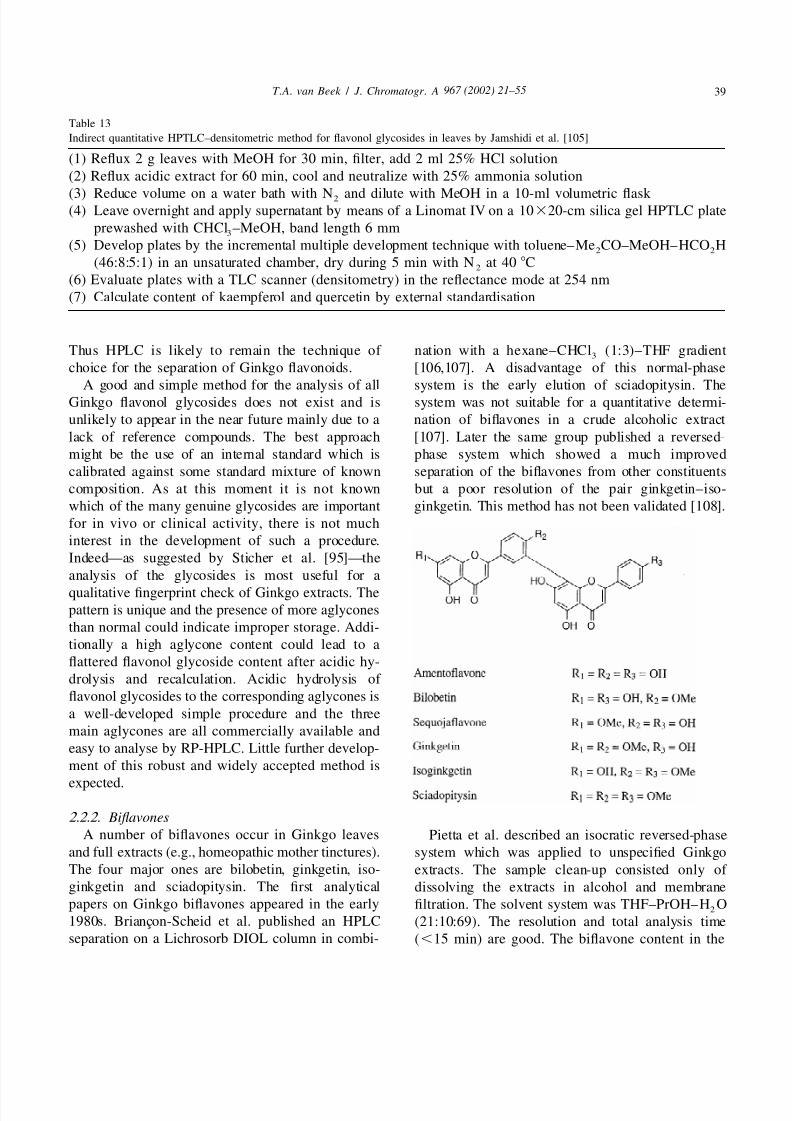
Thus HPLC is likely to remain the technique of nation with a hexane–CHCl (1:3)–THF gradient3
choice for the separation of Ginkgo flavonoids. [106,107]. A disadvantage of this normal-phase
A good and simple method for the analysis of all system is the early elution of sciadopitysin. TheGinkgo flavonol glycosides does not exist and is system was not suitable for a quantitative determi-
unlikely to appear in the near future mainly due to a nation of biflavones in a crude alcoholic extract
lack of reference compounds. The best approach [107]. Later the same group published a reversed-
might be the use of an internal standard which is phase system which showed a much improved
calibrated against some standard mixture of known separation of the biflavones from other constituents
composition. As at this moment it is not known but a poor resolution of the pair ginkgetin–iso-
which of the many genuine glycosides are important ginkgetin. This method has not been validated [108].
for in vivo or clinical activity, there is not much
interest in the development of such a procedure.
Indeed—as suggested by Sticher et al. [95]—the
analysis of the glycosides is most useful for a
qualitative fingerprint check of Ginkgo extracts. Thepattern is unique and the presence of more aglycones
than normal could indicate improper storage. Addi-
tionally a high aglycone content could lead to a
flattered flavonol glycoside content after acidic hy-
drolysis and recalculation. Acidic hydrolysis of
flavonol glycosides to the corresponding aglycones is
a well-developed simple procedure and the three
main aglycones are all commercially available and
easy to analyse by RP-HPLC. Little further develop-
ment of this robust and widely accepted method is
expected.
2.2.2. Biflavones
A number of biflavones occur in Ginkgo leaves Pietta et al. described an isocratic reversed-phaseand full extracts (e.g., homeopathic mother tinctures). system which was applied to unspecified GinkgoThe four major ones are bilobetin, ginkgetin, iso- extracts. The sample clean-up consisted only of ginkgetin and sciadopitysin. The first analytical dissolving the extracts in alcohol and membranepapers on Ginkgo biflavones appeared in the early filtration. The solvent system was THF–PrOH–H O
2
1980s. Briancon-Scheid et al. published an HPLC (21:10:69). The resolution and total analysis timeseparation on a Lichrosorb DIOL column in combi- (,15 min) are good. The biflavone content in the
5/13/2018 Chemical Analysis of Every Constituent - slidepdf.com
http://slidepdf.com/reader/full/chemical-analysis-of-every-constituent 20/35
967 (2002) 21 –5540 T . A. van Beek / J . Chromatogr . A
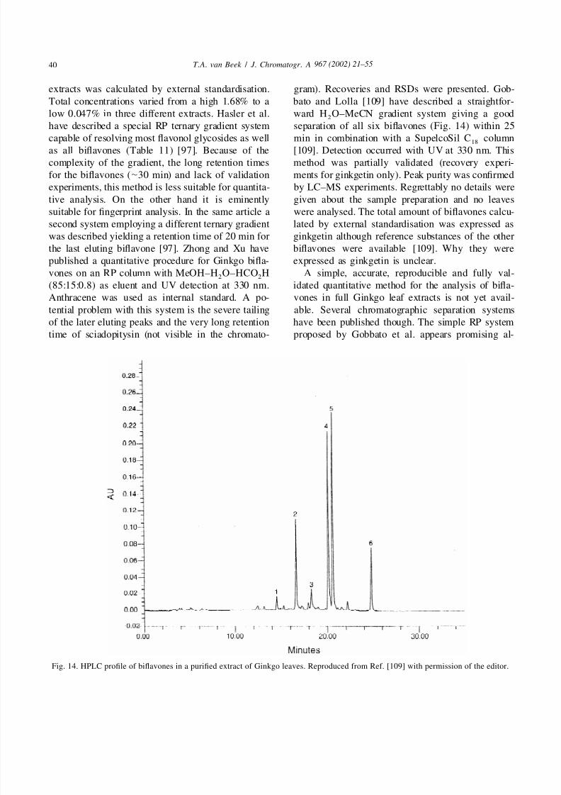
extracts was calculated by external standardisation. gram). Recoveries and RSDs were presented. Gob-
Total concentrations varied from a high 1.68% to a bato and Lolla [109] have described a straightfor-
low 0.047% in three different extracts. Hasler et al. ward H O–MeCN gradient system giving a good2
have described a special RP ternary gradient system separation of all six biflavones (Fig. 14) within 25
capable of resolving most flavonol glycosides as well min in combination with a SupelcoSil C column18
as all biflavones (Table 11) [97]. Because of the [109]. Detection occurred with UV at 330 nm. This
complexity of the gradient, the long retention times method was partially validated (recovery experi-
for the biflavones (|30 min) and lack of validation ments for ginkgetin only). Peak purity was confirmed
experiments, this method is less suitable for quantita- by LC–MS experiments. Regrettably no details were
tive analysis. On the other hand it is eminently given about the sample preparation and no leaves
suitable for fingerprint analysis. In the same article a were analysed. The total amount of biflavones calcu-
second system employing a different ternary gradient lated by external standardisation was expressed as
was described yielding a retention time of 20 min for ginkgetin although reference substances of the other
the last eluting biflavone [97]. Zhong and Xu have biflavones were available [109]. Why they were
published a quantitative procedure for Ginkgo bifla- expressed as ginkgetin is unclear.
vones on an RP column with MeOH–H O–HCO H A simple, accurate, reproducible and fully val-2 2
(85:15:0.8) as eluent and UV detection at 330 nm. idated quantitative method for the analysis of bifla-
Anthracene was used as internal standard. A po- vones in full Ginkgo leaf extracts is not yet avail-
tential problem with this system is the severe tailing able. Several chromatographic separation systems
of the later eluting peaks and the very long retention have been published though. The simple RP system
time of sciadopitysin (not visible in the chromato- proposed by Gobbato et al. appears promising al-
Fig. 14. HPLC profile of biflavones in a purified extract of Ginkgo leaves. Reproduced from Ref. [109] with permission of the editor.

5/13/2018 Chemical Analysis of Every Constituent - slidepdf.com
http://slidepdf.com/reader/full/chemical-analysis-of-every-constituent 21/35
967 (2002) 21 –55 41T . A. van Beek / J . Chromatogr . A
though it is unknown how much overlap this system methods (1)–(4), respectively [112]. According to
will give with other components present in Ginkgo them, methods (2) and (4) gave too high values,
extracts [109]. UV detection at 330 nm is the most while method (3) gave a too low value. Best results
selective and currently the best detection method were obtained with method (1) which was also used
available for Ginkgo biflavones. by Schennen [110]. However, even this best methodhas a high degree of uncertainty which is perhaps
best illustrated by the value of 7.0% given by Stumpf 2.2.3. Proanthocyanidins[6] for the proanthocyanidin content of the sameLarge amounts of proanthocyanidins occur in bothextract as investigated by Lang and Wilhelm. ClearlyGinkgo leaves (4–12% [110]) and standardisedthere is significant scope for improving existing orextracts (7.0% [6]). They are considered to be of developing new phytochemical methods for thisimportance for the beneficial properties of Ginkgoanalytically difficult class of compounds.leaf extracts. In spite of this, relatively little attention
has been paid to them in terms of phytochemical2.3. Alkylphenolspublications. Stafford et al. have identified four
dimers with the general structure given below [111].
Three different classes of alkylphenols (ginkgolicacids, ginkgols and bilobols) occur in various parts
of Ginkgo biloba. Only the two first mentioned
classes had been detected in Ginkgo leaves [114]
until recently Zarnowska et al. identified pentade-
cylresorcinol (dihydrobilobol) in Ginkgo leaves
[115]. In the latter study the main alkylresorcinol
(syn. cardol) occurring in Ginkgo fruits, bilobol (syn.
5-pentadec-8[ Z ]-enylresorcinol), could not be iden-
tified in leaves [115]. Synonyms for ginkgolic acids
are 2-hydroxy-6-alkylbenzoic acids, 6-alkylsalicylic
acids or anacardic acids. Synonyms for ginkgols are
3-alkylphenols or cardanols. The alkyl sidechainvaries from 13 to 17 carbons in length with zero to
Two publications have appeared with deal with the two double bonds. The double bonds possess the
quantitative analysis of Ginkgo proanthocyanidins Z -configuration. When only one double bond is
[110,112]. Due to a lack of knowledge on the precise present, the position is most frequently 8 [116]. The
structures of these compounds and the absence of most important structures of alkylphenols occurring
commercially available pure reference compounds, in Ginkgo leaves [24,114–120] are given below.
specific chromatographic separation and quantitation These compounds possess contact allergenic
is not (yet) possible. Instead four group reactions are [121], cytotoxic [122], mutagenic [123] and slight
used: neurotoxic properties [124] and their presence is
(1) acid hydrolysis in the presence of iron(III) and considered undesirable in Ginkgo special extracts
measurement of the formed anthocyanidins at 563 [7,125–127]. On the other hand also antitumornm [113]; activities have been reported for these compounds
(2) reaction with Folin Ciocalteus phenol reagent [24]. Furthermore it should be remarked that there is
(disadvantage: interference by other phenols); no solid proof of a strong allergic reaction to these
(3) reaction with vanillin (disadvantage: also alkylphenols when taken orally. For instance, no
monomeric flavanols react); and reports have been filed on adverse effects of Ginkgo
(4) reaction with proteins (hide powder) and homeopathic mother tinctures [128,129]. This in
gravimetric determination. spite of the fact that such extracts contain 2.2% of
Lang and Wilhelm found for one standardised ginkgolic acids (22 000 ppm !) [122] and that they
Ginkgo extract values of 2.3, 15.7, 0.9 and 22.2% for have been taken by many people during several

5/13/2018 Chemical Analysis of Every Constituent - slidepdf.com
http://slidepdf.com/reader/full/chemical-analysis-of-every-constituent 22/35
967 (2002) 21 –5542 T . A. van Beek / J . Chromatogr . A
years. Nevertheless these compounds are suspect and but also with more polar less selective solvents like
the larger manufacturers limit the total alkylphenol methanol. Water has—not unexpected—a negative¨concentration in the final standardised extract to 5 or influence on the extraction. Aye and Muller (1991)
10 ppm. Technically this poses no problems. Un- have shown that the concentration of ginkgolic acids
fortunately it is often not totally clear whether the (C and C ) in the initial crude extract is 0.1315:1 17:0
maximum limit relates only to all ginkgolic acids and 10.4% for acetone–H O (7:3) and pure acetone,2
combined or also includes ginkgols. The concen- respectively [50]. For the same two solvents Langtration of ginkgols is approximately a factor 10 lower and Stumpf gave values of 7.24 and 20%, respective-
than that of the ginkgolic acids and in an animal ly [49]. Supercritical carbon dioxide at 300 atm and
model for allergy the ginkgolic acids appear to be 55 8C can be used for a more selective extraction
more important than the ginkgols [127]. However in from leaves. Fatty acids are already extracted at
another publication it was claimed that the con- lower pressures while chlorophyll remains unex-
centration of ginkgols was equal to that of the tracted [118]. Purification is possible by partitioning,
gingkolic acids [130]. During the manufacturing silica gel column chromatography, ion-exchange
process the alkylphenols can be removed together chromatography, Sephadex LH-20 and RP-HPLC.
with other fat-soluble constituents by a partitioning Irie et al. have published concentrations for four
step with an apolar solvent like heptane. In ‘‘full’’ ginkgolic acids in Ginkgo leaves using the quantita-
extracts (e.g., alcoholic or acetone–water extracts tive procedure below (see Table 14) [119]. Theywithout multiple purification steps) these compounds reported 0.04% C , 1.20% C (8 Z ), 0.44% C
13:0 15:1 17:1
are still present in significant percentages (8 Z ) and 0.05% C (9 Z , 12 Z ). A similar procedure17:2
[49,50,122,127]. is used by the Pharmaton company (Switzerland)
Most phytochemical analyses on Ginkgo alkyl- except that they used an RP-8 column and detect at
phenols have been qualitative in nature and only 210 nm. The total content is expressed as the
during the last two years three validated quantitative combined amount of ginkgol and ginkgolic acid [7].
procedures for leaves or extracts have been published More recently a limiting test (#5 ppm) for
[131–134]. The alkylphenols are very apolar con- ginkgolic acids was published in draft Pharmacopeia
stituents which can be readily extracted with hexane monographs on standardised dry Ginkgo extract
5/13/2018 Chemical Analysis of Every Constituent - slidepdf.com
http://slidepdf.com/reader/full/chemical-analysis-of-every-constituent 23/35
967 (2002) 21 –55 43T . A. van Beek / J . Chromatogr . A
Table 14
Quantitative HPLC–UV method for alkylphenols by Irie et al. [119]
(1) Collect leaves and freeze dry them
(2) Extract 1-g sample 33 with 20 ml hexane each
(3) Pool the extracts and remove the solvent(4) Analyse with HPLC (YMC pak R-ODS-10, 4.63250 mm), MeCN–5% HOAc in H O (8:2) for 60 min,
2
then (9:1) for 10 min, 1 ml/min, detection UV 280 nm
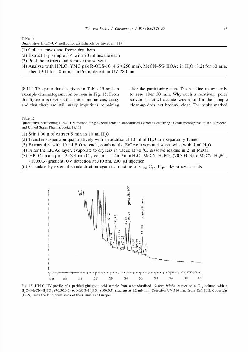
[8,11]. The procedure is given in Table 15 and an after the partitioning step. The baseline returns only
example chromatogram can be seen in Fig. 15. From to zero after 30 min. Why such a relatively polar
this figure it is obvious that this is not an easy assay solvent as ethyl acetate was used for the sample
and that there are still many impurities remaining clean-up does not become clear. The peaks marked
Table 15
Quantitative partitioning-HPLC–UV method for ginkgolic acids in standardised extract as occurring in draft monographs of the Europeanand United States Pharmacopeias [8,11]
(1) Stir 1.00 g of extract 5 min in 10 ml H O2
(2) Transfer suspension quantitatively with an additional 10 ml of H O to a separatory funnel2
(3) Extract 43 with 10 ml EtOAc each, combine the EtOAc layers and wash twice with 5 ml H O2
(4) Filter the EtOAc layer, evaporate to dryness in vacuo at 40 8C, dissolve residue in 2 ml MeOH
(5) HPLC on a 5 mm 12534-mm C column, 1.2 ml/ min H O–MeCN–H PO (70:30:0.3) to MeCN–H PO18 2 3 4 3 4
(100:0.3) gradient, UV detection at 310 nm, 200 ml injection
(6) Calculate by external standardisation against a mixture of C , C , C alkylsalicylic acids13 15 17
Fig. 15. HPLC–UV profile of a purified ginkgolic acid sample from a standardised Ginkgo biloba extract on a C column with a18
H O–MeCN–H PO (70:30:0.3) to MeCN–H PO (100:0.3) gradient at 1.2 ml/ min. Detection UV 310 nm. From Ref. [11], Copyright2 3 4 3 4
(1999), with the kind permission of the Council of Europe.
5/13/2018 Chemical Analysis of Every Constituent - slidepdf.com
http://slidepdf.com/reader/full/chemical-analysis-of-every-constituent 24/35
967 (2002) 21 –5544 T . A. van Beek / J . Chromatogr . A
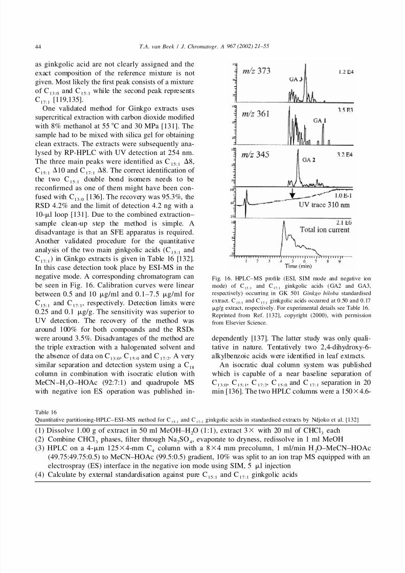
as ginkgolic acid are not clearly assigned and the
exact composition of the reference mixture is not
given. Most likely the first peak consists of a mixture
of C and C while the second peak represents13:0 15:1
C [119,135].17:1
One validated method for Ginkgo extracts uses
supercritical extraction with carbon dioxide modified
with 8% methanol at 55 8C and 30 MPa [131]. The
sample had to be mixed with silica gel for obtaining
clean extracts. The extracts were subsequently ana-
lysed by RP-HPLC with UV detection at 254 nm.
The three main peaks were identified as C D8,15:1
C D10 and C D8. The correct identification of 15:1 17:1
the two C double bond isomers needs to be15:1
reconfirmed as one of them might have been con-
fused with C [136]. The recovery was 95.3%, the13:0
RSD 4.2% and the limit of detection 4.2 ng with a
10-ml loop [131]. Due to the combined extraction–
sample clean-up step the method is simple. A
disadvantage is that an SFE apparatus is required.
Another validated procedure for the quantitative
analysis of the two main ginkgolic acids (C and15:1
C ) in Ginkgo extracts is given in Table 16 [132].17:1
In this case detection took place by ESI-MS in the
negative mode. A corresponding chromatogram can Fig. 16. HPLC–MS profile (ESI, SIM mode and negative ion
be seen in Fig. 16. Calibration curves were linear mode) of C and C ginkgolic acids (GA2 and GA3,15:1 17:1
respectively) occurring in GK 501 Ginkgo biloba standardisedbetween 0.5 and 10 mg/ml and 0.1–7.5 mg/ml forextract. C and C ginkgolic acids occurred at 0.50 and 0.1715:1 17:1C and C , respectively. Detection limits were15:1 17:1 mg/g extract, respectively. For experimental details see Table 16.
0.25 and 0.1 mg/g. The sensitivity was superior toReprinted from Ref. [132], copyright (2000), with permission
UV detection. The recovery of the method was from Elsevier Science.
around 100% for both compounds and the RSDs
were around 3.5%. Disadvantages of the method are dependently [137]. The latter study was only quali-
the triple extraction with a halogenated solvent and tative in nature. Tentatively two 2,4-dihydroxy-6-
the absence of data on C , C and C . A very alkylbenzoic acids were identified in leaf extracts.13:0 1 5:0 17:2
similar separation and detection system using a C An isocratic dual column system was published18
column in combination with isocratic elution with which is capable of a near baseline separation of
MeCN–H O–HOAc (92:7:1) and quadrupole MS C , C , C , C and C separation in 202 13:0 15:1 17:2 15:0 17:1
with negative ion ES operation was published in- min [136]. The two HPLC columns were a 15034.6-
Table 16
Quantitative partitioning-HPLC–ESI–MS method for C and C ginkgolic acids in standardised extracts by Ndjoko et al. [132]15:1 17:1
(1) Dissolve 1.00 g of extract in 50 ml MeOH–H O (1:1), extract 33 with 20 ml of CHCl each2 3
(2) Combine CHCl phases, filter through Na SO , evaporate to dryness, redissolve in 1 ml MeOH3 2 4
(3) HPLC on a 4-mm 12534-mm C column with a 834 mm precolumn, 1 ml/min H O–MeCN–HOAc4 2
(49.75:49.75:0.5) to MeCN–HOAc (99.5:0.5) gradient, 10% was split to an ion trap MS equipped with an
electrospray (ES) interface in the negative ion mode using SIM, 5 ml injection
(4) Calculate by external standardisation against pure C and C ginkgolic acids15:1 17:1
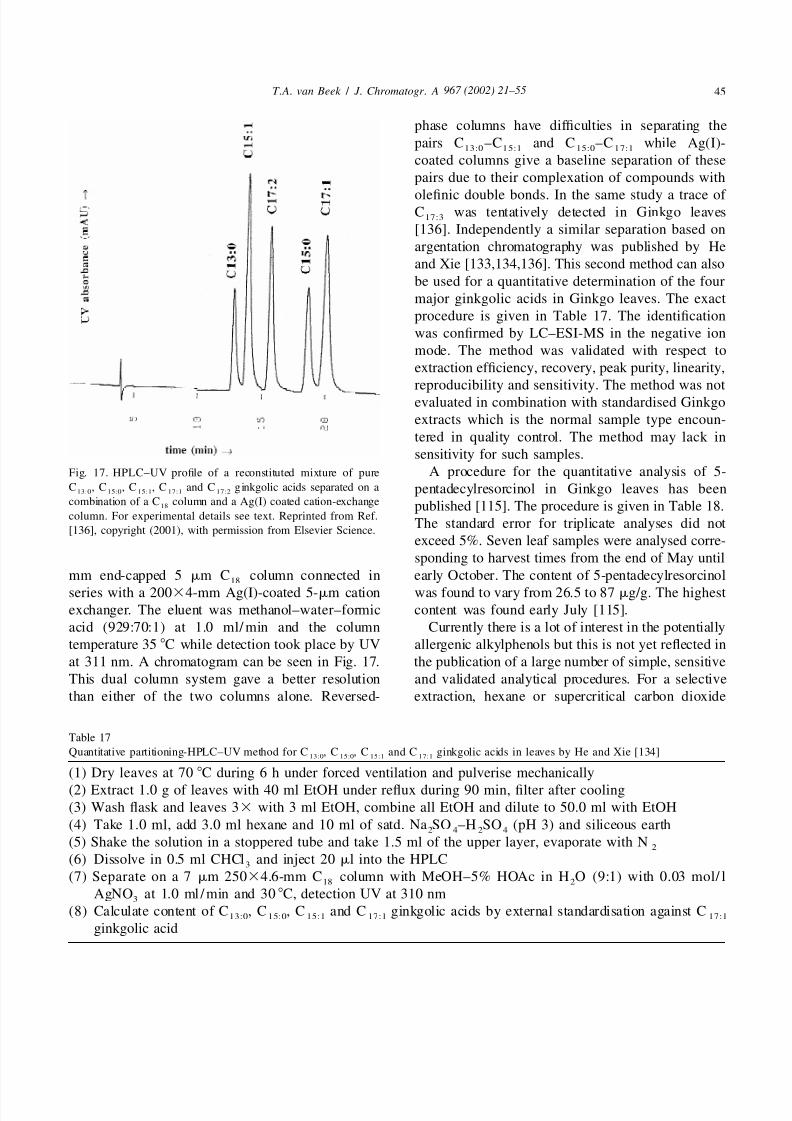
5/13/2018 Chemical Analysis of Every Constituent - slidepdf.com
http://slidepdf.com/reader/full/chemical-analysis-of-every-constituent 25/35
967 (2002) 21 –55 45T . A. van Beek / J . Chromatogr . A
phase columns have difficulties in separating the
pairs C –C and C –C while Ag(I)-13:0 1 5:1 15:0 1 7:1
coated columns give a baseline separation of these
pairs due to their complexation of compounds with
olefinic double bonds. In the same study a trace of C was tentatively detected in Ginkgo leaves
17:3
[136]. Independently a similar separation based on
argentation chromatography was published by He
and Xie [133,134,136]. This second method can also
be used for a quantitative determination of the four
major ginkgolic acids in Ginkgo leaves. The exact
procedure is given in Table 17. The identification
was confirmed by LC–ESI-MS in the negative ion
mode. The method was validated with respect to
extraction efficiency, recovery, peak purity, linearity,
reproducibility and sensitivity. The method was notevaluated in combination with standardised Ginkgo
extracts which is the normal sample type encoun-
tered in quality control. The method may lack in
sensitivity for such samples.
A procedure for the quantitative analysis of 5-Fig. 17. HPLC–UV profile of a reconstituted mixture of pure
C , C , C , C and C ginkgolic acids separated on a pentadecylresorcinol in Ginkgo leaves has been13:0 15:0 15:1 17:1 17:2
combination of a C column and a Ag(I) coated cation-exchange18 published [115]. The procedure is given in Table 18.
column. For experimental details see text. Reprinted from Ref.The standard error for triplicate analyses did not
[136], copyright (2001), with permission from Elsevier Science.exceed 5%. Seven leaf samples were analysed corre-
sponding to harvest times from the end of May until
mm end-capped 5 mm C column connected in early October. The content of 5-pentadecylresorcinol18
series with a 20034-mm Ag(I)-coated 5-mm cation was found to vary from 26.5 to 87 mg/g. The highestexchanger. The eluent was methanol–water–formic content was found early July [115].
acid (929:70:1) at 1.0 ml/ min and the column Currently there is a lot of interest in the potentially
temperature 35 8C while detection took place by UV allergenic alkylphenols but this is not yet reflected in
at 311 nm. A chromatogram can be seen in Fig. 17. the publication of a large number of simple, sensitive
This dual column system gave a better resolution and validated analytical procedures. For a selective
than either of the two columns alone. Reversed- extraction, hexane or supercritical carbon dioxide
Table 17
Quantitative partitioning-HPLC–UV method for C , C , C and C ginkgolic acids in leaves by He and Xie [134]13:0 15:0 15:1 17:1
(1) Dry leaves at 70 8C during 6 h under forced ventilation and pulverise mechanically
(2) Extract 1.0 g of leaves with 40 ml EtOH under reflux during 90 min, filter after cooling
(3) Wash flask and leaves 33 with 3 ml EtOH, combine all EtOH and dilute to 50.0 ml with EtOH
(4) Take 1.0 ml, add 3.0 ml hexane and 10 ml of satd. Na SO –H SO (pH 3) and siliceous earth2 4 2 4
(5) Shake the solution in a stoppered tube and take 1.5 ml of the upper layer, evaporate with N2
(6) Dissolve in 0.5 ml CHCl and inject 20 ml into the HPLC3
(7) Separate on a 7 mm 25034.6-mm C column with MeOH–5% HOAc in H O (9:1) with 0.03 mol/ l18 2
AgNO at 1.0 ml /min and 30 8C, detection UV at 310 nm3
(8) Calculate content of C , C , C and C ginkgolic acids by external standardisation against C13:0 15:0 15:1 17:1 17:1
ginkgolic acid
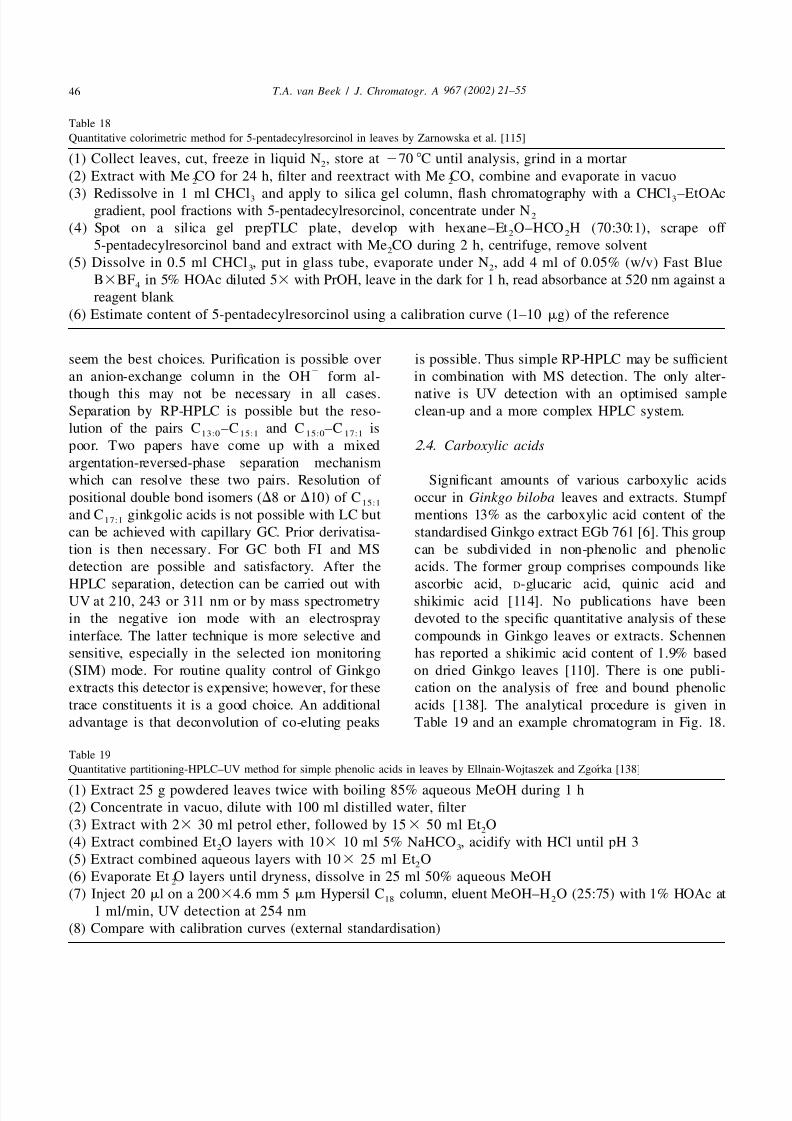
5/13/2018 Chemical Analysis of Every Constituent - slidepdf.com
http://slidepdf.com/reader/full/chemical-analysis-of-every-constituent 26/35
967 (2002) 21 –5546 T . A. van Beek / J . Chromatogr . A
Table 18
Quantitative colorimetric method for 5-pentadecylresorcinol in leaves by Zarnowska et al. [115]
(1) Collect leaves, cut, freeze in liquid N , store at 270 8C until analysis, grind in a mortar2
(2) Extract with Me CO for 24 h, filter and reextract with Me CO, combine and evaporate in vacuo2 2
(3) Redissolve in 1 ml CHCl and apply to silica gel column, flash chromatography with a CHCl –EtOAc3 3
gradient, pool fractions with 5-pentadecylresorcinol, concentrate under N2
(4) Spot on a silica gel prepTLC plate, develop with hexane–Et O–HCO H (70:30:1), scrape off 2 2
5-pentadecylresorcinol band and extract with Me CO during 2 h, centrifuge, remove solvent2
(5) Dissolve in 0.5 ml CHCl , put in glass tube, evaporate under N , add 4 ml of 0.05% (w/v) Fast Blue3 2
B3BF in 5% HOAc diluted 53 with PrOH, leave in the dark for 1 h, read absorbance at 520 nm against a4
reagent blank
(6) Estimate content of 5-pentadecylresorcinol using a calibration curve (1–10 mg) of the reference
seem the best choices. Purification is possible over is possible. Thus simple RP-HPLC may be sufficient2
an anion-exchange column in the OH form al- in combination with MS detection. The only alter-
though this may not be necessary in all cases. native is UV detection with an optimised sampleSeparation by RP-HPLC is possible but the reso- clean-up and a more complex HPLC system.
lution of the pairs C –C and C –C is13:0 15:1 15:0 17:1
poor. Two papers have come up with a mixed 2.4. Carboxylic acids
argentation-reversed-phase separation mechanism
which can resolve these two pairs. Resolution of Significant amounts of various carboxylic acids
positional double bond isomers (D8 or D10) of C occur in Ginkgo biloba leaves and extracts. Stumpf 15:1
and C ginkgolic acids is not possible with LC but mentions 13% as the carboxylic acid content of the17:1
can be achieved with capillary GC. Prior derivatisa- standardised Ginkgo extract EGb 761 [6]. This group
tion is then necessary. For GC both FI and MS can be subdivided in non-phenolic and phenolic
detection are possible and satisfactory. After the acids. The former group comprises compounds like
HPLC separation, detection can be carried out with ascorbic acid, D-glucaric acid, quinic acid andUV at 210, 243 or 311 nm or by mass spectrometry shikimic acid [114]. No publications have been
in the negative ion mode with an electrospray devoted to the specific quantitative analysis of these
interface. The latter technique is more selective and compounds in Ginkgo leaves or extracts. Schennen
sensitive, especially in the selected ion monitoring has reported a shikimic acid content of 1.9% based
(SIM) mode. For routine quality control of Ginkgo on dried Ginkgo leaves [110]. There is one publi-
extracts this detector is expensive; however, for these cation on the analysis of free and bound phenolic
trace constituents it is a good choice. An additional acids [138]. The analytical procedure is given in
advantage is that deconvolution of co-eluting peaks Table 19 and an example chromatogram in Fig. 18.
Table 19
´Quantitative partitioning-HPLC–UV method for simple phenolic acids in leaves by Ellnain-Wojtaszek and Zgorka [138]
(1) Extract 25 g powdered leaves twice with boiling 85% aqueous MeOH during 1 h
(2) Concentrate in vacuo, dilute with 100 ml distilled water, filter
(3) Extract with 23 30 ml petrol ether, followed by 153 50 ml Et O2
(4) Extract combined Et O layers with 103 10 ml 5% NaHCO , acidify with HCl until pH 32 3
(5) Extract combined aqueous layers with 103 25 ml Et O2
(6) Evaporate Et O layers until dryness, dissolve in 25 ml 50% aqueous MeOH2
(7) Inject 20 ml on a 20034.6 mm 5 mm Hypersil C column, eluent MeOH–H O (25:75) with 1% HOAc at18 2
1 ml/min, UV detection at 254 nm
(8) Compare with calibration curves (external standardisation)
5/13/2018 Chemical Analysis of Every Constituent - slidepdf.com
http://slidepdf.com/reader/full/chemical-analysis-of-every-constituent 27/35
967 (2002) 21 –55 47T . A. van Beek / J . Chromatogr . A
concentration of 0.25 mg/ g leaves at the end of
October [138].
Apart from these common phenolic acids, one rare
nitrogen-containing phenolic acid named 6-hydroxy-
kynurenic acid (6-HKA) occurs in Ginkgo leaves.Together with some other organic acids it is also
present in standardised extracts. Little is known
about the importance of his compound for the
clinical activity of Ginkgo. 6-HKA may be an
antagonist of N -methyl-D-aspartate (NMDA) [139].
6-HKA is probably a degradation product arising
from tryptophan [110]. Schennen was the first to
analyse 6-HKA in Ginkgo leaves. His procedure is
given in Table 20. It was discovered that the 6-HKA
content increases dramatically throughout the year to
a maximum of 1–2 mg/g in November. A similarprocedure was used by Chen et al. except that they
removed the spot of 6-HKA from a TLC plate and
then used off-line UV spectroscopy after elution to
determine the content [52].
Fig. 18. HPLC–UV profile of phenolic acids in a purified extract
of Ginkgo biloba leaves on a C column with MeOH–H O18 2
(25:75) with 1% HOAc at 1 ml/min: (1) protocatechuic acid; (2)
p-hydroxybenzoic acid; (3) vanillic acid; (4) caffeic acid; (5 )
isovanillic acid; (6) p-coumaric acid; (7a) E -ferulic acid; (7b)
Z -ferulic acid; (8) sinapic acid. Reprinted from Ref. [138] by
courtesy of Marcel Dekker Inc., 1999.
The sample clean-up appears time-consuming with
37 partitioning steps and has not been validated. The
authors followed the concentration of the phenolic
¨acids in leaves from June 28 until October 28. The Grasel and Reuter have used a more modernconcentration of the free acids increased until a approach by means of HPLC or HPTLC [139]. Theirmaximum at the end of October. The concentration HPLC procedure is given in Table 21. An exampleof bound phenolic acids (measured after acidic or chromatogram can be found in Fig. 19. The methodalkaline hydrolysis) showed a less clear pattern. The was validated with respect to extraction efficiency,major phenolic acid was p-coumaric acid with a recovery, reproducibility and linearity. The detection
Table 20
Quantitative partitioning-paper chromatography–fluorescence method for 6-HKA in leaves by Schennen [110]
(1) Extract 40–100 mg powdered leaves twice with 10 ml 70% MeOH under reflux
(2) Extract the combined filtrates with 20 ml petrol ether
(3) Evaporate the aqueous phase to dryness, dissolve residue in 2 ml 60% Me CO2
(4) Separate 20 ml solution by two-dimensional paper chromatography
(5) Extract 6-HKA and blank spot with 0.05 M H SO at 50 8C2 4
(6) Measure the fluorescence (ex. 350 nm, em. 540 nm) of both extracts
(7) Compare with calibration curve (also after two-dimensional paper chromatography)
5/13/2018 Chemical Analysis of Every Constituent - slidepdf.com
http://slidepdf.com/reader/full/chemical-analysis-of-every-constituent 28/35
967 (2002) 21 –5548 T . A. van Beek / J . Chromatogr . A
Table 21
¨Quantitative SPE–HPLC–UV method for 6-HKA in leaves by Grasel and Reuter [139]
(1) Extract 0.5–2 g powdered leaves in a Soxhlet with 80 ml petrol ether during 90 min
(2) Re-extract leaves in a Soxhlet with 80 ml 70% aqueous MeOH during 150 min
(3) Evaporate the aqueous extract to dryness, dissolve residue in 5 ml 70% MeOH(4) Filter and perform sample clean-up by SPE with an aromatic cation exchanger, wash solvents: H O and 1 M
2
HCl, elution solvent: 2 M HCl
(5) Collect 6-HKA fraction, evaporate and dissolve in 2 ml 70% MeOH
(6) Inject into HPLC, LiChrosorb RP 8 column, 25034 mm, MeCN–MeOH–H O (5:1:15) with H PO at pH2 3 4
3.2, 1 ml/min, UV 254 nm detection, calculate with calibration curve
limit was calculated as 12 ng. In the HPTLC method not be detected in Ginkgo leaves and was thus
(silica gel, CHCl –HOAc–H O (4:4:1)) the detec- assumed not to occur in Ginkgo phytopharmaceuti-3 2
tion took place by fluorescence. The detection limit cals [68]. This was supported by the absence of any
was 7 ng. Both methods gave similar results. The reports on the typical 4-O-methylpyridoxine intoxi-6-HKA content for three different commercial bat- cation signs (convulsions) in the millions of people
ches of Ginkgo leaves varied from 0.121 to 0.269 taking standardised Ginkgo extracts. However, re-
mg/g dry plant material. cently this compound was reported to occur in leaves
and could also be detected in Ginkgo drugs [142],
2.5. 4 -O- Methylpyridoxine but the concentrations are so low that no toxic effects
can be observed. The compound is extracted with
In Ginkgo seeds significant quantities of the boiling water and separated on a strong cation
antivitamin B6 4-O-methylpyridoxine occur. An exchanger. Due to the selectivity of the extraction
overconsumption of seeds has led to toxicity and solvent, the separation mechanism and the fluores-
even death in the past [140]. Especially children are cence detection, no sample clean-up was necessary.
vulnerable. A review on 4-O-methylpyridoxine is The entire procedure is given in Table 22 while a
available [141]. Until recently this compound could chromatogram is depicted in Fig. 20. The sensitivity
¨Fig. 19. HPLC–UV profile of an infusion of Ginkgo leaves. 6-HKA56-hydroxykynurenic acid. Analysed according to the method of Grasel
and Reuter [139]. Reprinted from Ref. [139], with permission from Georg Thieme Verlag.

5/13/2018 Chemical Analysis of Every Constituent - slidepdf.com
http://slidepdf.com/reader/full/chemical-analysis-of-every-constituent 29/35
967 (2002) 21 –55 49T . A. van Beek / J . Chromatogr . A
Table 22
Quantitative HPLC–fluorescence method for 4-O-methylpyridoxine in leaves by Arenz et al. [142]
(1) Freeze leaves in liquid nitrogen and subsequently powder them in a mortar
(2) Suspend 0.4 g leaves in 2 ml H O, heat in an almost closed test tube at 100 8C during 30 min2
(3) Centrifuge the suspension after cooling at 10 000 rpm(4) Inject the supernatant into an HPLC, Nucleosil 5SA column, 25034 mm, gradient from 100% 0.01 M HCl
to 100% 0.5 M KH PO (pH 5) in 15 min, 1 ml/min, fluorescence detection 330 nm excitation, 400 nm2 4
detection
was reported as ,0.5 mg. The method was validated. 2.6. Polyprenols
Concentrations in leaves varied from 2 to 5 mg/fresh
leaf. One fresh leaf equals approximately 0.25 g dry Polyprenols consist of a large number (10–30) of
weight. In standardised liquid Ginkgo drugs the isoprene units linked in a linear fashion. They occur
concentrations varied from 3.8 to 9.8 mg/ml. In in many plants besides Ginkgo leaves. They could be
contrast in the albumen of seeds a concentration of involved as sugar carriers in protein glycosylation. In105 mg/ g was measured [142]. This latter value Ginkgo the polyprenols are mostly present as ace-
corresponds well with values of 0.01–0.02% in raw tates. The chain consists of an v-unit, two trans
Ginkgo seeds reported by others [141,143]. In seeds isoprene units, a large number (10–20) of cis-iso-
also the 5-glucoside occurs [143]. prene units and a terminal (a) cis-isoprene unit with
the ester group. They are removed during the manu-
facturing process of standardised Ginkgo extracts
and therefore do not occur in such extracts. Thus the
two most relevant publications on Ginkgo polypre-
nols deal with the analysis of leaves only. Polypre-
nols are very apolar compounds which can be
extracted with solvents like hexane. Reviews on
Ginkgo polyprenols [144] and chromatography of polyprenols in general [145] have been published
before.
The initial work on Ginkgo polyprenols was
carried out by Ibata et al. [146]. Their method of
analysis is given in Table 23. The procedure is
preparative in nature and not very suitable for routine
quantitative analysis. They characterised the polypre-1
nols by a combination of IR, FD-MS, H NMR and13
C NMR, and saponification. The percentages by
weight of polyprenyl-14–23 acetates were given as
0.9, 1.6, 6.5, 24.9, 36.5, 17.7, 6.7, 2.9, 1.6 and 0.8%.During the growing season the total concentration of
polyprenol acetates steadily increased from an initial
0.05 to 2.0% in late autumn. The free polyprenol
concentration never increased above 0.05% [146].
A more simple assay has been described by Huh etFig. 20. HPLC profile of a German homeopathic Ginkgo drug al. and is summarised in Table 24 [147]. They(Ginkgo loges Tropfen). MPN54-O-methylpyridoxine. Analysed
extracted the leaves with the more selective hexaneaccording to the method of Arenz et al. [142]. For chromato-
and removed many slightly more polar constituentsgraphic details see Table 22, line 4. Reprinted from Ref. [142],
with permission from Georg Thieme Verlag. with 90% methanol in water. The extract was then

5/13/2018 Chemical Analysis of Every Constituent - slidepdf.com
http://slidepdf.com/reader/full/chemical-analysis-of-every-constituent 30/35
967 (2002) 21 –5550 T . A. van Beek / J . Chromatogr . A
saponified with base converting the acetates to the they found C polyprenol to be the most abundant.90
alcohols (free polyprenols). By means of supercriti- Quantities of C , C and C polyprenols (commer-85 90 95
cal fluid chromatography (SFC) with pure carbon cially available) were calculated via internal stan-
dioxide on an apolar capillary column, they were dardisation (I.S.5dodecaprenol (C ) from Rhus60
able to obtain a good separation of the individual C typhina). The method was partially validated. RSDs70
to C polyprenols present. Similarly to Ibata et al. were calculated (6–8%) and the calibration curves12 0
Table 23
Quantitative gravimetric method for polyprenols in leaves by Ibata et al. [146]
(1) Dry leaves at 50–60 8C during 1–2 days in an oven with forced ventilation, afterwards crush them(2) Extract 200 g with 600 ml hexane–Me CO (1:1) 33, each time for 3–9 days at 20 8C2
(3) Purify over silica gel with hexane–Et O (19:1), collect 72% pure polyprenol acetates2
(4) Purify by GPC on styrene–divinylbenzene gel with CHCl , collect 96.4% pure fraction3
(5) Purify by dissolving in a 20-fold volume of Me CO, discard precipitate2
(6) Separate into individual polyprenols by HPLC, Nucleosil 5C column, 300310 mm, Me CO–MeOH18 2
(9:1), 3 ml/min, RI detection
(7) Determine the weight of each fraction (weight is proportional to peak area in HPLC profile)
Table 24
Quantitative partitioning-SFC–FID method for polyprenols in leaves by Huh et al. [147]
(1) Dry leaves at 60 8C, pulverise them afterwards
(2) Extract 200 mg with hexane during 1 h after addition of 500 mg dodecaprenol (I.S.)
(3) Wash the hexane 33 with 90% aqueous MeOH, discard aqueous MeOH
(4) Stir hexane vigorously with K CO and MeOH during 12 h at room temperature2 3
(5) Wash once with H O and twice with saturated NaCl solution, dry over Na SO2 2 4
(6) Evaporate hexane, reconstitute in 1 ml hexane
(7) Inject 0.2 ml (time split 0.2 s) into an SFC apparatus equipped with a chemically bonded phenyl capillary
column (10 m350 mm I.D., 0.25 mm film) at 100 8C; fluid 100% CO ; pressure gradient from 200 to 4002
atm with 20 atm/min, FI detector at 325 8C

5/13/2018 Chemical Analysis of Every Constituent - slidepdf.com
http://slidepdf.com/reader/full/chemical-analysis-of-every-constituent 31/35
967 (2002) 21 –55 51T . A. van Beek / J . Chromatogr . A
were linear from 0.5 to 2.0 mg/ ml. Recovery of the scarce. This is even more true for the main bottle-
internal standard was 85.5%. The assumption was neck in terpene trilactone analysis: the sample clean-
made that the recoveries of the C , C and C up step. Nevertheless two methods appear to have a85 90 95
polyprenols were similar to that of dodecaprenol. slight edge over other published methods. One starts
The extraction efficiency was not determined. No with an aqueous solution which is purified overdistinction was made between polyprenol acetates diatomaceous earth (partitioning), the other starts
and free polyprenols. The polyprenol concentrations with a methanolic solution which is purified over
that were determined by Huh et al. in Ginkgo leaves silica gel (adsorption). Both methods use for this step
throughout the growing season were close to those a small SPE-type column with 0.5 g stationary phase.
published earlier by Ibata et al. An ontogenetic study Good separation and detection is not an issue and
of Ginkgo polyprenols was published separately by can take place equally well by RP-HPLC with
the same group [148]. refractive index or evaporative light scattering de-
Shen et al. have compared 19 different solvents tection or by GC–FID. The last technique has as
for the extraction of polyprenols from Ginkgo leaves disadvantage that a prior derivatisation step is neces-
[149]. Petroleum ether gave the most clean extract sary. Recently several LC–MS studies of terpene
(yield 0.92 g, purity 19.8%) while hexane–acetone trilactones appeared. This detector is so selective that(8:2) gave the highest yield (yield 1.16 g, purity it is possible to forgo any sample clean-up.
18.3%). Prior to quantitative HPLC analysis, a The analysis of the flavonol glycosides is straight-
sample clean-up on silica gel was necessary (no forward. First they are hydrolysed after which the
details given). Polyprenols were determined in the resulting three aglycones are quantified by RP-
purified extract on a Spherisorb C column with HPLC. A simple recalculation provides the original18
PrOH–MeOH (9:1) as solvent at 2.0 ml /min. The yield of the glycosides. The European and United
system was capable of resolving both the alcohols States Pharmacopeia have adopted the same pro-
and acetates of various chain length. The detection cedure. The direct analysis of the complex mixture
method was not given. The total content varied but of genuine flavonol glycosides is still in its infancy.
an average value of 1.5% was reported. Only fingerprint analysis is currently feasible. Al-
though biflavones do not occur in significant con-
centrations in Ginkgo standardised extracts, interestin them is rising because they possess interesting
3. Conclusion pharmacological properties as well. They are present
in full extracts and certain cosmetics. A good assay
Although a lot of different analytical methods has not yet been published. The analysis of Ginkgo
have been proposed by academia and industry for proanthocyanidins which do occur in standardised
various constituents of Ginkgo leaves during the last extracts is still in its infancy. Different assays give
20 years, in most cases no consensus has yet been widely different values.
reached about the use of one best method for each Lately there is a lot of interest in Ginkgo alkyl-
class of compounds (terpene trilactones, flavonol phenols (ginkgolic acids) because of their potentially
glycosides, etc.). In fact for the terpene trilactones, harmful effects. Most manufacturers limit the con-
one simple, reproducible method suitable for both centration of alkylphenols to 5 ppm. For the quanti-leaves and extracts has not yet been published. The tation of these trace constituents in extracts LC–MS
extraction of terpene trilactones from leaves can be is probably the method of choice. Due to the high
carried out with mixtures of acetone, methanol and selectivity of LC–MS a complete chromatographic
water, at room temperature or under reflux con- separation of all ginkgolic acids is not necessary.
ditions and with or without a pre-extraction with a Such a separation is difficult to achieve by RP-
highly non-polar solvent. In principle all give a HPLC. Further developments in the analysis of this
quantitative extraction but not all methods are equal- group are foreseen.
ly fast, environmentally friendly and yield equally From a phytopharmaceutical point of view the
clean extracts. Comparative assays are unfortunately interest in the remaining groups discussed in this

5/13/2018 Chemical Analysis of Every Constituent - slidepdf.com
http://slidepdf.com/reader/full/chemical-analysis-of-every-constituent 32/35
967 (2002) 21 –5552 T . A. van Beek / J . Chromatogr . A
review is less. Polyprenols and 4-O-methylpyridox- MeCN acetonitrile
ine do not occur in significant amounts in MeOAc methyl acetate
standardised Ginkgo extracts. Various simple pheno- MeOH methanol
lic carboxylic acids do occur in Ginkgo leaves and MS mass spectrometry
standardised extracts and some methods for their MW molecular massanalysis in leaves have been published. NaOAc sodium acetate
NMR nuclear magnetic resonance
PrOH propanol
4. Abbreviations RI refractive index
RP reversed-phase
APCI atmospheric pressure chemical ionisation RSD relative standard deviation
BP1 type of GC stationary phase SDS sodium dodecyl sulphate
BP10 type of GC stationary phase SFC supercritical fluid chromatography
BSTFA N ,O-bis(trimethylsilyl)trifluoroacetamide SFE supercritical fluid extraction
C type of HPLC stationary phase SIM selected ion monitoring18
C type of HPLC stationary phase SPE solid-phase extraction4
DB-1 GC stationary phase THF tetrahydrofuran
DMF dimethylformamide TLC thin-layer chromatography
ECD electron capture detector TMCS trimethylchlorosilane
ELSD evaporative light scattering detector TSP thermospray
ES electrospray UV ultraviolet
ESI electrospray ionisation
Et O diethylether2
EtOAc ethyl acetate AcknowledgementsEtOH ethanol
FD field desorption I wish to thank Professor Xie Peishan of theFI flame ionisation
Guangzhou Institute for Drug Control, Guanzhou,FID flame ionisation detector China, for providing me with photocopies of manyG-A ginkgolide A Chinese research articles.G-B ginkgolide B
G-C ginkgolide C
G-J ginkgolide JReferencesGC gas chromatography
GPC gel permeation chromatography[1] G. Warrier, A. Corzine, Ginkgo biloba, in: T.A. van Beek 6-HKA 6-hydroxykynurenic acid
(Ed.), Medicinal and Aromatic Plants—Industrial Profiles,HCO H formic acid
2 Vol. 12, Harwood, Amsterdam, 2000, p. 517.HOAc acetic acid [2] K. Nakanishi, Ginkgo biloba, in: T.A. van Beek (Ed.),
Medicinal and Aromatic Plants—Industrial Profiles, Vol. 12,HPLC high-pressure liquid chromato-
Harwood, Amsterdam, 2000, p. 143.graphy [3] F.V. DeFeudis, in: Ginkgo biloba Extract (EGb 761). FromHPTLC high-performance thin layer chromato-Chemistry to the Clinic, Ullstein Medical, Wiesbaden, 1998.
graphy[4] J. O’Reilly, H. Jaggy, European Patent 90123140.7
iPrOH isopropanol (03.12.90), Active component concentrates and new active
I.S. internal standard component combinations from Ginkgo biloba leaves, their
method of preparation and pharmaceuticals containing theIR infraredactive component concentrates or the active componentLC liquid chromatographycombinations, 1990.
Me CO acetone2 [5] E. Bombardelli, G. Mustich, M. Bertani, European Patent
MECC micellar electrokinetic capillary electro- 89309500.0 (19.09.89), New extracts of Ginkgo biloba and
phoresis their methods of preparation, 1989.
5/13/2018 Chemical Analysis of Every Constituent - slidepdf.com
http://slidepdf.com/reader/full/chemical-analysis-of-every-constituent 33/35
967 (2002) 21 –55 53T . A. van Beek / J . Chromatogr . A
[6] K.-H. Stumpf, in: Proceedings of ’97 international seminar [35] K. Nakanishi, Pure Appl. Chem. 14 (1967) 89.
on Ginkgo, The state science and technology commission, [36] K. Okabe, K. Yamada, S. Yamamura, S. Takada, J. Chem.
Beijing, 1997, p. 39. Soc. (1967) 2201.
[7] F.F. Camponovo, F. Soldati, Ginkgo biloba, in: T.A. van [37] K. Nakanishi, H. Habaguchi, Y. Nakadaira, M.C. Woods, M.
Beek (Ed.), Medicinal and Aromatic Plants—Industrial Maruyama, R.T. Major, M. Alauddin, A.R. Patel, K. Weing-
¨Profiles, Vol. 12, Harwood, Amsterdam, 2000, p. 245. es, W. Bahr, J. Am. Chem. Soc. 93 (1971) 3544.[8] Anonymous, Pharmacopeial Forum, 25 (1999) 7754. [38] K. Weinges, M. Hepp, H. Jaggy, Liebigs Ann. Chem. (1987)
[9] V.S. Srinivasan, Ginkgo biloba, in: T.A. van Beek (Ed.), 521.
Medicinal and Aromatic Plants—Industrial Profiles, Vol. 12, [39] T.A. van Beek, Ginkgo biloba, in: T.A. van Beek (Ed.),
Harwood, Amsterdam, 2000, p. 229. Medicinal and Aromatic Plants—Industrial Profiles, Vol. 12,
[10] European Pharmacopeia Commission, Pharmeuropa 11 Harwood, Amsterdam, 2000, p. 151.
(1999) 337. [40] Y. Komoda, H. Nakamura, M. Uchida, Iyo Kizai Kenkyusho
[11] European Pharmacopeia Commission, Pharmeuropa 11 Hokoku (Rep. Inst. Med. Dent. Eng.) 22 (1988) 83.
(1999) 333. [41] T.A. van Beek, H.A. Scheeren, T. Rantio, W.C. Melger, G.P.
[12] S.-R. Tang, A.-L. Zhou, Y.-Y. Zhao, J.-L. Wu, Zhiwu Ziyuan Lelyveld, J. Chromatogr. 543 (1991) 375.
Yu Huanjing (J. Plant Resources Environment) 1 (1992) 58. [42] Y. Yuzhen, X. Peishan, Yaowu Fenxi Zazhi (Chin. J. Pharm.
[13] T.A. van Beek, G.P. Lelyveld, J. Nat. Prod. 60 (1997) 735. Anal.) 21 (2001) 173.
[14] Q. Deng, Y. Gao, Zhongcaoyao 30 (1999) 419. [43] V. Flesch, M. Jacques, L. Cosson, B.P. Teng,V. Petiard, J.P.
[15] Y. Tang, F. Lou, J. Liq. Chrom. Related Technol. 23 (2000) Balz, Phytochemistry 31 (1992) 1941.
2897. [44] J.-P. Balz, D. Courtois, J. Drieu, K. Drieu, J.-P. Reynoird, C.´ ´[16] M. Xu, Z. Shi, L. Feng, J. Liu, R. Shi, M. Xu, Y. Lu, B. He, Sohier, B.P. Teng, A. Touche, V. Petiard, Planta Med. 65
React. Funct. Polym. 46 (2001) 273. (1999) 620.
[17] M. Xu, Z. Shi, R. Shi, J. Liu, Y. Lu, B. He, React. Funct. [45] H. Huh, E.J. Staba, Planta Med. 59 (1993) 232.
Polym. 43 (2000) 297. [46] H. Inoue, S. Kamoda, T. Terada, Y. Saburi, J. Wood Sci. 44
[18] Y. Tang, F. Lou, J. Wang, Y. Li, S. Zhuang, Phytochemistry (1998) 375.
58 (2001) 1251. [47] E. Lolla, A. Paletti, F. Peterlongo, Fitoterapia 69 (1998) 513.
[19] J. Xie, L. Zhu, H. Luo, L. Zhou, C. Li, X. Xu, J. Chroma- [48] X. Peishan, Y. Yuzhen, Q. Haoquan, in: Proceedings of ’97
togr. A 934 (2001) 1. International Seminar on Ginkgo, The State Science and
[20] H. Wagner, S. Bladt, in: Plant Drug Analysis: A Thin Layer Technology Commission, Beijing, 1997, p. 174.
Chromatography Atlas, Springer, Berlin, 1996. [49] F. Lang, H. Stumpf, Pharmeuropa 11 (1999) 268.
¨ ¨[21] R. Bauer, Drug Inform. J. 32 (1998) 101. [50] R.D. Aye, B. Muller, Munch. Med. Wschr. 133 (1991) S58.
[22] H.R. Petty, M. Fernando, A.L. Kindzelskii, B.N. Zarewych, [51] J.B. Fourtillan, A.M. Brisson, J. Girault, I. Ingrand, J.P.
´M.B. Ksebati, L.M. Hryhorczuk, S. Mobashery, Chem. Res. Decourt, K. Drieu, P. Jouenne, A. Biber, Therapie 50 (1995)
Toxicol. 14 (2001) 1254. 137.
[23] V. Taglioli, A.R. Bilia, C. Ghiara, G. Mazzi, V. Mercati, F.F. [52] Z. Chen, M. Ying, X. Mao, L. Hu, in: Proceedings of ’97
Vincieri, Pharmazie 56 (2001) 868. International Seminar on Ginkgo, The State Science and
[24] H. Itokawa, N. Totsuka, K. Nakahara, K. Takeya, J.-P. Technology Commission, Beijing, 1997, p. 154.
Lepoittevin, Y. Asakawa, Chem. Pharm. Bull. 35 (1987) [53] Q. Lang, C.M. Wai, Anal. Chem. 71 (1999) 2929.
3016. [54] T.A. van Beek, Anal. Chem. 72 (2000) 3396.
[25] E.J. Oliveira, D.G. Watson, FEBS Lett. 471 (2000) 1. [55] T.A. van Beek, L.T. Taylor, Phytochem. Anal. 7 (1996) 185.
[26] J. Wojcicki, B. Gawronska-Szklarz, W. Bieganowski, M. [56] A. Lobstein-Guth, F. Briancon-Scheid, R. Anton, J. Chroma-
Patalan, H. Smulski, L. Samochowiec, J. Zakrzewski, Mater. togr. 267 (1983) 431.
Med. Polona 27 (1995) 141. [57] H. Wagner, S. Bladt, U. Hartmann, A. Daily, W. Berkulin,
[27] P.G. Pietta, C. Gardana, P.L. Mauri, J. Chromatogr. B 693 Dtsch. Apoth. Ztg. 129 (1989) 2421.
(1997) 249. [58] H. Stumpf, Deutsche Apoth. Ztg. 129 (1989) 2794.
[28] P.G. Pietta, C. Gardana, P.L. Mauri, R. Maffei-Facino, M. [59] B.P. Teng, in: P. Braquet (Ed.), Ginkgolides: Chemistry,
Carini, J. Chromatogr. B 673 (1995) 75. Biology Pharmacology and Clinical Perspectives,Vol. 1, J.R.[29] X. Huang, W. Xie, Z. Gong, FEBS Lett. 478 (2000) 123. Prous Science Publishers, Barcelona, 1988, p. 37.
¨[30] G.D. Arthur, A.K. Jager, J. Van Staden, Environ. Exp. Bot. [60] T.A. van Beek, G.P. Lelyveld, Planta Med. 58 (1992) 413.
45 (2001) 55. [61] P. Pietta, P. Mauri, A. Rava, J. Pharm. Biomed. Anal. 10
[31] L. Tang, A. Okazawa, E. Fukusaki, A. Kobayashi, Z. (1992) 1077.
Naturforsch. 55C (2000) 923. [62] W. Yao, C. Yang, Y. Tian, T. Liu, Y. Xu, in: Proceedings of
[32] O. Sticher, Planta Med. 59 (1993) 2. ’97 International Seminar on Ginkgo, The State Science and
[33] T.A. van Beek, in: Proceedings of ’97 International Seminar Technology Commission, Beijing, 1997, p. 182.
on Ginkgo, The State Science and Technology Commission, [63] S.H. Sung, S.H. Jeon, Y.S. Moon, H.S. Lee, H. Huh, Y.C.
Beijing, 1997, p. 158. Kim, Yakhak Hoeji 38 (1994) 20.
[34] T.A. van Beek, E. Bombardelli, P. Morazzoni, F. Peterlongo, [64] M.H. Jeon, S.H. Sung, H. Huh, Y.C. Kim, Plant Cell Rep. 14
Fitoterapia 69 (1998) 195. (1995) 501.

5/13/2018 Chemical Analysis of Every Constituent - slidepdf.com
http://slidepdf.com/reader/full/chemical-analysis-of-every-constituent 34/35
967 (2002) 21 –5554 T . A. van Beek / J . Chromatogr . A
[65] F.F. Camponovo, J.-L.Wolfender, M.P. Maillard, O. Potterat, [94] E. Bombardelli, A. Cristoni, P. Morazzoni, Ginkgo biloba,
in: T.A. van Beek (Ed.), Medicinal and Aromatic Plants—K. Hostettmann, Phytochem. Anal. 6 (1995) 141.Industrial Profiles, Vol. 12, Harwood, Amsterdam, 2000, p.[66] P. Chen, N.-L. Su, L.-H. Nie, S.-Z. Yao, J.-G. Zeng, J.475.Chromatogr. Sci. 36 (1998) 197.
[95] O. Sticher, B. Meier, A. Hasler, Ginkgo biloba, in: T.A. van[67] D.J. Carrier, T.A. van Beek, R. van der Heijden, R. Ver-
Beek (Ed.), Medicinal and Aromatic Plants—Industrialpoorte, Phytochemistry 48 (1998) 89.Profiles, Vol. 12, Harwood, Amsterdam, 2000, p. 179.[68] K. Wada, K. Sakaki, K. Miura, M. Yagi, Y. Kubota, T.
[96] A.R. Hasler, Thesis: Flavonoide aus Ginkgo biloba L. undMatsumoto, M. Haga, Biol. Pharm. Bull. 16 (1993) 210.HPLC-Analytik von Flavonoiden in verschiedenen Arznei-[69] Q. Lang, H.K. Yak, C.M. Wai, Talanta 54 (2001) 673.
¨pflanzen, ETH Zurich, 1990.[70] P.G. Pietta, P.L. Mauri, A. Rava, Chromatographia 29 (1990)[97] A. Hasler, O. Sticher, B. Meier, J. Chromatogr. 605 (1992)
251.41.
[71] T.A. van Beek, A. van Veldhuizen, G.P. Lelyveld, I. Piron,[98] P. Pietta, P. Mauri, A. Bruno, A. Rava, E. Manera, P. Ceva, J.
P.P. Lankhorst, Phytochem. Anal. 4 (1993) 261.Chromatogr. 553 (1991) 223.
´ ´[72] D. Laurain, J. Tremouillaux-Guiller, J.-C. Chenieux, T.A.[99] P.G. Pietta, P.L. Mauri, A. Rava, G. Sabbatini, J. Chroma-
van Beek, Phytochemistry 46 (1997) 127.togr. 549 (1991) 367.
[73] X. Peishan, Y. Yuzhen, Q. Haoquan, L. Qiaoling, J. AOAC[100] P. Pietta, P. Mauri, Methods Enzymol. 335 (2001) 26.
Int. 84 (2001) 1232.[101] Y. Chen, W. Hu, B. Xie, Z. Yiang, in: Proceedings of ’97
[74] A.G. Jensen, K. Ndjoko, J.-L. Wolfender, K. Hostettmann, F.International Seminar on Ginkgo, The State Science and
Camponovo, F. Soldati, Phytochem. Anal. 13 (2002) 31. Technology Commission, Beijing, 1997, p. 206.[75] M. Ganzera, J. Zhao, I.A. Khan, Chem. Pharm. Bull. 49 [102] P. Mauri, P. Pietta, J. Pharm. Biomed. Anal. 23 (2000) 61.(2001) 1170. [103] C.-K. Kim, M.-K. Park, E.-J. Lee, S.-J. Hwang, J. Kor.
[76] A. Cartayrade, E. Neau, C. Sohier, J.-P. Balz, J.-P. Carde, J. Pharm. Sci. 19 (1989) 213.Walter, Plant Physiol. Biochem. 35 (1997) 859. [104] J. Li, L. Yu, Y. Zeng, Zhongguo Zhongyao Zazhi 21 (1996)
[77] J. Zhang, J. Pan, H. Xie, Z. Yang, X. Hu, K. Yang, Fenxi 106.Huaxue (Chin. J. Anal. Chem.) 28 (2000) 53. [105] A. Jamshidi, M. Adjvadi, S.W. Husain, J. Planar Chroma-
[78] H.-F. Wang, X.-R. Ju, Chin. J. Chromatogr. 18 (2000) 394. togr. 13 (2000) 57.[79] M.P. Maillard, F. Camponovo, J.-L. Wolfender, K. Hostet- [106] F. Briancon-Scheid, A. Guth, R. Anton, J. Chromatogr. 245
tmann, in: Poster 138, International Symposium of the (1982) 261.Phytochemical Society of Europe ‘‘Phytochemistry of plants [107] F. Briancon-Scheid, A. Lobstein-Guth, R. Anton, Plantaused in traditional medicine’’, 29 Sept.–1 Oct. 1993, Lausan- Med. 49 (1983) 204.ne, Switzerland, 1993. [108] A. Lobstein, L. Rietsch-Jako, M. Haag-Berrurier, R. Anton,
[80] P. Mauri, B. Migliazza, P. Pietta, J. Mass Spectrom. 34 Planta Med. 57 (1991) 430.(1999) 1361. [109] S. Gobbato, E. Lolla, Fitoterapia 67 (1996) 152.
[81] P. Mauri, P. Simonetti, C. Gardana, M. Minoggio, P. Moraz- ¨[110] A. Schennen, Thesis: Neue Inhaltsstoffe aus den Blattern14zoni, E. Bombardelli, P. Pietta, Rapid Commun. Mass ¨von Ginkgo biloba L. sowie Praparation C-markierter
Spectrom. 15 (2001) 929. ¨Ginkgo-Flavonoide, Philipps-Universitat, Marburg, 1988.[82] S.G. Tallevi, W.G.W. Kurz, J. Nat. Prod. 54 (1991) 634. [111] H.A. Stafford, K.S. Kreitlow, H.H. Lester, Plant Physiol. 82
¨[83] B. Steinke, B. Muller, H. Wagner, Planta Med. 59 (1993) (1986) 1132.155. [112] F. Lang, E. Wilhelm, Pharmazie 51 (1996) 734.
[84] T.A. van Beek, G.P. Lelyveld, Phytochem. Anal. 4 (1993) [113] L.J. Porter, L.N. Hrstich, B.G. Chan, Phytochemistry 25109. (1986) 223.
[85] Anonymous, Pharmacopeial Forum, 23 (1997) 3644. [114] A. Hasler, Ginkgo biloba, in: T.A. van Beek (Ed.), Medici-[86] A. Hasler, B. Meier, Pharm. Pharmacol. Lett. 2 (1992) 187. nal and Aromatic Plants—Industrial Profiles, Vol. 12,[87] D.J. Carrier, P. Coulombe, M. Mancini, R. Neufeld, M. Harwood, Amsterdam, 2000, p. 109.
Weber, J. Archambault, in: H.J.J. Nijkamp, L.H.W. van de [115] E.D. Zarnowska, R. Zarnowski, A. Kozubek, Z. Natur-
Plas, J. Aartrijk (Eds.), International Congress On Plant forsch. 55C (2000) 881.Tissue and Cell Culture, Kluwer, Amsterdam, 1990, p. 614. [116] J.L. Gellerman, H. Schlenk, Anal. Chem. 40 (1968) 739.[88] D.-J. Carrier, N. Chauret, M. Mancini, P. Coulombe, R. [117] T. Matsumoto, T. Sei, Agric. Biol. Chem. 51 (1987) 249.
Neufeld, M. Weber, J. Archambault, Plant Cell Rep. 10 [118] L. Verotta, F. Peterlongo, Phytochem. Anal. 4 (1993) 178.(1991) 256. [119] J. Irie, M. Murata, S. Homma, Biosci. Biotech. Biochem.
[89] N. Chauret, J. Carrier, M. Mancini, R. Neufeld, M. Weber, J. 60 (1996) 240.Archambault, J. Chromatogr. 588 (1991) 281. [120] L. Verotta, P. Morazzoni, F. Peterlongo, Ginkgo biloba, in:
[90] A. Biber, E. Koch, Planta Med. 65 (1999) 192. T.A. van Beek (Ed.), Medicinal and Aromatic Plants—
[91] J.B.T. Strode III, L.T. Taylor, T.A. van Beek, J. Chromatogr. Industrial Profiles, Vol. 12, Harwood, Amsterdam, 2000, p.
A 738 (1996) 115. 203.
[92] S.A. Oehrle, J. Liq. Chromatogr. 18 (1995) 2855. [121] J.-P. Lepoittevin, C. Benezra, Y. Asakawa, Arch. Dermatol.
Res. 281 (1989) 227.[93] C.L. Li, Y.Y. Wong, Planta Med. 63 (1997) 563.

5/13/2018 Chemical Analysis of Every Constituent - slidepdf.com
http://slidepdf.com/reader/full/chemical-analysis-of-every-constituent 35/35
967 (2002) 21 –55 55T . A. van Beek / J . Chromatogr . A
´[122] C.P. Siegers, Phytomedicine 6 (1999) 281. [138] M. Ellnain-Wojtaszek, G. Zgorka, J. Liq. Chromatogr. 22
[123] J. Westendorf, J. Regan, Pharmazie 55 (2000) 864. (1999) 1457.
[124] B. Ahlemeyer, D. Selke, C. Schaper, S. Klumpp, J. Kriegls- ¨[139] I. Grasel, G. Reuter, Planta Med. 64 (1998) 566.
tein, Eur. J. Pharmacol. 430 (2001) 1. [140] K. Wada, M. Haga, in: T. Hori, R.W. Ridge, W. Tulecke,[125] E. Koch, S.S. Chatterjee, H. Jaggy, Naunyn- ´P.D. Tredici, J. Tremouillaux-Guiller, H. Tobe (Eds.),
Schmiedeberg’s Arch. Pharmacol. 345 (Suppl.) (1992) R95. Ginkgo biloba—a Global Treasure. From Biology To[126] H. Jaggy, E. Koch, Pharmazie 52 (1997) 735. Medicine, Springer, Tokyo, 1997, p. 309.[127] E. Koch, H. Jaggy, S.S. Chatterjee, Int. J. Immuno- [141] K. Wada, Ginkgo biloba, in: T.A. van Beek (Ed.), Medicinal
pharmacol. 22 (2000) 229. and Aromatic Plants—Industrial Profiles,Vol. 12, Harwood,[128] H.J. Woerdenbag, T.A. van Beek, in: P.A.G.M. de Smet, K. Amsterdam, 2000, p. 451.
¨Keller, R. Hansel, R.F. Chandler (Eds.), Adverse effects of [142] A. Arenz, M. Klein, K. Fiehe, J. Groß, C. Drewke, T.herbal drugs, Vol. 3, Springer, Berlin, 1997, p. 51. Hernscheidt, E. Leistner, Planta Med. 62 (1996) 548.
[129] H.J. Woerdenbag, P.A.G.M. de Smet, Ginkgo biloba, in: [143] P.M. Scott, B.P.-Y. Lau, G.A. Lawrence, D.A. Lewis, J.T.A. van Beek (Ed.), Medicinal and Aromatic Plants– AOAC Int. 83 (2000) 1313.Industrial Profiles, Vol. 12, Harwood, Amsterdam, 2000, p. [144] H. Huh, Ginkgo biloba, in: T.A. van Beek (Ed.), Medicinal443. and Aromatic Plants—Industrial Profiles,Vol. 12, Harwood,
[130] T.-T. Nguyen Tu, S. Derenne, C. Largeau, A. Mariotti, H. Amsterdam, 2000, p. 215.Bocherens, Org. Geochem. 32 (2001) 45. [145] T. Rezanka, J. Votruba, J. Chromatogr. A 936 (2001) 95.
[131] G. Shen, W. Yao, Chin. J. Anal. Chem. 28 (2000) 985.[146] K. Ibata, M. Mizuno, T. Takigawa, Y. Tanaka, Biochem. J.[132] K. Ndjoko, J.-L. Wolfender, K. Hostettmann, J. Chroma- 213 (1983) 305.
togr. B 744 (2000) 249. [147] H. Huh, E.J. Staba, J. Singh, J. Chromatogr. 600 (1992)[133] J. He, B. Xie, Acta Pharm. Sinica 36 (2001) 609. 364.[134] J. He, B. Xie, J. Chromatogr. A 943 (2001) 303. [148] H. Huh, J. Singh, E.J. Staba, Planta Med. 59 (1993) 379.[135] H.A. Lloyd, C. Denny, G. Krishna, J. Liq. Chromatogr. 3 [149] Z. Shen, C. Wang, X. Chen, in: Proceedings of ’97
(1980) 1497. International Seminar on Ginkgo, The State Science and[136] T.A. van Beek, M.S. Wintermans, J. Chromatogr. A 930 Technology Commission, Beijing, 1997, p. 189.
(2001) 109.
[137] X. He, M.W. Bernart, G.S. Nolan, L. Lin, M.P. Linden-
maier, J. Chromatogr. Sci. 38 (2000) 169.