chem 224 slides - vanderbilt university · pdf file17 aa’s in the chromatogram?! ......
TRANSCRIPT

25
49
Rn
O NHFMOCCH2O
OCH3
OCH3 R1
OO
N
ONH
R2
1) Pd(0), Et3SiH2) piperidine
HN
O
RnR1
OCH2O
OCH3
OCH3N
ONH
R2
Rn
O NH2CH2O
OCH3
OCH3 R1
OHO
N
ONH
R2
PyBOP
CH3CO3H, HFanisole
HN
O
RnR1
OHN
ONH
R2
On-suppport cyclization
50
Peptide and Protein Analysis Primary (1°) structure of a peptide or protein is the amino acid sequence
Amino acid analyzer- automated instrument to determine the amino acid content of a peptide or protein. Individual amino acids are separated by hplc, then detected by post-column derivatization
1972 Nobel Prize in Chemistry William Stein Stanford Moore
peptide -or-
protein [H] reduce any
disulfide bonds
Enzymatic digestion
R CO2
NH3 individual amino acids -or-
H3O+, Δ liquid
chromatography derivatize w/
ninhydrin Detected w/
UV-vis
Different amino acids have different chromatographic mobilities (retention times)

26
51
Reaction of primary amines with ninhydrin
Intense purple color
So, why is it necessary to use a post- rather than pre-column derivatization protocol?
Why are there are only 17 AA’s in the chromatogram?
Amino Acid Analysis Chromatogram
O
O
N
O
O
R CO2
NH3O
O
O+
52
Fluorescence Detection- less background, greater sensitivity, lower detection limits
Absorption spectroscopy- wavelength that light absorbs, moloecules are in an electronically excited state
Emission spectroscopy- the excited molecules relax by emission of a photon.
Fluorescence- excitation wavelength and emission wavelength are different. Molecule will emit light at longer (lower energy) wavelength than is absorbs.

27
53
Fluorescent tags Dansyl- detected by UV or fluorescence
R CO2
N CH3H3C
SO OCl
+
NH3C CH3
S OONH
R CO2
NH3
Dansyl chloride
OPA (o-phthalaldehyde)- detected by fluorescence
R CO2
NH3 + CHO
CHO
SHHO
N
SR
CO2
OH
highly fluorescent
54
Reversed-phase (C-18) HPLC Trace 5 pmols amino acids w/ OPA, HOCH2CH2SH

28
55
Attomol detection w/ laser induced fluorescence
10-3 milli 10-6 micro 10-9 nano 10-12 pico 10-15 fempto 10-18 atto 10-21 zepto Avagadro’s number 1023
N CHO
O
CO2
NN
R
CO2S
OH
CO2
excitation: 488 nmemission: 560 nm
56
Peptide and Protein Sequences: primary (1°) structure- amino acid sequence
N-labeling with Sanger’s reagent: Sanger’s (2,4-dinitrofluorobenzene) reagent reacts with the N-terminal amino group and has a diagnostic UV absorbance that is detected after enzymatic digestion and amino acid analysis
N-terminal amino acid is specifically labeled with a unique UV chromophore
NO2
O2N
FH3N
O
R1 HN CO2+
Δ
NH O
R1 HN CO2nucleophilic
aromaticsubstitution NO2
O2N
NH O
R1OH
NO2
O2N
+
plus other unlabeled amino acids
enzymaticdigestion
-or-H3O+, Δ
H3NO
RnO

29
57
C-terminal sequencing: Carboxypeptidase- enzyme that hydrolyzed amide bonds of a peptide
or protein starting from the C-termial end (exopeptidase)
Hydrolyze peptide with hydazine (H2N-NH2)
NH O
HN
R2
O
NH
R1
O
OR3O
H3N NH O
HN
R2
OR3O
H3N O +Carboxypeptidase
Zn2+, H2OH3N
R1
O
O
derivatize and identify by HPLC
peptide has a new C-terminal AA
NH O
HN
R2
O
NH
R1
O
OR3O
H3N
H2NNH2 H2N
Rn
O
NHNH2H3N
R1
O
O +
C-terminal AAis still an amino acid
All other AA's areconverted to the
hydrazides
58
Edman Degradation: chemical method for the sequential cleavage and identification of the amino acids of a peptide, one at a time starting from the N-terminus. Reagent: Ph-N=C=S, phenylisothiocyanate)
H2N CO2
SCN
Ph+ H2N
O
R1 HN CO2
pH 9.0
then H+ HN
N OS
R1
Ph
+
N-phenylthiohydantoin:!separated by HPLC, !detected by UV-vis!
-1 peptide with a new !N-terminal amino acid !(repeat degradation cycle)!

30
59
Fluorescent Edman sequencing reagent
O
CO2H
OHO
Fluorescein(a common fluorescent dye)
O
CO2H
OHO
NC S
Fluorescein Isothiocyanate(a fluorescent Edman reagent)
Peptide sequencing by Edman degradation: Monitor the appearance of N-phenylthiohydantoin over time to get the peptide sequence. Good for peptides up to ~ 25 amino acids long. Longer peptides and proteins must be cut into smaller fragments before Edman sequencing
60
Enzymatic and chemical cleavage of peptides and proteins at defined sites
Enzymatic • trypsin: cleaves at the C-terminal side of basic residues,
Arg, Lys but not His
• chymotrypsin: cleaves at the C-terminal side of aromatic residues Phe, Tyr, Trp
NH O
HN
O
NH
R3
O
HN
R1O
H3NCO2
NH3
NH O
HN
O
H3N
R3
O
HN
R1O
H3NCO2
NH3
O +trypsin
H2O
NH O
HN
O
NH
R3
O
HN
R1O
H3NCO2 N
H O
HN
O
H3N
R3
O
HN
R1O
H3NCO2O +chymotrypsin
H2O

31
61
• thermolysin: cleaves at the N-terminal side of hydrophobic residues Phe, Trp, Leu
NH O
HN
O
NH
R3
O
HN
R1O
H3NCO2 +thermolysin
H2O
NH O
H3NO
NH
R3
O
HN
R1O
H3N CO2O
Chymotrypsin cleavage products Trypsin cleavage products
Tyr Arg Asp-Asn-Gln Leu-Lys Gly-Gly-Phe Ile-Arg-Pro-Lys Leu-Arg-Arg-Ile-Arg-Pro-Lys-Leu-Lys-Trp Tyr-Gly-Gly-Phe-Leu-Arg
Trp-Asp-Asn-Gln
Trypsin:
Chymotrypsin: Trp-Asp-Asn-Gln
Asp-Asn-Gln Leu-Arg-Arg-Ile-Arg-Pro-Lys-Leu-Lys-Trp
Leu-Lys Ile-Arg-Pro-Lys Arg Tyr-Gly-Gly-Phe-Leu-Arg
Gly-Gly-Phe Tyr
62
Other Commonly Used Protein Digest Reagents
Glu-C – cleaves to the C-terminal side of Glu residues (cleavage at Asp is 100-300 times slower)
Asp-N – cleaves to the N-terminal side of Asp residues and cysteic acid
Lys-N – cleaves to the N-terminal side of Lys residues
Lys-C – cleaves to the C-terminal side of Lys residues
Arg-C – cleaves to the C-terminal side of Arg residues
Non-specific proteases: pepsin, proteinase K, subtilysin
32
63
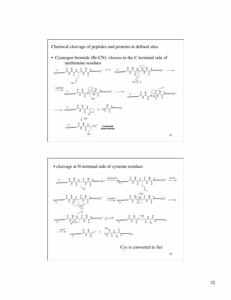
Chemical cleavage of peptides and proteins at defined sites
• Cyanogen bromide (Br-CN): cleaves to the C-terminal side of methionine residues
NH O
HN
O
NH
R3
O
HN
R1O
H3NCO2
SH3C
+NH O
H3N
R3
O
HN
R1O
H3NCO2
HN
Br-CNNH O
HN
O
NH
R3
O
HN
R1O
H3NCO2
SH3C CN
O
NH
R3
O
HN CO2
NH O
R1O
H3N
- H3CSCN
OH2
O
NH
R3
O
HN CO2
NH O
R1O
H3N OH
H+
NHNH
OO
NH O
R1O
H3NHN CO2
OH
C-terminalhomo-serine
H2O
64
• cleavage at N-terminal side of cysteine residues
Cys is converted to Ser
NH O
HN
O
NH
R3
O
HN
R1O
H3NCO2
SH
+
(H3CO)3PONH O
HN
O
NH
R3
O
HN
R1O
H3NCO2
SCH3
- H3CSCN
H+
H3O
NH O
HN
O
NH
R3
O
HN
R1O
H3NCO2
SCH3
CN
Br-CN
NH O
HN
O
NH
R3
O
HN
R1O
H3NCO2
O2H
NH O
HN
O
NH
R3
O
HN
R1O
H3NCO2
OH
NH
H2N
O
HN
R3
O
NH
R1O
H3N CO2O
O
NH O
HN
R3
O
NH
R1O
H3N CO2O
H3NO
HO

33
65
EPIDERMAL GROWTH FACTOR (EGF)!
H2N-ASN1•SER2•TYR3•PRO4•GLY5•CYS6•PRO7•SER8•SER9•TYR10•!ASP11•GLY12•TYR13•CYS14•LEU15•ASN16•GLY17•GLY18•VAL19•!CYS20•MET21•HIS22•ILE23•GLU24•SER25•LEU26•ASP27•SER28•!TYR29•THR30•CYS31•ASN32•CYS33•VAL34•ILE35•GLY36•TYR37•!SER38•GLY39•ASP40•ARG41•CYS42•GLN43•THR44•ARG45•ASP46•!LEU47•ARG48•TRP49•TRP50•GLU51•LEU52•ARG53-CO2H!
Trypsin Chymotrypsin Cyanogen Bromide
Disulfides bridges at: !Cys6 - Cys20! Cys14 - Cys31!
Cys33 - Cys42!
S. Cohen et al. J. Biol. Chem. 1972, 247, 5928-5934!! ! ! ! 1972, 247, 7612-7621! 1973, 248, 7669-7672!
66
Peptide sequencing by tandem mass spectrometry Ionization: SIMS (secondary ion mass spectrometry) Time-of-flight (TOF) mass spectrometer
Methods to get large, polar molecules into the gas phase for MS analysis
FAB: Fast Atom Bombardment MALDI: Matrix-Assisted Laser Desorption Ionization ESI: Electrospray Ionization
Mass spectrometry gives mass/charge (m/z) ratio
“Introduction to Proteomics: Tools for the New Biology,” Liebler, D. C., Humana Press: 2002

34
67
Mass spectrometry is a gas phase technique. Peptides (and proteins) are charged, polar, high molecular weight molecules (ions). How can peptides and proteins be coaxed into the gas phase?
Electrospray ionization (ESI): analyte is introduced into the mass spectrometer as an aerosol.
+++++
+++
++++
+++++
++++
+++
++++
+++++
+
+
- +
+to the mass
analyzer
liquid chromatography or capillary electrophoresis (separate the analytes)!
+++++
+++
++++
+++++
++++
+++
++++
+++++
+
+ +++++
- +
+ ++++++
+++
++++
+++++
++++
+++
++++
+++++
+
+ + ++++
- +
+ ++++++
+++
++++
+++++
++++
+++
++++
+++++
+
+ +++++
- +
+ ++++++
+++
++++
+++++
++++
+++
++++
+++++
+
+ +++ ++
- +
+ ++++++
+++
++++
+++++
++++
+++
++++
+++++
+
+ +++ ++
+
+
++
+
- +
+ + +
Coulombic fission
+++++
+++
++++
+++++
++++
+++
++++
+++++
+
+
+
+
++
+
- +
+++++++
+++
++++
+++++
++++
+++
++++
+++++
+
+
- +
++
++
+
+++++++
+++
++++
+++++
++++
+++
++++
+++++
+
+
- +
++
++
++
68
MALDI ionization (matrix-assisted laser desorption): analyte is co-crystallized with an organic molecule that has an intense UV absorption. A laser that is tuned to the absorption of the matrix, is “pulsed” at the MALDI matrix and energy is indirectly transferred to the analyte.
++ +
+
++
to the mass analyzer
Laser pulse
++ +
+
++
+
+
2002 Nobel Prize in Chemistry John Fenn (ESI) Koichi Tanaka (MALDI)
CO2H
CNHO
CO2H
HO
H3CO
OCH3
CO2HHO
OH2,5-dihydroxybenzoic acid (DHB)
α-cyano-4-hydroxycinnamic acid (CHCA)
Sinapinic acid

35
69
Mass Spectrometry (MS): measures the mass to charge ratio (m/z) Dalton (Da) or mass unit (u) = units for measuring molecular masses.
One Da. = 1/12 the mass of the 12C atom
Monoisotopic mass – sum of the exact masses of the most abundant isotope of each element in a molecule
Average mass – sum of the averaged masses of each element in a molecules, weighted according to isotopic abundance.
Nominal mass – mass calculated using the integer mass of the most abundant isotope for each element (H=1, C=12, O=16, N=14, etc.)
Isotope Mass Natural Abundance 1H 1.0078 99.99% 2H 2.0141 0.015 12C 12 98.89 13C 13.0034 1.11 14N 14.0031 99.64 15N 15.0001 0.36 16O 15.9949 99.76 17O 16.9991 0.04 18O 17.9992 0.2
Isotope Mass Natural Abundance 31P 30.9737 100 32S 31.9721 95 33S 32.9715 0.76 34S 33.9679 4.22 36S 35.9671 0.02
70
m/z
12C661H95
14N1916O26
Monoisotopic Mass 1569.66956
(100%)
Average Mass 1570.5722
1569 1570 1571 1572 1573 1574 1575 1576
Glu-Gly-Val-Asn-Asp-Asn-Glu-Glu-Gly-Phe-Phe-Ser-Ala-Arg (EGVNDNEEGFFSAR)
C66H95N19O26
12C65 13C1 1H95
14N1916O26 , etc.
(80.5%)
12C64 13C2 1H95
14N1916O26 , etc.
(37.3%) 12C63
13C3 1H9514N19
16O26 , etc. (12.7%)
Slide adapted from lecture material developed by A. Burlingame, S. Guan, and M. Baldwin (UCSF)
entitled “Mass Spectrometry and Proteomics”
http://www-personal.umich.edu/~junhuay/pattern.htm
12C63 13C4 1H95
14N1916O26 , etc.
(3.5%)

36
71
5730 3655 3660 2465 2095 1295
m/z 1000 2000 3000 4000 5000 6000
As the number of atoms in the molecule increases, the pattern of masses due to the presence of isotopes will change
Slide adapted from lecture material developed by A. Burlingame, S. Guan, and M. Baldwin (UCSF)
entitled “Mass Spectrometry and Proteomics”
72 Slide adapted from lecture material developed at the University of Lund
What does the isotopic distributions tell us?

37
73
Resolution and Resolving Power (RP): terms used interchangably
6130 6140 6150 6160 6170
Low resolution High
resolution
m/z
The smallest mass difference (ΔM) between peaks such that the valley between them is a specified fraction of the peak height
Full Width Half Maximum (FWHM): Width of a single peak measured at 50% peak apex.
ΔM
10%
ΔM 50% Slide adapted from lecture
material developed by A. Burlingame, S. Guan,
and M. Baldwin (UCSF) entitled “Mass Spectrometry
and Proteomics”
74
Mass Accuracy (MA) - the difference between the experimental mass (Mexp) and the theoretical value (Mcalc), calculated from elemental composition.
€
MA =Mexp −Mcalc
Mcalc
(ppm for high resolution MS)
Mexp = 1569.684, Mcalc = 1569.6696 accuracy = 9.2 ppm
783 785 787 783 785 787 783 785 787 783 785 787
RP=3200 (5.5 ppm)
m/z m/z m/z m/z
RP=1600 (16.2 ppm)
RP=800 (32.1 ppm)
RP=400 (76.8 ppm)
High resolution means better mass accuracy m/z: 784.775 784.860 784.848 784.830 M: 1569.549 1569.720 1569.695 1569.661
Slide adapted from lecture material developed
by A. Burlingame, S. Guan, and M. Baldwin (UCSF)
entitled “Mass Spectrometry and Proteomics”

38
75
Peptide Mass Fingerprinting : Proteins (or peptides) are digested in a predictable way and the masses of the resulting peptide fragments are unique enough to identify the protein.
Requires a database of known sequences and search software to compare (score) the experimentally observed masses with the calculated masses in the database .
m/z = 1529 ± 1 Da 478 peptide fragments from 1529.7 ± 0.1 164 mouse/human genome 1529.73 ± 0.01 25 1529.7340 ± 0.001 4 1529.7348 ± 0.0001 2
76
[M + H]+ accelerated into MS
collision cell
(He, Ar, Xe)
analyze fragments
to get sequence
Collision of the [M+H]+ ion with the gas causes it to fragment, analysis of these fragments ions gives sequence information
Many peptides and proteins give multiply charged ions
CID: collision induced dissociation
OCCH
RH2N
N-terminalfragment
CO2HCH
RH3N
C-terminalfragment
H2N CH CR1
O HN CH C
R2
O HN CH C
R3
O HN CH C
R4
OHO
a1 b1 c1
x1 y1 z1
a2 b2 c2
x2 y2 z2
a3 b3 c3
x3 y3 z3
charge to C-terminus
charge to N-terminus

39
77
Peptide sequencing by tandem mass spectrometry
to the detector
Q1 Electrospray Ion Source Q3 Collision Cell (Q2) Nanospray
Capillary
Select peptide to be analyzed
fragment the peptide
Analyze the peptide fragments
1000 1500 2000 2500 3000 m/z
1116
.67
1287
.73
1375
.76
1424
.85 1505
.77
1665
.89
1811
.85
2005
.07
2476
.21
2550
.52
2719
.48
1849
.12
1574
.20
1247
.70
Peptides fragment in a predictable manner
H2N CH CR1
O HN CH C
R2
O HN CH C
R3
O HN CH C
R4
OHO
b1
y1
b2
y2 y3 charge to C-terminus
charge to N-terminus b3
Select m/z 1505.8 for Q2
78
average exact - HN-CHR-CO Glycine G 75.07 75.03 57.1 Alanine A 89.10 89.05 71.1 Serine S 105.09 105.04 87.1 Proline P 115.13 115.05 97.1 Valine V 117.15 117.08 99.1 Threonine T 119.12 119.06 101.1 Cysteine C 121.16 121.02 103.1 Isoleucine I 131.18 131.09 113.2 Leucine L 131.18 131.09 113.2 Asparagine N 132.12 132.05 114.1 Aspartic Acid D 133.11 133.04 115.1 Glutamine Q 146.15 146.07 128.2 Lysine K 146.19 146.11 128.1 Glutamic Acid E 147.13 147.13 129.1 Methionine M 149.21 149.05 131.2 Histidine H 155.16 155.02 137.1 Phenylalanine F 165.19 165.19 147.2 Arginine R 174.20 174.11 156.2 Tyrosine Y 181.19 181.07 163.2 Tryptophan W 204.23 204.09 186.2
Amino Acids Sorted by Mass
H2N CH CR1
O HN CH C
R2
O HN CH C
R3
O HN CH C
R4OH
O
b1
y1
b2
y2 y3
b3

40
79
Some ambiguities with MS sequencing leucine (L) vs isoleucine (I): difficult to distinguish, must look at fragmentation of the sidechain
lysine (K, m/z=128.09) vs glutamine (Q, m/z = 128.06)
gly (G) + gly (G) = 114.04 = asn (N)= 114.04 ala (A) + gly (G) = 128.06 = gln (Q) = 128.06 = lys (K)= 128.09 gly (G) + val (V) = 156.09 = arg (R) = 156.10 ala (A) + asp (B) = glu (Z) + gly (G) = 186.06 = trp (N) = 186.08 ser (S) + val (V) = 186.1 = trp (N) = 186.08
CO2
NH3
CO2
NH3
CO2
NH3
CO2
NH3
H2N
O
H2N Ac2O
Ac2O
CO2
NH3
HN
O + 42 amu's
no reaction
80
H2N CH CCH2
O
CH2COH
O
HN CH C
CH2
O
HC CH3CH3
HN CH CHC
O
CH3CH3
HN CH CHC
O
CH3CH2CH3
NH
CH CCH2
O
OH
HN CH C
CH2
O
HC CH3CH3
HN CH CHC
O
CH3CH2CH3
HN CH CHC
O
CH3CH3
HN CH C
CH2
O
CH2COH
O
HN CH C
CH2
O
OH
HN CHC
CH2OH
O
CH2CH2CH2NH2
H2N CH CCH2
O
CH2COH
O
HN CH C
CH2
O
HC CH3CH3
HN CH CHC
O
CH3CH3
HN CH CHC
O
CH3CH2CH3
HN CH C
CH2O
OH
b5 fragment: m/z = 542.1
H3N CH CCH2
O
HC CH3CH3
HN CH CHC
O
CH3CH2CH3
HN CH CHC
O
CH3CH3
HN CH C
CH2
O
CH2COH
O
HN CH C
CH2
O
OH
HN CHC
CH2OH
O
CH2CH2CH2NH2
y6 fragment: m/z = 688.3
-or-
CID
H+

41
81
H2N CH CCH2
O
CH2COH
O
HN CH C
CH2
O
HC CH3CH3
HN CH CHC
O
CH3CH3
HN CH CHC
O
CH3CH2CH3
NH
CH CCH2
O
OH
HN CH C
CH2
O
HC CH3CH3
HN CH CHC
O
CH3CH2CH3
HN CH CHC
O
CH3CH3
HN CH C
CH2
O
CH2COH
O
HN CH C
CH2
O
OH
HN CHC
CH2OH
O
CH2CH2CH2NH2
b1
y10
b2
y9
b3
y8
b4
y7
b5
y6
b6
y5
b7
y4
b8
y3
b9
y2
b10
y1Glu Leu Val Ile Ser Leu Ile Val Glu Ser Lys129 113 99 113 87 113 113 99 129 87 145
82

42
83
DNA mRNA protein
Central Dogma
genome transcriptome proteome
post-translational modifications
84
Hierarchy of Protein Structure 20 Amino Acids - A protein of n residues 20n possible sequences!
100 residue protein has 10020 possibilities 1.3 X 10130! The latest estimates indicate on 40,000 sequences in the human genome
primary (1°) structure: the amino acid sequence
secondary (2°) structure: frequently occurring substructures or folds
tertiary (3°) structure: three-dimensional arrangement of all atoms in a single polypeptide chain
quaternary (4°) structure: overall organization of non-covalently linked subunits of a functional protein.