characterizationofthedna-bindingpropertiesofthe ... · repeat of the core mkx recognition motif....
TRANSCRIPT
Characterization of the DNA-binding Properties of theMohawk Homeobox Transcription Factor*□S
Received for publication, July 9, 2012 Published, JBC Papers in Press, August 24, 2012, DOI 10.1074/jbc.M112.399386
Douglas M. Anderson‡§1, Rajani George‡§, Marcus B. Noyes¶�, Megan Rowton‡§, Wenjin Liu**, Rulang Jiang**,Scot A. Wolfe¶�, Jeanne Wilson-Rawls‡, and Alan Rawls‡ ‡‡2
From the ‡School of Life Sciences, §Molecular and Cellular Biology Graduate Program, ‡‡Center for Evolutionary Medicine andInformatics, Biodesign Institute, Arizona State University, Tempe, Arizona 85287-4501, the ¶Program in Gene Function andExpression, �Department of Biochemistry and Molecular Pharmacology, University of Massachusetts Medical School, Worcester,Massachusetts 01605, and the **Department of Biomedical Genetics and Center for Oral Biology, University of Rochester School ofMedicine and Dentistry, Rochester, New York 14642
Background:Mkx is a transcriptional repressor that regulates muscle and tendon differentiation.Results:MKX binds to nnACA recognition sites as a homodimer. Mkx regulates transcription through recognition sites in theMkx and Sox6 loci.Conclusion:MKX has a novel DNA recognition mode and promotes slow muscle fiber type specification through Sox6.Significance:We provide insight into Mkx regulation of musculoskeletal-specific transcription.
Thehomeobox transcription factorMohawk (Mkx) is a potenttranscriptional repressor expressed in the embryonic precur-sors of skeletal muscle, cartilage, and bone. MKX has recentlybeen shown to be a critical regulator of musculoskeletal tissuedifferentiation and gene expression; however, the genetic path-ways throughwhichMKX functions and its DNA-binding prop-erties are currently unknown. Using a modified bacterialone-hybrid site selection assay, we determined the core DNA-recognitionmotif of the mousemonomericMkx homeodomainto be A-C-A. Using cell-based assays, we have identified a min-imal Mkx-responsive element (MRE) located within the Mkxpromoter, which is composed of a highly conserved invertedrepeat of the core Mkx recognition motif. Using the minimalMRE sequence, we have further identified conserved MREswithin the locus of Sox6, a transcription factor that repressesslow fiber gene expression during skeletal muscle differentia-tion. Real-time PCR and immunostaining of in vitro differenti-ated muscle satellite cells isolated fromMkx-null mice revealedan increase in the expression of Sox6 and down-regulation ofslow fiber structural genes. Together, these data identify theunique DNA-recognition properties of MKX and reveal a novelrole for Mkx in promoting slow fiber type specification duringskeletal muscle differentiation.
Mohawk (Mkx)3 is a transcriptional repressor expressed inthe embryonic progenitor cell populations of skeletal muscle,
tendon, cartilage, and bone (1–4).Mkx is a conserved homeo-box gene that has recently been shown to be a key regulator ofskeletal muscle and tendon differentiation (2, 5, 6). Targeteddisruption of Mkx in the mouse results in severe tendon hyp-oplasia and a significant decrease in the expression of extracel-lular matrix proteoglycans critical for musculoskeletal func-tion, including collagen type I and decorin (5). Parsing themechanism(s) by whichMkx regulates the development of themusculoskeletal system is dependent on characterizing itsDNA-binding properties and ultimately identifying its directgenetic targets.Mkx is a homeobox gene that belongs to the three-amino
acid loop extension superclass of atypical homeobox genes(includes PBC,MEIS, TGIF, IRO, andMKX) (1, 3 4, 7). Homeo-box genes contain a conserved �180-base pair (bp) sequencethat encodes an �60-amino acid three � helix DNA-bindingdomain, called the homeodomain. The homeodomain interactsdirectly with DNA such that the third � helix, the recognitionhelix, interacts with nucleotides in the major groove and theN-terminal arm makes contact with the minor groove. DNArecognition occurs through specific residues in the N-terminalarm (amino acids 3, 5–8) and the recognition helix (aminoacids 47, 50, 51, 54, and 55), such that an �6-bp recognitionmotif is formed (8–11).Among the atypical three-amino acid loop extension genes,
Mkx is most closely related to the Iroquois family. The Iroquoisgenes were first identified in Drosophila as a complex of threeclustered and highly related genes (Iro-C), including araucan(ara), caupolican (caup), and mirror (mirr). In mouse, six Iro-quois genes (Irx1–6) are involved in the development of adiverse set of tissues, including the central nervous system, ven-tricles of the heart, somites, lungs, gonads, and cartilage (12–16). MouseMkx shares 56% homology with Irx2 over the entirehomeodomain (35/63 residues) and 82% homology (14/17 res-
* This work was supported, in whole or in part, by National Institutes of HealthGrant 1R01HG004744 (to S. A. W.) and National Science Foundation (toJ. A. R. and J. W. R.).
□S This article contains supplemental Tables S1 and S2 and Fig. S1.1 Present address: Dept. of Molecular Biology, University of Texas Southwest-
ern Medical Center, 5323 Harry Hines Blvd., Dallas, TX 75390-9148.2To whom correspondence should be addressed: Box 874501, Life Sciences C
Bldg., Rm. 544, Arizona State University, Tempe, AZ, 85287-4501. Fax: 480-965-7892; E-mail: [email protected].
3 The abbreviations used are: Mkx, Mohawk; MRE, Mkx-responsive element;IBS, Iroquois-binding site; MBS, Mkx-binding sequence; Gal4DBD, Gal4
DNA-binding domain; CD-A, adjacent conserved domain; MHC, myosinheavy chain.
THE JOURNAL OF BIOLOGICAL CHEMISTRY VOL. 287, NO. 42, pp. 35351–35359, October 12, 2012© 2012 by The American Society for Biochemistry and Molecular Biology, Inc. Published in the U.S.A.
OCTOBER 12, 2012 • VOLUME 287 • NUMBER 42 JOURNAL OF BIOLOGICAL CHEMISTRY 35351
by guest on March 5, 2019
http://ww
w.jbc.org/
Dow
nloaded from

idues) specifically within Helix III (1). Among the amino acidsimportant for DNA recognition, mouseMkx and Irx factors areidentical at positions 8, 50, 51, 54, and 55, but differ at positions2, 3, 5, 6, 7, and 47 (1). Because it has been shownpreviously thatclosely related homeobox genes recognize similar DNA recog-nition motifs,Mkx and Irx homeodomains may have similarityin their DNA-binding specificity (11, 17).The minimal Iroquois-binding site (IBS) for Drosophila
Iro-C family members has been identified using in vitro siteselection assays (11, 18). The IBS is composed of an invertedrepeat (ACAnnTGT) that consists of two monomeric recogni-tion motifs for each Iro-C homeodomain (ACA). The arrange-ment of the two monomeric recognition motifs appears to beflexible, as Drosophila ara has been shown to recognize anopposite inverted repeat conformation (TGTnnACA), but nota direct repeat ACAnnACA (17–19). The monomeric recogni-tion motif of the Drosophila Mkx ortholog CG11617 has beencharacterized and is similar to Irx family members, except for astrong preference for a thymine (T) at position 1 of the bindingmotif (TnACA) (11).The genetic networks through whichMkx functions in the
normal differentiation and growth of the musculoskeletalsystem are currently unknown. To identify direct targets ofMKX regulation, we characterized the DNA-binding speci-ficity of mouse Mkx; its regulation of its own promotersequence; and its ability to form a homodimer. Additionally,we have identified two Mkx-responsive elements within thelocus of Sox6, a key repressor of slow fiber type specificationduring skeletal muscle differentiation. These characterizedrecognition elements provide a basis for identifying addi-tional direct targets of MKX regulation and further ourunderstanding of its role as a regulator of musculoskeletaldevelopment.
MATERIALS AND METHODS
Homeodomain-binding Site Selection—A fragment of mouseMkx containing the homeodomain and adjacent conserveddomain CD-A (amino acids 70–161) was amplified from theexpression plasmid CS2MT-Mkx (2) (see supplemental TablesS1 and S2 for primers). Additionally, a stop codon, TAA (itali-cized), was incorporated into the reverse primer. TheMkx frag-ment was subcloned into the pB1H2�2-12 vector as a fusion totheZif12 and the� subunit of bacterial RNApolymerase. Theseselections were performed as described previously, where therecognition motif was compiled from the sequences of 10-bplibrary members recovered from the binding site selection (11,19). Theweb-basedMotifsampler programwas used to developa position probability matrix for the Mkx-binding site (20, 21).Web-based Sequence Analysis—A genome-scale DNA pat-
tern search forMkx-binding siteswas performedusing theReg-ulatory Sequence Analysis Tools (22).Plasmids—The Mkx luciferase reporter, 3.5kbMkx-luc, was
constructed by cloning nucleotide �3576 to �127 of the Mkxlocus into the XhoI and HindIII cloning sites of pGL3Basic(Promega, Madison, WI). SmaI digest of the 3.5kbMkx-lucremoved most of the 5� flanking sequence, creating the113Mkx-luc (nucleotide �113 to �127 bp). Domain mappingof the �113 to �127-bp promoter region was done by cloning
each fragment in reverse orientation into theKpnI/XhoI sites infront of the SV40 promoter in pGL3Promoter. CS2MT-Mkx(1–204)-VP16 was created by cloning the Mkx fragmentinto the StuI/XhoI sites inCS2MTand the activation domain ofVP16 into the XhoI/XbaI sites (24). Primer sequences arelocated in supplemental Tables S1 and S2.Transient Transfections and Luciferase Reporter Assays—
Transcriptional activity of theMkx and Sox6 genomic sequenceswas measured in NIH3T3-transfected mouse fibroblast cells asdescribed in Anderson et al. (2). Luciferase activity was meas-ured using an FLx800 microplate reader (BioTek Instruments,Inc., Winooski, VT). Experiments were performed in triplicate,and each experiment was repeated at least three times.ElectrophoreticMobility Shift Assays (EMSA)—Recombinant
Myc-epitope-tagged proteins used in EMSA were expressed inmouse NIH3T3 cells using transient transfection with Lipo-fectamine and Plus reagent (Invitrogen), as described in Ref. 2.Double-stranded EMSA probes were created by annealingcomplementary oligonucleotides in sodium chloride-Tris-EDTA buffer (50 mM NaCl, 10 mM Tris, pH 8.0, and 1 mM
EDTA). Probe sequences are listed in supplemental Tables S1and S2. Binding reactionswere carried out at room temperaturein 4� binding buffer and separated using polyacrylamide gelelectrophoresis as described by Wilson-Rawls et al. (25).Mkx Knock-out Mouse Genotyping—Targeted disruption of
theMkx locus has been reported previously (6).Mkx knock-outmice were genotyped using a duplex PCR with the primers (seesupplemental Tables S1 and S2). Mice were maintained asheterozygotes for the Mkx-null allele and wild type, and nulllittermates from heterozygous crosses were used for experi-mental analysis.Primary Satellite Cell Isolation and Culture—Satellite cells
were isolated from hindlimb muscle from six 4-month-oldmice. Satellite cells were maintained in 20% fetal bovine serum(FBS) in Ham’s F-10 (supplemented with 10 ng/ml basic fibro-blast growth factor (BD Biosciences) and Primocin (InvivoGen,San Diego, CA). To induce differentiation, satellite cells wereplated in 60-mm dishes and changed to media containingDMEM containing 2% horse serum and Primocin. Differentia-tion medium was changed every day, and satellite cells wereallowed to differentiate for 3 days. Expression of fast twitchmyosin heavy chain isoforms (Myh1, 2, 4, and 8) and slowtwitch myosin heavy chain (Myh7) was detected by immuno-histochemistry using the MY32 and NOQ7.5.4D antibodies(Sigma-Aldrich), respectively. Immunohistochemistrywas per-formed as described in Ref. 26.Analysis of Satellite Cell Gene Expression Using Real-time
PCR—Total RNA was isolated and purified from satellite cellscultured in 60-mm dishes using 1 ml of ice-cold TRIzol (Invit-rogen) according to the manufacturer’s protocols. Reversetranscription of 2 �g of total RNA was performed using Super-script III (Invitrogen). Real-time quantitative PCR analysis ofthe cDNA was performed using qPCR MasterMix Plus w/oUNG (Eurogentec, Fremont, CA) on an ABI 7900HT quantita-tive Real-Time PCRMachine (Applied Biosystems, Foster City,CA).
Mkx DNA-binding Properties
35352 JOURNAL OF BIOLOGICAL CHEMISTRY VOLUME 287 • NUMBER 42 • OCTOBER 12, 2012
by guest on March 5, 2019
http://ww
w.jbc.org/
Dow
nloaded from
RESULTS
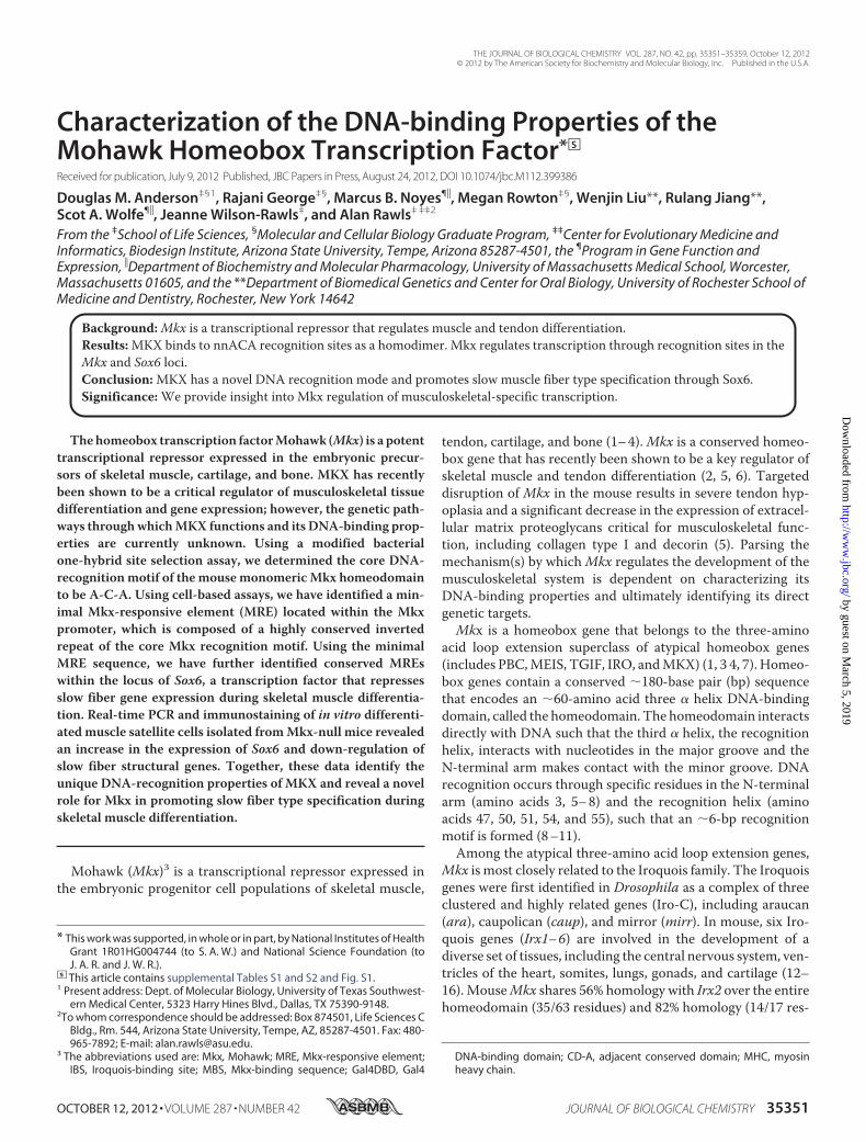
The Mouse Mkx Homeodomain Binds a Recognition MotifCommon to the Irx Family—To characterize the DNA-bindingspecificity of themouseMkx homeodomain, we utilized amod-ified bacterial one-hybrid assay to select recognition sequencesfrom a library of random decamer sequences. A fragment ofmouse Mkx including the homeodomain and adjacent con-served domainCD-A (amino acids 70–161)was cloned into thepB1H2�2-12 vector, as a fusion with the � subunit of RNApolymerase and the first and second zinc fingers of Zif268. Thisconstruct was co-transformed with a reporter plasmid thatcontains a region of random decamers adjacent to the bindingsite of the Zif268, and transformants were selected for theirability to grow under nutrient-deficient conditions (11, 19).A total of 21 sequences were recovered and analyzed using
the web-based programMotifsampler (Fig. 1A). The motif-de-tecting algorithm of this program uses Gibbs sampling to findthe position probability matrix of the motif (20, 21). The pre-ferred recognition sequence of mouseMKXwas determined tobe A-C-A (Fig. 1B). Thus, mouse MKX shares the same recog-nitionmotif as previously characterized Irx familymembers butdiverges from the recently reported binding site (T-n-A-C-A)of theDrosophilaMKXortholog, which has a strong affinity fora T at position 1 (11).Mkx Is Capable of Negative Autoregulation—DNA-binding
transcription factors often participate in the regulation of theirown transcription through autoregulatory feedback loops (27).Negative autoregulation is a well established mechanism forboth stabilizing transcription factor levels aswell asmodulatingspatial and temporal expression patterns (23, 28, 29). To deter-mine whether MKX is capable of directly regulating its owntranscription, we cloned a fragment of the mouse Mkx pro-
moter (�3,576 to �127 bp) upstream of the luciferase reportergene in pGL3Basic (3.5kbMkx-luc) (Fig. 2A). Full-length Mkx(MT-MKX) strongly repressed transcription (13.4-fold reduc-tion) when comparedwith background (Fig. 2B). Deletion of thethree MKX repression domains (2) (MT-Mkx(�MRD1-3)) orHelix III of thehomeodomain (MT-Mkx(�H3)) abrogated repres-sion of 3.5kbMkx-luc (Fig. 2B). To confirm the responsiveness of3.5kbMkx-luc toMkx, the C-terminal region containing the threeMkx repressor domains (amino acids 205–352) was replaced bythe potent transcriptional activation domain of VP16 (MT-Mkx-VP16) (30). TheMT-Mkx-VP16 fusion resulted in 8.9-fold activa-tion of 3.5kbMkx-luc. Additionally, neither a deletion mutant ofMT-Mkx-VP16 that does not contain Helix III of the homeodo-main (MT-Mkx(�H3)-VP16), nor VP16 alone, could activate the3.5kbMkx-luc reporter to the same extent (Fig. 2B). This demon-strates that MKX is capable of regulating its own promoter andthat this regulation is dependent upon amino acids within thehomeodomain.Similar repression levels were observed with a smaller frag-
ment of the Mkx promoter spanning �113 bp to �127 bp(113bpMkx-luc) (Fig. 2). This fragment was therefore furthersubdivided into three overlapping fragments (A, �113 to �17;B, �41 to �56; and C, �32 to �127) (Fig. 3A). Fragment Aalone was responsive to MT-Mkx-VP16, which we will hereinrefer to as the MRE (Fig. 3B). A multiple sequence alignmentrevealed that the MRE is highly conserved in the promoters ofvertebrate Mkx orthologs and is composed of a large invertedrepeat (see supplemental Fig. S1).MKX Interacts Directly with a Highly Conserved Sequence
within Its Promoter—The MRE includes a highly conservedinverted repeat, which contains the recognition motif we char-acterized, separated by 25 bases (ATGTT-N25-AACAT). Totest if MKX can bind this sequence, we performed an EMSAusing whole cell lysates from NIH3T3 cells expressing MT-MKX and a double-stranded DNA probe specific for the con-served inverted repeat sequence (Fig. 4). A band was consis-tently observed in the presence of MT-MKX and could befurther supershifted by an anti-Myc antibody (Fig. 4A). Amutant fusion protein, MT-MKX with the Helix III of thehomeodomain deleted (MT-Mkx(�H3)), was not able to shifttheMRE probe (Fig. 4A). Further,MT-MKX-VP16 also formeda complex with the probe andwas supershifted by the anti-Mycantibody (Fig. 4B). This demonstrated that MKX can form acomplex with this inverted repeat, which we will refer to as theMkx-binding sequence (MBS), and the homeodomain is neces-sary for this interaction.The ability ofMKX to regulate theMBS in cells was tested by
cloning this sequence upstream in pGL3Promoter (MBS-luc).MT-Mkx-VP16 activated the MBS-luc vector 2.5-fold, whencompared with pGL3Promoter alone (luc) (Fig. 4C). Reducingthe number of flanking residues and randomizing the interven-ing 21 bp did not alter the level of activation (artificial MBS-aMBS-luc) (Fig. 4C).Multimerizing three copies of the aMBS ina head-to-tail fashion resulted in additive increases in luciferaseactivation (3X-aMBS-luc). Point mutation of nucleotides withinboth inverted repeats (mut-aMBS; ACACT N25 AGTGT) abol-ished activation by MT-Mkx-VP16 (Fig. 4C). Multimerized ver-sionsof theMBSinwhichoneof thehalf-siteswasmutated (Lmut-
FIGURE 1. Characterizing the monomeric Mkx recognition motif. A frag-ment encoding the mouse Mkx homeodomain was used to screen a randomdecamer library using a modified bacterial one-hybrid site selection assay. A,21 clones from the selection were analyzed for overrepresented sequencemotifs. B and C, diagram of key determinants within the homeodomain thatare important for specifying the recognition motif for mouse Mkx is shown.Mkx showed little specificity for binding positions 1 and 2 but a strong pref-erence for A-C-A at positions 3–5.
Mkx DNA-binding Properties
OCTOBER 12, 2012 • VOLUME 287 • NUMBER 42 JOURNAL OF BIOLOGICAL CHEMISTRY 35353
by guest on March 5, 2019
http://ww
w.jbc.org/
Dow
nloaded from

aMBS or Rmut-aMBS) abrogated activation by MT-Mkx-VP16(Fig. 4C). This indicated that both half-sites of the MBS wererequired and given the inverted repeat nature of the MBS, sug-gested thatMKXmight bind theMBS as a homodimer.
MKX Can Homodimerize and Requires an Adjacent Con-served Domain to Recognize Its Binding Site—Homeodomain-containing proteins can function as monomers, homodimers,or heterodimers, which can lead to differential DNA-binding
FIGURE 2. Mkx is capable of negative autoregulation. Fragments of the mouse Mkx promoter cloned upstream of a luciferase reporter gene were assayed fortheir responsiveness to Mkx regulation in NIH3T3 cells. An Mkx promoter-luciferase construct encompassing �3,576 bp to �127 bp (3.5kbMkx-luc) wasstrongly repressed by co-transfection with a plasmid-encoding Myc tag full-length Mkx (MT-Mkx). Deletion of the three Mkx repressor domains (MT-Mkx(�MRD1-3)) or Helix III of the homeodomain (MT-Mkx(�H3)) abrogated this effect. An activated form of Mkx (MT-Mkx-VP16) resulted in specific activationof the Mkx promoter reporter. A smaller Mkx promoter construct encompassing �113 bp to �127 bp (113bpMkx-luc) responded in an identical manner.
FIGURE 3. Identifying the Mkx response element within the Mkx promoter. A, Mkx promoter sequence (�113 bp to �127) was cloned as three individualfragments (fragment A, �113 to �17; B, �41 to �56; and C, �32 to �127) upstream of SV40 in the luciferase pGL3Promoter. B, of the three smaller Mkxpromoter fragments, fragment A was the most responsive to MT-Mkx-VP16 activation. This sequence was renamed the MRE.
Mkx DNA-binding Properties
35354 JOURNAL OF BIOLOGICAL CHEMISTRY VOLUME 287 • NUMBER 42 • OCTOBER 12, 2012
by guest on March 5, 2019
http://ww
w.jbc.org/
Dow
nloaded from

specificity (31–33). To test whether MKX is capable ofhomodimerization,we used amodifiedmammalian two-hybridapproach using transient transfection in NIH3T3 cells. Gal4
DNA-binding domain (Gal4DBD)-fusion proteins with MKX-specific bait fragments were assayed for their ability to interactwith the MT-Mkx-VP16. Dimerization of Mkx bait fragments
FIGURE 4. Identification of the MBS within the MRE. A and B, MT-Mkx (A) and MT-Mkx-VP16 (B) were able to bind the MBS sequence but not a mutated MBSsequence in an electrophoresis mobility shift assay. The Mkx-MBS complex could be supershifted with an anti-Myc antibody (MT-Ab). Deletion mutants of Mkxlacking Helix III of the homeodomain (MT-Mkx(�H3) and MT-Mkx(�H3)-VP16) were not sufficient to shift MBS. EMSA primers are provided in supplementalTable S1. C, cell-based luciferase transcription assays demonstrate that the MBS sequence is responsive to Mkx-VP16 activation. An artificial MBS (aMBS) thatremoves 5�-flanking sequence and randomizes the central 21 bp was equally responsive. Increasing the copy number of the MBS resulted in increasedactivation by Mkx-VP16. Mutation of either half-site demonstrated that both are required for Mkx-mediated regulation.
Mkx DNA-binding Properties
OCTOBER 12, 2012 • VOLUME 287 • NUMBER 42 JOURNAL OF BIOLOGICAL CHEMISTRY 35355
by guest on March 5, 2019
http://ww
w.jbc.org/
Dow
nloaded from
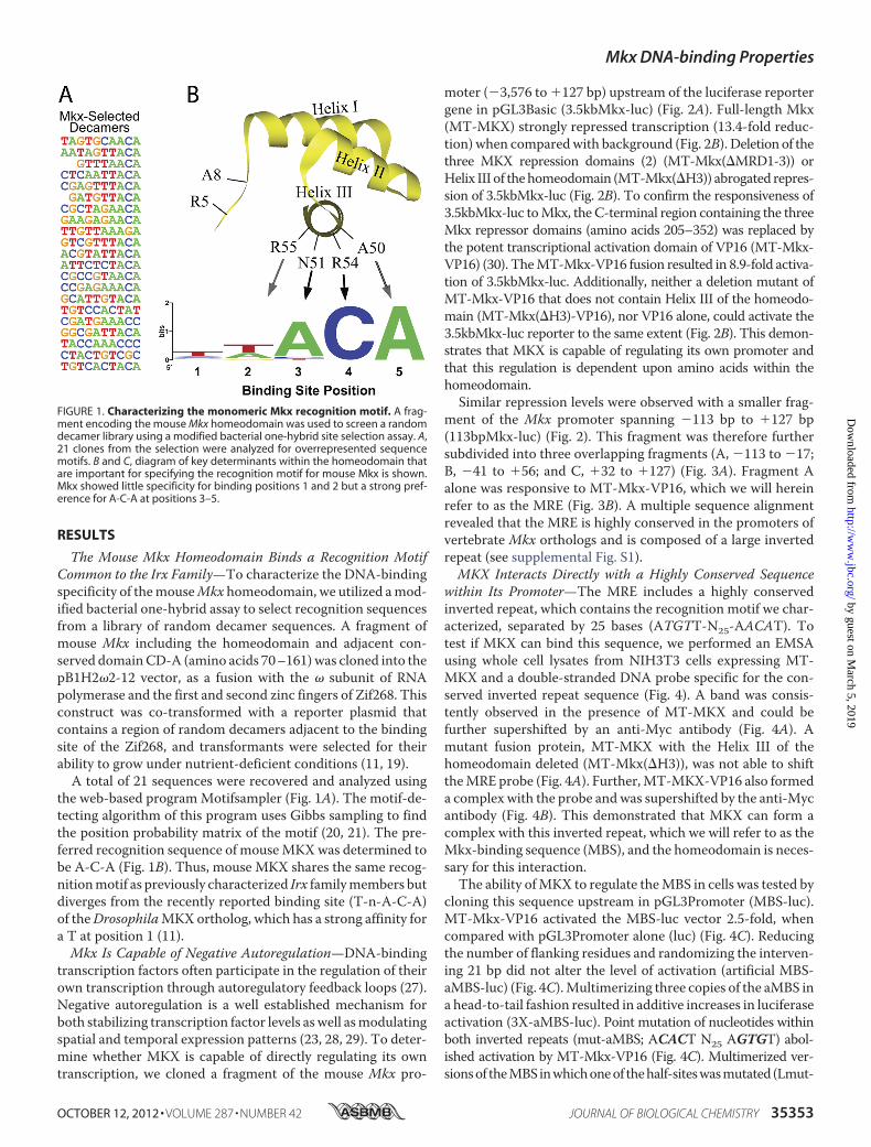
was scored by the level of activation of the Gal4DBD-respon-sive luciferase reporter, 5XUASpGL3Promoter, relative toGal4DBD alone (2). Initially, full-length Mkx resulted in areduction of reporter activity (Fig. 5A). Because the three Mkxrepression domains (MRD1-3) may suppress luciferase tran-scription, a fusion construct that lacks these domains(Gal4DBD-Mkx(�MRD1-3)) was examined. This resulted in a9-fold activation of the reporter, demonstrating that Mkx iscapable of homodimerization. A fusion construct containingthe Mkx homeodomain led to modest activation of thereporter (2.1-fold), whereas the inclusion of the adjacentconserved domain (CD-A) resulted in a 5.1-fold activation ofthe reporter (Fig. 5A). Neither the N-terminal amino acidsequence to the homeodomain nor CD-A domain alone wassufficient to activate the reporter (Fig. 5A). This demon-strates that MKX is capable of dimerization through thehomeodomain and that this interaction is enhanced by thepresence of the CD-A.The Sox6 Locus Contains Multiple Mkx-binding Sites—To
identify additional targets of MKX, a genome-scale DNA pat-tern search of the conserved MBS sequence was performedusing the Regulatory Sequence Analysis Tools (22). The num-ber of intervening nucleotides was allowed to vary from 0 to 25,and mismatches of 1 bp were allowed in either half-site(ATGTTN0–25AACAT).A list of putativeMRE sites identifiedby this approach is provided in supplemental Table S2. Withinthe intronic sequence in the Sox6 locus, we identified a wellconserved MBS with an intervening space of 11 nucleotidesbetween the inverted repeat sequences (ATGTT N11AACAT) (Fig. 6). Because of the overlap in theMKX and IRXDNA-recognition sequences, a similar genome-scale DNApattern search was performed using the Irx-bindingsequence (ACAnnTGT). This revealed an additional wellconserved element within the Sox6 promoter (AACATGT-GTT). Because Sox6 plays an important role in musculosk-eletal development, it represents a strong potential target forMKX (34–37).To validate these sequences as Mkx-binding sites, electro-
phoreticmobility shift assayswere performed.MKXwas able tobind to both the Sox6 MBS and IBS sequences similar to theMBS in the Mkx promoter (Fig. 6A). Multimerized (3�) Sox6
target sequences were cloned upstream of the SV40 promoterin the pGL3Promoter vector. Similar to the MBS identified inthe Mkx promoter, the Sox6 MBS and the Sox6 IBS wereresponsive to MT-MKX-VP16 activation (Fig. 6B). Deletion ofHelix III (MT-Mkx(�H3)-VP16) abrogated the activation at allthree sites (Fig. 6B). Transcriptional activationwas lost with thedeletion of CD-A from theMKX-VP16 fusion (MT-Mkx(�CD-A)-VP16), underscoring its importance in the function of Mkxregulation (Fig. 6B). This demonstrates that Mkx is capable ofregulating both MBS and IBS motifs within the Sox6 locus.BecauseMkx can recognize a binding site conformation sim-
ilar to the IBS, it is possible that MKX and IRX proteins mayco-regulate a subset of genes. To determine whether IRX pro-teins can similarly recognize both binding site conformations,we utilized an activated form ofmouse IRX2 (HA-IRX2-VP16).The HA-IRX2-VP16 fusion activated modestly the Sox6 IBS(2.6-fold) but did not activate either MBS-containing reporterplasmid (Fig. 6B).Sox6 Is Up-regulated in Satellite Cells Isolated from Mkx�/�
Mice—To validate that Mkx was a repressor of Sox6 transcrip-tion in vivo, the levels of endogenous Sox6mRNA were exam-ined in primary muscle cells deficient forMkx. Skeletal musclesatellite cells were isolated from the hindlimb muscles of5-month-oldMkx�/� and wild type littermate mice. Quantita-tive RT-PCR using Sox6-specific primers revealed that Sox6 isexpressed at a similar level in freshly isolated satellite cells fromMkx�/� and control muscle (Fig. 7A). However, satellite cellsdifferentiating into myotubes demonstrated that Sox6 expres-sion was 2.2-fold higher in theMkx�/� mice (Fig. 7A). Sox6 hasbeen shown to participate in fiber type specification in skeletalmuscle by repressing the transcription of the slow twitch myo-sin heavy chain (MHC) isoform (Myh7) in fast twitch musclefibers (34–36).Myh7 transcription was decreased in differenti-ating Mkx�/� myotubes but not the fast twitch myosin heavychain isoforms (Myh1 and Myh2) (Fig. 7A). The differentialreduction of the slow isoform was confirmed by immuno-stainingwith antibodies specific to slow and fastMHC isoforms(Fig. 7B). This is consistent withMkx regulating the transcrip-tion ofMyh7 through repression of Sox6 and predicts a role infiber type specificity.
FIGURE 5. Mkx is able to form a homodimer. The ability of Mkx to form a homodimer was assayed using a cell-based mammalian two-hybrid. Mkx is capableof homodimerization dependent upon amino acids within the homeodomain and CD-A. Data are presented for each Gal4-Mkx bait as -fold activation relativeto the level of luciferase activity obtained by co-transfection of the Gal4DBD alone with appropriate prey.
Mkx DNA-binding Properties
35356 JOURNAL OF BIOLOGICAL CHEMISTRY VOLUME 287 • NUMBER 42 • OCTOBER 12, 2012
by guest on March 5, 2019
http://ww
w.jbc.org/
Dow
nloaded from

DISCUSSIONThe genetic pathways controlling the patterning and differ-
entiation of the vertebrate musculoskeletal system require thecombined actions of multiple regulatory factors. The Mkxhomeobox gene has been shown to be an important regulator ofskeletalmuscle and tendon differentiation (5, 6).We have char-acterized the DNA recognition motif of the mouseMkx home-odomain and identified anMkx-responsive element within theproximal promoter ofMkx. Using the identifiedMkx regulatoryelement as a model, functional MREs within the Sox6 locuswere identified, suggesting that MKX can directly repress theexpression of Sox6. Combined with the observation of up-reg-ulation of Sox6 inMkx�/� satellite cells, this defines a potentialpathway by whichMkx promotes the expression of slow MHCduring muscle development.
The DNA recognition motif for members of the homeodo-main-containing transcription factor family is largely depen-dent on a core set of amino acids in Helix III and residues at theN terminus of the homeodomain that make direct contact withthe DNA. A comprehensive analysis performed in Drosophilaorganizes this superfamily of genes into 11 distinct specificitygroups (11). Consistent with sequence conservation in thehomeodomain, the Drosophila Mkx ortholog (CG11617) fallsinto the Iroquois group that is characterized by a monomericnnACA motif. Interestingly, CG11617 diverges from Iroquoisgenes such as mirr through the preference for T at position 1(TnACA) (11). Our data demonstrate that mouseMkx has lostspecificity for a T and therefore can bind half-sites such as thatfound in the vertebrateMBS (GAACATandAAACAT).Mouseand Drosophila Mkx orthologs may recognize different recog-
FIGURE 6. Mkx-binding sites within the Sox6 locus. Mkx (ATGTT-N11-AACAT)- and Irx (ACAnnTGT)-binding sites were identified in the locus of Sox6. A, EMSAsdemonstrate that MT-Mkx-VP16 can physically interact with the Sox6 MBS and IBS sequences in vitro. B, cell-based luciferase assays demonstrate that thesesequences are responsive to MT-Mkx-VP16 activation and are dependent upon sequences within Helix III of the homeodomain (MT-Mkx(�H3)-VP16) and CD-A(MT-Mkx(�CDA)-VP16). An activated form of mouse Irx2 (HA-Irx2-VP16) could only regulate the Sox6 IBS.
FIGURE 7. Sox6 and MHC expression in Mkx�/� satellite cells. Primary cultures of adult muscle satellite cells were isolated from Mkx knock-out (Mkx�/�) andwild type (Mkx�/�) mice and cultured in vitro. Total RNA was isolated from proliferating cultures (day 0) or cultures that were differentiated under low mitogenicconditions for 1 or 3 days (day 1 and day 3). A, quantitative real-time PCR revealed that Sox6 transcription is up-regulated in Mkx�/� satellite cells. Transcriptionof Myh7 was reduced in differentiated Mkx�/� myotubes, whereas fast MHC isoforms were up-regulated (Myh1) or unaffected (Myh2). B, expression of fast MHC(My32) and slow MHC (NOQ7.5.4D) were detected in differentiated myoblasts isolated from Mkx�/� and Mkx�/� mice.
Mkx DNA-binding Properties
OCTOBER 12, 2012 • VOLUME 287 • NUMBER 42 JOURNAL OF BIOLOGICAL CHEMISTRY 35357
by guest on March 5, 2019
http://ww
w.jbc.org/
Dow
nloaded from
nition motifs as the result of specific amino acid changes atcritical residues of the homeodomain. At position 8 of theN-terminal arm of the homeodomain, mouseMkx contains analanine (Ala) and theDrosophila ortholog contains a phenyala-nine (Phe) (7). Large hydrophobic residues in position 8 havebeen shown to influence binding specificity at position 1 of therecognition motif (11). The Iroquois factors, which share anaffinity for the same monomeric site as mouse Mkx, similarlycontain an Ala at position 8 of the N-terminal arm. Replace-ment of the Ala with the large hydrophobic Phe residue inDro-sophila caup partially added a preference for T at binding posi-tion 1 (11). Whereas the amino acids critical for DNArecognition are identical among vertebrate Mkx orthologs,position 8 in the N-terminal arm is different among cephalo-chordates, hemichordates, and echinoderms. This predicts thatthe alteration in position 8, coupled with the adaptation ofdimerization in vertebrates, expands the functionality of Mkxby increasing the complexity of binding sites and thereby targetgenes. Future studies will be required to determine whetherchanges in the amino acid identity at position 8 have alteredMkx DNA recognition among distantly related metazoans.Whereas the mouse MKX homeodomain shares a common
recognition motif with Irx family members, we have demon-strated that they differ in their ability to recognize half-site con-formations. Our data show that Mkx can recognize and bindbothMBS- and IBS-like conformations, whereas an IRX2-VP16fusion protein is only capable of regulating the IBS conforma-tion. MBS and IBS conformations are different in both thenumber of nucleotides separating the half-sites and the orien-tation of the half-sites. Inter-site spacing has been shown to bea critical parameter for homeobox gene regulation. The Dro-sophila bicoid homeobox transcription factor can bind stronglyto sites that are spaced 25 bp apart but only weakly to sitesseparated by 11 bp (38). This reveals an additional layer ofMkxregulation by which the level repression could be mediated byspecific MBS conformations that differ in spacer lengthbetween each half-site.The inverted repeat arrangement of the MBS and IBS
sequences is consistent withMkx-binding DNA as dimers. TheIroquois family has been shownpreviously to bindDNA in bothhomo- and heterodimer arrangements, and these interactionsare dependent upon sequences N-terminal to the homeodo-main (18). Similarly, MKX was capable of homodimerization,although this interaction was dependent upon a uniquesequence immediately C-terminal to the MKX homeodomain.Deletion of this conserved domain also diminished the ability ofMKX to regulate gene expression through theMBS, suggestingthat MKX binds DNA as a requisite homodimer. It is interest-ing to speculate on whether members of the Irx family andMKX form functional heterodimers because their expressionpatterns overlap in the somites, limb buds, and muscle duringdevelopment. Future studies will be required to determine theextent to which this interaction occurs in vivo and whetherthese closely related family members share functional redun-dancy during development.Our studies predict that Mkx is able repress its own tran-
scription through the MBS found in its promoter. Such nega-tive autoregulatory loops have been found at the core of several
developmental processes, including the anterior-posterior co-linear expression of the Hox genes, the oscillatory genetic net-works associated with circadian rhythms, and the segmentalclock of somitogenesis, stem cell potency, and cell differentia-tion (39–41). Further, it has been proposed that negative auto-regulation can suppress the transcriptional noise intrinsic ingenetic networks that reduces both the switching on andswitching off time of gene expression (42). TheMkx-responsiveelement is highly conserved among vertebrate Mkx orthologsand clustered with other conserved inverted repeat sequences.This suggests that negative autoregulation is a conserved fea-ture among Mkx orthologs and may be modulated in a largerregulatory complex. The significance of the negative autoregu-lation will become clearer as theMkx genetic network is betterunderstood.Our initial characterization of the MKX-binding properties
led to the identification of two well conserved MBS within theSox6 locus. Sox6 has been shown to be an important regulatorduring musculoskeletal development and fiber type specifica-tion (35–37, 43). Sox6 possesses a redundant function withSox5 during the differentiation of chondrocytes (44) and isrequired to block cells of the sclerotome from entering the ten-don lineage (45). In skeletal muscle, Sox6 suppresses slowtwitch fiber development through the direct repression ofMyh7 transcription (34, 35). In primary satellite cells isolatedfrom Mkx�/� mice, we observed that endogenous Sox6 tran-scription was elevated upon differentiation, suggesting it is adirect target of Mkx-mediated repression. Consistent with thisresult, Myh7 gene expression was reduced in these same cells,whereas fast MHC isoform expression levels were unchanged.This provides the first evidence for a role for Mkx in the regu-lation of fiber type specificity during skeletal muscle differenti-ation. Further, it provides a possible mechanism by whichMkxis able to participate in the integration of the differentiation ofmultiple cell types in the musculoskeletal system.
Acknowledgments—We thank Dr. Ron Allen for advice on culturingprimary satellite cells, Dr. Anthony Firulli for providing the CS2MTplasmid, Dr. Scott Bingham for assistance in DNA sequencing, andDr. James Elser for the generous use of the FLx800 microplate reader.
REFERENCES1. Anderson, D. M., Arredondo, J., Hahn, K., Valente, G., Martin, J. F., Wil-
son-Rawls, J., and Rawls, A. (2006) Mohawk is a novel homeobox geneexpressed in the developing mouse embryo. Dev. Dyn. 235, 792–801
2. Anderson, D. M., Beres, B. J., Wilson-Rawls, J., and Rawls, A. (2009) Thehomeobox geneMohawk represses transcription by recruiting the sin3A/HDAC co-repressor complex. Dev. Dyn. 238, 572–580
3. Liu, H., Liu, W., Maltby, K. M., Lan, Y., and Jiang, R. (2006) Identificationand developmental expression analysis of a novel homeobox gene closelylinked to the mouse twirler mutation. Gene Expr. Patterns 6, 632–636
4. Takeuchi, J. K., and Bruneau, B. G. (2007) Irxl1, a divergent Iroquois ho-meobox family transcription factor gene. Gene Expr. Patterns 7, 51–56
5. Ito, Y., Toriuchi, N., Yoshitaka, T., Ueno-Kudoh, H., Sato, T., Yokoyama,S., Nishida, K., Akimoto, T., Takahashi, M., Miyaki, S., and Asahara, H.(2010) The Mohawk homeobox gene is a critical regulator of tendon dif-ferentiation. Proc. Natl. Acad. Sci. U.S.A. 107, 10538–10542
6. Liu, W., Watson, S. S., Lan, Y., Keene, D. R., Ovitt, C. E., Liu, H., Schweit-zer, R., and Jiang, R. (2010) The atypical homeodomain transcription fac-tor Mohawk controls tendon morphogenesis. Mol. Cell. Biol. 30,
Mkx DNA-binding Properties
35358 JOURNAL OF BIOLOGICAL CHEMISTRY VOLUME 287 • NUMBER 42 • OCTOBER 12, 2012
by guest on March 5, 2019
http://ww
w.jbc.org/
Dow
nloaded from
4797–48077. Mukherjee, K., andBürglin, T. R. (2007)Comprehensive analysis of animal
TALE homeobox genes: new conserved motifs and cases of acceleratedevolution. J. Mol. Evol. 65, 137–153
8. Gehring, W. J., Affolter, M., and Bürglin, T. (1994) Homeodomain pro-teins. Annu. Rev. Biochem. 63, 487–526
9. Ades, S. E., and Sauer, R. T. (1995) Specificity of minor groove and majorgroove interactions in a homeodomain-DNA complex. Biochemistry 34,14601–14608
10. Wolberger, C. (1996)Homeodomain interactions.Curr. Opin. Struct. Biol.6, 62–68
11. Noyes,M. B., Christensen, R.G.,Wakabayashi, A., Stormo,G.D., Brodsky,M. H., and Wolfe, S. A. (2008) Analysis of homeodomain specificitiesallows the family-wide prediction of preferred recognition sites. Cell 133,1277–1289
12. Bosse, A., Zülch, A., Becker, M. B., Torres, M., Gómez-Skarmeta, J. L.,Modolell, J., and Gruss, P. (1997) Identification of the vertebrate Iroquoishomeobox gene family with overlapping expression during early develop-ment of the nervous system.Mech. Dev. 69, 169–181
13. Bellefroid, E. J., Kobbe, A., Gruss, P., Pieler, T., Gurdon, J. B., and Papa-lopulu, N. (1998) Xiro3 encodes a Xenopus homolog of the DrosophilaIroquois genes and functions in neural specification.EMBO J. 17, 191–203
14. Bao, Z. Z., Bruneau, B. G., Seidman, J. G., Seidman, C. E., and Cepko, C. L.(1999) Regulation of chamber-specific gene expression in the developingheart by Irx4. Science 283, 1161–1164
15. Cohen, D. R., Cheng, C.W., Cheng, S. H., andHui, C. C. (2000) Expressionof two novelmouse Iroquois homeobox genes during neurogenesis.Mech.Dev. 91, 317–321
16. Houweling, A. C., Dildrop, R., Peters, T., Mummenhoff, J., Moorman,A. F., Rüther, U., and Christoffels, V. M. (2001) Gene and cluster-specificexpression of the Iroquois family members during mouse development.Mech. Dev. 107, 169–174
17. Berger, M. F., Badis, G., Gehrke, A. R., Talukder, S., Philippakis, A. A.,Peña-Castillo, L., Alleyne, T. M., Mnaimneh, S., Botvinnik, O. B., Chan,E. T., Khalid, F., Zhang, W., Newburger, D., Jaeger, S. A., Morris, Q. D.,Bulyk, M. L., and Hughes, T. R. (2008) Variation in homeodomain DNAbinding revealed by high resolution analysis of sequence preferences. Cell133, 1266–1276
18. Bilioni, A., Craig, G., Hill, C., and McNeill, H. (2005) Iroquois transcrip-tion factors recognize a unique motif to mediate transcriptional repres-sion in vivo. Proc. Natl. Acad. Sci. U.S.A. 102, 14671–14676
19. Noyes, M. B., Meng, X., Wakabayashi, A., Sinha, S., Brodsky, M. H., andWolfe, S. A. (2008) A systematic characterization of factors that regulateDrosophila segmentation via a bacterial one-hybrid system.Nucleic AcidsRes. 36, 2547–2560
20. Thijs, G., Lescot, M., Marchal, K., Rombauts, S., De Moor, B., Rouzé, P.,and Moreau, Y. (2001) A higher order background model improves thedetection of promoter regulatory elements by Gibbs sampling. Bioinfor-matics 17, 1113–1122
21. Thijs, G., Marchal, K., Lescot, M., Rombauts, S., De Moor, B., Rouzé, P.,and Moreau, Y. (2002) A Gibbs sampling method to detect overrepre-sented motifs in the upstream regions of co-expressed genes. J. Comput.Biol. 9, 447–464
22. van Helden, J. (2003) Regulatory sequence analysis tools. Nucleic AcidsRes. 31, 3593–3596
23. Bateman E. (1998) Autoregulation of eukaryotic transcription factors.Prog. Nucleic Acids Res. Mol. Biol. 60, 133–168
24. Rupp, R. A., Snider, L., andWeintraub, H. (1994) Xenopus embryos regu-late the nuclear localization of XMyoD. Genes Dev. 8, 1311–1323
25. Wilson-Rawls, J., Rhee, J. M., and Rawls A. (2004) Paraxis is a basic helix-loop-helix protein that positively regulates transcription through bindingto specific E-box elements. J. Biol. Chem. 279, 37685–37692
26. Wilson-Rawls, J., Hurt, C. R., Parsons, S. M., and Rawls, A. (1999) Differ-ential regulation of epaxial and hypaxial muscle development by paraxis.Development 126, 5217–5229
27. Crews, S. T., and Pearson, J. C. (2009) Transcriptional autoregulation indevelopment. Curr. Biol. 19, R241–246
28. Irvine, K. D., Botas, J., Jha, S., Mann, R. S., and Hogness, D. S. (1993)Negative autoregulation by Ultrabithorax controls the level and pattern ofits expression. Development 117, 387–399
29. Semsey, S., Krishna, S., Erdossy, J., Horváth, P., Orosz, L., Sneppen, K., andAdhya, S. (2009) Dominant negative autoregulation limits steady-staterepression levels in gene networks. J. Bacteriol. 191, 4487–4491
30. Sadowski, I.,Ma, J., Triezenberg, S., and Ptashne,M. (1988) GAL4-VP16 isan unusually potent transcriptional activator. Nature 335, 563–564
31. Chang, C. P., Brocchieri, L., Shen, W. F., Largman, C., and Cleary, M. L.(1996) Pbx modulation of Hox homeodomain amino-terminal arms es-tablishes different DNA-binding specificities across the Hox locus. Mol.Cell. Biol. 16, 1734–1745
32. Neuteboom, S. T., Peltenburg, L. T., van Dijk, M. A., andMurre, C. (1995)The hexapeptide LFPWMR in Hoxb-8 is required for cooperative DNAbinding with Pbx1 and Pbx2 proteins. Proc. Natl. Acad. Sci. U.S.A. 92,9166–9170
33. Furukawa, K., Iioka, T.,Morishita,M., Yamaguchi, A., Shindo, H., Namba,H., Yamashita, S., and Tsukazaki, T. (2002) Functional domains of paired-like homeoprotein Cart1 and the relationship between dimerization andtranscription activity. Genes Cells 7, 1135–1147
34. Hagiwara, N., Ma, B., and Ly, A. (2005) Slow and fast fiber isoform geneexpression is systematically altered in skeletal muscle of the Sox6 mutant,p100H. Dev. Dyn. 234, 301–311
35. Hagiwara, N., Yeh,M., and Liu, A. (2007) Sox6 is required for normal fibertype differentiation of fetal skeletal muscle in mice. Dev. Dyn. 236,2062–2076
36. vonHofsten, J., Elworthy, S., Gilchrist,M. J., Smith, J. C.,Wardle, F. C., andIngham, P. W. (2008) Prdm1- and Sox6-mediated transcriptional repres-sion specifies muscle fiber type in the zebrafish embryo. EMBO Rep. 9,683–689
37. Quiat, D., Voelker, K. A., Pei, J., Grishin, N. V., Grange, R. W., Bassel-Duby, R., and Olson, E. N. (2011) Concerted regulation of myofiber-spe-cific gene expression and muscle performance by the transcriptional re-pressor Sox6. Proc. Natl. Acad. Sci. U.S.A. 108, 10196–10201
38. Fu, D., Zhao, C., and Ma, J. (2003) Enhancer sequences influence the roleof the amino-terminal domain of bicoid in transcription. Mol. Cell. Biol.23, 4439–4448
39. Bessho, Y., and Kageyama, R. (2003) Oscillations, clocks, and segmenta-tion. Curr. Opin. Genet. Dev. 13, 379–384
40. Pan, G., Li, J., Zhou, Y., Zheng, H., and Pei, D. (2006) A negative feedbackloop of transcription factors that controls stem cell pluripotency and self-renewal. FASEB J. 20, 1730–1732
41. Wong, W. F., Kurokawa, M., Satake, M., and Kohu, K. (2011) Down-regulation of Runx1 expression by TCR signal involves an autoregulatorymechanism and contributes to IL-2 production. J. Biol. Chem. 286,11110–11118
42. Zabet, N. R. (2011) Negative feedback and physical limits of genes. J.Theor. Biol. 284, 82–91
43. Dumitriu, B., Dy, P., Smits, P., and Lefebvre, V. (2006) Generation of miceharboring a Sox6 conditional null allele. Genesis 44, 219–224
44. Lefebvre, V., Behringer, R. R., and de Crombrugghe, B. (2001) L-Sox5,Sox6, and Sox9 control essential steps of the chondrocyte differentiationpathway. Osteoarthritis Cartilage 9, Suppl. A, S69–75
45. Brent, A. E., Braun, T., and Tabin, C. J. (2005) Genetic analysis of interac-tions between the somitic muscle, cartilage, and tendon cell lineages dur-ing mouse development. Development 132, 515–528
Mkx DNA-binding Properties
OCTOBER 12, 2012 • VOLUME 287 • NUMBER 42 JOURNAL OF BIOLOGICAL CHEMISTRY 35359
by guest on March 5, 2019
http://ww
w.jbc.org/
Dow
nloaded from

Rulang Jiang, Scot A. Wolfe, Jeanne Wilson-Rawls and Alan RawlsDouglas M. Anderson, Rajani George, Marcus B. Noyes, Megan Rowton, Wenjin Liu,
Transcription FactorCharacterization of the DNA-binding Properties of the Mohawk Homeobox
doi: 10.1074/jbc.M112.399386 originally published online August 24, 20122012, 287:35351-35359.J. Biol. Chem.
10.1074/jbc.M112.399386Access the most updated version of this article at doi:
Alerts:
When a correction for this article is posted•
When this article is cited•
to choose from all of JBC's e-mail alertsClick here
Supplemental material:
http://www.jbc.org/content/suppl/2012/08/24/M112.399386.DC1
http://www.jbc.org/content/287/42/35351.full.html#ref-list-1
This article cites 45 references, 15 of which can be accessed free at
by guest on March 5, 2019
http://ww
w.jbc.org/
Dow
nloaded from